Note: Descriptions are shown in the official language in which they were submitted.
CA 02613513 2007-12-24
WO 2007/002487 PCT/US2006/024623
Scleral Contact Lens with Grooves and Method of Making
Lens
CROSS REFERENCE
[0001] This application claims the benefit of priority to U.S. Provisional
Application
Serial No. 60/693857 filed June 24, 2005, the disclosure of which is
incorporated by
reference herein in its entirety. This application also claims the benefit of
priority to U.S.
Provisional Application Serial No. 60/753893 filed December 22, 2005, the
disclosure of
which is incorporated by reference herein in its entirety.
TECHNICAL FIELD
[0002] This disclosure relates generally to a scleral lens with grooves for
aspirating
tears to prevent suction when the lens is disposed on the eye. Also, this
disclosure relates
to a method of making the lens using laser technology.
BACKGROUND
[0003] The cornea is the transparent dome-shaped front part of our eyes and
their
most important focusing lens. Like the lens of a camera, its surface must be
perfectly
smooth in order to provide clear vision. When disease or injury causes the
corneal surface
to become irregular, the eye can no longer focus clearly, even with the
strongest glasses.
Hard contact lenses have the unique ability to improve the vision of these
eyes by
creating a smooth layer of tears that optically masks the irregular surface of
the cornea.
However, there are many eyes with damaged corneas that cannot be fitted with a
hard
contact lens that rests on their corneas. Moreover, the corneas of patients
who suffer from
severe ocular surface disease become so exquisitely fragile that they are
often unable to
withstand the pressure of blinking or the briefest exposure to air and even
less so the
friction of a hard contact lens.
[0004] A scleral lens, such as the Boston Scleral Lens offered by the Boston
Foundation for Sight, provides a device that addresses this condition. As
shown in FIGS
1- 3 and 3A, a scleral lens 10, which is about the size of a quarter, rests on
the relatively
insensitive white sclera 12 of the eye 14 and creates a space 16 over the
cornea 18 that is
filled with artificial tears. The eye contact location for the scleral lens is
different than
conventional contact lenses which rest on the cornea. As illustrated by the
contact length
L in FIG. 2, contact with the eye is limited to the sclera and the lens does
not contact the
cornea 18. By creating a fluid-filled space over the irregular surface of the
damaged
1
CA 02613513 2007-12-24
WO 2007/002487 PCT/US2006/024623
cornea, this lens device can be helpful in improving vision in eyes with
extremely
distorted corneas. Moreover, this fluid compartment becomes a liquid bandage
that
protects the raw and sensitive cornea from exposure to air and the rubbing
effects of
blinking. The therapeutic environment created between the lens and the cornea
nurtures
healing and can virtually eliminate pain and photosensitivity. It is this
unique "corneal
liquid bandage" that is responsible for the extraordinary healing experienced
by patients
who have used the scleral lens. However, by sealing a pool of liquid over the
cornea, the
scleral lens simulates a fluid pump by which micro quantities of fluid are
squeezed out of
the fluid compartment during lens compression as occurs during blinking. When
the lens
is decompressed after each blink, tears outside the lens are drawn into the
fluid
compartment. If the aspiration of tears is blocked during lens decompression,
negative
pressure develops in the fluid compartment that increases over time resulting
in
progressive lens suction that can be severely damaging to the eye. The
traditional
solution to scleral lens suction is to drill a hole in the lens through which
air is aspirated
(air ventilated). However, the presence of air bubbles in the fluid
compartment causes
desiccation of the corneal surface that is especially dangerous for diseased
corneas. The
purpose of this invention is to create a mechanism for preventing lens suction
by
facilitating the aspiration of outside tears while excluding the transit of
air bubbles.
[0005] A groove 22 is provided in that part of the posterior contact lens
suirface that
rests on the scleral surface of the eye (haptic). More than one groove is
typically used.
The groove(s) allows tears outside the lens to be aspirated into the central
fluid
compartment to prevent the development of suction.
[0006] One of the reasons for the effectiveness of the Boston Scleral Lens is
its highly
oxygen-porous plastic, developed under the leadership of Boston Foundation for
Sight
founder Dr. Perry Rosenthal, that allows the cornea to breathe through the
lens. The
cornea, unlike any other surface tissue of the human body breathes by
extracting oxygen
directly from the surrounding air rather than from the blood circulation and
the oxygen
porous plastic allows oxygen to reach the surface of the eye.
[0007] A scleral lens can be beneficial to treat a number of conditions. Among
the
most common conditions treated is severe dry eye. Some causes of dry eyes are
an
underlying medical condition, such as Sjogren's syndrome, graft versus host
disease
(following bone marrow transplants), radiation treatment in the eye area,
Stevens-
Johnson syndronae, and autoimmune disorders. In some cases the cause is
unknown
2
CA 02613513 2007-12-24
WO 2007/002487 PCT/US2006/024623
(idiopathic). The condition can be due to a diminished supply of tears,
excessively rapid
evaporation or both. Dry eyes is one of the most common ocular complaints.
Symptoms
vary from mildly annoying, intermittent dryness and burning and paradoxical
tearing
(worse in dry environments) to constant disabling pain, increased light
sensitivity and
blurred vision. Mild to moderate cases of dry eyes can usually be controlled
by the
frequent use of artificial tears, punctal plugs that slow the drainage of
tears from the eyes
and a new, prescription eye drop (Restasis) that may reduce the inflammation
associated
with dry eyes. This group constitutes the vast majority of dry eyes. However,
it is the
patient with severe, disabling dry eyes unresponsive to the above treatments
who can
benefit the most from the Boston Scleral Lens device (BSL) and for whom
scleral lens is
being prescribed.
[0008] It is irnportant to treat severe dry eyes, which is one of the
conditions known
as severe ocular surface disease. Such patients suffer constant disabling eye
pain and
photosensitivity and may develop erosions on the surface of their corneas that
impair
vision and can lead to scarring and permanent impairment of vision. In the
most severe
cases, corneal ulcers develop that are slow to heal (if they heal at all) and
can result in
perforation. Rarely, it may be necessary to suture the lids together in order
to save the
eye (tarsorrhaphy). These severely dry eyes are more prone to infection and
heal so
poorly that surgery, including corneal transplant, is rarely successful and
even dangerous.
Conventional treatments described above are of little value. The so-called
soft bandage
lens is often ineffective because it requires an adequate supply of tears to
maintain its
hydration.
[0009] Keratoconus is another eye condition can be addressed by the use of a
scleral
lens. Keratoconus is characterized by a progressive thinning and steepening of
the central
cornea. As the cornea steepens and thins, a patient experiences a decrease in
vision
which can be mild or severe depending on the severity of the disease.
Keratoconus has
no known cure.
[0010] Onset of keratoconus occurs during the teenage years--mean age of onset
is
age 16 years--but onset has been reported to occur at ages as young as 6
years.
Keratoconus shows no gender predilection and is bilateral in over 90% of
cases. In
general, the disease develops asymmetrically: diagnosis of the disease in the
second eye
lags about five years after diagnosis in the first. The disease process is
active for about
3
CA 02613513 2007-12-24
WO 2007/002487 PCT/US2006/024623
five to 10 years, then it may be stable for many years. During the active
stage, change
may be rapid.
[0011] Typically, early vision loss can be corrected by spectacles; later,
irregular
astigmatism requires optical correction with rigid contact lenses. Hard
contact lenses
provide a uniform refracting surface and therefore improve vision. However,
traditional
rigid contact lenses rest on the surface of the cornea and slide over this
surface with each
blink. As a result, patients with diseased or damaged corneas who are most
likely to
benefit from their unique vision-restoring properties are often unable to
tolerate them and
would face the potential serious complications and uncertain visual outcome of
corneal
transplant surgery.
[0012] An estimated hundreds of thousands of patients suffering from corneal
disorders in the U.S. alone can benefit from the Foundation's lenses. The vast
majority of
patients are young-some are children. The Boston Scleral Lens is their only
hope for
regaining function vision. For the remaining patients, our lenses replace the
need for
corneal transplant surgery. The cornerstone of the Boston Scleral Lens is the
liquid
bandage it sustains over the corneal surface. This device is the first fluid-
ventilated gas-
permeable scleral lens designed to maintain an oxygenated aqueous corneal
environment
free of air bubbles. Lens suction is avoided by incorporating a series of
radial channels in
the posterior haptic surface that facilitates the aspiration of tears (while
excluding air) as
the means of aborting the development of negative hydrostatic pressure.
[0013] In order to avoid obstructing the channels (a prerequisite to the
physiological
tolerance of the devices), the shape of each haptic bearing surface is adapted
to the
contour of the underlying sclera through the design/fitting process. This
flexibility and
precision has been made possible by a process described in U.S. patent No.
5,452,031, the
entire contents of which is incorporated by reference. Based on advanced
mathematical
functions known as "splines", the design program has been integrated with the
control
system of the state-of-the-art contact lens lathe. This on-site technology has
been an
essential resource for advancing the development of the Foundation's lenses
and enabling
us to reach a success rate exceeding 90%. The oxygen tension of the corneal
liquid
bandage is maintained by a highly oxygen permeable polymer from which the
devices are
fabricated. Specially manufactured discs of this material are made for the
Boston Scleral
Lenses by Bausch & Lomb.
4
CA 02613513 2007-12-24
WO 2007/002487 PCT/US2006/024623
[0014] The process of making and adapting the shape of scleral lenses to that
of the
individual eye is exacting and time consuming. Additionally, various channels
can be
used to enable tears outside the lens to be aspirated into the liquid
reservoir between the
lens and cornea to abort the development of lens suctions, a highly dangerous
situation.
At the same time, it is necessary to design the channels to avoid the
aspiration of air into
the fluid reservoir that would adversely affect the health of the cornea. In
order to
maintain the patency of the channels, it is necessary to match the shape of
the bearing
surface of the scleral lens (haptic) with that of the underlying eye surface
(sclera) in order
to avoid excessive eye compression that would obstruct the channels and render
them
ineffective. It would be beneficial to enhance the performance of the scleral
lens by
configuring the groves in manner that makes them less vulnerable to
compression or
enabling the transit of air bubbles and thereby reduce the precision now
required in
customizing the shape of their haptic bearing surface.
SUMMARY
[0015] Accordingly, the present invention provides a new scleral lens that
allows for
improved flow of liquid between the bearing surface of the lens (haptic) and
the
underlying sclera of the eye into the fluid reservoir between the lens and the
cornea. The
channels in the scleral lens extend radially from the inside border of the
scleral contacting
surface of the lens (haptic). The channels can have several different
configurations that
assist in the transit of fluid or tears between the fluid reservoir under the
central zone of
the lens (optic) and the tears external to the lens. The channels may have a U-
shaped
cross-section, a V- shaped cross section, a beveled cross-section and others.
Further, the
cross-section can vary along the radial extension of the channel. Plural
channels may be
used. Also, the channels may have a serpentine configuration or generally
arcuate
configuration.
[0016] The present invention also provides a novel method of allowing more
oxygen
to permeate through the lens to the corneal surface. Microchambers within the
lens
material can assist in increasing the permeability of the lens by decreasing
the volume of
plastic through which oxygen would have to travel to reach the cornea. The
microchambers would be in various configurations in the lens portion of the
scleral lens.
CA 02613513 2007-12-24
WO 2007/002487 PCT/US2006/024623
BRIEF DESCRIPTION OF THE DRAWINGS
In the Drawings:
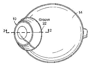
[0017] FIG. 1 is a perspective view of an eye with a Boston Foundation for
Sight
scleral lens;
[0018] FIG. 2 is a sectional view taken from lines 2-2 in Figure 1;
[0019] FIG. 3 is a top view of a scleral lens;
[0020] FIG. 3A is a sectional view of the lens taken from lines 3A-3A in
Figure 3;
[0021] FIG. 4 is a detail view of a channel of the present invention;
[0022] FIG. 4A is a sectional view taken from lines 4A-4A in FIG. 4;
[0023] FIGS. 4B-4E are,various configurations of the channel;
[0024] FIG. 4F is an expanded view of circle 4F in FIG. 4;
[0025] FIG. 4G is an expanded view of an alternative embodiment of a scleral
lens
taken at the same location as FIG. 4F.
[0026] FIGS. 5-7 are various configurations of multiple channels in the
scleral lens.
[0027] FIG. 8 is an alternate embodiment of the channels;
[0028] FIG. 9 is an illustration of the microchambers;
[0029] FIG. 9A is a sectional taken from 9A-9A in FIG. 9;
[0030] FIG. 10 is an illustration of the configuration of microchambers for
the scleral
lens;
[0031] FIG. 11 is a top view of a scleral lens with a microchambers;
[0032] FIG. 11A is a sectional view of a scleral lens taken from lines 11A-
11A.
6
CA 02613513 2007-12-24
WO 2007/002487 PCT/US2006/024623
DETAILED DESCRIPTION
[0033] The present invention provides a scleral lens which includes channels
that
improve the flow of tears outside the lens into the fluid reservoir occupying
the space
between the scleral lens and the cornea. The channels are disposed on the
inside surface
of the lens and extend generally radially from the inner limits of the scleral
contact
portion of the lens to the outside rim of the lens. Various configurations are
possible for
the channels, as described in more detail below. Additionally, the channels
can have a
serpentine or arcuate configuration to allow for the appropriate amount of
fluid flow
between the space under the lens and the scleral surface of the eye while
preventing the
transit of air bubbles. In another embodiment, microchannels can be formed in
the lens to
increase the oxygen transmissibility of the lens. The microchannels can have
many
configurations that reduce the volume of the lens material.
[0034] As illustrated in FIG. 4, a portion of a scleral lens 30 includes both
a scleral
contact surface 32 and a lens portion 34. The lens portion is disposed above
the cornea
when the scleral lens is applied to any eye. The scleral surface contacts the
lens at the
scleral contact surface 32. A channel 36 is disposed in the area of the
scleral contact
surface 32. The channel may extend between the inside limit of the haptic 38
and the
outside rim 40 of the contact lens scleral bearing surface. The channel
illustrated in FIG
3 extends the entire distance between the inside rim and the outside rim of
the scleral
bearing surfacd. In alternate embodiments the channel may extend the entire
distance
between the inside rim and the outside rim or it may terminate before the
outside rim as
illustrated in FIG. 4. When the channel terminated before the inside rim 38
and/or the
outside rim 40 an area 42 and 44 may be created which is discussed in more
detail below.
[0035] The channe136 may have one or more of several different cross-sections.
As
illustrated in FIG. 4A, the channel may have a generally square cross-section.
Of course,
one skilled in the art will be able to modify the choice of dimensions to
allow for
improved fluid flow into the central fluid compartment after the lens is
decompressed
following each blink while blocking the transit of air bubbles and tear
particulate matter.
The number of channels can be selected up to approximately 36. The channels
can be
evenly distributed along the scleral contact surface or, depending on desired
fluid flow
characteristics, can be placed unevenly around the scleral contact surface.
7
CA 02613513 2007-12-24
WO 2007/002487 PCT/US2006/024623
[0036] As illustrated in FIG 4B, a channel 48 may have a U shaped cross-
section.
This may provide advantages such as improved tear flow and a decrease in
particulate
matter getting caught in the "corners" of the channel. Of course, alternatives
to a
symmetrical U-share exist within the scope of this invention. For example,
depending on
the radius R of the curve that creates the U-shape the bottom of the channel
48 could be
shallow or have steeper edges. A preferred radius R is about one half the
width of the
channel.
[0037] Other cross-sections can be used. For example, FIG. 4C illustrates a
channel
50 with angled side walls 52, 54 and bottom 56. The side walls can create any
angle over
90 degrees. FIG. 4D illustrates a channel 58 with angled side walls 60 and 62
which
include an angle less than 90 degrees with the bottom 64 of the channel 58.
Another
alternative construction is illustrated in FIG. 4E where a channel 66 has
cross section is in
the shape of a diamond. Of course, the various shapes described can be
combined tÃ:., form
hybrid cross sections that provide the necessary fluid flow.
[0038] As described previously, the channels may terminate before the rims 38
and
40 as illustrated in FIG. 4. FIG 4F provides a detail view of the space 42
between the
channel 36 and the inside rim 38. The distance between the rim and the channel
can be
significant in developing the proper amount of fluid flow over the scleral
surface. In an
embodiment where the channels do not extend to the rim(s) the fluid a micro
channel 46
may be created to allow for the improved conduction of fluid from the inner
space above
the cornea and the channel 36. A microchannel can be disposed on the other
side of the
channel 36. The microchannel may have any of the cross sectional shaped
described
above. Figure 4G illustrates a groove 46 that is smaller than the groove 36
which can
assist in the hydrodynamics at the surface of the eye. As illustrated the
groove is smaller
than the groove 36 and extends the distance between the lens portion and the
end of the
groove. Other configurations are possible such the groove geometries disclosed
above.
[0039] The cross sections described above may vary in dimension along their
axial
length. For example, a channel in the radially inward portion of the scleral
surface may
increase as the channel extends radially outward. In one embodiment, the
channel may
increase in any dimension by 3 or more times along the length. Additionally,
the cross
sectional dimensions of a channel may decrease as the channel extends radially
outwardly.
8
CA 02613513 2007-12-24
WO 2007/002487 PCT/US2006/024623
[0040] As illustrated in FIG. 5-7, the radially extending channels can have
different
configurations. For example, FIG. 5 illustrates a scleral lens 70 with a
channel having a
serpentine configuration. The illustrated serpentine configuration has 3
curves, of course
more or fewer curves could be used in accordance with the present invention.
Multiple
serpentine patterns may be formed into the scleral contact area of the lens.
The preferred
number is up to 36. As illustrated in FIGS. 7 and 8 the scleral lenses 90 and
100 can have
arcuate grooves 92 and 102, 104. The arcuate channels may have a small radius
(which
makes a sharper curve) or a larger radius (which makes a gradually sloping
curve). These
curves may extend to the rims 38 and 40 of the scleral lens surfaces.
[0041] FIG. 8 illustrates still another embodiment of the present invention
where
channels are provided that are oriented both circumferentially and radially.
Specifically,
a scleral lens 110 includes arcuate channels 112, 114, and 116 that are
oriented
circumferentially. The channels may have any cross section as described above.
Further,
the arcuate channels can be connected by one or more radial channels 118, 120.
As
illustrated the number of radial channels may be selected to adjust the amount
of fluid
that moves between the inner rim 38 and the outer rim 40.
[0042] FIGS. 9- 11 illustrate another embodiment of the present invention
where
microchambers are formed in the scleral lens so that more oxygen can permeate
the lens
and provide more safety and comfort to the wearer. Because the cornea, unlike
all other
tissues of the human body, breathes by extracting oxygen directly from the
ambient air,
covering the outside surface of the cornea with a sheet of plastic can deprive
the cornea of
needed oxygen and cause undesirable side effects. Another aspect of the
present
invention provides microchambers in the optic portion of the scleral lens. In
FIG. 9 a
scleral lens 120 includes an inner rim 38 and outer rim 40. Microchambers are
provided
on the optic portion 122 of the scleral lens. The optic portion is disposed
above the
cornea when the lens is placed on the eye. As illustrated in FIG 9 and
detailed in FIG 9A,
a microchamber 124 is provided that form voids within the lens material. The
microcharnbers increase the gas transmissibility of the lens and enhances the
amount of
oxygen that can reach the cornea. The microchamber can have the dimensions
that allow
for increased gas transmissiblity while maintaining structural integrity of
the lens itself.
As illustrated, three microchambers extend radially along a rim portion of the
lens.
[0043] As illustrated in FIG. 10, the orientation of the microchamber may be
designed
to allow for maximum gas transmissibility enhancement while minimizing the
loss of
9
CA 02613513 2007-12-24
WO 2007/002487 PCT/US2006/024623
structural integrity to the lens. FIG. 10 illustrates a lens 130 with rims 38
and 40 and the
microchambers 132 are disposed in rectangular orientation with longer arcuate
array of
microchambers 132 being disposed at the portion that is disposed by the inner
rim 38.
[0044] FIGS. 11 and 11A illustrate a scleral lens 140 with an array of
microchambers
142 disposed the entire circumference at the outer portion the lens. FIG. 11A
illustrates
the zone of the array of microchambers that forms a circumferential ring. Such
a
configuration allows for viewing through the lens to be unobstructed in the
event that the
microchambers diffract light rays. Of course, interrupted circumferential
rings may be
used also.
[0045] The channels and microchambers can be constructed using an ultrafast
laser.
Such lasers use ultrafast pulses to process materials which may be in the nano
or even
femtosecond pulse range. Such pulse ranges instantaneously increase the
melting, boring
and vaporization temperature of the material. As such, a properly directed pui
=e can
create the appropriate channels and microchambers. Commercially available
suitable
ultrafast lasers would be Spectra-Physics of Mountain View CA.