Note: Descriptions are shown in the official language in which they were submitted.
CA 02614332 2014-09-08
CA 2614332
METHOD FOR THE PREPARATION OF PRRS VIRUS AND PROTEINS
OF AND DIAGNOSTIC TEST KITS FOR DETECTING THEM
FIELD OF THE INVENTION
This invention relates to kits, devices, and methods for the detection of
antibodies that recognize one or more proteins and/or antigens from porcine
reproductive and
respiratory syndrome virus (PRRSV). The antibodies may be in a biological
fluid of a PRRSV
infected or at risk subject. The invention may be advantageously applied to
both the diagnosis
and prevention of PRRSV infection.
BACKGROUND OF THE INVENTION
A major cause of economic losses in the U.S. swine industry is porcine
reproductive and respiratory syndrome (PRRS) virus, or PRRSV. PRRSV is the
causative agent
of reproductive failure and respiratory disorders in pigs. The economic losses
associated with
PRRS are mainly due to its involvement in abortion in pregnant females and
respiratory disease
complex (PRDC) in growing pigs. Different control measures including the use
of vaccine and
management change have been practiced. See U.S. Patent 5,690,940, for example.
Despite
routine vaccination, however, it is not uncommon for outbreaks of PRRSV to
occur on swine
farms.
The outbreaks are most commonly due to the failure of bio-security measures.
Poor detection of PRRSV infected replacement gilts into herds has been a
common source of
.. bio-security failure. These problems may be prevented if a simple and
inexpensive method to
test for PRRSV infection was available on farms. Such a method may also be
applied to testing
whether sows are producing negative weaned pigs and during acclimatization
procedures.
1
CA 02614332 2008-01-04
WO 2007/006031 PCT/US2006/026456
In order to test PRRSV antibody, veterinarians should collect blood samples
and send them to a veterinary diagnostic laboratory. At present, ELISA,
indirect fluorescent
antibody test and immunoperoxidase rnonolayer assay are common laboratory
methods to
detect anti-PRRSV antibody. However, these methods require expensive equipment
and
trained laboratory techniques. Using these techniques, at least 3 days
including mailing
time are required to obtain a result. In addition, swine farmers have to incur
costs for
collecting samples and shipping them to a diagnostic laboratory. A field-
based, simple and
rapid test for the detection of PRRSV antibodies would be very useful in
laboratories of
veterinary clinics or corporate swine farms. Eradication of the disease using
PRRSV
vaccine has not been routinely successful at the farm level. Methods such as
total
depopulation and repopulation have shown to be effective for on-farm
eradication.
However, such methods cannot be used in every farm and is relatively expensive
to
perform.
Moreover, such methods are dependent upon detection of PRRSV. The
nucleic acid sequences and encoded proteins of some PRRSV strains have been
described.
The detection of PRRSV via tissue samples, including lung tissue, has also
been discussed
(see WO 96/06619), which is consistent with the observation that PRRSV
preferentially
replicates in alveolar lung macrophages. After infection by the oronasal
route, PRRSV
replicated in lung macrophages proceed to the lung lymph nodes and then to
different
organs via blood stream.
Citation of documents herein is not intended as an admission that any is
pertinent prior art. All statements as to the date or representation as to the
contents of
documents is based on the information available to the applicant and does not
constitute any
admission as to the correctness of the dates or contents of the documents.
BRIEF SUMMARY OF THE INVENTION
The present invention relates to kits, devices, and methods for the detection
of infection of a subject by porcine reproductive and respiratory syndrome
(PRRS) virus, or
PRRSV. The detection is mediated by the use of compositions containing one or
more
PRRSV encoded proteins and/or antigens which binds antibodies against said
proteins.
Thus the invention may be viewed as based upon the principle of "antibody
capture"
followed by detection of the antibodies. The antibodies are those which are
present in a
subject infected with PRRSV but absent in uninfected individuals.
2
f - A
CA 2614332
Various embodiments of this invention relate to a method of preparing a
vaccine
composition containing porcine reproductive and respiratory syndrome (PRRS)
virus antigens from
PRRS virus infected cells, said method comprising: a) providing a population
of cells infected with
PRRS virus; b) isolating said infected cells away from cell-free PRRS virus to
form isolated cells
containing cell associated PRRS virus antigens; c) extracting or eluting PRRS
virus antigens from
the isolated cells with a detergent containing solution comprising a non-ionic
detergent, and a
divalent cation chelator, to form an extract or eluate comprising the PRRS
virus antigens, PRRS
virus-infected cell lysate comprising cellular components, the non-ionic
detergent, and the divalent
cation chelator; and d) combining said extract or eluate with an adjuvant to
form a vaccine
composition.
2a
CA 2614332 2020-02-26
CA 02614332 2008-01-04
WO 2007/006031 PCT/US2006/026456
The one or more PRRSV encoded proteins may include the nucleocapsid (N)
protein and/or one or more viral envelope ("E") proteins. The proteins may
also be
glycoproteins found on the surface of PRRSV infected cells. The proteins may
also be
considered PRRSV antigens used to detected antibodies in a subject, where the
antibodies
recognize the PRRSV proteins and thus PRRSV.
The PRRSV protein(s) and/or antigens may be used advantageously in kits,
devices, and methods of the invention to detect PRRSV infection at early time
points, based
upon the detection of anti-PRRSV antibodies. Such detection methods rely upon
the
presence of antibodies, in an infected subject at an early time post
infection, against at least
one PRRSV protein and/or antigens as described herein.
In a first aspect, the invention provides a method of using the compositions
containing one or more PRRSV expressed proteins and/or antigens in the
preparation or use
of a kit or device as described herein. In some embodiments, the device is a
dish that is
coated with a composition of the invention. Non-limiting examples of such
dishes include
Petri dishes of various sizes as well as the wells of a microtiter plate. The
dish may be for
use in a diagnostic test for antibodies against one or more PRRSV proteins
and/or antigens
contained in the composition. Thus, such devices may be used to detect the
presence of
antibodies against one or more PRRSV proteins and/or antigens in the
composition. The
antibodies to be detected may be in a sample of a biological fluid, and if the
antibodies are
present, it serves as an indicator of PRRSV infection in the subject from
which the sample
was taken. Thus the devices may be used as a rapid means of diagnosing the
presence of
PRRSV infection.
At the heart of such a device of the invention is the presence of an
immobilized form of one or more PRRSV proteins and/or antigens present in the
composition. A PRRSV protein and/or antigen may be immobilized directly, such
as by
adsorption (e.g. coating) or by conjugation to a surface of a device as non-
limiting
examples. Alternatively, the protein and/or antigen may be indirectly
immobilized, such as
by use of an agent which binds the protein and/or antigen or by use of a
linker that binds
both the surface and the protein or antigen. Thus the immobilized PRRSV
protein(s) and/or
antigen(s) may be viewed as a "capture agent" for binding by an antibody
against the
protein(s) and/or antigen(s) to form an immobilized complex containing the
protein(s)
and/or antigen(s). The immobilized "capture agent" may of course be viewed as
an agent to
"capture" an antibody which binds the agent.
3
CA 02614332 2008-01-04
WO 2007/006031 PCT/US2006/026456
The complex may then be detected by use of a "detector agent", such as a
labeled reagent which binds the complex, as described herein. If bound to a
complex, the
"detector agent" indicates the presence of anti-PRRSV antibody in a sample. In
some
embodiments, the "detector agent" is a labeled secondary antibody which binds
the
antibodies, if present in the sample, which have been immobilized as part of
the complex.
The presence of anti-PRRSV antibody may be used as an indication of PRRSV
infection in
the subject from which the sample was obtained. The absence of anti-PRRSV
antibody
would indicate the absence of infection. The sample is preferably from a
porcine subject, or
other subject suspected of being infected with PRRSV, but any subject which
may be
infected by PRRSV or a PRRSV carrier may be used in the devices of the
invention,
The "detector agent" may be labeled to permit direct detection, such as by
conjugation to a particulate label which is visible to the eye upon sufficient
aggregation.
Alternatively, the agent may be labeled for indirect detection, such as by
conjugation to an
enzyme which is detected based upon its activity on a detectable substrate or
to produce a
detectable product. Of course the "detector agent" may be a "secondary agent"
which binds
a labeled or unlabeled "primary" binding agent which binds the complex of the
invention.
A device of the invention may also contain a control site or control region
within the device of the invention which confirms the proper functioning of
the device
regardless of whether PRRSV antibodies were present in a sample applied to the
device.
Such a control may be based upon the detection of another molecular or
macromolecular
entity present in the sample.
Without being bound by the format of the device used, the PRRSV protein(s)
and/or antigen(s), and compositions comprising it/them, of the invention may
also be used
in a method of detecting antibodies against the PRRSV protein(s) and/or
antigen(s). Such
methods may be designed to detect such antibodies in a biological fluid from a
subject, such
as an individual suspected of being infected with PRRSV. The method comprises
contacting the sample, or a diluted form thereof, with the PRRSV encoded
protein(s) and/or
antigen(s) and determining whether there are any antibodies in the sample
which bind the
protein(s) and/or antigen(s). The binding of an antibody to a PRRSV protein
and/or antigen
forms a complex, which may be detected to indicate the presence of the
antibody, and thus
indicated the presence of a PRRSV infection in the subject from which the
sample was
obtained, The sample is preferably from a porcine subject, but any subject
which may be
infected by PRRSV or a PRRSV carrier may be used in relation to the present
invention.
4
CA 02614332 2008-01-04
WO 2007/006031
PCT/US2006/026456
The range of biological fluids which may be used in the practice of the
invention includes any fluid in which antibodies against a PRRSV protein
and/or antigen
may be cletectably present. Non-limiting examples include the bodily
secretions of a
subject, such as blood, serum, plasma, saliva, tears, mucous, nasal discharge,
and vaginal
secretions. Dilutions of such fluids may of course also be used as the sample
in the practice
of the invention. In some embodiments, a diluent containing EDTA or other
divalent cation
chelator used in a 1:1 ratio with a sample of biological fluid from a subject.
In
embodiments with serum or plasma samples, dilution may be omitted.
The sample is preferably from an individual suspected of being infected with
PRRSV due to the presence of symptoms indicative of an infection.
Alternatively, the
methods of the invention may be used as part of routine screening of animals,
such as those
of a farm to permit rapid identification and isolation of infected
individuals. The methods
may also be used in specific instances, such as prior to transport or transfer
of an animal
from one location to another to permit identification of infection and prevent
spread of
infection. In many embodiments of the invention, the subject is a pig, and
thus the sample
may be of a bodily fluid or secretion from a pig. Non-limiting examples of
pigs that from
which samples may be obtained for use with the present invention include boar,
gilt, sow,
fattener, nursery and suckling pigs. The pigs may range in any age from birth
to death.
Non-limiting examples include pigs of about 30 to about 40, 41 to about 50, or
51 to about
60 days or older may be used in the practice of the invention.
In some embodiments, the method is a vertical immunodiffusion enzyme assay
(VIDEA) based format. The format utilizes immobilized PRRSV protein and/or
antigen
which is separated by a permeable barrier from a sample containing, or
suspected of
containing, anti-PRRSV antibodies. The sample is presented to the barrier via
a porous
material, and anti-PRRSV antibodies in the sample diffuse through the
permeable barrier to
come into contact with the PRRSV protein and/or antigen. Contact between the
antibodies
and the protein/antigen results in the formation of a complex, which is
subsequently
detected to indicate the presence of the antibodies in the sample. The
detection may be in
the form of an area wherein the complexes are present. In some embodiments,
the
permeable barrier is agar or agarose.
In other embodiments, the method is a horizontal immunodiffusion enzyme
assay (HIDEA) based format. The format is similar to that of VIDEA described
above in
that both utilize immobilized PRRSV protein and/or antigen. The HIDEA format
includes
the separation, by a permeable barrier, of the immobilized protein/antigen
from a sample
5
CA 02614332 2008-01-04
WO 2007/006031
PCT/US2006/026456
containing, or suspected of containing, anti-PRRSV antibodies. The sample is
presented to
the barrier directly, such that the anti-PRRSV antibodies diffuse through the
permeable
barrier to come into contact with the PRRSV protein and/or antigen. The
diffusion may
also radiate outward from the point of sample contact to the barrier. Contact
between the
antibodies and the protein/antigen results in the formation of a complex,
which may be
detected to indicate the presence of the antibodies in the sample. The
detection may be in
the form of a circle or ring in which the complexes are present. In some
embodiments, the
permeable barrier is agar or agarose.
The various aspects of the invention are contemplated for use in relation to
all PRRSV strains that are antigenically related to the North American
strains. Therefore,
the invention may be more generally viewed as based on the protein(s) of any
PRRSV virus.
Additionally, methods relating to the production of such devices are
provided. Thus additional aspects of the invention related to methods of
preparing
components of the kits, devices and methods described herein. The invention
includes a
method of preparing PRRSV proteins from cells infected therewith. In some
embodiments,
the proteins are harvested at an early time point after infection when the
majority, or
entirety, of the proteins remain associated with the infected cells. Stated
differently, the
majority or entirety of PRRSV encoded proteins are either within the infected
cells or
associated with the cell membrane of the infected cells. Under such
conditions, relatively
.. few, if any, PRRSV particles are present in the extracellular environment
outside the cells.
Another aspect provides methods for the immobilization of a first binding
agent, such as, but not limited to, the N protein and/or one or more envelope
proteins of
PRRSV, on at least one surface of a device as described herein.
In other aspects of the invention, kits comprising the devices of the
invention
.. or for the practice of the methods described herein are provided.
Additional aspects include
compositions and articles of manufacture for use in the practice of one or
more methods
provided by the invention. Non-limiting examples of such compositions include
those for
use in the preparation of a kit or device of the invention, Non-limiting
examples of kits
include those comprising one or more compositions or agents of the invention,
or one or
.. more devices of the invention, for use in the methods disclosed herein.
The details of one or more embodiments of the invention are set forth in the
accompanying drawings and the description below, Other features, objects, and
advantages
of the invention will be apparent from the drawings and detailed description,
and from the
claims.
6
CA 02614332 2008-01-04
WO 2007/006031
PCT/US2006/026456
BRIEF DESCRIPTION OF THE DRAWINGS
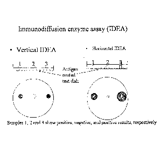
Figure 1 is a schematic representation of two immunodiffusion enzyme
assays (IDEAs). The vertical (VIDEA) and horizontal (HIDEA) embodiments are
shown,
.. along with a representation of how positions 1, 2, and 3 show positive,
negative, and
positive results, respectively.
DEFINITIONS
As used herein, the terms porcine reproductive and respiratory syndrome
(PRRS) virus, or PRRSV, refer to a virus which causes PRRS, Mystery Swine
Disease
(MSD), Swine Infertility and Respiratory Syndrome (SIRS) which was previously
known as
"blue-eared syndrome", porcine epidemic abortion and respiratory syndrome
(PEARS),
Wabash syndrome, mystery pig disease (MPD), swine plague, blue abortion
disease or blue
ear disease in the United Kingdom, abortus blau in the Netherlands,
seuchenhafter spatabort
der schweine in Germany, and Heko-Heko disease.
PRRSV protein refers to any polypeptide product encoded by the PRRSV
genome and/or produced as only as a result of PRRSV infection or the PRRSV
lifecycle.
Thus PRRSV specific, and thus not encoded by or expressed by a PRRSV infected
cell,
polypeptides are within the scope of the term. Endogenous polypeptides encoded
by a
PRRSV infected cell, but not expressed in the absence of PRRSV infection
and/or lifecycle,
are not intended. However, endogenous polypeptides expressed only as a
consequence of
PRRSV infection and/or lifecycle are within the scope of the term. The term
also includes
alternative forms of the polypeptides due to changes in secondary and/or
tertiary structure,
such as those resulting from partial or substantial protein denatufation as a
non-limiting
example. Thus denatured forms of the polypeptides are within the scope of the
term.
PRRSV antigen refers to any portion or fragment of a PRRSV polypeptide
that is recognized by an anti-PRRSV antibody. In some cases, the portion or
fragment may
be a peptide with an attached moiety, such as, but not limited to, a sugar
moiety, a
phosphate moiety, or a lipid moiety. Alternatively, the portion or fragment
may be a
peptide without any attached non-peptide moieties.
7
CA 02614332 2008-01-04
WO 2007/006031
PCT/US2006/026456
DETAILED DESCRIPTION OF MODES OF PRACTICING THE INVENTION
The invention relates to kits, devices, and methods directed to the detection
anti-PRRSV antibodies. The detection is based upon the use of one or more
PRRSV
encoded proteins and/or antigens which binds antibodies against said proteins.
The
antibodies are those present in a PRRSV infected subject but absent in
uninfected
individuals. The invention may be viewed as an "antibody capture" assay
wherein the
captured antibody is detected. The invention also may be considered as
providing
immuno diffusion based methods for the detection of anti-PRRSV antibodies.
The invention is based in part upon the recognition that the structural
proteins of PRRSV, including the nucleocapsid (N), membrane (M) associated and
at least 4
envelope (E) proteins may be used to detect antibodies in a PRRSV infected
subject. Stated
differently, the invention is based in part on the discovery that antibodies
against these
proteins, and/or antigenic portions thereof, are present in PRRSV infected
subjects such that
detection of the antibodies provides an advantageous means to indicate that a
subject is
infected with PRRSV.
The invention is also based in part on the discovery that certain conditions
and protocols may be used to obtain a composition of PRRSV proteins and/or
antigens that
contains a high concentration of E proteins relative to other PRRSV proteins
and/or
antigens. Thus the invention includes a method of preparing PRRSV proteins
and/or
antigens from cells infected therewith. The proteins and/or antigens are
harvested at an
early time point after infection when the majority, or entirety, of the
proteins remain
associated with the infected cells or are otherwise part of a cell associated
viral component
(CAVC) of the invention. Stated differently, the majority or entirety of PRRSV
proteins
and/or antigens are either within the infected cells or associated with the
cell membrane of
the infected cells. Under such conditions, relatively few, if any, PRRSV
particles are
present in the extracellular environment outside the cells. The invention is
based in part on
the unexpected discovery that cells infected for a relatively short period of
time can produce
sufficient amounts of PRRSV encoded proteins that are suitable for use in the
detection
methods of the invention. The preparation of CAVC from an early time point,
before the
production of PRRSV particles and/or the release thereof into the
extracellular environment
also has the benefit of increased safety in that no infectious viral particles
are present as a
contaminant.
8
CA 02614332 2008-01-04
WO 2007/006031
PCT/US2006/026456
The infected cell may be any that is capable of being productively infected
by PRRSV. Non-limiting examples include porcine cells, either in vitro or in
vivo. One
non-limiting example of cells in vitro is primary cells from a porcine subject
that are
infected with PRRSV. A non-limiting example of cells in vivo are porcine
alveolar
macrophages (PAMs) which are infected with PRRSV via an oronasal route and
then
subsequently harvested for preparation of PRRSV proteins and/or antigens.
Another non-
limiting example is with the use of the simian cell line MARC-145. The PRRSV
proteins
and/or antigens produced by the method include at least one of the PRRSV N, M,
and E
proteins. Combinations of these proteins may also be prepared from cells of
the invention.
In some embodiments, the prepared proteins/antigens have a high ratio of E
proteins relative
to N and M proteins.
Thus the invention provides a method of preparing PRRS virus proteins and
antigens from PRRS virus infected cells, where the method comprises a)
providing a
population of cells infected with PRRS virus; b) isolating the infected cells
away from cell-
free PRRS virus to form cells containing cell associated PRRS virus proteins
and antigens;
and c) extracting or eluting PRRS virus proteins and antigens from the cells
isolated in part
b). In cases wherein there is no cell-free virus present, then part b) may be
modified to
simply be an act of isolating the cells from other materials which may
interfere with the
method, such as the culture medium used with the cells. Part b) may be
performed by any
means known in the art, such as by use of centrifugation to generate a cell
pellet and
supernatant that is removed or discarded or by use of a membrane filtration to
remove the
supernatant and retain the cells. Part c) is optionally performed by
resuspension of cells in a
buffer. In some embodiments, the extraction or elution is with a detergent
containing
solution, thus the buffer used to resuspend cells may contain detergent.
In other embodiments, the method comprises use of a population of cells that
has been infected with PRRS virus for a sufficient time to produce little to
undetectable
amounts of infectious units per ml of supernatant, such as the culture media
used with the
cells. Non-limiting examples include less than 101s tissue culture infective
dose (TCID)so
/ml). The population of cells used in the method may optionally have been
infected with
PRRS virus for a sufficient time to observe CPE. Of course cells that have not
been
infected long enough for CPE, or early stages of CPE, to be observed may also
be used in
the practice of the invention.
In a further embodiment, the invention provides a method of preparing PRRS
virus proteins and antigens from PRRS virus infected pig alveolar macrophage
(PAM) cells.
9
CA 02614332 2014-09-08
The method may comprise preparing said proteins and antigens from a population
of PAM
cells obtained by lung lavage of a pig oronasally inoculated with PRRS virus.
The PRRS
virus proteins and antigens may be extracted or eluted from the cells as
described above.
Thus the cells may be resuspended in an extraction buffer. In some
embodiments, the
extraction or elution is with a detergent containing solution, thus the buffer
used to
resuspend cells may contain detergent,
The detergent containing solution may be any that is suitable for extracting
or eluting PRRSV proteins and/or antigens. One non-limiting example is the use
of a non-
ionic detergent, like poly(ethylene glycol) p-isooetyl-phenyl ether,
octylphenoxypolyethoxyethanol (Nonidet P-40Tm), or Triton X-100Tm. In some
embodiments of
the invention, the detergent is at a concentration of about 0.5% in solution,
such as a
solution of about 0.5% Triton X-100.
The invention also provides a method of preparing PRRS virus proteins and
=
antigens from PRRS virus infected cells, Such a method comprises preparing
said proteins
and antigens from a population of the cells prepared by in vitro and in vivo
methods. For
the in vitro method, pig alveolar macrophage (PAM) cells or MARC-145 line
cells are
cultured, and the cells are harvested following an infection of PRRSV. For the
in vivo
method without using a cell culture system, PAM cells obtained by lung lavage
of a pig
following inoculation of PRRS virus oronasally. . The antigen yields were
compared using
different PRRSV isolates and different days after virus inoculation, and
optimal conditions
for the highest antigenic yields were predetermined by comparative testings.
The invention of course includes compositions containing the isolated
PRRSV proteins and/or antigens prepared by any of the methods disclosed
herein. The
compositions may be used for any purpose for which PRRSV proteins and/or
antigens are
used. Non-limiting examples include use to prepare antibodies against the
proteins/antigens; use as reference markers for PRRSV proteins; and use as an
immunogenic compositions, such as in the case of a vaccine formulation,
optionally with a
suitable adjuvant, that is administered to an animal to generate an immune
response.
Additional non-limiting examples of the compositions include those where the
protein(s)/antigen(s) is/are in soluble or lyophilized (freeze dried) form.
In some embodiments, the composition is in a solution suitable for coating a
surface, such as a surface of a device of the invention. Such solutions allow
the proteins to
be coated on the bottom of Petri dishes. Non-limiting examples include
solutions of dilute
PRRSV proteins/antigens in 0,06M carbonate buffer solution at pH 9.6. The
solutions may
CA 02614332 2008-01-04
WO 2007/006031
PCT/US2006/026456
be used to coat the surface of a dish or well, such as those of polystyrene or
glass Petri
dishes or multi-well plates, respectively. Non-adsorbed material may be poured
off,
optionally followed by one or more washes in buffered solution without the
proteins/antigens. The coating may be conducted at various temperatures,
including those
below 25 C, for various time periods. In some embodiments, coating may be at
or about
4 C for about 72 hours.
Thus the invention also provides a method of coating a surface with virus
protein(s) and/or antigen(s), said method comprising a) providing a solution
containing one
or more PRRS virus proteins and antigens prepared as described herein; and b)
contacting
said solution with a surface for a period of time to allow said proteins and
antigens to adsorb
to the surface of said dish. In some embodiments, the protein(s) and/or
antigen(s) were
extracted or eluted from PRRSV infected cells by use of a detergent containing
solution as
described herein. In further embodiments, the surface to be coated is made of
polystyrene
or glass or similar material. The surface is preferably washed with ethanol
which is then
dried off.
A solution of PRRSV proteins/antigens is applied to the clean surface and
allowed to adsorb for from about 2 to about 4 hours to overnight. The
proteins/antigens
may be diluted to an optimum concentration between about 0.01 to 0.001%,
preferably in a
sodium bicarbonate-sodium carbonate coating buffer to promote adherence of the
proteins/antigens to the surface. The coating buffer should preferably be
between about
0.01M to 0.1M (pH 9 to 10) containing from about 0.84 to 8.4 grams per liter
of NaHCO3
and 0.11 to 10.6 grams per liter of Na2CO3. After the coating period, the
excess solution is
poured off or otherwise removed. The coated surface may be washed with
distilled water or
buffer without the proteins/antigens. The coating with PRRSV protein(s) and/or
antigen(s)
may be optionally followed by coating with a blocking agent, such as, but not
limited to,
1% bovine serum albumin (BSA) in phosphate-buffered saline (PBS), pH 7.2. The
coating
may be conducted at various temperatures, including those of about 37 C, for
various time
periods. In some embodiments, coating may be at or about 37 C for about 1
hour. After the
application of blocking agent, the solution is removed and optionally followed
by one or
more water washed or washes comprising buffer solution without the agent.
After coating, a permeable barrier layer is applied to the coated surface. The
barrier may be applied as a solution which later forms a permeable barrier
after drying.
Non-limiting examples include solutions of agar and/or agarose which when
dried form a
permeable barrier composed of a gel like material. Thus a melted agar or
agarose layer may
11
CA 02614332 2008-01-04
WO 2007/006031
PCT/US2006/026456
be applied and permitted to solidify. The depth of the agar layer is not
critical and may vary
from at least about 1.5 mm and preferably about 2 mm up to about 10 mm. The
layer is
applied from solution between about 0.75 to 1.5% and preferably about 1%.
As a non-limiting example with polystyrene petri-dishes that are 60 mm in
diameter, 4 ml of 0.6% agarose in PBS is used for VIDEA based methods as
described
herein. Six (6) ml of the same solution is used for HIDEA based methods. The
solution is
allowed to solidify, such as at 4 C, overnight. Devices that are so coated may
be stored,
such as at 4 C in a moisture chamber, for up to 4 months.
A device with a coated surface as described herein may be used as all or part
of a diagnostic kit for the detection of anti-PRRSV antibodies. The invention
thus also
provides a method of detecting antibodies to PRRS virus which method comprises
a)
collecting a blood or blood serum sample from a subject; b) contacting said
sample with a
permeable barrier of a coated device as described herein; c) incubating said
sample to allow
diffusion of molecules such as antibodies through the permeable barrier and
bind the
protein(s)/antigen(s) used to coat the device; d) removing the permeable
barrier and
optionally washing the device to remove unbound antibodies; e) detecting
complexes
formed by the binding of antibodies to the protein(s)/antigen(s). With respect
to the device
in part b), one non-limiting example is where the device is a dish test plate
comprising (1) a
dish having a flat supporting surface; (2) a coating adsorbed on said surface
of PRRSV
protein(s)/antigen(s); and (3) a layer of agar overlaying said coating.
Regarding part e), the method may be practiced with application of a detector
agent that binds the formed complexes. One non-limiting example is with the
use of a
secondary antibody that binds antibodies that may be present in the sample.
Non-limiting
examples of such secondary antibodies include those from specific animals,
e.g., mouse, rat,
goat, rabbit, etc., which recognize the Fe portions of the antibodies in the
sample tested.
The secondary antibody may be conjugated to a label to facilitate its
detection. The conjugation modifies the antibody by attachment of another
moiety thereto.
The moiety is preferably a detectable label, including a directly detectable
label such as a
radioactive isotope, a fluorescent label (Cy3 and Cy5 as non-limiting
examples) or a
particulate label. Non-limiting examples of particulate labels include latex
particles, metal
sols, and colloidal gold particles. Alternatively, the label may be for
indirect detection.
Non-limiting examples include an enzyme, such as, but not limited to,
peroxidase,
luciferase, alkaline phosphatase, and horse radish peroxidase. Other non-
limiting examples
12
CA 02614332 2008-01-04
WO 2007/006031
PCT/US2006/026456
include a molecule bound by another molecule, such as, but not limited to,
biotin, an affinity
peptide, or a purification tag. Preferably, the label is covalently attached.
In some embodiments, the secondary antibody is an enzyme conjugated anti-
swine immunoglobulin which is allowed to bind the antibodies in the complexes
for about
30 to about 60 minutes. After binding, unbound secondary antibody may be
removed by
one or more optional washes. The enzyme may be any suitable enzyme, such as
the
enzymes used in enzyme linked immunosorbent assays (ELISAs), including a
peroxidase.
Peroxidase produces a purple color when reacted with aminosalicylic acid and
hydrogen
peroxide, or p-phenylene diamine and hydrogen peroxide. Alkaline phosphatase
produces a
yellow color when reacted with dinitrophenylphosphate. Beta-galactosidase
reacts with 0-
nitrophenyl,beta.-D-galactopyranoside to give a purple color.
Continuing with the non-limiting example of an enzyme linked secondary
antibody, detection of a complex by detection of the bound secondary antibody
may be
mediated by overlaying an agar solution containing a substrate which is
reacted upon by the
enzyme to produce a detectable signal, such as color production as a non-
limiting example.
In some embodiments, the detectable signal is visually observable, such as by
the unaided
eye. The signal may be compared to the color reaction observed with the use of
positive
controls (containing known anti-PRRSV antibodies which bind the coated
surface) and/or
negative controls containing no such anti-PRRSV antibodies.
In some embodiments of the invention, particularly the VIDEA as described
herein, the sample is collected and applied to (or is collected by) a porous
material such as,
but not limited to, filter paper or a filter paper disc. With the use of paper
discs as a non-
limiting example, the disc is placed flat on the permeable barrier to allow
molecules to
diffuse from the disc, through the barrier, and to the coated surface. At the
surface,
molecules (like antibodies) from the sample which bind to PRRSV
proteins/antigens of the
invention form complexes with the proteins/antigens. Thus the molecules from
the sample
become immobilized to the coated surface. In some embodiments, the period of
time for
diffusion and complex formation is about 2 to about 3 hours at either about
room
temperature (25 C) or about 37 C.
After complex formation, the permeable barrier is removed. In some
embodiments comprising an agar or agarose barrier, the layer of agar or
agarose may be
peeled off followed by the optional washes.
The methods of the invention are based upon the fonnation of a complex
comprising the PRRSV proteins/antigens bound to anti-PRRSV antibodies of a
sample as
13
CA 02614332 2008-01-04
WO 2007/006031
PCT/US2006/026456
described herein. The anti-PRRSV antibodies may be detected to improve the
ease of
detecting the complex. Detection of a complex of PRRSV proteins/antigens and
anti-
PRRSV antibodies from a sample indicates the presence of PRRSV infection in
the subject
from which the sample was obtained.
The invention further provides a vertical immunodiffusion enzyme assay
(VIDEA) method, said method comprising a) providing a diagnostic device
comprising at
least one surface coated with one or more PRRSV proteins and antigens as
described herein,
said surface having been overlaid with a permeable barrier as described
herein; b)
contacting the surface of the barrier with a porous material comprising an
antibody
containing sample derived from a subject; c) incubating said device for a
period sufficient to
allow diffusion of material from said sample to said one or more surface, said
period
optionally occurring after removal of the porous material; and d) detecting
the presence of
antibodies at the surface of the dish after removal of the permeable barrier.
In some
embodiments, the porous material is a paper disk.
The invention further provides a horizontal immunodiffusion enzyme assay
(HIDEA) method, said method comprising a) providing a diagnostic device
comprising at
least one surface coated with one or more PRRSV proteins and antigens as
described herein,
said surface having been overlaid with a permeable barrier as described herein
but
comprising one or more indentations, for receiving a sample, on said surface;
b) contacting
the one or more indentations with an antibody containing sample derived from a
subject; c)
incubating said device for a period sufficient to allow diffusion of material
from said sample
to said one or more surface; and d) detecting the presence of antibodies at
the surface of the
dish after removal of the permeable barrier. In some embodiments, from one to
a plurality
of small diameter holes are punched out of the permeable barrier to form
indentations that
function as test sample wells. The wells may be about 1 to 4 mm in diameter,
for example,
and penetrate through the agar coating. The use of a template with seven 3-mm
circular
wells to punched holes onto the agar of each testing dish for HIDEA is one non-
limiting
example.
In both VIDEA and HIDEA embodiments, the sample is a serum sample or a
whole blood sample. The methods may be applied to samples from a variety of
animals
and/or subjects that may have been infected with PRRSV or that are suspected
of being
infected with PRRS virus. The diagnostic device may be a dish, plate, or well
of a plate.
Both positive and negative controls may be run with each set of test samples.
The same
amount of control serum is placed in other wells on the test device.
14
CA 02614332 2008-01-04
WO 2007/006031
PCT/US2006/026456
After incubation of the plates, the agar gel layer is peeled off and the
plates
are washed, such as with a washing buffer such as Tween 20 in phosphate
buffered saline as
a non-limiting example. Plates may be washed with distilled or tap water
rather than
washing buffer. Washing removes unbound (non-specific) antibodies as well as
other
contaminants.
Both the VIDEA and HIDEA formats include the use of an antigen-antibody
reaction to form a complex, which may be detected by use of a detecting agent,
such as a
secondary antibody, to bind the complex, such as by binding the antibody
portion of the
complex. The detector agent may be kept in contact with complexes for about 30
minutes
to about 2 hours at room temperature. The plates are then washed, such as with
buffered
washing solution as a non-limiting example to remove unbound conjugate. The
washing
liquid may be added slowly from the edge of the test plate with a syringe or
pipette and
poured off. This is optionally repeated up to three times or more.
While the detector agent is incubating, an agar or agarose coating is
prepared. As a non-limiting example, a 1% solution of agar, preferably in
phosphate
buffered saline, is melted and a substrate for the enzyme of the conjugate is
incorporated.
In some embodiments, a catalyst is incorporated as needed. As a non-limiting
example,
when the enzyme is a peroxidase, the I% agar solution may contain between
about 0.05 to
0.10% of 5-aminosalicylic acid as the substrate and between about 0.002 and
0.01%
hydrogen peroxide as catalyst. The use of about 0.08% substrate and about
0.005% catalyst
may also be used. The agar is poured over the washed surface and allowed to
solidify.
A color reaction between the enzyme of the secondary antibody occurs
within the agar support layer. The reaction aids in the visualization by an
enzyme-substrate
reaction. The diameters of dark purple circular zones were measured for HIDEA
and the
presence or absence or color density were recorded for VIDEA to determine
antibody
quantities.
In the case of HIDEA, the color develops in the form of a circular zone or
ring. The rings are dark enough to measure within about 5 to about 30 minutes
of the
substrate reaction. Upon standing for longer periods, the ring will become
darker but will
not enlarge. The diameter of the ring produced is related to the amount of
specific antibody
present in the blood (i.e., virus neutralization titer). The diameter of the
dark colored
circular zone is measured and used to correlate with a standard virus
neutralization test
antibody value. The values determined are related to the size of the sample
well in the test
CA 02614332 2008-01-04
WO 2007/006031
PCT/US2006/026456
plate and the size of the sample used. A table of values and/or depictions of
representative
rings may be included with each device of the invention or with each kit of
the invention.
The detecting of the presence of antibodies at the surface in both the VIDEA
and HIDEA formats is meant to detect antibodies bound to PRRSV
protein(s)/antigen(s) on
the surface. The detection may be performed by any means described herein,
including the
use of a labeled secondary antibody. Other non-limiting means include the
visualization of
an antigen-antibody reaction via an enzyme-substrate reaction, while the
presence of color
reaction or density from the reaction is used to indicate the amount
(quantitative or semi-
quantitative) or presence (qualitative) of anti-PRRSV antibodies.
The PRRSV proteins/antigens as well as compositions, methods, and devices
comprising the proteins/antigens are suitable for the preparation of kits
produced in
accordance with well known procedures. The invention thus provides kits
comprising the
PRRSV proteins/antigens as described herein, or compositions or devices
comprising them,
for use in one or more methods as disclosed herein. Such kits optionally
further comprise
an identifying description or label or instructions relating to their use in
the methods of the
present invention. Such a kit may comprise containers, each with one or more
of the
various reagents (typically in concentrated form) or devices utilized in the
methods. A set
of instructions will also typically be included.
Kits comprising a device of the invention may further comprise one or more
additional reagents or pieces of equipment for use with the device in a method
of the
invention. Non-limiting examples of additional materials for inclusion are
sample diluent
solution, diluent vial, and a dropper for transfer of sample. Other non-
limiting examples
include porous materials for use with the VIDEA format and secondary
antibodies.
Having now generally described the invention, the same will be more readily
understood through reference to the following examples which are provided by
way of
illustration, and are not intended to be limiting of the present invention,
unless specified.
EXAMPLES
Example 1: Preparation of PRRSV proteins and dishes
PRRSV proteins may be prepared by use of a PRRSV strain to inoculate
susceptible cells in vitro or in vivo and harvesting infected cells at an
optimal time to
prepare cell associated viral components. For in vitro methods, the antigen
may be prepared
16
CA 02614332 2008-01-04
WO 2007/006031
PCT/US2006/026456
by a cell culture system or by the use of recombinant technologies.
Alternatively, PRRSV
infected pig alveolar macrophage (PAM) cells can be obtained from pigs
following oronasal
inoculation with PRRSV and washing the lung (lung lavage).
The cells were pelleted by centrifugation, and the supernatant removed or
discarded. The pellets may be optionally washed. Cell pellets were suspended
in a 0.05M
tris (hydroxymethyl) aminomethane 0.025M EDTA buffer containing 0.5% Triton X-
100 at
a volume of 5-10 times that of the packed cells. The mixture was stirred for 2-
15 hours at
4 C and centrifuged at 10,000g for 1 hour. The supernatant was used as PRRSV
antigen.
The antigen is non-infectious allowing wide use without a risk of viral
spreading.
A dilution of PRRSV protein/antigen in 0.06M carbonate buffer solution at
about pH 9.6 was pre-determined by comparative tests. The antigen was coated
on
polystyrene petri-dishes (60 mm in diameter) by adsorbing at 4 C for 72 hours.
Unadsorbed
antigen was poured off, and the dishes were incubated with a blocking agent
(e.g. 1%
bovine serum albumin, BSA) in phosphate-buffered saline (PBS, pH 7.2) for 1
hour at 37 C.
After removing the BSA, 6 ml for HIDEA and 4 ml for VIDEA of 0.6% agarose in
PBS
was overlayed in the petri-dishes. The agar was allowed to solidify at 4 C
overnight. Using
a template, seven 3-mm circular wells were punched onto the agar of each
testing dish for
HIDEA. For VIDEA, the dishes without wells were used. All of the test dishes
were stored
at 4 C in a moisture chamber for up to 4 months.
Example 2: VIDEA
Test plates were prepared by coating viral antigens on the bottom of Petri
dishes followed by an overlaying of agar gel generally as described above. In
use, a filter
paper disc is soaked with test serum from pigs and placed on the agar of the
test plate. The
flatness of the disc presents a nearly two dimensional starting area such that
the contents of
a sample like a test serum would move generally in one direction through the
agar and
toward the coated surface.
The gel was peeled off after an incubation period and the dishes were washed
3 times with 5 to 8 ml of washing solution (PBS containing 0.05% Tween-20).
Then 3 ml
of commercially available peroxidase-conjugated rabbit anti-pig IgG (1 to 2
g/ml) diluted
in PBS was applied for 45 minutes at 25 C. After the dishes were washed again
3 times
with washing solution, 5 ml of 1% agar in PBS containing substrate (5-
aminosalicylic acid)
and H202 at concentrations of 0.08% and 0.005%, respectively, was overlaid
onto the
dishes. The presence or absence or color density were recorded for VIDEA
17
CA 02614332 2008-01-04
WO 2007/006031 PCT/US2006/026456
In one experiment, the incubation was at 25 or 37 C for about 2 to 3 hours
after which the agar was peeled off. The peroxidase reaction results were
determined after
about 5 to 10 minutes based on positive samples showing a color reaction.
Various serum
samples with known ELISA S/P ratios or history of PRRS virus infection were
tested along
with samples from naïve animals. The sensitivity and specificity of VIDEA was
evaluated
with respect to ELISA. Table 1 below summarizes the results of this VIDEA and
the
corresponding ELISA S/P ratios for various pig sera. The percent agreement
between the
assay results are shown in parentheses. VIDEA showed 100% specificity for pig
sera found
to be negative in the ELISA. Incubation at 25 C for about 3 hours showed 100%
agreement
-- for all samples when compared to the ELISA.
Table 1
VIDEA VIDEA VIDEA VIDEA
37 C, 2 hrs 37 C, 2 hrs 25 C, 3 hrs 25 C, 3
hrs
ELISA OD No. of sera positive negative positive negative
Naive 40 0 40 (100%) 0 40 (100%)
<0.4 30 20 (66.6%) 10 (33.3%) 30 (100%) 0
0,4-0.9 32 31(96.9%) 1(3.1%) 32 (100%) 0
1.0-1.9 17 17(100%) 0 17(100%) 0
>2.0 10 10(100%) 0 10(100%) 0
Antigen-antibody reactions occurred in as little as 2 hours, however, longer
-- times appear to beneficial in cases of low antibody titers, which may be
observed as false
negatives at short incubation times. The high correlation of VIDEA to ELISA
S/P rations
indicates the ability of VIDEA as an assay for detecting anti-PRRS virus
antibodies. The
correlation with the ELISA S/P ratio of <0.4, which is the standard cut-off
suggests that
VIDEA may have equivalent sensitivity to ELISA.
Example 2: HIDEA
Devices of the invention with a coated surface and a permeable barrier are
used wherein the barrier material is modified such that it defines one or more
indentations in
the surface of the barrier. Using agarose as a non-limiting example, one or
more wells may
be made in the agarose.
18
CA 02614332 2008-01-04
WO 2007/006031 PCT/US2006/026456
Each well was filled with 0.015 ml of test serum. Devices were incubated
for about 12 to about 24 hours at room temperature (25 C) to allow antibody
diffusion and
antigen-antibody reaction. Additional uses of the devices were conducted with
24 hour
incubation times.
The gel was peeled off after an incubation and the dishes were washed 3
times with 5 to 8 ml of washing solution (PBS containing 0.05% Tween-20). Then
3 ml of
commercially available peroxidase-conjugated rabbit anti-pig IgG (1 to 2
jig/m1) diluted in
PBS was applied for 45 minutes at 25 C. After the dishes were washed again 3
times with
washing solution, 5 ml of 1% agar in PBS containing substrate (5-
aminosalicylic acid) and
-- H202 at concentrations of 0.08% and 0.005%, respectively, was overlaid onto
the dishes.
The diameters of dark purple circular zones were measured in mm after 5 to 30
minutes of
reaction for HIDEA. Tables 2 and 3 show different diameters of 14 sera tested
under
different conditions. The diameters of <7 mm are considered negative in HIDEA.
Some of
the ELISA negative sera showed positive results indicating better sensitivity
for HIDEA.
-- Results in Table 3 indicate a high repeatability of HIDEA.
Table 2. HIDEA diameters of swine sera with different ELISA S/P ratios using
various
incubation hours at room temperature (25 C)
Incubation hours
Serum tested 15h 18 h 20h 24h
1. PRRS-free 3* 6 4 5
2. PRRS-free 4 6 4 5
3. ELISA S/P ratio** 0.06 7 8 9 9
4. ELISA S/P ratio 0.21 8 10
10 10
5. ELISA S/P ratio 0.73 8 11
11 11
6. ELISA S/P ratio 1.96 11 13
14 14
7. ELISA S/P ratio 2.83 12 14
15 15
* Diameter (mm) in HIDEA; greater than 7 mm are positive
** ELISA S/P ratio greater than 0.4 are positive
Table 3. Repeatability of HIDEA diameters for 7 sera with different ELISA S/P
ratios and
various incubation hours at room temperature (25 C)
__________________________________________________________________
Serum tested 3 h 6 h 12 h 15 h 21 h 24 h
1. a. PRRS-free 3* 3 3 3 4 4
b. PRRS-free 3 3 3 3 4 4
2. a, PRRS-free 3 3 3 3 4 4
b. PRRS-free 3 3 3 3 4 4
19
CA 02614332 2014-09-08
3. a. ELISA-neg 3 3 3 . 3
4 4
b. ELISA-neg 3 3 3 3 4 4
4. a. ELISA-neg 3 3 4 4
4 4
b. ELISA-neg 3 3 4 4 4 4
5. a. ELISA S/P ratio 0.54 5 7 7 8 9 9
= b. ELISA S/P ratio 0.54 5 7 8
8 9 9
6. a. ELBA S/P ratio 1.80 5 7 7 7 10 10
= b. ELISA S/P ratio 1.80 5 7 7
8 10 10
7, a. ELISA S/P ratio 2.59 5 7 7 8 10 11
b. ELISA S/P ratio 2.59 5 7 8 8 11 12
a 8c b, each serum sample is tested twice by HIDEA for reproducibility
* Diameter (mm) hi HIDEA; greater than 7 mm are positive .
** ELISA S/P ratio greater than 0.4 are positive
As used herein, the terms
"a", "an", and "any" are each intended to include both the singular and plural
forms.
Having now fully described this invention, it will be appreciated by those
skilled in the art that the same can be performed within a wide range of
equivalent
parameters, concentrations, and conditions without departing from the scope
of
the invention and without undue experimentation. While this invention has been
described
in connection with specific embodiments thereof, it will be understood that it
is capable of
further modifications. This application is intended to cover any variations,
uses, or
adaptations of the invention following, in general, the principles of the
invention and
. including such departures from the present disclosure as come
within known or customary
practice within the art to which the invention pertains and as may be applied
to the essential
features hereinbefore set forth.
=