Note: Descriptions are shown in the official language in which they were submitted.
CA 02615160 2013-12-17
=
MEDICAL-TREATMENT ELECTRODE ASSEMBLY
AND METHOD FOR MEDICAL TREATMENT
[0003] Field of the Invention
[0004] The present invention is related generally to medical systems, and more
particularly to a
medical-treatment electrode assembly and to a method for medical treatment.
[0005] Background of the Invention
[0006] A medical-treatment electrode assembly is known wherein a tube having
two electrodes
is inserted into a patient's esophagus, wherein the two electrodes are
operatively connected to a
medical radio-frequency (RF) generator, and wherein the two electrodes are
brought into contact
with esophageal tissue to treat gastro-esophageal reflux disease and other
diseases of mucosal
tissue.
[0007] Still, scientists and engineers continue to seek improved medical-
treatment electrode
assemblies and methods for medical treatment.
1
CA 02615160 2008-01-11
WO 2007/011634
PCT/US2006/027154
2
[0008] Summary
[0009] A first expression of a first embodiment of a medical-treatment
electrode assembly of the invention includes a flexible tube and a first
medical-
treatment flexible electrode. The flexible tube includes a sidewall having an
outer surface and includes a distal end insertable into a patient. The first
medical-treatment flexible electrode is fixedly supported on the outer surface
of
the sidewall of the flexible tube proximate the distal end of the flexible
tube.
The first medical-treatment flexible electrode is contactable with patient
tissue.
The first medical-treatment flexible electrode is operatively connectable to a
medical radio-frequency (RF) generator.
=
[0010] A second expression of a first embodiment of a medical-treatment
electrode assembly of the invention includes a flexible tube and two medical-
treatment electrodes. The flexible tube has an outer surface and has a distal
end
insertable into a patient. The two medical-treatment electrodes are supported
on
the outer surface of the flexible tube proximate the distal end, are
contactable
with patient tissue, and are operatively connectable to a medical radio-
frequency (RF) generator, wherein the two medical-treatment electrodes are
spaced apart, and wherein a video camera of a flexible endoscope inserted into
the flexible tube proximate the distal end can view the patient tissue between
the two medical-treatment electrodes.
[0011] A first expression of a second embodiment of a medical-treatment
electrode assembly of the invention includes a first medical-treatment
electrode
body. The first medical-treatment electrode body is insertable into a patient
and
is operatively connectable to a medical radio-frequency (RF) generator. The
first medical-treatment electrode body has an outer surface contactable with
patient tissue, has a central lumen operatively connectable to a vacuum
source,
and has an opening extending from the outer surface to the central lumen.
10012] An expression of a third embodiment of a medical-treatment electrode
assembly of the invention includes a tube, a medical-treatment electrode, and
CA 02615160 2008-01-11
WO 2007/011634
PCT/US2006/027154
3
patient-tissue-moving apparatus. The tube is insertable into a patient and
includes a sidewall having an opening. The medical-treatment electrode is
supported by the tube, is contactable with patient tissue which is outside the
tube, and is operatively connectable to a medical radio-frequency (RF)
generator. The patient-tissue-moving apparatus moves patient tissue into the
opening to tighten patient tissue outside the tube against the medical-
treatment
electrode.
[0013] A method of the invention is for medical treatment and includes
inserting a tube into a hollow body organ of a patient, wherein the tube
supports
a medical-treatment electrode and has a sidewall opening. The method also
includes then moving patient tissue into the sidewall opening to tighten
patient
tissue outside the tube into substantially full contact with the medical-
treatment
electrode. The method also includes then activating the medical-treatment
electrode.
[0014] Several benefits and advantages are obtained from one or more of the
expressions of one or more of the embodiments and method of the invention. In
one application, having a medical-treatment electrode which is flexible
provides
more intimate contact between the electrode and patient tissue which reduces
charring of patient tissue and which improves non-visual monitoring of tissue
treatment. In the same or a different application, being able to have a video
camera of a flexible endoscope view patient tissue between two medical-
treatment electrodes allows the user to visually monitor tissue treatment for
patient tissue between the two medical-treatment electrodes. In one
implementation, having a medical-treatment electrode body with a central
lumen operatively connectable to a vacuum source and with an opening
extending from the outer surface of the medical-treatment electrode body to
the
central lumen provides a vacuum to draw patient tissue into more intimate
contact with the electrode. In one employment, having patient-tissue-apparatus
apparatus for moving patient tissue into a sidewall opening of a tube
supporting
a medical-treatment electrode tightens patient tissue outside the tube
against,
and into substantially full contact with, the medical-treatment electrode.
CA 02615160 2008-01-11
WO 2007/011634
PCT/US2006/027154
4
[0015] Brief Description of the Figures
[0016] FIGURE 1 is a schematic, longitudinal cutaway view of an
embodiment of a medical instrument which includes a first embodiment of a
medical-treatment electrode assembly of the invention and which includes a
medical radio-frequency (RF) generator and an embodiment of a flexible
endo scope;
[0017] FIGURE 2 is a view of a portion of the medical-treatment electrode
assembly of Figure 1, taken along lines 2-2 in Figure 1;
[0018] FIGURE 3 is a schematic, top planar view of a second embodiment of
a medical-treatment electrode assembly of the invention;
[0019] FIGURE 4 is a cross-sectional view of the medical-treatment electrode
assembly of Figure 3 taken along lines 4-4 in Figure 3;
[0020] FIGURES 5-7 are schematic, radial cross-sectional views of a patient's
esophagus and a third embodiment of a medical-treatment electrode assembly of
the invention showing various stages of patient esophageal tissue being drawn
into a tube opening and of patient esophageal tissue outside the tube being
tightened against two medical-treatment electrodes;
[0021] FIGURE 8 is a schematic, longitudinal cross-sectional view of a
portion of a patient's esophagus and a cutaway view of a fourth embodiment of
a medical-treatment electrode assembly of the invention showing patient tissue
being drawn in a tube opening by grasping forceps and showing patient tissue
outside the tube being tightened against a medical-treatment electrode
supported
by the tube;
[0022] FIGURE 9 is a side elevational view of a corkscrew retractor; and
[0023] FIGURE 10 is a side elevational view of a hook retractor.
CA 02615160 2008-01-11
WO 2007/011634
PCT/US2006/027154
[0024] Detailed Description
[0025] Before explaining several embodiments of the present invention in
detail, it should be noted that each embodiment is not limited in its
application
or use to the details of construction and arrangement of parts and steps
illustrated in the accompanying drawings and description. The illustrative
embodiments of the invention may be implemented or incorporated in other
embodiments, variations and modifications, and may be practiced or carried out
in various ways. Furthermore, unless otherwise indicated, the terms and
expressions employed herein have been chosen for the purpose of describing the
illustrative embodiments of the present invention for the convenience of the
reader and are not for the purpose of limiting the invention.
[0026] It is further understood that any one or more of the following-
described embodiments, expressions of embodiments, examples, etc. can be
combined with any one or more of the other following-described embodiments,
expressions of embodiments, examples, etc.
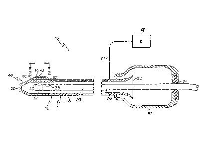
[0027] A first embodiment of a medical-treatment electrode assembly 10 of
the invention is shown in Figures 1 and 2. A first expression of the
embodiment
of Figures 1 and 2 is for a medical-treatment electrode assembly 10 including
a
flexible tube 12 and a first (i.e., at least one) medical-treatment flexible
electrode 14. The flexible tube 12 includes a sidewall 16 having an outer
surface 18 and includes a distal end 20 insertable into a patient (such as,
without
limitation, insertable into the esophagus 22 of a patient 24 shown in Figure 5
for
a third embodiment to be described later). The first medical-treatment
flexible
electrode 14 is fixedly supported (directly or indirectly) on the outer
surface 18
proximate the distal end 20, is contactable with patient tissue (such as
patient
tissue 26 shown in Figure 5), and operatively connectable to a medical radio-
frequency (RF) generator 28.
[0028] In one application, having a medical-treatment electrode which is
flexible provides more intimate contact between the electrode and patient
tissue
CA 02615160 2013-12-17
which reduces charring of patient tissue and which improves non-visual
monitoring of
tissue treatment.
[0029] In one enablement of the first expression of the embodiment of Figures
1 and 2,
the flexible tube 12 has a proximal end 30 disposable outside the patient 24.
In this
enablernent, the medical-treatment electrode assembly 10 also includes a
handle 32
surrounding, and attached to, the flexible tube 12 proximate the proximal end
30 and
includes an annular seal 34 attached to the handle 32. The annular seal 34 is
adapted to
sealing receive a flexible endoscope 36 insertable into the flexible tube 12.
[0030] In one arrangement, wherein a flexible endoscope 36 is employed, the
flexible
endoscope 36 includes an aspiration port 38, and the sidewall 16 includes a
through hole
40 disposed proximate the distal end 20 and in fluid communication with the
aspiration
port 38 of the flexible endoscope 36. In one variation, the first medical-
treatment flexible
electrode 14 includes a through hole 42 aligned with the through hole 40 of
the sidewall
16. In one application, providing a vacuum draws patient tissue into more
intimate
contact with the electrode.
[0031] In the same or a different arrangement, wherein a flexible endoscope 36
is
employed, the flexible endoscope 36 includes a video camera 44, and the
flexible tube 12
includes a distal end cap (e.g., 46) attached to the distal end of the
sidewall 16. In one
construction, the sidewall 16 is a monolithic sidewall extending from the
distal end cap
to the proximal end 30 of the flexible tube 12. In one variation, the distal
end cap and the
sidewall 16 are portions of a monolithic flexible tube 12. In one example, the
distal end
cap is chosen from the group consisting of a flexible tapered closed end cap
46, an open
end cap (not shown) adapted to allow passage therethrough of the video camera
of the
flexible endoscope and an end cap (not shown) adapted to open to allow passage
therethrough of the video camera of the flexible endoscope and to close upon
removal of
the flexible endoscope therefrom.
6
CA 02615160 2013-12-17
[0032] In one employment of the first expression of the embodiment of Figures
1 and
2, a lead 52 operatively connects the first medical-treatment flexible
electrode 14 to
the radio-frequency (RF) generator 28. In one illustration, the lead 52
extends through
a longitudinal channel in the sidewall 16 of the flexible tube 12. In one
variation, the
lead 52 from the electrode exits the flexible tube 12 through the sidewall 16
before
reaching the handle 32. In another variation, not shown, the lead from the
electrode
exits the flexible tube 12 inside the handle 32 and then exits the handle 32.
In one
modification, not shown, the lead is operatively connected to a control button
on the
handle to start and stop the medical treatment. In one method, the lead and
substrate
are manufactured using etched circuit technology, wherein the lead is
substantially flat
and comprises, consists essentially of, or consists of copper, and the
substrate is
substantially flat and comprises, consists essentially of, or consists of
polyester. In one
deployment, shrink wrap, not shown, surrounds the longitudinal juncture of the
flexible
tube and the distal end of the handle.
100331 In one extension of the first expression of the embodiment of Figures 1
and 2,
the medical-treatment electrode assembly 10 also includes a second medical-
treatment
flexible electrode 48. The second medical-treatment flexible electrode 48 is
supported
(directly or indirectly) on the outer surface 18 proximate the distal end 20
and is spaced
apart from the first medical-treatment flexible electrode 14. The second
medical-
treatment flexible electrode 48 is contactable with the patient tissue 26 and
is
operatively connectable to the medical radio-frequency (RF) generator 28. It
is noted
that when only a single electrode is present, the assembly is operated as a
monopolar
assembly, and when two (or more) electrodes are present, the assembly can be
operated
as a monopolar and/or a bipolar assembly as can be appreciated by the artisan.
100341 A second expression of the embodiment of Figures 1 and 2 is for a
medical-treatment electrode assembly 10 including a flexible tube 12 and two
medical-
treatment (flexible or rigid) electrodes 14 and 48. The flexible tube 12 has
an outer
surface 18 and has a distal end 20 insertable into a patient 24. The two
medical-
treatment electrodes 14 and 48 are supported (directly or
7
CA 02615160 2008-01-11
WO 2007/011634
PCT/US2006/027154
8
indirectly) on the outer surface 18 proximate the distal end 20, are
contactable
with patient tissue 26, and are operatively connectable to a medical radio-
frequency (RF) generator 28. The two medical-treatment electrodes 14 and 48
are spaced apart, and a video camera 44 of a flexible endoscope 36 inserted
into
the flexible tube 12 and translated proximate the distal end 20 can view the
patient tissue 26 between the two medical-treatment electrodes 14 and 48.
[0035] In one application, being able to have a video camera of a flexible
endoscope view patient tissue between two medical-treatment electrodes allows
the user to visually monitor tissue treatment for patient tissue between the
two
medical-treatment electrodes.
[0036] It is noted that the enablements, arrangements, variations, etc. of the
previously-described first expression of the embodiment of Figures 1 and 2 are
equally applicable to the second expression of the embodiment of Figures 1 and
2.
[0037] In one construction of the second expression of the embodiment of
Figures 1 and 2, the flexible tube 12 is chosen from the group consisting of a
transparent tube (as shown), a tube (not shown) having a solid transparent
window disposed between the two medical-treatment electrodes, and a tube (not
shown) having a tube cutout disposed between the two medical-treatment
electrodes. In one example, when the flexible tube 12 is a transparent tube,
the
flexible tube 12 comprises, consists essentially of, or consists of
polyethylene,
polyurethane, or polyester. In one variation, the medical-treatment electrode
assembly 10 also includes a transparent substrate 50 bonded to the outer
surface
18 of the flexible tube 12, wherein the two medical-treatment electrodes 14
and
48 are bonded to the substrate 50. In one modification, the lead 52 is bonded
to
the substrate 50, and the substrate 50 (with the bonded lead 52) extends (not
shown) on the outer surface 18 to proximate the proximal end 30 of the
flexible
tube 12. In one example, the substrate 50 comprises, consists essentially of,
or
consists of polyester. It is noted that additional unmarked through holes
(similar to through holes 40 and 42) are shown as small circles on the second
CA 02615160 2008-01-11
WO 2007/011634
PCT/US2006/027154
9
medical-treatment electrode 48, on the substrate 50, and on the flexible tube
12
in Figure 2. The number and layout of the through holes are left to the
artisan.
[0038] A third expression of the embodiment of Figures 1 and 2 is for a
medical-treatment electrode assembly including a flexible tube 12 and two
medical-treatment flexible electrodes 14 and 48. The flexible tube 12 has an
outer surface 18 and has a distal end 20 insertable into a patient 24. The two
medical-treatment flexible electrodes 14 and 48 are fixedly supported on the
outer surface 18 proximate the distal end 20, are contactable with patient
tissue
26, and are operatively connectable to a medical radio-frequency (RF)
generator
28. The two medical-treatment flexible electrodes 14 and 48 are spaced apart.
A video camera 44 of a flexible endoscope 36 inserted into the flexible tube
12
and translated proximate the distal end 20 can view the patient tissue 26
between the two medical-treatment flexible electrodes 14 and 48.
[0039] It is noted that the enablements, arrangements, variations,
constructions, etc. of the previously-described first and/or second
expressions of
the embodiment of Figures 1 and 2 are equally applicable to the third
expression
of the embodiment of Figures 1 and 2.
[0040] A second embodiment of a medical-treatment electrode assembly 54 of
the invention is shown in Figures 3 and 4. A first expression of the
embodiment
of Figures 3 and 4 is for a medical-treatment electrode assembly 54 including
a
first medical-treatment electrode body 56 which is insertable into a patient
24.
The first medical-treatment electrode body 56 is operatively connectable to a
medical radio-frequency (RF) generator 58 and has an outer surface 60
contactable with patient tissue 26. The first medical-treatment electrode body
56 has a central lumen 62 operatively connectable to a vacuum source 64 and
has an opening 66 extending from the outer surface 60 to the central lumen 62.
[0041] In one application, providing a vacuum draws patient tissue into more
intimate contact with the electrode. Examples of vacuum sources 64 include,
without limitation, syringes, squeeze bulbs, and pump motors.
CA 02615160 2008-01-11
WO 2007/011634
PCT/US2006/027154
[0042] In one extension of the first expression of the embodiment of Figures 3
and 4, the medical-treatment electrode assembly 54 also includes a second
medical-treatment electrode body 68 insertable into the patient 24 and spaced
apart from the first medical-treatment electrode body 56. The second medical-
treatment electrode body 68 is operatively connectable to the medical radio-
frequency (RF) generator 58 and has an outer surface 60 contactable with
patient tissue 26. The second medical-treatment electrode body 60 has a
central
lumen (similar to the central lumen 62 of the first medical-treatment
electrode
body 56) operatively connectable to the vacuum source 64 and has an opening
66 extending from the outer surface 60 of the second medical-treatment
electrode body 68 to the central lumen of the second medical-treatment
electrode body 68.
[0043] In one application, the first and second medical-treatment electrode
bodies 56 and 68 are supported on the outside of a flexible tube (not shown)
in a
manner similar to that shown for the embodiment of Figures 1 and 2.
[0044] A third embodiment of a medical-treatment electrode assembly 70 of
the invention is shown in Figures 5-7. A first expression of the embodiment of
Figures 5-7 is for a medical-treatment electrode assembly 70 including a
flexible tube 72 (shown in cross-section), a medical-treatment electrode 74,
and
two rollers 76 and 78. The flexible tube 72 is insertable into a patient 24
and
includes a sidewall 80 having an opening 82. The medical-treatment electrode
74 is supported by the flexible tube 72 and is contactable with patient tissue
26'
which is outside the flexible tube 72. The medical-treatment electrode 74 is
operatively connectable to a medical radio-frequency (RF) generator (such as
RF generator 58 of Figure 3), and the flexible tube 72 is operatively
connectable
to a vacuum source (such as vacuum source 64 of Figure 3) to draw patient
tissue 26" into the opening 82. The two rollers 76 and 78 are disposed inside
the flexible tube 72 and are adapted to rollingly engage patient tissue 26",
drawn into the opening 82 by the vacuum source, to draw more patient tissue
26" into the flexible tube 72 through the opening 82 to tighten patient tissue
26'
outside the flexible tube 72 against the medical-treatment electrode 74.
CA 02615160 2008-01-11
WO 2007/011634
PCT/US2006/027154
11
[0045] In one configuration of the first expression of the embodiment of
,
Figures 5-7, the opening 82 is located closer to the two rollers 76 and 78
than to
the medical-treatment electrode 74. In the same or a different configuration,
the
two rollers 76 and 78 are translatable toward and away from each other.
Mechanisms for rotating and translating the two rollers 76 and 78 are left to
the
artisan.
[0046] In one procedure employing the first expression of the embodiment of
Figures 5-7, the flexible tube 72 of the medical-treatment electrode assembly
70
is inserted into the esophagus 22 of a patient 24 to have a few cellular
layers of
patient tissue 26" outside the flexible tube 72 be medically treated by the
medical-treatment electrode 74. In one example, as shown in Figure 5, at first
some patient tissue 26" is drawn through the opening 82 and into the flexible
tube 72 by vacuum alone with the two rollers 76 and 78 spaced apart enough so
as not to contact such patient tissue 26". In this example, as shown in Figure
6,
then the two rollers 76 and 78 are brought toward each other to rollingly
engage
such patient tissue 26" to begin to draw more patient tissue 26" through the
opening 82 and into the flexible tube 72. This action increases the tightening
around the flexible tube 72 of patient tissue 26' outside the flexible tube
72. In
this example, as shown in Figure 7, the two rollers 76 and 78 have drawn
enough patient tissue 26" through the opening 82 and into the flexible tube 72
to cause patient tissue 26' outside the flexible tube 72 to more intimately
contact the medical-treatment electrode 74. Notice how the gap 84 between the
outside of the flexible tube 72 and the patient tissue 26' outside the
flexible tube
72 decreases from Figure 5 to Figure 6 to Figure 7, and that the gap 84
disappears in Figure 7 between the patient tissue 26' outside the flexible
tube 72
and the outside of the flexible tube 72 at the medical-treatment electrode 74.
[0047] It is noted that, in the example, the direction of rotation of the two
rollers 76 and 78 to draw more patient tissue 26" through the opening 82 and
into the flexible tube 72 is shown by unmarked arrows in Figures 6 and 7. In
one construction, the surface of the two rollers 76 and 78 is textured to
better
grip patient tissue 26".
CA 02615160 2008-01-11
WO 2007/011634
PCT/US2006/027154
12
[0048] A second expression of the embodiment of Figures 5-7 is for a
medical-treatment electrode assembly 70 including a flexible tube 72, two
medical-treatment electrodes 74 and 86, and two rollers 76 and 78. The
flexible
tube 72 is insertable into a patient 24 and includes a sidewall 80 having an
opening 82. The two medical-treatment electrodes 74 and 86 are supported by
the flexible tube 72, are spaced apart, and are contactable with patient
tissue 26'
which is outside the flexible tube 72. The two medical-treatment electrodes 74
and 86 are operatively connectable to a medical radio-frequency (RF) generator
(such as RF generator 58 of Figure 3), and the flexible tube 72 is operatively
connectable to a vacuum source (such as vacuum source 64 of Figure 3) to draw
patient tissue 26" into the opening 82. The two rollers 76 and 78 are disposed
inside the flexible tube 72 and are adapted to rollingly engage patient tissue
26", drawn into the opening 82 by the vacuum source, to draw more patient
tissue 26" into the flexible tube 72 through the opening 82 to tighten patient
tissue 26' outside the flexible tube 72 against the two medical-treatment
electrodes 74 and 86.
[0049] In one configuration of the second expression of the embodiment of
Figures 5-7, the opening 82 is located closer to the two rollers 76 and 78
than to
the two medical-treatment electrodes 74 and 86. In the same or a different
configuration, the two rollers 76 and 78 are translatable toward and away from
each other.
[0050] In one deployment of the second expression of the embodiment of
Figures 5-7, the two rollers 76 and 78 are adapted to rollingly engage
esophageal patient tissue 26", and wherein the two medical-treatment
electrodes 74 and 86 are contactable with esophageal patient tissue 26' which
is
outside the flexible tube 72.
[0051] A 'third expression of the embodiment of Figures 5-7 is for a medical-
treatment electrode assembly 70 which includes a tube 72 and a medical-
treatment electrode 74. The tube 72 is insertable into a patient 24 and
includes a
sidewall 80 having an opening 82. The medical-treatment electrode 74 is
CA 02615160 2008-01-11
WO 2007/011634
PCT/US2006/027154
13
supported by the tube 72, is contactable with patient tissue 26' which is
outside
the tube 72, and is operatively connectable to a medical radio-frequency (RF)
generator (such as RF generator 28 of Figure 1 or RF generator 58 of Figure
3).
The medical-treatment electrode assembly 70 also includes means 88 for
moving patient tissue 26" into the opening 82 to tighten patient tissue 26'
outside the tube 72 against the medical-treatment electrode 74.
[0052] In one example, the patient-tissue-moving means 88 includes a
vacuum source (such as vacuum source 64 shown in figure 3). In the same or a
different example, the patient-tissue-moving means 88 includes two rollers 76
and 78. In one variation, the means 88 includes only one of the vacuum source
and the rollers. In a different variation, the means 88 includes both the
vacuum
source and the rollers. Other patient-tissue-moving means 88 includes a tissue
gasper, a tissue pincher, a tissue pusher, tissue tweezers, and the like.
[0053] In one enablement, the patient-tissue-moving means 88 includes a
tissue engaging device 90 (such as, but not limited to, rollers 76 and 78)
adapted
to mechanically pull and/or push patient tissue 26" into the opening 82 to
tighten patient tissue 26' outside the tube 72 against the medical-treatment
electrode 74. In one implementation, the tissue engaging device 90 is
translatable and/or rotatable to mechanically pull and/or push patient tissue
26"
into the opening 82 to tighten patient tissue 26' outside the tube 72 against
the
medical-treatment electrode 74. In one construction, the tissue engaging
device
90 is disposed at the distal end of a flexible or rigid endoscope (not shown)
which is insertable into the tube 72 of Figures 5-7 and which is operable from
outside the patient.
[0054] Examples of tissue engaging devices 90 include, without limitation,
the previously-described rollers 76 and 78, tissue gripping devices, tissue
tweezing devices, tissue clamping devices, tissue squeezing devices, tissue
reversibly penetrating devices, tissue holding devices, tissue pressing
devices,
and the like. Examples of tissue clamping devices include, without limitation,
grasping forceps, forked jaw grasping forceps, rat tooth grasping forceps,
three
CA 02615160 2008-01-11
WO 2007/011634
PCT/US2006/027154
14
prong grasping forceps, tripod grasping forceps, fenestrated cup forceps,
ellipsoid forceps, and the like. Examples of tissue reversibly penetrating
devices include, without limitation, corkscrew retractors, hook retractors,
and
the like.
[0055] One embodiment of a medical-treatment electrode assembly 92
employing grasping forceps 94 is shown in Figure 8. The grasping forceps 94
are attached to the end of a translatable shaft 96 extending from the distal
end of
a flexible endoscope 98 disposed in a tube 100 having an opening 102. The
grasping forceps 94 are shown pulling patient tissue 26" into the opening 102
to
tighten patient tissue 26' outside the tube 100 against the medical-treatment
electrode 104 which is supported by the tube 100. An embodiment of a
corkscrew retractor 106 extending from the distal end of an endoscope 108 is
shown in Figure 9, and an embodiment of a hook retractor 110 extending from a
distal end portion of an endoscope 112 is shown in Figure 10. In one
variation,
the corkscrew retractor 106 and the hook retractor 110 are made of a shape
memory alloy for easier insertion within the endoscope 108/112.
[0056] A first method of the invention is for medical treatment and includes
inserting a tube into a hollow body organ of a patient, wherein the tube
supports
a medical-treatment electrode and has a sidewall opening. The first method
includes then moving patient tissue into the sidewall opening to tighten
patient
tissue outside the tube into substantially full contact with the medical-
treatment
electrode. The first method includes then activating the medical-treatment
electrode.
[0057] A second method of the invention is for medical treatment and
includes inserting a tube into an abdominal or thoracic cavity of a patient,
wherein the tube supports a medical-treatment electrode and has a sidewall
opening. The second method includes then moving patient tissue into the
sidewall opening to tighten patient tissue outside the tube into substantially
full
contact with the medical-treatment electrode. The second method includes then
activating the medical-treatment electrode.
CA 02615160 2008-01-11
WO 2007/011634
PCT/US2006/027154
[0058] Several benefits and advantages are obtained from one or more of the
expressions of one or more of the embodiments and method of the invention. In
one application, having a medical-treatment electrode which is flexible
provides
more intimate contact between the electrode and patient tissue which reduces
charring of patient tissue and which improves non-visual monitoring of tissue
treatment. In the same or a different application, being able to have a video
camera of a flexible endoscope view patient tissue between two medical-
treatment electrodes allows the user to visually monitor tissue treatment for
patient tissue between the two medical-treatment electrodes. In one
implementation, having a medical-treatment electrode body with a central
lumen operatively connectable to a vacuum source and with an opening
extending from the outer surface of the medical-treatment electrode body to
the
central lumen provides a vacuum to draw patient tissue into more intimate
contact with the electrode. In one employment, having patient-tissue-moving
apparatus for moving patient tissue into a sidewall opening of a tube
supporting
.a medical-treatment electrode tightens patient tissue outside the tube
against,
and into substantially full contact with, the medical-treatment electrode.
[0059] While the present invention has been illustrated by a description of
several embodiments and examples, etc. thereof, it is not the intention of the
applicants to restrict or limit the spirit and scope of the appended claims to
such
detail. Numerous other variations, changes, and substitutions will occur to
those skilled in the art without departing from the scope of the invention. It
will
be understood that the foregoing description is provided by way of example,
and that other modifications may occur to those skilled in the art without
departing from the scope and spirit of the appended Claims.