Note: Descriptions are shown in the official language in which they were submitted.
CA 02615491 2012-09-06
WO 2007/010394
PC171B2006/002296
INHIBITION OF THI TUMORIGENIC POTENTIAL OF
TUMOR STEM CELLS BY LrF AND BMPS
BACKGROUND OF THE INVENTION
[0001) Reference to any prior art in this specification is not, and should
not be taken
as, an acknowledgment or any form of suggestion that this prior art forms part
of the
common general knowledge in any country.
Neural Stem Cells
[0002] Traditionally, stem cells were thought to be located only in tissues
where
differentiated cells were most susceptible to loss and the need for
replacement great, such
as the skin (Huelsken et al., Cell 105: 533-45, 2001), intestinal epithelia
(Potten et al.,
Development 110: 1001-20, 1990) and the blood (Morrison et al., Annu Rev Cell
Dev
Biol 11: 35-71, 1995). Indeed, the best-known example of an adult stem cell is
the
hematopoietic stem cell (HSC), which is found in the bone marrow and is
ultimately
responsible for the generation of all blood cell types throughout the life of
the animal
(Morrison et al., supra.; Weissman, Cell 100: 157-68, 2000; Weissman, Science
287:
1442-6, 2000). Since the adult central nervous system (CNS) was thought not to
exhibit
a significant amount of neuronal death, and have no regenerative capacity, the
existence
of neural stem cells seemed both unlikely, and unnecessary. However, in 1992
two
independent groups successfully demonstrated the existence of precursor cells
within the
adult mammalian CNS with the ability to give rise to new neurons (Reynolds and
Weiss,
Science 255: 1707-10, 1992; Richards et al., Proc Natl Acad Sci U S A 89: 8591-
5,
1992).
[0003J The source of the new neurons was identified as stem cells that line
the entire
ventricular neuroaxis of the adult mammalian CNS (Reynolds and Weiss, 1992).
Like
stem cells found in other tissues, CNS stem cells (or neural stem cells
(NSCs)) have been
shown to demonstrate the defining in vitro stem cell characteristics (Hall et
al.,
Development 106: 619-33, 1989; Potten et al, supra.) of proliferation,
extensive self-
- 1¨
CA 02615491 2008-01-14
WO 2007/010394
PCT/1B2006/002296
renewal, generation of a large number of progeny, multi-lineage
differentiation potential
and the in vivo characteristic of regenerating tissue after injury.
[0004] One of the roles of a stem cell is to divide and give rise to more
committed
precursor cells with the ability to proliferate and generate a large number of
undifferentiated cells. Ultimately it is the progeny of these more committed
precursor cell
types that give rise to differentiated progeny. Thus, stem cells can be
thought of as a
relatively quiescent reservoir of uncommitted cells with the ability to divide
throughout
the lifespan of the animal and hence with an extensive proliferation
potential, while
progenitor cells are more committed and divide more frequently but have a more
limited
proliferation potential over time. Both during development, and in the adult,
the
proliferation of stem and progenitor cells underpins cell genesis.
[0005] Due to the lack of any specific morphological, molecular or
antigenic
signature stem cells are identified based on a functional criterion. Hence, to
study the
regulation of stem cells in vitro a tissue culture methodology must be
developed that
induces stem cell division. Few such assays exist, however, in the nervous
system a
culture methodology referred to as the Neurosphere Assay (NA) (Reynolds and
Weiss,
supra.) is commonly used to identify, propagate and enumerate NSCs in vitro.
Briefly,
the NA involves the microdissection of embryonic through to adult CNS tissue
followed
by the disruption of cell to cell contacts and the generation of a suspension
of single cells.
Cells are plated (typically at a low density) in tissue cultureware in a
defined serum-free
medium in the presence of at least one proliferation-inducing growth factor
(ie.
Epidermal Growth Factor [EGF], basic Fibroblastic Growth Factor [bFGF] etc.).
Under
these conditions within 2-5 days a multipotent NSC begins to divide giving
rise to a
clonally derived cluster of undifferentiated cells referred to as a
neurosphere. In the
continued presence of the proliferation inducing factor the cells in the
neurosphere
continue to divide resulting in an increase in the number of cells comprising
the
neurosphere and consequently the size of the neurosphere. Neurospheres can be
collected, disrupted in to a single cell suspension, and the cells replated in
culture to
generate new neurospheres. Passaging of NSC in this manner results in an
arithmetic
increase in viable CNS precursor cells. The NA assay allows for NSCs to be
isolated and
expanded in defined conditions so the behavior of the putative stem cells can
be studied
-2¨
CA 02615491 2008-01-14
WO 2007/010394
PCT/1B2006/002296
under different experimental conditions. The NA has become the standard assay
for the
isolation of mammalian NSC and forms the core of many assays used to
understand the
cellular and molecular biology of stem cells in the nervous system.
[0006] The concept of tumors arising from a small population of cells with
stem cell
characteristics that contribute to the growth and propagation of the tumor is
not new to
the cancer biology field. The idea was proposed in early 1970's and
experimentally
confirmed in studies on acute myelogenousleukaemia (AML) where low frequency
tumor
initiating cells were demonstrated to resemble normal haematopoietic stem
cells (HSCs).
These studies suggested that leukemia stem cells were the direct descendents
of HSC or
the produce of a more differentiated cell that had acquired HSC features.
Discovery of
stem cells outside of the blood system raised the possibility that cancers of
solid tissues
may also contain stem like cells. The existence and isolation of tumor
initiating stem-like
cells in solid tumors was first demonstrated in human breast cancer tissue, an
approach
that has also been applied to tumors of the CNS.
[0007] Several groups have recently reported on the ability of cells
derived from
human glioma tissue to generate neurosphere-like cells in culture, suggesting
the
presence of NSCs within CNS tumors. Interestingly, it has been demonstrated,
based on
fluorescence activated cell sorting (FACS) isolation of "side-population"
cells, that the
well-established C6 glioma cell line contains a minor population of
neurosphere-forming
cells that retain in vivo malignancy. Galli and colleagues (Galli et al.,
Cancer Research
(2004) 64: 7011-7021) reported on the isolation, propagation and serial
transplantation of
tumor neural stem cells (tNSCs) from human glioblastoma multiforme (GBM) that
exhibit near identical functional properties as NSC derived from the embryonic
and adult
CNS. These GBM tNSCs are prominin positive precursors, which display the
critical
neural stem cell features in vitro, can be expanded in a stable fashion and,
throughout
serial transplantation-culturing cycles reproduce the original tumor-
initiating
characteristics. Together, these studies strongly support the hypothesis that
CNS tumors
contain a population of stem cells that may be responsible for tumor
initiation and
malignancy. The tNSCs can be sorted from other GBM cells using FACS by virtue
of
the expression on the tNSCs of CD133 (Singh et al., Nature (2004) 432:396-
401).
-3¨
CA 02615491 2008-01-14
WO 2007/010394
PCT/1B2006/002296
[0008] GBM is the most common adult malignant brain tumor, with a median
survival time of 9-12 months. The vast majority of patients die by two years
from
diagnosis. There is essentially no cure, and management therapy is commonly
based on
the combination of surgery, radiotherapy and chemotherapy. Survival rates have
changed
very little in over thirty years, which has prompted the active search for new
treatments
such as gene therapy, antiangiogenesis, immunotherapy and small molecule
transduction
inhibitors.
LIF
[0009] Leukemia inhibitory factor (LIF) is a polyfunctional glycoprotein
cytokine
whose inducible production can occur in many, perhaps all, tissues. LIF is
also
sometimes referred to as Cholinergic Differentiation Factor (CDF). LIF acts on
responding cells by binding to a heterodimeric membrane receptor composed of a
low-
affinity LIP-specific receptor (LIFR) and the gp130 receptor chain also used
as the
receptor for interleukin-6, oncostatin M, cardiotrophin-1, and ciliary
neurotrophic factor.
LIF is essential for blastocyst implantation and the normal development of
hippocampal
and olfactory receptor neurons. LIF is used extensively in experimental
biology because
of its key ability to induce embryonic stem cells to retain their
totipotentiality. LIF has a
wide array of actions, including acting as a stimulus for platelet formation,
proliferation
of some hematopoietic cells, bone formation, adipocyte lipid transport,
adrenocorticotropic hormone production, neuronal survival and formation,
muscle
satellite cell proliferation, and acute phase production by hepatocytes (for
review see
Metacalf, Stem Cells 2003;21:5-14).
BMP
[0010] Bone morphogenetic proteins (BMPs) are members of the TGF-h
superfamily
(Hoodless et al., Cell 85:489-500, 1996). There are more than 20 members known
that
can be subgrouped according to the homology in their sequence (Hoodless et
al., supra,
Wozney et al. J Cell Sci, Suppl 13:149-156, 1990). BMPs play crucial roles
during the
embryonic development. For example, they influence gastrulation, neurogenesis,
apoptosis and hematopoiesis (see Nohe et al., Cellular Signalling 16, 291-299
(2004) for
-4¨
CA 02615491 2008-01-14
WO 2007/010394
PCT/1B2006/002296
review). BMP receptors are hereinafter referred to as BMPRs. BMPRs from humans
include BMPR1a, BMPR1b, and BMPR2.
[0011] In accordance with the present disclosure, it has now been
determined that
LIF and BMPs regulate progenitor and stem cell survival, self-renewal,
proliferation
and/or differentiation and in particular can reduce the numbers of
proliferating cells in
cancerous tissues.
SUMMARY OF THE INVENTION
[0012] In one aspect, the disclosure provides methods for the treatment or
prevention
of a disease or disorder characterized by excessive or misregulated cellular
proliferation.
The methods comprise administering a therapeutically effective amount of a
Leukemia
inhibitory factor (LIF) preparation and/or at least one Bone Morphogenetic
Protein
(BMP) preparation to a subject or tissue thought to be undergoing such
excessive or
misregulated cellular proliferation.
[0013] In another aspect the disclosure provides a method for reducing the
growth of
a tumor comprising administering a therapeutically effective amount of a
Leukemia
inhibitory factor (LIF) preparation and/or a Bone Morphogenetic Protein (BMP)
preparation to said tumor. Included is a method for reducing the growth of a
tumor in a
human patient, including brain tumors (for example, glioblastoma multiforme)
by
administering a BMP-4 preparation to a human patient.
[0014] In a further aspect, the disclosure provides a method of decreasing
the number
of tumor stem cells and/or tumor progenitor cells in a tumor comprising
contacting the
tumor with a LIF preparation and/or a BMP preparation.
[0015] In another aspect, the disclosure provides LIF preparations and BMP
preparations which are capable of increasing LIF receptor (LIFR) mediated
signalling or
BMP receptor (BMPR) mediated signalling, respectively, in a tumor stern cell
or a tumor
progenitor cell.
[0016] In another aspect, the present disclosure provides agents,
hereinafter referred
to as "LIFR signalling activators" and "LIF Receptor signalling activators"
which are
-5¨
CA 02615491 2008-01-14
WO 2007/010394
PCT/1B2006/002296
capable of increasing LIF receptor (LIFR)-mediated signalling in a tumor stem
cell or
tumor progenitor cell.
[0017] In another aspect the disclosure provides a method of reducing the
growth of a
tumor by increasing LIFR or BMPR-mediated signalling in said tumor. LIFR
mediated
signalling may be activated, for example, using a LIP preparation and/or a
LIFR
signalling activator; BMPR mediated signalling may be activated, for example,
using a
BMP preparation and/or a BMPR signalling activator.
[0018] In another aspect, the disclosure provides methods for the
identification of
LIFR signalling activators and BMPR signalling activators.
[0019] In another aspect, the disclosure provides methods for the treatment
or
prevention of a disease or disorder characterized by excessive or misregulated
cellular
proliferation. The methods involve administering a therapeutically effective
amount of
LIFR signalling activator and/or BMPR signalling activator to a subject or
tissue thought
to be undergoing such excessive or misregulated cellular proliferation.
[0020] In another aspect, the disclosure provides a method of decreasing
the number
of tumor stem cells and/or tumor progenitor cells in a tumor comprising
contacting the
tumor with a LIFR signalling activator and/or a BMPR signalling activator.
[0021] In another aspect the disclosure provides a method for reducing the
growth of
a tumor comprising administering a therapeutically effective amount of a LIFR
signalling
activator and/or a BMPR signalling activator to said tumor.
[0022] In another aspect, the disclosure provides methods for reducing the
likelihood
that a tumor stem cell or tumor progenitor cell undergoes a symmetrical
division, the
method comprising contacting the tumor stem cell or tumor progenitor cell with
at least
one agent selected from the group consisting of a LIP preparation, a BMP
preparation, a
BMPR signalling activator, and a LIFR signalling activator.
[0023] In another aspect, the disclosure provides methods for reducing
neural stem
cell frequency and neural progenitor cell frequency in serially passaged
neural stem cells
comprising contacting the neural stem cells with a LIP preparation and/or a
BMP
preparation.
-6¨
CA 02615491 2008-01-14
WO 2007/010394
PCT/1B2006/002296
[0024] In another aspect, the disclosure provides pharmaceutical
compositions
comprising at least one agent selected from the group consisting of a LIF
preparation, a
BMP preparation, a BMPR signalling activator, and a LIFR signalling activator.
[0025] In a further aspect, the use of BMP or LIF preparation in the
manufacture of a
medicament for the treatment of a tumor is disclosed, including use of a BMP-4
preparation in the manufacture of a medicament for the therapeutic and/or
prophylactic
treatment of brain tumors (for example, glioblastoma multiforme).
[0026] In a further aspect, the disclosure provides methods for the
treatment of
tumors that comprise tumor stem cells, comprising contacting the tumor stem
cells with
an agent that induces differentiation of the tumor stem cells. Suitable
differentiating
agents include LIF preparations, BMP preparations, BMPR signalling activators,
and
LIFR signalling activators. For example, a glioblastoma multiforme may be
treated
according to the methods of the disclosure by contacting tumor neural stem
cells in the
tumor (or remaining in the resection cavity following surgical de-bulking of
the tumor)
with a BMP-4 preparation in an amount sufficient to induce differentiation of
the tumor
neural stem cells.
BRIEF DESCRIPTION OF THE FIGURES
[0027] FIGURE 1A shows a plot of the theoretical total number of cells at
the end of
each passage vs. passage number for human neural stem cells. FIGURE 1B shows
the
log of the total number of cells at the end of each passage vs. passage number
and a best-
fit trend line for the linear log plot.
[0028] FIGURE 2A shows a plot of the theoretical total number of cells at
the end of
each passage vs. passage number for human neural stem cells with and without
the
addition of LIF. FIGURE 2B shows the log of the total number of cells at the
end of each
passage vs. passage number and a best-fit trend line for the linear log plot.
FIGURE 2C
shows graphically the reduction in stem cell and progenitor cell frequency in
the presence
of LIF.
-7--
CA 02615491 2008-01-14
WO 2007/010394
PCT/1B2006/002296
[0029] FIGURE 3A depicts a phase contrast 20X view of a tumor cell
neurosphere.
FIGURE 3B depicts the theoretical total cell number at each division for two
different
GBM cell lines.
[0030] FIGURE 4A shows a plot of the theoretical total number of cells at
the end of
each passage vs. passage number for serially passed GBM tumor cells. FIGURE 4B
shows the log of the total number of cells at the end of each passage vs.
passage number
and a best-fit trend line for the linear log plot. FIGURE 4C shows shows
graphically the
tumor stem cell and tumor progenitor cell frequency.
[0031] FIGURE 5 main graph shows the % of total cells plated (y-axis) that
form
colonies in the following diameter size ranges in the Neural Colony Forming
Cell Assay
(NCFCA) for serially passed GBM tumor cells: 1,500-2,000 um; 1,000-1,500 um;
500-
1,000 um; and <500 um). The inset to the main graph in FIGURE 5 shows the % of
total
cells plated (y-axis) that form colonies in the 1,500-2,000 um and 1,000-1,500
um
diameter categories, using a different y-axis scale than the main graph.
[0032] FIGURE 6 shows real time PCR results in primary human tumor
specimens
and human tumor neural stem cell lines for BMPR1a, BMPR1b, and BMPR2.
[0033] FIGURE 7 depicts the % of stem cells and progenitor cells in GBM
cell lines
treated with LIF or BMP-4.
[0034] FIGURE 8 depicts the % of stem cells and progenitor cells in GBM
cell lines
treated with LIF, BMP-4, or LIF+BMP-4.
[0035] FIGURE 9 depicts the % of stem cells and progenitor cells in GBM
cell lines
treated with BMP-2 continuously during serial passage or treated with BMP-2
transiently
for one passage ("post BMP").
[0036] FIGURE 10 illustrates GBM in immunodeficient mice caused by the
transplantation of tumor neural stem cells from human GBM (top panel) and also
illustrates that GBM formation is reduced when human tumor neural stem cells
are pre-
treated with BMP-4 or LIF prior to transplantation (lower panel).
[0037] Figure 11A depicts transcript levels for BMPR1A, BMPR1B, BMPR2, and
BMP-4 for cells from acutely dissociated and cultured GBM cells. FIGURE 1B-D
- 8 ¨
CA 02615491 2008-01-14
WO 2007/010394
PCT/1B2006/002296
depicts BMPR1A, BMPR1B, and BMPR2 immunoreactivity in freshly isolated GBM
cells. FIGURE 11E-G depicts BMPR1A, BMPR1B, and BMPR2 immunoreactivity in
cultured GBM cells. FIGURE 1H-I depicts phosphoSmad 1,5,8 immunoreactivity in
GBM cells. FIGURE 1K-P shows Western blot analysis of BMP-4, Smadl,
phosphoSmad 1,5,8 and Smad 4 in GBM cells.
[0038] FIGURE 12A depicts measurements of cell death, apoptosis, Ki67
immunoreactivity, and CD133 immunoreactivity in GBM cultures in the presence
and
absence of BMP-4. FIGURE 12B shows the clonogenic index of GBM cells in the
presence and absence of BMP-4. FIGURE 12C depicts the propagation of GBM cells
in
the Neurosphere Assay in the presence and absence of BMP-4.
[0039] FIGURE 13A depicts GBM cells in the absence of BMP-4. Figure 13B
depicts GBM cells cultured with BMP-4. FIGURE 13C (control GBM cells) and
FIGURE 13D (BMP-4 treated GBM cells) show GFAP-immunoreactivity (IR); FIGURE
13E (control GBM cells) and FIGURE 13F (BMP-4 treated GBM cells) show 13III-
tubulin IR; FIGURE 13G (control GBM cells) and FIGURE 13H (BMP-4 treated GBM
cells) show GalC IR. FIGURE 13I-K shows cytofluorometric analysis of control
GBM
cells and BMP-4 treated GBM cells for GFAP IR ,13III-tubulin IR, and GalC IR.
[0040] FIGURE 14A shows a tumor mass in mice injected with untreated GBM
cells.
FIGURE 14B shows the absence of a comparable tumor mass in mice injected with
BMP-4 treated GBM cells. FIGURE 14C shows tumors in mice co-treated with
control
beads that lack BMP-4. FIGURE 14D shows the absence of comparable tumors in
mice
co-treated with BMP-4 beads. FIGURE 14 E shows tumors in mice post-treated
with
control beads. FIGURE 14F shows the absence of comparable tumors in mice post-
treated with BMP-4 beads. FIGURE 14G shows the cellular morphology of
untreated
GBM tumors in mice. FIGURE 14H shows the cellular morphology of BMP-4 treated
GBM tumors in mice. FIGURE 14J shows survival graphs for GBM injected mice
treated pre- (left panel), co- (centre panel) and post-(right panel) GBM
injection with
either control beads or BMP-4 beads.
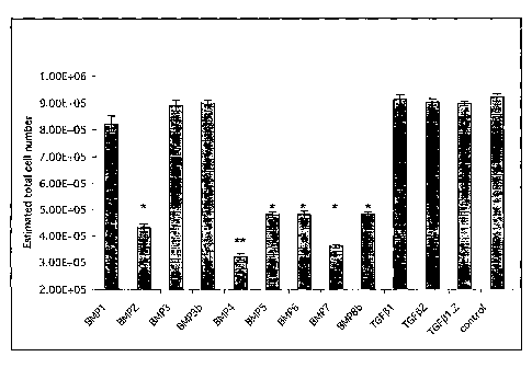
[0041] FIGURE 15 depicts the effects of various BMPs on the growth of GBM
cells.
-9¨
CA 02615491 2008-01-14
WO 2007/010394
PCT/1B2006/002296
DETAILED DESCRIPTION OF THE PREFERRED EMBODIMENTS
[0042] Prior to describing the present disclosure in detail, it is to be
understood that
unless otherwise indicated, the subject disclosure is not limited to specific
formulation
components, manufacturing methods, dosage regimens, or the like, as such may
vary. It is
also to be understood that the terminology used herein is for the purpose of
describing
particular embodiments only and is not intended to be limiting.
[0043] It must be noted that, as used in the subject specification, the
singular forms
"a", "an" and "the" include plural aspects unless the context clearly dictates
otherwise.
Thus, for example, reference to a "agent" includes a single agent, as well as
two or more
agents; reference to a "stem cell" includes a single stem cell, as well as two
or more stem
cells; and so forth.
[00441 As used herein, a "therapeutically effective amount" refers to that
amount of a
therapeutic agent sufficient to treat or manage a disease or disorder
characterized by
excessive or misregulated cellular proliferation and, preferably, the amount
sufficient to
destroy, modify, control or remove primary, regional or metastatic cancer
tissue. A
therapeutically effective amount may refer to the amount of therapeutic agent
sufficient
to delay or minimize the onset of the disease or disorder characterized by
excessive or
misregulated cellular proliferation , e.g., delay or minimize the spread of
cancer or the
growth of a tumor. A therapeutically effective amount may also refer to the
amount of
the therapeutic agent that provides a therapeutic benefit in the treatment or
management
of a tumor or of cancer. Further, a therapeutically effective amount with
respect to a
therapeutic agent of the disclosure means that amount of therapeutic agent
alone, or in
combination with other therapies, that provides a therapeutic benefit in the
treatment or
management of hyperproliferative cell disease or cancer. The term can
encompass an
amount that improves overall therapy, reduces or avoids unwanted effects, or
enhances
the therapeutic efficacy of or synergies with another therapeutic agent.
-10¨
CA 02615491 2008-01-14
WO 2007/010394
PCT/1B2006/002296
[0045] The terms "agent", "compound", "active agent", "active compound,"
"therapeutic agent," "pharmacologically active agent", "medicament", "active"
and
"drug" are used interchangeably herein to refer to a substance that induces a
desired
pharmacological and/or physiological effect. The terms also encompass
pharmaceutically
acceptable and pharmacologically active ingredients of those active agents
specifically
mentioned herein including but not limited to salts, esters, amides, prodrugs,
active
metabolites, analogs and the like. When the terms "agent", "compound", "active
agent",
"pharmacologically active agent", "medicament", "active" and "drug" are used,
then it is
to be understood that this includes the active agent per se as well as
pharmaceutically
acceptable, pharmacologically active salts, esters, amides, prodrugs,
metabolites, analogs,
etc. The agents of the present disclosure may be any proteinaceous molecules
such as
peptides, polypeptides and proteins or non-proteinaceous molecules such as
nucleic acid
molecules and small to large natural or synthetically derived organic and
inorganic
molecules. The agents can generally cross the blood-brain barrier or may be
suitable for
direct administration to the CNS.
[0046] Reference herein to "treatment" may mean a reduction in the severity
of an
existing disease or condition. The term "treatment" is also taken to encompass
"prophylactic treatment" to prevent the onset of a disease or condition. The
term
"treatment" does not necessarily imply that a subject is treated until total
recovery.
Similarly, "prophylactic treatment" does not necessarily mean that the subject
will not
eventually contract a disease or condition.
[0047] "Stem cell" as used herein refers to an undifferentiated cell
capable of, (a)
proliferation, (b) self renewal over an extended period of time, (c) able to
generate a large
number of progeny, and (d) the ability to give rise to all the cell types of
the tissue from
which it is obtained.
[0048] As used herein, a "tumor stem cell" is a stem cell obtained from a
tumor. A
tumor stem cell is capable of (a) proliferation, (b) self renewal over an
extended period
of time, (c) able to generate a large number of progeny, and (d) the ability
to give rise to
all the cell types of the tumor from which it is obtained. A "tumor neural
stem cell," also
-11¨
CA 02615491 2008-01-14
WO 2007/010394
PCT/1B2006/002296
referred to herein as a "tNSC," refers to tumor stem cell obtained from a
tumor of the
CNS.
[0049] "Progenitor cell" as used herein refers to an undifferentiated cell
capable of,
(a) proliferation, (b) limited self renewal ability, (c) generation of a
limited number of
progeny and (d) the ability to give rise to at least one type of progeny.
[0050] As used herein, a "tumor progenitor cell" is a progenitor cell
obtained from a
tumor. A tumor progenitor cell is capable of (a) proliferation, (b) limited
self renewal
ability, (c) generation of a limited number of progeny and (d) the ability to
give rise to at
least one cell type found in the tumor from which it is obtained.
LIF Preparations and BMP Preparations
[0051] In one aspect, the disclosure provides methods for the treatment or
prevention
of a disease or disorder characterized by excessive or misregulated cellular
proliferation.
The methods comprise administering a therapeutically effective amount of a
Leukemia
inhibitory factor (LIF) preparation and/or at least one Bone Morphogenetic
Protein
(BMP) preparation to a subject or tissue thought to be undergoing such
excessive or
misregulated cellular proliferation.
[0052] Preferably the disorder characterized by excessive proliferation is
a benign
tumor or a malignant tumor (cancer). For example, the tumor may be a brain
tumor
including, but not limited to, acoustic neuroma, adenoma, astrocytoma,
juvenile pilocytic
astrocytoma, brain stem glioma, chordoma, choroid plexus, craniopharyngioma,
ependymoma, ganglioglioma, ganglioglioneurocytoma, glioblastoma multiforme
(GBM),
glioma, lymphoma, medulloblastoma, meningioma, oligodendroglioma, optic nerve
glioma, pituitary tumors, pineal tumors, or pineoblastoma. In preferred
embodiments, the
brain tumor is GBM.
[0053] In another aspect the disclosure provides a method for reducing the
growth of
a tumor comprising administering a therapeutically effective amount of a
Leukemia
inhibitory factor (LIF) preparation and/or at least one Bone Morphogenetic
Protein
(BMP) preparation to said tumor. In some embodiments, a therapeutically
effective
-12¨
CA 02615491 2008-01-14
WO 2007/010394
PCT/1B2006/002296
amount of a BMP-4 preparation is administered to GBM in a human patient in
order to
reduce the growth of the GBM.
10054] In a further aspect, the disclosure provides a method of decreasing
the number
of tumor stem cells and/or tumor progenitor cells in a tumor comprising
contacting the
tumor with a LIF preparation and/or a BMP preparation. Without being limited
by theory
or hypothesis, it is believed that when administered to a tumor, LIF
preparations and
BMP preparations lead to an increase in LIFR or BMPR-mediated signalling,
which
results in the modulation of any one or more of the following tumor stem cell
or tumor
progenitor cell properties such as, but not limited to, cell survival, self-
renewal,
symmetric division, proliferation and/or differentiation properties. In
particular, and
without being limited by theory or hypothesis, it is believed that the
increase in LIFR or
BMPR-mediated signalling results in a reduction in the proliferation
properties of stem
and progenitor cells and in particular a reduction in the probability of
symmetric division
exhibited by proliferating stem cells or progenitor cell thereby reducing
their numbers.
Accordingly, in another aspect the disclosure provides a method for reducing
the
likelihood that a tumor stem cell or tumor progenitor cell undergoes a
symmetrical
division, the method comprising contacting the tumor stem cell or tumor
progenitor cell
with a LIF preparation and/or a BMP preparation.
[0055] In another aspect the disclosure provides a method of reducing the
growth of a
tumor by increasing LIFR or BMPR-mediated signalling in said tumor. LIFR
mediated
signalling may be activated, for example, using a LIF preparation and/or a
LIFR
signalling activator (see below); BMPR mediated signalling may be activated,
for
example, using a BMP preparation and/or a BMPR signalling activator (see
below).
[0056] Current treatments aimed at eradicating tumorigenic cells using
conventional
treatments are designed to eliminate rapidly cycling cells. For example,
traditional
chemotherapy agents are most effective against dividing cells. Like their non-
transformed counterpart, tNSCs cycle infrequently and thereby escape the toxic
effects of
treatment and may easily re-initiate tumor expansion after treatment. The
intrinsic
longevity of adult stem cells and their inherent ability to express drug
resistance and anti-
apoptotic genes may be found in their malignant counterpart, compounding the
difficulty
-13¨
CA 02615491 2008-01-14
WO 2007/010394
PCT/1B2006/002296
in developing effective treatment strategies aimed at eradicating tumour stem
cells. The
methods and compositions disclosed herein overcome this difficulty by
targeting the
tNSC cells. Without being limited by theory or hypothesis, it is believed that
the
methods and compositions described herein have a pro-differentiation effect on
tNSCs
(as evidenced by the upregulation of neural differentiation markers,
particularly astroglial
antigens, as show in the Examples), and thus permanently reduce the stem cell
pool
without effecting cell viability or eliciting apoptosis. As a result (as shown
in the
Examples below), even a transient exposure to the compositions of the
disclosure
(particuarly BMP-4 compositions for GBM treatment) irreversibly inhibits the
tumorigenic potential of tNSCs.
[0057] Inducing differentiation of tumor cells, rather than trying to kill
them, is an
entirely new approach to cancer treatment. Thus, in another aspect the
disclosure
provides a method of treating a tumor comprising tumor stem cells, the method
comprising contacting the tumor stem cells with an agent (such as a BMP
preparation)
that induces their differentiation. In one such embodiment, tumor neural stem
cells in a
brain tumor, such as glioblastoma multiforme, are contacted with BMP-4 in
order to
induce their differentiation.
[0058] The terms "LIF preparation" and "BMP preparation" includes the LIF
polypeptide or a BMP polypeptide as produced in nature, preferably in humans,
with or
without any post-translational modifications. This includes, for human LIF,
the
polypeptide encoded by the mRNA having the GenBank accession number NM_002309.
For human BMPs, this includes the BMP polypeptides encoded by the human genes:
BMP1, BMP2, BMP3, BMP4, BMP5, BMP6, BMP7, BMP8A, BMP8B, GDF10 (BMP-
3b), GDF11 (BMP11),GDF2 (BMP9), BMP10, BMP15, and by the mRNAs having the
GenBank accession numbers: NM 001719 (BMP7); NM 001201 (BMP3); NM 001200
(BMP2); NM 005448 (BMP15); NM_001720 (BMP8B); NM 014482 (BMP10);
NM 006132 (BMP1-4); NM 006131 (BMP1-5); NM 006130 (BMP1-6); NM 006129
(BMP1-3): NM_006128 (BMP1-2): NM_001718 (BMP6); NM_001199 (BMP1-1);
NM 130851 (BMP4-3); NM 130850 (BMP4-2); NM 001202 (BMP4-1); NM_181809
(BMP8A); NM 021073 (BMP5). Note that BMP-4 polypeptide is also sometimes
referred to as BMP-2B.
-14¨
CA 02615491 2008-01-14
WO 2007/010394
PCT/1B2006/002296
[0059] Preferred BMPs for use in the methods and compositions of the
disclosure
include BMP-2, BMP-4, BMP-5, BMP-6, BMP-7, and BMP-8b. In particular, exposure
to BMP-4 is shown in the Examples below to enforce the maturation of cells
isolated
from human GBM while not affecting overall viability and apoptosis. This
results in the
upregulation of neuronal and glial markers and also results in a major
reduction in
proliferation ability. BMP-4 exposure -- even transiently -- is shown in the
Examples
below to greatly reduce the size of GBM tNSCs populations (CD133+ GBM cells)
in
GBM cultures, to greatly reduce the clonogenic index of GBM cells, and to
dramatically
reduce the kinetics of expansion of GBM tNSCs. These effects are irreversible
and
extinguish the in vivo tumour-initiating ability of human GBM cells.
[0060] The term "LIF preparation" or "BMP preparation" as used herein also
includes fragments of LIF or BMP polypeptides or glycopolypeptides which at
least
partially retain the ability to attenuate excessive cellular proliferation in
the assays and
treatment methods of the disclosure e.g. which retain between 1-100% of the
activity of
full length LIF or a full length BMP in the assays or treatment methods of the
disclosure.
Such fragments may have increased activity relative to full length LIF or a
full length
BMP in the assays or treatment methods of the disclosure. Such fragments may
have a
continuous series of deleted residues from the amino or the carboxy terminus,
or both, in
comparison to the full length protein. The fragments may be characterized by
structural
or functional domains, such as fragments that comprise alpha-helix and alpha-
helix
forming regions, beta-sheet and beta-sheet-forming regions, turn and turn-
forming
regions, coil and coil-forming regions, hydrophilic regions, hydrophobic
regions, alpha
amphipathic regions, beta amphipathic regions, flexible regions, surface-
forming regions,
and substrate binding regions. The fragments may be produced by peptide
synthesis
techniques, or by cleavage of full length LIF or BMP polypeptide. The
fragments may be
linked at their N termini, C termini, or both their N and C termini to other
polypeptide
sequences, thus forming fusion proteins.
[0061] The term "LIF preparation" or "BMP preparation" as used herein also
includes a polypeptide or glycopolypeptide having an amino acid sequence which
is
partially homologous with the amino acid sequence of LIF or a BMP polypeptide,
or a
fragment thereof, as disclosed above, and which at least partially retain the
ability to
-15¨
CA 02615491 2008-01-14
WO 2007/010394
PCT/1B2006/002296
attenuate excessive cellular proliferation in the assays and treatment methods
of the
disclosure. Homologues may be 50%, 70%, 80%, 80.6%, 83%, 85%, 90%, 91%, 92%,
93%, 94%, 95%, 96%, 97%, 98%, 99%, 99.1%, 99.2%, 99.3%, 99.4%, 99.5%, 99.6%,
99.7%, 99.8%, or 99.9% identical to LIF or BMP, or fragments thereof.
[0062] The term "LIF preparation" or "BMP preparation" also includes
variants of
LIF or BMP full length polypeptide, and variants of LIP or BMP fragments. Such
variants at least partially retain the ability to attenuate excessive cellular
proliferation in
the assays and treatment methods of the disclosure. Variants may include
deletions,
insertions, inversions, repeats, and substitutions selected according to
general rules
known in the art so as have little effect on activity. For example, guidance
concerning
how to make phenotypically silent amino acid substitutions is provided in
Bowie et al.,
Science 247: 1306-1310 (1990), incorporated by reference herein in its
entirety. For
example, variants can be obtained by site directed mutagenesis or alanine-
scanning
mutagenesis (introduction of single alanine mutations at every residue in the
molecule).
(Cunningham and Wells, Science 244: 1081-1085 (1989). Variants may also have
amino
acid substitutions that contain, for example, one or more non-peptide bonds
(which
replace the peptide bonds) in the protein or peptide sequence. Variants may
also have
substitutions that include amino acid residues other than naturally occurring
L-amino
acids, e.g., D-amino acids or non-naturally occurring or synthetic amino
acids, e.g., B or
y amino acids. Variants may also include crosslinking groups which impose
conformational constraints on the polypeptide. Variants may also include
glycosylations,
acetylations, phosphorylations and the like. Variants may also include (i)
substitutions
with one or more of the non-conserved amino acid residues, where the
substituted amino
acid residues may or may not be one encoded by the genetic code, or (ii)
substitution with
one or more of amino acid residues having a substituent group, or (iii) fusion
of the
mature polypeptide with another compound, such as a compound to increase the
stability
and/or solubility of the LIF or BMP preparation (for example, polyethylene
glycol), or to
target the LIF or BMP preparation to a specific cell type (such as a tumor
neural stem
cell), or to allow the LIF or BMP preparation to cross the blood-brain barrier
(BBB)
and/or the blood-tumor barrier (BTB), or (iv) fusion of the polypeptide with
additional
amino acids or additional peptides or additional polypeptides, or (v) fusion
to a cytoxic
- 16¨
CA 02615491 2008-01-14
WO 2007/010394
PCT/1B2006/002296
agent, for example to a toxin or radioactive compound, or (vi) fusion to a
marker that
may be used for imaging purposes, for example, a radiolabel.
[00631 The LIF preparations and BMP preparations of the disclosure can be
prepared
in any suitable manner, including through the isolation of naturally occurring
polypeptides, by recombinant techniques, by polypeptide synthesis techniques,
or by a
combination of these methods. Methods for preparing such polypeptides are well
understood in the art. The LIF or BMP preparations may be in the form of a
larger
protein, such as a fusion protein. It is often advantageous to include an
additional amino
acid sequence which contains secretory or leader sequences, pro-sequences,
sequences
which aid in purification, such as multiple histidine residues, or an
additional sequence
for stability during recombinant production.
[00641 The LIF preparations and BMP preparations of the present disclosure
are
preferably provided in an isolated form, and preferably are substantially
purified. A
recombinantly produced version of a LIF or BMP preparation can be
substantially
purified using techniques described herein or otherwise known in the art, such
as, for
example, by the one-step method described in Smith and Johnson, Gene 67: 31-40
(1988). LIF or BMP preparations of the disclosure also can be purified from
natural,
synthetic or recombinant sources using protocols known in the art, such as,
for example,
antibodies of the disclosure raised against the fall-length LIF or BMP.
[0065] In some embodiments of the present disclosure a LIF preparation
and/or BMP
preparation may be administered to a subject directly such that endogenous
tumor stern
cells and tumor progenitor cells are regulated in vivo. For example, a BMP-4
preparation
may be administered to a brain tumor, such as GBM, in a human patient. In
alternative
embodiments of the present disclosure, tumor stem cells and tumor progenitor
cells may
be contacted with the agents of the present disclosure in vitro. For example,
an isolated
tumor, which comprises tumor stem cells and tumor progenitor cells, may be
contacted
with the agents of the disclosure in vitro.
[00661 Methods for administering the LIF and/or BMP preparations to a
subject,
including to a tumor in a subject, along with pharmaceutical compositions
comprising
-17¨
CA 02615491 2008-01-14
WO 2007/010394
PCT/1B2006/002296
LIF and/or BMP preparations, are provided below in the section entitled
"Administration
and Pharmaceutical Compositions."
LIF Receptor Signalling Activators and BMP Receptor Signalling Activators
[0067] In another aspect embodiment, the present disclosure provides
agents,
hereinafter referred to as "LIFR signalling activators" and "LIF Receptor
signalling
activators" which are capable of increasing LIF receptor (LIFR)-mediated
signalling in a
tumor stem cell or tumor progenitor cell. The disclosure also provides methods
for the
identification of such LIFR signalling activators, and pharmaceutical
compositions
comprising such LIFR signalling activators. The LIFR signalling activators of
the
present disclosure may increase LIFR-mediated signalling in a stem or
progenitor cell by
activating LIFR directly (e.g. an agonist), or indirectly, such as by
increasing the
expression or activity of a secondary molecule or compound (e.g. by increasing
expression of LIF itself, or by increasing the activity or expression a
downstream
component of LIFR-mediated signalling, such as JAK or STAT) in the tumor stem
cell or
tumor progenitor cell which in turn increases LIFR-mediated signalling in a
tumor stem
cell or tumor progenitor cell.
[0068] In an additional aspect, the present disclosure provides agents,
hereinafter
referred to as "BMPR signalling activators" and "BMP receptor signalling
activators"
which are capable of increasing BMP receptor (BMPR)-mediated signalling in a
tumor
stem cell or tumor progenitor cell. The disclosure also provides methods for
the
identification of such BMPR signalling activators, and pharmaceutical
compositions
comprising such BMPR signalling activators. The BMPR signalling activators of
the
present disclosure may increase BMPR-mediated signalling in a tumor stem cell
or tumor
progenitor cell by activating BMPR directly (eg an agonist), or indirectly,
such as by
increasing the expression or activity of a secondary molecule or compound (eg
by
increasing expression of BMP itself, or by increasing expression or activity
of a
downstream component of BMPR-mediated signalling) in the tumor stem cell or
tumor
progenitor cell which in turn increases BMPR-mediated signalling on a tumor
stem cell
or tumor progenitor cell.
-18¨
CA 02615491 2008-01-14
WO 2007/010394
PCT/1B2006/002296
[0069] Reference herein to "LIFR" includes reference to all forms of LIFR
such as
LIFR homologs, paralogs, orthologs, derivatives, fragments and functional
equivalents.
Reference herein to "BMPR" includes reference to all forms of BMPR such as
BMPR
homologs, paralogs, orthologs, derivatives, fragments and functional
equivalents.
[0070] In the context of the present disclosure, an increase in LIFR or
BMPR-
mediated signalling refers to an increase of one to about 1000% of the normal
level of
LIFR or BMPR-mediated signalling. Alternatively, the LIFR or BMPR signalling
activator can return the level of LIFR or BMPR-mediated signalling to normal
in cases
where signalling is less than normal.
[0071] Preferably, the increase in LIFR or BMPR-mediated signalling results
in the
modulation of any one or more of tumor stem cell and tumor progenitor cell
properties
such as, but not limited to, survival, self-renewal, proliferation, symmetric
division and/or
differentiation properties. Most preferably, the increase in LIFR or BMPR-
mediated
signalling alters the division properties of tumor stem cells and tumor
progenitor cells
and in particular a reduction in the probability of symmetric division or
reduction in cell
cycle frequency exhibited by proliferating tumor stern cells and tumor
progenitor cells
thereby leading to a reduction in the numbers of tumor stem cells and tumor
progenitor
cells.
[0072] The LIFR and BMPR signalling activators of the disclosure may be any
proteinaceous molecules such as peptides, polypeptides and proteins, or they
may be non-
proteinaceous molecules. Methods for the isolation of LIFR and BMPR signalling
activators are provided herein.
[0073] In relation to the present disclosure, mimetics are a particularly
useful group
of LIFR and BMPR signalling activators. The term is intended to refer to a
substance
which has some chemical similarity to the molecule it mimics, such as, for
example, LIF,
but which agonizes (mimics) its interaction with a target, such as, for
example, a LIFR. A
peptide mimetic is one class of mimetics, and may be a peptide-containing
molecule that
mimics elements of protein secondary structure (Johnson et al., Peptide Turn
Mimetics in
Biotechnology and Pharmacy, Pezzuto et al., Eds., Chapman and Hall, New York,
1993).
The underlying rationale behind the use of peptide mimetics is that the
peptide backbone
- 19¨
CA 02615491 2008-01-14
WO 2007/010394
PCT/1B2006/002296
of proteins exists chiefly to orient amino acid side chains in such a way as
to facilitate
molecular interactions such as those of antibody and antigen, enzyme and
substrate or
scaffolding proteins. A peptide mimetic, therefore, is designed to permit
molecular
interactions similar to the natural molecule.
[0074] The designing of mimetics to a pharmaceutically active compound is a
known
approach to the development of pharmaceuticals based on a "lead" compound.
This
might be desirable where the active compound is difficult or expensive to
synthesize or
where it is unsuitable for a particular method of administration, e.g.
peptides are
unsuitable active agents for oral compositions as they tend to be quickly
degraded by
proteases in the alimentary canal. Mimetic design, synthesis and testing is
generally used
to avoid randomly screening large numbers of molecules for a target property.
A
mimetic of BMP-4, including a peptide mimetic for example, is specifically
contemplated
herein.
[0075] The goal of rational drug design is to produce structural analogs of
biologically active polypeptides of interest or of small molecules with which
they interact
in order to fashion drugs which are, for example, more active or stable forms
of the
polypeptide, or which, for example, enhance or interfere with the function of
a
polypeptide in vivo (see, e.g. Hodgson, Bio/Technology 9:19-21, 1991). In one
approach,
one first determines the three-dimensional structure of a protein of interest
by x-ray
crystallography, by computer modelling or most typically, by a combination of
approaches. Useful information regarding the structure of a polypeptide may
also be
gained by modelling based on the structure of homologous proteins. An example
of
rational drug design is the development of HIV protease inhibitors (Erickson
et al.,
Science 249:527-533, 1990).
[0076] The capability of the LIFR and BMPR signalling activators of the
present
disclosure, whether they be proteinaceous or non-proteinaceous, to interact
with LIFR or
BMPR and/or increase LIFR or BMPR-mediated signalling (either directly or
indirectly)
in a stem or progenitor cell may be assessed via a number of screening methods
which
would be well known to a person skilled in the art. These may include
screening naturally
- 20 ¨
CA 02615491 2015-08-17
WO 2007/010394
PCT/1B2006/002296
produced libraries, chemical produced libraries, as well as combinatorial
libraries, phage
display libraries and in vitro translation-based libraries.
[0077] Antibodies raised against LIFR and BMPR may be particularly useful
as
agonists that mimic the active configuration of LIP and BM1' respectively.
Suitable
antibodies include polyclonal, monoclonal, monovalent, bispecific,
heteroconjugate,
multispecific, human, humanized or chimeric antibodies, single chain
antibodies, Fab
fragments, F(ab') fragments, fragments produced by a Fab expression library,
anti-
idiotypic (anti-Id) antibodies, and epitope-binding fragments of any of the
above. The
teim "antibody," as used herein, refers to immunoglobulin molecules and
immunologically active portions of immunoglobulin molecules, i.e., molecules
that
contain an antigen binding site that immunospecifically binds an antigen. The
immunoglobulin molecules can be of any type (e.g., IgG, IgE, IgM, IgD, IgA and
IgY),
class (e.g., IgGl, igG2, IgG3, IgG4, IgAl and IgA2) or subclass of
immunoglobulin
molecule. Moreover, the term "antibody" (Ab) or "monoclonal antibody" (Mab) is
meant
to include intact molecules, as well as, antibody fragments (such as, for
example, Fab and
F(ab')2 fragments) which are capable of specifically binding to protein. Fab
and F(ab')2
fragments lack the Fc fragment of intact antibody, clear more rapidly from the
circulation
of the animal or plant, and may have less non-specific tissue binding than an
intact
antibody (Wahl et al., J. Nucl. Med, 24: 316-325 (1983)). Methods for
producing
antibody agonists are described in, for example, PCT publication WO 96/40281;
U.S.
Pat. No. 5,811,097; Deng et al., Blood 92(6): 1981-1988 (1998); Chen etal.,
Cancer Res.
58 (16): 3668-3678 (1998); Harrop et al., J. Immunol. 161 (4): 1786-1794
(1998); Zhu et
al., Cancer Res. 58 (15): 3209-3214 (1998); Yoon etal., J. Immunol. 160 (7):
3170-3179
(1998); Prat et al., J. Cell. Sci, ill (Pt2): 237-247 (1998); Pitard et al.,
J. Immunol.
Methods 205 (2): 177-190 (1997); Liautard et al., Cytokine 9 (4): 233-241
(1997);
Carlson et al., J. Biol. Chem. 272 (17): 11295-11301(1997); Taryman et al,,
Neuron 14
(4): 755-762 (1995); Muller et al., Structure 6 (9): 1153-1167 (1998);
Bartunek et al.,
Cytokine 8(1): 14-20 (1996); Harlow et al., Antibodies: A Laboratory Manual,
(Cold
Spring Harbor Laboratory Press, 2nd ed. 1988); Hammerling, et al., in:
Monoclonal
Antibodies and T-Cell Hybridomas 563-681 (Elsevier, N.Y., 1981).
-21¨
CA 02615491 2008-01-14
WO 2007/010394
PCT/1B2006/002296
[0078] Nucleic acid ligands (also known as "aptamers") may also be
particularly
useful as agonists that mimic the active configuration of LIF and BMP
respectively. For
example, aptamers can be selected using the SELEX (Systematic Evolution of
Ligands
by Exponential Enrichment) method (Tuerk and Gold, 1990, Science 249: 505-510,
which is incorporated by reference herein in its entirety). In the SELEX
method, a large
library of nucleic acid molecules (e.g., 1015 different molecules) is produced
and/or
screened with the target molecule, in this case BMPR, LIFR, or portions
thereof. The
target molecule is allowed to incubate with the library of nucleotide
sequences for a
period of time. Several methods can then be used to physically isolate the
aptamer target
molecules from the unbound molecules in the mixture and the unbound molecules
can be
discarded. The aptamers with the highest affinity for the target molecule can
then be
purified away from the target molecule and amplified enzymatically to produce
a new
library of molecules that is substantially enriched for aptamers that can bind
the target
molecule. The enriched library can then be used to initiate a new cycle of
selection,
partitioning, and amplification. After 5-15 cycles of this selection,
partitioning and
amplification process, the library is reduced to a small number of aptamers
that bind
tightly to the target molecule. Individual molecules in the mixture can then
be isolated,
their nucleotide sequences determined, and their properties with respect to
binding
affinity and specificity measured and compared. Isolated aptamers can then be
further
refined to eliminate any nucleotides that do not contribute to target binding
and/or
aptamer structure (i.e., aptamers truncated to their core binding domain).
See, e.g.,
Jayasena, 1999, Clin. Chem. 45: 1628-1650 for review of aptamer technology,
the entire
teachings of which are incorporated herein by reference.
[0079] Essentially any chemical compound can be employed as a candidate
LIFR or
BMPR signalling activator. High throughput screening methodologies are
particularly
envisioned for the detection of such candidate activators. Such high
throughput screening
methods typically involve providing a combinatorial chemical or peptide
library
containing a large number of potential therapeutic compounds (e.g., ligand or
modulator
compounds). Such combinatorial chemical libraries or ligand libraries are then
screened
in one or more assays to identify those library members (e.g., particular
chemical species
or subclasses) that display a desired characteristic activity. The compounds
so identified
-22¨
CA 02615491 2008-01-14
WO 2007/010394
PCT/1B2006/002296
can serve as conventional lead compounds, or can themselves be used as
potential or
actual therapeutics.
[0080] A combinatorial chemical library is a collection of diverse chemical
compounds generated either by chemical synthesis or biological synthesis, by
combining
a number of chemical building blocks (i.e., reagents such as amino acids). As
an
example, a linear combinatorial library, e.g., a polypeptide or peptide
library, is formed
by combining a set of chemical building blocks in every possible way for a
given
compound length (i.e., the number of amino acids in a polypeptide or peptide
compound).
Millions of chemical compounds can be synthesized through such combinatorial
mixing
of chemical building blocks.
[0081] The preparation and screening of combinatorial chemical libraries is
well
known to those having skill in the pertinent art. Combinatorial libraries
include, without
limitation, peptide libraries (e.g. U.S. Pat. No. 5,010,175; Furka, 1991, Int.
J. Pept. Prot.
Res., 37: 487-493; and Houghton et al., 1991, Nature, 354: 84-88). Other
chemistries for
generating chemical diversity libraries can also be used. Nonlimiting examples
of
chemical diversity library chemistries include, peptides (PCT Publication No.
WO
91/019735), encoded peptides (PCT Publication No. WO 93/20242), random bio-
oligomers (PCT Publication No. WO 92/00091), benzodiazepines (U.S. Pat. No.
5,288,514), diversomers such as hydantoins, benzodiazepines and dipeptides
(Hobbs et
al., 1993, Proc. Natl. Acad. Sci. USA, 90: 6909-6913), vinylogous polypeptides
(Hagihara et al., 1992, J. Amer. Chem. Soc., 114: 6568), nonpeptidal
peptidomimetics
with glucose scaffolding (Hirschmann et al., 1992, J. Amer. Chem. Soc., 114:
9217-
9218), analogous organic synthesis of small compound libraries (Chen et al.,
1994, J.
Amer. Chem. Soc., 116: 2661), oligocarbamates (Cho et al., 1993, Science, 261:
1303),
and/or peptidyl phosphonates (Campbell et al., 1994, J. Org. Chem., 59: 658),
nucleic
acid libraries (see Ausubel, Berger and Sambrook, all supra), peptide nucleic
acid
libraries (U.S. Pat. No. 5,539,083), antibody libraries (e.g., Vaughn et al.,
1996, Nature
Biotechnology, 14 (3): 309-314) and PCT/US96/10287), carbohydrate libraries
(e.g.,
Liang et al., 1996, Science, 274-1520-1522) and U.S. Pat. No. 5,593,853),
small organic
molecule libraries (e.g., benzodiazepines, Baum C&EN, Jan. 18, 1993, page 33;
and U.S.
Pat. No. 5,288,514; isoprenoids, U.S. Pat. No. 5,569,588; thiazolidinones and
-23¨
CA 02615491 2008-01-14
WO 2007/010394
PCT/1B2006/002296
metathiazanones, U.S. Pat. No. 5,549,974; pyrrolidines, U.S. Pat. Nos.
5,525,735 and
5,519,134; morpholino compounds, U.S. Pat. No. 5,506,337; and the like).
[0082] Devices for the preparation of combinatorial libraries are
commercially
available (e.g., 357 MPS, 390 MPS, Advanced Chem Tech, Louisville Ky.;
Symphony,
Rainin, Woburn, Mass.; 433A Applied Biosystems, Foster City, Calif.; 9050
Plus,
Millipore, Bedford, Mass.). In addition, a large number of combinatorial
libraries are
commercially available (e.g., ComGenex, Princeton, N.J.; Asinex, Moscow,
Russia;
Tripos, Inc., St. Louis, Mo.; ChemStar, Ltd., Moscow, Russia; 3D
Pharmaceuticals,
Exton, Pa.; Martek Biosciences, Columbia, Md., and the like).
[0083] Candidate LIFR and BMPR signalling activators may first be screened
for
their ability to bind to LIFR or BMPR, or to downstream components of the LIFR
or
BMPR signalling pathway, using a binding assay, and those candidates that bind
may
then be screened in a functional assay. Suitable binding assays include the
fluorescence
based thermal shift assay (3-Dimensional Pharmaceuticals, Inc., 3DP, Exton,
Pa.) as
described in U.S. Pat. Nos. 6,020,141 and 6,036,920 to Pantoliano et al.; see
also, J.
Zimmerman, 2000, Gen. Eng. News, 20 (8)).
[0084] An example of a method for functionally screening candidate LIFR and
BMPR signalling activators includes the following steps:
(i) Isolating a sample of tumor stem cells and/or tumor progenitor cells;
(ii) placing aliquots of the tumor stem cells and/or tumor progenitor cells
into
suitable receptacles; and
(iii) exposing the aliquots of tumor stem cells and/or tumor progenitor
cells to
candidate agents for a particular period of time and under particular
conditions;
and
(iv) screening for morphological, physiological and genetic changes to the
tumor stem cells and/or tumor progenitor cells.
[0085] Morphological, physiological and genetic changes includes screening
for
states of survival, self-renewal, proliferation and/or differentiation. An
example of an
assay that can be used is the Neural Colony Forming Cell Assay (NCFCA)
described in
-24¨
CA 02615491 2008-01-14
WO 2007/010394
PCT/1B2006/002296
United States Patent Application Publication No. 2005/0112546, incorporated
herein by
reference in its entirety. The NCFCA is able to distinguish stem cells from
progenitor
cells, both which have a proliferative potential and are capable of forming
spheres in
suspension culture (Neurosphere Assay) or colonies in the NCFCA. Briefly,
primary or
cultured cells obtained from a tumor are plated in a serum-free 3-D collagen
matrix
containing the mitogens FGF2 and EGF. Under these culture conditions only stem
cells
and progenitor cells with a proliferative potential divide forming well-
defined colonies
whose size can be measured after 1-4 weeks. Differences in colony size
positively
correlate to the proliferative potential of the founding cell and provide a
readout of stem
and progenitor cell frequency. Under these conditions only colonies greater
than 2 mm in
diameter are derived from a stem cell while those less than 2mm in diameter
are derived
from progenitor cells. A meaningful and accurate readout of stem cell and
progenitor
cells allows one to screen for genetic and epigenetic elements that alter the
frequency of
these two cell types.
[0086] Another example of an assay for survival, self-renewal,
proliferation and/or
differentiation which may be used to screen for LIFR and BMPR receptors is
performed
as follows. First, cells from a disaggregated glioblastoma multiforme tumor
are plated in
serum free medium containing the mitogens FGF2 (fibroblast growth factor 2)
and EGF
(epidermal growth factor) as described by Gritti et al., J. Neurosci. (1996)
16(3):109-
1100, incorporated herein by reference. This culture system selects away
differentiating/differentiated cells from primary tumor cultures, leaving only
the tumor
stem cells free to proliferate and expand exponentially, thereby forming
primary
neurospheres. The primary neurospheres may dissociated and plated again in
serum free
medium at clonal density in the presence of EGF and FGF2 in microtitre plates.
Candidate LIFR and BMPR signalling activators are added to each well of the
microtitre
plate, and the plates are incubated for a period of time sufficient to allow
untreated cells
to proliferate. At the end of incubation, the neurospheres are again
disocciated and the
process can be repeated for a predetermined number of additional passages in
the
presence of the candidate LIFR and BMPR signalling activators. At the end of
the
predetermined number of passages, the wells of the microtitre plate may be
examined
using a microscope for the presence of neurospheres, and the number and size
of the
- 25 ¨
CA 02615491 2008-01-14
WO 2007/010394
PCT/1B2006/002296
neurospheres are determined, providing a measure of the effect of the
candidate LIFR or
BMPR signalling activator on stem and progenitor cells. The mathematical
algorithms of
Example I may be used to determine the number of stem cells and progenitor
cells at the
end of each passage. Comparison with untreated cells that were also serially
passaged
allows for the identification of candidate LIFR and BMPR signalling activators
e.g.
agents that attenuate the proliferation properties of stern and progenitor
cells. The
candidate LIFR and BMPR signalling activators may then be assayed on
differentiated or
differentiating cells to determine if the effect of the candidate agents is
specific for tumor
stem cells, rather than being generally cytotoxic.
[0087] LIFR and BMPR signalling activators may also include RNA
interference
(RNAi) molecules, ribozymes, or antisense oligonucleotides. Such molecules may
reduce the expression of inhibitors of LIFR and BM-PR signalling, and thus
have the
effect of activating LIFR and BMPR signalling.
[0088] The LIFR and BMPR signalling activators of the disclosure are useful
for
increasing LIFR or BMPR-mediated signalling in a tumor stem cell or tumor
progenitor
cell. Accordingly, the present disclosure provides a method of increasing LIFR
or
BMPR-mediated signalling in a tumor stem cell or tumor progenitor cell, the
method
comprising contacting the tumor stem cell or tumor progenitor cell with a LIFR
and/or
BMPR signalling activator for a time and under conditions sufficient to
increase LIFR or
BMPR-mediated signalling in the tumor stem cell or tumor progenitor cell. The
LIFR
and/or BMPR signalling activators may also used in combination with a LIF
preparation
and/or a BMP preparation as disclosed herein.
[0089] The disclosure also provides methods for the treatment or prevention
of a
disease or disorder characterized by excessive or misregulated cellular
proliferation. The
methods involve administering a therapeutically effective amount of LIFR
and/or BMPR
signalling activator to a subject or tissue thought to be undergoing such
excessive or
misregulated cellular proliferation. Preferably, the disorder characterized by
excessive
cellular proliferation is a brain disorder, more preferably a brain tumor
including, but not
limited to, acoustic neuroma, adenoma, astrocytoma, juvenile pilocytic
astrocytoma,
brain stem glioma, chordoma, choroid plexus, craniopharyngioma, ependymoma,
-26¨
CA 02615491 2008-01-14
WO 2007/010394
PCT/1B2006/002296
ganglioglioma, ganglioglioneurocytoma, glioblastoma multiforme (GBM), glioma,
lymphoma, medulloblastoma, meningioma, oligodendroglioma, optic nerve glioma,
a
pituitary tumor, a pineal tumor, or pineoblastoma. The LIFR and/or BMPR
signalling
activators may also administered in combination (either at the same time or at
different
times) with a LIF preparation and/or a BMP preparation as disclosed herein.
100901 In another aspect the disclosure provides a method for reducing the
growth of
a tumor comprising administering a therapeutically effective amount of a LIFR
signalling
activator and/or a BMPR signalling activator to said tumor.
[0091] In a further aspect, the disclosure provides a method of decreasing
the number
of tumor stem cells and/or tumor progenitor cells in a tumor comprising
contacting the
tumor with a LIFR signalling activator and/or a BMPR signalling activator.
Without
being limited by theory or hypothesis, it is believed that when administered
to a tumor,
LIFR signalling actvators and BMPR signalling activators lead to an increase
in LIF or
BMP-mediated signalling, which results in the modulation of any one or more of
the
following tumor stem cell or tumor progenitor cell properties such as, but not
limited to,
cell survival, self-renewal, symmetric division, proliferation and/or
differentiation
properties. In particular, it is believed that the increase in LIFR or BMPR-
mediated
signalling results in a reduction in the proliferation properties of stem and
progenitor cells
and in particular a reduction in the probability of symmetric division
exhibited by
proliferating stem cells or progenitor cell thereby reducing their numbers.
Accordingly,
in another aspect the disclosure provides methods for reducing the likelihood
that a tumor
stem cell or tumor progenitor cell undergoes a symmetrical division, the
method
comprising contacting the tumor stem cell or tumor progenitor cell with a BMPR
signalling activator and/or a LIFR signalling activator.
[0092] In some embodiments of the present disclosure the LIFR and/or BMPR
signalling activator may be administered to a subject directly such that
endogenous tumor
stem cells and tumor progenitor cells are regulated in vivo. In alternative
embodiments of
the present disclosure, tumor stem cells and tumor progenitor cells may be
contacted with
the agents of the present disclosure in vitro.
- 27 ¨
CA 02615491 2008-01-14
WO 2007/010394
PCT/1B2006/002296
[0093] Methods for administering the LIFR signalling activators and/or BMPR
signalling activators to a subject, including to a tumor, along with
pharmaceutical
compositions comprising LIFR signalling activators and/or BMPR signalling
activators,
are provided below in the section entitled "Administration and Pharmaceutical
Compositions"
Administration and Pharmaceutical Compositions
[0094] As disclosed above, therapeutically-effective amounts of a LIF
preparation
and/or a BMP preparation and/or LIFR signalling activator and/or BMPR
signalling
activator may be, inter alia, administered to a subject or tissue to treat or
prevent a disease
or disorder characterized by excessive or misregulated cellular proliferation.
For
example, in one embodiment a therapeutically effective amount of a BIVLP-4
preparation
or a BMP-4 mimetic is administered to a human patient suffering from GBM. In
addition
to the tumors and cancers described supra, other cancers that may be treated
or prevented
according to the methods disclosed herein include, but are not limited to,
carcinoma,
lymphoma, blastoma, sarcoma, and leukemia. More particular examples of such
cancers
include squamous cell cancer, lung cancer (including small-cell lung cancer,
non-small
cell lung cancer, adenocarcinoma of the lung, and squamous carcinoma of the
lung),
cancer of the peritoneum, hepatocellular cancer, gastric or stomach cancer
(including
gastrointestinal cancer), pancreatic cancer, cervical cancer, ovarian cancer,
liver cancer,
bladder cancer, hepatoma, breast cancer, colon cancer, colorectal cancer,
endometrial or
uterine carcinoma, salivary gland carcinoma, kidney or renal cancer, liver
cancer,
prostate cancer, vulval cancer, melanoma, thyroid cancer, hepatic carcinoma
and various
types of head and neck cancer, as well as B-cell lymphoma (including low
grade/follicular non-Hodgkin's lymphoma (NHL); small lymphocytic (SL) NHL;
intermediate grade/follicular NHL; intermediate grade diffuse NHL; high grade
immunoblastic NHL; high grade lymphoblastic NHL; high grade small non-cleaved
cell
NHL; bulky disease NHL; mantle cell lymphoma; AIDS-related lymphoma; and
Waldenstrom's Macroglobulinemia); chronic lymphocytic leukemia (CLL); acute
-28 ¨
CA 02615491 2008-01-14
WO 2007/010394
PCT/1B2006/002296
lymphoblastic leukemia (ALL); hairy cell leukemia; chronic myeloblastic
leukemia; and
post-transplant lymphoproliferative disorder (PTLD).
[0095] In one embodiment, a LIF preparation and/or a BMP preparation and/or
LIFR
signalling activator and/or BMPR signalling activator is administered to a
subject in the
form of a pharmaceutical composition. Accordingly, in another aspect the
present
disclosure also provides pharmaceutical compositions which are useful for the
treatment
or prevention of a disease or disorder characterized by excessive or
misregulated cellular
proliferation. The pharmaceutical compositions of the disclosure comprise at
least one
agent selected from the group consisting of a LIF preparation, a BMP
preparation, a LIFR
signalling activator, and a BMPR signalling activator. For example, in one
embodiment a
pharmaceutical composition comprising a therapeutically effective amount of a
BMP-4
preparation is provided for the treatment of GBM. In another embodiment, a
pharmaceutical composition comprising a therapeutically effective amount of a
BMP-2
preparation is provided for the treatment of GBM. In another embodiment, a
pharmaceutical composition comprising a therapeutically effective amount of a
BMP-5
preparation is provided for the treatment of GBM. In another embodiment, a
pharmaceutical composition comprising a therapeutically effective amount of a
BMP-6
preparation is provided for the treatment of GBM. In another embodiment, a
pharmaceutical composition comprising a therapeutically effective amount of a
BMP-7
preparation is provided for the treatment of GBM. In another embodiment, a
pharmaceutical composition comprising a therapeutically effective amount of a
BMP-8b
=
preparation is provided for the treatment of GBM.
[0096] In another aspect, the disclosure discloses the use of a LIF
preparation and/or
a BMP preparation and/or LIFR signalling activator and/or BMPR signalling
activator in
the manufacture of a medicament for the treament or prevention of a disease or
disorder
characterized by excessive or misregulated cellular proliferation. For
example, the use of
a BMP-4 preparation or a BMP-4 mimetic in the manufacture of a medicament for
the
treatment of glioblastoma multiforme is specifically contemplated.
[0097] The pharmaceutical compositions of the disclosure may comprise a
single
agent or they may comprise any combination of the aforementioned agents, for
example a
- 29¨
CA 02615491 2008-01-14
WO 2007/010394
PCT/1B2006/002296
combination of LIF and BMP-4. Moreover, the pharmaceutical compositions may
comprise more than one agent of a particular class, for example, two different
LIF
preparations ,or two different BMPR signalling activators, for example BMP-2
and BMP-
4.
[0098] The pharmaceutical compositions preferably comprise at least one
pharmaceutically acceptable carrier. In such pharmaceutical compositions, a
LIF
preparation and/or a BMP preparation and/or a LIFR signalling activator and/or
a BMPR
signalling activator forms the "active compound." As used herein the language
"pharmaceutically acceptable carrier" includes solvents, dispersion media,
coatings,
antibacterial and antifungal agents, isotonic and absorption delaying agents,
and the like,
compatible with pharmaceutical administration. Supplementary active compounds
can
also be incorporated into the compositions. A pharmaceutical composition is
formulated
to be compatible with its intended route of administration. Examples of routes
of
administration include parenteral, e.g., intravenous, intradermal,
subcutaneous, oral (e.g.,
inhalation), transdermal (topical), transmucosal, and rectal administration.
Solutions or
suspensions used for parenteral, intradermal, or subcutaneous application can
include the
following components: a sterile diluent such as water for injection, saline
solution, fixed
oils, polyethylene glycols, glycerine, propylene glycol or other synthetic
solvents;
antibacterial agents such as benzyl alcohol or methyl parabens; antioxidants
such as
ascorbic acid or sodium bisulfite; chelating agents such as
ethylenediaminetetraacetic
acid; buffers such as acetates, citrates or phosphates and agents for the
adjustment of
tonicity such as sodium chloride or, dextrose. pH can be adjusted with acids
or bases, such
as hydrochloric acid or sodium hydroxide. The parenteral preparation can be
enclosed in
ampoules, disposable syringes or multiple dose vials made of glass or plastic.
[0099] Note that in embodiments where a tissue or cell thought to be
undergoing
excessive or misregulated cell proliferation is treated with a LIF preparation
and/or a
BMP preparation and/or LIFR signalling activator and/or BMPR signalling
activator in
vitro, a LIF preparation and/or a BMP preparation and/or LIFR signalling
activator
and/or BMPR signalling activator may or may not be in the form of a
pharmaceutical
composition.
-30---
CA 02615491 2008-01-14
WO 2007/010394
PCT/1B2006/002296
[0100] Subject as used herein refers to humans and non-human primates (e.g.
guerilla, macaque, marmoset), livestock animals (e.g. sheep, cow, horse,
donkey, pig),
companion animals (e.g. dog, cat), laboratory test animals (e.g. mouse,
rabbit, rat, guinea
pig, hamster), captive wild animals (e.g. fox, deer) and any other organisms
who can
benefit from the agents of the present disclosure. There is no limitation on
the type of
animal that could benefit from the presently described agents. The most
preferred subject
of the present disclosure is a human. A subject regardless of whether it is a
human or
non-human organism may be referred to as a patient, individual, animal, host
or recipient.
[0101] Pharmaceutical compositions suitable for injectable use include
sterile
aqueous solutions (where water soluble) or dispersions and sterile powders for
the
extemporaneous preparation of sterile injectable solutions or dispersion. For
intravenous
administration, suitable carriers include physiological saline, bacteriostatic
water,
Cremophor EL.TM. (BASF, Parsippany, N.J.) or phosphate buffered saline (PBS).
In all
cases, the composition must be sterile and should be fluid to the extent that
easy
syringability exists. It should be stable under the conditions of manufacture
and storage
and must be preserved against the contaminating action of microorganisms such
as
bacteria and fungi. The carrier can be a solvent or dispersion medium
containing, for
example, water, ethanol, polyol (for example, glycerol, propylene glycol, and
liquid
polyetheylene glycol, and the like), and suitable mixtures thereof. The proper
fluidity can
be maintained, for example, by the use of a coating such as lecithin, by the
maintenance
of the required particle size in the case of dispersion and by the use of
surfactants.
Prevention of the action of microorganisms can be achieved by various
antibacterial and
antifungal agents, for example, parabens, chlorobutanol, phenol, ascorbic
acid,
thimerosal, and the like. In many cases, it will be preferable to include
isotonic agents,
for example, sugars, polyalcohols such as manitol, sorbitol, sodium chloride
in the
composition. Prolonged absorption of the injectable compositions can be
brought about
by including in the composition an agent which delays absorption, for example,
aluminum monostearate and gelatin.
[0102] Sterile injectable solutions can be prepared by incorporating the
active
compound in the required amount in an appropriate solvent with one or a
combination of
ingredients enumerated above, as required, followed by filtered sterilization.
Generally,
-31¨
CA 02615491 2008-01-14
WO 2007/010394
PCT/1B2006/002296
dispersions are prepared by incorporating the active compound into a sterile
vehicle
which contains a basic dispersion medium and the required other ingredients
from those
enumerated above. In the case of sterile powders for the preparation of
sterile injectable
solutions, the preferred methods of preparation are vacuum drying and freeze-
drying
which yields a powder of the active ingredient plus any additional desired
ingredient
from a previously sterile-filtered solution thereof.
[0103] Oral compositions generally include an inert diluent or an edible
carrier. For
the purpose of oral therapeutic administration, the active compound can be
incorporated
with excipients and used in the form of tablets, troches, or capsules, e.g.,
gelatin capsules.
Oral compositions can also be prepared using a fluid carrier for use as a
mouthwash.
Pharmaceutically compatible binding agents, and/or adjuvant materials can be
included
as part of the composition. The tablets, pills, capsules, troches and the like
can contain
any of the following ingredients, or compounds of a similar nature: a binder
such as
microcrystalline cellulose, gum tragacanth or gelatin; an excipient such as
starch or
lactose, a disintegrating agent such as alginic acid, Primogel, or corn
starch; a lubricant
such as magnesium stearate or Sterotes; a glidant such as colloidal silicon
dioxide; a
sweetening agent such as sucrose or saccharin; or a flavoring agent such as
peppermint,
methyl salicylate, or orange flavoring.
[0104] For administration by inhalation, the compounds are delivered in the
form of
an aerosol spray from pressured container or dispenser which contains a
suitable
propellant, e.g., a gas such as carbon dioxide, or a nebulizer.
[0105] Systemic administration can also be by transmucosal or transdermal
means.
For transmucosal or transdermal administration, penetrants appropriate to the
barrier to
be permeated are used in the formulation. Such penetrants are generally known
in the art,
and include, for example, for transmucosal administration, detergents, bile
salts, and
fusidic acid derivatives. Transmucosal administration can be accomplished
through the
use of nasal sprays or suppositories. For transdermal administration, the
active
compounds are formulated into ointments, salves, gels, or creams as generally
known in
the art. The compounds can also be prepared in the form of suppositories
(e.g., with
-32¨
CA 02615491 2008-01-14
WO 2007/010394 PCT/1B2006/002296
_
conventional suppository bases such as cocoa butter and other glycerides) or
retention
enemas for rectal delivery.
[0106] In one embodiment, the active compounds are prepared with carriers
that will
protect the compound against rapid elimination from the body, such as a
controlled
release formulation, including implants and microencapsulated delivery
systems.
Biodegradable, biocompatible polymers can be used, such as ethylene vinyl
acetate,
polyanhydrides, polyglycolic acid, collagen, polyorthoesters, and polylactic
acid.
Methods for preparation of such formulations will be apparent to those skilled
in the art.
The materials can also be obtained commercially from Alza Corporation and Nova
Pharmaceuticals, Inc. Liposomal suspensions (including liposomes targeted to
infected
cells with monoclonal antibodies to cell-specific antigens) can also be used
as
pharmaceutically acceptable carriers. These can be prepared according to
methods known
to those skilled in the art, for example, as described in U.S. Pat. No.
4,522,811.
[0107] It is advantageous to formulate oral or parenteral compositions in
dosage unit
form for ease of administration and uniformity of dosage. Dosage unit form as
used
herein refers to physically discrete units suited as unitary dosages for the
subject to be
treated; each unit containing a predetermined quantity of active compound
calculated to
produce the desired therapeutic effect in association with the required
pharmaceutical
carrier.
[0108] Toxicity and therapeutic efficacy of such compounds can be
determined by
standard pharmaceutical procedures in cell cultures or experimental animals,
e.g., for
determining the LD50 (the dose lethal to 50% of the population) and the ED50
(the dose
therapeutically effective in 50% of the population). The dose ratio between
toxic and
therapeutic effects is the therapeutic index and it can be expressed as the
ratio
LD50/ED50. Compounds which exhibit high therapeutic indices are preferred.
While
compounds that exhibit toxic side effects can be used, care should be taken to
design a
delivery system that targets such compounds to the site of affected tissue in
order to
minimize potential damage to uninfected cells and, thereby, reduce side
effects.
[0109] The data obtained from the cell culture assays and animal studies
can be used
in formulating a range of dosage for use in humans. The dosage of such
compounds lies
-33¨
CA 02615491 2008-01-14
WO 2007/010394
PCT/1B2006/002296
preferably within a range of circulating concentrations that include the ED50
with little or
no toxicity. The dosage can vary within this range depending upon the dosage
form
employed and the route of administration utilized. For any compound used in
the method
of the disclosure, the therapeutically effective dose can be estimated
initially from cell
culture assays. A dose can be formulated in animal models to achieve a
circulating
plasma concentration range that includes the IC50 (i.e., the concentration of
the test
compound which achieves a half-maximal inhibition of symptoms) as determined
in cell
culture. Such information can be used to more accurately determine useful
doses in
humans. Levels in plasma can be measured, for example, by high performance
liquid
chromatography.
[0110] As defined herein, a therapeutically effective amount of protein or
polypeptide
(i.e., an effective dosage) may range from about 0.001 to 30 mg/kg body
weight,
preferably about 0.01 to 25 mg/kg body weight, more preferably about 0.1 to 20
mg/kg
body weight, and even more preferably about 1 to 10 mg/kg, 2 to 9 mg/kg, 3 to
8 mg/kg,
4 to 7 mg/kg, or 5 to 6 mg/kg body weight. The protein or polypeptide can be
administered one time per week for between about 1 to 10 weeks, preferably
between 2 to
8 weeks, more preferably between about 3 to 7 weeks, and even more preferably
for
about 4, 5, or 6 weeks. The skilled artisan will appreciate that certain
factors can
influence the dosage and timing required to effectively treat a subject,
including but not
limited to the severity of the disease or disorder, previous treatments, the
general health
and/or age of the subject, and other diseases present. Moreover, treatment of
a subject
with a therapeutically effective amount of a protein, polypeptide, or antibody
can include
a single treatment or, preferably, can include a series of treatments.
[0111] In embodiments of the disclosure where a proliferative disorder of
the brain is
treated, for example GBM, it is preferable that the pharmaceutical composition
and/or
method of administration are tailored to overcome the blood-brain barrier
(BBB) and/or
the blood-tumor barrier (BTB). Methods for delivering pharmaceutical
compostions
across the BBB are known in the art. For example, see Misra et al, (2003) J
Pharm
Pharm Sci 6:252-273, incorporated herein by reference in its entirety.
Suitable methods
include, but are not limited to, trans-cranial brain drug delivery methods
such as
intracerebral implantation, intracerebroventricular (ICY) infusion, and or
convection
-34¨
CA 02615491 2008-01-14
WO 2007/010394
PCT/1B2006/002296
enhanced diffusion (CED). Intracerebral implantation may be carried out using,
for
example, polymer beads (such as heparin acrylic beads) or polymer wafers (such
as
polifeprosan 20) impregnated with a LIF preparation and/or a BMP preparation
and/or
LIFR signalling activators and/or BMPR signalling activators). For example,
intracerebral implantation may be carried out by stereotactically injecting
BMP-4 loaded
heparin acrylic beads into the tumor or into the resection cavity.
[0112] The pharmaceutical compositions of the disclosure, for example
pharmaceutical compositions comprising BMP-2, BMP-4, BMP-5, BMP-6, BMP-7, or
BMP-8b preparations (or combinations thereof), may be administered to an
unresected
tumor using these methods; alternatively, the pharmaceutical compositions may
be
administered to the resection cavity following tumor resection. In some
embodiments,
the pharmaceutical compositions are administered first intratumorally, and are
then
administered postoperatively following tumor resection.
[0113] In some embodiments, a LIF preparation and/or a BMP preparation
and/or
LIFR signalling activator and/or BMPR signalling activator is associated with
a molecule
that can bind to an exofacial epitope on a component of the blood-brain
barrier (BBB)
receptor-mediated transport (RMT) system. In this way, LIF preparation and/or
a BMP
preparation and/or a LIFR signalling activator and/or a BMPR signalling
activator, can be
transported across the BBB using the endogenous RMT system. For example, a LIF
or
BMP preparation may be conjugated to a monoclonal antibody (such as 0X26) to
the
transferrin receptor (TfR) to enable trans-membrane transport of the
conjugate. See, for
example, Pardridge, Neurorx 2(1):3-14 (2005), incorporated herein by reference
in its
entirety. Nanoparticles may also be induced to cross the BBB by conjugation
to, for
example, 0X26; such nanoparticles may also be conjugated to a LIF preparation
and/or a
BMP preparation and/or LIFR signalling activator and/or BMPR signalling
activator.
See for example, Olivier et al., Pharm Res. (2002) 19(8):1137-43, incorporated
herein by
reference in its entirety. In addition, liposomes conjugated to, for example,
0X26 may be
used to deliver encapsulated LIF preparation and/or a BMP preparation and/or
LIFR
signalling activator and/or BMPR signalling activator across the BBB. See
Huwyler et
al., Proc Natl Acad Sci U S A. (1996) 93(24):14164-9, incorporated herein by
reference
in its entirety. In addition, agents and treatments that disrupt the BBB and
the BTB may
-35¨
CA 02615491 2008-01-14
WO 2007/010394
PCT/1B2006/002296
also be used to pass a LIF preparation and/or a BMP preparation and/or a LIFR
signalling
activator or a BMPR signalling activator through the BBB or BTB. For example,
intracarotid infusion of the vasoactive agent bradykinin can selectively
increase
permeability in brain tumor capillaries. See Matsukado et al., Brain Res.
(1998)
792(1):10-5, incorporated herein by reference in its entirety.
[01141 In one embodiment, nucleic acid molecules encoding for the LIF and
BMP
preparations, or for polypeptide and peptide LIFR or BMPR signalling
activators, or for
ribozymes, RNAi molecules, and antisense molecules that are LIFR or BMP
signalling
activators, are inserted into vectors and used as gene therapy vectors. Gene
therapy
vectors can be delivered to a subject by, for example, intravenous injection,
local
administration (see U.S. Pat. No. 5,328,470) or by stereotactic injection (see
e.g., Chen et
al. (1994) Proc. Natl. Acad. Sci. USA 91: 3054-3057; Voges et al. (2003), Ann.
Neurol.
54:479-487, each of which is incorporated herein by reference in its
entirety). The
pharmaceutical preparation of the gene therapy vector can include the gene
therapy
vector in an acceptable diluent or encapsulant (such as a liposome which
contains the
vector), or can comprise a slow release matrix in which the gene delivery
vehicle is
imbedded. Alternatively, where the complete gene delivery vector can be
produced intact
from recombinant cells, e.g., retroviral vectors, the pharmaceutical
preparation can
include one or more cells which produce the gene delivery system. In one
embodiment,
nucleic acid molecules encoding LIF and BMP preparations, or for polypeptide
and
peptide LIFR or BMPR signalling activators, are transfered into mammalian
neural
cancer stem cells by lentiviral vectors, as described in Consiglio et al, Proc
Natl Acad Sci
U S A. 2004 Oct 12; 101(41):14835-14840, incorporated herein by reference in
its
entirety.
[0115] In some embodiments, a single active compound according to the
disclosure is
administered e.g. a single LIF preparation or a single BMP preparation. In
other
embodiments, multiple active compounds are co-administered e.g. two different
LIF
preparations; or a LIF preparation and a BMP preparation; or a BMPR signalling
activator and two different BMP preparations. In addition, the active
compounds of the
disclosure may be co-administered with other pharmaceuticals, such as
chemotherapeutic
agents, radiation sensitizers, radiotherapeutics, and the like. Reference
herein to "co-
- 36 ¨
CA 02615491 2008-01-14
WO 2007/010394
PCT/1B2006/002296
administered" means simultaneous administration in the same formulation or in
two
different formulations via the same or different routes or sequential
administration by the
same or different routes. Reference herein to "sequential" administration is
meant a time
difference of from seconds, minutes, hours or days between the administration
of the two
types of agents and/or pharmaceutical compositions. Co-administration of the
agents
and/or pharmaceutical compositions may occur in any order.
[0116] The treatment methods and pharmaceutical compositions disclosed
herein
may be used in conjunction with other treatments, including chemotherapy (such
as
carmustine, cisplatin, paclitaxol, temozolomide, PCV (procarbazine, lomustine,
and
vincristine) and IL13-PE38QQR) treatment with radiotherapeutics (such as
radiolabelled
antibodies or radiolabelled nucleic acid ligands), radiotherapy, and surgery
(including
tumor resection/surgical de-bulking). For example, BMP-4 preparations may be
co-
administered with chemotherapeutic agents following tumor resection surgery
and
radiation therapy for the treatment of GBM. These additional treatments may be
used
before and/or during and/or after treatment according to the methods disclosed
herein.
EXAMPLES
[0117] The present disclosure is further described by the following non-
limiting
examples. Note that for Examples 19-23, the protocols used to obtain the
recited results
are provided in Examples 24-29.
Example 1: Serial Passage Of Fetal Human Neural Stem Cells And Analysis Of
Stem And Progenitor Cell Frequency Based On Mathematical Modelling.
[0118] An algorithm was derived to calculate the number of neural stem
cells and the
number of neural progenitor cells present in serially passaged neurosphere
cultures. For
the purposes of this algorithm, all cells are divided into three categories:
1. Stem cells are defined as: undifferentiated cells capable of, (a)
proliferation, (b) self renewal over an extended period of time, (c) able to
-37¨
CA 02615491 2008-01-14
WO 2007/010394
PCT/1B2006/002296
generate a large number of progeny, and (d) the ability to give rise to all
the cell types of the tissue from which it is obtained,
2. Progenitor cells are defined as: undifferentiated cells capable of, (a)
proliferation, (b) limited self renewal ability, (c) generation of a limited
number of progeny and (d) the ability to give rise to at least one type of
progeny; and
3. Differentiated cells are defined as a cell with limited or no
proliferation
ability and expression of both lineage specific markers and mature
functional properties.
[0119] The terms "stem cell" and NSC may be used interchangeably herein.
Similarly, the terms "progenitor cell" and NPC may also be used
interchangeably herein.
In describing this algorithm, a number of assumptions have been made:
1. A neurosphere is composed of stem cells, progenitor cells and
differentiated cells.
2. Every neurosphere is grown from either a single stem cell or single
progenitor cell.
3. A stem cell has an infinite lifetime and a progenitor cell has a finite
lifetime. The finite lifetime is defined to be /passages.
4. A stem cell always forms a neurosphere and a progenitor cell will form a
neurosphere unless it has reached the end of its lifetime.
5. Every neurosphere has a total of c cells which are of one of two
possible
compositions. The possible compositions are:
a. in each neurosphere derived from a single stem cell the
composition is s stem cells, p progenitor cells and the
remainder are differentiated cells; and
-38--
CA 02615491 2008-01-14
WO 2007/010394
PCT/1B2006/002296
b. in each neurosphere derived from a single progenitor
cell
the composition is p progenitor cells with the remainder
being differentiated cells.
[0120] An algorithm that describes the total number of cells, in, at each
passage, n,
has been derived. At the beginning of the experiment a stem cell derived
neurosphere is
dissociated, i.e., there are s stem cells, p progenitor cells and c - s -p
differentiated cells.
The stem and progenitor cells are allowed to grow into neurospheres of which
the total
number is s + p and the differentiated cells die. The total number of cells at
the time of
the first TI passage, is then given by the product of the total number of
neurospheres with
the number of cells in each neurosphere:
(1)=(s+ p)c sc + pc
[0121] The second equality of the above equation is interesting because s c
represents
the total number of cells in stem cell derived neurospheres and p c represents
the total
number of cells in progenitor cell derived neurospheres.
[0122] Assuming that progenitor cells live for two generations, these
neurospheres
are now dissociated. At the first passage there are s stem cell derived
neurospheres, each
of which must contain s stem cells, p progenitor cells and c ¨ s ¨ p
differentiated cells.
Also at the first passage are the p progenitor derived neurospheres each of
which in turn
contains p progenitor cells and c ¨p differentiated cells. These newly
dissociated cells
will now grow into their own neurospheres, except the differentiated cells
which die. The
total number of cells at the time of the second passage, T2, is then given by:
(2) = sc + pc
(3) T2 = Asc + (Aps = s2 c + spc + p2 c
[0123] The first column on the right hand side of the equality contains the
terms that
represent the stem cell derived neurospheres. The second column represents the
progenitor derived neurospheres.
[0124] The second equality in the T2 equation represents the total number
of cells in
an expanded form. The s2c terms denotes the total number of cells in the stem
cell
-39¨
CA 02615491 2008-01-14
WO 2007/010394
PCT/1B2006/002296
derived neurosphere which will in the next generation become stem cell derived
neurospheres. The second term, spc, represents the total number of cells in
stem cell
derived neurospheres which in the next generation become progenitor cell
derived
neurospheres. The last term, p2 c, represents the progenitor cell derived
neurospheres
which, depending on their lifetime, will either become progenitor cell derived
neurospheres or die.
[0125] These cells are now passaged leading to the third generation. The
total number
of cells in this generation depends on the lifetime of the progenitor cells.
If the lifetime is
only two generations then the progenitor derived progenitor cells will now die
rather than
create a new neuro sphere. Under this assumption, the total number of cells at
the time of
the third passage, T3 is given by:
(4) = sc + pc
(5) T2 = S2C + spc + pc
(6) T3 = + As2c + (p)psc + 0 = s3 c + s2 pc + p2 sc
[0126] Similarly the total number of cells at the time of the fourth
passage can be
determined. Rewriting these equations yields:
(7) T = c s(1 +
2\
(8) T2 = CS2 1+ +
s s
2\
(9) T3 = CS3 1+19¨ +
S s
( )2 \
(10) T4 = CS4 1 + +
s
[0127] Thus, by induction, we can say that at the nth passage the total
number of cells
will be given by
-40¨
CA 02615491 2008-01-14
WO 2007/010394
PCT/1B2006/002296
( )2 \
01) Tõ=csn 1+P-4P- where n 2.
S S
[0128] It should be repeated that this equation is based on the assumption
that the
progenitor lifetime is two generations.
[0129] If the lifetime of the progenitor cells is longer than two
generations, the total
number of cells at the time of the third passage, T3 is given by
(12) Ti = sc + pc
(13) T2 = S2 C spc + p2 c
(14) T3 = p)s2 c + (p)psc + (p)p2 c = s3 c + s2 pc + p2 s c + p3 c
[0130] If the progenitor lifetime is three generations then the total
number of cells by
the fourth generation is given by:
(15) = cs(1-4-;-')
( (2
(16) T2 = CS2 1+¨P + R)
S \s
2 3
(17) T cs3 14-3 +(-E) 4E))
3
S S
2 3
(18) T4 =CS4[1-1-2-411) 4-1
S S
[0131] Thus with this specific assumption, by induction, we can say that
the total
number of cells at the nth passage is given by
( 2
3
(19) T= cs" 1+ 2- 4¨) + )
V- where n 3
s Ps sj
-41-
CA 02615491 2008-01-14
WO 2007/010394
PCT/1B2006/002296
[0132] In general, by a similar argument we can show that if the progenitor
cell
lifetime is 1 generations then the total number of cells at the e passage is
given by
(20) Tn = cs" (¨P where n <1
S
(21) Tn = csn E(E) where n
1=0 s
[0133] There is a well known simplification for the summation in the above
equation,
'1+1
(22) Er' 1-1
[0134] Thus the Tn expression simplifies to:
(ein+i
(23) Tn = csn where n <1
1_ ily+1
(24) Tn = csn where n?..1.
1-11
[0135] Since, for a given cell type, p, s, c and lare fixed, we can replace
the term in
square brackets by a constant, thus:
(25) Tn = Bps" where n 1
and
0+1]
(26) c ks)
1¨P¨
,
[0136] This equation can be represented in a linear form by taking the
logarithm of
Equation (25) to get
(27) log Tn = n log s + log B where n
-42¨
CA 02615491 2008-01-14
WO 2007/010394
PCT/1B2006/002296
[0137] Examining Equation (27), the slope of this straight line is logs and
the y-
intercept is log B. From an experimental point of view this means that if the
log of the
total number of cells is plotted against the passage number then the number of
stem cells
in a neurosphere can be calculated by examining the slope of the plot and the
number of
progenitor cells can be calculated by examining the y-intercept. When
constructing this
plot one must be careful to only include the data points which are at passage
numbers
greater than the lifetime of the progenitor cells. The lifetime of the
progenitor cells is
calculated by noting that for 1 > n, the ratio of the total number of cells at
given passage
to the previous passage is constant (¨Tn+1 = s) but that for 1<n this ratio is
not constant.
Tn
Hence 1=n+1, where n is the largest integer for which this ratio is not equal
to s. This
technique can be adapted to handle noisy data.
[0138] It is important to note that the first 1 - I passages, (1,log T
do not lie on the straight line. This can be seen by examining Equations (23)
and (24).
[0139] From Equation (27) it can be seen that the number of progenitor
cells in a
neurosphere, p does not affect the slope of the line. If the slope changes,
this must mean
that the number of stem cells change. If the slope is 0, i.e., if the line is
horizontal, then
there is only one stem cell in a neurosphere and hence the total numbers of
cells at each
passage does not expand.
[0140] If the conditions under which the neurospheres grow are changed, for
example
if the growth factor is changed from only EGF to only EGF + FGF, then the
slope of the
line is still solely determined by the number of stem cells in a neurosphere.
The proof of
this assertion constitutes the remainder of this section.
[0141] Suppose that the conditions change after the rth passage and the
number of
stem cells that are produced changes to q, the number of progenitor cells to
w, and the
lifetime of the progenitor cells to m generations (for convenience we will
assume that
m 1 but the case where m > 1 is similar). The total number of cells after r
+ 1 passages
is given by:
-43¨
CA 02615491 2008-01-14
WO 2007/010394
PCT/1B2006/002296
m.p 1
(28) Tr+i + w)s r c + wl)
Z ¨ where m
S
[0142] After r + in ¨ 1 passages the total number is:
(29) = csrqm-1 + wn"(12)
i.o q
and after r + m passages the total number is:
m w
(30) Tr+m = CSr gm )
i=0 q
[0143] By induction, the total number of cells after r + N passages is
then:
m
(31) Tr+N =qNsrcJ
i=0 q
(32) = BrqN
where:
(33) Br=
z=o q
[0144] Linearising,
(34) log TN N log q+ log Br
[0145] Thus, as before, it can see that after the change of conditions the
slope of this
line is only affected by the number of stem cells.
[0146] Based on the above algorithm stem cell and progenitor cell frequency
can be
calculated from the following steps:
1. serially
passing cells in the neurosphere assay and plotting total number of
cells generated at each passage based on multiplying the total cells of the
-44 ¨
CA 02615491 2012-09-06
WO 2007/010394
PCT/1B2006/002296
previous passage by the fold increase in cells generated of the current
passage;
2. linearising the logarithmic growth curve by taking the log of the total
cells
generated at each passage;
3. calculating the line of best fit and then using the formula for a
straight line
(y=mx+b) to calculate the slope of the line and the y-intercept, which in
turn reveals the number of stem cells and the number of progenitor cells
using the formulae above.
Example 2: Serial Passage of Fetal Human Neural Stem Cells and Analysis of
Stem
and Progenitor Cell Frequency Based on Mathematical Modeling.
[0147] The mathematical model of Example 1 was applied to serially passaged
fetal
human neural stem cells. Fetal human neural stem cells were serially passaged
using
conventional techniques (i.e. cells are plated at clonal density at each
passage using a
fraction of the cells from the previous passage, and using serum free medium
supplemented with the mitogens EGF and FGF2), as described in Vescovi et al.
(1999)
Exp.Neurol. 156:71-83. At the end of
each passage, the total number of cells was determined by dissociating the
cells into a
single cell suspension and counting the cells single cell suspension and
counting the
number of viable and dead cells by trypan blue exclusion cells. The
theoretical total
number of cells at the end of each passage (which is a function of the total
number of
cells counted at the end of the passage and the fraction of the immediately
prior passage
that was replated to yield the counted cells) was plotted vs. passage number
(FIGURE
1A) to yield a growth curve. The log of the total number of cells at the end
of each
passage was also plotted vs. passage number (FIGURE 1B). A best-fit trend line
for the
linear log plot was generated (FIGURE 1B) and the formula for a straight line
(y=mx+b)
was used to determine the slope and the y-intercept. Stem cell and progenitor
cell
frequency were then calculated according to equations (26) and (27) in Example
1 (with
n=1, /=n+1=2, c=1,000; these values are also used in all of the following
examples), Stem
- 45 ¨
CA 02615491 2012-09-06
WO 2007/010394
PCT/1B2006/002296
cell frequency was calculated to be 0.24% of the total cell population, and
progenitor cell
frequency was calculated to be 1.15% of the total cell population.
Example 3: Analysis Of Stem And Progenitor Cell Frequency Of Fetal Human
Neural Stem Cells In The Neural Colony Forming Cell Assay.
[0148] The Neural Colony Forming Cell Assay (NCFCA) may be used to
determine
stem and progenitor cell frequency based on an analysis of neural colony size.
The
NCFCA is described in United States Patent Application Publication Serial No.
2005/0112546, published May 26, 2005.
Briefly, the NCFCA is performed by suspending neural cells in a semi-solid
medium,
preferably a collagen-based or methycellulose-based (IMDM, DMEM/F12, McKoy's,
Iscoves) semi-solid medium. The semi-solid medium may comprise the same
suitable
medium used to culture the neural cells (e.g. Neurocultm4 [StemCell
Technologies, Inc.]
serum free medium without cytokines plus NeurocultTm Proliferation Supplements
plus
EGF) to which collagen or methylcellulose is added. The medium is preferably
serum
free. Cells in the semi-solid medium are plated at a concentration that will
allow
sufficient number of colonies for statistical analyses of the data (e.g. 1,000-
25,000 cells,
preferably 2,500-7,500 per 35 mm culture dish). The colonies which are formed
arise
from a single cell--either a neural stem cell or progenitor cell. The colonies
are cultured
until size and differences can be discerned between colonies sizes (e.g. about
10-30
days), then colonies are counted and colony size is estimated using grids on a
scoring
dish. Colonies which were generated from a single neural stem cell will
continue to grow
in size over time, while colonies generated from a neural progenitor cell will
have a
limited ability to grow and hence not continue to grow in size over time.
Colony size will
distinguish between High Proliferative Potential--NSC (HPP-NSC), Low
Proliferative
Potential--NSC (LPP-NSC) and Neural Progenitors cells. Therefore, the size of
the
colony generated can be indicative of whether the colony was generated from a
neural
stem cell or neural progenitor cell and further whether the NSC have high or
low
proliferative potentials. In particular, the larger colonies (as compared to
the other
colonies on the dish) are indicative of high proliferative potential neural
stem cells, mid-
- 46 ¨
CA 02615491 2008-01-14
WO 2007/010394
PCT/1B2006/002296
sized colonies are indicative of low proliferative potential neural progenitor
cells, and the
smaller colonies are indicative of neural progenitor cells. The actual
diameter of the
"larger colonies" or "smaller colonies" will depend on many factors, such as
how long the
colonies are cultured etc. For example, after culturing 2,500 cells/dish for
14-28 days,
colonies were classified into one of four categories based on diameter: (1)
>2.0 mm, (2)
1-2 mm, (3) 0.5-1 mm and (4) <0.5 mm. Therefore, assuming the colonies are
cultured
for at least 14 days, a diameter of greater than 2.0 mm is indicative of a
colony generated
from a neural stern cell. Cell types in the NCFCA can also be distinguished
based on
morphologies they produce. Undulatated colonies are produced by neural stem
cells,
whereas colonies with a smooth periphery are produced by neural progenitor
cells. Kits
for performing the NCFCA are commercially available from StemCell
Technologies, Inc.
[0149] Stem and progenitor cell frequency for fetal human neural stem cells
was
calculated by plating cells in the NCFCA. Passage 12 fetal neural stem cell
progeny were
dissociated into a single cell suspension and plated at a density of 2000
cells/ml together
with 2Ong/m1 of EGF and lOng/m1 of bFGF. Cells were cultured in the semi-solid
medium for 3 weeks after which the frequency and size of colonies were
calculated. Less
than 2 % of the total cells plated formed colonies exhibiting stem cells
characteristics.
Thus, the results in Example 2 from the mathematical analysis method are
consistent with
the results of the NCFCA.
Example 4: Leukemia Inhibitor Factor (LIF) Reduces Stem And Progenitor Cell
Frequency In Serially Passed Fetal Human Neural Stem Cells
[0150] Serially passed fetal human neural stem cells were cultured in
normal
proliferation conditions as in Example 2 (including the mitogens FGF2 and EGF)
with or
without the addition of 2Ong/m1 of human LIF. Growth curves were generated for
both
groups (FIGURE 2A), converted to a linear scale by taking the log of the
theoretical total
number of cells generated at the end of each passage (see Example 2) and the
slope and
y-intercept was determined based on best fit trendline (FIGURE 2B). Analysis
of stem
and progenitor cell frequency according to the method of Example 1 revealed a
48%
reduction in stem cell frequency and an 18% reduction in progenitor cell
frequency in the
-47¨
CA 02615491 2012-09-06
WO 2007/010394
PCT/1B2006/002296
presence of LIF (FIGURE 2C; y-axis represents % of control value). Hence, LIF
can be
used to reduce neural stem cell and neural progenitor cell frequency in
serially passed
human neural stem cells,
Example 5: Bone Morphogen Protein 2 (BMP-2) Reduces Stem Cell And Progenitor
Cell Frequency In Serially Passed Fetal Human Neural Stem Cells
[0151] Serially passed fetal human neural stem cells were cultured in
normal
proliferation conditions (including the mitogens FGF2 and EGF) as in Example 2
with or
without the addition of 2Ong/m1 of human BMP-2 protein. Growth curves were
generated for both groups, converted to a linear scale by taking the log of
the theoretical
total number of cells generated at the end of each passage and determining the
slope and
y-intercept based on best fit trendline. Analysis of stem and progenitor cell
frequency by
the method of Example 1 revealed a reduction in both cell types in the
presence of BMP-
2. Hence, BMP can be used to reduce neural stem cell frequency and neural
progenitor
cell frequency in serially passed human neural stem cells.
Example 6: Serially Passaged Cells Derived From Glioblastoma Multiforme (GBM)
Exhibit Key Stem Cell Features
[0152] Human GBM contain tumor neural stem cells (tNSC) that proliferate
under
conditions tailored to allow ex vivo growth of neural stem cells. See Galli
etal. (2004)
Cancer Research 64:7011-7021, incorporated herein by reference in its
entirety. The
tNSCs may be isolated from tumor samples according to the method of Galli et
al (2004),
supra, and Vescovi et al., Exp. Neurol. 156:71-83 (1999).
Briefly, tNSCs may be isolated by first dissociating the tumor
samples by trituration in Dulbecco's modified Eagle's medium (DMEM)/F12
containing
07. mg/mL ovomucoid. Cells are collected by centrifugation and resuspended in
growth
factor-free, chemically defined NS-A medium (Stem Cell Technologies,
Vancouver, BC,
Canada) containing 2mM glutamine, 0.6% glucose, 9.6 gm/mL putrescine, 6.3
ng/ml,
progesterone, 5.2 ng/mL sodium selenite, 0.025 mg/mL insulin, 0.1 mg/mL
transferrin.
-48¨
CA 02615491 2008-01-14
WO 2007/010394
PCT/1B2006/002296
Viable cells are then plated in 25 cm2 tissue culture flasks at density (2,500-
5,000
cells/cm2) in the same chemically defined NS-A medium plus 20 ng/mL EGF and 20
ng/mL FGF2. The medium is replaced with fresh medium every 24-48 hours if
necessary. Five to 20 days after plating, tumor cell neurospheres can be
observed (see
FIGURE 3A, which depicts a phase contrast 20X view) that resemble the
classical
neurospheres formed in vitro by normal neural stem cells, as described by
Reynolds &
Weiss (1992) Science 255:1707-1710, incorporated herein by reference in its
entirety.
The tumor cell neurospheres may be dissociated and serially re-passaged under
the same
conditions, whereupon the tNSCs establish new tumor cell neurospheres. The
tNSCs
stably self-renew and expand in culture (see FIGURE 3B which depicts the
theoretical
total cell number at each division for two different GBM cell lines). The
tNSCs are
multipotential in vitro since their progeny produce neurons, oligodendrocytes,
and
astrocytes under differentiating conditions. For example, staining of
differentiated
progeny with an anti-beta-tubulin antibody, an anti-glial fibrillary acidic
protein (GFAP)
antibody-(GFAP is a marker of astroglial differentiation), and with an anti-
galactocerebroside (GalC) antibody was observed (GalC is a marker for
oligodendrocytes).
[0153] When transplanted into the brains of immuno-deficient mice, the
tNSCs
generate GBM with immunoreactivity for astroglial-specific markers, just as in
typical
human GBM. When these tumors are themselves removed and tNSCs recultured, the
secondary tNSCs regenerate GBM when transplanted in the brains of immuno-
deficient
mice. Thus, human tNSCs are unipotential in vivo, multipotential in vitro, can
act as
tumor-founding cells down to the clonal level, and can establish tumors in
mice that
closely resemble key features of the human disease even after serial
retransplantation.
Human tNSCs therefore seem to be involved in the growth and recurrence of
human
GBM.
Example 7: Use of Mathematical Modelling Of Serially Passed GBM Tumor Cells to
Calculate Tumor Stem Cell And Tumor Progenitor Cell Frequency.
-49¨
CA 02615491 2008-01-14
WO 2007/010394
PCT/1B2006/002296
[0154] Stem cell and progenitor cell frequency was determined for three GBM
cell
lines using the analysis method of Example 1. The cell lines were derived from
the
tumors of three different patients (tumors obtained from the Neurological
Institute C.
Besta and classified according to World Health Organization (WHO) guidelines).
The
tumor cells were serially passed using the methods of Example 6, and the
theoretical total
number of cells generated was plotted for each passage for each cell line
(627, 913, 1022)
(see FIGURE 4A). The log of the theoretical total number of cells generated at
the end of
each passage was calculated and based on a best fit-treadline, the slope and y-
intercept
were calculated (see FIGURE 4B). The frequency of the number of stem and
progenitor
cells was calculated using the method of Example 1. The tumor stem cell
frequency was
0.44% of the total cells, and the tumor progenitor cell frequency was 1.56% of
the total
cells (depicted for each of the three GBM cell lines in FIGURE 4C).
Example 8: Calculation Of Tumor Stem Cell And Tumor Progenitor Cell
Frequency From GBM Tumor Stem Cell Derived Progeny In The Neural Colony
Forming Cell Assay (NCFCA'
[0155] Passage 15 GBM cells were plated in the Neural Colony Forming Cell
Assay
(NCFCA) as described in Example 3. Cultures were fed weekly with fresh media
and
after 3 weeks colony diameter was determined. In this assay only the largest
colonies
exhibit stem cell characteristics following extraction from the semi-solid
medium and
sub-cloning. Between 0.25% to 0.65% of the total cells plated formed large
colonies.
See main graph in FIGURE 5 which shows the % of total cells plated (y-axis)
that form
colonies in the following diameter size ranges: 1,500-2,000 m; 1,000-1,500
ttm; 500-
1,000 p.m; and <500 [tm). The inset to the main graph in FIGURE 5 shows the %
of total
cells plated (y-axis) that form colonies in the 1,500-2,000 p.m and 1,000-
1,500 gm
diameter categories, using a different y-axis scale than the main graph. Thus,
the
NCFCA and the mathematical analysis of Example 7 yield a similar frequency for
tumor
stem cells in the GBM tumor cell lines.
Example 9: Presence Of All Of The Forms Of BMP Receptors In Primary Tumor
Samples and tNSCs.
-50¨
CA 02615491 2008-01-14
WO 2007/010394
PCT/1B2006/002296
[0156] Expression of the three BMP receptors (BMPR1a, BMPR1b and BMPR2) was
determined by real time PCR in:
1. Primary human brain tumor specimens: anaplastic astrocytoma (AA
031217); glioblastoma (GBM050203, GBM 040114, GBM 040202, GBM
050207, GBM 050208); and disembryoblastic neuroepithelial neoplasia
(NND 040115);
2. normal human fetal neural stem cells (fNSCs); and
3. human glioblastoma tumor neural stem cell (tNSC) lines (GBM010627,
GBM 020913, GBM 021022) prepared according to the method of
Example 6
[0157] The following method was used. First, total RNA was isolated from
fNSCs,
tNSCs and from the primary tumor samples using the TRIzol reagent (Life
Technologies,
Rockville, MD), and then reverse-transcribed using SuperScript Rnase H-
Reverse
Transcriptase (Life Technologies, Rockville, MD). All cDNAs used as templates
were
previously normalized using Glyceraldehyde-3-phosphate dehydrogenase (GAPDH)
as a
housekeeping gene and MCF-7 cell line as a positive control. Quantitative Real-
Time
PCR reactions were then run in triplicate using primers specific for each BMP
receptor
and using Brilliant SYBR Green QPCR Core Reagent Kit (STRATAGENE, La Jolla,
CA, USA). SYBR Green dye binds to any PCR product, and therefore does not
require
the use of sequence specific probes. The Brilliant SYBR Green master mixes
contain
dUTP for use with the UNG decontamination protocol. The primer sequences used
are as
follows:
Primer Sequences x Reverse transcribed (RT) PCR
hBMPR-1A Fw: 5'-AATGGAGTAACCTTAGCACCAGAG-3'
hBMPR-1A RW: 5'-AGCTGAGTCCAGGAACCTGTAC-3'
hBMPR-1B Fw: 5'-GGTTGCCTGTGGTCACTTCTGG-3'
hBMPR-1B Rw: 5'-TAGTCTGTGATTAGGTACAACTGG-3'
hBMPR-2 Fw: 5'-TCAGATATATGGCACCAGAAGTG-3'
-51--
CA 02615491 2008-01-14
WO 2007/010394
PCT/1B2006/002296
hBMPR-2 Rw: 5'-GTGGAGAGGCTGGTGACACTTG-3'
Primer Sequences x Real Time PCR
hBMPRla Fw: 51-caggttectggactcagetc-3'
hBMPRla Rw: 5'-ctttecttgggtgccataaa-3'
hBMPR1b Fw: 5'-aaaggtcgctatggggaagt-3'
hBMPR1b Rw: 5'-gcagcaatgaaacccaaaat-3'
hBMPR2 Fw: 5'-gctaaaatttggcagcaagc-3'
hBMPR2 Rw: 5'-cttgggccetatgtgtcact-31
[0158] Fluorescent emission was recorded in real-time (Chromo 4 Four-Color
Real-
Time PCR Detector, MJ Research, BIO-RAD, USA). Gene expression profiling was
completed using the Comparative Ct method of relative quantification (Higuchi
et al).
For each gene, relative RNA quantities were normalized to two endogenous
controls,
GAPDH and 18s rRNA. Each replicate was normalized and the average relative
quantity
(RQ) is reported for each gene. The mean fold changes were calculated along
with
standard deviation and 95% confidence intervals of the three replicates.
[0159] FIGURE 6 depicts the results. The results indicate that different
tumors
having different degrees of aggressiveness have characteristic BMPR expression
profiles.
Thus, the BMPR expression profile of a tumor may be used in a diagnostic
method to
characterize the tumor and to predict the aggressiveness of the tumor.
Example 10: Leukemia Inhibitory Factor (LIF) And BMPs Reduce Stein Cell
Frequency In Serially Passed Human GBM Cell Lines.
[0160] GBM tumor cells obtained from three different patients were serially
passed
in control medium (including the mitogens EGF + FGF2; see Example 6) with or
without
either LIF (20ng/m1) or BMP-4 (20ng/m1). Analysis of stem and progenitor cell
- 52¨
CA 02615491 2008-01-14
WO 2007/010394
PCT/1B2006/002296
frequency using the method of Example 1 revealed a significant reduction in
tumor stem
cell numbers by addition of LIF or BMP-4 (a statistical significance of
p<0.01), a
significant reduction in tumor progenitor cell numbers by addition of BMP-4 (a
statistical
significance of p<0.05) but no significant change in tumor progenitor cell
population
frequency by addition of LIF. These results indicate that BMP molecules reduce
tumor
stem cell and tumor progenitor cell proliferation while LIF selectively
reduces tumor
stem cell numbers with no effect on the tumor progenitor population. FIGURE 7
depicts
the results graphically (as % of untreated control values). These results
indicate that LIF
and BMP are each effective in reducing tNSC proliferation, even in the
presence of the
mitogens EGF and FGF2. This shows that LIF and/or BMP treatment will be
effective in
treating brain tumors in humans.
Example 11: Application Of LIF And BMP-2 To Serially Passed GBM Derived Cells
Reduce Stem And Progenitor Cell Frequency
[0161] Serially passed GBM cells (see Example 6) in control medium
(including the
mitogens EGF + FGF2; see Example 6) were treated with LIF, BMP-2 or a
combination
of BMP-2+LIF. Control serially passaged GBM cells were treated with neither
protein.
Growth curves were compared using the method of Example 1 to calculate stem
cell and
progenitor cell frequency. The data reveals a greater reduction in tumor stem
cell and
tumor progenitor cell frequency when LIF and BMP-2 are used together than when
used
alone. FIGURE 8 depicts the results graphically (y-axis represents % of
untreated control
value). These results show that co-administration of LIF and BMP-2 is
effective in
reducing the proliferation of tNSCs, even in the presence of the mitogens EGF
and FGF2,
and that co-administration of LIF and BMP-2 will be effective in treating
brain tumors in
humans.
Example 12: Transient Treatment Of Cultured GBM Cells With BMP-2
Permanently Reduces Stem And Progenitor Cell Frequency
-53¨
CA 02615491 2008-01-14
WO 2007/010394
PCT/1B2006/002296
[0162] GBM tumor cells obtained from three different patients were serially
passed
in control medium (including EGF + FGF2; see Example 6) with the addition of
BMP-2
(20ng/m1) (indicated as "BMP" in FIGURE 9) or a transient exposure to BMP-2
(indicated as "post BM?" in FIGURE 9) for one passage. Analysis of tumor stem
cell
and tumor progenitor cell frequency using the method of Example 1 revealed a
significant reduction in tumor stem cell and tumor progenitor cell numbers in
both BMT
groups. These results indicate that a transient exposure to BMP-2 produces a
permanent
reduction in tumor stem cell and tumor progenitor cell frequency, even in the
presence of
the mitogens EGF and FGF2. These results indicate that BMP-2 treatment will
have a
lasting effect on brain tumor growth in humans.
Example 13: Reduced Tumorgenicity Of Cultured GBM Cells After Prior
Treatment With LIF And BMP
[0163] Transplantation of 100,000 tumor neural stem cells from human GBM
into
the brain (ventrolateral striatum) of immunodeficient mice was performed using
stereotactic injection. The tumor neural stem cells established GBM in the
mice. These
GBM lesions became apparent as broad hyper-nucleated areas clearly
distinguished by
the normal tissue (see FIGURE 10, top panel). When the same tumor neural stem
cells
were pre-treated with 100 ng/mL BMP-4 or 100 ng/mL LIF for 48 hours prior to
transplantation, the formation of the tumor is enormously reduced
(approximately by
80%) and the hyper-nucleated areas are often difficult to detect (see FIGURE
10, bottom
panel). These results further indicate that BM? and/or LIF treatment is
effective in the
treatment of human brain tumors. See also Example 22 for an additional
Example.
=
Example 14: Assay For Tumor Stem Cell Frequency In Breast Carcinoma Tissue
Following LIF And BMP-2 Treatment
[0164] Tumor specimens are obtained from consenting patients undergoing
biopsies
for tumor resection and are enzymatically digested in a 1:1
collagenase/hyaluronidase
solution for 1 hour at 37 C followed by filtration through a 40iim filter and
plating of
-54¨
CA 02615491 2012-09-06
WO 2007/010394
PCT/1B2006/002296
cells at a clonal density in serum free DMEM/F12 hormone mixture (NeuroCult,
Stem
Cell Technologies) with the mitogens EGF and FGF2 (20ng/m1). After 3-5 days
clonally
derived clusters of cells are observed floating in suspension. These cells are
collected,
enzymatically dissociated and cells replated in fresh growth medium. Cell
passaged in
this manner every 4-7 days exhibit a geometric increase in the total numbers
of cells
generated.
[0165] Serial passaged mammary tumor derived stem cells are exposed to up
and/or
BMP-2 and compared to control cultures. Tumor stem cell and tumor progenitor
cell
frequency between the treatment and control groups is analysed by plating
cells from
dissociated mammary spheres in the Neural Colony Forming Cell Assay and
analysing
the serial growth curves with the mathematical model of Example 1.
Example 15: Assay For Tumor Stem Cell Frequency In Prostate Carcinoma Tissue
Following LIF And BMP-2 Treatment
[0166] Prostate cancer cells are obtained from lymph node metastasis and
cultured in
DMEM/F12 hormone mixture (StemCell Technologies) with the addition of 5% serum
and the stem cell mitogens EGF and FGF2. Clonally derived spheroids are
serially
passaged following trypsinization and expansion data from passaged cells
(untreated, or
treated with LIF and/or BMP-2) is used to calculate tumor stem cell and tumor
progenitor
cell frequency using the mathematical model of Example 1.
Example 16: Assay For Tumor Stem Cell Frequency In Melanoma Tissue
Following LIF And BMP-2 Treatment.
[01671 Melanoma cells are obtained from resected tumors and stem cells are
isolated
as per methods used to culture multipotent stem cells from mammalian skin (see
Toma et
al, Nat Cell Biol. 2001 Sep;3(9):778-84).
Briefly, tissue is cut into small (< 2mm) pieces, washed with HEM and digested
with 0.1
trypsin for 30 min at 37 C. Samples are rinsed in PBS, mechanically
dissociated into a
single cell suspension, filtered through a 40um cell strainer and plated at a
density of
-55¨
CA 02615491 2008-01-14
WO 2007/010394
PCT/1B2006/002296
50,000 cell per ml in Nunc T-80 tissue culture flasks. Growth medium is serum-
free
DMEM/F12 with hormone mix (StemCell Technologies) with the addition of the
stem
cell mitogens EGF and FGF2. After 7-10 divisions, clonally derived clusters of
cells are
identified in culture. These melanospheres are collected, dissociated into a
single cell
suspension and replated using fresh medium stem cell mitogens. Cultures
passaged in
this manner are treated with LIF or BMP-2 and the total numbers of cell
generated over
time is plotted on a log scale. Tumor stem cell and tumor progenitor cell
frequency
between the treatment and control groups is analysed using the mathematical
model of
Example 1.
Example 17: Treatment of Recurrent Glioblastoma Multiforme Using LIF
Administration By Convection Enhanced Delivery (CED) Following Tumor
Resection
[0168] Patients with recurrent glioblastoma multiforme are selected.
Following
tumor resection, two to three catheters are placed in the brain parenchyma
surrounding
the resection cavity using image guidance to avoid entrance into the sulci or
ventricles.
Seventy-two milliters of human LIF at 1[1.g/mL is infused through the
catheters over 96
hours using a syringe pump.
Example 18: Treatment of Recurrent Glioblastoma Multiforme Using BMP-4
Administration With Polymer Beads
[0169] Patients with recurrent glioblastoma multiforme are selected.
Following
tumor resection, BMP-4 saturated polyacrylic beads (releasing BMP-4 for over a
week)
are implanted at the resection site.
Example 19: Expression of BMPRs In Cells From GBM Specimens
[0170] The expression of BMPs and their receptor (BMPR) transcripts and
proteins
in cells derived from GBM tissue were evaluated on the CD133+ tNSC population
derived from GBM tissue (Singh, S. K. et al., Nature 432, 396-401 (2004)).
While the in
- 56¨
CA 02615491 2008-01-14
WO 2007/010394
PCT/1B2006/002296
vitro data below illustrate data obtained cells from one representative GBM
specimen,
equivalent results were obtained with four additional samples, which include
CD133+
sorted or unsorted cells, either acutely isolated from GBMs or following brief
culturing
with mitogens (cultured cells) (Galli et al, Cancer Res 64, 7011-21(2004);
Singh, S. K. et
al., Nature 432, 396-401 (2004)). Transcripts for all three BMPR subtypes
(BMPR1A, -
1B, -2) and BMPs were found in both acutely dissociated and in cultured GBM
cells. See
FIGURE 11A where lane 1 is a negative control, lane 2 is acutely dissociated
GBM
CD133+ cells, lane 3 is cultured GBM CD133+ cells, and lane 4 is MCF7 cells as
a
positive control. Also, the cognate receptor and BMP-4 proteins were found in
both
populations, in both CD133+ and CD133- fractions. See FIGURE 11B-G which
depicts
immuno fluorescence of the indicated proteins in freshly isolated (FIGURE 11B-
D) and
cultured (FIGURE 11E-G) CD133+ GBM cells.
[0171] BMPRs were functional, as Smad 1-5-8 phosphorylation was observed
following addition of saturating concentrations of BMP-4 (10Ong/m1) ¨ one of
the most
effective ligands ¨ while no activation of the p38 MAPK pathway was ever
detected.
See FIGURE 11H-J which shows the phosphorylation and nuclear translocation of
the
receptor-activated Smad proteins (antiphosphoSmad 1,5,8) at the indicated
times (in
minutes) in GBM cells.
[0172] FIGURE 11K-P shows Western blot analysis of the indicated proteins.
FIGURE 11K shows BMP-4 protein in acutely dissociated (left lane) and cultured
CD133+ GBM cells (right lane). FIGURE 11L shows that Smadl levels were
unchanged
in BMP-4 treated cultured cells (left lane is control, right lane is BMP-4
treated).
FIGURE 11M-N shows increased Smad 1,5,8 phosphorylation in the presence of BMP-
4
(left lanes controls; right lanes are BMP-4 treated) in freshly dissociated
(FIGURE 11M)
and cultured (FIGURE 11N) cells. FIGURE 110-P shows increased Smad4 expression
in cultured (FIGURE 110) and acutely dissociated (FIGURE 11P) GBM cells
following
BMP-4 treatment (control in left lanes; BMP-4 treatment in right lanes).
[0173] This result also indicates that the Smad 1,5,8 complex may also be a
useful
therapeutic target for the treatment of GBM. For example, therapeutic agents
that
increase the phosphorylation of the complex will have the same effect on tNSCs
as BMP-
- 57 ¨
CA 02615491 2008-01-14
WO 2007/010394
PCT/1B2006/002296
4. The screening methods disclosed elsewhere in this disclosure may be used to
obtain
such therapeutic agents.
Example 20: Study Of The Effect Of BMP-4 Exposure On GBM tNSCs
[0174] The nature of the effect of BMP-4 on cells isolated from GBMs was
studied.
Different from other cell systems (Hallahan et al, Nat Med 9, 1033-8 (2003);
Zuzarte-
Luis & Hurle, Semin Cell Dev Biol 16, 261-9 (2005); Hruska et al, Circ Res 97,
105-14
(2005)), including medulloblastomas (Graham et al., Mol Cell Neurosci 8, 76-83
(1996)),
BMP-4 did not produce cell death (as measured by propidium iodide exclusion)
or
apoptosis (as measured by the TUNEL assay), but significantly reduced the
proliferation
(as measured by Ki67 immunofluorescence) of GBM cells in response to mitogens.
FIGURE 12A shows graphically the effects of BMP-4 in GBM cultures (empty
columns
are control cultures, black columns are BMP-4 treated; mean+ SE, n=3; *
p,0.005;
PI=propidium iodide).
[0175] The effects of other BMPs on proliferation were also assayed. See
FIGURE
15 which shows the effects of other BMPs (100 ng/ml) on the growth of GBM
cells.
BMP-2, -4,-5,-6,-7,-8b inhibit cell growth, whereas BMP1, -3 and -3b appear to
be
ineffective, similar to TGFP1 and 2 and to TGFf31.2 (a chimeric TGFP agonist
polypeptide) (all at 100 ng/ml), which were also ineffective.* p<0.005 BMPs vs
control,
mean SE n=3, two-tailed Student's t-test; ** p<0.001 BMP4 vs control, mean
SE
n=3, two-tailed Student's t-test. Thus, BMP-2, -4,-5,-6,-7,-8b will also be
useful in the
methods and compositions of the disclosure, particularly for the treatment of
GBM.
[0176] The anti-proliferative effect of BMP-4 was corroborated by cell
cycle
analysis, showing a significant increase in the number of GBM cells in GO/G1
phase and
a decrease in the percentage of cells in S phase in response to BMP-4. Unlike
BMP4,
TGFPs¨ found in GBM cells (Kjellman et al., Int J Cancer 89, 251-8 (2000)),
using
signalling pathways overlapping with those of BMPs (Canalis et al., Endocr Rev
2, 218-
35 (2003);Golestaneh et al, Oncogene 24, 5722-30 (2005)) and eliciting pro-or
antimotic
effects (Jennings et al., J Neurooncol 36, 123-40 (1998)) ¨ did not affect GBM
cell
proliferation, underlying the specificity of the BMP4 actions described here.
- 58¨
CA 02615491 2008-01-14
WO 2007/010394
PCT/1B2006/002296
[0177] Two classic assays ¨ one which determines the clonogenic index,
thus
measuring the percentage of clone-forming neural precursors (Gritti et al., J
Neurosci 16,
1091-100 (1996); Reynolds et al., Dev Biol 175, 1-13 (1996)) and a second
providing a
measurement of the expansion of the size of NSC pool based on growth kinetics
data
(Galli et al., Development 129, 1633-44 (2002); Reynolds et al., Nat Methods
2, 333-6
(2005)) ¨ confirmed that the cytostatic effect of BMP-4 impinged upon the tNSC
population in GBM cells. A 48-hour exposure to BMP-4, produced a greater than
70%
reduction in the clonogenic index (% clones formed relative to total cells
plated) in GBM
cells, in vitro (see FIGURE 12B which shows graphically the clonogenic index
for
control and BMP-4 treated GBM cells, both acutely dissociated and cultured;
mean SE,
n=3; * p<0.005). Also, exposure of GBM cells to BMP-4 soon after isolation
from the
primary tumor specimen abolished their ability to undergo expansion in
culture, while
addition of BMP4 to GBM cells that were already expanding in vitro greatly
decreased
their growth rate. See FIGURE 12C which depicts graphically the propagation of
GBM
cells in vitro using the Neurosphere Assay, revealing that cells from acutely
dissociated
GBM tissue could not be serially subcultured in the presence of BMP-4 (black
dotted line
and black solid triangle). FIGURE 12C also shows that after brief expansion
under the
same conditions (rhombuses) cells from the same acutely dissociated tissue
which were
treated with BMP-4 also showed a significant reduction in the slope of their
growth curve
(squares). A similar effect was seen for BMP4-treated human fetal NSCs (see
FIGURE
12C, black stars (control) versus black circles (BMP-4)), while U87 human
glioma lines
(which do not bear BMPRs) were unaffected (see FIGURE 12C open triangles,
control
versus crosses (BMP-4)).
[0178] Cytofluorimetric analysis showed that treatment with BMP4 resulted
in a
nearly halved size of the CD133+ population (see FIGURE 12A), both in acutely
dissociated and cultured GBM cells. Together, these data indicate that BMP-4
is able to
target the tNSC population in GBM cells.
[0179] Exposure of GBM cells to BMP-4 resulted in overt morphological
changes, in vitro. Relative to cells grown with mitogens alone, cells
receiving also BMP4
took on a more differentiated (flat, phase dark, with elaborated processes)
morphology.
See FIGURE 13A which shows control cells and FIGURE 13B which shows cells
-59¨
CA 02615491 2008-01-14
WO 2007/010394
PCT/1B2006/002296
following 48 hour exposure to BMP-4. Accordingly, a considerable increase in
the
expression of astroglial markers (GFAP-immunoreactivity [IR]) was observed,
together
with a less intense augment of the labelling for neuronal (Bill-tubulin and
MAP5) or
oligodendroglial (GalC) markers. See FIGURE 13C-H. Specifically, FIGURE 13C
(control) and FIGURE 13D (BMP-4) show GFAP-IR; FIGURE 13E (control) and
FIGURE 13F (BMP-4) show BIII-tubulin IR; FIGURE 13G (control) and FIGURE 13H
(BMP-4) show GalC IR.
[0180] Quantification of the effect of BMP4 by counting the number of cells
expressing specific differentiation markers, was difficult, due to the
aberrant expression
of neuronal and glial antigens in undifferentiated proliferating GBM cultures
and within
the same cell¨ ¨ two phenomena that are not observed in normal neural stern
cells.
Therefore, cytofluorimetric analysis was used to measure fluorescence signal
intensity
and found that, relative to control, BMP-4-treated cultures exhibited a
greater than two
fold increase in GFAP-IR (see FIGURE 131) and a consistent, yet not
significant
increase in f3III-tubulin (FIGURE 13J) and GalC (FIGURE 13K) immunoreactivity
(MEFL = molecules of equivalent fluorescein; MEFE = molecules of equivalent
phycoerthyrin).
[0181] Without being limited by theory or hypothesis, it is believed that
the presently
described effects of BMP-4 result either through a reduction in the numbers of
symmetric
divisions yielding two identical daughters that are, themselves tNSCs, or a
differentiation
-of a fraction of the tNSCs such that they no longer are able to retain stem
cell properties.
Altogether, this suggests BMP-4 can override mitogenic stimulation, enforcing
the
acquisition of a more mature and less tumorigenic phenotype by tNSCs. This
effect is
unexpected because BMP-4 exerts an anti-differentiation effect on human ES
cells, which
increases the stem cell pool and expansion rate.
Example 21: BMP-4 Inhibits The Tumorigenic Potential Of GBSCs And Can Be
Delivered In Vivo
[0182] Both acutely dissociated and cultured CD133+ GBM cells were exposed
to
BMP-4 for 48 hours in culture, prior to unilateral, intrastriatal injection (3
x 105 viable
- 60¨
CA 02615491 2008-01-14
WO 2007/010394
PCT/1B2006/002296
cells) in immunodeficient scid/bg mice. Tumor formation and expansion were
compared
to those of control animals receiving GBM cells, maintained under identical
conditions,
but without BMP-4.
[0183] All animals receiving untreated GBM cells developed well-
established
tumor masses on the injected side (see FIGURE 14A). These showed
characteristic
glioblastoma features, including marked nuclear atypia, expression of aberrant
glial
elements, extensive neovascularization and high mitotic activity (Galli et
al.Cancer Res
64, 7011-21(2004)), and invaded the lateral, third and fourth ventricles.
Conversely,
BMP4-treated cells did not form invasive tumors, but small, delimited lesions,
which
were confined to the injection site, had a low mitotic index and showed no
ventricular
invasion (see FIGURE 14B). Between three and four months post-injection, all
control
animals died, whereas virtually all mice receiving BMP-4 pre-treated cells
survived (see
FIGURE 14J which depicts survival graphically in pre- (left panel), co-
(centre panel)
and post-treatment (right panel) paradigms (Logrank test, p<0.001, p<0.001,
and
p<0.005, respectively). Identical results were observed when the residual
CD133+
fraction from BMP-4-treated GBM cultures was purified by FACS and its
tumorigenicity
compared to that of an equal number (1.5 x 10⁵ CD133+ cells/animal) of CD133+
cells
purified from control GBM cells. Also, as shown previously (Galli et al.
Cancer Res 64,
7011-21(2004); Singh et al., Nature 432, 396-401 (2004)), it was always
possible to re-
culture CD133+ tNSCs (average clonogenic frequency: 9.0 1.3% [n=2, 90 days
post-
transplant]) from the brain of mice receiving acutely dissociated control GBM
cells.
These, when re-transplanted into the brain of scid/bg mice intracerebrally,
gave rise to
large secondary tumors. Conversely, this was never possible when animals
received
BMP-4-pretreated GBM cells in the primary transplant nor was it possible to
establish
secondary tumors by direct injection of 3 x 10⁵ cells, which were acutely
dissociated
from the primary tumors from these same mice.
[0184] Taken together, these findings demonstrate that even transient
exposure to
BMP-4 depletes the GBM tNSC population and produces a prominent decrease of
the in
vivo tumor-initiating ability of GBM cells. See also Example 13.
-61¨
CA 02615491 2008-01-14
WO 2007/010394
PCT/1B2006/002296
Example 22: In Vivo Delivery Of BMP-4 Prevents Intercerebral Tumor
Establishment and Growth
[0185] In vivo delivery of BMP-4 was then evaluated as a treatment to
prevent
intercerebral tumor establishment and growth. Transplantation of GBM was
accompanied by injection of vehicle- (control) or BMP-4-saturated polyacrylic
beads
(releasing BMP-4 for over 1 week), at the site of cells' engraftment, either
at the same
time as cells (co-treatment paradigm) or ten days later (post-treatment
paradigm).
Histological, serial reconstruction in both treatments revealed a significant
reduction in
the maximal extension of the tumor mass in the BMP-4-treated animals versus
controls.
Specifically, in all experimental settings, animals receiving control beads
developed
large, malignant tumors (FIGURE 14C (cultured GBM cells, 4 weeks post-
injection, co-
treatment paradigm); FIGURE 14E (freshly isolated GBM cells, 30 days after
cell
injection, post-treatment paradigm) and soon died (FIGURE 14J), whereas mice
implanted with BMP4-releasing beads displayed small, confined lesions (FIGURE
14D
(cultured GBM cells, 4 weeks post-injection, co-treatment paradigm); FIGURE
14F
(freshly isolated GBM cells, 30 days after cell injection, post-treatment
paradigm); and
survived significantly longer (FIGURE 143). Control tumors contained
pleiomorphic,
highly neoplastic elements, with reactive chromatin and highly malignant,
infiltrating
cells (FIGURE 14G). Conversely, BMP-4-treated animals displayed lesions
embodying
little neoplastic cells, many highly differentiated elements and numerous
macrophages
(FIGURE 14H). The mitotic index was significantly higher in controls relative
to BMP-
4-treated animals (co-treatment: control 3.8 0.2 versus BMP-4 0.20 0.1;
p<0.01.
Post-treatment: control 4.3 0.3 versus BMP4 0.7 0.3; p<0.05. Mean SE, n=4,
two-
tailed Student's t-test). Immunofluorescence in vivo, revealed the presence of
astrocytes
and nestin-positive cells, but not oligodendroglial or neuronal cells, in both
control and
BMP4-treated tumors.
[01861 These results indicate that intracerebral implantation of a device
that releases
or otherwise delivers BMP-4 will be effective in the treatment of GBM in
humans.
Example 23: Neutralizing BMP-4 Antibody Studies
- 62¨
CA 02615491 2012-09-06
WO 2007/010394
PCT/1B2006/002296
[0187] Example 19 shows that endogenous BMP4 and other BMPs are found in
GBM cells. In agreement with the findings presented in the foregoing Examples,
neutralizing anti-BMP4 antibodies increase tNSC proliferation. This suggests
that
endogenous BMPs may act physiologically on 013M cells in vivo, though their
effects are
insufficient to halt tumor growth. Studying the mechanisms behind this
phenomenon, for
example the possible presence of endogenous BM? antagonists in the tumor
(Canalis et
al., Endocr Rev 2, 218-35 (2003)), might point to alternative strategies for
the cure of
GBMs. Also, BMPs, their cognate receptors and their associated intracellular
transduction mechanisms, particularly the Smad pathway, emerge as promising
target for
therapies aiming more specifically at the cells mainly responsible for GBM
establishment
and expansion.
Example 24: Primary Culture, Culture Propagation, Cloning And Cell Line
Establishment
[0188] GBM cells were obtained by processing tumor samples as described by
Galli
et al., Cancer Research (2004) 64: 7011-7021. Acutely dissociated cells were
sorted for
their immunoreactivity to CD133 (see below) and plated in 25 cm2 tissue
culture flasks at
a final density of 2500-cells/cm2 in NeuroCulte NS-A serum-free medium (Stem
Cell
Technologies). Culture propagation, clonogenic assay and population analysis
were
performed using the same conditions described previously Galli et al.,
Development 129,
1633-44 (2002) . For pre-treatment of acutely dissociated CD133+ GBM cells
with
BMP4, these were plated in the same medium devoid of mitogens (control) or
containing
100 ng/ml of BMP4 for 48 hours prior to transplantation.
Example 25: Immunocytochemistry
[0189] 2.5 x 10⁴ cells/cm2 GBM cells were plated onto MatrigelTm-coated
glass
coverslips in the presence of FGF2/ EGF and treated with 13MP4 (100 ng/ml) for
48h.
Cells were then washed and fixed in 4% paraformaldehyde (10 minutes). Multiple
immunofluorescence for neural antigens (GFAP, Dako Corporation; Tujl, Babco;
Gale,
- 63 ¨
CA 02615491 2008-01-14
WO 2007/010394
PCT/1B2006/002296
Chemicon) was performed as described by Galli et al., Cancer Research (2004)
64: 7011-
7021. Ki67 staining (1:1000, NovoCastra, Newcastle, UK) detected proliferating
cells.
[0190] After fixation in 4% paraformaldehyde, immunostaining for BMPR-1A, -
1B
and -2 was carried out according to the manufacturer's instructions (1:50 R&D
Systems).
When staining for phospho-Smad 1(1:100 Cell Signaling, Beverly, MA) cells were
treated with BMP-4 (100 ng/ml) at different times (from 5 minutes to 2 hours).
Appropriate isotypic or negative controls were always included throughout
these
procedures. Apoptotic cells were detected using digoxigenin-based modification
of the
original TUNEL method introduced by Gavrieli et al., J Cell Biol 119, 493-501
(1992)
using the fluorescein-dUTP TUNEL assay (In Situ Cell Death Detection Kit,
Fluorescein,
Roche Applied Science). Briefly, cells grown on 12-mm coverslips were fixed in
4%
paraformaldehyde for 10 min at room temperature and then rinsed in PBS. Cells
were
then penneabilized for 2 min on ice before labeling with 50 ul of TUNEL
reaction
mixture and incubating at 37 C for 1 hour in a humidified chamber under
parafilm
coverslips. After washing with PBS, slides were mounted in DAPI-containing
VectashieldTM and examined by fluorescence microscopy. For propidium iodide
(PI)
staining, cells were fixed in 4% paraformaldehyde for 10 min at room
temperature, rinsed
in PBS and incubated with P1(1 ug/ml) for 5 min at room temperature. After
washing,
coverslips were mounted in DAPI-containing VectashieldTM and analysed by
fluorescence microscopy. PI exclusion denoted viable cells. For the assays in
Example
19, samples were run in six replicates for each condition tested.
Example 26: Conventional And Real-Time PCR
[01911 Total RNA was isolated from cultured and acutely dissociated GBM
cells
using TRIzol reagent (Life Technologies, Rockville, MD), and reverse-
transcribed using
SuperScript RNAse H- Reverse Transcriptase (Life Technologies). The amounts of
cDNA used as templates in the conventional PCR reactions were normalized with
reference to glyceraldehyde-3-phosphate dehydrogenase (GAPDH). MCF-7 cell
lines
were used as positive controls for BMIRs. PCR products were visualized by
electrophoresis in agarose (1%) gels stained with ethidium bromide.
- 64¨
CA 02615491 2008-01-14
WO 2007/010394
PCT/1B2006/002296
[0192] Quantitative RT-PCR reactions were run in triplicate using Brilliant
SYBR Green QPCR Core Reagent Kit (Stratagene, La Jolla, CA). SYBR Green dye
binds to any PCR product, and therefore does not require the use of sequence-
specific
probes. Fluorescent emission was recorded in real-time (Chromo 4 Four-Color
Real-Time
PCR Detector, MJ Research, Bio-Rad). Gene expression profiling was completed
using
the comparative Ct method of relative quantification. Relative RNA quantities
were
normalized to two endogenous controls, GAPDH and 185 ribosomal RNA (18S rRNA).
[0193] For conventional PCR, the following primers were used: BMP4,
forward: 5'-
cttcagtctggggaggag-3', reverse: 5'-gatgaggtgcccaggcac-3'; BMPR1A, forward: 5'-
aatggagtaaccttagcaccagag-3', reverse: 51-agctgagtccaggaacctgtac-3'; BMPR1B,
forward:
5'- ggttgcctgtggtcacttctgg-3', reverse: 5'-tagtctgtgattaggtacaactgg-3'; BMPR2,
forward: 5'-
tcagatatatggcaccagaagtg-3', reverse: 51-gtggagaggctggtgacacttg-3'; GAPDH,
forward: 5'-
cggagtcaacggatttggtcgtat-3', reverse: 5'-agccttctccatggtggtgaagac-3'. PCR
amplification
conditions consisted of 35 cycles with primers annealing at 56 C.
[0194] For RT-PCR, the following primers were used: BMPR1A, forward: 5'-
caggttcctggactcagctc-3' , reverse: 5' -ctttcettgggtgccataaa-3'; BMPR1B,
forward: 5'-
aaaggtcgctatggggaagt-3', reverse: 5'-gcagcaatgaaacccaaaat-3'; BMPR2, forward:
5'-
gctaaaatttggcagcaagc-3', reverse: 5'-cttgggccctatgtgtcact-3'; GAPDH: the same
primers
as described for conventional PCR were used; 18S rRNA, forward: 5'-
agtecctgccetttgtacaca-3', reverse: 5'-gatccgagggcctcactaaac-3'. The
specificity of the
primers was confirmed for every PCR run by dissociation curve analysis
(OpticonC2
and Chromo4TM Real-Time System Software, MJ Research). RT-PCR amplification
conditions consisted of 40 cycles with primers annealing at 56 C.
Example 27: Western Blotting
[0195] Proteins were harvested by washing cultured and acutely dissociated
GBM
cells in cold PBS and lysing with 500 111 of 1X Sample Buffer ( 62.5 mM Tris
HC1, pH
6.8 at 25 C; 2% w/v SDS; 10% Glycerol; 50 mM DTT; 0.01% w/v Bromophenol Blue).
Samples were incubated on ice and stored at -20 C. Aliquots were boiled for 5
minutes,
incubated on ice and loaded 20 ill/lane onto SDS-PAGE gel (10 cm x 10 cm) .
Proteins
- 65 ¨
=
CA 02615491 2008-01-14
WO 2007/010394
PCT/1B2006/002296
were then transferred to nitrocellulose membranes. Membranes were blocked in
5% milk
powder/0.1% Tween in TBS for 1 h at room temperature and washed 3 times with
15 ml
of TBS/0.1% Tween. Blots were then incubated with anti-Smad1-5-8 (1:1000; Cell
Signaling), anti-phospho Smad1-5-8 (1:1000; Cell Signaling), anti-Smad4 ( 1:
200; Santa
Cruz), anti-BMP4 (1:400, Chemicon) in 5% milk powder/0.1% Tween in TBS. For
all
blots, membranes were washed 3 times with 15 ml of TBS/0.1% Tween and then
incubated with the appropriate horseradish conjugated secondary antibody
(1:1000,
Amersham) in 5% milk powder/0.1% Tween in TBS for 1 hour at room temperature.
Bands were visualised by chemiluminescence (ECL; Amersham).
Example 28: Flow Cvtometrv
[0196] To determine the phosphorylation status of p38 and Smad 1-5-8, cell
preparations were centrifuged and resuspended in 0.5 ml PBS and 0.5 ml 4%
paraformaldehyde for 10 min at 37 C. GBM cells were then permeabilized by
slowly
adding ice-cold 100% methanol to pre-chilled cells while gently vortexing,
giving a final
concentration of 90% methanol. Following incubation for 30 min at 4 C and
centrifugation, the cells were washed twice with 3 ml of 0,5% bovine serum
albumin
(BSA, Sigma) in PBS, resuspended in 150 ul PBS and incubated for 10 min at
room
temperature. After incubation, they were exposed to a 1:50 dilution of anti-
phospho Smad
1-5-8 rabbit polyclonal antibody (Cell Signaling) or 1:50 of phospho-p38 MAP
kinase
rabbit polyclonal antibody (Cell Signaling) for 1 hour in the dark at room
temperature.
After extensive washes, a 1:800 dilution of goat anti-rabbit Ig FITC-labeled
antibody
(BD, Pharmingen) was added and each tube was incubated for 30 min in the dark
at room
temperature. After two washes with 3 ml of 0.5% BSA (Sigma) in PBS cells were
resuspended in 0.5 ml PBS and analysed by flow cytometry. Autofluorescence and
isotype controls were run routinely for all of these assays.
[0197] For cell cycle analysis, 1 million cultured and acutely dissociated
GBM
cells/sample were treated with BMP4 (100 ng/ml) for the indicated time. GBM
cells were
then resuspended in equal volumes of ice-cold PBS and 100% ethanol, and
incubated on
ice for 30 minutes. After centrifugation, the cell pellet was washed 3 X with
PBS and
- 66¨
CA 02615491 2008-01-14
WO 2007/010394
PCT/1B2006/002296
centrifuged for 5 minutes. GBM cells were then incubated overnight in the dark
in 1 ml
PBS containing RNAse (12.5 ug /m1; Sigma) and propidium iodide (3 ug/ml;
Sigma) and
analysed by flow cytometry.
[0198] For quantification of CD133 expression, double-staining flow
cytometry was
performed, using 7-amino actinomycin D (7AAD) to identify viable cells. After
washing
in PBS, GBM cells were resuspended in 7AAD labeling buffer (0.1 M phosphate-
citrate
buffer containing 0.15 M NaC1, 5 mM EDTA, 0.5% BSA and 0.004% saponin, pH 6.0)
before 7AAD was added in a final concentration of 20 uM as described by Toba
et al., J
Immunol Methods 182, 193-207 (1995). Following 7AAD incubation for 5-7 min,
GBM
cells were incubated with monoclonal CD133/1 (CD133)-PE conjugate antibody
(1:40,
Miltenyi Biotec) for 30 min at 4 C and washed with 1 ml of growth medium.
Cells were
then centrifuged at 500 x g for 5 min, resuspended in 0.5 ml growth medium and
analysed by flow cytometry. The same analysis was also performed using another
monoclonal CD133/2 (293C3)-PE conjugate antibody, yielding identical results.
[0199] GBM cell sorting (MoFlo High Performance Cell Sorter,
DakoCytomation)
was performed using the same dilution of CD133/1 (CD133)-PE conjugate and
CD133/2
(293C3)-PE conjugate antibodies. As described by Singh et al. (Nature (2004)
432:396-
401), sorted cells were analysed for purity by flow cytometry with a
FACSCalibur
machine (BD Biosciences) using the same antibodies. Purity was at least 87%
for both
positive and negative CD133 fractions.
[0200] For glial fibrillary acidic protein (GFAP),13III-tubulin, and
galactocerebroside
C (GalC, also sometimes referred to as "GC") quantification, a rainbow
calibration
particle mixture (8 peaks), 3.0-3.4 urn (BD Biosciences) was used for
calibration, and the
intensity of cell labeling was expressed as molecules of equivalent
phycoerythrin
(MEPE) or molecules of equivalent fluorescein (MEFL). Briefly, for
intracellular staining
cells were permeabilized by in 0.5 ml of Cytofix/Cytoperm solution (BD
Biosciences) at
room temperature for 20 min. Cells were washed with 2 ml of BD Perm/Wash 1X
(BD
Biosciences) and incubated at room temperature for 10 min. After
centrifugation, they
were resuspended in 0.2 ml BD Perm! Wash solution 1X (BD Biosciences)
containing the
appropriate primary antibody mix. For membrane antigens, cells were
resuspended in 0.2
- 67¨
CA 02615491 2008-01-14
WO 2007/010394
PCT/1B2006/002296
ml of growth medium and then incubated for 30 min at 4 C with the following
primary
antibodies: 1:400 polyclonal anti-GFAP (Dako Corporation), monoclonal anti-
f3III-
tubulin (Babco), and monoclonal anti-galC (Chemicon). The cells were then
washed and
exposed for 30 min at 4 C to secondary antibody. In the case of intracellular
antigens
these were 1:800 goat anti-rabbit Ig FITC-labeled or goat anti-mouse IgG R-PE-
labeled
antibody (BD, Pharmingen), while for membrane antigens 1:1000 FITC-conjugated
F(ab')2 goat anti-mouse IgM or FITC- conjugated goat anti-mouse IgM (Jackson
ImmunoResearch) were used. After extensive washing, cells were resuspended and
analysed by flow cytometry.
[0201] For all the above assays, analyses were performed by flow cytometry
(FACSCalibur, BD Biosciences) using CellQuest software (BD Biosciences).
Background fluorescence was estimated by substituting the specific primary
antibodies
with specific isotype controls. Measurement of autofluorescence was also
routinely
conducted for each condition tested.
Example 29: Evaluation of tumorigenicity by orthotopic injection and
immunohistochemistry
[0202] Tumorigenicity was determined by orthotopic transplantation of GBM
cells
either grown under control conditions or with the further addition of 10Ong/m1
of BMP-4
for 48 hours. Prior to transplantation, cells were washed and resuspended in
PBS (108
cells/m1). Three microliters were injected stereotactically into the right
striatum of
immunodeficient mice as described previously in Galli et al., Cancer Research
(2004) 64:
7011-7021. Polymer-based delivery of BMP-4 was performed using BMP-4-loaded
heparin acrylic beads (100 beads/animal; Sigma-Aldrich). Prior to
transplantation, beads
were incubated for lhour at 37 C in PBS alone or containing 0.65p,g/p,1 of
BMP4 and
thoroughly rinsed 2 X 3 times with PBS prior to implantation. Hematoxylin and
eosin
staining and immunohistochemistry were performed on paraffin-embedded, 4 urn-
thick
mycrotome sections, processed as described previously Galli et al., Cancer Res
64, 7011-
21 (2004).
- 68 ¨
CA 02615491 2012-09-06
WO 2007/010394
PCT/1B2006/002296
[0203] A downward sloping plot of the cumulative chance (y-axis) of
surviving
during time periods (x-axis) was performed using the software MedCaic 'm (
Manakerke,
Belgium). Significant differences in survival were determined by the Logrank
test,
- 69 ¨