Note: Descriptions are shown in the official language in which they were submitted.
CA 02619308 2008-02-13
WO 2007/020555 PCT/IB2006/052705
METHOD AND APPARATUS FOR AUTOMATIC 4D CORONARY MODELING
AND MOTION VECTOR FIELD ESTIMATION
The present embodiments relate generally to computer-aided reconstruction of a
three-dimensional anatomical object from diagnostic image data and more
particularly, to a
method and apparatus for automatic 4D coronary modeling and motion vector
field
estimation.
Coronary arteries can be imaged with interventional X-ray systems after
injection
of contrast agent. Due to coronary motion, the generation of three-dimensional
(3D)
reconstructions from a set of two-dimensional (2D) projections is only
possible using a
limited number of projections belonging to the same cardiac phase, which
results in very
poor image quality. Accordingly, methods have been developed to derive a 3D
model of
the coronary tree from two or more projections. Some of the methods are based
on an
initial 2D centreline in one of the X-ray angiograms and the search for
corresponding
centreline points in other angiograms of the same cardiac phase, exploiting
epipolar
constraints. As a result, the algorithms are very sensitive to respiratory and
other residual
non-periodic motion.
Another method is based on a front propagation algorithm in 3D. In the later
method, a speed function, for controlling the front propagation, is defined by
the
probability that a boundary voxel of the front belongs to a vessel. The
probability is
evaluated by forward projecting the voxel into every vesselness-filtered
projection of the
same cardiac phase and multiplying the response values. It is noted that such
an algorithm
is less sensitive to residual motion inconsistencies between different
angiograms. However,
such a front propagation algorithm in 3D is only semi-automatic.
For example, the 3D seed point, which is the starting point of the front
propagation,
has to be defined manually. The 3D end point for each vessel has to be defined
manually.
From end point to seed point, the 3D front propagation algorithm searches
automatically
the fastest connecting path with respect to the speed function. In one aspect
of the 3D front
propagation algorithm, an end point is derived from the considered size of the
reconstruction volume. However, this is very unspecific criteria causing the
algorithm to
miss vessel-branches if set too small; or the front propagates beyond the
borders of the
vessel tree volume if the value is set too high. It is likely that in most
cases, there is not a
1
CA 02619308 2008-02-13
WO 2007/020555 PCT/IB2006/052705
single value of the criteria avoiding the above-mentioned artifacts for the
whole vessel tree.
A much more specific criterion, optimized for each vessel, is needed.
In addition, with respect to the 3D front propagation algorithm, the search
and
ranking of different vessels and vessel-segments according to their relevance
is referred to
as "structuring." In a workflow of the 3D front propagation algorithm, a user
performs a
ranking by manually selecting specific vessels and manually defining the seed
point and
the end points for every vessel, thus manually attaining the "structuring."
Furthermore, the 3D front propagation algorithm extracts coronary models and
centerlines for single cardiac phases, only. In order to derive a four-
dimensional (4D)
motion field from a set of models or center lines from different cardiac
phases, a method
must be given to derive corresponding points on the 3D centerlines.
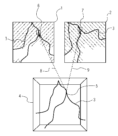
Figure 1 shows schematically a diagnostic projection data set consisting of
two (2)
two-dimensional (2D) projections 1 and 2 which were acquired by means of X-ray
fluoroscopy in the same cardiac phase. Note that any suitable type of cardiac
phase
monitoring can be used, for example, the recording of an electrocardiogram
(ECG) in
parallel with acquisition of the X-ray projections. Each of the projections 1
and 2, recorded
at different projection angles, shows a branched blood vessel 3 of a patient.
The projection
images 1 and 2 accordingly show the same blood vessel 3 from different
perspectives. To
acquire the projection data set, a contrast agent was administered to the
patient, such that
the blood vessel 3 shows up dark in the projections.
To reconstruct the three-dimensional structure of the blood vessel 3 according
to
the 3D front propagation method, a seed point 5 is initially set within a
reconstruction
volume 4. The blood vessel 3 is then reconstructed in the volume 4, by
locating adjacent
points in the volume 4 in each case belonging to the blood vessel 3 in
accordance with a
propagation criterion. To this end, local areas 6 and 7 belonging to the
respective point 5
within the two-dimensional projections 1 and 2, respectively, are in each case
subjected
individually to mathematical analysis. After location of a point adjacent to
the seed point 5,
the procedure is repeated for points in turn adjacent to this point, until the
entire structure
of the blood vessel 3 has been reconstructed within the volume 4.
The point investigated in each case with each propagation step is identified
as
belonging to the blood vessel if the mathematical analysis of the local areas
6 and 7 gives a
positive result for all or the majority of the projections belonging to the
projection data set
2
CA 02619308 2008-02-13
WO 2007/020555 PCT/IB2006/052705
(i.e., in this example projections 1 and 2, respectively). The local areas 6
and 7 are
determined by projecting the point 5, in accordance with the projection
directions in which
the two projections 1 and 2 were recorded, into the corresponding planes of
these two
projections. This is indicated in Figure 1 by arrows 8 and 9, respectively.
Note the while
this known 3D front propagation method has been described with respect to two
(2)
projections of the same heart phase, it is not limited to two (2) projections.
Accordingly, an improved method and system for overcoming the problems in the
art is desired.
According to an embodiment of the present disclosure, a method for computer-
aided automatic four-dimensional (4D) modeling of an anatomical object
comprises
acquiring automatically a set of three-dimensional (3D) models representing a
plurality of
static states of the object throughout a cycle. A 4D correspondency estimation
is performed
on the set of 3D models to determine which points of the 3D models most likely
correspond to each other, wherein the 4D correspondency estimation includes
one or more
of (i) defming a reference phase, (ii) performing vessel-oriented
correspondency
estimation, and (iii) post-processing of 4D motion data. The method can also
be
implemented by an imaging system, as well as in the form of a computer program
product.
Furthermore, the method according to one embodiment of the present disclosure
also
includes enabling automatic 3D modeling with a front propagation algorithm.
Figure 1 shows schematically a diagnostic projection data set consisting of
two (2)
two-dimensional (2D) projection images;
Figure 2 is an example of fully automatically extracted 3D centerlines back-
projected into two projection images of an underlying cardiac phase, obtained
with the
modeling method according to one embodiment of the present disclosure;
Figure 3 is an illustrative view showing examples of projections along three
orthogonal axes of extracted vessels at two different cardiac phases, obtained
with the
modeling method according to one embodiment of the present disclosure; and
Figure 4 is a partial block diagram view of an imaging apparatus according to
another embodiment of the present disclosure.
In the figures, like reference numerals refer to like elements. In addition,
it is to be
noted that the figures may not be drawn to scale.
3
CA 02619308 2008-02-13
WO 2007/020555 PCT/IB2006/052705
Automatic 3D Modelin2:
According to one embodiment of the present disclosure, a method comprises
automatic 3D vessel centerline extraction from gated rotational angiography X-
ray
projections using a front propagation method. In particular, the method
includes a non-
interactive algorithm for the automatic extraction of coronary centerline
trees from gated
3D rotational X-ray projections, i.e., without human interaction. The method
utilizes the
front propagation approach to select voxels that belong to coronary arteries.
The front
propagation speed is controlled by a 3D vesselness probability, which is
defined by
forward projecting the considered voxel into every vesselness-filtered
projection of the
same cardiac phase, picking the 2D response pixel values and combining them.
The
method further includes different ways of combining 2D response values to a 3D
vesselness probability. The method still further includes utilizing several
single-phase
models to build a combined multi-phase model.
Stated another way, the method includes a fully automatic algorithm for the
extraction of coronary centerline trees from gated 3D rotational X-ray
projections. The
algorithm is feasible when using good quality projections at the end-diastolic
cardiac
phase. Shortcut-artifacts from almost kissing vessels in systolic phases and
ghost vessel
artifacts can be significantly reduced by use of alternative versions of the
front propagation
algorithm. All algorithm versions have limited motion compensation ability,
thus after
fmding an optimal cardiac phase, centerline extraction of projections with
residual
respiratory motion is possible. In addition, single-phase models can also be
combined in
order to determine the best cardiac phase and to reduce the probability of
incorrectly traced
vessels. Furthermore, corresponding points in different single-phase models
can be found
in order to generate a fu114D coronary motion field with this approach.
Accordingly, the front propagation methods as discussed herein enable
automatic
extraction of a coronary vessel centerline tree without human interaction.
Further as noted
above, the front propagation models are relatively insensitive to residual
motion, especially
caused by respiration. According to one embodiment, it is necessary to
determine a model
that represents the coronary vessel shape at the cardiac phase of least motion
from a set of
ECG gated models. In the centerline extraction algorithm, the algorithm
enables a fully
automatic coronary vessel centerline extraction based on the front propagation
approach.
4
CA 02619308 2008-02-13
WO 2007/020555 PCT/IB2006/052705
As discussed herein, the automatic 3D front propagation algorithm uses gated
projections as input. The gating is performed according to a simultaneously
recorded
electrocardiogram (ECG) signal. The algorithm consists of multiple preparation
and
analysis steps, including (i) prefiltering of the gated projections; (ii)
finding seed point, (iii)
front propagation; (iv) for all vessel candidates: (a) finding end points, (b)
backtracing, and
(c) cropping and structuring; (v) finding the "root arc"; (vi) linking; (vii)
weighting; and
(viii) output and linking for output.
Prefiltering of the gated projections
In a first step, the projections are sorted into groups of same delay with
respect to
the R-peak of the ECG signal. A gated projection data set consists of the
nearest neighbor
projections to a given gating point from every heart cycle. All following
steps of the
algorithm are carried out on gated projection sets. In the next step, the
projections are
filtered using a multiscale vesselness filter, with filter widths from 1 to 7
pixels. The result
is a set of 2D response matrices R2D, which provide a probability for each
pixel to belong
to a vessel or not. The multiscale vesselness filter is defined as the maximum
of the
eigenvalues of the hessian matrices of all scales. To avoid border artifacts,
the vessel-
filtered projections can be cropped by a circular mask with a radius of about
(0.98 *
projection width).
Finding seed point
For each voxel x3D , a corresponding pixel on each projection can be
calculated by
using a cone-beam forward projection. The cone-beam forward projection can be
characterized where n denotes the current projection, eõ x, eõ , , and eõ Z,
are the normal
vectors of the detector plane, Dõ is the detector origin, Fõ the focus point,
defining the
trajectory data for each projection. x3D is the considered voxel and Põ its
projection. The
dimensions of the detector plane are determined by wX and wY (width and height
in mm)
and pX and pY (width and height in pixels).
The projected pixel on the detector plane in 3D is computed as follows:
~
*
~ - (DF~~ ~Tt ) ~' . it 'y3~e p.x 1.
'.F, .... i ....~a ....... .. ~ ~: L ~:i'. ..~~.Y ~
. _ f. . ~
~~ ~~~,,5
5
CA 02619308 2008-02-13
WO 2007/020555 PCT/IB2006/052705
Then the corresponding (x,y) -coordinates on a projection are:
(1) T)
~~ ' ,, _ z
1 a, (~'_ c~ra ,
_
~ "
Because the system geometry data is specific for each projection, the pixel
coordinates v
also depend on the current projection n.
Assuming there is no motion between different projections, the probability R3D
of a voxel
x3D to be located within a vessel can be obtained by multiplying the 2D
vesselness result
values R2D for all corresponding pixels:
~ ~ ... ' 1 ~...~ . .. ' ..... ~
(R..?aFa~Y ~)
A seed point is consequently found by choosing the voxel with the largest
response within
a certain subvolume.
Currently, a subvolume of about 11 % of the whole volume is examined this way,
because
the main vessels (ideally the root arc) are assumed to be located within the
cranial half of
the volume and in the centre, so the subvolume is determined as follows:
0.2", 1t~,...X ~' '; 5 ..~:
=~ 1~ :1 x
(Eq. 4~
3 ? ~~,~~~~
~. ~ - .. . 15 where the y-axis is oriented in caudo-cranial direction. The
maximum y value should not
reach y,Y,ax, because residual border artifacts of the vessel-filtered
projections may affect the
search for an appropriate seed point.
For further acceleration, the 3D response value for each voxel is not
completely
calculated using all N projections. If, after calculating the product of n
projections, the
intermediate value falls below the currently highest response value, the
remaining N-n
projections don't need to be calculated, because with every additional
multiplication, the
intermediate response value can only decrease further. This results in an
additional
acceleration factor of 2 to 5 depending on the source data.
6
CA 02619308 2008-02-13
WO 2007/020555 PCT/IB2006/052705
Front propagation
After an appropriate seedpoint has been found, the front propagation can be
started.
For each voxel that has been examined before, a characteristic value will be
stored, which
indicates how "quickly" the front has propagated towards this voxel starting
from the seed
point. Consequently, this value is called time value and set to zero at the
seed point. The
increase of these time values following an arbitrary path should therefore be
lower for
probably good vessels and higher (steeper) for "bad" vessels and artifacts.
At each iteration step, starting from the voxel on the front with the
currently lowest
time value, the 3D vessel response values of every neighboring voxel is
calculated, and its
reciprocal is added to the time value of the considered start voxel. If a
neighbouring voxel
has been considered before, it's value won't be recalculated again. Thus, the
time value
T(x3D (ko )) for a voxel x3D (ko ) reached after ko steps, represents the
history of the best
possible path beginning at the seed point, because it contains the response
values of all
preceding voxels:
2t1 .:'y ~'~_Ye7~ lJ.~ ~ ~.
x 1 {Eq.
There are several ways to compute an appropriate response value R3D for each
voxel. The
overall quality of the algorithm mainly depends on the quality of the approach
used here.
Thus, different approaches have been tried out, but only three of them proved
to be
feasible.
First front propagation approach (FP1)
A simple and stable way is to multiply all response values of the
corresponding
pixels on each filtered projection:
,,.
~
~t ~ ~...... ~ .~~ (70. ~~~~~* 6)
where n covers the gated projections and R2D is the corresponding pixel value
on the
current filtered projection, whose coordinates are given by võ as mentioned
herein above.
Thus, R3D is higher for better response and vice versa. The multiplication is
practically no
7
CA 02619308 2008-02-13
WO 2007/020555 PCT/IB2006/052705
problem with very low R2D responses, because even apart from vessel
structures, the R2D
response does not actually reach zero.
This approach gives reasonable results if the vessels on almost all
projections of the
set are of similar and relatively high quality. It has problems to trace weak
and thin vessels,
consequently even larger vessels might not be traced until their actual
ending, as they are
getting finer. The front propagates quickly towards the "good" vessels, but as
they are
getting weaker, the front progress becomes more and more indifferent and tends
to
propagate towards the border of the vessels. Therefore, reasonable tracing of
the whole
vessel tree using relatively poor-quality projections will consume much
computing power
by doing many iterations (e.g., about 3-5 million for 5123 resolution).
Nevertheless, the
outer ends of the vessels might still not be traced completely.
Second front propagation approach (FP2)
A solution for the problem of tracing thin vessels as described in the
preceding
section might be to prefer voxels with low response to those that are
obviously not lying on
a vessel at all. The second front propagation approach therefore tries to
emphasize voxels
with a relatively even response on all projections compared to those whose
response values
of the backprojected pixels differ more. This decision may be wrong, because
even
"correct" voxels might have bad response values on some projections because of
movement or bad projection/prefiltering quality. Because every filtered
projection is
normalized to 1, the result can be emphasized by raising it to a power below 1
and
suppressed by raising it to a power above 1. In order to describe how
uniformly the 2D
response values of a certain voxel x3D are distributed, the exponent 71 (x3D )
is now
calculated as normalized variance:
777) :':..
with
8
CA 02619308 2008-02-13
WO 2007/020555 PCT/IB2006/052705
R "'
~~. t~~
H
and used as follows:
.i +..
-- ~ i :1..
(Eq* 9,)
,==1:
This approach prefers weak vessels but will decrease the motion compensation
ability. It
tends to be unstable in some cases.
Third front propaggation approach (FP3)
A third front propagation approach is to account for the projection angle
difference
a,Y,-aõ between two projections m and n to prefer information extracted from
perpendicular
views to those taken from views of similar angle. This should minimize
misinterpretations
of depth information within two projections. Because there are more than two
projections
available, all projections (1 ... no) are considered by pairs and the
respective results are
combined by multiplication. The response value for each pair of projections is
calculated
by multiplying their according 2D response values and weighting them by the
sine of
projection difference angle:
The sine is obtained by calculating the cross product of the vectors pointing
from the
volume centre Mto the detector D divided by their respective length:
(L fi. I P
D,r vI T~' I
This third front propagation approach performs well when tracing thin vessels
and
compensates residual motion. In addition, the third front propagation approach
may be
more stable than the second front propagation approach.
9
CA 02619308 2008-02-13
WO 2007/020555 PCT/IB2006/052705
Terminating the front propaggation
Depending on the volume resolution and the quality of the projections, there
is a
rule-of-thumb value of the number of iterations that are reasonable:
i - 0.03 ~ ~~iuln(~ov o 1 ti ~~xel s. 12)
With respect to the first front propagation, for 2563 voxels, about 500k
iterations are
sufficient, while 5123 will need about 4,000k iterations to let the front
propagate into
similar regions. However, the later number of iterations consumes about eight
(8) times
more memory and computation time. The second and third FP approach only need
about
half as many iterations to get similar results.
Finding vessel segments
After finding an end point, the vessel centerline is traced, cropped and its
parts are
stored separately. Consecutive vessels are treated the same way. The following
three steps
of (1) finding end points, (2) backtracing, and (3) cropping and structuring
are therefore
done for each vessel candidate and its subvessels respectively.
(1) Finding end points
After the front propagation has finished, for every vessel an appropriate end
point
has to be found. This is achieved by dividing the whole volume into n3
subvolumes where
n=50 at this stage. Within each volume, the voxel with the highest time values
is chosen.
This voxel is located on the outer edge of a vessel, because the front is
propagating quickly
at the centre of each vessel and then broadens slowly (causing high time
values) towards its
border.
(2) Backtracing
The backtracing is performed using a steepest gradient method. Given an end
point,
the backtracing is directed towards the voxel with the largest time value
decrease with
respect to the current one. By following the largest decrease at every step,
an optimal path
back to the seed point is calculated. Starting at the surface of the front
propagation, it leads
directly to the vessel center and then along the centerline to the seed point.
If a path has
already been traced before by an earlier iteration, it will not be traced
again. This is
managed by a 3D bitmap in which the traced voxels are marked plus an
additional safety
area of two voxels at each side. This prevents doubled tracing of similar
(parallel) paths.
CA 02619308 2008-02-13
WO 2007/020555 PCT/IB2006/052705
(3) Cropping and Structuring
It is noted that voxels located at the border of a vessel do not belong to the
centerline and thus such voxels need to be cropped. Cropping is done by a
recursive
algorithm, wherein the recursive algorithm's task is to split the traced
centerline into
segments of different quality. The segment at the point where backtracing has
begun, has
worst quality and is thereby eliminated.
The recursive cropping algorithm assumes that the quality of every vessel is
best
close to the seed point and decreases towards its backtracing start point. The
mean value of
the first quarter of the current vessel voxels is calculated, wherein the
calculated value is
then used as threshold while scanning towards the tracing start point. The
threshold may be
occasionally exceeded several times, but if the number of those exceeding gets
beyond a
tolerance value (for example, a maximum of ten (10) consecutive times), then
the
particular spot is considered a significant quality breach and the vessel is
split into two
parts. This means, the worst quality segments are cut away from the vessel
segment of
better quality and then stored as an independent vessel. This second vessel is
then treated
the same way, thus the segment for the independent vessel is separated and so
on. The
recursive algorithm is aborted if the remaining part is shorter than a minimal
length (for
example, on the order of ten (10) voxels). The border voxels located at the
tracing start
point are either cut away by the minimum length criterion or, if their length
exceeds ten
(10) voxels, then they are rated negligible by the weighting algorithm
discussed later
herein.
Finding the "root arc"
As mentioned herein, the seed point for the front propagation does not
necessarily
correspond to the root arc, which is the inflow node of the coronary artery
tree. As a
consequence, every vessel is traced back to this "wrong" starting point. To
estimate the real
position of the root arc, the most cranial point of the longest three single
vessels segments
is used. The linking vessel segment between the seed point and the new top
point is then
used to extend other vessels, if necessary.
Linking
Up to now, the vessels have no relation to each other. Each vessel ending is
caused
by one of the following three reasons: i) the root arc has been reached, thus
no linking is
needed; ii) the vessel was formerly a part of a longer vessel and has been
separated by the
11
CA 02619308 2008-02-13
WO 2007/020555 PCT/IB2006/052705
cropping and structuring algorithm described herein above; and iii) there is a
bifurcation,
which means that there is another vessel crossing, which has been detected at
backtracing
stage. Up to this point, it is only known whether a path has been traced
before, but not
which vessel uses it. The correct successor vessel is determined by choosing
the point that
is geometrically closest to the end point of every vessel segment. Because at
the
backtracing stage all vessels were indexed in an ascending order, it is only
necessary to
search for points on vessels of a lower index than the considered one. After
linking, the
total length of every vessel (from end point to root arc) can easily be
calculated by adding
the length of all vessel segments along a link path.
Wei tin
In the steps described herein above, a large number of paths have been
extracted,
but only a few of them really represent existing vessels, while the majority
are caused by
artifacts such as lack of projection quality, residual motion, foreshortening
etc. Therefore,
it must be determined, which of them most probably represent real vessels. A
measure S
for the overall significance of an extracted path candidate can be composed of
several
factors: i) length of vessel segment or total length, ii) quality, determined
by time values,
iii) 3D position (probably with the assistance of a pre-defined model), and
(iv) shape.
According to the significance value S, all path candidates can be sorted,
which enables one
to choose the most significant path for output, where the maximum number of
paths to
output can be set by a system user. The calculation of the significance value
S is still to
improve, because a misjudgement here can lead to the output of a wrong
("ghost") vessel.
In one embodiment, S is calculated as follows:
V ,~
_ y PUPt -
s
7f ~: ~
where yeõa and yroot arc are the y coordinates (along the caudo-cranial
rotational axis) of the
current vessel segment end point and of the root arc determined as described
herein above,
respectively. The quantity lpa,.t is the length of the vessel segment in
voxels and
T(x3D (keõd)) is the time value of the end point of the vessel segment. It may
be possible to
automatically estimate a reasonable number of extractable vessel centerlines
using, for
example, gradient criteria.
12
CA 02619308 2008-02-13
WO 2007/020555 PCT/IB2006/052705
Output and linking for output
When saving the centerline data into a file, it may be necessary to check the
links
and to re-link some parts of the vessels, because one or more segments of a
linked path
may not be selected for output.
According to an embodiment of the present disclosure, an improved front
propagation algorithm transforms the prior known method of a semi-automatic 3D
algorithm into a fully automatic 4D algorithm. The method addresses various
problems
discussed herein above and provides solutions as follows:
1. Seed point: According to one embodiment, the seed point is defined
automatically by evaluating the above mentioned 3D vessel response in a
centered cranial
sub-volume of the 3D volume observable in every angiogram, and selecting the
point with
a maximum 3D response. Any suitable type of cardiac phase monitoring can be
used in
parallel with acquisition of the X-ray projections of a corresponding 3D
response, for
example, the cardiac phase monitoring may include the recording of an
electrocardiogram
(ECG). The maximum 3D response point is located on the vessel tree, but not
necessarily
at the inflow node of the main bifurcation. An alternative method is to select
the point with
maximum 3D response on the cranial part of the surface of the above mentioned
volume.
In the later instance, this provides a seed point located on the catheter
filled with contrast
agent, which comes in from the cranial side via the aorta.
2. Stopping the front propagation: The number of performed iterations of the
front
propagation is derived from either (i) the voxel resolution of the front
propagation volume
or (ii) by analysing the decrease of the 3D response values along an extracted
vessel.
3. End Points: Potential end points of vessels can be determined automatically
by
one or more different methods. In a first embodiment, the front propagation
volume is
divided into a large number of sub-volumes (e.g. 503 or 50*50*50). Within
every sub-
volume, the point with the latest front arrival is selected as the start point
for a back tracing
algorithm. The back tracing algorithm follows a speed field backwards along
the path with
the steepest gradient to the seed point. In a second embodiment, during a
front propagation,
the algorithm tracks the path along the steepest gradient and stops if a major
decrease of
the 3D vessel response is detected. In any event, the accurate estimation of
potential vessel
end points is not extremely critical, because in the following structuring
step, the vessel-
segments are analysed and weighted according to their relevance.
13
CA 02619308 2008-02-13
WO 2007/020555 PCT/IB2006/052705
4. Structuring: The vessels are divided into different segments by a dynamic
structuring algorithm. The dynamic structuring algorithm determines sections
of the
extracted centrelines with homogenous 3D vessel response. A weighting of each
vessel-
segment is performed according to different criteria: (i) length, (ii) 3D
vessel response
(corresponding to quality), (iii) shape and position of the centreline (or
optionally based on
an a-priori coronary model). The most relevant weighted vessels are
automatically selected
and constitute the output of the 3D algorithm. Figure 2 contains examples (20)
of fully
automatically extracted 3D centerlines back-projected into two projections (22
and 24) of
an underlying cardiac phase, obtained with the modeling method according to
one
embodiment of the present disclosure.
4D alwithm=
According to one embodiment of the present disclosure, the automatic 4D
coronary
modeling and motion vector field estimation method needs at input a set of 3D
models
representing all static states throughout the whole cardiac cycle by repeating
the above
described procedure for every distinguishable cardiac phase. The method
determines
corresponding points of different models by matching bifurcations and other
shape
properties of the different models. A possible application in which to exploit
the 4D
information is to derive an optimal cardiac phase for gated or motion-
compensated 3D
reconstruction.
The method according to the embodiments of the present disclosure provides a
fully
automatic, robust 4D algorithm for coronary centreline extraction and
modeling. The
method is capable to handle inconsistencies in angiograms of the same heart
phase due to
residual motion. Furthermore, the method according to the embodiments of the
present
disclosure provides improvements over the prior known 3D front propagation
algorithm,
wherein the improvements enable new applications such as 4D motion compensated
reconstructions and modeling.
A set of 3D models representing all static states throughout the whole cardiac
cycle
can be obtained by repeating the 3D modeling procedure for every
distinguishable cardiac
phase. Depending on the minimum heart beat rate during the rotational run
fi,n,;,, (in beats
per minute, bpm) and the acquisition frame rate fa (in 1/s), the number of
distinguishable
cardiac phases pN equals to:
14
CA 02619308 2008-02-13
WO 2007/020555 PCT/IB2006/052705
~ J t:z
~~~
which means that PN independent 3D models have been created. This value ranges
from
about about 15 for an acquisition frame rate fa of 25 fps (frames per second)
and heart beat
rate fh of 100 bpm (beats per minute) to about 40 for fa 30 fps and fh 45 bpm.
The task of
4D correspondency estimation is to determine which points of the models most
likely
correspond to each other, which enables to estimate the motion of certain part
of the vessel
tree throughout the cardiac cycle. Problems like longitudinal motion of the
vessels and
ambiguities caused during the 3D modeling process, which make 4D
correspondency
estimation more difficult, have to be taken into consideration. The
correspondency
estimation is performed by executing the following steps:
1. Definition of reference phase (stable phase)
2. Vessel-oriented correspondency estimation
3. Post-processing of 4D motion data
1. Definition of reference phase
To estimate stable 4D correspondencies, it is necessary to decide which of the
many
potential vessels structures extracted during the steps are of highest
significance during the
whole cardiac cycle. During the 3D algorithm, the vessel segments are weighted
according
to their presumed significance, but this is done independently for every
single 3D model,
which results in fluctuation of the extracted vessels at different cardiac
phases. Therefore, a
reference phase pr (stable phase) with all desired vessels extracted must be
defined prior to
the correspondency estimation. This can either be done automatically or
manually.
Automatic definition: Either, the 3D model representing the phase nearest to
35%
RR is chosen, which is in practice very likely a phase of low motion and
consequently
phase of good extraction quality or the model containing the three longest
vessels is
chosen. Note that RR represents a time interval defined by two subsequent R-
peaks of an
ECG, wherein the ECG is dominated by R-peaks and each R-peak represents an
electrical
impulse which precedes the contraction of the heart.
Manual definition: According to visual inspection of all extracted 3D models
(e.g.
using an overview plot 30 with projections of all models as shown in Figure
3), one can
manually define the most suitable cardiac phase and restart the algorithm.
Figure 3 shows
CA 02619308 2008-02-13
WO 2007/020555 PCT/IB2006/052705
an example 30 of two projections of extracted vessels at different cardiac
phases. The
upper row 32, representing cardiac phase of 43.5% RR, shows three correctly
extracted
vessels which qualifies that phase as potential reference phase, while the
quality of the
vessels shown in the bottom row 34 (5% RR) is worse.
2.Vessel-oriented correspondency estimation
The correspondence estimation is performed independently for every extracted
vessel at
the reference phase pr using one stable point at each model. When performing
this step for
the first time, the main bifurcation ("root arc") serves as stable point while
during later
iterations, sub-bifurcation points with probably higher precision are used.
The algorithm
exploits the fact that, during a cardiac cycle, the vessel's arc length k does
not change
considerably (less than 2% in total). The 3D coordinates:
Pl)
of any vessel point are parameterized by the vessel's arc length X, which
depends on the
considered phase number p, the considered vessel number v and the voxel number
i along
the vessel path: k =%(p, v, i). If, in the following, the text refers to
entire vessel, the voxel
number i is omitted.
Equally spaced versions of both the currently considered reference phase
vessel k
(pr , vr) and the current target phase vessel X (p, v), maintaining a
predefined spacing s
(currently set to 2 mm), are created, because the point-to-point distances of
the original 3D
models vary by factor of ~ 3 and more, caused by diagonal voxel distances and
linking
gaps. They represent the whole path from the stable point to the vessel's end.
The vessel
point coordinates are low-pass filtered prior to the equidistant spacing to
eliminate
quantization effects originating from the voxel representation of the front
propagation and
thus to provide a stable arc length criterion. The low-pass version of the
vessel k (p, v) is
denoted by V(p, v). The two vessels are compared point by point and an overall
similarity
criterion C is computed :
16
CA 02619308 2008-02-13
WO 2007/020555 PCT/IB2006/052705
{ AlSk
3,T)
p, v1t ir )'~ :1.~ ~ 1-) P.. V, ft '
L~i?. .. 5. .
?.-O
V) ' V 3
V3 f.>'of'~
Smaller similarity criteria C indicate better correspondence between the two
current
vessels. Consequently, the vessel combination with smallest C is considered to
be
equivalent. This procedure is repeated for every combination of source vessels
vr and target
phase vessels v and every possible target phase p:~pr. All corresponding
coordinates of the
corresponding vessels are finally stored in a dynamic array A(p,i) (called
motion field)
with indices [O..pN_1] (phase) and [0..ima,,_1] (corresponding 3D points).
3. Post-processin2 of 4D motion data
During the correspondency estimation procedure every corresponding vessel is
represented beginning from the reference point (normally the root arc), which
causes
several parts of the vessel tree to be represented multiple times. This
results in high local
point densities, which need to be thinned out to avoid singularities and other
ambiguities.
The reduction is achieved by computing the Euclidean distance d between each
combination of points belonging to a certain phase and erasing one of them if
the distance
falls below a threshold, which is defined as t = 0.5 s =1mm
-4
12~ - 4 (p.A (p2)
The resulting corresponding "root arc" points throughout all cardiac cycles
can be checked
for outliers. If the distance of the root arc in a specific phase to the
median (or mean)
position is above a given threshold, this cardiac phase is excluded from the
model. In a
similar manner all other bifurcation and single points can be treated.
17
CA 02619308 2008-02-13
WO 2007/020555 PCT/IB2006/052705
Turning now to Figure 4, the imaging apparatus illustrated therein is a C-arm
X-ray
apparatus, which comprises a C-arm 10, which is suspended by means of a holder
11, for
example, from a ceiling (not shown). An X-ray source 12 and an X-ray image
converter 13
are guided movably on the C-arm 10, such that a plurality of two-dimensional
projection
X-ray images of a patient 151ying on a table 14 in the center of the C-arm 10
may be
recorded at different projection angles. Synchronous movement of the X-ray
source 12 and
the X-ray image converterl3 is controlled by a control unit 16. During image
recording, the
X-ray source 12 and the X-ray image converter 13 travel synchronously around
the patient
15. The image signals generated by the X-ray image converter 13 are
transmitted to a
controlled image processing unit 17. The heart beat of the patient 15 is
monitored using an
ECG apparatus 18. The ECG apparatus 18 transmits control signals to the image
processing unit 17, such that the latter is in a position to store a plurality
of two-
dimensional projections in each case in the same phase of the heart beat cycle
to perform
an angiographic investigation of the coronary arteries. The image processing
unit 17
comprises a program control, by means of which three-dimensional models of a
blood
vessel tree detected with the projection data set thus acquired can be
performed, according
to a 3D front propagation method. In addition, the image processing unit 17
comprises a
further program control, by means of which 4D modeling can be performed,
according to
the embodiments of the present disclosure. The 4D modeling, as well as one or
more
reconstructed blood vessel, may then be visualized in any suitable manner on a
monitor 19
connected to the image processing unit 17.
Although only a few exemplary embodiments have been described in detail above,
those skilled in the art will readily appreciate that many modifications are
possible in the
exemplary embodiments without materially departing from the novel teachings
and
advantages of the embodiments of the present disclosure. For example, the
embodiments of
the present disclosure can be applied to other periodically moving structures
such as
cardiac venes or more general to tree-like structures. Accordingly, all such
modifications
are intended to be included within the scope of the embodiments of the present
disclosure
as defined in the following claims. In the claims, means-plus-function clauses
are intended
to cover the structures described herein as performing the recited function
and not only
structural equivalents, but also equivalent structures.
18
CA 02619308 2008-02-13
WO 2007/020555 PCT/IB2006/052705
In addition, any reference signs placed in parentheses in one or more claims
shall
not be construed as limiting the claims. The word "comprising" and
"comprises," and the
like, does not exclude the presence of elements or steps other than those
listed in any claim
or the specification as a whole. The singular reference of an element does not
exclude the
plural references of such elements and vice-versa. One or more of the
embodiments may be
implemented by means of hardware comprising several distinct elements, and/or
by means
of a suitably programmed computer. In a device claim enumerating several
means, several
of these means may be embodied by one and the same item of hardware. The mere
fact that
certain measures are recited in mutually different dependent claims does not
indicate that a
combination of these measures cannot be used to an advantage.
19