Note: Descriptions are shown in the official language in which they were submitted.
CA 02619455 2008-02-15
WO 2007/059000 PCT/US2006/043921
MODULATION OF ANGIOGENESIS BY A-BETA PEPTIDE FRAGMENTS
CROSS REFERENCE TO RELATED APPLICATIONS
[0001] This application claims the benefit of priority to U.S. Provisional
Application
No. 60/735,472, filed November 10, 2005, the disclosure of which is
incorporated
herein.
FIELD OF THE INVENTION
[0002] The present invention is related to compositions and methods for
treating
diseases and pathological conditions or processes mediated by pathological
angiogenesis by administering biologically active fragments of full length A(3
peptides to a patient suffering from such diseases, conditions, or processes.
DESCRIPTION OF RELATED ART
[0003] Alzheimer's disease (AD) is the major cause of dementia in the elderly
in
Western countries, and is characterized by the progressive accumulation of
intracellular neurofibrillary tangles, extracellular parenchymal senile
plaques, and
cerebrovascular deposits (Sissodia, et al. F.A.S.E.B. J. 9:366-370 (1995)).
The
principal component of senile plaques and cerebrovascular deposits is the [3-
amyloid
peptide, the aggregated form of which consists of the 39-43 amino acid residue
A(3
peptides that are proteolytically derived from the amyloid precursor protein
(APP)
(Naidu, et al. 1995 J. Biol. Chem. 270:1369-1374; Gorevic, et al. 1986 J.
Neuropathol. Exp. Neurol. 45, 647-64; Selkoe, et al. 1986 J. Neurochem. 46,
1820-
34). The primary protein component of senile plaques is beta/A4 amyloid, a 42-
43
amino acid peptide.
[0004] Vascular pathology is the norm in advanced cases of AD, with cerebral
amyloid angiopathy (CAA) being one of the most common abnormalities detected
at
autopsy (Ellis, et al. Neurology 46:1592-1596 (1996)). Certain vascular
lesions, such
as microvascular degeneration affecting the cerebral endothelium and
periventricular
white matter lesions, are evident in most AD cases (Ellis, et al. Neurology
46:1592-
1596 (1996); Kalaria, Ann. N.Y. Acad. Sci. 893:113-125 (1999)). Furthei7nore,
CA 02619455 2008-02-15
WO 2007/059000 PCT/US2006/043921
morphological alterations have been observed in AD brain microvessels and
capillaries; in particular, terminal arterioles frequently have focal
constriction and
smootli muscle cells with an irregular shape and arrangement (Hashimura et al.
Jpn. J.
Psychiatry Neurol. 45:661-665 (1991)). Capillaries in AD brain typically show
an
abnoimal abluminal surface with irregular constriction and dilatation along
their paths
(Kimura et al. Jpn. J. Psychiatry Neurol. 45:671-676 (1991)). Functional
imaging
techniques including positron emission tomography (PET) and single photon
emission
computerized tomography (SPECT) have revealed the existence of hypoperfusion
in
individuals prior to the time that they meet clinical criteria for AD
suggesting that
vascular abnormalities occur early during the disease process (Nagata et al.
Neurobiology of Aging 21:301-307 (2000); Johnson et al. Neurobiology of Aging
21:289-292 (2000)). In other disorders involving cerebrovascular damage (such
as
traumatic brain injury, stroke and brain arteriovenous malformation),
angiogenesis is
a prominent response (Mendis et al. Neurocheni. Res. 23:1117-23 (1998); Slevin
et al.
Stroke 31:1863-70 (2000); Hashimoto et al. Circ. Res. 89:111-3 (2001)). Given
the
plethora of reports on cerebrovascular damage in AD brain, the induction of an
angiogenic reparative response would be expected, although there has been very
little
work in this area.
[0005] Several assays have been developed to study the specific steps involved
in
the angiogenic process (adhesion, migration, growth, invasion and
differentiation).
Knowledge of the effects of A(3 on angiogenesis would be of value in
understanding
its role in the micro-cerebrovascular abnormalities observed in AD. In the AD
brain,
A(3 peptides are known to form fibrillar deposits around blood vessels,
leading to
cerebral amyloid angiopathy (CAA) (Pardridge, et al. 1987 J. Neurochem. 49,
1394-
401; Jellinger K.A., Attems J. 2005 J. Neurol. Sci. 229-230, 37-41). The
increased
levels of soluble and deposited A(3 in the AD brain can induce vascular
damage,
inflammation/gliosis, and reduced cerebral blood flow (Paris, et al. 2000 Ann.
N.Y.
Acad. Sci. 903, 97-109; Johnson, et al. 2005 Radiology. 234, 851-9). Numerous
studies have shown that vascular functional impairments and reduced blood flow
are
characteristic features of the AD brain (Nicoll, et al. 2004 Neurobiol. Aging.
25, 589-
97 and 603-4; Paris, et al. 2004 Brain Res. 999, 53-61; Beckmann, et al. 2003
J.
Neurosci. 23, 8453-9; Farkasm, et al. 2001 "Cerebral microvascular pathology
in
aging and Alzheimer's disease" Prog. Neurobiol. 64, 575-611). Recently, it has
been
2
CA 02619455 2008-02-15
WO 2007/059000 PCT/US2006/043921
shown that angiogenesis is impaired in AD, and that this is associated with
alterations
in genes involved in vascular differentiation (Wu, et al. 2005 Nat. Med. 11,
959-65).
A reduced brain capillary density is known in transgenic mouse models of AD
(Paris,
et al. 2004 Neurosci. Lett. 360, 80-5; Lee, et al. 2005 Brain Res. Bull. 65,
317-22). An
impaired formation of capillary lilce structures on reconstituted basement
membrane
by endothelial cells and arterial explants harvested fiom the brains of
TgAPPsw mice,
suggesting abnormal alterations in the angiogenic response in TgAPPsw mice was
recently demonstrated. (Paris, et al. 2004 Neurosci. Lett. 360, 80-5).
[0006] U.S. Patent Publication No. 2003/0077261 to Paris et al. discloses that
A(3
peptides can be used as anti-angiogenic agents, and discloses the sequences of
A-Beta
peptides and APP as well as the nucleic acids encoding them, which are shown
in the
attached Sequence Listing shown in Figure 10.
[0007] Angiogenesis is inhibited by A(3 peptides in multiple different in-
vitro and in-
vivo assays (Paris, et al. 2004 Angiogenesis. 7, 75-85). In-vitro, A(31.4o and
A(3i_42 can
dose dependently inhibit capillary tube formation by human brain microvascular
endothelial cells when plated on Matrigel, and can promote capillary
degeneration at
high doses. Mutants of the full-length A(3 peptide, including 1 or 2 amino
acid
substitutions, were also found to be biologically active anti-angiogenics.
However at
low doses, A(3 appears to be pro-angiogenic (Paris, et al. 2004 Angiogenesis.
7, 75-85;
Cantara, et al. 2004 F.A.S.E.B. J. 18, 1943-5).
SUMMARY OF THE INVENTION
[0008] It has been surprisingly discovered that biologically active fragments
of full
length A(3 peptides are useful as anti-angiogenic agents. These anti-
angiogenic A(3
peptide fragments may be used to treat pathological conditions mediated by
undesired
and/or uncontrolled angiogenesis (characterized as "angiogenic diseases"), as
described further herein.
[0009] Thus, in a first aspect, the present invention provides a variety of
anti-
angiogenic A(3 peptide fragnients as well as compositions which include one or
more
such fragments. In one embodiment, the biologically active A(3 peptide
fragment may
be 8, 9, 10, 11, 12, 13, 14, 15, 16, 17, 18, 19, 20, 21, 22, 23, 24, 25, 26,
27, 28, 29, 30,
31, 32, 33, 34, 35, 36, 37, 38 or 39 amino acids in length.
3
CA 02619455 2008-02-15
WO 2007/059000 PCT/US2006/043921
[0010] In a particular embodiment, the anti-angiogenic A(3 peptide fragment is
the
A(i1-28 peptide fragment, the A(3I0-35 peptide fragment, the A(312-2$ peptide
fragment,
the A(313-zo peptide fragment, or other biologically active fragments or
variants or
homologs thereof.
[0011] In a specific embodiment, the anti-angiogenic A(3 peptide fragment is
A(312-ZS
and contains the amino acid sequence HHQKLVFF, or biologically active
fragments,
variants or homologs thereof.
[0012] In another specific embodiment, the anti-angiogenic A(3 peptide
fragment is
A(313-20 or the amino acid sequence HHGKLVFF, or biologically active variants
or
homologs thereof. The variants may include, for example, amino acid
substitutions.
[0013] In another embodiment, the present invention is a pharmaceutical
coniposition comprising an anti-angiogenic A(3 peptide fragment and one or
more
pharmaceutically acceptable carriers, diluents, or excipients.
[0014] In a second aspect, the present invention provides a method for
treating a
disease or disorder mediated by pathological angiogenesis by administering to
a
subject in need thereof an effective amount of a biologically active A(3
peptide
fragment, wherein the fragment is between 8 and 39 amino acids in length. The
anti-
angiogenic A(3 peptide fragment is optionally administered in combination or
alternation with one or more therapeutic agents. The subject may be, for
example, a
mammal such as a human.
[0015] In one embodiment, the present invention is a method for treating
cancer by
administering to a subject in need thereof an effective amount of a
biologically active
A(3 peptide fragment, optionally, in combination or alternation with one or
more
cheinotherapeutic agents.
[0016] In a particular embodiment, the present invention is a method of
treating
cancer by administering to a subject in need thereof an effective amount of a
A012_38
peptide fragment containing the amino acid sequence HHQKLVFF or biologically
active fragments, variants or homologs thereof.
[0017] In another particular embodiment, the method of treating cancer
involves
administering to a subject in need thereof an effective amount of A(313-20
peptide
fragment or the amino acid sequence HHQKLVFF or biologically active variants
or
homologs thereof.
4
CA 02619455 2008-02-15
WO 2007/059000 PCT/US2006/043921
100181 The biologically active A(3 peptide fragment can be administered by any
suitable means including, but not limited, to oral, parenteral, intravenous,
intraarterial,
pulmonary, mucosal, topical, transdermal, subcuteaneous, intramuscular,
intrathecal
or intraperitoneal administration.
[0019] A third aspect of the present invention provides diagnostic methods and
kits
for detection and measurement of anti-angiogenic Ap peptide fragment activity
in
biological fluids and tissues.
[0020] A fourth aspect of the present invention provides diagnostic methods
and lcits
to screen for compounds that are potentially therapeutic in treatment of
Alzheimer's
disease by interfering with the anti-angiogenic effect of the A(3 peptide
fragment.
[0021] Another aspect of the invention are uses of the peptide fragments in
DESCRIPTION OF DRAWINGS
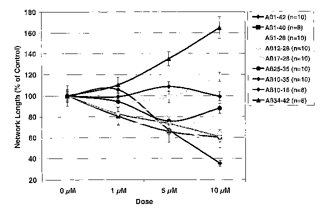
[0022] Figure 1 is a graph of the total length of capillary tubes expressed as
a
percentage of control treatment for 0, 1, 5 and 10 M doses of various AP
peptide
fragments as described in Example 8.
[0023] Figures 2A and 2B are chats of the cellular proliferation and cellular
adhesion of HUVEC samples, expressed as a percentage of the control, after
incubation with various A[3 peptide fragments as described in Example 9.
[0024] Figure 3 is a chart of the total length of capillary tubes expressed as
a
percentage of control treatment versus treatment with heparin (0.5 or 1
mg/ml),
A(31-42 peptide, A[i + heparin (500 g/ml) and A(3 + heparin (lmg/ml) as
described in
Example 10.
[0025] Figure 4 is a graph of the total length of capillary tubes expressed as
a
percentage of control treatment for 0, 1, 5 and 10 M doses of A(31_28,
A(31_28 GGQGL
and A(31_28 AAQAL as described in Example 11.
[0026] Figure 5 provides photographs (at 4X magnification) of capillaries
tubes
formed following incubation with A(3 peptide fragments as described in Example
11.
CA 02619455 2008-02-15
WO 2007/059000 PCT/US2006/043921
[0027] Figure 6 is a graph of the total length of capillary tubes expressed as
a
percentage of control treatment for 0, 1, 5 and 10 M doses of the peptides
HHHQKLVFF, VHHQKLVII, and VHHQKLVKK as described in Example 12.
[00281 Figure 7 is a chart of the Angiogenic Index (Al) for the rat corneal
micropocket assay in response to 200ng VEGF, VEGF + 0.5 g A(312_28, VEGF +
2.5 g A012_28 and VEGF + 5.O g A012_2$ as described in Example 13.
[0029] Figure 8 is a chart of the Angiogenic Index (AI) for the rat corneal
micropocket assay in response to VEGF, 5ug A(31_28 GGQGL, and 0.5ug, 2.5ug and
5ug of A012_28 and HHH-peptide (HHHQKLVFF), as described in Example 14.
[0030] Figure 9 provides representative photographs of rat corneal
micropockets
following a seven day incubation as described in Example 14, including a VEGF
control and 0.5 g, 2.5 g and 5.0 g of A(312_28=
[0031] Figure 10 is a listing of sequences of A-Beta peptides and APP as well
as the
nucleic acids encoding them, as described in U.S. Patent Publication No.
2003/0077261 to Paris et al.
DETAILED DESCRIPTION OF THE INVENTION
[0032] Anti-angiogenic therapy is an attractive approach for inhibition of
tumor
progression, as tumors depend upon an adequate blood supply for growth. It is
disclosed herein that short peptides derived from the A(3 sequence inhibit
angiogenesis, and can be used for anti-cancer therapy.
[0033] Provided are anti-angiogenic A(3 peptide fragments that can be used to
treat
pathological conditions mediated by undesired and/or uncontrolled or
pathological
angiogenesis. Provided herein is a particular anti-angiogenic motif (HHQKLVFF)
which may be used in anti-tumor or anti-angiogenic therapies.
Anti-An io eg nic Peptide Fragments
[0034] The present invention provides anti-angiogenic fragments of A(3
peptides
useful for the treatment of disorders or diseases associated with pathological
or
unwanted angiogenesis.
6
CA 02619455 2008-02-15
WO 2007/059000 PCT/US2006/043921
[0035] The term "A(3 peptide fragment" as used herein refers to an anti-
angiogenic
fragment of a full length A(3 peptides (e.g., A(31_40, AR1_42, A(3i-43) and
includes A(3
peptide fragment variants, homologs (such as mammalian orthologs) and
isofonns,
unless otherwise noted. The term also includes fragments with substitutions of
one or
more equivalent amino acids, or non-natural amino acids.
[0036] In one embodiment, the A(3 peptide fragment is at least one amino acid
less in
number than the total nuinber of amino acids found in the full-length A(3
peptide. Full
length A(3 peptides are derived from proteolytic processing of one or more
isoforms of
the amyloid precursor protein (APP), a transmembrane glycoprotein (Kang, J. et
al.
Nature (Lond.). (1987) 325: 733-736). The 39-43-amino acid-long A(i peptide
amino
acid sequence begins in the ectodomain of APP and extends into the
transmembrane
region. A(3 is formed after sequential cleavage of APP by the (i- and y-
secretases.
A(31_42 and A(31_43 forms are specifically found in all kinds of AD plaques,
indicating
that those forms are critically important in AD pathology.
[0037] In a particular embodiment, the A(3 peptide fragment is at least one
amino
acid less in number than the total number of amino acids found in the full
length A(31_
40 peptide. The A(31_40 peptide fragment consists of, for example, 8, 9, 10,
11, 12, 13,
14, 15, 16, 17, 18, 19, 20, 21, 22, 23, 24, 25, 26, 27, 28, 29, 30, 31, 32,
33, 34, 35, 36,
37, 38 or 39 amino acids.
[0038] In another particular embodiment, the A(3 peptide fragment is at least
one
amino acid less in number than the total number of amino acids found in the
full
length AP1_42 peptide. The A(31_42 fragment consists of, for example, 8, 9,
10, 11, 12,
13, 14, 15, 16, 17, 18, 19, 20, 21, 22, 23, 24, 25, 26, 27, 28, 29, 30, 31,
32, 33, 34, 35,
36, 37, 38, 39, 40 or 41 amino acids.
[0039] In another particular embodiment, the A(3 peptide fragment is at least
one
amino acid less in number than the total number of amino acids found in the
full
length A(31_43 peptide. The A(31_43 fragment consists of, for example, 8, 9,
10, 11, 12,
13, 14, 15, 16, 17, 18, 19, 20, 21, 22, 23, 24, 25, 26, 27, 28, 29, 30, 31,
32, 33, 34, 35,
36, 37, 38, 39, 41, or 42 amino acids.
[0040] In one embodiment, the fragment consists of, for example, 8, 9, 10, 11,
12,
13, 14, 15, 16, 17, 18, 19, 20, 21, 22, 23, 24, 25, 26, 27, 28 or more amino
acid
residues, and includes the sequence HHQKLVFF.
7
CA 02619455 2008-02-15
WO 2007/059000 PCT/US2006/043921
[0041] In one embodiment, one or more of the following biologically active A(3
peptide fragments may be used to treat diseases or disorders associated with
unwanted
or pathological angiogenesis: the A[iI-28 peptide, the A(31o-35 peptide, the
A(312-2$
peptide, the A(313-z0 peptide, or biologically active fragments or variants
thereof.
[0042] The anti-angiogenic A(3 peptide fragment preferably contains the HHQK
proteoglycan binding region, since fragments without that sequence (A(325-35,
AP17-28,
and A(334-42) were not active, suggesting that the heparin binding motif HHQK
is
required to mediate the anti-angiogenic activity of A. The A(31o-16 fragment
was
inactive even though it contains the HHQK sequence, suggesting that the HHQK
proteoglycan binding motif is not sufficient to inhibit angiogenesis and that
other
neighboring residues are required. In particular, the LVFF sequence
immediately
following the HHQK domain is also required for inhibition of angiogenesis.
Thus,
preferred A(3 peptide fragments contain the anlino acid sequence HHQKLVFF.
[0043] In one embodiment, the fragment consists of, for example, 8, 9, 10, 11,
12,
13, 14, 15, 16, 17, 18, 19, 20, 21, 22, 23, 24, 25, 26, 27, 28, 29, 30, 31,
32, 33, 34, 25,
36, 37, 38 or more amino acid residues, and includes the sequence HHQKLVFF.
Such fragnients may include one or more (e.g. 2, 3 or 4) substitutions of
equivalent
amino acids, including, e.g., non-natural amino acids.
[0044] In one embodiment, the A(3 peptide fragment is a A012-28 peptide
containing
the amino acid sequence HHQKLVFF, or a biologically active fragment or variant
thereof.
[0045] In another embodiment, the A[i peptide fragment is a A(313-zo peptide
fragment or the amino acid sequence HHQKLVFF, or a biologically active
fragment
or variant thereof.
[0046] In another embodiment, the A(3 peptide fragment is, e.g., a 10, 20, 30,
or 40
amino acid fragment of the A[3 peptide.
[0047] The peptide fragments are obtained, for example, by chemical synthesis,
or
are recombinantly produced by host cells.
[0048] Likewise, the terms variant and homologous are also used
interchangeably.
"Variant" or "homologous" peptide fragments will be understood to designate
those
containing, in relation to the native polypeptide sequence, modifications such
as
deletion, addition, or substitution of at least one amino acid, truncation,
extension, or
8
CA 02619455 2008-02-15
WO 2007/059000 PCT/US2006/043921
the addition of chimeric heterologous polypeptides. Optionally, "variant" or
"homologous" peptide fragments can contain a mutation or post-translational
modifications.
[0049] Among the "variant" or "homologous" polypeptides or peptide fragments,
those whose amino acid sequence exhibits 80.0% to 99.9% (inclusive) identity
to the
native polypeptide sequence are preferred. These percentages are purely
statistical and
differences between two peptide sequences can be distributed randomly and over
the
entire sequence length.
[0050] "Variant" or "homologous" polypeptide sequences exhibiting a percentage
identity with the polypeptides of the present invention can, alternatively,
have 80, 81,
82, 83, 84, 85, 86, 87, 88, 89, 90, 91, 92, 93, 94, 95, 96, 97, 98, or 99
percent identity
with the polypeptide sequences of the instant invention. The expression
equivalent
amino acid is intended here to designate any amino acid capable of being
substituted
for one of the amino acids in the basic structure without, however,
essentially
modifying the biological activities of the corresponding peptides and as
provided
below.
[0051] Several substitutions could be made to HHQKLVFF (motif) region of A(3
while potentially retaining the substitution antiangiogenic properties of the
peptide.
Specifically, the following expression indicates such equivalent substitutions
for
HHQKLVFF:
[RH]-H-[NQ]-[RK]-[ILV]-[ILV]-F-F
[0052] Exemplary sequences with such motifs are listed in Table 1. Sources are
noted if a particular peptide sequence is a part of a naturally occurring
protein.
Table 1
Amino acid Source, if
sequence naturally-
curring
HHQKLVFF human APP/ A(3
RHQKLVFF rat/mouse APP/ Ap
HHNKLVFF
RHNKLVFF
HHQRLVFF
RHQRLVFF
HHNRLVFF
RHNRLVFF
HHQKIVFF
9
CA 02619455 2008-02-15
WO 2007/059000 PCT/US2006/043921
RHQKIVFF
HHNKIVFF
RHNKIVFF
HHQRIVFF
RHQRIVFF tr:Q3YB12_BACST
putative regulator
[Geobacillus
stearothermo hilus
HHNRIVFF
RHNRIVFF
HHQKVVFF
RHQKVVFF
HHNKWFF
RHNKWFF
HHQRWFF [Q6CET0] Yarrowia
lipolytica
chromosome B of
strain CLIB99 of
Yarrowia lipolytica
(trembl).
RHQRVVFF
HHNRVVFF
RHNRWFF
HHQKLIFF
RHQKLIFF
HHNKLIFF
RHNKLIFF
HHQRLIFF
RHQRLIFF
HHNRLIFF
RHNRLIFF
HHQKIIFF
RHQKIIFF
HHNKIIFF
RHNKIIFF
HHQRIIFF
RHQRIIFF
HHNRIIFF
RHNRIIFF
HHQKVIFF
RHQKVIFF
HHNKVIFF
RHNKVIFF
HHQRVIFF
RHQRVIFF
HHQKLLFF
RHQKLLFF
HHNKLLFF
RHNKLLFF
HHQRLLFF
RHQRLLFF
HHNRLLFF
RHNRLLFF Trembl sequence
CA 02619455 2008-02-15
WO 2007/059000 PCT/US2006/043921
entry
tr:Q7QS20 GIALA
HHQKILFF
RHQKILFF
HHNKILFF
RHNKILFF
HHQRILFF
RHQRILFF
HHNRILFF
RHNRILFF
HHQKVLFF
RHQKVLFF
HHNKVLFF
RHNKVLFF
HHQRVLFF
RHQRVLFF
HHNRVIFF
RHNRVIFF
100531 The motif search from http://motif:genome.jp/MOTIF2.html was used to
search the peptide combinations in the NR-AA TrembUSwissprot database. The
substitution of physico-chemical equivalent amino acids in peptide sequences
is
known in the art. (Eisenberg, et al. 1984 "Amino acid scale: Nonnalized
consensus
hydrophobicity scale." J. Mol. Biol. 179:125-142; and Mathura, et al. 2001,
"New
quantitative descriptors for amino acids based on multidimensional scaling of
a large
number of physical-chemical properties", J. Mol. Modeling 7:445-453).
[0054] In one embodiment, the A(3 peptide fragment consists of or comprises
one of
the peptide sequences listed in Table 1, with optional equivalent amino acid
substitutions.
[0055] The subject invention also provides biologically active peptide
fragments
capable of eliciting an immune response. The immune response can provide
components (either antibodies or components of the cellular immune response
(e.g.,
B-cells, helper, cytotoxic, and/or suppressor T-cells) reactive with the
peptide
fragment.
[0056] Fragments, as described herein, can be obtained by cleaving a
polypeptide
with a proteolytic enzyme (such as trypsin, chymotrypsin, or collagenase) or
with a
chemical reagent, such as cyanogen bromide (CNBr). Alternatively, polypeptide
fragments can be generated in a highly acidic environment, for example at pH
2.5.
11
CA 02619455 2008-02-15
WO 2007/059000 PCT/US2006/043921
Such polypeptide fragments may be also prepared by chemical synthesis or using
hosts transformed with an expression vector containing nucleic acids encoding
polypeptide fragments. The transformed host cells contain a nucleic acid and
are
cultured according to well-known methods; thus, expression of these fragments
is
possible, under the control of appropriate elements for regulation and/or
expression.
[0057] The peptides can be modified by variation in the splicing of
transcriptional
products of the A(3 gene, genetic recombination, or by chemical synthesis.
Such
peptides can contain at least one modification in relation to the polypeptide
sequence
being modified. These modifications can include the addition, substitution,
deletion of
amino acids contained within the polypeptides.
[0058] Conservative substitutions whereby an amino acid of one class is
replaced
with another amino acid of the same type fall within the scope of the subject
invention
so long as the substitution does not materially alter the biological activity
of the
polypeptide. For example, the class of nonpolar amino acids include Ala, Val,
Leu,
Ile, Pro, Met, Phe, Gly and Trp; the class of uncharged polar amino acids
include Ser,
Thr, Cys, Tyr, Asn, and Gln; the class of acidic amino acids includes Asp and
Glu;
and the class of basic amino acids includes Lys, Arg, and His. In some
instances, non-
conservative substitutions can be made where these substitutions do not
significantly
detract from the biological activity of the polypeptide.
[0059] In order to extend the life of the polypeptides provided, it may be
advantageous to use non-natural amino acids, for example in the D form, or
alternatively amino acid analogs, such as sulfur-containing forms of amino
acids.
Alternative means for increasing the life of polypeptides can also be used.
For
example, peptide fragments can be recombinantly modified to include elements
that
increase the plasma, or serum half-life. These elements include, and are not
limited to,
antibody constant regions (see for example, U.S. Patent No. 5,565,335, hereby
incorporated by reference in its entirety, including all references cited
therein), or
other elements such as those disclosed in U.S. Patent Nos. 6,319,691;
6,277,375; or
5,643,570, each of which is incorporated by reference in its entirety,
including all
references cited within each respective patent. Alternatively, the
polynucleotides and
genes can be recombinantly fused to elements that are useful in the
preparation of
immunogenic constructs for the purposes of vaccine formulation or elements
useful
for the isolation of the polypeptides provided.
12
CA 02619455 2008-02-15
WO 2007/059000 PCT/US2006/043921
[0060] The peptide fragments disclosed may further contain linkers that
facilitate the
attachment of the fragments to a carrier molecule for delivery or diagnostic
purposes.
The linkers can also be used to attach fragments to solid support matrices for
use in
affmity purification protocols. In one embodiment, the linlcers specifically
exclude
where the fragment is a subsequence of another peptide, polypeptide, or
protein as
identified in a search of protein sequence databases. In other words, the non-
identical
portions of the other peptide, polypeptide, of protein is not considered to be
a"linlcer"
in this aspect. Non-limiting examples of "linkers" suitable for the practice
of the
invention include chemical linkers (such as those sold by Pierce, Roclcford,
Ill.),
peptides that allow for the connection of the immunogenic fragment to a
carrier
molecule (see, for example, linkers disclosed in U.S. Patent Nos. 6,121,424;
5,843,464; 5,750,352; and 5,990,275, hereby incorporated by reference in their
entirety). In various embodiments, the linkers can be up to 50 ainino acids in
length,
up to 40 amino acids in length, up to 30 amino acids in length, up to 20 amino
acids in
length, up to 10 amino acids in length, or up to 5 amino acids in length.
[0061] In other specific embodiments, the peptides may be expressed as a
fusion, or
chimeric protein product ( joined via a peptide bond to a heterologous protein
sequence (e.g., a different protein)). Such a chimeric product can be made by
ligating
the appropriate nucleic acid sequences encoding the desired amino acid
sequences to
each other by methods known in the art, in the proper coding frame, and
expressing
the chimeric product by methods commonly known in the art (see, for exaniple,
U.S.
Patent No. 6,342,362, hereby incorporated by reference in its entirety;
Altendorf, et al.
1999-WWW, 2000 "Structure and Function of the Fo Complex of the ATP Synthase
from Escherichia Coli," J. of Experimental Biology 203:19-28, G. B.; Baneyx
1999
Biotechnology 10:411-21; Eihauer, et al. 2001 J. Biochem. Biophys. Methods
49:455-
65; Jones, et al. 1995 J. Chromatography 707:3-22; Jones, et al. 1995 J. of
Chromatography A. 707:3-22; Margolin, et al. 2000 Methods 20:62-72; Puig, et
al.
2001 Methods 24:218-29; Sassenfeld, et al. 1990 Tib. Tech. 8:88-93; Sheibani,
et al.
1999 Prep. Biochem. & Biotechnol. 29(1):77-90; Skerra, et al. 1999
Biomolecular
Engineering 16:79-86; Smith, et al. 1998 The Scientist 12(22):20; Smyth, et
al. 2000
Methods in Molecular Biology, 139:49-57; Unger, et al. 1997 The Scientist
11(17):20; each of which is hereby incoiporated by reference in their
entireties).
Alternatively, such a chimeric product may be made by protein synthetic
techniques,
13
CA 02619455 2008-02-15
WO 2007/059000 PCT/US2006/043921
e.g., by use of a peptide synthesizer. Fusion peptides can comprise
polypeptides and
one or more protein transduction domains, as described above. Such fusion
peptides
are particularly useful for delivering the cargo polypeptide through the cell
membrane.
[0062] Increasing the ainount of A(3 peptide fragment activity within a tissue
is
useful in treating a variety of angiogenic diseases, such as cancers, tumors,
and/or
malignancies. Thus, according to the methods provided, the amount of A(3
peptide
fragment activity can be increased within a tissue by directly administering
the A(3
peptide fragment to a patient suffering from an angiogenic disease (such as
exogenous
delivery of the A(3 peptide fragment) or by indirect or genetic means (such as
delivery
of a polynucleotide encoding the A(3 peptide fragment or upregulating the
endogenous
A(3 peptide fragment activity). Non-limiting examples of such cancers, tumors,
and/or
malignancies that can be treated using the methods of the invention include
prostate
cancer, breast cancer, melanoma, chronic myelogenous leukemia, cervical
cancer,
adenocarcinomas, lymphoblastic leukemia, colorectal cancer, and lung
carcinoma.
[0063] The peptide fragments or nucleic acids encoding them can be used in
screening, or aiding in the diagnosis of, an individual suspected of having an
angiogenic or angiogenesis-mediated disease. The peptide fragments disclosed
herein
and nucleic acids encoding them can be used to detect the A(3 peptide in
hybridization
assays by the use of complementary sequences. The presence of a significantly
increased amount of A(3 peptide fragment is associated with an indication of
Alzheimer's disease. The presence of a significantly decreased amount of A(3
peptide
is associated with an indication of an angiogenic disease, such as a
malignancy or
cancer. A[i gene product can be detected by well-known methodologies
including, and
not limited to, Western blots, enzyme linked immunoassays (ELISAs),
radioimmunoassays (RIAs), Northern blots, Southern blots, PCR-based assays, or
other assays for the quantification of gene product known to the skilled
artisan. This
information, in conjunction with other information available to the skilled
practitioner, assists in making a diagnosis.
[0064] In one aspect, the subject invention concerns a method of inhibiting
angiogenesis in a patient in need of anti-angiogenesis therapy by
administration of
biologically active A(3 peptide fragment to the patient.
14
CA 02619455 2008-02-15
WO 2007/059000 PCT/US2006/043921
[0065] In one embodiment, a treatment for a pathological condition selected
from
the group consisting of cancer, arthritis, atherosclerosis, psoriasis, macular
degeneration, and diabetic retinopathy by administering to the patient a
therapeutically effective amount of an A(3 peptide fragment.
[0066] In one embodiment, biologically active variants of the A(3 peptide
fi=agments
are utilized, wherein the variants have a substitution at the 21 amino acid
position, or
the 22 amino acid position, or 23 amino acid position, or combinations
thereof. In a
specific embodiment, the substitution(s) is a conservative substitution which
does not
materially alter the biological activity of the polypeptide.
[0067] Various means for delivering polypeptides to a cell can be utilized to
carry
out the methods provided. For example, protein transduction domains (PTDs) can
be
fused to the polypeptide, producing a fusion polypeptide, in which the PTDs
are
capable of transducing the polypeptide cargo across the plasma membrane
(Wadia, J.
S. and Dowdy, S. F., Curr. Opin. Biotechnol. 2002, 13(1), 52-56). Examples of
PTDs
include the Drosophila homeotic transcription protein antennapedia (Antp), the
herpes
simples virus structural protein VP22, and the human immuno-deficiency virus 1
(HIV-1) transcriptional activator Tat protein.
[0068] According to the method of angiogenesis inhibition provided,
recombinant
cells can be administered to a patient, wherein the recombinant cells have
been
genetically modified to express A(3 peptide fragments disclosed herein.
[0069] The method of angiogenesis inhibition provided can be used to treat a
patient
suffering from cancer, or as a cancer preventative. The method of tumor
inhibition
provided can be used to treat patients suffering from a variety of cancers
including,
but not limited, to cancer of the breast, prostate, melanoma, chronic
myelogenous
leukemia, cervical cancer, adenocarcinoma, lymphoblastic leukemia, colorectal
cancer, and lung carcinoma. According to the methods provided, various other
anti-
cancer or anti-tumor compounds, such as cytotoxic agents, can be administered
in
conjunction with A(3 peptide fragments.
CA 02619455 2008-02-15
WO 2007/059000 PCT/US2006/043921
Nucleotide sequences encoding A(3 fragments
[0070] In another aspect, the subject invention provides isolated andlor
purified
nucleotide sequences comprising a polynucleotide sequence encoding the amino
acid
sequence of the peptide fragments disclosed herein,
[0071] Also provided are isolated nucleic acid inolecules comprising
polynucleotides encod'uig the A(3 peptide fragments. One aspect of the
invention
provides isolated nucleic acid molecules comprising polynucleotides having a
nucleotide sequence selected from the group consisting of: (a) a nucleotide
sequence
encoding any of the amino acid sequences of the polypeptides described herein
including in Table 1; and (b) a nucleotide sequence complementary to any of
the
nucleotide sequences in (a).
[0072] Further embodiments of the invention include isolated nucleic acid
molecules
that comprise a polynucleotide having a nucleotide sequence at least 90%
identical,
and more preferably at least 95%, 96%, 97%, 98% or 99% identical to any of the
nucleotide sequences in (a) or (b) above.
[0073] Nucleotide, polynucleotide, or nucleic acid sequences(s) are understood
to
mean, according to the present invention, either a double-stranded DNA, a
single-
stranded DNA, or products of transcription of the said DNAs (e.g., RNA
molecules).
The nucleic acid, polynucleotide, or nucleotide sequences can be isolated,
purified (or
partially purified), by separation methods including, but not limited to, ion-
exchange
chromatography, molecular size exclusion chromatography, affinity
chromatography,
or by genetic engineering methods such as amplification, cloning or
subcloning.
100741 Optionally, the polynucleotide sequences can also contain one or more
polynucleotides encoding heterologous polypeptide sequences (e.g., tags that
facilitate
purification of the polypeptides of the invention (see, for example, U.S.
Patent No.
6,342,362, hereby incorporated by reference in its entirety; Altendorf, et al.
1999-
WWW, 2000 "Structure and Function of the Fo Complex of the ATP Synthase from
Escherichia Coli," J. of Experimental Biology 203:19-28, G. B.; Baneyx 1999
Biotechnology 10:411-21; Eihauer, et al. 2001 J. Biochem. Biophys. Methods
49:455-
65; Jones, et al. 1995 J. Chromatography 707:3-22; Jones, et al. 1995 J. of
Chromatography A. 707:3-22; Margolin, et al. 2000 Methods 20:62-72; Puig, et
al.
2001 Methods 24:218-29; Sassenfeld, et al. 1990 Tib. Tech. 8:88-93; Sheibani,
et al.
16
CA 02619455 2008-02-15
WO 2007/059000 PCT/US2006/043921
1999 Prep. Biochem. & Biotechnol. 29(1):77-90; Slcerra, et al. 1999
Biomolecular
Engineering 16:79-86; Smith, et al. 1998 The Scientist 12(22):20; Smyth, et
al. 2000
Methods in Molecular Biology, 139:49-57; Unger, et al. 1997 The Scientist
11(17):20; each of which is hereby incorporated by reference in their
entireties), or
commercially available tags from vendors such as such as STRATAGENE (La Jolla,
Calif.), NOVAGEN (Madison, Wis.), QIAGEN, Inc., (Valencia, Calif.), or
INVITROGEN (San Diego, Calif.).
Vectors
[0075] Other aspects provide vectors containing one or more of the
polynucleotides
provided, such as vectors containing nucleotides encoding biologically active
A(3
peptide fragments. The vectors can be vaccine, replication, or amplification
vectors.
In some embodiments, the polynucleotides are operably associated with
regulatory
elements capable of causing the expression of the polynucleotide sequences.
Such
vectors include, among others, chromosomal, episomal and virus-derived
vectors,
e.g., vectors derived fiom bacterial plasmids, from bacteriophage, from
transposons,
from yeast episomes, from insertion elements, from yeast chromosomal elements,
from viruses such as baculoviruses, papova viruses, such as SV40, vaccinia
viruses,
adenoviruses, fowl pox viruses, pseudorabies viruses and retroviruses, and
vectors
derived from combinations of the aforementioned vector sources, such as those
derived from plasmid and bacteriophage genetic elements (e.g., cosmids and
phagemids).
[0076] As indicated above, vectors can also comprise elements necessary to
provide
for the expression and/or the secretion of a polypeptide, such as a fragment
of the A(3
peptide, encoded by the nucleotide sequences provided in a given host cell.
The
vector can contain one or more elements selected from the group consisting of
a
promoter, signals for initiation of translation, signals for termination of
translation,
and appropriate regions for regulation of transcription. In certain
embodiments, the
vectors can be stably maintained in the host cell and can, optionally, contain
signal
sequences directing the secretion of translated protein. Other embodiments
provide
vectors that are not stable in transformed host cells. Vectors can integrate
into the host
genome or be autonomously-replicating vectors.
17
CA 02619455 2008-02-15
WO 2007/059000 PCT/US2006/043921
[0077] In a specific embodiment, a vector comprises a promoter operably linked
to a
protein or peptide-encoding nucleic acid sequence, one or more origins of
replication,
and, optionally, one or more selectable markers (e.g., an antibiotic
resistance gene).
Non-limiting exemplary vectors for the expression of polypeptides include pBr-
type
vectors, pET-type plasmid vectors (PROMEGA), pBAD plasmid vectors
(INVITROGEN) or those provided in the examples below. Furthermore, vectors are
useful for transfonning host cells for the cloning or expression of the
nucleotide
sequences provided.
[0078] Promoters which may be used to control expression include, but are not
limited to, the CMV promoter, the SV40 early promoter region (Bemoist and
Chambon 1981 Nature 290:304-310), the promoter contained in the 3' long
terminal
repeat of Rous sarcoma virus (Yamamoto, et al. 1980 Cell 22:787-797), the
herpes
thymidine kinase promoter (Wagner et al. 1981 Proc. Natl. Acad. Sci. USA
78:1441-
1445), the regulatory sequences of the metallothionein gene (Brinster et al.
1982
Nature 296:39-42); prokaryotic vectors containing promoters such as the (3-
lactamase
promoter (Villa-Kamaroff, et al. 1978 Proc. Natl. Acad. Sci. USA 75:3727-
3731), or
the tac promoter (DeBoer, et al. 1983 Proc. Natl. Acad. Sci. USA 80:21-25);
see also,
"Useful Proteins from Recombinant Bacteria" in Scientific American, 1980,
242:74-
94; plant expression vectors comprising the nopaline synthetase promoter
region
(Herrera-Estrella et al. 1983 Nature 303:209-213) or the cauliflower mosaic
virus 35S
RNA promoter (Gardner, et al. 1981 Nucl. Acids Res. 9:2871), and the promoter
of
the photosynthetic enzyme ribulose biphosphate carboxylase (Herrera-Estrella
et al.
1984 Nature 310:115-120); promoter elements from yeast or fungi such as the
Ga14
promoter, the ADC (alcohol dehydrogenase) promoter, PGK (phosphoglycerol
kinase) promoter, and/or the alkaline phosphatase promoter.
Homologous nucleotide sequences
[0079] Provided herein are "homologous" or "modified" nucleotide sequences.
Modified nucleic acid sequences will be understood to mean any nucleotide
sequence
obtained by mutagenesis according to techniques well known to persons slcilled
in the
art, and exhibiting modifications in relation to the noi7nal sequences. For
example,
mutations in the regulatory and/or promoter sequences for the expression of a
18
CA 02619455 2008-02-15
WO 2007/059000 PCT/US2006/043921
polypeptide that result in a modification of the level of expression of a
polypeptide
provide for a "modified nucleotide sequence". Likewise, substitutions,
deletions, or
additions of nucleic acid to the polynucleotides provide for "homologous" or
"modified" nucleotide sequences. In various embodiments, "homologous" or
"modified" nucleic acid sequences have substantially the same biological or
serological activity as the native (naturally occurring) A(3 peptide
fragments. A
"homologous" or "modified" nucleotide sequence will also be understood to mean
a
splice variant of the polynucleotides of the instant invention or any
nucleotide
sequence encoding a "modified polypeptide" as defined below.
[0080] A homologous nucleotide sequence, as described herein, encompasses a
nucleotide sequence having a percentage identity with the bases of the
nucleotide
sequences of between at least (or at least about) 80.0% to 99.9% (inclusive),
or 85%
to 99%, or 90% to 99%, or 95% to 99%.
[0081] In various embodiments, homologous sequences exhibiting a percentage
identity with the bases of the nucleotide sequences described can have 80, 81,
82, 83,
84, 85, 86, 87, 88, 89, 90, 91, 92, 93, 94, 95, 96, 97, 98, or 99 percent
identity with the
polynucleotide sequences of the instant invention.
[0082] Both protein and nucleic acid sequence homologies may be evaluated
using
any of the variety of sequence comparison algorithms and programs known in the
art.
Such algorithms and programs include, but are by no means limited to, TBLASTN,
BLASTP, FASTA, TFASTA, and CLUSTALW (Pearson and Lipman 1988 Proc.
'Natl. Acad. Sci. U.S.A. 85(8):2444-2448; Altschul, et al. 1990 J. Mol. Biol.
215(3):403-410; Thompson, et al. 1994 Nucleic Acids Res. 22(2):4673-4680;
Higgins, et al. 1996 Methods Enzymol. 266:383-402; Altschul, et al. 1990 J.
Mol.
Biol. 215(3):403-410; Altschul, et al. 1993 Nature Genetics 3:266-272).
[0083] Also provided are nucleotide sequences complementary to any of the
polynucleotide sequences disclosed herein. Thus, the invention is understood
to
include any DNA whose nucleotides are complementary. to those of the sequence
of
the invention, and whose orientation is reversed (e.g., an antisense
sequence).
[0084] Further provided are fragments of the polynucleotide sequences
disclosed
herein. Representative fragments of the polynucleotide sequences will be
understood
to mean any nucleotide fragment having at least 8 or 9 successive nucleotides,
19
CA 02619455 2008-02-15
WO 2007/059000 PCT/US2006/043921
preferably at least 12 successive nucleotides, and still more preferably at
least 15 or at
least 20 successive nucleotides of the sequence from which it is derived. The
upper
limit for such fragments is the total number of polynucleotides found in the
sequence
encoding for A(3I_42 peptide, (or, in certain embodiments, the open reading
frame
(ORF) identified herein). The appropriate fragments thereof encoding for a
specific
peptide are also useful. For example, nucleotide sequences that are A(3
peptide
fragment homologs, or fragments thereof, which have been previously
identified, can
be utilized to carry out the method for inhibiting angiogenesis of the subject
invention.
Hybridization and detection probes
[0085] Among these representative fragments, those capable of hybridizing
under
stringent conditions with a nucleotide sequence are preferred. Conditions of
high or
intennediate stringency are provided infra and are chosen to allow for
hybridization
between two complementary DNA fragments. Hybridization conditions for a
polynucleotide of about 300 bases in size will be adapted by persons skilled
in the art
for larger- or smaller-sized oligonucleotides, according to methods well known
in the
art (see, for example, Sambrook, et al. 1989 Molecular Cloning, A Laboratory
Manual, Second Edition, Cold Spring Harbor Press, N.Y., pp. 9.47-9.57).
[0086] Also provided are detection probes (e.g., fragments of the disclosed
polynucleotide sequences) for hybridization with a target sequence or an
amplicon
generated from the target sequence. Such a detection probe will advantageously
have
as sequence a sequence of at least 9, 12, 15, 20, 25, 30, 35, 40, 45, 50, 55,
60, 65, 70,
75, 80, 85, 90, 95, or 100 nucleotides. Alternatively, detection probes can
comprise 8,
9, 10, 11, 12, 13, 14, 15, 16, 17, 18, 19, 20, 21, 22, 23, 24, 25, 26, 27, 28,
29, 30, 31,
32, 33, 34, 35, 36, 37, 38, 39, 40, 41, 42, 43, 44, 45, 46, 47, 48, 49, 50,
51, 52, 53, 54,
55, 56, 57, 58, 59, 60, 61, 62, 63, 64, 65, 66, 67, 68, 69, 70, 71, 72, 73,
74, 75, 76, 77,
78, 79, 80, 81, 82, 83, 84, 85, 86, 87, 88, 89, 90, 91, 92, 93, 94, 95, 96,
97, 98, 99,
100, 101, 102, 103, 104, 105, 106, 107, 108, 109, 110, 111, 112, 113, 114,
115, 116,
117, 118, 119, 120, 121, 122, 123, 124, 125, 126, 127 and up to, for example,
128
consecutive nucleotides of the disclosed nucleic acids. The detection probes
can also
be used as labeled probe or primer in the subject invention. Labeled probes or
primers
CA 02619455 2008-02-15
WO 2007/059000 PCT/US2006/043921
are labeled with a radioactive compound or with another type of label.
Alternatively,
non-labeled nucleotide sequences may be used directly as probes or primers;
however,
the sequences are generally labeled with a radioactive element (32P, 35S, 3H,
125I) or
with a molecule such as biotin, acetylaminofluorene, digoxigenin, 5-bromo-
deoxyuridine, or fluorescein to provide probes that can be used in numerous
applications.
[0087] The nucleotide sequences disclosed may also be used in analytical
systems,
such as DNA chips. DNA chips and their uses are well known in the art and (see
for
example, U.S. Patent Nos. 5,561,071; 5,753,439; 6,214,545; Schena, et al. 1996
BioEssays 18:427-431; Bianchi, et al. 1997 Clin. Diagn. Virol. 8:199-208; each
of
which is hereby incorporated by reference in their entireties) and/or are
provided by
commercial vendors such as AFFYMETRIX, Inc. (Santa Clara, Calif.).
[0088] Various degrees of stringency of hybridization can be employed. The
more
severe the conditions, the greater the complementarity that is required for
duplex
formation. Severity of conditions can be controlled by temperature, probe
concentration, probe length, ionic strength, time, and the like. Preferably,
hybridization is conducted under moderate to high stringency conditions by
techniques well known in the art, as described, for example, in Keller, G. H.,
M. M.
Manak 1987 DNA Probes, Stockton Press, New York, N.Y., pp. 169-170.
[0089] By way of example, hybridization of immobilized DNA on Southern blots
with 32P-labeled gene-specific probes can be performed by standard methods
(Maniatis, et al. 1982 Molecular Cloning: A Laboratory Manual, Cold Spring
Harbor
Laboratory, New York). In general, hybridization and subsequent washes can be
carried out under moderate to high stringency conditions that allow for
detection of
target sequences with homology to the exemplified polynucleotide sequence. For
double-stranded DNA gene probes, hybridization can be carried out overnight at
20-
25 C below the melting temperature (Tm) of the DNA hybrid in 6x SSPE, 5x
Denhardt's solution, 0.1% SDS, 0.1 mg/ml denatured DNA. The melting
temperature
is described by the following forrnula (Beltz et al. 1983 Methods of
Enzymology, R.
Wu, L. Grossman and K. Moldave [eds.] Academic Press, New York 100:266-285).
[0090] T,ri 81.5 C.+16.6 Log[Na+]+0.41(%G+C)-0.61(% fonnamide)-600/length of
duplex in base pairs.
21
CA 02619455 2008-02-15
WO 2007/059000 PCT/US2006/043921
[0091] Washes are typically carried out as follows:
[0092] (1) twice at room temperature for 15 minutes in lx SSPE, 0.1% SDS (low
stringency wash);
[0093] (2) once at Tn, -20 C. for 15 minutes in 0.2x SSPE, 0.1% SDS (moderate
stringency wash).
[0094] For oligonucleotide probes, hybridization can be carried out overnight
at 10-
20 C. below the melting temperature (Tm) of the hybrid in 6x SSPE, 5x
Denhardt's
solution, 0.1% SDS, 0.1 mg/ml denatured DNA. Tm for oligonucleotide probes can
be
determined by the following formula:
[0095] Tm ( C)=2 (number T/A base pairs)+4 (number G/C base pairs) (Suggs et
al.
1981 ICN- UCLA Symp. Dev. Biol. Using Purified Genes, D. D. Brown [ed.],
Academic Press, New York, 23:683-693).
[0096] Washes can be carried out as follows:
[0097] (1) twice at room temperature for 15 minutes lx SSPE, 0.1% SDS (low
stringency wash;
[0098] 2) once at the hybridization temperature for 15 minutes in lx SSPE,
0.1%
SDS (moderate stringency wash). -
[0099] In general, salt and/or temperature can be altered to change
stringency. With
a labeled DNA fragment >70 or so bases in length, the following conditions can
be
used:
[00100] 1 Low: 1 or 2X SSPE, room temperature Low: 1 or 2X SSPE, 42 C.
Moderate: 0.2X or 1X SSPE, 65 C. High: 0.1X SSPE, 65 C.
[00101] By way of another non-limiting example, procedures using conditions of
high stringency can also be performed as follows: Pre-hybridization of filters
containing DNA is carried out for 8 h to overnight at 65 C. in buffer
composed of 6x
SSC, 50 mM Tris-HCl (pH 7.5), 1 mM EDTA, 0.02% PVP, 0.02% Ficoll, 0.02%
BSA, and 500 g/ml denatured salmon sperm DNA. Filters are hybridized for 48 h
at
65 C., the preferred hybridization temperature, in pre-hybridization mixture
containing 100 g/ml denatured salmon sperm DNA and 5-20x106 cpm of 32P-
labeled
probe. Alternatively, the hybridization step can be performed at 65 C. in the
presence
of SSC buffer, lx SSC corresponding to 0.15M NaCI and 0.05 M Na citrate.
22
CA 02619455 2008-02-15
WO 2007/059000 PCT/US2006/043921
Subsequently, filter washes can be done at 37 C. for 1 h in a solution
containing 2x
SSC, 0.01% PVP, 0.01% Ficoll, and 0.01% BSA, followed by a wash in 0.1x SSC at
500 C. for 45 min. Alternatively, filter washes can be perfonned in a solution
containing 2x SSC and 0.1% SDS, or 0.5x SSC and 0.1% SDS, or 0.lx SSC and 0.1%
SDS at 68 C. for 15 minute intervals. Following the wash steps, the
hybridized
probes are detectable by autoradiography. Otlier conditions of higli
stringency which
may be used are well known in the art (see, for example, Sambrook, et al. 1989
Molecular Cloning, A Laboratory Manual, Second Edition, Cold Spring Harbor
Press,
N.Y., pp. 9.47-9.57; and Ausubel, et al. 1989 Current Protocols in Molecular
Biology,
Green Publishing Associates and Wiley Interscience, N.Y., each incorporated
herein
in its entirety).
[00102] A further non-limiting example of procedures using conditions of
intermediate stringency are as follows: Filters containing DNA are pre-
hybridized,
and then hybridized at a temperature of 60 C. in the presence of a 5x SSC
buffer and
labeled probe. Subsequently, filters washes are performed in a solution
containing 2x
SSC at 50° C. and the hybridized probes are detectable by
autoradiography.
Other conditions of intermediate stringency which may be used are well known
in the
art (see, for example, Sambrook et al. 1989 Molecular Cloning, A Laboratory
Manual,
Second Edition, Cold Spring Harbor Press, N.Y., pp. 9.47-9.57; and Ausubel et
al
1989 Current Protocols in Molecular Biology, Green Publishing Associates and
Wiley
Interscience, N.Y., each of which is incorporated herein in its entirety).
[00103] Duplex formation and stability depend on substantial complementarity
between the two strands of a hybrid and, as noted above, a certain degree of
mismatch
can be tolerated. Therefore, the probe sequences of the subject invention
include
mutations (both single and multiple), deletions, insertions of the described
sequences,
and combinations thereof, wherein said mutations, insertions and deletions
permit
formation of stable hybrids with the target polynucleotide of interest.
Mutations,
insertions and deletions can be produced in a given polynucleotide sequence in
many
ways, and these methods are known to an ordinarily skilled artisan. Other
methods
may become known in the future.
[00104] It is also well lcnown in the art that restriction enzymes can be used
to obtain
functional fragments of the subject DNA sequences. For example, Ba131
exonuclease
can be conveniently used for time-controlled limited digestion of DNA
(commonly
23
CA 02619455 2008-02-15
WO 2007/059000 PCT/US2006/043921
referred to as "erase-a-base" procedures). See, for example, Maniatis, et al.
1982
Molecular Cloning: A Laboratory Manual, Cold Spring Harbor Laboratory, New
Yorlc; Wei, et al. 1983 J. Biol. Chem. 258:13006-13512. The nucleic acid
sequences
disclosed can also be used as molecular weight markers in nucleic acid
analysis
procedures.
Host cells
[00105] Provided are liost cells transfonned by a polynucleotide according to
the
invention and the production of A(3 peptide fragments by the transformed host
cells.
In some embodiments, transformed cells comprise an expression vector
containing
polynucleotide sequences for an A(3 peptide fragment. Other embodiments
provide for
host cells transformed with nucleic acids. Yet other embodiments provide
transformed
cells comprising an expression vector containing fragments of AP
polynucleotide
sequences. Transformed host cells can be cultured under conditions allowing
the
replication and/or the expression of the nucleotide sequences provided.
Expressed
polypeptides are recovered from culture media and purified, for further use,
according
to methods known in the art.
[00106] The host cell may be chosen from eukaryotic or prokaryotic systems,
for
example bacterial cells (Gram negative or Gram positive), yeast cells, animal
cells,
plant cells, and/or insect cells using baculovirus vectors. In some
embodiments, the
host cell for expression of the polypeptides include, and are not limited to,
those
taught in U.S. Patent Nos. 6,319,691; 6,277,375; 5,643,570; 5,565,335; Unger,
et al.
1997 The Scientist 11(17):20; or Smith, et al. 1998 The Scientist 12(22):20,
each of
which is incorporated by reference in its entirety, including all references
cited within
each respective patent or reference. Other exemplary, and non-limiting, host
cells
include Staphylococcus spp., Enterococcus spp., E. coli, and Bacillus
subtilis; fungal
cells, such as Streptomyces spp., Aspergillus spp., S. cerevisiae,
Schizosaccharomyces pombe, Pichia pastoris, Hansela polymorpha, Kluveromyces
lactis, and Yarrowia lipolytica; insect cells such as Drosophila S2 and
Spodoptera Sf9
cells; animal cells such as CHO, COS, HeLa, C127, 3T3, BHK, 293 and Bowes
melanoma cells; and plant cells. A great variety of expression systems can be
used to
produce the polypeptides provided and polynucleotides can be modified
according to
24
CA 02619455 2008-02-15
WO 2007/059000 PCT/US2006/043921
methods lrnown in the art to provide optimal codon usage for expression in a
particular expression system.
[00107] Furthermore, a host cell strain may be chosen that modulates the
expression
of the inserted sequences, modifies the gene product, and/or processes the
gene
product in the specific fashion. Expression from certain promoters can be
elevated in
the presence of certain inducers; thus, expression of the genetically
engineered
polypeptide may be controlled. Furthermore, different host cells have
characteristic
and specific mechanisms for the translational and post-translational
processing and
modification (e.g., glycosylation, phosphorylation) of proteins. Appropriate
cell lines
or host systems can be chosen to ensure the desired modification and
processing of
the foreign protein expressed. For example, expression in a bacterial system
can be
used to produce an unglycosylated core protein product whereas expression in
yeast
will produce a glycosylated product. Expression in mammalian cells can be used
to
provide "native" glycosylation of a heterologous protein. Furthermore,
different
vector/host expression systems may effect processing reactions to different
extents.
[00108] Nucleic acids and/or vectors can be introduced into host cells by well-
known methods, such as, calcium phosphate transfection, DEAE-dextran mediated
transfection, transfection, microinjection, cationic lipid-mediated
transfection,
electroporation, transduction, scrape loading, ballistic introduction and
infection (see,
for example, Sambrook, et al. 1989 Molecular Cloning: A Laboratory Manual,
2<sup>nd</sup> Ed., Cold Spring Harbor Laboratory Press, Cold Spring Harbor, N.Y.).
[001091 The subject invention also provides for the expression of a
polypeptide,
derivative, or a variant (e.g., a splice variant) encoded by a polynucleotide
sequence
disclosed herein. Alternatively, the invention provides for the expression of
a
polypeptide fragment obtained from a polypeptide, derivative, or a variant
encoded by
a polynucleotide fragment derived from the polynucleotide sequences disclosed
herein. In either embodiment, the disclosed sequences can be regulated by a
second
nucleic acid sequence so that the polypeptide or fragment is expressed in a
host
transformed with a recombinant DNA molecule according to the subject
invention.
For example, expression of a protein or peptide may be controlled by any
promoter/enhancer element known in the art.
CA 02619455 2008-02-15
WO 2007/059000 PCT/US2006/043921
[00110] The subject invention also provides nucleic acid-based methods for the
identification of the presence of the A(3 gene, or fragments or variants
thereof, in a
sample. These methods can utilize the nucleic acids provided and are well
known to
those slcilled in the art (see, for example, Sambrook, et al. 1989 Molecular
Cloning, A
Laboratory Manual, Second Edition, Cold Spring Harbor Press, N.Y., pp. 9.47-
9.57,
or Abbaszadega, et al. 2001 Reviews in Biology and Biotechnology, 1(2):21-26).
Among the techniques useful in such methods are enzymatic gene amplification
(or
PCR), Southern blots, Northern blots, or other techniques utilizing nucleic
acid
hybridization for the identification of polynucleotide sequences in a sample.
The
nucleic acids can be used to screen individuals for disorders associated with
dysregulation of the A(3 gene or its transcriptional products.
[00111] The subject invention also provides polypeptides encoded by nucleotide
sequences of the invention. The subject invention also provides fragments of
at least 5
amino acids of a polypeptide encoded by the polynucleotides of the instant
invention.
Pharmaceutical Formulations and Administration
[00112] As used herein, the term "administration" or "administering" refers to
the
process of delivering an agent to a patient. The process of administration can
be
varied, depending on the agent, or agents, and the desired effect.
Administration can
be accomplished by any means appropriate for the therapeutic agent, for
example, by
oral, parenteral, mucosal, pulmonary, topical, catheter-based, rectal,
intracranial,
intracerebroventricular, intracerebral, intravaginal or intrauterine delivery.
Parenteral
delivery can include for example, subcutaneous intravenous, intrauscular,
intra-
arterial, and injection into the tissue of an organ, particularly tumor
tissue. Mucosal
delivery can include, for example, intranasal delivery. Oral or intranasal
delivery can
include the administration of a propellant. Pulmonary delivery can include
inhalation
of the agent. Catheter-based delivery can include delivery by iontropheretic
catheter-
based delivery. Oral delivery can include delivery of a coated pill, or
administration
of a liquid by mouth. Administration can generally also include delivery with
a
pharmaceutically acceptable carrier, such as, for example, a buffer, a
polypeptide, a
peptide, a polysaccharide conjugate, a liposome, and/or a lipid. Gene therapy
protocol
is also considered an administration in which the therapeutic agent is a
polynucleotide
26
CA 02619455 2008-02-15
WO 2007/059000 PCT/US2006/043921
capable of accomplishing a therapeutic goal when expressed as a transcript or
a
polypeptide into the patient.
[00113] In one embodiment, the A(3 peptide fragment is administered in an
effective
amount to inhibit pathological angiogenesis. As used herein, the term
"angiogenesis"
is intended to refer to the process by which new blood vessels are formed and
which
is essential to a variety of normal body activities (such as reproduction,
development,
and wound repair). The process is believed to involve a complex interplay of
molecules which both stimulate and inhibit the growth of endothelial cells,
the
primary cells of the capillary blood vessels. Under normal conditions, these
molecules
appear to maintain the microvasculature in a quiescent state (i.e., one of no
capillary
growth) for prolonged periods. When necessary, however (such as during wound
repair), these cells can undergo rapid proliferation and turnover within a
short period
of time. Although angiogenesis is a highly regulated process under normal
conditions,
many conditions (characterized as "angiogenic diseases") are driven by
persistent
unregulated angiogenesis. Otherwise stated, unregulated angiogenesis may
either
cause a particular pathological condition directly or exacerbate an existing
pathological condition. For example, ocular neovascularization has been
implicated as
the most common cause of blindness and dominates approximately twenty eye
diseases. In certain existing conditions, such as arthritis, newly formed
capillary blood
vessels invade the joints and destroy cartilage. In diabetes, new capillaries
formed in
the retina invade the vitreous, bleed, and cause blindness. Growth and
metastasis of
tumors are also angiogenesis-dependent (Folkman, J., Cancer Research, 46:467-
473,
1986; Folkman, J., Journal of the National Cancer Institute, 82:4-6, 1989). It
has been
shown, for example, that tumors which enlarge to greater than 2 mm, must
obtain
their own blood supply and do so by inducing the growth of new capillary blood
vessels. Once these new blood vessels become embedded in the tumor, they
provide a
means for tunlor cells to enter the circulation and metastasize to distant
site, such as
liver, lung or bone (Weidner, N. et al., The New England Journal of Medicine,
324(1):1-8, 1991).
[00114] The pharmaceutical compositions of the subject invention can be
formulated
according to known methods for preparing pharmaceutically useful compositions.
Formulations are described in a number of sources which are well known and
readily
available to those skilled in the art. For example, Remington's Phannaceutical
27
CA 02619455 2008-02-15
WO 2007/059000 PCT/US2006/043921
Sciences (Martin E W 1995 Easton Pennsylvania Maclc Publishing Company,
19<sup>th</sup> ed.) describes formulations which can be used in connection with the
subject invention. Formulations suitable for parenteral administration
include, for
example, aqueous sterile injection solutions, which may contain antioxidants,
buffers,
bacteriostats, and solutes which render the formulation isotonic with the
blood of the
intended recipient; and aqueous and nonaqueous sterile suspensions which may
include suspending agents and thickening agents. The formulations maybe
presented
in unit-dose or multi-dose containers, for example sealed ampoules and vials,
and may
be stored in a freeze dried (lyophilized) condition requiring only the
condition of the
sterile liquid carrier, for example, water for injections, prior to use.
Extemporaneous
injection solutions and suspensions may be prepared from sterile powder,
granules,
tablets, etc. It should be understood that in addition to the ingredients
particularly
mentioned above, the formulations of the subject invention can include other
agents
conventional in the art having regard to the type of formulation in question.
[00115] In one embodiment, the A(3 peptide fragments are delivered in a
sustained
release formulation. The formulations provide extended release and extended
half-
life. Controlled release systems suitable for use include, without limitation,
diffusion-
controlled, solvent-controlled and chemically-controlled systems. Diffusion
controlled systems include, for example reservoir devices, in which the AJ3
peptide
fragment or fragments are enclosed within a device such that release of the
peptide
fragments is controlled by permeation through a difussion barrier. Common
reservoir
devices include, for example, membranes, capsules, microcapsules, liposomes,
and
hollow fibers. Monolithic (matrix) device are a second type of diffusion
controlled
system, wherein the Ap peptide fragment(s) are dispersed or dissolved in an
rate-
controlling matrix (e.g., a polymer matrix). The peptide fragments are
homogeneously
dispersed throughout a rate-controlling matrix and the rate of drug release is
controlled by diffusion through the matrix. Polymers suitable for use in the
monolithic matrix device include naturally occurring polymers, synthetic
polymers
and synthetically modified natural polymers, as well as polymer derivatives.
[00116] Therapeutically effective and optimal dosage ranges for the A(3
peptide
fragments can be determined using methods known in the art. Guidance as to
appropriate dosages to achieve an anti-angiogenesis and/or anti-tumor effect
is
provided from the exemplified assays disclosed herein. The minimal amounts of
A(3
28
CA 02619455 2008-02-15
WO 2007/059000 PCT/US2006/043921
peptide fragment to achieve a therapeutic effect can likewise be determined.
In one
embodiment, the A(3 peptide fragment is administered in an equivalent amount
to be
within the M dose range. In another embodiment, an amount equivalent to about
1
M to about 100 M A(3 peptide fragment is administered. In another embodiment,
an
amount equivalent to about 2 M to about 10 M A(3 peptide fragment is
administered. Pharmaceutical formulations that can be administered can
comprise,
e.g., 1-10,000 mg, 10-1000 mg, 50-900 mg, 100-800 mg, or 200-500 mg.
[00117] The subject invention also pertains to diagnostic and/or screening
methods
and kits to screen for compounds that are potentially therapeutic in treatment
of
Alzheimer's disease by interfering with the anti-angiogenic effect of an Ap
peptide
fragment.
[00118] In one aspect, included is a method for identifying compounds that
interfere
with A(3-induced angiogenesis inhibition, wherein the method includes the
steps of (a)
contacting a first biological sample capable of undergoing angiogenesis with a
test
compound, a biologically active amount of an A(3 peptide fragment, and an
angiogenic agent; and (b) determining the extent of angiogenesis that occurs
in the
first biological sample. Optionally, the method can include the steps of= (c)
separately
contacting a second biological sample capable of undergoing angiogenesis with
a
biologically active amount of an A(3 peptide fragment and an angiogenic agent;
(d)
determining the extent of angiogenesis that occurs in the second biological
sample;
and (e) comparing the extent of angiogenesis that occurs in the first
biological sample
with that which occurs in the second biological sample. In this way, steps (c)-
(d) can
be utilized as a control. Preferably, the same A(3 peptide fragment is used in
the first
and second biological samples. i
[00119] Determining the extent of angiogenesis can be carried out using
methods
lcnown in the art, such as those described herein, and can be done
qualitatively or
quantitatively. For example, molecular or cellular markers of cancer or tumor
growth
can be utilized. The extent of angiogenesis can also be determined by
measuring the
amount of endothelial cell proliferation or the extent of blood vessel growth
within a
biological sample.
[00120] The biological samples utilized in the methods and kits can include
various
biological fluids and tissues that can exhibit angiogenesis and/or tumor
development.
29
CA 02619455 2008-02-15
WO 2007/059000 PCT/US2006/043921
For example, the biological sample can be arterial tissue, corneal tissue,
endothelial
cells, umbilical cord tissue, chorionic allantoid membrane, and the like.
[00121] The angiogenic agent can be any molecule, compound, or cell that is
capable of inducing angiogenesis in the biological sample. For example, the
angiogenic agent can be a trophic factor, such as a neurotrophic factor. The
angiogenic factor can be a cytokine or growth factor such as vascular
endothelial
growth factor, platelet-derived growth factor, and basic fibroblast growth
factor. The
diagnostic and/or screening methods of the subject invention can be carried
out in
vivo, such as in an animal model, or in vitro.
[00122] In another aspect, the subject invention includes a kit for
identifying
compounds that interfere with A[i-induced angiogenesis inhibition. The kit can
include a compartment containing at least one AR peptide fragment and,
optionally, a
compartment containing an angiogenic agent. Furthermore, the kit can
optionally
include a compartnient containing one or more biological samples.
[00123] In another aspect, a method is provided for identifying compounds that
interfere with A(3-induced anti-tumor activity, including the steps of (a)
contacting a
first tumor tissue with a test compound and a biologically active amount of an
A(3
peptide fragment; and (b) determining the extent of tumor growth that occurs
in the
tuinor tissue. Optionally, the method can further include the steps of: (c)
separately
contacting a second tumor tissue with a biologically active amount of an A(3
peptide
fragment; (d) determining the extent of tumor growth that occurs in the second
tumor
tissue; and (e) comparing the extent of tumor growth that occurs in the first
tumor
tissue with that which occurs in the second tumor tissue. In this way, steps
(c)-(d) can
be utilized as a control. The extent of tumor growth can be determined
quantitatively
or qualitatively using methods known in the art, including methods described
herein.
For example, molecular or cellular markers of cancer or tumor growth can be
utilized.
[00124] In another aspect, the subject invention includes a kit for
identifying
compounds that interfere with Ap-induced anti-tumor activity. The kit can
include a
comparhnent containing at least one A(3 peptide fragment and, optionally, a
compartment containing at least one tumor tissue. Furthermore, the kit can
optionally
include a compartment containing one or more biological samples.
CA 02619455 2008-02-15
WO 2007/059000 PCT/US2006/043921
[00125] The test compounds that can be screened using the methods and kits of
the
subject invention can include any substance, agent, or molecule, including,
for
example, small molecules and living or dead cells.
[00126] A variety of patients may be treated including any vertebrate species.
Preferably, the patient is of a mammalian species. Mammalian species which
benefit
from the disclosed methods of treatment include, and are not limited to, apes,
chimpanzees, orangutans, humans, monkeys; domesticated animals (e.g., pets)
such as
dogs, cats, guinea pigs, hanlsters, Vietnamese pot-bellied pigs, rabbits, and
ferrets;
domesticated farm animals such as cows, buffalo, bison, horses, donkey, swine,
sheep, and goats; exotic animals typically found in zoos, such as bear, lions,
tigers,
panthers, elephants, hippopotamus, rhinoceros, giraffes, antelopes, sloth,
gazelles,
zebras, wildebeests, prairie dogs, koala bears, kangaroo, opossums, raccoons,
pandas,
hyena, seals, sea lions, elephant seals, otters, porpoises, dolphins, and
whales.
Treatment of Tumors and Cancer
[00127] In one embodiment, a method for treating tumors, cancers or otlier
proliferative disorders in animals or humans in need of such treatment is
provided,
comprising administering a therapeutically effective amount, optionally in
unit dosage
form, of an A(3 peptide fragment described herein. Also provided are methods
for
inhibiting angiogenesis in animals or humans in need thereof, comprising
administering a therapeutically effective amount, optionally in unit dosage
form, of a
an A(3 peptide fragment disclosed herein.
[00128] A(3 peptide fiagments and pharmaceutical compositions comprising the
fragments, are provided, that can be used in one embodiment to treat tumors
and
cancers, including, but not limited to cancers or tumors in the following
tissues or
organs: breast, prostate, lung, bronchus, colon, urinary tract, bladder,
kidney,
pancreas, thyroid, stomach, brain, esophagus, liver, intrahepatic bile duct,
cervix,
skin, larynx, heart, testis, small intestine, thyroid, vulva, gallbladder,
pleura, eye,
nose, ear, nasopharnx, ureter, gastrointestineal system, rectal tissue,
pancreas, head
and neck. Cancers that can be treated include without limitation non-Hodgkin
lymphoma, melanoma, multiple myeloma, acute myeloid leukemia, chronic
lymphatic
31
CA 02619455 2008-02-15
WO 2007/059000 PCT/US2006/043921
leukemia, Hodgkin lymphoma, chronic myeloid leukemia, acute lymphatic
leukemia,
carcinomas, adenocarcinomas; sarcomas; lymphomas, and leukemias.
[00129] In one subembodiment, the A(3 peptide fragments can be used to treat,
for
example, prostate cancer, lung cancer, colorectal cancer, bladder cancer,
cutaneous
melanoma, pancreatic cancer, leukemia, breast cancer, endometrial cancer, non-
Hodgkin's lymphoma, and ovarian cancer.
[00130] In another subembodiment, the A(3 peptide fiagments can be used to
treat
epithelial cell cancers and tumors including: slcin cancer, cervical cancer,
anal
carcinoma, esophageal cancer, hepatocellular carcinoma (in the liver),
laryngeal
cancer, renal cell carcinoma (in the kidneys), stomach cancer, testicular
cancers, and
thyroid cancer.
[00131] In another subembodiment, the A(3 peptide fragments are used to treat
hematological malignancies (blood and bone marrow) including leukemia,
lymphoma,
and multiple myeloma.
[00132] In a fu.rther subembodiment, the A(3 peptide fragments are used to
treat
sarcomas including: osteosarcoma (in bone), chondrosarcoma (arising from
cartilage),
and rhabdomyosarcoina (in muscle).
[00133] In another subembodiment, the Afi peptide fragments are used to treat
cancers and tumors of miscellaneous origin including: brain tumors,
gastrointestinal
stromal tumors (GIST), mesothelioma (in the pleura or pericardium), thymoma
and
teratomas, and melanoma.
[00134] Examples of tumors that can be treated include, without limitation,
malignant brain tumors, such as glioblastomas; malignant lung tumors, such as
adenocarcinomas; or malignant tumors of the breast, colon, kidney, bladder,
head or
neck.
[00135] Proliferative disorders that can be treated include, without
limitation,
hematopoietic disorders, such as leukemias, lymphomas or polycythemias; and
ocular
disorders, such as diabetic retinopathy, macular degeneration, glaucoma or
retinitis
pigmentosa. Inflammatory disorders that can be treated include, without
limitation,
rheumatoid arthritis, osteoarthritis, pulmonary fibrosis, sarcoid granulomas,
psoriasis
or asthma.
32
CA 02619455 2008-02-15
WO 2007/059000 PCT/US2006/043921
[00136] In one embodiment, the A[3 peptide fragments can be used to treat a
carcinoma, sarcoma, lymphoma, leulcemia, and/or myeloma. In other embodiments,
the A(3 peptide fragments disclosed herein can be used to treat solid tumors.
[00137] In other embodiments, the A(3 peptide fragments described herein can
be
used for the treatment of cancer, including, but not limited to the cancers
listed in
Table 2a below.
Table 2a: Types of Cancer
Acute Lymphoblastic Leukemia, Adult Hairy Cell Leukemia
Acute Lymphoblastic Leulcemia, Head and Neck Cancer
Childhood Hepatocellular (Liver) Cancer, Adult
Acute Myeloid Leukemia, Adult (Primary)
Acute Myeloid Leukemia, Childhood Hepatocellular (Liver) Cancer,
Adrenocortical Carcinoma Childhood (Primary)
Adrenocortical Carcinoma, Childhood Hodgltin's Lymphoma, Adult
AIDS-Related Cancers Hodgkin's Lymphoma, Childhood
AIDS-Related Lymphoma Hodgkin's Lymphoma During Pregnancy
Anal Cancer Hypopharyngeal Cancer
Astrocytoma, Childhood Cerebellar Hypothalamic and Visual Pathway
Astrocytoma, Childhood Cerebral Glioma, Childhood
Basal Cell Carcinoma Intraocular Melanoma
Bile Duct Cancer, Extrahepatic Islet Cell Carcinoma (Endocrine
Bladder Cancer Pancreas)
Bladder Cancer, Childhood Kaposi's Sarcoma
Bone Cancer, Osteosarcoma/Malignant Kidney (Renal Cell) Cancer
Fibrous Histiocytoma Kidney Cancer, Childhood
Brain Stem Glioma, Childhood Laryngeal Cancer
Brain Tumor, Adult Laryngeal Cancer, Childhood
Brain Tumor, Brain Stem Glioma, Leukemia, Acute Lymphoblastic, Adult
Childhood Leukemia, Acute Lymphoblastic,
Brain Tumor, Cerebellar Astrocytoma, Childhood
Childhood Leukemia, Acute Myeloid, Adult
Brain Tumor, Cerebral Leukemia, Acute Myeloid, Childhood
Astrocytoma/Malignant Glioma, Leukemia, Chronic Lymphocytic
Childhood Leukemia, Chronic Myelogenous
Brain Tumor, Ependymoma, Childhood Leukemia, Hairy Cell
Brain Tumor, Medulloblastoma, Lip and Oral Cavity Cancer
Childhood Liver Cancer, Adult (Primary)
Brain Tumor, Supratentorial Primitive Liver Cancer, Childhood (Primary)
Neuroectodermal Tumors, Childhood Lung Cancer, Non-Small Cell
Brain Tumor, Visual Pathway and Lung Cancer, Small Cell
Hypothalamic Glioma, Childhood Lymphoma, AIDS-Related
Brain Tumor, Childhood Lymphoma, Burkitt's
Breast Cancer Lymphoma, Cutaneous T-Cell, see
Breast Cancer, Childhood Mycosis Fungoides and Sezary
Breast Cancer, Male Syndrome
Bronchial Adenomas/Carcinoids, Lymphoma, Hodgkin's, Adult
33
CA 02619455 2008-02-15
WO 2007/059000 PCT/US2006/043921
Table 2a: Types of Cancer
Childhood Lymphoma, Hodgkin's, Childhood
Burkitt's Lymphoma Lymphoma, Hodgkin's During
Carcinoid Tumor, Childhood Pregnancy
Carcinoid Tumor,Gastrointestiinal Lymphoma, Non-Hodgkin's, Adult
Carcinoma of Unknown Primary Lymphoma, Non-Hodgkin's, Childhood
Central Nervous System Lymphoma, Lymphoma, Non-Hodgkin's During
Primary Pregnancy
Cerebellar Astrocytoma, Childhood Lymphoma, Primary Central Nervous
Cerebral Astrocytoma/Malignant Glioma, System
Childhood Macroglobulinemia, Waldenstr6m's
Cervical Cancer Malignant Fibrous Histiocytoma of
Childhood Cancers Bone/Osteosarcoma
Chronic Lymphocytic Leulcemia Medulloblastoma, Childhood
Chronic Myelogenous Leulcemia Melanoma
Chronic Myeloproliferative Disorders Melanoma, Intraocular (Eye)
Colon Cancer Merkel Cell Carcinoma
Colorectal Cancer, Childhood Mesothelioma, Adult Malignant
Cutaneous T-Cell Lymphoma, see Mesothelioma, Childhood
Mycosis Fungoides and Sezary Syndrome Metastatic Squamous Neck Cancer with
Endometrial Cancer Occult Primary
Ependymoma, Childhood Multiple Endocrine Neoplasia
Esophageal Cancer Syndrome, Childhood
Esophageal Cancer, Childhood Multiple Myeloma/Plasma Cell
Ewing's Family of Tumors Neoplasm
Extracranial Germ Cell Tumor, Childhood Mycosis Fungoides
Extragonadal Germ Cell Tumor Myelodysplastic Syndromes
Extrahepatic Bile Duct Cancer Myelodysplastic/Myeloproliferative
Eye Cancer, Intraocular Melanoma Diseases
Eye Cancer, Retinoblastoma Myelogenous Leukemia, Chronic
Gallbladder Cancer Myeloid Leukemia, Adult Acute
Gastric (Stomach) Cancer Myeloid Leukemia, Childhood Acute
Gastric (Stomach) Cancer, Childhood Myeloma, Multiple
Gastrointestinal Carcinoid Tumor Myeloproliferative Disorders, Chronic
Germ Cell Tumor, Extracranial, Nasal Cavity and Paranasal Sinus Cancer
Childhood Nasopharyngeal Cancer
Germ Cell Tumor, Extragonadal Nasopharyngeal Cancer, Childhood
Genn Cell Tumor, qvarian Neuroblastoma
Gestational Trophoblastic Tumor Non-Hodgkin's Lymphoma, Adult
Glioma, Adult Non-Hodglcin's Lymphoma, Childhood
Glioma, Childhood Brain Stem Non-Hodgkin's Lymphoma During
Glioma, Childhood Cerebral Astrocytoma Pregnancy
Glioma, Childhood Visual Pathway and Non-Small Cell Lung Cancer
Hypothalamic Oral Cancer, Childhood
Oral Cavity Cancer, Lip and
Skin Cancer (Melanoma) Oropharyngeal Cancer
Skin Carcinoma, Merkel Cell Osteosarcoma/Malignant Fibrous
Small Cell Lung Cancer Histiocytoma of Bone
Small Intestine Cancer Ovarian Cancer, Childhood
Soft Tissue Sarcoma, Adult Ovarian Epithelial Cancer
34
CA 02619455 2008-02-15
WO 2007/059000 PCT/US2006/043921
Table 2a: Types of Cancer
Soft Tissue Sarcoma, Childhood Ovarian Germ Cell Tumor
Squamous Cell Carcinoma, see Skin Ovarian Low Malignant Potential Tumor
Cancer (non-Melanoma) Pancreatic Cancer
Squamous Neck Cancer with Occult Pancreatic Cancer, Childhood
Primary, Metastatic Pancreatic Cancer, Islet Cell
Stomach (Gastric) Cancer Paranasal Sinus and Nasal Cavity Cancer
Stomach (Gastric) Cancer, Childhood Parathyroid Cancer
Supratentorial Primitive Neuroectodermal Penile Cancer
Tumors, Childhood Pheochromocytoma
T-Cell Lymphoma, Cutaneous, see Pineoblastoma and Supratentorial
Mycosis Fungoides and Sezary Syndrome Primitive Neuroectodermal Tumors,
Testicular Cancer Cliildhood
Thymoma, Childhood Pituitary Tumor
Thymoma and Thymic Carcinoma Plasma Cell Neoplasm/Multiple
Thyroid Cancer Myeloma
Thyroid Cancer, Childhood Pleuropulmonary Blastoma
Transitional Cell Cancer of the Renal Pregnancy and Breast Cancer
Pelvis and Ureter Pregnancy and Hodglcin's Lymphoma
Trophoblastic Tumor, Gestational Pregnancy and Non-Hodgkin's
Unknown Primary Site, Carcinoma of, Lymphoma
Adult Primary Central Nervous System
Unknown Primary Site, Cancer of, Lymphoma
Childhood Prostate Cancer
Unusual Cancers of Childhood Rectal Cancer
Ureter and Renal Pelvis, Transitional Cell Renal Cell (Kidney) Cancer
Cancer Renal Cell (Kidney) Cancer, Childhood
Urethral Cancer Renal Pelvis and Ureter, Transitional
Uterine Cancer, Endometrial Cell Cancer
Uterine Sarcoma Retinoblastoma
Vaginal Cancer Rhabdomyosarcoma, Childhood
Visual Pathway and Hypothalamic Salivary Gland Cancer
Glioma, Childhood Salivary Gland Cancer, Childhood
Vulvar Cancer Sarcoma, Ewing's Family of Tumors
Waldenstrom's Macroglobulinemia Sarcoma, Kaposi's
Wilms' Tumor Sarcoma, Soft Tissue, Adult
Sarcoma, Soft Tissue, Childhood
Sarcoma, Uterine
Sezary Syndrome
Skin Cancer (non-Melanoma)
Skin Cancer, Childhood
Anti-angiogenic activity of the A(.3 peptide fragment
[00138] Without being limited to any theory, it is possible that the sequence
HHQKLVFF is the sequence of AR that confers anti-antiogenic activity.
CA 02619455 2008-02-15
WO 2007/059000 PCT/US2006/043921
[00139] Numerous studies have shown that heparin and various proteoglycans on
the
cell surface can bind to A(i peptides (Snow, et al. 1995 Arch. Biochem.
Biophys. 320,
84-95; McLaurin, et al. 2000 Eur. J. Biochem. 267, 6353-61; McKeon J, Holland
LA.
2004 Electrophoresis 25, 1243-8), and heparan sulfate proteoglycans have been
shown to be associated with amyloid deposits in AD brain (van Horssen, et al.
2001
Acta Neuropathol. (Berl). 102, 604-14). Heparin sulfate proteoglycans also
play a
prominent role during angiogenesis by allowing the interaction of specific
growth
factors such as basic fibroblast growth factor (bFGF) and vascular endothelial
growtli
factor (VEGF) with the cell surface. In this way, proteoglycans are thought to
modulate the interaction of growth factors with receptors (Rusnati M, Presta
M. 1996
Int. J. Clin. Lab. Res. 26, 15-23; Dougher, et al. 1997 Growth Factors. 14,
257-68). It
is shown herein that the addition of exogenous heparin is able to effectively
reverse
the anti-angiogenic activity of AJ31_42. Addition of heparin alone caused a
slight
inhibition of angiogenesis, which is consistent with studies indicating the
inhibitory
effect of excess heparins on angiogenesis. The mechanism of this effect has
been
suggested to be via an increased release of tissue factor pathway inhibitor
(Mousa SA,
Mohamed S. 2004 Thromb. Haemost. 92, 627-33).
[001401 Cell surface proteoglycans such as heparan sulfate proteoglycans can
bind
to and allow the activity of various growth factors including VEGF and bFGF
(lozzo
RV, San Antonio JD. 2001 J. Clin. Invest. 108, 349-55=, Presta, et al. 2005
Cytokine
Growth Factor Rev. 16, 159-78; Sanderson, et al. 2005 J. Cell Biochem. Sep 7,
(advance electronic publication)). It is possible that A(3 binds to these
proteoglycans,
impacting the binding and interaction of growth factors with the cell.
Therefore, as the
angiogenesis assays contain heparin binding growth factors, the addition of
excess
heparin may act to bind out A[i peptides and prevent their binding to the cell
surface,
hence opposing the anti-angiogenic activity of A. Alternatively, A(3 has also
been
shown to directly interact with the heparin binding motif on VEGF (Yang, et
al. 2005
J. Neurochem. 93, 118-27); hence it is possible that the binding of Ap to
heparin can
prevent it fiom binding to VEGF, reversing the anti-angiogenic activity of
A(.3. It is
also possible that heparin (and other glycosaminoglycans) affect the
conformational
properties of A(3 peptides, changing the rate of fibril formation (Castillo,
et al. 1999 J.
Neurochem. 72, 1681-7; Cohlberg, et al. 2002 Biochemistry. 41, 1502-11)
thereby
rendering the peptide unable to block angiogenesis. The anti-angiogenic
activity of
36
CA 02619455 2008-02-15
WO 2007/059000 PCT/US2006/043921
A(3 peptides in-vitro seems to be related to their conformational properties,
as
preparations of A(3 containing higher 0-sheet content are more potently anti-
angiogenic (Gebbinlc, et al. 2000 Biochim. Biophys. Acta. 1502, 16-30.
Additionally,
soluble oligomers of the peptide are particularly anti-angiogenic whereas
fibrillar
forms are inactive (Paris, et al. 2005 Brain Res. Mol. Brain Res. 136, 212-30;
Slcovseth, et al. 2005 Blood 105, 1044-51) suggesting that particular residues
in the
Ap peptide need to be exposed in order to inhibit angiogenesis.
[00141] One motif within the A(3 peptide sequence which may be important for
imparting anti-angiogenic activity is the putative proteoglycan binding
region, HHQK
(Cardin, A.D.; Weintaub, H.J.R. 1989 Ai-teriosclerosis. 9, 21-32; Snow, et
al.1995
Arch. Biochem. Biophys. 320, 84-95; McLaurin , et al. 2000 Eur. J. Biochem.
267,
6353-61; McKeon, et al. 2004 Electrophoresis. 25, 1243-8). Proteoglycans are
known
to play a regulatory role during angiogenesis (Moon, et al. 2005 J. Cell
Physiol. 203,
166-76; Tkachenko , et al. 2005 Circ. Res. 96, 488-500; Presta, et al. 2005)
Cytokine
Growth Factor Rev. 16, 159-78). Also, numerous studies have indicated an
important
role for heparan sulfate proteoglycans in AD pathogenesis, and it has been
suggested
that interfereiice with the binding of these molecules to A(3 may be
beneficial
therapeutically (Leveugle, et al. 1994) Neuroreport. 5, 1389-92; Kisilevsky,
et al.
2002 J. Mol. Neurosci. 19, 45-50).
[00142] Another potentially significant sequence for anti-angiogenic activity
is the
four amino acids adjacent to the HHQK motif, towards the C-terminal portion
(LVFF). This region is known to constitute part of the 0 strand and hence is
important
for oligomerization of the peptide (Morimoto , et al. 2004 J. Biol. Chem. 279,
52781-
8; Irie, et al. 2005 J. Biosci. Bioeng. 99, 437-47). It has recently been
shown in a
conformational model of A(3io-42 that the highly hydrophobic residues 17-20
are
exposed in the dimeric form, while studies by another group reveal that this
region is
buried in the fibrillar form (Mathura, et al. 2005 Biochem. Biophys. Res.
Commun.
332, 585-92; Olofsson, et al. 2005 J. Biol. Chem. Oct 7, (advance electronic
publication)).
[00143] To further investigate the possibility that A[i could be acting by
preventing
the binding of growth factors to proteoglycans on the cell surface, three
residues were
substituted in the putative proteoglycan binding sequence, HHQK of AJ312_28.
It is
shown herein that the neutral amino acid substitutions GGQG or AAQA in place
of
37
CA 02619455 2008-02-15
WO 2007/059000 PCT/US2006/043921
the wildtype HHQK completely abolish the anti-angiogenic affect of the
wildtype
A(312-2$ peptide. Further, the anti-angiogenic potency of A(312_28 in-vivo was
confirmed
by using a rat coineal micro-pocket model of VEGF-induced angiogenesis. Levels
of
VEGF are increased in the brain of AD patients (Kalaria, et al. 1998 Brain
Res. Mol.
Brain Res. 62, 101-5; Tarkowski, et al. 2002 Neurobiol Aging. 23, 237-43), but
this is
not associated with an increased brain vascularization (Buee, et al. 1997 Ann.
N. Y.
Acad. Sci. 826, 7-24). The accumulation of A(3 in AD brains may therefore
result in
the inhibition of VEGF activity. VEOF is neurotrophic; it is important for
maintaining
vascular integrity, and also a key factor in vascular remodeling following
stroke or
head injury (Slevin, et al. 2000 Neuroreport 11, 2759-64; Shore, et al. 2004
Neurosurgery. 54, 605-12). The antagonistic action of A(3 towards VEGF in the
AD
brain may explain why AD patients and transgenic mouse models of AD do poorly
following stroke (Koistinaho, et al. 2002 Proc. Natl. Acad. Sci. U.S.A. 99,
1610-5;
Wen, et al. 2004 J. Biol. Chem. 279, 22684-92; Koistinaho M, Koistinaho J.
2005
Brain Res. Brain Res. Rev. 48, 240-50).
[00144] Examples provided herein support that the proteoglycan binding motif
alone
may not be sufficient to elicit anti-angiogenic effects, and that the amino
acids
immediately adjacent to this sequence (LVFF) are required to mediate the anti-
angiogenic activity of A(3. In a conformational model of A(3 oligomers, it has
been
shown that the LVFF sequence (amino acids 17-20) is an exposed region of the
peptide (Mathura, et al. 2005 Biochem. Biophys. Res. Commun. 332, 585-92).
[00145] The pro-angiogenic affects of A(334-42 have also been noted. The pro-
angiogenic activity of the A034-42 fragment observed in the networle assay
described
herein is consistent with the pro-angiogenic activity of A(314o / 42 peptides
at low
concentrations that has previously observed (Paris, et al. 2004 Angiogenesis.
7, 75-85;
Cantara, et al. 2004 F.A.S.E.B. J. 18, 1943-5). The folding of A(3 may be such
that the
C-terminal 34-42 sequence is left exposed when monomers and dimers are formed.
Subsequently this region may be buried upon higher order oligomer or fibril
fonnation. A recent NMR study of AP1-42 fibrils confirmed that the residues 28-
42 are
solvent inaccessible and the back bone amides were not amenable for a
deuterium
exchange even after a long time period (Olofsson, et al. 2005 J. Biol. Chem.
Oct 7,
(advance electronic publication)). Thus, the pro-angiogenic effect of A(31-
4oi42 peptides
at low concentrations may be due to their predominantly monomeric or dimeric
states
38
CA 02619455 2008-02-15
WO 2007/059000 PCT/US2006/043921
exposing a pro-angiogenic motif (Olofsson, et al. 2005 J. Biol. Chem. Oct 7,
(advance
electronic publication); Fraser, et al. 1994 J. Mol. Biol. 244, 64-73; van
Horssen, et al.
2001 Acta Neuropathol. (Berl). 102, 604-14; Rusnati, M; Presta, M. 1996 Int.
J. Clin.
Lab. Res. 26, 15-23; Dougher, et al. 1997 Growth Factors. 14, 257-68; Mousa,
et al.
2004 Thromb. Haemost. 92, 627-33; lozzo, et al. 2001 J. Clin. Invest. 108, 349-
55;
Presta, et al. 2005 Cytokine Growth Factor Rev. 16, 159-78; Sanderson, et al.
2005 J.
Cell Biochem. Sep 7, (advance electronic publication)) in the C-terminal
region.
Combination Therapy
[0001] In one aspect, the peptide fragments disclosed herein can be used in
combination with at least one additional chemotherapeutic agent in order to
treat a
cancer, tumor or other proliferative disorder. The additional agents can be
administered in combination or alternation with the compounds disclosed
herein. The
drugs can form part of the same composition, or be provided as a separate
composition for administration at the same time or a different time.
[0002] Examples of second therapeutic agents include but are not limited to,
IL-12,
retinoids, interferons, angiostatin, endostatin, thalidomide, thrombospondin-
1,
thrombospondin-2, captopryl, anti-neoplastic agents such as alpha interferon,
COMP
(cyclophosphamide, vincristine, methotrexate and prednisone), etoposide,
mBACOD
(methortrexate, bleomycin, doxorubicin, cyclophosphamide, vincristine and
dexamethasone), PRO-MACB/MOPP (prednisone, methotrexate (w/leucovin rescue),
doxorubicin, cyclophosphamide, taxol, etoposide/mechlorethamine, vincristine,
prednisone and procarbazine), vincristine, vinblastine, angioinhibins, TNP-
470,
pentosan polysulfate, platelet factor 4, angiostatin, LM-609, SU-101, CM-101,
Techgalan, thalidomide, SP-PC~ and radiation.
[0003] Other example include agents with antimitotic effects (antimitotic
inhibitors), such as those which target cytoskeletal elements, including
microtubule
modulators such as taxane drugs (such as taxol, paclitaxel, taxotere,
docetaxel),
podophylotoxins or vinca alkaloids (vincristine, vinblastine); antimetabolite
drugs
(such as 5-fluorouracil, cytarabine, gemcitabine, purine analogues such as
pentostatin,
methotrexate); allcylating agents or nitrogen mustards (such as nitrosoureas,
cyclophosphamide or ifosphamide); drugs which target DNA such as the
antracycline
diugs adriamycin, doxorubicin, pharnlorubicin or epirubicin; drugs which
target
39
CA 02619455 2008-02-15
WO 2007/059000 PCT/US2006/043921
topoisomerases (topoisomerase inhibitors) such as etoposide; hormones and
hormone
agonists or antagonists such as estrogens, antiestrogens (tamoxifen and
related
compounds) and androgens, flutamide, leuprorelin, goserelin, cyprotrone or
octreotide; drugs which target signal transduction in tumor cells including
antibody
derivatives such as herceptin; allcylatin.g drugs such as platinum drugs (cis-
platin,
carbonplatin, oxaliplatin, paraplatin) or nitrosoureas; drugs potentially
affecting
metastasis of tumours such as matrix metalloproteinase inhibitors; gene
therapy and
antisense agents; antibody therapeutics; other bioactive compounds of marine
origin,
such as the didemnins such as aplidine; corticosteroids; steroid analogues,
such as
dexamethasone; anti-inflammatory drugs, including nonsteroidal agents (such as
acetaminophen or ibuprofen) or steroids and their derivatives in particular
dexamethasone; and anti-emetic drugs, including 5HT-3 inhibitors (such as
gramisetron or ondasetron).
[0004] Other examples of second therapeutic agents include those disclosed
below
in Table 1 a.
Table la:
Chemotherapeutic Agents
- 13-cis-Retinoic Acid - Neosar
-2-Arnino-6-Mercaptopurine - Neulasta
- 2-CdA - Neumega
- 2-Chlorodeoxyadenosine - Neupogen
- 5-fluorouracil - Nilandron
- 5-FU - Nilutamide
- 6 - TG - Nitrogen Mustard
- 6 - Thioguanine - Novaldex
- 6-Merca to urine - Novantrone
- 6-MP - Octreotide
- Accutane - Octreotide acetate
- Actinoinycin-D - Oncospar
- Adriamycin - Oncovin
- Adrucil - Ontak
- Agrylin - Onxal
- Ala-Cort - Oprevelkin
- Aldesleukin - Orapred
- Alemtuzumab - Orasone
- Alitretinoin - Oxaliplatin
- Alkaban-AQ - Paclitaxel
- Alkeran - Painidronate
- All-transretinoic acid - Panretin
- Alpha interferon - Para latin
- Altretamine - Pediapred
CA 02619455 2008-02-15
WO 2007/059000 PCT/US2006/043921
- Amethopterin - PEG Interferon
- Ainifostine - Pe as ar ase
- Aininoglutethimide - Pegfilgrastim
- Anagrelide - PEG-INTRON
- Anandron - PEG-L-as ara 'nase
- Anastrozole - Phenylalanine Mustard
- Arabinosylcytosine - Platinol
- Ara-C - Platinol-AQ
- Aranes - Prednisolone
- Aredia - Prednisone
- Arimidex - Prelone
- Aromasin - Procarbazine
- Arsenic trioxide - PROCRIT
- Asparaginase - Proleukin
- ATRA - Prolifeprospan 20 witli Carmustine implant
- Avastin - Purinethol
- BCG - Raloxifene
- BCNU - Rheumatrex
- Bevacizumab - Rituxan
- Bexarotene - Rituximab
- Bicalutamide - Roveron-A (interferon alfa-2a)
- BiCNU - Rubex
- Blenoxane - Rubidomycin hydrochloride
- Bleomycin - Sandostatin
- Bortezomib - Sandostatin LAR
- Busulfan - Sargramostim
- Busulfex - Solu-Cortef
- C225 - Solu-Medrol
- Calcium Leucovorin - STI-571
- Cam ath - Streptozocin
- Camptosar - Tainoxifen
- Cainptothecin-11 - Targretin
- Capecitabine - Taxol
- Carac - Taxotere
- Carboplatin - Temodar
- Carmustine - Temozolomide
- Carmustine wafer - Teniposide
- Casodex - TESPA
- CCNU - Thalidomide
- CDDP - Thalomid
- CeeNU - TheraCys
- Cerubidine - Thio anine
- cetuximab - Thio anine Tabloid
- Chlorambucil - Thiophosphoamide
- Cisplatin - Thioplex
- Citrovorum Factor - Thiotepa
- Cladribine - TICE
- Cortisone - Toposar
- Cosmegen - Topotecan
- CPT-1 1 - Toremifene
- C clo hos hamide - Trastuzumab
- Cytadren - Tretinoin
- Cytarabine - Trexall
41
CA 02619455 2008-02-15
WO 2007/059000 PCT/US2006/043921
- Cytarabine liposomal - Trisenox
- Cytosar-U - TSPA
- Cytoxan - VCR
- Dacarbazine - Velban
- Dactinom cin - Velcade
- Darbepoetin alfa - VePesid
- Daunomycin - Vesanoid
- Daunorubicin - Viadur
- Daunorubicin hydrochloride - Vinblastine
- Daunorubicin liposomal - Vinblastine Sulfate
- DaunoXome - Vincasar Pfs
- Decadron - Vincristine
- Delta-Cortef - Vinorelbine
- Deltasone - Vinorelbine tartrate
- Denileukin diftitox - VL$
- D oCyt - VP-16
- Dexamethasone - Vumon
- Dexamethasone acetate - Xeloda
- dexamethasone sodium phosphate - Zanosar
- Dexasone - Zevalin
- Dexrazoxane - Zinecard
- DHAD - Zoladex
- DIC - Zoledronic acid
- Diodex - Zometa
- Docetaxel - Gliadel wafer
- Doxil - Glivec
- Doxorubicin - GM-CSF
- Doxorubicin liposomal - Goserelin
- Droxia - granulocyte - colony stimulating factor
- DTIC - Granulocyte macrophage colony stimulating
factor
- DTIC-Dome - Halotestin
- Duralone - Herceptin
- Efudex - Hexadrol
- Eligard - Hexalen
- Ellence - Hexamethylmelamine
- Eloxatin - HMM
- Elspar - Hycamtin
- Emcyt - H drea
- Epirubicin - Hydrocort Acetate
- Epoetin alfa - Hydrocortisone
- Erbitux - Hydrocortisone sodium phosphate
- Erwinia L-asparaginase - Hydrocortisone sodium succinate
- Estraniustine - Hydrocortone phosphate
- Ethyol - Hydroxyurea
- Eto o hos - Ibritumomab
- Etoposide - Ibritumomab Tiuxetan
- Etoposide phosphate - Idamycin
- Eulexin - Idarubicin
- Evista - Ifex
- Exemestane - IFN-alpha
- Fareston - Ifosfamide
- Faslodex - IL - 2
42
CA 02619455 2008-02-15
WO 2007/059000 PCT/US2006/043921
- Femara - IL-11
- Fil astim - Imatinib mesylate
- Floxuridine - Imidazole Carboxamide
- Fludara - Interferon alfa
- Fludarabine - Interferon Alfa-2b (PEG con'u ate
- Fluoro lex - Interleukin - 2
- Fluorouracil - Interleukin-11
- Fluorouracil (cream) - Intron A (interferon alfa-2b)
- Fluoxymesterone - Leucovorin
- Flutamide - Leukeran
- Folinic Acid - Leukine
- FUDR - Leuprolide
- Fulvestrant - Leurocristine
- G-CSF - Leustatin
- Gefitinib - Liposomal Ara-C
- Gemcitabine - Liquid Pred
- Gemtuzumab ozogamicin - Lomustine
- Gemzar - L-PAM
- Gleevec - L-Sarcolysin
- Lupron - Meticorten
- Lupron Depot - Mitomycin
- Matulane - Mitomycin-C
- Maxidex - Mitoxantrone
- Mechlorethainine - M-Prednisol
-Mechlorethamine Hydrochlorine - MTC
- Medralone - MTX
- Medrol - Mustargen
- Megace - Mustine
- Megestrol - Mutamycin
- Megestrol Acetate - Myleran
- Mel halan - Iressa
- Merca to urine - Irinotecan
- Mesna - Isotretinoin
- Mesnex - Kidrolase
- Methotrexate - Lanacort
- Methotrexate Sodium - L-asparaginase
- Methylprednisolone - LCR
- Mylocel
- Letrozole
[00146] Assays useful for the Peptides Disclosed herein
[00147] Angiogenesis assays lcnown in the art may be used. See, for example,
U.S.
Patent Application 2003/0077261A1 to Paris, et al. wherein rat aortic ring,
bovine,
mouse and human angiogenesis assays are described.
[00148] Quantification of ring microvessel outgrowths as described, for
example, in
U.S. Patent Application 2003/0077261A1 to Paris, et al. may be used wherein
ring
cultures are photographed using a digital video camera linked to an OLYMPUS
BX60
43
CA 02619455 2008-02-15
WO 2007/059000 PCT/US2006/043921
microscope and the outgrowth area is selectively measured and detected with
the
Image Pro Plus software.
[00149] Endothelial Cell Migration Assays, described in U.S. Patent
Application
2003/007726lA1 to Paris, et al. may be used, where migration of human brain
adult
endotlielial cells is evaluated using a modified Boyden chamber assay (BD
BioCoat
MATRIGEL Invasion Chamber), as described (Soker et al. 1998; Nakamura et al.
1997).
[00150] Nude Mouse Tumor Xenograft models as described, for example, in U.S.
Patent Application 2003/0077261A,1 to Paris, et al. may be used wherein A-549
(human lung adenocarcinoma) and U87-MG (human glioblastoma) cells are
implanted into 8-week-old female nude mice. Tumors grown in the animals are
measuring before, after and during treatment with A(3 peptides. On the
termination
day of each in vivo antitumor study, tumors are extracted and microvessels are
quantified.
[00151] The invention will be understood in further detail in view of the
following
nonlimiting examples.
44
CA 02619455 2008-02-15
WO 2007/059000 PCT/US2006/043921
Examules
(Materials and Methods are provided in Examples 1-7).
Example 1: Cell Culture and Reagents
[00152] All in-vitro experiments were performed using primary Human Umbilical
Vein Endothelial Cells (HUVEC) at passages 3 - 4, purchased from American
Tissue
Type Culture Collection (ATCC, VA). Cells were cultured in F12K Medium (ATCC,
VA) supplemented with 10% fetal bovine serum (Invitrogen, CA), 0.1 mg/ml
Heparin
and 0.03 mg/ml endothelial cell growth supplement (Signia-Aldrich, MO). At all
times, cells were maintained in a sterile cell culture incubator at 37 C and
5% CO2.
Example 2: Preparation of A(3 Peptides
[00153] All peptides were prepared by and purchased from Biosource, CA upon
request. 1 mg of lyophilized peptides were dissolved in 1 ml of 1,1,1,3,3,3-
hexafluoro-2-propanol (HFIP) in order to minimize formation of (3-sheet
structures
and promote a-helical secondary structure. Peptides were allowed to air dry in
a
chenzical fume hood for one hour, followed by further drying in a speed-vac
(Thermo-
Savant, NY) for 30 minutes. The resulting clear film was re-suspended in 100%
dimethylsulfoxide (DMSO) to a concentration of 1mM. Peptides were subsequently
aliquoted and stored at -80 C.
Example 3: Capillary Tube Formation Assay
[00154] HUVEC (7.5 x 10⁴ cells/ml) in 500 1 of medium were seeded in 24-well
plates, on top of a layer of Matrigel basement membrane matrix (Invitrogen,
CA) in
F12K medium (ATCC, VA) containing 4% serum (Invitrogen, CA), 0.1 mg/ml
Heparin and 0.03 mg/ml endothelial cell growth supplement (Sigma-Aldrich, MO).
Cells were incubated with peptides (or control conditions) for 24 hours.
Control wells
received the same volume of vehicle (DMSO) used to dilute the peptides.
Network
formation experiments were performed in triplicate, and at least 2 randomly
chosen
fields were photographed for each well using a 4X objective., Capillary length
was
measured using Image Pro Plus software (Media Cybernetic, Inc., MD).
CA 02619455 2008-02-15
WO 2007/059000 PCT/US2006/043921
Example 4: Cell Proliferation Assay
[00155] HUVEC (5 x 10³ cells per well) were seeded in a 96 well plate. Cells
were
incubated with peptides (or control conditions) for 24 hours. A quick cell
proliferation
assay was performed as per the manufacturer's protocol (Biovision Inc., CA).
Example 5: Cell Adhesion Assay
HUVEC (1 x 10⁴ cells per well) were seeded in a 96 well plate pre-coated with
basement membrane protein complex. Cells were incubated with peptides (or
control
conditions) for 2.5 hours. For measurement of cell adhesion, the Innocyte cell
adhesion assay was used (Calbiochem, CA) and the protocol followed as per the
manufacturer's recommendations.
Example 6: Rat Corneal Micropocket Assay
[00156] This assay was carried out as described previously (Paris D, Townsend
K,
Quadros A, Humphrey J, Sun J, Brem S, Wotoczek-Obadia M, DelleDonne A, Patel
N, Obregon DF, Crescentini R, Abdullah L, Coppola D, Rojiani AM, Crawford F,
Sebti SM, Mullan M. (2004) Angiogenesis. 7, 75-85), using hydron pellets
containing
either VEGF (200ng) alone, or in combination with different amounts of A(3
peptide
fragments. The vascular growth response was measured seven days post
implantation.
The lengths and widths of vessel outgrowths were measured and the angiogenic
index
(Al) calculated using the formula L x W = AI. Rats were perfused with
colloidal
carbon, eyes enucleated and fixed in 10% buffered formalin. Comeas were
removed
under an Olympus dissecting microscope and mounted on glass slides with
Crystal
Mount media.
Example 7: Statistical analysis
[00157] Statistical analyses were performed using ANOVA with post-hoc
comparisons using Scheffe's or Bonferroni's using SPS S for Windows release
10.1.
Example 8: Effect of A(3 peptide fragments on capillary tube formation
[00158] Various A(3 peptides and peptide fragments were tested for their
ability to
inhibit capillary networlc formation in the assay described in Example 3,
including
AP1-42, AR1-40, AP1-28, A012-28, A017-28, A-025-35, AR1o-35, A(J1o-16 and
A[334-42) at 1, 5 and
46
CA 02619455 2008-02-15
WO 2007/059000 PCT/US2006/043921
M. Total length of capillary tubes was quantified for each treatment group (n
>_ 8),
and expressed as a percentage of control treatment (Figure 1).
[00159] Post hoc analysis revealed significant differences between control and
A(31-
40, A(1i-42, A(lr-2s, APio-35 and A012-28 at the 5 and 10 M doses (P <
0.005). Of the
peptides tested, A01-2s, A(312-2sa APio-35, ARi-40 and A(31-42 were the most
active. A(325-35
was slightly active at 5 M, but not at 10 M, and the other peptides (A(3Io-16,
APi7-28
and A(334-42) did not display any anti-angiogenic activity (Figure 1). On the
contrary,
A034-42 promoted angiogenesis in a dose dependent manner. These data suggest
that
the minimal sequence required to preserve the anti-angiogenic activity of the
A(3
peptide is included in residues 12-28. Furthermore, the observation that the
AP12-28
fragment is anti-angiogenic whereas the 17-28 fraginent (missing the HHQK
motif) is
inactive suggests that the proteoglycan binding region (HHQK) present between
residues 13-16 is required for anti-angiogenic activity.
Example 9: Effect of A(3 peptide fragments on cell proliferation and cell
adhesion
[00160] Various A(3 peptide fragments were tested for their ability to inhibit
cellular
proliferation and cellular adhesion to a basement membrane complex using the
assays
described in Example 4 and 5, respectively.
[00161] All peptide treatments significantly inhibited cellular proliferation
(P <
0.005). ANOVA revealed no significant main effects between any of the peptides
tested. Post hoc testing revealed no significant differences between the
different
peptides (P > 0.005). Whilst all the fragments tested were able to inhibit
cell
proliferation of HiJVEC, there were no appreciable differences in potency
between
the different peptides (Figure 2a). However, both ANOVA and post hoc testing
revealed that none of the fiagments tested were able to significantly affect
cellular
adhesion to a basement membrane complex comprising laminin, collagen IV,
heparan
sulfate proteoglycans and entactin (P > 0.005) (Figure 2b). The differences in
anti-
angiogenic activity of the A(3 peptide fragments could therefore not be
related to
effects on cellular proliferation or adhesion.
47
CA 02619455 2008-02-15
WO 2007/059000 PCT/US2006/043921
Example 10: Effect of heparin on capillary tube formation
[00162] In order to verify the importance of the putative heparin binding
sequence
within the A(3 peptide, heparin was added to the samples and the anti-
angiogenic
activity of A(3 peptide was quantified using the capillary tube formation
assay
described in Example 3.
[00163] Total length of capillary tubes was quantified for each treatment
group (n >
8), and expressed as a percentage of control treatment. Post hoc analysis
revealed
significant differences between control and all treatment groups (P < 0.001),
between
A(3 and A(3 + heparin 500 g/ml (P < 0.001), A(3 and A(3 + heparin lmg/ml (P <
0.001). The addition of 500 g/ml and lmg/ml of heparin effectively reversed
inhibition of capillary tube formation induced by A(31_42 (Figure 3). Addition
of
heparin alone also caused a slight inhibition of angiogenesis.
Example 11: Effect of proteoglycan binding region mutant A(3 peptide
fragments on capillary tube formation
[00164] Since the addition of heparin reversed the anti-angiogenic activity of
A(31_42,
it was hypothesized that the proteoglycan binding region within the peptide
may be
critical for imparting anti-angiogenic activity. To test this hypothesis,
amino acid
substitutions, that are known to effectively prevent the binding of A(3 to
heparan
sulfate proteoglycans (substitution of three amino acids present in the HHQK
proteoglycan binding motif for either GGQG, or AAQA) (McLaurin et al. Eur. J.
Biochem. 2000, 267, 6353-61; Olofssen, et al. J. Biol. Chem 2005, Oct 7,
advance
electronic publication), were made to one of anti-angiogenic peptide fragments
(A(31_
28). The effect of the mutant A(3 peptide fragments were then tested in the
capillary
tube formation assay described in Example 3.
[00165] Total length of capillary tubes was quantified for each treatment
group (n >
8), and expressed as a percentage of control. ANOVA revealed significant dose
dependent main effects of wildtype A[31_28 (P < 0.001), but no main effect of
AP1_28
GGQGL (P = 0.566), or A(31_28 AAQAL (P = 0.380). Post hoc analysis revealed
significant effects of wildtype A(31_2$ at 1, 5 and 10 M (P < 0.005), but no
significant
effects of the mutant A(31_28 peptides at any of the doses tested.
48
CA 02619455 2008-02-15
WO 2007/059000 PCT/US2006/043921
[00166] Amino acid substitutions in the proteoglycan binding region of A(3
completely abolished the anti-angiogenic activity of the A(31-2g peptide
(Figure 4) (see
Table 2 for a list of peptide sequences).
Table 2. Summary of anti-angiogenic activity of A(3 peptide sequences at 10 M
Peptide Amino Acid Sequence Anti-
angiog
enic?
A(31-42 DAEFRHDSGYEVHHQKLVFFAEDVGSNKGAIIGLMVGGVVIA Y
A(31-4o DAEFRHDSGYEVHHQKLVFFAEDVGSNKGAIIGLMVGGW Y
A(312-2$ VHHQKLVFFAEDVGSNK Y
A(317-28 LVFFAEDVGSNK N
ARio-35 YEVHHQKLVFFAEDVGSNKGAIIGLM Y
A(I25-35 GSNKGAIIGLM N
A(31o-16 YEVHHQK N
A034-42 LMVGGVVIA N
A01-2$ DAEFRHDSGYEVHHQKLVFFAEDVGSNK Y
W ildt e
Aj31-28 DAEFRHDSGYEVGGQGI.,VFFAEDVGSNK N
Mutant I
A(31_28 DAEFRHDSGYEVAAQALVFFAEDVGSNK N
Mutant 2
Fragment HHHQKLVFF Y
1
Fragment VHHQKLVH N
2
Fragment VHHQKLVKK N
3
Example 12: Effect of LVFF mutant A(3 peptide fragments on capillary tube
formation
[00167] The role of the VFF amino acid sequence adjacent on the C-terminal
side of
the HHQK sequence was established by testing peptide fragments consisting of 9
amino acids starting at the HHQK sequence (table 2, fragments 1-3) in the
capillary
tube formation assay described in Example 3.
[00168] Total length of capillary tubes was quantified for each treatment
group (n
6), and expressed as a percentage of control. ANOVA revealed significant main
effect
for the wildtype (HHHQKLVFF), but not for the mutant peptides. Post hoc
analysis
49
CA 02619455 2008-02-15
WO 2007/059000 PCT/US2006/043921
revealed significant effects of wildtype peptide at 1, 5 and 10 M (P < 0.005),
but no
significant effects of the LVFF mutant peptides at any of the doses tested.
[00169] These results support that only a peptide sequence containing both the
HHQK and the VFF motif effectively inhibits angiogenesis in capillary tube
formation assay (Figure 6). However, the A(31o-16 fragment containing only the
YEVHHQK sequence is inactive, showing that this region alone is not sufficient
for
anti-angiogenic activity.
Example 13: Effect of A(i12-28 peptide fragments in the rat corneal
micropocket
assay
[00170] In order to determine whether A012-2$ peptide fragment that appeared
to be
anti-angiogenic in-vitro was also anti-angiogenic in-vivo, A012-2$ was tested
in the rat
comeal micropocket assay described in Example 6. Corneal micropockets were
incubated for 7 days. Quantification of data from the rat corneal micropocket
assay in
response to 200ng VEGF, VEGF + 0.5 g A(31Z-28, VEGF + 2.5 g A(312-2$ and VEGF
+
5.0 g A(31Z-28. ANOVA revealed significant main effect of A(3 dose and post
hoc
analysis revealed a significant effect at the 5 g dose (P < 0.001).
Angiogenesis
indexes are represented as mean +/- SEM.
[00171] These results support that AP12-28 is able to dose dependently inhibit
VEGF-
induced angiogenesis in this in-vivo assay (Figure 7), confirming data from in-
vitro
experiments.
Example 14: Effect of A[312-28, A(312-28 mutants and A(313-2o in the rat
corneal
micropocket assay
[00172] Following the rat comeal micropocket assay method described in Example
6, the effect of various A(3 peptide fragments and mutants was tested.
Quanitification
of data from the assay in response to response to 200ng VEGF, 5.0 g of the
A012-28
GGQGL mutant peptide and 0.5 g, 2.5 g and 5.0 g of A(312-28 and A(313-20
(HHH-
peptide or HHQKLVFF). A(312-Z$ GGQGL mutant is inactive at inhibiting
angiogenesis in vivo. The shorter HHH-peptide appears antiangiogenic in vivo
(P<0.05 in a dose dependent manner). Results are shown in Figure 8. 4x
magnified
photographs of the capillaries are shown in Figure 9.