Note: Descriptions are shown in the official language in which they were submitted.
DEMANDE OU BREVET VOLUMINEUX
LA PRESENTE PARTIE DE CETTE DEMANDE OU CE BREVET COMPREND
PLUS D'UN TOME.
CECI EST LE TOME 1 DE 2
CONTENANT LES PAGES 1 A 33
NOTE : Pour les tomes additionels, veuillez contacter le Bureau canadien des
brevets
JUMBO APPLICATIONS/PATENTS
THIS SECTION OF THE APPLICATION/PATENT CONTAINS MORE THAN ONE
VOLUME
THIS IS VOLUME 1 OF 2
CONTAINING PAGES 1 TO 33
NOTE: For additional volumes, please contact the Canadian Patent Office
NOM DU FICHIER / FILE NAME:
NOTE POUR LE TOME / VOLUME NOTE:
CA 02619825 2008-02-19
WO 2007/024743 PCT/US2006/032458
Proteolysis Resistant Antibody Preparations
Field of the Invention
The invention relates to evaluating the glycoform content of antibodies
and, more particularly, to methods of preparing and using antibody
preparations that are
substantially homogeneous glycoform, for example, unsialylated glycoforms.
Background
Antibodies are soluble serum glycoproteins that play a significant role in
innate immunity. The carbohydrate structures of all naturally produced
antibodies at
conserved positions in the heavy chain constant regions vary with isotype
(Fig. 1).
Each isotype possesses a distinct array of N-linked oligosaccharide
structures, which
variably affect protein assembly, secretion or functional activity (Wright,
A., and
Morrison, S. L., Trends Biotech. 15:26-32 (1997)). The structure of the
attached N-
linked oligosaccharides (Fig. 2) varies considerably, depending on the degree
of
processing, and can include high-mannose, as well as complex biantennary
oligosaccharides with or without bisecting G1cNAc and core Fucose residues
(Wright,
A., and Morrison, S. L., supra). Typically, there is heterogeneous processing
of the core
oligosaccharide structures attached at a particular glycosylation site such
that even
monoclonal antibodies exist as multiple glycoforms. Likewise, it has been
shown that
major differences in antibody glycosylation occur between antibody-producing
cell
lines, and even minor differences are seen for a given cell line grown under
different
culture conditions.
Among antibody isotypes (e.g., IgE, IgD, IgA, IgM, and IgG), IgGs are
the most abundant with the IgGl subclasses exhibiting the most significant
degree and
array of effector functions. IgGl-type antibodies are the most commonly used
antibodies in cancer immunotherapy where ADCC and CDC activity are often
deemed
important. Structurally, the IgG hinge region and CH2 domains play a major
role in the
antibody effector functions. The N-linked oligosaccharides present in the Fc
region
(formed by the dimerization of the hinge, CH2 and CH3 domains) affect the
effector
1
CA 02619825 2008-02-19
WO 2007/024743 PCT/US2006/032458
functions. These covalently bound oligosaccharides are complex biantennary
type
structures and are highly heterogeneous (see Fig. 2); NANA, 5-N-
acetylneuraminic
acid, (NeuAc) or NGNA, 5-N- glycoly 1 neuraminic acid (NeuGc) is typically
"sialic
acid." Other sialic acids have been found or can be chemically synthesized. A
conserved N-linked glycosylation site at Asn2971ies in each CH2 domain. In the
mature antibody, the two complex bi-antennary oligosaccharides attached to
Asn297 are
buried between the CH2 domains, forming extensive contacts with the
polypeptide
backbone. It has been found that their presence is essential for the antibody
to mediate
effector fiinctions, such as ADCC (Lifely, M. R., et al., Glycobiology 5:813-
822 (1995);
Jefferis, R., et al., Immunol Rev. 163:59-76 (1998); Wright, A. and Morrison,
S. L.,
supra).
The biological presence and significance of individual saccharides at
specific positions has also begun to be explored. For example, the extent of
galactosylation of antibodies is affected by age, gender, and disease (Raju,
T.S., et al.
Glycobiology 2000. 10(5): 477-86). In general, oligosaccharide structures are
somewhat
species-specific and vary widely. Further, the biological significance of
oligosaccharide
structures with and without bisecting GIcNAc and core fucose residues has also
been
studied. Human IgG and many of the recombinantly-produced IgG's contain minor
amounts of sialylated (or unsialylated or asialylated) oligosaccharides,
however, the
vast majority of IgG's contain non-sialylated oligosaccharide structures.
Proteolytic cleavage of antibodies naturally occurs under physiological
conditions and can also be an industrial processing step in the production of
biologic
therapeutics based on antibody structure. Papain-generated or processed
therapeutic
antibody fragments, Fabs, are gaining more widespread use. Although papain is
a
plant-derived enzyme, there are a number of protease cleavage sites identified
in the
IgGl hinge and they are summarized in Figure 3.
Recombinant IgGs can be converted to IgG fragments, such as Fab and
F(ab')2 and Fc, using various proteolytic enzymes (Figs. 1 and 3). The
digestion
fragments represent a major biotherapeutic classs useful in managing and
treating
2
CA 02619825 2008-02-19
WO 2007/024743 PCT/US2006/032458
human diseases. Abciximab ((c7E3 Fab, marketed as REOPRO ), is one example of
a
Fab therapeutic. The 47,615 dalton Fab fragment is purified from cell culture
supematant, digestion with papain and column chromatography. Other examples
include: DigiFab (DigiTAB), a preparation of Fab fragments from sheep
polyclonal
antibodies, for the potential treatment of digoxin poisoning; CroFAb, a
preparation of
monovalent Fab fragments obtained from sheep immunized with snake venoms, as
an
antivenom against bites by the four most common North American crotalids (pit
vipers)
approved in the US in October 2000; and EchiTAb, an antivenom based on Fab
fragments of monospecific sheep polyclonal antibodies, for the treatment of
bites by the
carpet viper (Echis Ocellatus), a snake prevalent in West Africa. Other Fabs
in
development include: ranibizumab (rhuFab V2; AMD-Fab; Lucentis), a high
affmity
Fab variant of Genentech's bevacizuma.b, as a potential treatment for age-
related
macular degeneration; and 5G1.1, an intravenous humanized monoclonal antibody
that
prevents the cleavage of human complement component C5 into its pro-
inflammatory
components, as a potential treatment for several chronic inflammatory
diseases,
including rheumatoid arthritis (RA), membranous and lupus nephritis,
dermatomyositis,
and paroxysmal nocturnal hemoglobinuria (PNH).
Other Fab-containing compositions with potential therapeutic use
include chemically modified Fabs, such as CDP-870 a humanized anti-TNFalpha-
Fab
fragment linked to polyethylene glycol (PEG). CDP-870 is derived from a mouse
anti-
human TNFalpha antibody that was selected for its high-affmity binding and
neutralizing potential. Fab fragments of this antibody were constructed by
recombinant-DNA technology, humanized and synthesized by fed-batch
fermentation in
E coli. The yield of this fermentation procedure reached between 300 and 1200
mg
protein/1 bacterial culture. To enhance plasma half-life, a PEG moiety was
added to the
Fab fragments. For this purpose, a site-specific conjugation method was
developed in
which a single cysteine residue was introduced into the hinge region of the
Fab
fragment for the covalent addition of the hydrophilic polymer (PEG) moiety for
the
purposes of increasing its circulating half-life. Using a low-cost E coli
technology to
produce the Fab fragments, allowed the manufacturer (Celltech) to lower the
3
CA 02619825 2008-02-19
WO 2007/024743 PCT/US2006/032458
manufacturing costs of CDP-870 by 10- to 20-fold compared with antibodies that
are
conventionally produced in mammalian cell culture. E. coli do not express
glycosylated
proteins.
To date, the relationship between glycan presence and composition on
the susceptibility of IgGs to proteolytic cleavage from human or other species
has not
been studied. Therefore, there is a need to understand the relationship
between the
proteolytic pattern and glycan structure of therapeutically relevant antibody
structures
for the purposes of efficient antibody production and as a tool for
identifying the
presence and/or composition of antibody glycans.
SUMMARY OF THE INVENTION
The present invention comprises a method for enhancing the ability of an
antibody preparation to resist cleavage by a protease and methods of using
such
antibody preparations to treat pathological conditions associated with the
presence of
elevated levels of proteases, such as cancer. In one embodiment of the method
of the
invention, the antibody preparation is substantially free of sialylated
glycoforms in the
Fc region of the antibody. In another aspect of the invention, the protease is
selected
from the group consisting of papain, pepsin, a matrix metalloproteinase
including
MMP-7, neutrophil elastase (HNE), stromelysin (MMP-3), macrophage elastase
(MMP-
12), trypsin, chymotrypsin, and other proteases, including glycosylation
modification
enzymes, e.g., sialidase-A, galactosidase, etc. In one embodiment, the
antibody
preparation is modified so to be substantially homogeneous with respect to
glycoform
GO.
In another embodiment, the present invention comprises a method for
increasing or reducing the ability of an antibody preparation to resist
cleavage by a
protease and methods of using such antibody preparations in diseases or
conditions
associated with the presence of increased or reduced levels of proteases.
The present invention also comprises a method of enhancing the stability
of an antibody with a protease, such as papain, pepsin, a matrix
metalloproteinase
4
CA 02619825 2008-02-19
WO 2007/024743 PCT/US2006/032458
including MMP-7, neutrophil elastase (HNE), stromelysin (NAU-3), macrophage
elastase (MMP-12), trypsin, chymotrypsin, and other proteases, including
glycosylation
modification enzymes by treating an antibody preparation in vitro with a
sialidase and,
optionally, fiarther treating the antibody with a(3-galactosidase and/or a-
galactosidase to
remove galactose residues.
The present invention further comprises a method for detecting or
diagnosing a disease state in a cell or subject, comprising determining the
state of
glycosylation of antibodies in the cell or subject. The method of determining
or
diagnosing the disease state may rely on an analysis of the glycoform of the
natural or
therapeutically administered antibodies in a subject along with a
determination of the
presence of Fab, F(ab')2, Fv, facb, or Fc fragments in a biological sample
from said
subj ect.
The present invention further provides any invention described herein.
BRIEF DESCRIPTION OF THE SEVERAL VIEWS OF THE DRAWING
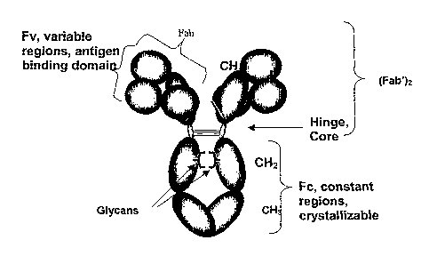
Fig. 1 depicts antibody IgG domains showing the relationship between the
domains and
the major designated cleavage fragments.
Fig. 2 is a schematic depiction of the variations in the biantennary
oligosaccharide
structure found in human IgG.
Fig. 3 shows the amino acid sequence in the human IgGl hinge region and
cleavage
sites for various enzymes.
Figs. 4A-G are MALDI-TOF-MS recordings with peak information on the
identification of species superimposed (+1 is singly charged molecular ion, +2
is
doubly charged molecular ion, +3 is triply charged molecular ion and LC is
free
light chain) and showing the formation of IgG fragments over time during
digestion with papain for a glycosylated and deglycosylated preparation: (A)
undigested, (B) % hour, (C) 1 hour, (D) 2 hours, (E) 4 hours, (F) 8 hours, and
(G) after 24 hours.
Fig. 5 shows a comparison of the percent peak area of +1 molecular ions of
intact IgGs
5
CA 02619825 2008-02-19
WO 2007/024743 PCT/US2006/032458
(A) and Fc fragments (B) of glycosylated and deglycosylated IgG samples
during papain digestion.
Fig. 6 shows tracings of MALDI-TOF-MS analysis of intact homogeneous IgG
glycoform preparations described in Example 5 for GO, G2, and G2S2
glycoforms.
Figs. 7A-D show tracings of MALDI-TOF-MS analysis of the PGNase released
glycans
from the various homogeneous glycoform preparations and from the control
sample.
Fig. 8 shows tracings of MALDI-TOF-MS analysis of papain digests of
homogeneous
IgG glycoform preparations GO, G2 and G2S2 along with a control sample
subjected to papain digestion at 50:1 ratio at 37 C after 15 minutes with the
various peak identities labeled.
Fig. 9 is a graphical representation of the integrated peak area of the intact
IgG from
MALDI-TOF-MS analysis of papain digests of homogeneous IgG glycoform
preparations GO, G2 and G2S2 relative to a control subjected to papain
digestion
at 50:1 ratio at 37 C at various times.
Fig. 10 is a graphical representation of the integrated peak area of the Fc
domain from
MALDI-TOF-MS analysis formed during papain digestion of homogeneous IgG
glycoform preparations GO, G2 and G2S2 along with a control sample at various
times.
DETAILED DESCRIPTION OF THE INVENTION
Abbreviations
AA, anthranilic acid; al,3GT, a-1,3-galactosyltransferase; 01,4GT, (3-
1,4-galactosyltransferase; a2,3ST, a-2,3-sialyltransferase; ADCC, antibody-
dependent
cellular cytotoxicity; CDC, complement-dependent cytotoxicity; CMP-Sia,
cytidine
monophosphate N-acetylneuraminic acid; FBS, fetal bovine serum; IgG,
immunoglobulin G; MALDI-TOF-MS, matrix-assisted laser/desorption ionization
time-
6
CA 02619825 2008-02-19
WO 2007/024743 PCT/US2006/032458
of-flight mass spectrometry; NANA, N-acetylneuraminic acid isomer of sialic
acid;
NGNA, N-glycolylneuraminic acid isomer of sialic acid; PNGase F, peptide N-
glycosidase F; HPLC, reversed phase high-performance liquid chromatography;
SA,
Sinapic acid; Sia, sialic acid; SDHB, dihydroxybenzoic acid containing sodium
chloride; UDP-Gal, uridine diphosphate galactose; UDP-G1cNAc, uridine
diphosphate
N-acetylglucosamine.
Definitions
The terms "Fc," "Fc-containing protein" or "Fc-containing molecule" as
used herein refer to a monomeric, dimeric or heterodimeric protein having at
least an
immunoglobulin CH2 and CH3 domain. The CH2 and CH3 domains can form at least a
part of the dimeric region of the protein/molecule (e.g., antibody).
The term "antibody" is intended to encompass antibodies, digestion
fragments, specified portions and variants thereof, including, without
limitation,
antibody mimetics or comprising portions of antibodies that mimic the
structure and/or
function of an antibody or specified fragment or portion thereof, including,
without
limitation, single chain antibodies, single domain antibodies, minibodies, and
fragments
thereof. Functional fragments include antigen-binding fragments that bind to
the target
antigen of interest. For example, antibody fragments capable of binding to a
target
antigen or portions thereof, including, but not limited to, Fab (e.g., by
papain digestion),
Fab' (e.g., by pepsin digestion and partial reduction) and F(ab')2 (e.g., by
pepsin
digestion), facb (e.g., by plasmin digestion), pFc' (e.g., by pepsin or
plasmin digestion),
Fd (e.g., by pepsin digestion, partial reduction and reaggregation), Fv or
scFv (e.g., by
molecular biology techniques) fragments, are encompassed by the term antibody
(see,
e.g., Colligan, Immunology, supra).
The term "monoclonal antibody" as used herein is a specific form of Fc-
containing fusion protein comprising at least one ligand binding domain which
retains
substantial homology to at least one of a heavy or light chain antibody
variable domain
of at least one species of animal antibody.
7
CA 02619825 2008-02-19
WO 2007/024743 PCT/US2006/032458
Enzymatic Digestion of Antibodies
Due to their high affmity target binding, Fabs also provide an ideal
targeting moiety, for e.g., conjugation of toxins or to embed in more complex
structures,
such as liposomes. As an enhancement to long circulating lipid vesicles
carrying
encapsulated drug, targeted or immunoliposomes are expected to enable more
precise
delivery of actives to diseased or pathogenic tissue while sparing normal
cells thereby
reducing side effects. The use of IgG and Fab as targeting moieties for
therapeutic
liposomes is disclosed in US4957735 and Maruyama et al. (1995) Biochim Biophys
Acta 1234: 74-80.
Papain is a sulfliydryl protease that has been used to digest IgG
antibodies into either Fab or F(ab')2 fragments, depending on whether L-
cysteine is
present or absent during the reaction, respectively. Prolonged treatment, or
excessive
amounts of papain, typically results in overdigestion of the Fc domain,
although the Fab
domains often remain resistant to overdigestion with papain. This is because
the Fc
domain contains additional (secondary) papain cleavage sites (Fig. 1). The
histidine
residue is the C-terminal position of abcixima.b when papain digestion is
performed in
the presence of cysteine.
Human IgGl: A-E-P-K-S-C-D-K-T-H-T-C-P-P-C-P-A-P-E-L-L-G-G
Human IgG2: C-P-P -L-K-E-C-P-P-C-P-A-P-P-_-V-A-G
Human IgG3: C-D-T-P-P-P-C-P-R-P-C-P-A-P-E-L-L-G
Human IgG4: S-K-Y-G-P-P- C-P-S-C-P-A-P
While papain is an industrially useful enzyme, it is of plant origin
originally isolated from the green fruit and leaves of Carica papaya
(Caricaceae spp).
An industrially useful mammalian enzyme, is pepsin. Pepsin is autoactivated
and active
at low pH as it is a normal component of the gastric fluid secreted into the
lumen of the
stomach after eating. Low levels of the precursor enzyme pepsinogen can be
found in
the serum but, since activation and activity are acid dependent, is not
physiologically
8
CA 02619825 2008-02-19
WO 2007/024743 PCT/US2006/032458
relevant to circulating antibodies. Pepsin cleaves human IgGl between the
leucine234-
leucine235 in the lower hinge. This cleavage site is downstream from the hinge
core (-C-
P-P-C-) containing two cysteine residues that link the two heavy chains via
disulfide
bonds creating a F(ab')2 molecule which is bivalent for antigen binding.
The lower hinge/CH2 region, P-A-P-E-L-L-G-G-P-S-V-F is within the
domain where cleavage sites exist for MMP-3 and MMP-12 (P-A-P*E-L-L-G for
each)
as well as pepsin and MMP-7 (P-A-P-E-L*L-G for each). In addition, a group of
physiologically relevant enzymes; neutrophil elastase (HNE), stromelysin (MMP-
3) and
macrophage elastase (MMP-12) cleave IgG at different positions to generate
subtly
different F(ab')2, Fab and Fc fragments (Fig. 3).
It was unexpectedly found that the level of glycoslylation of the Fc alters
the susceptibility to enzymatic degradation of said antibodies, resulting in
modulation of
various aspects of the production processes and biological actions of said
antibodies.
More specifically, during the course of these experiments it was discovered
that the Fc
of glycosylated Abs is more resistant to papain digestion than that of
deglycosylated,
aglycosylated or non-glycosylated Abs. Substantially deglycosylated,
aglycosylated or
non-glycosylated shall mean that most of the actual and/or potential
glycosylation sites
are unoccupied (with glycan), i.e., are not glycosylated.
The present invention further comprises a method for controlling the
properties of an Fc-containing molecule by altering the glycosylation of the
Fc's CH2
domains and the use of the altered Fc-containing molecules.
The presence or absence of glycan in the Fc-containing molecule affects
the affinity for one or more of the FcyRI, FcyRIIA, and FcyRIIIA receptors,
ADCC
activity, macrophage or monocyte activation, and serum half-life (Lifely et
al., Jeffreis,
and Wright and Morrison, supra). Therefore, since proteolytic degradation is a
measure
of glycosylation and glycosylation is a requirement for the secondary
functions of an
IgG-class antibody, susceptibility to proteolysis becomes a marker for the
above
mentioned functions of said IgG-class antibody. For example, sialic acid has a
net
negative charge at physiological pH and, thus, the presence of sialic acid in
the Fc-
9
CA 02619825 2008-02-19
WO 2007/024743 PCT/US2006/032458
bound carbohydrate might be expected to alter the three-dimensional structure
and
hence conformation of the CH2 domain and thereby affect Fc accessibility by
proteolytic enzymes. Accordingly, the sialic acid content of the
oligosaccharide
attached to the CH2 domain is a detenninant of proteolytic susceptibility and
proteolytic
cleavage rate is a measure of sialic acid content of the IgG or other Fc-
containing
protein.
Enrichment of Glycoforms of Fc-containing Proteins
One approach to preparing sublots of a particular Fc-containing protein
that differ in glycan content and structure is to take an Fc-containing
protein preparation
with heterogeneous Fc oligosaccharides, including both glycosylated and
aglycosylated
molecules, and pass it over a column containing an immobilized lectin that has
differential affinity for, for example, sialylated and asialylated
oligosaccharides. The
nonbinding flow-through (T, through) or the column unbound fraction can be
separated
from the bound fraction (B, bound), the latter collected while passing elution
buffer
through the column. It may also be possible to separately collect a weakly
bound
fraction or the column retarded fraction (R, retarded), for example, by
collecting Fc-
containing protein that elutes during continued washing of the column with the
original
sample buffer. Depending on the lectin used, the binding fraction is expected
to have a
higher saccharide, e.g., sialic acid, content therefore oligosaccharide
content, than the
non-binding fraction.
Examples of lectins that may enrich for sialylated or asialylated Fc-
containing proteins are the lectin from Maackia anaurensis (MAA), which
specifically
binds oligosaccharides with terminal sialic acid, and the lectin wheat germ
agglutinin
(WGA), which specifically binds oligosaccharides with either terminal sialic
acid or
terminal N-acetylglucosamine (G1cNAc). Another example is the lectin Ricin I
(RCA),
which binds oligosaccharides with tenninal galactose. In the latter example,
the non-
binding flow-through fraction may be enriched for sialylated Fc-containing
molecules.
Other lectins known in the art include those provided by Vector labs and EY
labs.
CA 02619825 2008-02-19
WO 2007/024743 PCT/US2006/032458
Enzymatic Modification of Fc-containing proteins
An alternative approach for preparing sublots of an Fc-containing protein
that differ in glycan content is to treat a portion of an Fc-containing
protein preparation
with a saccharase, such as a fucosidase or sialidase enzyme, thereby removing
specific
sugar residues, e.g., fucose or sialic acids. The resulting afucosylated or
asialylated
material can be compared to the original, partially fucosylated or sialylated
material for
differences in biological activity.
Addition of saccharides to the Fc region can also be achieved using in
vitro glycosylation methods. Glycosyltransferases naturally function to
synthesize
oligosaccharides. They produce specific products with excellent stereochemical
and
regiochemical geometry. The transfer of glycosyl residues results in the
elongation or
synthesis of an oligo- or polysaccharide. A number of glycosyltransferase
types have
been described, including sialyltransferases, fucosyltransferases,
galactosyltransferases,
mannosyltransferases, N-acetylgalactosaminyltransferases, N-
acetylglucosaminyltransferases and the like.
Glycosyltransferases which are useful in the present invention include,
for example, a-sialyltransferases, a-glucosyltransferases, a-
galactosyltransferases, a-
fucosyl- transferases, a-mannosyltransferases, a-xylosyltransferases, a-N-
acetylhexosaminyltransferases, 0-sialyltransferases, (3-glucosyltransferases,
(3-
2 0 galactosyltransferases, (3-fucosyltransferases, (3-mannosyltransferases,
(3-
xylosyltransferases, and (3-N-acetylhexosaminyltransferases, such as those
from
Neisseria meningitidis, or other bacterial sources, and those from rat, mouse,
rabbit,
cow, pig, human and insect and viral sources. Preferably, the
glycosyltransferase is a
truncation variant of glycosyltransferase enzyme in which the membrane-binding
domain has been deleted.
Exemplary galactosyltransferases include a(1,3) galactosyltransferase
(E.C. No. 2.4.1.151, see, e.g., Dabkowski et al., Transplant Proc. 25:2921
(1993) and
Yamamoto et al. Nature 345:229-233 (1990)) and a(1,4) galactosyltransferase
(E.C. No.
2.4.1.38). Other glycosyltransferases can be used, such as a
sialyltransferase.
11
CA 02619825 2008-02-19
WO 2007/024743 PCT/US2006/032458
. An a(2,3)sialyltransferase, often referred to as the sialyltransferase, can
be used in the production of sialyl lactose or higher order structures. This
enzyme
transfers sialic acid (NeuAc) from CMP-sialic acid to a Gal residue with the
formation
of an a-linkage between the two saccharides. Bonding (linkage) between the
saccharides is between the 2- position of NeuAc and the 3-position of Gal. An
exemplary a(2,3)sialyltransferase referred to as a (2,3)sialyltransferase (EC
2.4.99.6)
transfers sialic acid to the non- reducing terminal Gal of a Gal(31-->3Glc
disaccharide or
glycoside. See, Van den Eijnden et al., J. Biol. Chem., 256:3159 (1981),
Weinstein et
al., J. Biol. Chem., 257:13845 (1982) and Wen et al., J. Biol. Chem.,
267:21011 (1992).
Another exemplary a-2,3- sialyltransferase (EC 2.4.99.4) transfers sialic acid
to the
non- reducing ternlinal Gal of the disaccharide or glycoside. See, Rearick et
al., J. Biol.
Chem., 254:4444 (1979) and Gillespie et al., J. Biol. Chem., 267:21004 (1992).
Further
exemplary enzymes include Gal-0-1,4- G1cNAc a-2,6 sialyltransferase (See,
Kurosawa
et al. Eur. J. Biochem. 219: 375-381 (1994)).
Other glucosyltransferases particularly useful in preparing
oligosaccharides of the invention are the mannosyltransferases including
a(1,2)
mannosyltransferase, a(1,3) mannosyltransferase, (3(1,4) mannosyltransferase,
DoI-P-
Man synthase, OChl, and Pmtl.
Still other glucosyltransferases include N-
acetylgalactosaminyltransferases including a(1,3) N-
acetylgalactosaminyltransferase,
(3(1,4) N- acetylgalactosaminyltransferases (Nagata et al. J. Biol. Chem.
267:12082-
12089 (1992) and Smith et al. J. Biol Chem. 269:15162 (1994)) and polypeptide
N-
acetylgalactosaminyltransferase (Homa et al. J. Biol Chem. 268:12609 (1993)).
Suitable N-acetylglucosaminyltransferases include GnTI (2.4.1.101, Hull et
al., BBRC
176:608 (1991)), GnTII, and GnTIII (Ihara et al. J. Biolchem. 113:692 (1993)),
GnTV
(Shoreiban et al. J. Biol. Chem. 268: 15381 (1993)).
For those embodiments in which the method is to be practiced on a
commercial scale, it can be advantageous to immobilize the glycosyl
transferase on a
support. This immobilization facilitates the removal of the enzyme from the
batch of
12
CA 02619825 2008-02-19
WO 2007/024743 PCT/US2006/032458
product and subsequent reuse of the enzyme. Immobilization of glycosyl
transferases
can be accomplished, for example, by removing from the transferase its
membrane-
binding domain, and attaching in its place a cellulose-binding domain. One of
skill in
the art will understand that other methods of immobilization could also be
used and are
described in the available literature.
Because the acceptor substrates can essentially be any monosaccharide
or oligosaccharide having a terminal saccharide residue for which the
particular
glycosyl transferase exhibits specificity, substrate may be substituted at the
position of
its non-reducing end. Thus, the glycoside acceptor may be a monosaccharide, an
oligosaccharide, a fluorescent-labeled saccharide, or a saccharide derivative,
such as an
aminoglycoside antibiotic, a ganglioside, or a glycoprotein including
antibodies and
other Fc-containing proteins. In one group of preferred embodiments, the
glycoside
acceptor is an oligosaccharide, preferably, Gal(3(1-3)G1cNAc, Gal(3(1-
4)GlcNAc,
Gal(3(1- 3)Ga1NAc, Gal(3(1-4)Ga1NAc, Man a(1,3)Man, Man a(1,6)Man, or
GaINAcp(1-4)-mannose. In a particular preferred embodiment, the
oligosaccharide
acceptor is attached to the CH2 domain of an Fc-containing protein.
The use of activated sugar substrate, i.e., sugar-nucleoside phosphate,
can be circumvented by either using a regenerating reaction concurrently with
the
glycotransferase reaction (also known as a recycling system). For example, as
taught
in, e.g., U.S. Pat. 6,030,815, a CMP-sialic acid recycling system utilizes CMP-
sialic
acid synthetase to replenish CMP-sialic acid (CMP-NeuAc) as it reacts with a
sialyltransferase acceptor in the presence of a a(2,3)sialyltransferase to
form the sialyl-
saccharide. The CMP-sialic acid regenerating system useful in the invention
comprises
cytidine monophosphate (CMP), a nucleoside triphosphate (for example,
adenosine
triphosphate (ATP), a phosphate donor (for example, pliosphoenolpyruvate or
acetyl
phosphate), a kinase (for example, pyruvate kinase or acetate kinase) capable
of
transferring phosphate from the phosphate donor to nucleoside diphosphates and
a
nucleoside monophosphate kinase (for example, myokinase) capable of
transferring the
terminal phosphate from a nucleoside triphosphate to CMP. The
a(2,3)sialyltransferase
13
CA 02619825 2008-02-19
WO 2007/024743 PCT/US2006/032458
and CMP- sialic acid synthetase can also be viewed as part of the CMP-sialic
acid
regenerating system as removal of the activated sialic acid serves to maintain
the
forward rate of synthesis. The synthesis and use of sialic acid compounds in a
sialylation procedure using a phagemid comprising a gene for a modified CMP-
sialic
acid synthetase enzyme is disclosed in international application WO 92/16640,
published October 1, 1992.
An alternative method of preparing oligosaccharides is through the use
of a glycosyltransferase and activated glycosyl derivatives as donor sugars
obviating the
need for sugar nucleotides as donor sugars as taught in U.S. Pat. 5,952,203.
The
activated glycosyl derivatives act as alternates to the naturally-occurring
substrates,
which are expensive sugar-nucleotides, usually nucleotide diphosphosugars or
nucleotide monophosphosugars in which the nucleotide phosphate is a-linked to
the 1-
position of the sugar.
Activated glycoside derivatives which are useful include an activated
leaving group, such as, for example, fluoro, chloro, bromo, tosylate ester,
mesylate
ester, triflate ester and the like. Preferred embodiments of activated
glycoside
derivatives include glycosyl fluorides and glycosyl mesylates, with glycosyl
fluorides
being particularly preferred. Among the glycosyl fluorides, a-galactosyl
fluoride, a-
mannosyl fluoride, a-glucosyl fluoride, a- fucosyl fluoride, a-xylosyl
fluoride, a-sialyl
fluoride, alpha-N-acetylglucosaminyl fluoride, a-N-acetylgalactosaminyl
fluoride, (3-
galactosyl fluoride, P-mannosyl fluoride, 0-glucosyl fluoride, (3-fucosyl
fluoride, (3-
xylosyl fluoride, beta-sialyl fluoride, (3-N-acetylglucosaminyl fluoride and
(3-N-
acetylgalactosaminyl fluoride are most preferred.
Glycosyl fluorides can be prepared from the free sugar by first
acetylating the sugar and then treating it with HF/pyridine. Acetylated
glycosyl
fluorides may be deprotected by reaction with mild (catalytic) base in
methanol (e.g.,
NaOMe/MeOH). In addition, many glycosyl fluorides are commercially available.
Other activated glycosyl derivatives can be prepared using conventional
metliods
known to those of skill in the art. For example, glycosyl mesylates can be
prepared by
14
CA 02619825 2008-02-19
WO 2007/024743 PCT/US2006/032458
treatment of the fully benzylated hemiacetal form of the sugar with mesyl
chloride,
followed by catalytic hydrogenation to remove the benzyl groups.
A further component of the reaction is a catalytic amount of a nucleoside
phosphate or analog thereof. Nucleoside monophosphates which are suitable for
use in
the present invention include, for example, adenosine monophosphate (AMP),
cytidine
monophosphate (CMP), uridine monophosphate (UMP), guanosine monophosphate
(GMP), inosine monophosphate (IMP) and thymidine monophosphate (TMP).
Nucleoside triphosphates suitable for use in accordance with the present
invention
include adenosine triphosphate (ATP), cytidine triphosphate (CTP), uridine
triphosphate
(UTP), guanosine triphosphate (GTP), inosine triphosphate (ITP) and thymidine
triphosphate (TTP). A preferred nucleoside triphosphate is UTP. Preferably,
the
nucleoside phosphate is a nucleoside diphosphate, for example, adenosine
diphosphate
(ADP), cytidine diphosphate (CDP), uridine diphosphate (UDP), guanosine
diphosphate
(GDP), inosine diphosphate (IDP) and thymidine diphosphate (TDP). A preferred
nucleoside diphosphate is UDP. As noted above, the present invention can also
be
practiced with an analog of the nucleoside phosphates. Suitable analogs
include, for
example, nucleoside sulfates and sulfonates. Still other analogs include
simple
phosphates, for example, pyrophosphate.
One procedure for modifying recombinant proteins produced, in e.g.,
murine cells wherein the hydroxylated fomi of sialic acid predominates (NGNA),
is to
treat the protein with sialidase, to remove NGNA-type sialic acid, followed by
enzymatic galactosylation using the reagent UDP-Gal and betal,4 Galtransferase
to
produce highly homogeneous G2 glycoforms. The preparation can then,
optionally, be
treated with the reagent CMP-NANA and alpha-2,3 sialyltransferase to give
highly
homogeneous G2S2 glycoforms.
For purposes of this invention, substantially homogeneous for a
glycoform shall mean about 85% or greater of that glycoform and, preferably
about
95% or greater of that glycoform.
CA 02619825 2008-02-19
WO 2007/024743 PCT/US2006/032458
Structural Characterization of Sialic Acid Variants
For structural characterization of sialic acid variants containing
oligosaccharides, the glycoprotein preparations including antibody
preparations were
treated with peptide-N-glycosidase F to release the N-linked oligosaccharides.
The
enzyme peptide-N-glycosidase F (PNGase F) cleaves asparagines-linked
oligosaccharides. The released oligosaccharides are fluorescently labeled with
anthranilic acid (2-aminobenzoic acid), purified and analyzed by HPLC as
described
(see Anumula, K. R. and Dhume ST. Glycobiology. 1998 Jul;8(7):685-94).
Alternatively, the oligosaccharides released can be subjected to MALDI-TOF-MS,
as
described herein or to EsI-MS. The oligosaccharides separated as various
discreet
molecular weights, such as GO, G1, G2, G2S1 and G2S2, by these methods can be
detected and quantified.
Biological Characterization of Glycoform Variants
Fc-containing proteins can be compared for functionality by several
well-known in vitro assays. In particular, affinity for members of the FcyRI,
FcyRII,
and FcyRIII family of Fc7 receptors is of interest. These measurements could
be made
using recombinant soluble forms of the receptors or cell-associated forms of
the
receptors. In addition, affinity for FcRn, the receptor responsible for the
prolonged
circulating half-life of IgGs, can be measured, for example, by BlAcore using
recombinant soluble FcRn. Cell-based functional assays, such as ADCC assays
and
CDC assays, provide insights into the likely functional consequences of
particular
variant structures. In one embodiment, the ADCC assay is configured to have NK
cells
be the primary effector cell, thereby reflecting the functional effects on the
FcyRIIIA
receptor. Phagocytosis assays may also be used to compare immune effector
functions
of different variants, as can assays that measure cellular responses, such as
superoxide
or inflammatory mediator release. In vivo models can be used as well, as, for
example,
in the case of using variants of anti-CD3 antibodies to measure T cell
activation in mice,
an activity that is dependent on Fc domains engaging specific ligands, such as
Fc7
receptors.
16
CA 02619825 2008-02-19
WO 2007/024743 PCT/US2006/032458
Protein Production Processes
Different processes involved with the production of Fc-containing
proteins can impact Fc oligosaccharide structure. In one instance, the host
cells
secreting the Fc-containing protein are cultured in the presence of serum,
e.g., fetal
bovine serum (FBS) that was not previously subjected to an elevated heat
treatment (for
example, 56 C for 30 minutes). This can result in Fc-containing protein that
contains
no, or very low amounts of, sialic acid, due to the natural presence in the
serum of
active sialidase enzymes that can remove sialic acid from the Fc-containing
proteins
secreted from those cells. In another embodiment, the cells secreting the Fc-
containing
protein are cultured either in the presence of serum that was subjected to an
elevated
heat treatment, thereby inactivating sialidase enzymes, or in the absence of
serum or
other medium components that may contain sialidase enzymes, such that the Fc-
containing protein has higher or lower levels of glycosylation or
glycosylation variants.
In another embodiment, the conditions used to purify and further process
Fc-containing proteins are established that will favor optimal glycan content.
In one
embodiment, the conditions produce maximal or minimal oligosaccharide content
or
cause the transformation of the expressed Fc-containing polypeptide in a
predominant
glycoform. For example, because sialic acid is acid-labile, prolonged exposure
to a low
pH environment, such as following elution from protein A chromatography column
or
viral inactivation efforts, may lead to a reduction in sialic acid content.
Host Cell Selection or Host Cell Engineering
As described herein, the host cell chosen for expression of the
recombinant Fc-containing protein or monoclonal antibody is an important
contributor
to the final composition, including, without limitation, the variation in
composition of
the oligosaccharide moieties decorating the protein in the immunoglobulin CH2
domain. Thus, one aspect of the invention involves the selection of
appropriate host
cells for use and/or development of a production cell expressing the desired
therapeutic
protein.
17
CA 02619825 2008-02-19
WO 2007/024743 PCT/US2006/032458
In one embodiment in which the sialic acid content is controlled, the host
cell is a cell that is naturally deficient or devoid of sialyltransferases. In
another
embodiment, the host cell is genetically modified to be devoid of
sialyltransferases. In
a further embodiment, the host cell is a derivative host cell line selected to
express
reduced or undetectable levels of sialyltransferases. In yet another
embodiment, the
host cell is naturally devoid of, or is genetically modified to be devoid of,
CMP-sialic
acid synthetase, the enzyme that catalyzes the formation of CMP-sialic acid,
which is
the source of sialic acid used by sialyltransferase to transfer sialic acid to
the antibody.
In a related embodiment, the host cell may be naturally devoid of, or is
genetically
modified to be devoid of, pyruvic acid synthetase, the enzyme that forms
sialic acid
from pyruvic acid.
In an additional embodiment, the host cell may be naturally devoid of, or
is genetically modified to be devoid of, galactosyltransferases, such that
antibodies
expressed in said cells lack galactose. Without galactose, sialic acid will
not be
attached. In a separate embodiment, the host cell cell may naturally
overexpress, or be
genetically modified to overexpress, a sialidase enzyme that removes sialic
acid from
antibodies during production. Such a sialidase enzyme may act intracellularly
on
antibodies before the antibodies are secreted or be secreted into the culture
medium and
act on antibodies that have already been secreted into the medium and may
further
contain a galactase. Methods of selecting cell lines with altered glycosylases
and
which express glycoproteins with altered carbohydrate compositions have been
described (Ripka and Stanley, 1986. Somatic Cell Mol Gen 12:51-62;
US2004/0132140). Methods of engineering host cells to produce antibodies with
altered glycosylation patterns resulting in enhanced ADCC have been taught in,
e.g.,
U.S. Pat. 6,602,864, wherein the host cells harbor a nucleic acid encoding at
least one
glycoprotein modifying glycosyl transferase, specifically (3 (1,4) N-
acetylglucosaminyltranferase III (GnTI1I).
Other approaches to genetically engineering the glycosylation properties
of a host cell through manipulation of the host cell glycosyltransferase
involve
18
CA 02619825 2008-02-19
WO 2007/024743 PCT/US2006/032458
elinvinating or suppressing the activity, as taught in EP1,176,195,
specifically, alphal,6
f-ucosyltransferase (FUT8 gene product). It would be known to one skilled in
the art to
practice the methods of host cell engineering in other than the specific
examples cited
above. Further, the engineered host cell may be of mammalian origin or may be
selected from COS-1, COS-7, HEK293, BHK21, CHO, BSC-1, Hep G2, 653, SP2/0,
293, HeLa, myeloma, lymphoma, yeast, insect or plant cells, or any derivative,
irnmortalized or transformed cell thereof.
In another embodiment, the method of suppressing or eliminating the
activity of the enzyme required for oligosaccharide attachment may be selected
from the
group consisting of gene silencing, such as by the use of siRNA, genetic knock-
out, or
addition of an enzyme inhibitor, such as by co-expression of an intracellular
Ab or
peptide specific for the enzyme that binds and blocks its enzymatic activity,
and other
known genetic engineering techniques. In another embodiment, a method of
enhancing
the expression or activity of an enzyme that blocks saccharide attachment, or
a
saccharidase enzyme that removes sugars that are already attached, may be
selected
from the group consisting of: transfections with recombinant enzyme genes,
transfections of transcription factors that enhance enzyme RNA synthesis, and
genetic
modifications that enhance stability of enzyme RNA, all leading to enhanced
activity of
enzymes, such as sialidases, that result in lower levels of sialic acid in the
purified
product. hi another embodiment, specific enzyme inhibitors may be added to the
cell
culture medium. Alternatively, the host cell may be selected from a species or
organism incapable of glycosylating polypeptides, e.g. a prokaryotic cell or
organism,
such as and of the natural or engineered E. coli spp, Klebsiella spp., or
Pseudomonas
spp.
Antibodies
An antibody described in this application can include or be derived from
any marnnal, such as, but not limited to, a human, a mouse, a rabbit, a rat, a
rodent, a
primate, or any combination thereof and includes isolated human, primate,
rodent,
mammalian, chimeric, humanized and/or CDR-grafted antibodies,
inrnmunoglobulins,
19
CA 02619825 2008-02-19
WO 2007/024743 PCT/US2006/032458
cleavage products and other specified portions and variants thereof. The
invention also
relates to antibody encoding or complementary nucleic acids, vectors, host
cells,
compositions, formulations, devices, transgenic animals, transgenic plants,
and methods
of making and using thereof, as described herein together as combined with
what is
known in the art.
The antibodies or Fc-fusion proteins described herein can be derived in
several ways well known in the art. In one aspect, the antibodies are
conveniently
obtained from hybridomas prepared by immunizing a mouse with the target
peptides.
The antibodies can thus be obtained using any of the hybridoma techniques well
known
in the art, see, e.g., Ausubel, et al., ed., Current Protocols in Molecular
Biology, John
Wiley & Sons, Inc., NY, NY (1987-2001); Sambrook, et al., Molecular Cloning: A
Laboratory Manual, 2d Edition, Cold Spring Harbor, NY (1989); Harlow and Lane,
antibodies, a Laboratory Manual, Cold Spring Harbor, NY (1989); Colligan, et
al., eds.,
Current Protocols in Inununology, John Wiley & Sons, Inc., NY (1994-2001);
Colligan
et al., Current Protocols in Protein Science, John Wiley & Sons, NY, NY, (1997-
2001),
each entirely incorporated herein by reference.
The antibodies or Fc-fusion proteins or components and domains thereof
may also be obtained from selecting from libraries of such domains or
components, e.g.,
a phage library. A phage library can be created by inserting a library of
random
oligonucleotides or a library of polynucleotides containing sequences of
interest, such
as from the B-cells of an immunized animal or human (Smith, G.P. 1985. Science
228:
1315-1317). Antibody phage libraries contain heavy (H) and light (L) chain
variable
region pairs in one phage allowing the expression of single-chain Fv fragments
or Fab
fragments (Hoogenboom, et al. 2000, Irmnunol. Today 21(8) 371-8). The
diversity of a
phagemid library can be manipulated to increase and/or alter the
immunospecificities of
the monoclonal antibodies of the library to produce and subsequently identify
additional, desirable, human monoclonal antibodies. For example, the heavy (H)
chain
and light (L) chain immunoglobulin molecule encoding genes can be randomly
mixed
(shuffled) to create new HL pairs in an assembled immunoglobulin molecule.
CA 02619825 2008-02-19
WO 2007/024743 PCT/US2006/032458
Additionally, either or both the H and L chain encoding genes can be
mutagenized in a
complementarity determining region (CDR) of the variable region of the
immunoglobulin polypeptide, and subsequently screened for desirable affinity
and
neutralization capabilities. Antibody libraries also can be created
synthetically by
selecting one or more human framework sequences and introducing collections of
CDR
cassettes derived from human antibody repertoires or througli designed
variation
(Kretzschmar and von Ruden 2000, Current Opinion in Biotechnology, 13:598-
602).
The positions of diversity are not limited to CDRs, but can also include the
framework
segments of the variable regions or may include other than antibody variable
regions,
such as peptides.
Other libraries of target binding components which may include other
than antibody variable regions are ribosome display, yeast display, and
bacterial
displays. Ribosome display is a method of translating mRNAs into their cognate
proteins while keeping the protein attached to the RNA. The nucleic acid
coding
sequence is recovered by RT-PCR (Mattheakis, L.C. et al. 1994. Proc. Natl.
Acad. Sci.
USA 91, 9022). Yeast display is based on the construction of fusion proteins
of the
membrane-associated alpha-agglutinin yeast adhesion receptor, agal and aga2, a
part of
the mating type system (Broder, et al. 1997. Nature Biotechnology, 15:553-7).
Bacterial display is based on fusion of the target to exported bacterial
proteins that
associate with the cell membrane or cell wall (Chen and Georgiou 2002.
Biotechnol
Bioeng, 79:496-503).
In comparison to hybridoma technology, phage and other antibody
display methods afford the opportunity to manipulate selection against the
antigen
target in vitro and without the limitation of the possibility of host effects
on the antigen
or vice versa.
Also described is a method for producing an antibody or Fc-fusion
protein, comprising translating the encoding nucleic acid under conditions in
vitro, in
vivo or in situ, such that the peptide or antibody is expressed in detectable
or
recoverable amounts.
21
CA 02619825 2008-02-19
WO 2007/024743 PCT/US2006/032458
While having described the invention in general terms, the embodiments
of the invention will be further disclosed in the following examples.
EXAMPLE 1: ISOLATION OF FC DOMAIN FROM IGG
Papain was obtained from Sigma and PNGase F (peptide N-glycosidase
F) was obtained from New England Biolabs. Sinapic acid was obtained from
Fluka.
MALDI-TOF-MS analyses were carried out using Voyager DE Biospectrometry
workstation (Applied BioSystems, Foster City, CA).
Antibody samples were deglycosylated by treating with PNGase F in 20
mM Tris-HCl buffer, pH 7Ø The deglycosylated antibody samples were purified
on a
protein A column (HiTrap Protein A cartridges were obtained from Amersham
Biosciences) and analyzed by MALDI-TOF-MS for purity.
Antibody samples (at -1 mg/ml, before and after deglycosylation) were
treated with papain (1:50, w/w) in 20 mM Tris-HCl buffer, pH 7.0, containing 2
mM L-
cysteine and aliquots were withdrawn at fixed time intervals (0, 15, 30, 60,
90 minutes
followed by at 2, 3, 4, 5, 6, 8 and 24 hrs). The aliquots (about 2 l) were
immediately
mixed with 2 l of matrix solution (the matrix solution was prepared by
dissolving 10
mg sinapic acid in 1.0 ml of 50% acetonitrile in water containing 0.1%
trifluoroacetic
acid) and 2 l of this solution was loaded onto the MALDI target plate and
allowed to
air dry prior to the analysis.
MALDI-TOF-MS data indicates that the deglycosylation of IgG under
native conditions with PNGase F is complete and non-destructive (see Figs. 4A-
G).
MALDI-TOF-MS analysis of aliquots withdrawn at fixed time intervals
suggest that most of the deglycosylated IgG was digested by papain within one
hour
whereas complete digestion of control IgG (glycosylated) takes more than 4
hours. The
Fc fragments (at 10 KDa and 23.7 KDa) of deglycosylated IgG are observed at 30
minutes into digestion with papain and most of the deglycosylated Fc was
digested into
fragments within 4 hours. Fc fragments of glycosylated IgG are observed after
only 4
hours of digestion with papain at 1:50 (w/w) enzyme to substrate ratio and
requires
22
CA 02619825 2008-02-19
WO 2007/024743 PCT/US2006/032458
more than 24 hours to completely convert glycosylated Fc into smaller
fragments at 10
KDa and 23.7 KDa.
During attempts to isolate Fc fragments from different IgG antibodies, it
was observed that prior removal of CH2 domain glycans increased the rate of
papain-
mediated degradation of the Fc domain, making it more difficult to obtain
intact Fc
from deglycosylated (or aglycosylated) IgGs. Subsequent time course
experiments
comparing glycosylated and deglycosylated versions of both IgG antibodies and
purified Fc domains showed that, in both cases, the rate of degradation of Fc
in the
deglycosylated molecules was at least 4-8 times faster compared to their
glycosylated
counterparts. These results indicate that the presence of CH2 domain glycans
increases
resistance to papain-mediated degradation of Fc domains. It also suggests
that, given
how IgGs that lack glycosylation do not seem to have a defined Fc structure
and do not
bind Fc receptors, papain sensitivity may constitute an additional means of
assessing
proper Fc structure.
EXAMPLE 2: PAPAIN DIGESTION OF HOMOGENEOUS GLYCOFORMS
To assess the paraineters of papain digestion of antibody preparations
substantially homogenous with respect to their glycosylation patterns,
antibody samples
are enzymatically modified to produce such preparation for testing as
described below.
To galactosylate purified antibody samples via enzymatic method, bovine 20 1,4-
galactosyltransferase ((31,4GT) and LTDP-Gal obtained from Sigma Chemical Co.
(St.
Louis, MO) are added to the antibody samples. Recombinant rat liver a-2,3-
sialyltransferase
(a2,3ST), recombinant a-l,3-galactosyltransferase (a1,3GT) and CMP-Sia were
obtained from
Calbiochem (San Diego, CA). PNGase F was obtained from New England Biolabs
(Beverly,
MA) or from Prozyrne (San Leandro, CA) or from Selectin BioSciences (Pleasant
Hill, CA). 25 Galactosidase and (3-glucosaminidase from Diplococcus
pneunaoniae were obtained from either
ProZyme or from Selectin BioSciences. (3-Galactosidase from bovine kidney and
all other
enzymes were either from ProZyme or from Selectin BioSciences. NAP-5 and
HiTrap
protein A colurruis were from Pharmacia Biotech (Piscataway, NJ). All other
reagents
were of analytical grade.
23
CA 02619825 2008-02-19
WO 2007/024743 PCT/US2006/032458
Test antibodies for these analyses include monoclonal IgG Abs with
human IgGl and kappa constant regions expressed in transfected Sp2/0 mouse
myeloma cells. The Mabs are fully human or a mouse/human chimeric MAb specific
for, e.g., human TNF.
Fully galactosylated but not sialylated biantennary structures are
designated G2 glycoforms. G2 antibodies were prepared by subjecting IgG
samples in
100 mM MES buffer (pH 7.0) (-10 mg in 1.0 mL of buffer) to 50 milliunits of
(31,4GT,
5 mol of UDP-Gal, and 5 mol of MnC12 at 37 C for 24 hours. Another aliquot
of
enzyme and UDP-Gal was added and the mixture was incubated for an additiona124
hours at 37 C. The regalactosylated IgG samples were purified using a HiTrap
protein
A column. The oligosaccharides were released by PNGase F and characterized by
MALDI-TOF-MS and by HPLC as described below. The resulting preparations of
Mabs were found to contain 0% sialic acid.
Fully sialylated and galactosylated antibodies are designated G2S2. The
G2S2 glycoform was made by bringing IgG samples into 100 mM MES buffer, pH
7.0,
(or 10 mg in 1.0 mL of buffer) using NAP-5 columns according to the
manufacturer's
suggested protocol. To this solution were added 50 milliunits each of (31,4GT
and
a2,3ST and 5 mol each of UDP-Gal, CMP-Sia (NANA isomer), and MnC12. The
mixture was incubated at 37 C. After 24 hours, another aliquot of enzymes was
added
along with the nucleotide sugars and the mixture incubated for an additiona124
hours at
37 C. The G2S2 glycoform of IgG samples was purified as described above. Using
this method, sialation of an antibody preparation reached 90 to 98% G2S2
glycoform.
Test Abs were structurally analyzed by different methods. To perform
MALDI-TOF-MS analysis of intact IgG Abs, IgG samples were brought into 10 rnM
Tris-HC1 buffer, pH 7.0 and adjusted concentration to -1 mg/mL buffer. About 2
1 of
IgG solution was mixed with 2 1 of matrix solution (the matrix solution was
prepared
by dissolving 10 mg sinnapinic acid in 1.0 ml of 50% acetonitrile in water
containing
0.1 % trifluoroacetic acid) and 2 ml of this solution was loaded onto the
target and
allowed to air dry. MALDI-TOF-MS was acquired using a Voyager DE instrument
24
CA 02619825 2008-02-19
WO 2007/024743 PCT/US2006/032458
from Applied BioSystems (Foster City, CA).
To perform MALDI-TOF-MS analysis of released Fc glycans, IgG
samples (-50 g), before and after in vitro glycosylation reactions, were
digested with
PNGase F in 10 mM Tris-HCl buffer (50 l) pH 7.0 for 4 hours at 37 C. The
digestion
was stopped by acidifying the reaction mixture with 50% acetic acid (- 5 l)
and then
passing it through a cation-exchange resin column as described previously
(Papac et al.,
1996; Papac et al., 1998; Raju et al., 2000). These samples containing a
mixture of
acidic and neutral oligosaccharides were analyzed by MALDI-TOF-MS in the
positive
and negative ion modes, as described elsewhere (Papac et al., 1996; Papac et
al., 1998;
Raju et al., 2000) using a Voyager DE instrument from Applied BioSystems
(Foster
City, CA).
HPLC analysis of Fc glycans was done by digesting IgG samples (50 g)
in 10 mM Tris-HCl buffer (50 l) pH 7.0 with PNGase F at 37 C for 4-8 hours.
Derivatization of the released oligosaccharides with anthranilic acid (2-
aminobenzoic
acid) was carried out as described (Anumula KR. Anal Biochem. 2000 Jul
15;283(l):17-26). Briefly, a solution of 4% sodium acetate'3H2O (w/v) and 2%
boric
acid (w/v) in methanol was prepared first. The derivatization reagent was then
freshly
prepared by dissolving -30 mg of anthranilic acid (Aldrich) and -20 mg of
sodium
cyanoborohydride (Aldrich) in 1.0 ml of methanol-sodium acetate-borate
solution. IgG-
derived oligosaccharides (<3 nmol in 20-50 l of water) were mixed with 0.1 ml
of the
anthranilic acid (AA) reagent solution in 1.6 ml polypropylene screw cap
freeze vials
with'O" rings (Sigma) and capped tightly. The vials were heated at 80 C in an
oven or
heating block (Reacti-Therm, Pierce) for 1-2 hours. After cooling the vials to
room
temperature, the samples were diluted with water to bring the volume to -0.5
ml.
Derivatized oligosaccharides were purified by using NAP-5 columns.
Using a preparation that is at least 90% in the G2 or G2S2 glycoform, a
papain digestion experiment as described in Example 1 is performed and
analyzed to
demonstrate the effect of sialic acid content on the cleavage rate and
specificity of
papain. Additional cleavage analyses are perfonmed with otlier proteolytic
enzymes
CA 02619825 2008-02-19
WO 2007/024743 PCT/US2006/032458
using the samples as prepared in this example.
EXAMPLE 3: PRODUCTION OF ANTIBODY FRAGMENTS USING MATRIX
METALLOPROTEINASE-3
Metalloproteinases (MMPs) were purified from the supematant of cell
clones expressing recombinant human MMPs. The enzyme was activated with either
1
mM 4-aminophenylmercuric acetate (APMA; Sigma) for 1 hour at 37 C or by
treating
with chymotrypsin. The activated enzyme was stored at -70 C. Immunoglobulin
preparations (0.5-1.0 mg/ml) were incubated with digest buffer (250 mM Tris-
HCI, pH
7.4, containing 1.5 M NaCI, 50 mM CaC12 containing 15-60 g/ml activated MMP)
for
0-24 hours at 37 C. Aliquots were withdrawn at 0, 15, 30, 45, 60, and 120
minutes
followed by 3, 4, 5, 6, 8, 12, and 24 hours. The aliquots were (about 2
microliters) with
matrix solution (about 2 microliters) and 2 microliters of this mixture was
loaded onto
the MALDI-TOF-MS target plate and then analyzed by MALDI-TOF-MS using a
Voyager DE spectrometer.
EXAMPLE 4: PROTEOLYTIC CLEAVAGE OF PURIFIED FC
Papain was obtained from Sigma and PNGase F (peptide N-glycosidase
F) was obtained from New England Biolabs. Sinapic acid was obtained from
Fluka.
MALDI-TOF-MS analyses were carried out using Voyager DE Biospectrometry
workstation (Applied BioSystems, Foster City, CA).
Antibody (IgG) samples were deglycosylated by treating with PNGase F
in 20 mM Tris-HCl buffer, pH 7Ø The deglycosylated antibody samples were
purified
on a protein A column (HiTrap Protein A cartridges were obtained from Amersham
Biosciences) and analyzed by MALDI-TOF-MS for purity. Fc fraginents of IgG
were
isolated and purified as described elsewhere.
Fe fragment samples (about 1 mg/ml, before and after deglycosylation)
were treated with papain (1:50, w/w) in 20 mM Tris-HCl buffer, pH 7.0,
containing 2
mM L-cysteine and aliquots were withdrawn at fixed time intervals (0, 15, 30,
60, 90
minutes followed by at 2, 3, 4, 5, 6, 8 and 24 hrs). The aliquots (about 2 l)
were
26
CA 02619825 2008-02-19
WO 2007/024743 PCT/US2006/032458
immediately mixed with 2 l of matrix solution (the matrix solution was
prepared by
dissolving 10 mg sinapic acid in 1.0 ml of 50% acetonitrile in water
containing 0.1%
trifluoroacetic acid) and 2 l of this solution was loaded onto the MALDI
target plate
and allowed to air dry prior to the analysis.
The MALDI-TOF-MS data of glycosylated and deglycosylated IgG
digests was analyzed to obtain the relative % peak area data of intact IgGs
and Fc
fragments at all of the time points (Fig. 5). For the IgG samples, the time-
course
experiments indicated that within 15 minutes of digestion, more than 70% of
the
deglycosylated IgG was converted into Fab, Fc, and smaller Fc fragments (at
m/z 10.5
KDa and 12 KDa fragments) whereas less than 50% of the glycosylated IgG was
converted into fragments. After 60 minutes of incubation, no deglycosylated
IgG was
detectable, whereas approximately 80% of glycosylated IgG was still intact
according to
MALDI-TOF-MS analysis. For the Fc fragments, after 4 hrs of digestion, more
than
95% of the deglycosylated Fc fragment was converted into the smaller fragments
of
10.5 and 12 KDa, whereas no more than 10% of the glycosylated Fc converted
into
these smaller fragments at that time point. In fact, nearly 50 % of the
glycosylated Fc
remained undigested even after 24 hrs.
These data indicate that the glycosylated IgG is significantly more
resistant to papain digestion than the deglycosylated IgG, and that the
glycosylated Fc is
much more resistant than deglycosylated Fc. The time-course experiment also
revealed
that the amount of Fab fragments from the deglycosylated and glycosylated IgGs
were
equivalent, suggesting that only the Fc fragments undergo overdigestion and
conversion
into the 10.5 KDa and 12 KDa fragments.
EXAMPLE 5: PREPARATION OF MABS WITH SPECIFIC GLYCOFORMS
To better understand the role of oligosaccharides in increasing antibody
resistance to papain, IgG preparations with homogeneous GO, G2 and G2S2
oligosaccharides were prepared using in vitro methods.
Preparation of GO Glycoform To prepare homogeneous GO glycoform,
27
CA 02619825 2008-02-19
WO 2007/024743 PCT/US2006/032458
IgG samples were first treated with sialidase A to remove minor amounts of
terminal
sialic acid residues followed by treating with (3-galactosidase to remove
terminal (3-
galactose residues. IgG samples (10 mg in 1.0 mL) in 100 mM MES buffer (pH
7.0)
were treated with 100 milliunits each of sialidase A(A. ureafaciens) and (3-
galactosidase (D. pneumoniae) for 24 hours at 37 C. After 24 hours, another
aliquot of
enzymes was added and incubated for an additiona124 hours at 37 C.
After purification on a protein A column, the resulting GO glycoform
was characterized by MALDI-TOF-MS for intact mass (Fig. 6A). The mass spectrum
contained a singly charged molecular ion at m/z 147.7 KDa, a doubly charged
molecular ion at m/z 73.9 KDa and a triply charged molecular ion at m/z 49.3
KDa.
The mass spectrum also contained an ion at 23.4 KDa representing the free
light chain
produced during the laser desorption ionization. This mass spectral data
indicated that
the antibody was intact after treatment with enzymes to modify the Fc glycans
into
homogeneous GO oligosaccharide.
The modified oligosaccharide chain, released by treating the IgG
samples with PNGase F, was analyzed by MALDI-TOF-MS in the positive mode using
sDHB as matrix (after purification using a cation-exchange column) and also by
HPLC
(after derivatizing with anthranilic acid using a reductive amination
procedure as
described by Anumula (1998 supra). The MALDI-TOF-MS analysis showed a
molecular ion at m/z 1486.8 that corresponds to the molecular weight of
sodiated core
fucosylated complex biantennary oligosaccharide terminated with G1cNAc
residues.
The normal phase HPLC analysis of AA-derivatized oligosaccharide afforded a
single
peak eluting at 20.5 min and corresponds to the elution time of AA-labeled
standard
core fucosylated complex biantennary oligosaccharide terminated with G1cNAc
residues (data not shown) indicating that the GO IgG glycoform sample
contained more
than 99% GO oligosaccharide.
Preparation of G2 Glycoform The IgG samples were first treated with
sialidase A and purified as described above. The sialidase A treated IgG
samples (10
mg in 1.0 mL) in 100 mM MES buffer (pH 7.0)) were treated with 50 milliunits
of
28
CA 02619825 2008-02-19
WO 2007/024743 PCT/US2006/032458
(31,4GT, 5 mol of UDP-Gal, and 5 mol of MnC12 at 37 C for 24 hours. Another
aliquot of enzyme and UDP-Gal was added and the mixture was incubated for an
additiona124 hours at 37 C.
After purification on a protein A column, the antibody sample was
analyzed by MALDI-TOF-MS for intact mass. The mass spectrum showed a singly
charged molecular ion at m/z 148.7 KDa, a doubly charged molecular ion at m/z
74.2
KDa and a triply charged molecular ion at 49.5 KDa. The mass spectrum also
contained a singly charged molecular ion at m/z 23.4 KDa due to free light
chain
produced during laser desorption ionization. The G2 glycoform was then
subjected to
PNGase F treatment to release the N-linked oligsosaccharides and the released
oligosaccharides were analyzed by MALDI-TOF-MS in the positive mode using sDHB
as matrix. The mass spectrum showed a molecular ion at m/z 1812.1 (Fig. 6B)
and this
molecular ion at m/z 1812.1 corresponds to the inolecular weight of sodiated
core
fucosylated complex biantennary oligosaccharide terminated with galactose
residues.
The normal phase HPLC analysis of the PNGase F released oligosaccharides after
derivatization with AA exhibited a single peak and the elution time of this
peak
corresponds to the elution time of standard G2 oligosaccharide indicating that
the G2
glycoform preparation contained intact IgG molecule with more than 99% G2
oligosaccharide.
Preparation of G2S2 Glycoform For the preparation of G2S2 glycoform,
antibody samples were treated with a mixture of (3-galactosyltransferase and
a2,3-
sialyltransferase in the presence of UDP-Gal, CMP-NANA and MnC12 in a single
step.
IgG samples were brouglht into 100 mM MES buffer (pH 7.0) (10 mg in 1.0 mL)
using
NAP-5 columns according to the manufacturer's suggested protocol. To this
solution,
were added 50 milliunits each of P1,4GT and a2,3ST and 5 mol each of UDP-Gal,
CMP-Sia, and MnC12. The mixture was incubated at 37 C. After 24 hours,
another
aliquot of enzymes was added along with the nucleotide sugars and the mixture
incubated for an additiona124 hours at 37 C.
29
CA 02619825 2008-02-19
WO 2007/024743 PCT/US2006/032458
MALDI-TOF-MS analysis of the enzyme treated sample, after
purification on a protein A column, showed a singly charged molecular ion at
m/z
-148.8 KDa, a doubly charged molecular ion at m/z - 74.3 KDa and a triply
charged
molecular ion at m/z -49.5 Kda (Fig. 6C) suggesting that the antibody was
intact and
the enzyme treatment did not alter the primary structure of the antibody. The
negative
mode MALDI-TOF-MS and HPLC analysis of the PNGase F released oligosaccharide
(Figs. 7A-D) showed that the antibody contained greater than 85% G2S2
glycoform
along with minor amounts of G2S1 structure (monosialylated structures).
Further, 99%
of the Fc glycans contained at least one sialic acid residue. Fig. 7A shows
the control
(Voyager Spec #l=>NFO.7=>NR(2.OO)[BP = 1485.0,6636]), Fig. 7B shows the G2
glycoform (Voyager Spec #1=>NF0.7=>NR(2.00)[BP = 1811.4, 5403]), Fig. 7C shows
the GO glycoform (Voyager Spec #1=>NF0.7=>NF0.7=>NR(2.00)[BP =1486.5,
7006]), and Fig. 7D shows the G2S2 glycoform (-ve mode) (Voyager Spec
#1=>NF0.7[BP = 2385.4, 2769]).
Size-Exclusion Chromatogrraphy To assess the amounts of aggregates
present in antibody samples before and after in vitro modification of the
glycan
structures, Ab samples were analyzed by size-exclusion chromatography using an
Agilent 1100 HPLC system (Agilent). A TOSO HAAS TSK 3000SWXL (Tosoh Biosep
LLC) 7.8 rnm x 30 cm, 5 pm column was used at ambient temperature. The mobile
phase was phosphate buffered saline (PBS), and the flow rate was 0.5 mUmin.
The
modified IgG glycoform preparations exhibited similar chromatographic profiles
to the
control antibody profile indicating that the modification procedures did not
create any
additional aggregation and/or change in the primary structure of the protein.
EXAMPLE 6: PAPAIN DIGESTION OF MABS WITH SPECIFIC
GLYCOFORMS
To assess the relative resistance of GO, G2 and G2S2 glycoforms to
papain cleavage, the IgG glycoforms and a control sample were treated with
papain in
the presence of cysteine at 37 C for a period of 24 hours and the digests
analyzed by
MALDI-TOF-MS.
CA 02619825 2008-02-19
WO 2007/024743 PCT/US2006/032458
Time-Course analysis of papain digests
The GO, G2 and G2S2 glycoform along with the control IgG samples
were treated with papain at 1:50, enzyme to substrate ratio at 37 C. From
these
reactions, aliquots at 0, 15, 30, 60 and 90 minutes, and at 2, 3, 4, 5, 6, 8,
and 24 hours
were examined by MALDI-TOF-MS. All three IgG glycoforms along with the control
antibody sample were cleaved into Fab and Fc fragments, as evidenced by the
presence
of molecular ions at mlz -47.3 and -52.5 KDa for Fab and Fc fragments,
respectively
(Fig. 8).
A comparison of peak height of intact IgG observed at m/z - 147.5 KDa
is shown in Fig. 9. Intact IgG peak at m/z - 147.5 KDa was measured from 0 to
120
minutes, however, after 2 hours, very little intact IgG peak was observed. At
0 minute,
the peak height of all of the IgG glycoforms along with the control IgG was
observed to
be the same. At 15 minutes, about 50% of GO, 45% of control and 35% of G2
glycoform remained undigested. In contrast, only about 25% of G2S2 glycoform
remained undigested. At 30 minutes, about 45% of GO glycoform, 40% of control
antibody and -20% of G2 glycoform remained undigested, whereas only about 10%
of
G2S2 glycoform remained undigested. Therefore, the data presented in Fig. 9
suggest
that the GO glycoform is more resistant to the cleavage by papain in the CH1
domain,
that produces Fab and Fc fragments as the primary products, than the other
glycoforms.
In addition to the primary papain cleavage site in the CH1 domain, IgGs
can also undergo secondary cleavage in the CH2 domain of the Fc by reduced
papain.
To examine the resistance of IgG glycoforms to papain digestion at the
secondary
cleavage site, the peak heights of Fc fragments observed at m/z - 52.5 KDa
were
compared. The peak height data of Fc fragments of GO, G2, G2S2 and control IgG
is
shown in Fig. 10. The relative peak height of Fc fragments of G2 and G2S2 from
0.25
hours (15 minutes) to 1 hour was about 5% more than the relative peak height
of GO
glycoform; the peak heights of the Fc fragments of GO glycoform and control
IgG were
almost similar. Thus, both intact IgG as G2 and G2S2 glycoforms and the Fc
product
31
CA 02619825 2008-02-19
WO 2007/024743 PCT/US2006/032458
itself comprising these glycoforms are more sensitive to digestion by papain.
The data
shown in Fig. 10 therefore exemplifies the competing rates of formation and
degradation of the Fc product: at 1.5 hours time point, the peak heights of
the Fc
fragments of all of the glycoforms and the control IgG were almost the same.
After 1.5
hours time point, the peak heights of the Fc fragments of the G2 and G2S2
glycoforms
were gradually less than the peak height of the Fc fragments of the GO
glycoform and
control IgG. At 6 hours time point, the peak height of the G2S2 glycoform was
about
30% less than the peak height of the GO glycoform; the G2 glycoform peak
height was
about 25% less than the peak height of the GO glycoform. At 8 hours time
point, the
peak height of the Fc fragment of the G2S2 glycoform was about 60% less than
the Fc
fragment peak height of the GO glycoform; the peak height of the Fc fragment
of the G2
glycoform was about 50% less than the Fc fragment peak height of the GO
glycoform.
After 0.5 hours digestion, at all the time points the Fc fragment peak height
of GO was
greater than those of the G2, G2S2 and control IgG. At 24 hours time point, no
appreciable Fc fragments were observed for the G2 and G2S2 glycoforms, whereas
about 70% of the Fc fragments of GO and control IgG were observed. These data
indicate that the Fc fragment of the GO glycoform is more resistant to papain
digestion
at the secondary cleavage site present in the CH2 domain of the Fc. Further,
the data
also suggest that the G2S2 glycoform is the most sensistive of the glycoforxns
to both
primary digestion in the CH1 domain and secondary digestion in the CH2 domain.
These data suggest that the G2S2 glycoform may be more sensitive to
papain digestion and the GO glycoform may be more resistant to papain
digestion than
the G2 glycoform. The G2 glycoform was more resistant to papain digestion than
the
G2S2 glycoform, but about 50% less resistant than the GO glycoform. These
results
suggested that there was differential sensitivity of IgG glycoforms to papain
digestion.
These differences in sensitivity seems to be both at the primary cleavage site
as well as
at the secondary cleavage site in the Fc.
It will be clear that the invention can be practiced otherwise than as
particularly described in the foregoing description and examples. Numerous
32
CA 02619825 2008-02-19
WO 2007/024743 PCT/US2006/032458
modifications and variations of the present invention are possible in light of
the above
teachings and, therefore, are within the scope of the appended claims.
33
DEMANDE OU BREVET VOLUMINEUX
LA PRESENTE PARTIE DE CETTE DEMANDE OU CE BREVET COMPREND
PLUS D'UN TOME.
CECI EST LE TOME 1 DE 2
CONTENANT LES PAGES 1 A 33
NOTE : Pour les tomes additionels, veuillez contacter le Bureau canadien des
brevets
JUMBO APPLICATIONS/PATENTS
THIS SECTION OF THE APPLICATION/PATENT CONTAINS MORE THAN ONE
VOLUME
THIS IS VOLUME 1 OF 2
CONTAINING PAGES 1 TO 33
NOTE: For additional volumes, please contact the Canadian Patent Office
NOM DU FICHIER / FILE NAME:
NOTE POUR LE TOME / VOLUME NOTE: