Note: Descriptions are shown in the official language in which they were submitted.
CA 02619983 2008-02-20
WO 2008/013552 PCT/US2006/033607
FULLY HUMAN HYBRIDOMA FUSION PARTNER CELL LINES
CROSS-REFERENCE TO RELATED APPLICATIONS
[0001] This application claims benefit of Provisional Application number
60/711,819,
filed August 26, 2005, the entire contents of which are hereby incorporated by
reference as if
fully set forth herein, under 35 U.S.C. 119(e)
FIELD OF THE INVENTION
[0002] This invention is in the field of fusion partner cell lines for use in
making
hybridomas that secrete fully human monoclonal antibodies.
STATEMENT OF GOVERNMENTAL INTEREST
[0003] This invention was made with Government support under Grant No.
OC010016
awarded by the U.S. Army Department of Defense Program Project. The Government
has
certain rights in the invention.
BACKGROUND OF THE INVENTION
[0004] The seminal discovery by Kohler and Milstein (Kohler G, and Milstein
C., Nature
1975; 256:495) of mouse hybridomas capable of secreting specific monoclonal
antibodies
(MAbs) against predefined antigens ushered in a new era in experimental
immunology. Many
problems associated with antisera were circumvented. Clonal selection and
immortality of
hybridoma cell lines assured monoclonality and permanent availability of
antibody products.
At the clinical level, however, the use of such antibodies is clearly limited
by the fact that
they are foreign proteins and act as antigens to humans.
[0005] Since the report of Kohler and Milstein, the production of mouse
monoclonal
antibodies has become routine. However, the application of xenogenic MAbs for
in vivo
diagnostics and therapy is often associated with undesirable effects such as a
human anti-
mouse immunoglobulin response. Progress in making fully human monoclonal
antibodies has
been hampered by the absence of human myelomas suitable for use as fusion
partners with
the desirable attributes of mouse myeloma cells such as stability, and high
antibody
production. The use of Epstein-Barr virus (EBV) has proved to be quite
efficient for human
lymphocyte immortalization (Kozbor D, and Roder J., J.Immunology 1981;
127:1275; Casual
0, Science 1986; 234:476), but has certain limitations such as low antibody
secretion rate,
1
CA 02619983 2008-02-20
WO 2008/013552 PCT/US2006/033607
poor clonogenicity of antibody-secreting lines and chromosomal instability
necessitating
frequent subcloning.
[0006] Among the best potential fusion partners are syngenic myeloma cells
with well-
developed protein synthesis machinery. Nilsson K. and Ponten J., Int.J.Cancer
1975; 15:321.
However, culturing difficulties explain why few lines have been conditioned
for in vitro
growth and capability to produce viable hybrids. Goldman-Leikin R E,
J.Lab.Clin.Med.
1989: 113:335. Existing syngenic myelomas have low fusion yield and slow
hybrid growth,
although MAb production is relatively stable. Brodin T, J.Immunol.Meth. 1983;
60: l.
Genetic instability, such as that which occurs when a mouse myeloma is used as
the
immortalizing partner with a human cell, is a major disadvantage of
interspecies hybrids.
Production of mouse-human cell hybrids is not difficult. In vitro these cells
have growth
characteristics similar to those of conventional mouse-mouse hybridomas. Teng
N N H,
Proc.Natl.Acad.Sci. (USA) 1983; 80:7308. However, spontaneous elimination of
human
chromosomes considerably reduces the probability of stable MAb secretion.
Weiss M C, and
Green H. Proc.Natl.Acad.Sci. (USA) 1967; 58:1104. In order to improve growth
characteristics and stability of Hu-MAb production, heterohybrids between
mouse myeloma
cells and human lymphocyte (Oestberg L, and Pursch E., Hybridoma 1983; 2:361)
as well as
heteromyelomas (Kozbor D, et. al., J.Immunology 1984; 133:3001) have been used
as the
fusion partners. However, the problem remains that hybridomas made using
murine/human
heteromyelomas do not produce fully human antibodies.
[0007] Only one fully human fusion partner cell line has been reported.
Abraham Karpas,
et al. developed a fusion partner cell line (designated Karpas 707) from a
patient who had
multiple myeloma; it was not the product of cell fusion. Abraham Karpas, et
al., PNAS
February 13, 2001, Vol. 98, No. 4, 1799-1804, and Vaisbourd, M., et al.,
Hybridoma and
Hybridomics, Vol. 20, No. 5, 2001, 287-292, the entire contents of which are
hereby
incorporated by reference as if fully set forth herein. An ideal fusion
partner cell line would
not secrete any immunoglobulin and would have a short doubling time.
Unfortunately Karpas
707 secretes gamma light chain and has a very slow doubling time of about 35
hours.
[0008] One attempt to overcome these problems has been to modify mouse
monoclonal
antibodies by linking rodent variable regions and human constant regions to
make chimeric
antibodies, or by grafting the complementarity-determining region gene
segments from
mouse antibodies into human genes to make humanized antibodies. These
modifications
reduce but do not eliminate the immunogenicity of the antibody. Phage display
technology
was developed for the in vitro generation of human monoclonal antibodies, and
transgenic
2
CA 02619983 2008-02-20
WO 2008/013552 PCT/US2006/033607
mice strains that contain human instead of mouse Ig genes have been developed.
Bruggemann, M., et al. (1996) Immunol. Today 17, 391-97. These mice strains
have human
genes and make human antibodies, but the diversity in the strain is selected
not in a human
but in a mouse host, and the antibodies undergo affinity maturation in the
mouse not a human
environment. Immortalization of beta-lymphocytes with Epstein Barr Virus has
also been
tried, but the derived cells are typically unstable and secrete very small
amounts of
antibodies.
100091 Thus there is a great need for fully human, natural fusion partner cell
lines that do
not produce any immunoglobulin, are stable, fuse well with human lymphocytes,
and result
in hybridomas that stably produce fully humanized antibodies.
SUMMARY OF THE INVENTION
[0010] Certain aspects of the present invention are directed to methods to
make new fully
human fusion partner cell lines called Karyochi cells that can be fused with
antibody-
secreting cells to make fully human hybridomas called Karyochi-based
hybridomas, that
likewise secrete fully human monoclonal antibodies. Some aspects are directed
to the fully
human antibodies made by the Karyochi-based hybridomas. Other aspects are
directed to
certain parent cells that can be used to make the Karyochi cells, including
the human
lymphoma cell line FPO having Patent Deposit Designation Number PTA-7466, and
the
human myeloma cell line FP1.0 having Patent Deposit Designation Number PTA-
7465.
Certain aspects are further directed to the human chimeric fusion partner cell
lines Karyochi-
XX, which has Patent Deposit Designation Number PTA-7468, and Karyochi-7,
which has
Patent Deposit Designation Number PTA-7467.
[0011] An aspect of the invention is directed to Karyochi fusion partner cell
lines
(chimeric cells having nuclei from two different cells) and to methods for
making them using
cells from a single animal species, preferably from humans. Karyochi cells are
made by
isolating a donor nucleus that is substantially free of cytoplasm from either
a first malignant
B-lymphocyte cell line or a normal B-lymphocyte in the single animal species,
transferring
the donor nucleus into the cytoplasm of a recipient cell from a second T- or B-
lymphoid cell
line in the single animal species, and allowing time for the synchronization
and fusion of the
two nuclei in the recipient cell to form the chimeric Karyochi fusion partner
cell line. Nuclear
transfer can be accomplished using intra-cytosolic nucleus injection or by
impact-induced
nucleus administration. In some aspects of the invention the first and second
human
lymphoid cell lines are different human cell lines selected from the group
including myeloma,
3
CA 02619983 2008-02-20
WO 2008/013552 PCT/US2006/033607
lymphoma, multiple myeloma, lymphoblastoma and leukemia cell lines. While the
preferred
aspects of the invention involve making and using fully human Karyochi cells
and Karyochi-
based hybridomas to obtain fully human monoclonal antibodies, Karyochi cells
and
Karyochi-based hybridomas can be made using cells of any species of animal
that makes
antibodies, including all mammals, birds and reptiles.
[0012] Some embodiments of the invention are directed to Karyochi-based
hybridoma
that produce and secrete monoclonal antibodies, and to methods of making them.
In a
preferred embodiment the Karyochi-based hybridomas are fully human and make
fully
human monoclonal antibodies; however, Karyochi-based hybridomas can be made
for any
animal species. Human Karyochi-based hybridomas, for example, are made by
obtaining a
human Karyochi fusion partner cell that is made as described above, fusing it
with a human
antibody-producing lymphoid cell, and allowing time for the nucleus of the
Karyochi cell and
the nucleus of the lymphoid cell to synchronize and fuse to form the Karyochi-
based
hybridoma. The human antibody-producing lymphoid cell can be a splenocyte, a
lymph node
cell, a cell from Peyer's Patches, a peripheral blood lymphocyte, a B cell, a
T cell, or a tonsil
gland lymphocyte. In an aspect of the invention, the Karyochi-based hybridoma
is made
using lymphoid cell lines that each express a different selection marker
including 8-
Azaguanine resistance, 5-Bromouracil, 5-Fluorouracil or G-418 resistance. In
some aspects
of the invention the human antibody-producing lymphoid cell used to make a
Karyochi-based
hybridoma comes from a human having a condition causing the expression of an
antigen
associated with the condition, for example the condition is a disease such as
bacterial
infection and the antigen is a bacterial endotoxin, or the condition is cancer
and the antigen is
a cancer antigen. The Karyochi-based hybridoma will then be selected that
produces human
monoclonal antibodies that are specific or have high affinity for the antigen
that is associated
with the condition. In an aspect of the invention the lymphoid cells used to
make the
hybridomas come from an animal, preferably a human, that has a condition
including
diseases such as cancer, an infectious disease, an autoimmune disease, a
disease associated
with overepression of hormones or enzymes, graft vs. host disease, and
cardiovascular
disease. In certain embodiments the fully human monoclonal antibodies are
specific or have
high affinity for the antigen that can be a tumor-associated antigen, a cell
specific antigen, a
tissue-specific antigen, a hormone, an enzyme, a nucleic acid, a toxin, a
viral antigen, a
bacterial antigen, a fungal antigen, a parasitic antigen, a pyron, an enzyme,
or a eukaryotic
antigen.
4
CA 02619983 2008-02-20
WO 2008/013552 PCT/US2006/033607
BRIEF DESCRIPTION OF THE DRAWING
[0013] The present invention is illustrated by way of example, and not by way
of
limitation, in the figures of the accompanying drawings and in which:
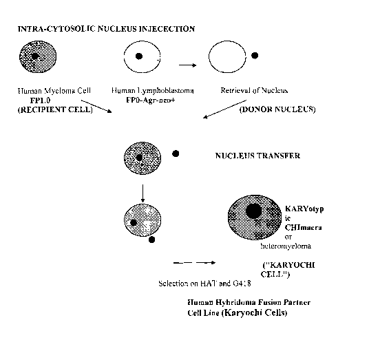
[0014] FIG. 1. is a cartoon illustrating the method for constructing a
Kayrochi Cell. FIG.
lA illustrates the intra-cytosolic nucleus injection technique (ICN), and FIG.
1B illustrates
the impact-induced nucleus administration method (IINA).
DEFINITIOINS
[0015] Human Lymphoid cell line ("HuLCL") includes myeloma, multiple myeloma,
lymphoma, and lymphoblastoma, and leukemia cell lines Unless otherwise
defined, scientific
and technical terms used in connection with the present invention shall have
the broadest
meanings that are commonly understood by those of ordinary skill in the art.
Further, unless
otherwise required by context, singular terms shall include pluralities and
plural terms shall
include the singular.
[0016] Generally, nomenclatures utilized in connection with, and techniques
of, cell and
tissue culture, molecular biology, and protein and oligo- or polynucleotide
chemistry and
hybridization described herein are those well known and commonly used in the
art, as
described in various general and more specific references such as those that
are cited and
discussed throughout the present specification. See e.g. Singleton et al.,
Dictionary of
Microbiology and Molecular Biology 2<sup>nd</sup> ed., J. Wiley & Sons (New York,
N.Y. 1994);
Sambrook et al. Molecular Cloning: A Laboratory Manual (2d ed., Cold Spring
Harbor
Laboratory Press, Cold Spring Harbor, N.Y. (1989)), which are incorporated
herein by
reference.
[0017] Herein, "mammal" means any mammal, preferably a human.
[0018] The term "mammals other than humans" and "non-human mammals" used
herein,
are synomic to each other, meaning all mammals other than humans defined
above.
[0019] The term "monoclonal antibody" as used herein refers to an antibody
obtained
from a population of substantially homogeneous antibodies, i.e., the
individual antibodies
comprising the population are identical except for possible naturally
occurring mutations that
may be present in minor amounts, and the term includes any fragment or portion
thereof.
Monoclonal antibodies are highly specific, being directed against a single
antigenic site or
epitope. Furthermore, in contrast to polyclonal antibody preparations which
include different
antibodies directed against different determinants (epitopes), each monoclonal
antibody is
directed against a single determinant on the antigen. As used herein,
monoclonal antibodies
CA 02619983 2008-02-20
WO 2008/013552 PCT/US2006/033607
are produced and secreted by Karyochi-based hybridomas. A fully human
monoclonal
antibody of this invention made by a fully human Karyochi-based hybridoma may
be any
human monoclonal antibody or a portion thereof having any isotype belonging to
any class
and any subclass of immunoglobulin including: IgG (IgGI, IgG2, IgG3, and
IgG4), IgM, IgA
(IgAI and IgA2), IgD, IGD, and IgE or IgM. Examples of particularly preferable
immunoglobulin of the present invention are those belonging to human-derived
IgG (IgM,
IgGI, IgG2, IgG3, or IgG4).
[0020] The term "epitope" is used to refer to binding sites for antibodies on
protein
antigens. Epitopic determinants usually consist of chemically active surface
groupings of
molecules such as amino acids or sugar side chains and usually have specific
three-
dimensional structural characteristics, as well as specific charge
characteristics.
[0021] An "isolated" monoclonal antibody within the scope of the present
invention is
one that has been identified and separated and/or recovered from a component
of its natural
environment.
[0022] A "neutralizing monoclonal antibody" as used herein is a monoclonal
antibody
molecule that is able to eliminate or significantly reduce an effector
function of a target
antigen, such as a cancer antigen or tumor antigen, to which it binds. In an
embodiment, a
neutralizing antibody will reduce an effector function by 1-10, 10-20, 20-30,
30-50, 50-70,
70-80, 80-90, 90-95, 95-99, 99-100%.
[0023] Karyotype means the complete set of chromosomes of a cell or organism.
[0024] Karyogamy means the fusion of two or more nuclei.
[0025] Human Lymphoid cell line ("HuLCL") includes myeloma, multiple myeloma,
lymphoma, lymphoblastoma, and leukemia cell lines.
[0026] Heteromyeloma means a cell line that combines genetic material from two
different lymphoid cell lines by fusing whole cells from each lymphoid cell
line; the term
includes heterolymphomas. By contrast Karyochi cells are not formed from the
fusion of two
whole cells and therefore the resulting cells cannot be defined as
heterohybridomas or
heteromyelomas.
[0027] Hybridoma means an immortal antibody-producing cell line that stably
produces
antibodies, made by fusing cells from an immortal cell line (transformed) with
an antibody-
producing cell such as a beta lymphocyte.
[0028] Human-murine hybridoma means an immortal, antibody-producing cell line
which results from the fusion of a murine heteromyeloma cell line with a human
beta
lymphocyte.
6
CA 02619983 2008-02-20
WO 2008/013552 PCT/US2006/033607
[0029] Donor Nucleus means an isolated nucleus from a lymphoid cell or
lymphoid cell
line (preferably human) which nucleus is substantially free of cytoplasm.
[0030] Recipient cell means a whole cell from a lymphoid cell line (preferably
human).
[0031] Chimeric cell means a cell with chromosomes from two different
heterogeneous
cells.
[0032] Karyochi cell means a chimeric cell for use as a fusion partner cell
line that is
made using cells from a single animal species, preferably a human. To make a
Karyochi cell,
an isolated donor nucleus that is substantially free of cytoplasm is obtained
from a normal B-
cell or a B-cell line, and is then transferred into a whole recipient cell
taken from a T- or B-
lymphoid cell line. With time, the donor nucleus and the nucleus of the
recipient cell fuse
into a single nucleus thus making the chimeric Karyochi fusion partner cell.
Karyochi cells
are preferably made from donor nuclei and recipient cells that come from the
same species,
preferably human, however any species of animal that makes antibodies can be
used
including mammals, birds and reptiles. If the donor nucleus and the recipient
cell are both
taken from malignant B-cell lines, then they must be from different
(heterogeneous) cell
lines. Human Karyochi cells can be used to make fully human antibody-secreting
hybridomas
called Karyochi-based hybridomas. Various cell combinations can be used to
make Karyochi
Cells:
RECIPIENT CELL DONOR NUCLEUS KARYOCHI CELL TPE
Malignant T-cell Malignant B-cell T/B chimeric cell
Malignant T-cell Normal B-cell TB chimeric cell
Malignant B-cell, type 1 Malignant B-cell, type II B/B chimeric cell
Malignant B-cell Normal B-cell B/B chimeric cell
[0033] Trioma means a cell that has three nuclei.
[0034] Antibody-producing lymphoid cell means any lymphoid cell from any
species of
animal that is capable of producing antibodies, such as a peripheral blood
lymphocyte, a
splenocyte, a lymph node cell, a B cell, a tonsil gland lymphocyte, or a
Peyer's patch cell,
preferably a human lymphoid cell.
[0035] Karyochi-based hybridoma means a monoclonal antibody-producing cell
line,
which results from the fusion of a Karyochi cell with an antibody-producing
lymphoid cell.
Preferably the Karyochi cell and the antibody-producing lymphoid cell come
from the same
7
CA 02619983 2008-02-20
WO 2008/013552 PCT/US2006/033607
species, preferably from a human, such that the Karyochi-based hybridoma
produces and
secretes fully human monoclonal antibodies. Karyochi-based hybridomas can be
made using
cells from any animal that makes antibodies including mammals, reptiles and
birds.
[0036] T-cell (or T lymphocyte) means any of the lymphocytes that mature in
the thymus
and have the ability to recognize specific peptide antigens through the
receptors on their cell
surface.
[0037] B-cell means a type of lymphocyte that is capable of producing
antibodies in
response to detecting the presence of a particular antigen.
[0038] Specific monoclonal antibody means a type of antibody which binds
specifically
to a particular and certain antigen, epitope, cell or tissue and does not bind
to other antigens,
epitopes, cells or tissues that are not particular and certain for the given
antibody. High
affinity monoclonal antibodies means antibodies which bind strongly to
particular and certain
antigens, epitopes, cells and tissues with an affinity constant (Ka) in the
range 10"' - 10"13 M.
DETAILED DESCRIPTION OF THE INVENTION
[0039] Certain embodiments of the present invention are directed to new
chimeric fusion
partner cell lines named "Karyochi cells," which are defined herein, and to
methods for
making and using them. Karyochi cells are made by obtaining an isolated donor
nucleus that
is substantially free of cytoplasm; and transferring the isolated nucleus into
the cytoplasm of
a whole recipient cell. In a preferred embodiment the donor nucleus and the
recipient cell
come from the same animal species, preferably from a human. Where human cells
are used,
the donor nucleus comes from a lymphoid cell line, thus making either T/B or
B/B chimeric
Karyochi cells. The use of T-cells in constructing chimeric cells can be
beneficial because T-
cells secrete an array of autocrine and paracrine growth factors and cytokines
that stimulate
cell growth and antibody secretion. The CHImeric cell thus formed has two
different
KARYOtypes; hence the name "Karyochi cells." Because Karyochi cells are not
formed from
the fusion of two whole cells; the resulting cells cannot be defined as
heterohybridomas or
heteromyelomas. Karyochi cells are chimeric cells carrying two different sets
of
chromosomes derived from different cell types that preferably come from the
same species.
With time, the nuclei in the Karyochi cells synchronize and fuse to form a
single nucleus.
Human Karyochi cells are ideal fusion partner cell lines for forming fully
human monoclonal
antibody-producing hybridomas called "Karyochi-based hybridomas," which are
defined
herein. Karyochi cells can come from any animal that has antibody-producing
cells, including
all mammals, birds and reptiles.
8
CA 02619983 2008-02-20
WO 2008/013552 PCT/US2006/033607
[0040] The invention is further directed to methods for making Karyochi cells.
The
method includes: obtaining an isolated donor nucleus that is substantially
free of cytoplasm
from either a normal or malignant B-cell, for example using the techniques for
nucleus
isolation pioneered in in vitro fertilization. The preferred method for
transferring the donor
nucleus is intra-cytosolic nucleus injection of the donor nucleus into the
cytoplasm of the
recipient cell. Nuclear transfer can also be made by using impact-induced
nucleus
administration (IINI).
[0041] Certain embodiments of the invention are directed to Karyochi-based
hybridomas,
preferably fully human hybridomas capable of producing fully human monoclonal
antibodies,
and to methods for making them. In an embodiment, Karyochi-based hybridomas
are
obtained by fusing the described Karyochi cell (preferably human) with an
antibody-
producing lymphoid cell (preferably human), and selecting monoclonal antibody-
producing
Karyochi-based hybridomas that produce antibodies against an antigen of
interest. The
invention is further directed to the fully human monoclonal antibodies
(hereafter "HuMAbs")
made by human Karyochi-based hybridomas, and to any monoclonal antibodies made
by
Karyochi-based hybridomas in any species. Certain embodiments are also
directed to
monoclonal antibodies that are made from cells that all derive from the same
species, thus
making the antibodies fully compatible with that species. According to an
embodiment of this
invention, hybridoma replication is effective both in vitro or in vivo.
Karyochi-based
hybridoma cells can therefore grow in Petri dishes, flasks, wells or
bioreactors as well as in
vivo in immunocompromised mice, for example. In certain embodiments the human
antibody-producing lymphoid cell is taken from a patient having a condition,
such as a
disease including an infection or cancer, which condition results in
expression of at least one
antigen associated with the presence of the condition in the animal (for
example a disease-
specific, condition-specific or tumor-associated antigens). In certain
embodiments human
Karyochi-based hybridomas are made using antibody-secreting cells from a
subject known to
be infected or diseased or having the condition of interest. Isolated fully
HuMAbs made by
these hybridomas are then screened for specificity or affinity for an antigen
known to be
associated with the infection, disease or condition. Such HuMAbs can then be
used
therapeutically or diagnostically using methods known in the art. In certain
embodiments the
isolated fully human (or other species) monoclonal antibodies are bound to a
toxin or a
radionuclide that can kill the target cells expressing the antigen. All of the
examples herein
are for making fully human Karyochi cells, Karyochi-based hybridomas and
monoclonal
antibodies.
9
CA 02619983 2008-02-20
WO 2008/013552 PCT/US2006/033607
[0042] A good fusion partner cell line for making fully human monoclonal
antibody-
producing hybridomas should ideally meet the following requirements. It
should:
- be fully human in origin;
- produce no or negligible amounts of endogenous immunoglobulin or individual
immunoglobulin chains;
- have a short doubling time;
- grow in suspension culture;
- be suitable for high efficiency fusion with B-cells of different
histological origin;
- be non-biased (non-selective in terms of Ig type) in fusion to B-cells
producing
different
Ig isotypes;
- yield stable Ig-producing hybrids capable of long-term stable production of
specific
Ig's;
and
- be easily adaptable to serum- and protein-free media and culturing in
bioreactors for
mass production of monoclonal antibodies.
Until now there was no fully human fusion partner cell line that met these
criteria.
[0043] In our earlier work we developed the heteromyeloma fusion partner cell
line
called MFP2 (ATCC Designation Number HB-12482), which is one of the better
fusion
partner cell lines presently available. MFP2 cells have been studied
thoroughly and are the
subject of U.S. Patent No. 6,197,582, the entire contents of which are hereby
incorporated by
reference as if fully set forth herein. Yet as good as MFP2 cells are, they
are not fully human
as they are formed from fusing mouse myeloma 653 cells and human myeloma RPMI
8226
cells. Table I compares the general characteristics of several fusion partner
cell lines (MFP2,
X63, 653, RPMI-8226, and B6B11) of animal and human origin. As Table I shows,
RPMI-
8226 by itself is not a good fusion partner cell line. It produces IgG light
chain, has
insignificant levels of fusibility with other cells, and very low fusion
efficiency. However
RPMI fused with mouse Myeloma 653 made the MFP2 cell line, which is a good
fusion
partner cell line (FPCL) despite the fact that it is not fully human.
[0044] Abraham Karpas, et al. have reported the only potentially useful fully
human
fusion partner cell line, which is designated Karpas 707. Abraham Karpas, et
al., PNAS
February 13, 2001, Vol. 98, No. 4, 1799-1804, and Vaisbourd, M., et al.,
Hybridoma and
Hybridomics, Vol 20, No. 5, 2001, 287-292, the entire contents of which are
hereby
incorporated by reference as if fully set forth herein. Karpas 707 was
established from a
CA 02619983 2008-02-20
WO 2008/013552 PCT/US2006/033607
patient who had multiple myeloma, however, it was not the product of cell
fusion. Karpas
707 cells secrete gamma light chain and have a very slow doubling time of
about 35 hours. A
report analyzing the variable regions of antibody heavy and light chains from
the Karpas 707
heterohybridomas showed that they were representative of human B lymphocytes
with
respect to family use, segment use, somatic mutation and chain pairings. The
combination of
long doubling time and the production of IgG light chain by Karpas 707 make it
less than
ideal as a human fusion partner cell line. Fusion efficiencies for Karpas 707
cells have not
been reported.
Karyochi Cells: Formation and Characteristics
[0045] Certain embodiments of the present invention are directed to new human
fusion
partner cell lines (FPCL) called Karyochi cells for making fully human,
antibody-producing
hybridomas called Karyochi-based hybridomas. Karyochi cells meet all of the
criteria listed
above for an ideal human fusion partner cell line. Unlike the known hybrid
fusion partner
cells that are made using cells from two different species and from the fusion
of two whole
cells, Karyochi cells are made using cells from the same species and they are
not formed
from the fusion of two whole cells. Instead, Karyochi cells are made by
transferring an
isolated donor nucleus that is substantially free of cytoplasm taken, for
example from a
normal or a malignant B cell, into the cytoplasm of a whole recipient cell
taken from a
different lymphoid cell line. The chimeric cell thus formed has two different
karyotypes;
hence the name "Karyochi cells" to distinguish them from cells made by the
fusion of two
whole cells. In the preferred embodiment Karychi cells are fully human. After
nuclear
transfer, the two nuclei in the Karyochi cell eventually synchronize and fuse.
Karyochi cells
are aneuploid, i.e. human Karyochi cells don't have a typical 23 homologous
pair set of
human chromosomes. In those Karyochi cells where both the donor and recipient
cells come
from transformed cells, significant chromosome instability is usual. Human
Karyochi cells
typically have more than 46 chromosomes. When two karyotypes are combined they
don't
form a stable karyotypic chimera in which the number of chromosomes is a
simple arithmetic
sum. The cells undergo chromosome elimination over time after chimerization
until the
karyotype is stabilized. The karyotpe for chimeras of transformed cells
usually is presented as
a mode, i.e. a range of chromosome number which can be found in individual
cells from the
same cell line. Preliminary studies estimate that human Karyochi cells have a
modal number
of between about 120 to about 140 chromosomes.
11
CA 02619983 2008-02-20
WO 2008/013552 PCT/US2006/033607
[0046] The decision to use heterogeneous cell types for the donor and
recipient cell
nuclei was based on previous experience in traditional whole cell fusion which
showed that
heterohybridomas and heteromyelomas perform much better in fusing with human
lymphocytes to make hybridomas than either parental cell lines separately.
Ostberg L. Human
monoclonal antibodies in transplantation, Transplant Proc. 1992 Aug;24(4 Suppl
2):26-30;
Ostberg L, Pursch E. Human X (mouse X human) hybridomas stably producing human
antibodies, Hybridoma. 1983;2(4):361-7; Nilsson K, et al., Entrapment of
animal cells for
production of monoclonal antibodies and other biomolecules, Nature. 1983 Apr
14;302(5909):629-30; Ostberg L. Human X (mouse X human) hybridomas, Methods
Enzymol. 1986;121:228-34; Isaacson C. et al., Human and primate monoclonal
antibodies for
in vivo therapy, Clin Chem. 1988 Sep;34(9):1681-8, the entire contents of
which are hereby
incorporated by reference as if fully set forth herein. Lymphoid cell lines
suitable for making
Karyochi cells include myeloma, lymphoma, lymphoblastoma and leukemia. In an
embodiment, Karyochi cells are formed by fusing a donor cell from a T- or B-
lymphoid cell
that is not transformed with a cell from a lymphoid cell line.
[0047] In a preferred embodiment, the donor nucleus is isolated using the
Intra-Cytosolic
Nucleus Injection (ICNI) technique that is used to isolate nuclei from a sperm
for in vitro
fertilization (IVF). Trokoudes KM, et al., Pregnancy with spermatozoa from a
globozoospermic man after intracytoplasmic sperm injection treatment Hum
Reprod, 1995
Apr;10(4):880-2; Hlinka D, et al., A modified method of intracytoplasmic sperm
injection
without the use of polyvinylpyrrolidone, Hum Reprod. 1998 Jul;13(7):1922-7;
Katayose H,
et al., Efficient injection of bull spermatozoa into oocytes using a Piezo-
driven pipette
Theriogenology, 1999 Nov; 52(7):1215-24, the entire contents of which are
hereby
incorporated by reference as if fully set forth herein. This method involves
removing the
nucleus from the donor cell so that it is substantially free of cytoplasm
using an ultra thin
micromanipulator needle (diameter < 5 um) and injecting the nucleus into the
cytoplasm of
the whole recipient cell. The recipient cell typically has a diameter of about
30-60
micrometers and a volume at average of about 50,000 micrometers3. The volume
of the donor
nucleus (largely free of cytoplasm) varies substantially but is typically
about 150
micrometers3, which is about 0.3% of the recipient cell volume. Thus, the new
methods of the
present invention that use a donor nucleus substantially free of cytoplasm
(rather than a
whole donor cell) to form fusion partner cell lines cause negligible
disruption to the
infrastructure of the cytoplasm and negligible alteration of the volume of the
recipient cell.
Moreover, disruption of the cytoplasm of the recipient cell is limited to the
site of insertion of
12
CA 02619983 2008-02-20
WO 2008/013552 PCT/US2006/033607
the donor nucleus. Endoplasmic reticulum and Golgi apparatus throughout the
recipient cell
remain essentially intact. It is speculated that the minimum disruption of the
recipient cell in
making Karyochi cells accounts at least in part for their improved success for
making stable
monoclonal antibody-producing Karyochi-based hybridomas.
[0048] Methods for ICNI and other methods of nucleus transfer are described
more fully
in: Khalili MA, et al., J Assist Reprod. Genet. 2002; 19: 84-6; and in K.D.
Nusser, et al.,
Human Reproduction, Vol. 16, No. 1, 130-137, the entire contents of which are
hereby
incorporated by reference as if fully set forth herein. Regarding methods for
the isolation and
purification of nuclei, we reference Deborah L. Hodge, et al., Molecular and
Cellular
Biology, 2002, p. 1742-1753, Vol. 22, No. 6; and Dijkwel, P. A., et al., Mol.
Cell Biol. 1991,
11,3850-3859, the entire contents of which are hereby incorporated by
reference as if fully
set forth herein.
[0049] In another embodiment, the donor nucleus is forced into the recipient
cell
cytoplasm using Impact-induced Nucleus Administration (IINA). Wallace DC, et
al., J Cell
Biol. Cytoplasmic transfer of chloramphenicol resistance in human tissue
culture cells.1975
Oct;67(1):174-88; Jeon, K.W. J Selective effects of enucleation and transfer
of heterologous
nuclei on cytoplasmic organelles in Amoeba proteus Protozool, 1975
Aug;22(3):402-5;
Appels R, et al., The first division of HeLa times chick erythrocyte
heterokaryons. Transfer
of chick nuclei to daughter cells, Exp Cell Res. 1975 Apr;92(1):79-86, the
entire contents of
which are hereby incorporated by reference as if fully set forth herein. In
the IINI method
purified donor nuclei substantially free of cytoplasm are isolated by lysing
cells and
separating the nuclei in a sucrose gradient. After several washings the nuclei
are pelleted,
resuspended in 0.5% albumin and 10% sucrose/PBS, counted and prepared for
IINA. The
isolated nuclei are then pelleted onto a bed of recipient cells under the
several hundreds of G-
force using a centrifuge. When a force of between 400-500 g is applied, a
certain fraction of
the donor nuclei penetrate the recipient cell membrane and integrate with the
cell cytoplasm
without disrupting or damaging the recipient cell. FIG. 1 A is a cartoon of
the method of
making a Karyochi cell using ICNI; FIG. 1B is a cartoon of the method of
making a Karyochi
cell using IINA. Cells that receive more than one nucleus usually do not
survive. The
selection of a Karyochi cell is done by culturing the recipient cells in the
presence of HAT
and G418, two selective markers that allow for the survival of only chimeric
cells while
recipient cells that did not receive a donor nucleus will die.
[0050] Various combinations of cells can be used to make Karyochi cells. Some
combinations are shown in Table 2.
13
CA 02619983 2008-02-20
WO 2008/013552 PCT/US2006/033607
TABLE 1
GENERAL CHARACTERISTICS OF FUSION PARTNER CELL LINES
Karyochi-7 MFP-2
Origin human heteromyeloma trioma {(mouse x human)
heteromyeloma x human
lymphocyte }
Karyotype (modality) 120-140 80-90
Doubling Time (hours) 20 20-22
Product (Ig) none none
Fusibility human PMNC, Lymph Node human, monkey LN,
PBL, tonsils, spleen
Fusion Efficiency >1 per 105 1 per 105
Fusion Efficiency PEG high (>1 per 105) high
Fusion Efficiency ELEC high (>1 per 104) high
Hybridoma Ig-Products IgG, IgM IgM, IgG, IgA, IgE(D)?
Ig-Levels up to 30 ug/ml/24 hrs/106 cells up to 400 ug/ml/24 rs/106
cells (one instance)
Glycosylation likely human (galactose rich) likely human (galactose
rich)
Clonogenicity high high
Stability (hybridomas) 9-10 months more than 5 years
Serum-free medium yes yes
Ascites Production yes, in immunodeficient mice no
Bioreactor Production yes yes
Resistance to alloreactivity yes (G418CeS) yes for a MFP-2-S clone
Transfectable ND yes (at least one example)
ND = no data
14
CA 02619983 2008-02-20
WO 2008/013552 PCT/US2006/033607
TABLE 2
Cell Combinations for Making Karyochi Cells
RECIPIENT CELL DONOR NUCLEUS KARYOCHI CELL TPE
Malignant T-cell Malignant B-cell T/B chimeric cell
Malignant T-cell Normal B-cell T/B chimeric cell
Malignant B-cell, type 1 Malignant B-cell, type II B/B chimeric cell
Malignant B-cell Normal B-cell B/B chimeric cell
[0051] Twelve fully human Karyochi fusion partner cell lines have been made
thus far.
They are named Karyochi 1-6, Karyochi XX, and descendants of Karyochi XX named
XX1,
XX-3, XX-5, XX-7 and XX-10. The lineage of Karyochi cell lines 1-6 and XX are
set forth
in Table 3. Karyochi-XX was a population of karyotypic hybrid cells generated
using FPO
lymphoblastoma as the donor nucleus and FP1.0 myeloma as the recipient cell.
This
population (Karyochi-XX) was then cloned using a single cell cloning procedure
that is well
known in the art and widely used in hybridoma development. 10 subclones
labeled Karyochi-
XX-1 through Karyochi-XX-10 were selected for further evaluation of their
fusion efficiency
and ability to form stable hybridomas. One of these subclones, Karyochi-XX-7
(hereafter
"Karyochi-7") manifested superior properties and was chosen for further work.
Karyochi-7 is
a stable cloned karyotypic hybrid fusion partner cell line derived from parent
cells FPO and
FP1Ø Certain embodiments are directed to Karyochi-7 cells, which have been
deposited
with the ATCC and have Patent Designation Number PTA-7467; and to Karyochi-XX
cells
that have also been deposited with the ATCC with Patent Designation Number PTA-
7468.
CA 02619983 2008-02-20
WO 2008/013552 PCT/US2006/033607
TABLE 3
GENEALOGY OF KARYOCHI CELLS
Karyochi cell Donor Nucleus Recipient Nucleus
line Origin Origin
Karyochi-1 Normal B-cell FPO lymphoblastoma cell
Karyochi-2 Normal B-cell RPMI 8226 lymphoblastoma cell
Karyochi-3 FP1.0 myeloma cell FP3 lymphoblastoma cell
Karyochi-4 FP2 lymphoblastoma cell FP3 lymphoblastoma cell
Karyochi-5 FP3 lymphoblastoma cell RPMI 8226 lymphoblastoma
Karyochi-6 FPO lymphoblastoma FP2 myeloma cell
Karyochi- FPO cells FP1.0 cells
XX(clones from Hodgkin's lymphoma biopsy myeloma biopsy
1to10)
[0052] Eight fully human Karyochi-based hybridoma cell lines secreting fully
human
monoclonal antibodies were made using Karyochi fusion partner cell lines 1, 2,
5, XX-1, XX-
3, XX-5, Karyochi-7, and XX-10 fused with human lymphoid antibody-producing
cells from
PBL, spleen and lymph nodes. Tables 4 and 5 show various characteristics of
these Karyochi-
based hybridomas. Based on the data in Tables 4 and 5, the Karyochi-7 cell
line (an FP.O x
FP1.0 descendant of Karyochi XX) was chosen as the preferred fusion partner
cell line for
optimizing monoclonal antibody-producing hybridomas. It is expected that
various Karyochi
cell lines will be preferred depending on the human monoclonal antibody-
producing
lymphoid cell selected for hybridoma fusion.
16
CA 02619983 2008-02-20
WO 2008/013552 PCT/US2006/033607
TABLE 4
KARYOCHI-BASED HYBRIDOMAS
Karyochi- Fusion Human Fusion Stability Production
based Partner Cell Lymphoid Efficiency Over ug/ml/24
hybridoma
series # Line = Cell (PEG, 10'5) Time hrs/106 cells
Karyochi (mo) (range)
Cell
Karyochi- Karyochi 1 PBL, 0.1-0.3 1.5 0.5-1
based
hybridoma spleen,
#1 node
Karyochi- Karyochi 2 PBL, <0.1 1 1-2
based
hybridoma spleen,
#2 node
Karyochi- Karyochi 5 PBL, 0.3-0.4 2.5 0.5-1
based spleen,
hybridoma
#3 PBL froz
Karyochi- Karyochi PBL, 1-2 5 2-3
based XX.1 spleen,
hybridoma
#4 PBL froz
Karyochi- Karyochi PBL, node 1-2 5 0.5-1
based
hybridoma XX.3
#5
Karyochi- Karyochi PBL, 0.5-0.6 4 8-10
based XX.5 spleen,
hybridoma
#6 node
Karyochi- Karyochi PBL, 5-10 9-10 25-30
based XX.7 spleen,
hybridoma
#7 [Karyochi 7] node, PBN
froz
Karyochi- Karyochi Spleen, 2-3 5-6 5-8
based xx,10 node
hybridoma
#8
17
CA 02619983 2008-02-20
WO 2008/013552 PCT/US2006/033607
[0053] An example of making Karyochi cells is described with reference to the
fully
human Karyoci-XX cell line that was made with donor nuclei from human FPO
cells
established in culture from a biopsy taken from a Hodgkin's lymphoma patient.
Karyochi 7
fusion partner cells are a descendant of Karyochi XX. FPO cells have a mixed
phenotype
indicating their T-cell origin: CD3+CD4+CD19-CD20-CD45-CD38-CD33-CD34-CD138-k-
x- a; these cells have been deposited with the American Type Culture
Collection (ATCC) and
have Patent Deposit Designation number PTA-7466 to which an embodiment of the
invention is directed. FPO cells have an irregular shaped morphology, and they
grow in
clumps in suspension. FPO cells were mutagenized by ultraviolet light, and
those cells with a
dual resistance to 8-Azaguanine (8-Ag) (designated "FPO-AgR cells") and
sensitivity to HAT
(Hypoxanthine, Aminopurine, and Thymidine) were selected. FPO stands for
Fusion Partner
Zero. FPO-AgR cells were then transfected with Neo+ plasmid that confers
Geneticin G418
resistance, and Geneticin G418-resistant clones were selected. The resulting
cells designated
"FPO-AgR-neo+ cells" were used as nuclei donors. Certain embodiments of the
present
invention are directed to the FPO-AgR-neo+ cell line, deposited with the ATCC.
18
CA 02619983 2008-02-20
WO 2008/013552 PCT/US2006/033607
TABLE 5
KARYOCHI FUSION PARTNER CELLS : GROUP CELL CHARACTERISTICS
Karyochi- Karyochi- Karyochi-XX.3 Karyochi-XX.1
XX.7, XX.10
(Karyochi-
7)
Doubling Time 20-22 24-26 30-32 20-22
(h r)
Product None None None Traces of lambda chain
(endogenous
I
Fusibility SPL, LNL, LNL, PBL SPL, LNL SPL, LNL
PBL
Fusion >1 per 10 2-3 per 10 1-2 per 10 1-2 per 105
Efficiency
Hybridoma IgG, IgM, IgG, IgM, IgG, IgM IgM
Ig Products IgA IgA
Hybridoma 25-30, one 5-8 1-2 1-2
Ig Levels
ug/ml/24h/106 instance of
cells up to 300
Clonogenicity high medium medium medium
Stability 9-10 5-6 5 5
(hybridoma),
mo
Serum-free Yes NT NT No
medium
Ascites Yes in NT NT NT
Production
immuno-
deficient
mice
Bioreactor Yes YES YES NT
Production
19
CA 02619983 2008-02-20
WO 2008/013552 PCT/US2006/033607
[0054] The whole recipient cells used to make Karyochi XX and Karyochi-7 cells
were
human FP1.0 cells, which were established in culture from a biopsy taken from
a myeloma
patient. FPl.0 cells were not mutagenized and the original "wild" type was
used as the
recipient cell to the Karyochi cells. An embodiment is directed to FP1.0 cells
that have been
deposited with the ATCC and have been given Patent Deposit Designation Number
PTA-
7465; they have the following phenotype: CD38+CD56+CD138+CD45"CD19-CD20"CD3-
CD4-CD10+CD33"CD34-7 x. FP1.0 wild type cells have a round shape appearance;
and they
grow in suspension reaching densities close to 2x10⁶ cells/ml in standard RPMI-
1640 media.
To make the Karyochi cells, the isolated donor nucleus from the FPO-AgR-neo+
cell was
microinjected into the cytoplasm of the whole recipient FP1.0 cell. The
injection series
included 20-30 cells at approximately 3 cells in a microdrop.
[0055] Both the ICNI and IINA methods for transferring the isolated donor
nucleus to the
whole recipient cell result in a dikaryon (a cell carrying two nuclei); one is
the recipient's
original nucleus and another one is the donor nucleus. During metaphase of the
first mitotic
division following donor nucleus injection, the nuclei fuse and the
chromosomes from both
nuclei mix up. After the recipient cell divides, the resulting daughter cells
carry the mixed
karyotype consisting of chromosomes from both parental cells in a single
nucleus. To select
true Karyochi cells, recipient cells carrying the donor nucleus were incubated
for 48 hours
after which time they were put in media with the selective agents HAT and
G418. Only true
chimeras called Karyochi cells could live on this selective medium. The cells
that did not
receive the donor nucleus die in the presence of HAT. Similarly, cells that
received the donor
nucleus but for some reason lost their own nucleus die in the presence of
G418. Only
Karyochi cells having a donor nucleus (FPO-AgR-neo+) and a nucleus from the
recipient cell
(FP1.0 ) can live in the presence of HAT and G418.
CA 02619983 2008-02-20
WO 2008/013552 PCT/US2006/033607
TABLE 6
KARYOCHI-7 - vs - KARPAS 707 COMPARISON
Ka ochi-7 Kar as 707
Karyotype 120-140 210-220
(Modal Number)
Doubling Time 20 hours 35 hours +
IgG Product NONE IgG light chain
Fusibility PMNC, lymphocytes, lymph human tonsils, Epstein-Barr
nodes, splenocytes, epithelial transformed cells (164 cells)
cells
> 1 per 10 No information
Fusion Efficiency
(general)
Fusion Efficiency high (>5 per 105) No information, apparently
PEG sensitive to PEG
Fusion Efficiency high (> 10 per 104) High
ELECTRO
Hybridoma IgG IgG, IgM, IgA IgG, IgM
Products
Hybridoma IgG 25-30 ug with one instance of up 21.0 ug/ml/24 hrs/10 cells
levels to 300 ug/ml/24 06 cells
Clono enici High No information
Stability FPCLs High, 10 months at least without No information
cloning
Stability 9-10 months or more 5 months or more
Hybridomas
Serum-free Media Yes No information
Ascites No, except in immunodeficient No information
mice
Bioreactor Yes No information
Glycosylation of likely human, galactose rich no No information
MAbs direct data
Resistance to Yes, G418 marker No information
alloreactivity
[0056] As Tables 4 and 5 show, not all Karyochi cells have the same
characteristics with
respect to fusibility, fusion efficiency, clonogenicity, the ability to thrive
in serum-free
medium, and the ability to be produced in ascitic fluid or in a bioreactor.
Table 6 shows that
Karyochi-7 cells compare favorably to fully human Karpas 707 fusion partner
cell lines.
21
CA 02619983 2008-02-20
WO 2008/013552 PCT/US2006/033607
Karyochi-7 cells have a doubling time of about twenty (20) hours which is
slightly better than
the doubling time of 20-22 hours for MFP2 cells (Table 1). Moreover, Karyochi-
7 cells
reproduce substantially faster than Karpas-707 cells that have a thirty-five
(35) hour doubling
time. Like MFP2 cells, Karyochi-7 cells produce no immunoglobulin which is
highly
desirable in a fusion partner cell line. By contrast Karpas 707 cells produce
light lambda
chain IgG molecules. Karyochi-7 cells are capable of fusing with human
polymorphonuclear
cells (PMNC), lymph node cells, lymphocytes and splenocytes. Karpas 707 cells
have been
reported to form heterohybridomas with an Epstein-Barr virus-transformed cell
line (164
cells), with fresh tonsil cells and white blood cells from peripheral blood to
produce stable
hybrids that did not loose immunoglobulin secretion over five months of
continuous growth.
The Karyochi-7 cell line has been stable over a period of 12 months
maintaining its doubling
time and fusion efficiency. Importantly, Karyochi-based hybridomas made using
Karyochi-7
cells as the fusion partner cell line have been comparably stable over a
period averaging 7
months; the longest hybridoma monitored was stable for 10 months, see Table 4.
Karyochi
cells, especially Karyochi-7 cells to which certain embodiments are directed,
are ideal fusion
partners. They are fully human in origin, produce no or negligible amounts of
endogenous
immunoglobulin or individual immunoglobulin chains, have a short doubling
time, grow in
suspension, have high efficiency fusion with B-cells of different histological
origin, are non-
biased (non-selective in terms of Ig type) in fusion to B-cells producing
different Ig isotypes,
yield stable Ig-producing hybrids capable of long term stable production of
specific
immunoglobulins, and are easily adaptable to serum-free media and culturing in
bioreactors
for mass production of monoclonal antibodies. Fusion Efficiency is very
important in a
fusion partner cell line. Karyochi-7 cells have good fusion efficiencies of>1
per 105
lymphoid cells, which compares favorably to MFP2 cells, X63.653 mouse
plasmacytoma
cells (See Table 1), and B6B11 heteromyeloma cells (See Table 1). The fusion
efficiency of
Karayochi-7 cells in PEG is high (>1 per 105), and it is even better using
electrofusion (>1
per 104). Zimmerman U., et al. Hum.Antibodies Hybridomas 1995; 6(2):77-80, the
contents
of which are hereby incorporated by reference.
[0057] Importantly, Karyochi-based hybridomas produce high levels of IgG and
IgM (up
to 300 ug/ml/24 hrs/106 cells), which is comparable to the levels produced by
MFP2
hybridomas and Karpas 707 cells. Karyochi-based hybridomas are non-biased (non-
selective
in terms of Ig type) in fusion to B-cells producing different Ig isotypes.
Karyochi-based
hybridomas made with Karyochi-7 cells produce high levels of IgG, IgA and IgM
(up to 300
ug/ml/24 hrs/106 cells), which is comparable to the levels produced by MFP2
hybridomas
22
CA 02619983 2008-02-20
WO 2008/013552 PCT/US2006/033607
and Karpas 707 cells. Karyochi-based hybridomas made with Karyochi-7 cells are
also
adaptable to serum-free media and culturing in bioreactors for mass production
of fully
human monoclonal antibodies. Moreover, Karyochi-7 cells fuse very well with
lymphocytes
using PEG format of fusion, while the Karpas 707 cell line is sensitive to
PEG.
Formation of Karyochi-based hybridomas
[0058] Certain embodiments of the present invention are directed to Karyochi-
based
hybridomas made by fusing a Karyochi cell, preferably human, with an antibody-
producing
lymphoid cell (preferably human), including a peripheral blood lymphocyte, a
splenocyte, a
lymph node cell, a B cell, a T cell, a tonsil gland lymphocyte, a monocyte, a
macrophage, an
erythroblastoid cell or a Peyer's patch cell. Karyochi-based hybridomas have
been made
using various Karyochi cells and human lymphoid cell combinations as indicated
in Table 5.
[0059] One particular hybridoma series designated Karyochi-based hybridoma #7
(Table
4) was made by fusing Karyochi-7 cells with human spleen cells. Karyochi-based
hybridoma
#7 was cloned by limiting dilutions using Hybridoma Cloning Factor (Origen 50-
0615)
according to methods that are known in the art. Fazekas de St. Groth, S., et
al. Journal of
Immunological Methods 35: 1-21 (1980); Sugasawara, R., Journal of Tissue
Culture Methods
12: 93-95, (1989); and Sugasawara, R., Bio/Technology 6: 895-902 (1988), the
entire
contents of which are hereby incorporated by reference as if fully set forth
herein. The
supernatants of the hybridomas were screened for the presence of nonspecific
immunoglobulin secretion according to methods known in the art that are
described in
Example 1. Karyochi-based hybridoma #7 (not to be confused with Karyochi-7, a
fusion
partner cell line) made all classes of fully human monoclonal antibodies (IgG,
IgM and IgA)
at a level of up to about 300 ug/ml/24 hrs/106 cells (one instance), and has
been stable for 9
months in culture. It continues to thrive, multiply, produce and secrete
antibodies. In one
embodiment the invention is directed to a method for making human monoclonal
antibody-
producing Karyochi-based hybridomas by obtaining a human Karyochi cell, fusing
the
Karyochi cell with a human lymphoid cell, allowing time for the nuclei from
the Karyochi
cell and the lymphoid cell to synchronize and fuse, incubating the fused cell
under conditions
permissive to the production of antibody, determining whether the fused cell
produces
monoclonal antibody, and if it does, selecting and identifying the cell as a
Karyochi-based
hybridoma.
[0060] In our earlier work we showed that lymph node-derived hybridomas from a
thyroid cancer patient produced anti-thyroglobulin antibodies. Kalantarov G,
Rudchenko S,
23
CA 02619983 2008-02-20
WO 2008/013552 PCT/US2006/033607
Trakht I, Human Antibodies, 11, 3, 2002, pp. 85-96, the contents of which are
hereby
incorporated by reference. This was an unexpected result because the patient
had no known
history of autoimmune (i.e. anti-thyroid antibodies) disease. This showed that
the antibodies
produced in this patient to thyroglobulin were induced by the presence of
cancerous thyroid
adenocarcinoma cells, which are known to secrete thyroglobulin. Thus it was
shown that
tumor cells in a patient can induce a humoral immune response to tumor-
associated antigens.
It also showed that antibody-producing cells can be identified and
immortalized by fusing
lymphocytes from a patient having cancer with a fusion partner cell line in
order to produce a
hybridoma that secretes anti-tumor monoclonal antibodies.
[0061] Similar results were obtained and are described in U.S. Patent No.
6,197,582 for
human breast cancer, the entire contents of which are hereby incorporated by
reference as if
fully set forth herein. Axillary lymph nodes were excised from breast cancer
patients who
underwent mastectomy or lumpectomy. Lymphocytes isolated from these lymph
nodes were
fused to MFP-2 fusion partner cells. Monoclonal antibodies produced and
secreted by the
resulting hybridomas were then screened against breast cancer cell lines MCF7,
SK-BR-3,
ZR-75-1. Nearly all the hybridomas produced IgG or IgM (approximately 85% and
10%
respectively). Nearly 15% of the hybridomas assayed against breast cancer cell
lines
produced antibodies specifically directed against breast cancer cells. The
hybridoma
supernatants were tested in two ways: (1) on live cells in the CELISA
(cellular ELISA) assay
and (2) by Western blotting using cell lysates. Even a patient who had
received 77 cycles of
chemotherapy which would reasonably be expected to have a depressing effect on
the
patient's immune system, none-the-less produced anti-cancer antibodies
suitable for fusing
with fusion partner cell lines to make hybridomas. Trakht L, et al.
unpublished data. These
methods are known in the art and can be used to test isolated fully HuMAbs
made by
Karyochi-based hybridomas.
[0062] Karyochi-based hybridomas can be similarly made using B-lympohocytes
taken
from an animal, preferably a human, having a disease or condition such as
cancer or an
infection. The molecular weight range of the specific antigens recognized by
human
monoclonal antibodies can be determined using known methods. In order to
delineate the
nature of the antigenic target, immunoprecipitation followed by
microsequencing can be
performed. In addition, random peptide combinatorial libraries can be used to
identify the
molecular targets of the cancer-specific antibodies. Birch-Machin I., et al.
J.Virol.
Methods.2000; 88(1): 89-104, the contents of which are hereby incorporated by
reference.
Human monoclonal antibodies can also be screened against known cancer-specific
antigens
24
CA 02619983 2008-02-20
WO 2008/013552 PCT/US2006/033607
that have been described as potential targets for the immunotherapy of cancer,
including
HER2/neu, Mucin 1 and Mucin 2, p53, c-myc, blood antigens T, Tn and sialyl-Tn,
tuncated
form of EGF, Lewis-Y antigen and others. The presence of circulating
antibodies to these
antigens has been described in cancer patients. Moller G., 1995, the contents
of which are
hereby incorporated by reference. HuMAbs can be made against any cancer
antigen,
including lung cancer, liver cancer, leukemia, lymphoma, neuroblastoma,
glioma,
meningioma, bone cancer, thyroid cancer, breast cancer or prostate cancer.
[0063] Infectious diseases are commonly accompanied by a well-developed
humoral and
cellular immune response. Patients with certain infections often contain large
numbers of
specific antibody-producing lymphocytes that can be used to generate Karyochi-
based
hybridomas. Infected individuals also tend to over express the proinflammatory
cytokines
and lymphokines, including tumor necrosis factor alpha and interleukin-1a,
which are
involved for example in septic shock. These cytokines can be neutralized by
the isolated
human monoclonal antibodies from Karyochi-based hybridomas. Additional targets
for
antibody neutralization therapy include infectious agents and their toxins,
such as tetanus
toxin, anthrax toxin, botulinum toxin, and lipid A. The peripheral blood of
patients infected
with bacteria, fungi, protozoa or viruses typically contains circulating
antibody-producing
cells that can be isolated and fused with Karyochi cells to make Karyochi-
based hybridomas
that produce fully human monoclonal antibodies against antigens that are
specifically
produced in the infected host, including those produced in response to
infection, or antigens
expressed by the infectious agents themselves, for example bacterial
endotoxins. As an
example, PBLs can be obtained from patients with septic shock, Aids, Hanta
virus infection,
HIV, HTLV-I, HTLV-II, influenza, Ebola virus, human papilloma virus,
Staphlococcus,
Streptococcus, Klebsiella, E. coli, anthrax or cryptococcus, Hepatitis B and
C, or herpes
virus. Karyochi-based hybridomas made by fusing these PBLs (or other antibody-
producing
cell) with Karyochi cells can be screened against the respective antigens to
select hybridomas
that make monoclonal antibodies with therapeutic value and specificity. Using
the cells and
methods of the present invention bulk quantities of anti-HIV antibodies for
use in passive
immunotherapy for treating AIDS can be made. Such antibodies can be used in an
autologous
or heterologous manner. Therefore another embodiment of the invention is
directed to human
Karyochi-based hybridomas made by fusing a lymphoid cell from a patient having
an
infection, disease, or condition to a Karyochi fusion partner cell, and to the
HuMAbs they
produce that are specific for an antigen associated with the respective
infection, disease or
condition. The antigen can be specific for the pathogen causing the infection,
or it can bind to
CA 02619983 2008-02-20
WO 2008/013552 PCT/US2006/033607
a protein made by the pathogen, or to an antigen made by the infected host
according to
pathogen DNA. The antigen can also be directed to cytokines and lymphokines
that are
produced in abnormal amounts in an infected individual.
[0064] Human monoclonal antibodies can also be used therapeutically to treat
patients
having an autoimmune disease by using them to block autoantibodies, or to
block the
patient's own CD4+ T cells which are involved in autoimmune cellular
cytotoxicity. In one
embodiment of the invention, human monoclonal antibodies against CD4+cells can
be
generated by Karyochi-based hybridomas made by fusing a Karyochi cell with a
patient's
CD4+cell. The resulting hybridomas will then be screened for the production
and secretion of
HuMAbs directed to CD4. These HuMAbs can be administered therapeutically to
reduce or
deplete the patient's excess CD4+cells, thereby relieving autoimmune cellular
attack. The
antibodies can also be used in other patients suffering from overexpression of
CD4 because
the antibodies are fully human and should be well tolerated. In another
embodiment,
Karyochi cells can be used to generate Karyochi-based hybridoma cells capable
of producing
anti-idiotypic HuMAbs directed to specific autoantibodies. For example,
autoimmune
thyroiditis is an autoimmune dysfunction in which there is a high titer of
anti-thyroglobulin
antibodies in a patient's plasma. PBL-derived lymphocytes can be isolated from
such patients for fusion with Karyochi cells. The resultant Karyochi-based
hybridoma cells can be screened
to identify those capable of producing HuMAbs with a substantial anti-
idiotypic immune
response directed against the autoantibodies reactive with thyroglobulin.
These anti-idiotypic
antibodies can then be used to modulate the autoimmune disease by neutralizing
and thereby
reducing or depleting the anti-thyroglobulin antibodies in the patient. Such
an approach may
be used autologously or heterologously. In an autologous approach, the anti-
idiotypic
antibody-producing cells are identified in peripheral blood of the patient to
be treated, then
isolated and fused with Karyochi cells. Following selection for specific anti-
anti-
thyroglobulin HuMAbs antibodies produced by Karyochi-based hybridomas, the
antibodies
are passively administered to the original patient. In a heterologous
approach, the anti-anti-
thyroglobulin antibodies are administered to a different patient.
[0065] HuMAbs produced by Karyochi-based hybridomas can be used in prevention
of
organ transplant rejection by blocking T cells through the OKT-3 (anti-CD3)
marker.
Antibodies to adhesion molecules (anti-integrin antibodies) can also be made
that prevent
migration of immune cells, which is important, for example in rheumatoid
arthritis. Blood
clotting may be modulated, for example, in acute cardiac ischemia following
coronary
angioplasty, using human monoclonal antibodies against GPIIb/IIIa of platelet.
Intravenous
26
CA 02619983 2008-02-20
WO 2008/013552 PCT/US2006/033607
infusion of immunoglobulins helps to neutralize the Fc-receptor mediated cell
aggregation of
platelet or other blood cells (e.g. thromobytopenic purpura). Hu-MAbs may be
also be used
to detoxify or neutralize toxin or venom exposure. Such exposures include, but
are not
limited to snake, spider or poison toad bites, and yellow jacket or scorpion
stings. To do this,
lymphoid cells are isolated from a patient exposed to the toxin/venom and
these cells are
fused with Karyochi cells to make Karyochi-based hybridomas, the Hu-MAbs of
which are
screened for affinity for the toxin/venom. Alternatively, lymphocytes can be
immunized with
the toxin/antigen at non-toxic doses in vitro as is described below, and these
cells can be used
for fusion. There is a shortage of natural human immunoglobulin required for
these kinds of
treatments. The horse anti-serum currently used to neutralize rattlesnake
venom causes serum
sickness disease in 30% of cases. The human monoclonal antibody production
system
described herein facilitates in vitro production of essentially unlimited
quantities of fully
human immunoglobulins that can be selected to fit particular needs. For
example, in the case
of immunoglobulin which blocks Fc receptors, instead of treating the patient
with the pooled
preparation of immunoglobulins where only a small fraction of molecules
possess the
required qualities, the immunoglobulin preparation of the molecules with the
required
properties can be produced using Karyochi fusion partners and Karyochi-based
hybridomas.
[0066] Previous attempts to generate human anti-tumor antibodies or antibodies
against
infectious agents required forced or artificial immunization of a subject with
purified or
isolated antigen. Using the Karyochi cells and hybridomas of the present
invention, the
antigen may be unknown. The starting material for developing antibodies is the
pool of
immunocompetent lymphocytes which evolved as a part of natural immune response
to the
foreign antigens presented in its natural form and environment in vivo.
Lymphocytes to be
used in forming Karyochi-based hybridomas can be immunized in vitro against
antigens of
interest as was described in Trakht, U.S. Patent No. 6,197,582. Hybridomas can
then be
selected for their ability to make HuMAbs against the antigens using
procedures well known
in the art. Basically, freshly isolated lymphocytes will be resuspended in the
appropriate
culture medium such as RPMI-S- containing 2.5 mM L-leucine methyl ester (Leu-
OMe)
(Borrebaeck, CAK, et al., 1987), and cultured to a final concentration of
about 107 cells per
ml. The suspended lymphocytes can then be incubated with a mitogen such as
pokeweed
mitogen (PWM) and the antigen of interest in different concentrations. After
immunization
the immunized lymphocytes can be fused with Karyochi cells to make Karyochi-
based
hybridomas. Assays such as enzyme-linked immunoassay (ELISA) can be used to
test
27
CA 02619983 2008-02-20
WO 2008/013552 PCT/US2006/033607
Karyochi-based hybridoma supernatants for the presence of antibodies against
the antigens of
interest.
[0067] Therefore, in certain embodiments, the human antibody-producing
lymphoid cell
to be fused with the Karyochi cells of the present invention is obtained from
a patient having
a condition such as a disease condition (hereafter "the condition") for which
at least one
antigen associated with the condition is produced. This antigen can be one
that has been
identified and is known, or an unknown antigen that causes host lymphocytes to
make
antibodies against it. Another embodiment is directed to a human Karyochi-
based
hybridomas made by fusing a lymphoid cell taken from a patient having the
condition with a
Karyochi cell, such that the resulting Karyochi-based hybridoma secretes a
human
monoclonal antibody having specific or high binding affinity for the disease-
specific antigen.
Other embodiments of this invention are directed to the HuMAbs produced by the
Karyochi-
base hybridomas and their therapeutic use, and to any other monoclonal
antibodies made by
any species of Karyochi hybridoma. According to another embodiment of this
invention, the
disease-specific antigen is a tumor-associated antigen, a cell-specific
antigen, a tissue-specific
antigen, an enzyme, a nucleic acid, an immunoglobulin, a toxin, a viral
antigen, a bacterial
antigen or a eukaryotic antigen. In an embodiment of this invention, the
antigen is a
mammalian, insect, E. coli or Klebsiella antigen. In some embodiments, the
HuMAbs made
by the Karyochi-based hybridomas are coupled to an effector compound such as a
cytotoxic
agent, drug, enzyme, dye or radioisotope to be used therapeutically or
diagnostically.
[0068] The mechanisms underlying the stable production of HuMAbs produced by
human Karyochi-based hybridomas are unknown. It has been suggested by others
that human
chromosomes or their fragments retained in the partner line after the first
fusion modify the
intracellular environment in such a way that the human lymphocyte chromosomes
or
fragments after the second fusion are stabilized. Oestberg L, and Pursch E.,
1983. The fully
human Karyochi cells of the present invention are a significant improvement
over previous
fusion partner cell lines because they can be used to make Karyochi-based
hybridomas that
produce fully human monoclonal antibodies, and because Karyochi cells compare
favorably
to MFP2 cells as fusion partners in other parameters. The fully human Karyochi
cells and the
Karyochi-based hybridomas of the present invention include provide the basis
for studying
various repertoires of natural human antibodies under normal and
pathophysiological
conditions. The cells and methods described herein provide the basis to
identify novel tumor
or infectious disease-associated markers, and provide fully human monoclonal
antibodies for
in vivo therapeutic and in vitro diagnostic use with insignificant risk of
side effects even with
28
CA 02619983 2008-02-20
WO 2008/013552 PCT/US2006/033607
multiple administrations. According to an embodiment of this invention, the
HuMABs are
coupled to a detectable marker such as a radiolabeled molecule, a fluorescent
molecule, an
enzyme, a ligand, a colorimetric marker or a magnetic bead for easy detection.
[0069] The present invention also provides an isolated nucleic acid encoding
the human
monoclonal antibody produced by the described human Karyochi-based hybridomas.
The
nucleic acid may include, but is not limited to DNA, RNA, cDNA,
oligonucleotide analogs,
vectors, expression vectors or probes. Additionally, the present invention
contemplates a
DNA construct for expressing the nucleic acid encoding the monoclonal antibody
in a host
cell capable of expressing the monoclonal antibody or portions thereof.
[0070] There has long been a need for human monoclonal antibodies for
diagnosis,
treatment, and monitoring of various conditions including diseases such as
cancer. Attempts
to employ xenoantibodies in clinical trials have not produced promising
results. Non-human
antibodies from mice, for example, cause development of a human anti-mouse
immune
response, sensitization to foreign protein which may eventually result in
anaphylactic
reaction, and lack of biological effect since the effector properties of the
xenoantibodies may
mismatch the components of the human immune system. Human monoclonal
antibodies have
numerous advantages. One is that human monoclonal antibodies can identify
those tumor-
associated antigens (TAA) which are immunogenic only in humans, while
xenoantibodies in
most cases recognize those antigens and antigenic epitopes which express
immunodominance
in a host and are often the tissue specific epitopes. Another advantage is the
well-developed
interaction of human monoclonal antibodies with the effector components (such
as
complement) of the host immune system. In addition, allergic and/or
anaphylactic reaction to
the injectible human monoclonal antibodies is less of a concern since human
monoclonal
antibodies are syngenic in human subjects. The Karyochi cells fusion partner
cell lines,
Karyochi-based hybridomas and HuMAbs of the present invention facilitate the
identification, immortalization, and ex-vivo expansion of fully human
monoclonal antibody-
producing cells that emerge in vivo from natural humoral immune responses to
an antigen.
Since the human lymphoid cells used to make the human Karyochi-based
hybridomas are a
part of the natural immune system response, the fully human monoclonal
antibodies they
produce are compatible with other components of the immune system, and are
able to induce
a safe, effective and specific biological response in a human subject.
[0071] As described above, specific monoclonal antibody-producing cells are
identified
and may be produced in unrestricted fashion, ex-vivo (using bioreactors, SCID
mice, etc).
The antibodies may be used therapeutically as passive immunotherapy either
autologously in
29
CA 02619983 2008-02-20
WO 2008/013552 PCT/US2006/033607
the same subject or heterologously in a different subject. Syngenic or
allogenic use of human
monoclonal antibody can be highly effective since the same patient can be
infused with fully
HuMAbs many times without the risk of developing an anti-xenogenic immune
response.
The infused HuMAbs, depending on their effector functions, can initialize
complement
dependent cytolysis of the target tumor cells, or antibody-dependent cellular
cytotoxicity
antibody dependent cellular cytotoxicity (ADCC) (by NK or CTL cells), or
provide direct
cytotoxic effect through apoptosis. The developed antibodies may also be
applied both to
invasive diagnostics (imaging, immunoscintigraphy) or therapy (drug targeting,
radioimmunotherapy, complement-dependent cytolysis, ADCC, apoptotic cytolysis
etc.)
[0072] Native antibodies and immunoglobulins, which include human monoclonal
antibodies produced by Karyochi hybridomas, are usually heterotetrameric
glycoproteins of
about 150,000 daltons, composed of two identical light (L) chains and two
identical heavy
(H) chains. Each light chain is linked to a heavy chain by one covalent
disulfide bond, while
the number of disulfide linkages varies between the heavy chains of different
immunoglobulin isotypes. Each heavy and light chain also has regularly spaced
intrachain
disulfide bridges. At one end of each heavy chain there is a variable domain
(VH) that is
followed by a number of constant domains. Each light chain has a variable
domain at one end
(VL) and a constant domain at its other end; the constant domain of the light
chain is aligned
with the first constant domain of the heavy chain, and the light chain
variable domain is
aligned with the variable domain of the heavy chain. Particular amino acid
residues are
believed to form an interface between the light- and heavy-chain variable
domains. Chothia et
al. J. Mol. Biol. 186:651 (1985); Novotny and Haber, Proc. Natl. Acad. Sci.
U.S.A. 82:4592
(1985); Chothia et al., Nature 342:877-883 (1989).
[0073] Monoclonal antibodies or portions thereof, include "F(ab')2 " and
"Fab"' fragments
that are produced by treating monoclonal antibody with a protease such as
pepsin and papain.
"F(ab')Z " and "Fab"' fragments means an antibody fragment generated by
digesting
immunoglobulin near the disulfide bonds in the hinge regions existing between
each of the
two H chains. For example, papain cleaves IgG upstream of the disulfide bonds
in the hinge
regions existing between each of the two H chains to generate two homologous
antibody
fragments. Each of these two homologous antibody fragments is called Fab'. In
another
example, pepsin cleaves IgG downstream of the disulfide bonds in the hinge
regions existing
between each of the two H chains to generate an antibody fragment slightly
larger than the
fragment in which the two above-mentioned Fab' are connected at the hinge
region. This
antibody fragment is called F(ab')2.
CA 02619983 2008-02-20
WO 2008/013552 PCT/US2006/033607
[0074] An "isolated" monoclonal antibody within the scope of the present
invention is
one that has been identified and separated and/or recovered from a component
of its natural
environment. Contaminant components of its natural environment are materials
that would
interfere with diagnostic or therapeutic uses for the antibody, and may
include enzymes,
hormones, and other proteinaceous or nonproteinaceous solutes. In certain
preferred
embodiments, the monoclonal antibody will be purified (1) to greater than 95%
by weight of
antibody as determined by the Lowry method, and terminal or internal amino
acid sequence
by use of a spinning cup sequenator, or (2) to homogeneity by SDS-PAGE under
reducing or
nonreducing conditions using Coomassie blue or, preferably, silver stain.
Isolated antibody
includes the antibody in situ within recombinant cells since at least one
component of the
antibody's natural environment will not be present. Ordinarily, however,
isolated antibody
will be prepared by at least one purification step.
[0075] The Karyochi-based hybridomas producing monoclonal antibodies can be
screened by cultivating the cells in microtiter plates, for example, and by
measuring the
reactivity of the culture supernatant in the well in which hybridoma growth is
observed, to
any antigen of interest including an immunogen, for example, by enzyme
immunoassay such
as radio immunoassay (RIA) and enzyme-linked immuno-solvent assay (ELISA). The
monoclonal antibodies can be produced from Karyochi-based hybridomas by
cultivating the
hybridomas in vitro or in vivo such as in the ascites fluid of a mouse, rat,
guinea pig, hamster,
or rabbit, preferably a rat or more preferably a mouse, and isolating the
antibodies from the
resulting culture supernatant or ascites fluid. For example, in the ascites
transudate method, a
mineral oil such as pristane (2,6,10,14-tetramethylpentadecane) is
administrated by i.p. to the
mammal in which the hybridoma is to be grown, for example, the mammal can be
the same
species from which the myeloma cells were derived. Then, the hybridoma, from
about 1 x 10⁷
to lx 10 9 cells, are administrated i.p. to the animal, and a large amount of
hybridoma cells
are grown in the animal. After 1 to 4 weeks, preferably 2 to 3 weeks, ascites
fluid or serum is
collected from the animal. If it is necessary to purify the antibody from the
ascites fluid or
serum, it can be purified by conventional methods such as salting-out with
ammonium
sulfate, ion-exchange chromatography on anion exchanger e.g. DEAE cellulose,
affinity
chromatography on Protein A sepharose and gel filtration, and these may be
used singly or in
combination. Other methods for growing hybridomas and isolating monoclonal
antibodies are.
described in US Patent No. 6,605,705 that is incorporated by reference herein.
[0076] Cultivating the Karyochi-based hybridomas in vitro can be performed
depending
on, e.g., the property of cells to be cultured, the object of a test study,
and the various
31
CA 02619983 2008-02-20
WO 2008/013552 PCT/US2006/033607
conditions of a cultivating method, by using known nutrient media or any
nutrient media
derived from known basal media for growing, maintaining, and storing the
hybridomas to
produce monoclonal antibodies in culture supernatant. Monoclonal antibodies
can be isolated
and purified from the hybridoma culture supernatant or ascites fluid mentioned
above by any
method known in the art, including, saturated ammonium sulfate precipitation,
euglobulin
precipitation method, caproic acid method, caprylic acid method, ion exchange
chromatography (DEAE or DE52), affinity chromatography using anti-
immunoglobulin
column, or protein A column. Human monoclonal antibodies of the present
invention can
also be isolated using conventional methods such as cell culture method,
ascites transudate
method, etc. For example, in the cell culture method, the hybridoma is
cultured for 2 to 14
days in a medium such as RPMI-1640, MEM, or E-RDF containing 10 to 20% calf
serum or
in a serum-free medium under conventional culture conditions, e.g. 37 degrees
Centigrade,
5% CO2. The antibody can then be obtained from the culture.
[0077] "Binding rate constant (Ka)" herein means a value indicating the
binding strength
(degree) of the monoclonal antibody to the target antigen calculated based on
the antibody
antigen reaction kinetics. "Dissociation rate constant (Kd)" means a value
indicating the
dissociation strength (degree) of the monoclonal antibody from the target
antigen.
"Dissociation constant (Kd)" is a value obtained by dividing the "dissociation
rate constant
(Kd)" by the "binding rate constant (Ka)" value. These constants are used to
represent the
affinity of the monoclonal antibody to antigen and its activity to neutralize
antigen. The
constants can be analyzed according to various methods, and can be easily
analyzed using a
commercial assay kit BiacoreX (Amersham Pharmacia) or a similar kit according
to the
manual and experimental method attached to the kit. ka, kd and Kd values
obtained using the
kit are expressed in 1/M.Sec, l/Sec and M (mol) units, respectively. Higher ka
values
indicate stronger antigen binding activity of monoclonal antibody tested, and
smaller Kd
values show stronger antigen neutralizing activity of antibody.
[0078] The invention has been described in the foregoing specification with
reference to
specific embodiments. It will however be evident that various modifications
and changes may
be made to the embodiments without departing from the broader spirit and scope
of the
invention. The specification and drawings are to be regarded in an
illustrative rather than a
restrictive sense.
32
CA 02619983 2008-02-20
WO 2008/013552 PCT/US2006/033607
EXAMPLES
Example 1 Preparing Human Chimeric Karyochi Cells
Donor Nucleus Isolation for IINI
[0079] A digitonin-based protocol was used to isolate nuclei as described
previously.
Briefly, cells in monolayer were rinsed twice with ice-cold cell washing
buffer (CWB)
composed of 5 mM Tris-HCI, pH 7.4, 50 mM KCI, 0.5 mM EDTA, 0.05 mM spermine,
0.125 mM spermidine, 0.5% thioglycol, and 0.25 mM PMSF. The cells were then
scraped off
with a plastic cell lifter (Fisher Scientific, Springfield, NJ, USA) after CWB
containing 0.1%
digitonin (water-soluble form; Sigma) was placed on the monolayer (5 mL per 15
cm plate).
The cell suspension was forced three times through a 21-gauge hypodermic
needle and
layered over 2 mL of 12.5% glycerol in CWB-digitonin in a 15-mL conical
plastic tube.
Nuclei were pelleted by centrifugation at 3000g for 10 min. This nuclei
isolation protocol
was used to prepare the nuclei for IINI.
Isolation of lymphocytes: splenocytes, lymph cells and PBLs
[0080] Lymph node: Media: Any serum-free media supplemented with Sodium
Pyruvate,
non-essential amino acids and vitamins can be used to isolate human
lymphocytes. Lymph
node tissue was placed in 100 mm Petri dish, dissected with scissors, and then
pushed
through a sieve with glass pestle. The pellets were suspended in DMEM medium,
with 0.5%
Fetal Calf Serum and the cell suspension is transferred to 50 ml tube,
underlayed with
Histopaque-1.077 (Sigma, H8889), (1/3 of the volume of cell suspension) and
centrifuged at
400g room temperature (RT) fir 40 minutes. Lymphocytes form an opalescent ring
on the
border of the Hystopaque that facilitates their identification for aspiration.
The lymphocytes
are then aspirated, washed twice with the same media and counted.
[0081] Blood: Blood is diluted 1:1 with any serum-free media supplemented with
Sodium Pyruvate, non-essential amino acids and vitamins, and dispensed over 50
ml tubes.
Histopaque-1.077 (Sigma, H8889) is underlayed (1/3 of the volume of cell
suspension) and
centrifuged at 400g room temperature for 40 minutes. Peripheral blood
lymphocytes form an
opalescent ring on the border of the Hystopaque that facilitates their
identification for
aspiration. The PBLs are then aspirated, washed twice with the same media and
counted.
33
CA 02619983 2008-02-20
WO 2008/013552 PCT/US2006/033607
B. Preparation of Karyochi
[0082] FPO cells were established in culture from a biopsy taken from a
Hodgkin's
lymphoma patient. The FPO cells have a mixed phenotype indicating their T-cell
origin:
CD3+CD4+CD19-CD20-CD45"CD38"CD33'CD34"CD138"X-x. The cells have an irregular
shaped morphology, and they grow in clumps in suspension. These cells were
mutagenized
by ultraviolet light in several rounds of 30 seconds each, and selected for 8-
Azaguanine (8-
Ag) resistance by gradually increasing the 8-Ag concentration from 5 ug/ml to
20 ug/ml. The
final mutant FPO-AgR cells were able to grow in the presence of 20 ug/mI of 8-
Ag. These
FPOAgR cells were sensitive to HAT (Hypoxanthine, Aminopurine, and Thymidine)
such
that all cells died in the presence of HAT within 72 hours. FPO-AgR cells were
then
transfected with Neo+ plasmid, and Geneticin G418-resistant clones were
selected in the
presence of 1,000 ug/ml of G418 (total mass concentration with effective
concentration
around 600 ug/ml). The resulting FPO-AgR-neo+cells were used as nuclei donors.
[0083] Human FP1.0 cells were established in culture from a biopsy taken from
a
myeloma patient. The cells were adopted in culture and grew in the presence of
10% FCS
(fetal calf serum). These cells were not mutagenized and the original "wild"
type was used as
the recipient cell to create a karyotypic chimera. FP1.0 cells were subjected
to phenotyping
and found to have the following phenotype: CD38+CD56+CD]
38+CD45"CD19"CD20"CD3"
CD4-CD 10+CD33-CD34Xx. The cells have a round shape; and they grow in
suspension
reaching densities close to 2x10⁶ cells/ml in standard RPMI-1640 media
supplemented with
vitamins, glutamine, non-essential amino acids and 10%FCS.
[0084] Karyochi (KARYOtypic CHImeras) cells were generated by removing the
nucleus
from an FPO-AgR-neo+ cell such that the nucleus is substantially free of
cytoplasm. The
isolated FPO nucleus is the donor nucleus. The donor nucleus is transferred
into the
cytoplasm of recipient FP1.0 cells where it ultimately fuses with the nucleus
of the FP1.0 cell
to form the Karyochi cell. Donor nucleus transfer can be accomplished using
two different
techniques. Intracytosolic injection of donor nucleus into recipient cell
cytosol is the
preferred method for nuclear transfer. (Intra Cytosolic Nucleus Injection -
ICNI). This
method involves removing the nucleus from the donor cell using ultra thin
micromanipulator
needle (diameter < 6 um) and injecting the nucleus into the cytoplasm of the
recipient cell.
[0085] One of the cell fusion methods we used is a variation of the impact-
induced
administration of donor nucleus into the recipient cell cytoplasm. Impact-
induced
administration of donor nucleus into the recipient cell cytoplasm (Impact-
Induced Nucleus
Administration - IINA) uses the preparation of purified nuclei isolated from
donor cells that
34
CA 02619983 2008-02-20
WO 2008/013552 PCT/US2006/033607
are pelleted onto a bed of recipient cells under the several hundreds of G-
force using a
centrifuge. The relevant references are: Hahn C. et al., 1990; Smith P. et
al., 1988; Hill M. et
al., 1985, Nahava K. et al., 1977; and Jett M. et al. 1977, these references
are incorporated by
reference herein. When a force of between 400-500 g is applied, a certain
fraction of nuclei
penetrate the recipient cell membrane and integrate with the cell cytoplasm
preserving their
intactness and functionality. Dikaryons typically survive well, while cells
with three and
more nuclei die. Karyotyping the cells to determine the number of chromosomes
then is done
to positively identify Karyochi cells that received two nuclei.
[0086] Both methods result in a dikaryon, which is a cell carrying two nuclei,
one is the
recipient's original nucleus and another one is the donor nucleus. During the
metaphase of the
first mitotic division following the nucleus transfer, the nuclei fuse and the
chromosomes
from both nuclei mix. After the recipient cell divides, the resulting daughter
cells will carry
the mixed karyotype consisting of chromosomes of both parental cells in a
single nucleus. To
select the true chimeras (heteromyelomas) the cells were incubated for 48
hours after which
time they were put in media with the selective agents HAT and G418. Only true
chimeras
called Karyochi cells could live on this selective medium. The cells that did
not receive the
donor nucleus die in the presence of HAT. Similarly, cells that received the
donor nucleus but
for some reason lost their own nucleus will also die in the presence of G418.
Only Karyochi
cells can live in the presence of HAT and G418.
C. The Karyochi-7 Adoptive Fusion Protocol
[0087] The protocol for fusion of Karyochi Cells (Karyochi-7) with Lymphocytes
isolated from Spleen, Lymph Node or Blood to make Karyochi-based hybridomas is
set forth
below:
1. Both lymphocytes and Karyochi-7 cells need to be washed 4 times by
centrifugation at
200g, RT for 10 min. and counted prior to fusion. Media suitable for washing
include any
serum-free media supplemented with L-glutamine, Na Pyruvate, NEAA and
Vitamins.
2. Cells are counted using a hemacytometer in a presence of Trypan Blue
(Cellgro, 25-900-
LI) to determine cell viability. Trypan blue is supplied as stock solution
0.4%. To prepare
0.1% working solution, mix 1 part Trypan Blue and 3 parts of PBS. To count the
cells, mix
equal volumes of cell suspension and 0.1% Trypan Blue.
3. After the cells are counted, mix Karyochi-7 and lymphocytes at a ratio 1:4
or 1:5
(Karyochi-7:lymphocytes), add washing media up to 50 ml and pellet the cell
mixture at
200g, RT for 10 min.
CA 02619983 2008-02-20
WO 2008/013552 PCT/US2006/033607
4. Decant the supernatant and leave the tube upside down for 30 sec. Aspirate
all the
remaining media.
5. Resuspend the pellet by shaking the tube with the thumb.
6. Add pre-warmed at 37 C Polyethylene glycol (PEG-1500) (Sigma, P7181), 300
ul for a
cell mixture of about 10-300x106 cells, and 400 ul for a cell number above
300x106. Incubate
with constant shaking for 3 min. at room temperature (RT).
7. Wash out the PEG solution with washing media using the following schedule:
10 ml for 10
minutes, and 5 ml for 5 minutes.
8. Continue washing with the PEG washing procedure by adding C-RPMI:--10 ml
for 5
minutes, and 5 ml for 1 minute.
9. Centrifuge the cell suspension at 200g at RT for 10 min. Resuspend the
cells in C-RPMI ,
containing lx HAT (Sigma, H0262) and lx HT (Sigma, H0137) at a concentration
1x10⁶
lymphocytes/ml and spread the suspension over 96 well plates in an amount of
about 200
ul/well (Falcon, 3872).
10. Change 1/2 of the media with the same medium containing HAT and HT every 3-
4 days.
Let the Karyochi-based hybridoma cells grow for about 2-4 weeks prior to
screening.
Screening should begin when the Karyochi-based hybridoma cells occupy approx.
1/5 of the
volume of the well.
D. Cloning of Karyochi-based hybridoma Cells
[0088] Karyochi-based hybridoma cells are cloned by limiting dilutions using
Hybridoma
Cloning Factor (Origen, 50-0615) as a feeder (20% in C-RPMI). Cloning takes 2-
4 weeks
depending on the cells.
E. Screening Karyochi-based hybridoma Supernatant For the Presence of
Nonspecific
Ig Secretion.
[0089] The screening method for detecting the presence of nonspecific
immunoglobulin
secretion is set forth below:
1. Prepare carbonate-bicarbonate buffer (0.1M Na2CO3, 1 part, 0.1M NaHCO3, 9
parts), pH
9.6.
2. Capturing antibodies Goat-Anti-Human IgG (Fc-specific) (Sigma, 12136) or
Goat-Anti-
Human IgM (Sigma, 12386) were used.
36
CA 02619983 2008-02-20
WO 2008/013552 PCT/US2006/033607
3. Prepare a capturing antibody solution of lug/ ml carbonate-bicarbonate
buffer and spread
it over a 96 well plate (non sterile, Nunc, 439454) using 100 ul/well (100
ng/well). Cover
the plate with parafilm and incubate overnight at +4 C.
4. Wash the plate out 5 times with deionized (DI) water and block nonspecific
binding with
0.3% dry milk (Foodclub) in Phosphate buffered saline (PBS) for 1 hr at RT.
5. Apply the Karyochi-based hybridoma supernatants in an amount of 100
ul/well. Apply
standard Human IgM (Sigma 18260) or standard Human IgG (Sigma 12511) in an
amount
of 5 ug/well, the highest concentration and incubate 2 hrs at RT.
6. Wash the plate 6 times with DI water.
7. Prepare a solution of secondary antibodies (Goat-Anti-Human polyvalent Ig's-
Peroxidase
labeled, Sigma, A8400) 1:2000 in PBS/milk (0.3%). Apply 100 ul of labeled
antibodies to
the plate and incubate 1 hr at RT.
8. Wash the plate 8 times with DI water.
9. Dissolve 1 tablet of substrate (Tetramethylbenzidine, Sigma T5525) in 1 ml
DMSO and
add 9 ml of 0.05 phosphate-citrate buffer, pH 5.0 (Sigma P4809) containing
0.03%
sodium perborate (Sigma P4922).
10. Add substrate solution to the wells (100 ul/well).
11. Read the plate at 655 nm (filter #7).
12. The isotype of the secreted monoclonal antibodies can be determined by
ELISA using
murine anti-human light and heavy chains (MAH-L, H) and goat anti-mouse
immunoglobulin (25 ug/ml) conjugated to peroxidase and absorbed with human
immunoglobulin. Production of cytoplasmic light and/or heavy chains in
Karyochi-based
hybridomas, can be estimated immunocytochemically using the peroxidase-anti-
peroxidase system (PAP) as described in U.S. Patent No. 6,197,582, the entire
contents of
which is hereby incorporated by reference as if set forth fully herein.
F. Fusion Protocols
[0090] Fusion protocols were used in accordance with those described for the
respective
cell lines. In those cases when such protocols were not available, the
Karyochi-7 adopted
fusion protocol was used. The following parameters were monitored over time
during the
course of each experiment:
- fusion efficiency, expressed as number of wells showing positive cell growth
and
frequency of hybrids per number of human lymphocytes;
37
CA 02619983 2008-02-20
WO 2008/013552 PCT/US2006/033607
- number of wells showing Ig production;
- stability of Ig production in non-cloned Karyochi-based hybridoma
populations over the 6
week period;
- clonogenicity; and
- stability of Ig production in individual clones over a period of at least 6
weeks.
G. Chromosomal Analysis
[0091] Preparations of metaphase chromosomes can be obtained by the following
technique. Adding colchicine to cells during exponential growth. Cells were
then trypsinized
and stained for G-banding as described (Seabright S., Lancet 1971; 2:971.) (10-
15 plates
from each line). To count chromosome number, at least 50 metaphase figures are
then
analyzed for each cell line.
H. DNA Analysis by Flow Cytometry
[0092] To estimate the DNA content the cells (l×l0<sup>6</sup>) can be fixed
with 1 ml
70% ethanol, washed, incubated for 2-3 hours with 0.3 mg/ml Ribonuclease A
(Serva) in
Hank's solution (pH 7.4) and stained for 2 hours with propidium iodide (0.05
mg/ml, Sigma)
in Hank's solution. The DNA content is then measured in a FACS-II
cytofluorometer (Becton
Dickinson). Fluorescence was excited by an argon ion laser at 488 nm (164-05
Model,
Spectra-Physics) at a power of 400 mW and registered behind a 600 nm long pass
interference filter (Ditric Optica).
38
CA 02619983 2008-02-20
WO 2008/013552 PCT/US2006/033607
REFERENCES
Antonov A S, et al., Atherosclerosis 1986; 59:1.
Appels R, Bell PB, Ringertz NR.
Borrebaeck C A K, et al., Biochem.Biophys.Res.Commun. 1987; 148:941.
Birch-Machin I., et al. J.Virol. Methods.2000; 88(l): 89-104.
Brodin T, J.Immunol.Meth. 1983; 60:1.
Casual 0, Science 1986; 234:476.
Dijkwel, P. A., Vaughn, J. P., Hamlin, J. L., Mol. Cell Biol. 1991, 11,3850-
3859.
Fazekas de St. Groth, S., et al. Journal of Immunological Methods 35: 1-21
(1980).
Friedman H, Klein T W, Nakano M, Nowotny A, and Eds. Advances in
Galanos G, et al., Eur.J.Biochem 1969; 9:245.
Glassy M C, Proc.Natl.Acad.Sci (USA) 1983; 80:6327.
Goldman-Leikin RE, J.Lab.Clin.Med. 1989: 113:335.
Hahn CG, Covault J. Anal Biochem. 1990, 190(2):193-7.
Hill M, Hillova J, Mariage-Samson R, Marx M. Exp Cell Res. 1985, 156(1):127-
39.
Hlinka D, et al, Hum Reprod. 1998 Jul;l3(7):1922-7.
Hodge, Deborah L., et al., Molecular and Cellular Biology, 2002, p. 1742-1753,
Vol. 22, No.
6.
Isaacson C, et al., Clin Chem. 1988 Sep;34(9):1681-8.
Jeon KW. Protozool. 1975 Aug;22(3):402-5.
Jett M, Seed TM, Jamieson GA. J Biol Chem. 1977, 252(6):2134-42.
Kalantarov G, Rudchenko S, Trakht I. Human Antibodies, 11, 3, 2002, pp. 85-96.
Katayose H, et al., Theriogenology. 1999 Nov;52(7):1215-24.
Khalili MA, et al., J Assist Reprod. Genet. 2002; 19: 84-6.
Kohler G, and Milstein C., Nature 1975; 256:495
Kozbor D, et. al., J.Immunology 1984; 133:3001.
Kozbor D, and Roder J., J.Immunology 1981; 127:1275.
Kyriacou K.I, Hum Reprod. 1995 Apr;10(4):880-2.
Levy, R., and Miller R A. Federation Proceedings 1983; 42:2650.
Moller, G, 1995. (editor) Immunological Reviews Vol 145: Tumor Immunology
Nakaya K, et al. Cancer Res. 1977, 37(10):3701-6.
Nilsson K. and Ponten J., Int.J.Cancer 1975; 15:321.
Nilsson K, et al. Nature. 1983 Apr 14;302(5909):629-30.
Nusser K.D., et al., Human Reproduction, Vol. 16, No. 1, 130-137.
39
CA 02619983 2008-02-20
WO 2008/013552 PCT/US2006/033607
Oestberg L, and Pursch E., Hybridoma 1983; 2:361.
Ostberg L., Transplant Proc. 1992 Aug; 24(4 Suppl 2):26-30.
Ostberg L, Methods Enzymol. 1986;121:228-34.
Ollson L, et al., J.Immunol.Methods 1983; 61:17.
Posner M R, et al., Hybridoma 1983; 2:369.
Reading C L., J.Immunol. Meth. 1982; 53:261.
Raison R L, et al., J.Exp.Medicine 1982; 156:1380.
Rokhlin 0 V, 8th Int. Congress of Immunology, Berlin. Abstracts 1989; 6.
Seabright S., Lancet 1971; 2:971.
Shnyra A A, et al., In: Exp. Medicine & Biology Endotoxin New York: Plenum,
1990;
256:681.
Smith PJ, Friede MH, Scott BJ, von Holt C. Anal Biochem. 1988, 169(2):390-4.
Sugasawara, R., Bio/Technology 6: 895-902 (1988).
Sugasawara, R., Journal of Tissue Culture Methods 12: 93-95, (1989).
Teng N N H, Proc.Natl.Acad.Sci. (USA) 1983; 80:7308.
Trokoudes KM, et al.. J Cell Biol. 1975 Oct;67(1):174-88.
Weiss M C, and Green H. Proc.Natl.Acad.Sci. (USA) 1967; 58:1104.
Zimmerman U., et al. Hum.Antibodies Hybridomas 1995; 6(2):77-80