Note: Descriptions are shown in the official language in which they were submitted.
CA 02620848 2008-02-25
WO 2007/023296
PCT/GB2006/003181
- 1 -
=
Surgical Scaffold
The present invention relates to a scaffold for
reshaping an ear or nose of an animal, preferably a human,
wherein the scaffold is formed at least in part from a
shape-memory material or a plastic material.
Prominent ear and nose deformity is common amongst the
human population.
Firstly the problem of ear deformity will be
considered. An ear which projects more than 17 mm from the
side of the head is usually perceived as prominent. By this
estimate, up to 5%; of the population may be affected. Both
ears are commonly affected, although occasionally just one
side is prominent. The prominence may be the result of a
poorly formed or absent antihelical fold (Figures 1 and 2).
Or it may be the result of a deep conchal fossa (Figures 1
and 3). Alternatively, both of these abnormalities may need
to be addressed when correcting prominent ears.
,
There are a number of known methods for addressing the
problem of prominent ears. These methods may be divided
into two categories, those involving otoplasy surgery (a
procedure to change the shape of the ear) and those avoiding
surgery. Examples of each of these categories will now be
briefly discussed.
A number of operations (otoplasty surgery) are
available to correct ear deformities. These vary from very
invasive procedures to reshape the cartilage to minimally
CA 02620848 2008-02-25
WO 2007/023296
PCT/GB2006/003181
- 2 -
invasive procedures. = The principle involved in all of these
procedures is reshaping of the cartilage which gives the ear
its prominence.
Standard, invasive, otoplasty surgery is a lengthy
procedure which takes approximately 90 minutes (45 minutes
for one ear). A large number of complications have been
associated with this type of surgery. These include:
problems with infection, bleeding, skin necrosis, death from
general anaesthesia, recurrence of the prominence, keloid or
hypertrophic scarring, asymmetry, palpable sharp edges
(where the cartilage has been cut), pain, numbness and cold
intolerance/sensitivity.
Minimally invasive otoplasty procedures (using needles
or similar instruments) to reshape the cartilage have fewer
complications and take less time (15 minutes for each ear),
but are also less successful at achieving corrections of ear
prominence. Asymmetry and palpable sharp edges are also
more common compared with standard otoplasty surgery.
further disadvantage of both standard otoplasty
surgery and minimally invasive otoplasty procedures is that
surgeons must undergo lengthy and costly training to learn
the relevant surgical techniques. Furthermore, the results
of the first 10-20 cases are likely to be unpredictable.
There is currently ne5 means by which this can be avoided.
To avoid some of the problems associated with otoplasty
surgery several devices have been developed to correct
prominent ears, which avoid surgery altogether.
CA 02620848 2013-03-25
, 31763-4
3
=
An example of suah a device is known.as Earbuddiesn'.
At birth and for a variable time afterwards (up to six
months), the cartilage of the human ear remains soft and
deformable. Therefore, external forces applied to the
cartilage can result in permanent changes to its shape.
After six months, the cartilage becomes more firm and more
resistant to deformation. In the firstsfew years of life,
EarbuddiesTM take advantage of the deformability of the
cartilage. = A piece of soft wire coated in silicone (for
comiort) is moulded and placed onto the outside of the ear
and taped into position (Figures 4a to 4c). The cartilage
moulds its shape to that of the ear buddy and any prominence
is cOrrected. Earbuddiee are
very succeSsful When used in chiidre-n Up to'the age of about
15. 6 months. Thereafter, the cartilage becomes more firm and
=. the length of time that the splint needs to remain in place
to exert an effect makes it impractical to use. This is
compounded by the increasing dexterity of the- child .who will
try and usually succeed) in removing Ehe splint, thereby
reducing its effectiveness.
=
An alternative device, which avoids the need for.
surgery is known as Auri Clip. The Auri Clip applies
gentle, continuous,. external pressure to the cartilage of
the ear in the region of the antihelical fold (Figures lr 51
6). This deforms the cartilage in this area over a
prolonged= period of time to make the ears lie flatter
against the. head. The AurioClip forms part of the patented
Auri(Nethod which consists of three products:
=
CA 02620848 2013-03-25
' 31763-4
- 4 -
i) The Auri Clip.
ii) The Auri Strip, a special plaster.
iii) The Auri Protective Spray.
According to the manufacturer, the AurioClip is a brace
measuring 1 inch (2.5 cm) on all sides which is fixed to the
ear during the night or day (Figures 5a and 5b). It
consists of three parts: the part behind the ear, the part
in front of the ear, and a lock. The Auri Strip is a very
thin (0.2 mm thick), transparent and double-sided medical
adhesive material that is invisible when worn and can also
be used to reshape the antihelical fold (Figures 6a to 6c).
The Auri Protective Spray is used together with the
Auri Clip and Auri Strip to prevent problems with skin
irritation due to prolonged usage of the Auri Clip. The =
makers claim that 3 to 6 months treatment is enough to have
a permanent effect.
= This technique has the disadvantage that.the clips
cause skin irritation in some patients. Furthermore,
correction of the deformities may not be complete.
Nose deformities are also common in the human
population. Deformities of the nose include, for example,
having a broad tip, bifid tip or cleft tip. Rhinoplasty
(nose shaping surgery) has conventionally been used to
address these deformities. Noses may be made smaller using
reduction rhinoplasty, or enlarged using augmentation
rhinoplasty. Such surgery usually involves separating the
skin of the nose from its supporting framework of bone and
cartilage. In conventional rhinoplasty both the bone and
=
=
CA 02620848 2013-03-25
31763-4
=
the cartilage may need to be reshaped. Bone, which forms
approximately one-third of the nose, is relatively easy to
reshape. In contrast, cartilage, which forms the remaining
two-thirds, is relatively difficult to reshape. This is
5 particularly true for the tip of the nOse.
There are several disadvantages of conventional
rhinoplasty. For example, traumatic dissection of the nose
may damage nasal cartilages. There is also a risk of skin
necibsis. Furthermore, asymmetry may be made worse by -
surgery. Cartilage grafts are often in short supply,
especially in revision procedure and in cleft lip noses.
Furthermore, the operations are often lengthy and the
surgeon must be highly skilled. Training of a sufficiently
skilled surgeon to perform rhinoplasty is time consuming and
costly. Moreover, there are disadvantages of conventional
rhinoplasty to the patient. The operation may be painful
and' there is a risk of adverse reaction, or even death due
to the general anaesthetic. Furthermore, the results of
surgery may be unpredictable and irregularities may be
ob6erved, particularly on the tip or dorsum. There'is also
a risk of recurrence of the deformity.
CA 02620848 2013-03-25
31763-4
- 5a -
According to an aspect of the present invention,
there is provided a scaffold for reshaping an ear or a nose,
the scaffold comprising a body portion and a plurality of
engaging members in the form of prongs extending from the body
portion, the scaffold being configured to be i) attached to the
cartilaginous portion of an ear or ii) attached to the
cartilaginous portion of a nose, wherein the scaffold is formed
at least in part from a shape-memory material and is capable of
transforming from a first configuration to a second pre-
programmed configuration.
According to another aspect, there is provided a
scaffold for reshaping an ear or a nose, the scaffold being
configured to be i) attached to the cartilaginous portion of an
. ear or ii) attached to the cartilaginous portion of the nose,
wherein the scaffold is formed at least in part from a shape-
= memory material and/or
CA 02620848 2013-03-25
. 31763-4
- 6 -
a plastic material, having a first configuration and a pre-
programmed second configuration.
Preferably, the scaffold for reshaping an ear or a
nose comprises a body portion and at least one engaging member
for engaging the cartilaginous portion of an ear or a nose,
wherein the scaffold is formed at least in part from a shape-
memory material and/or a plastic material and is capable of
transforming from a first configuration to a second, pre-
programmed configuration.
Another aspect provides a method of reshaping an ear
or a nose comprising providing a scaffold as described above,
introducing at least part of the scaffold into an ear or a nose
and altering the scaffold to cause the scaffold to transform
from its first configuration to its second, pre-programmed
configuration.
Another aspect provides an applicator for inserting
the scaffold as defined herein into an ear or nose, the
apparatus comprising means for releasably retaining the
scaffold and means for deploying the scaffold into the ear or
nose.
By the term "scaffold" as used herein is meant any
biocompatible structure or framework, which may be used to
reshape an ear or a nose. Preferably, upon implantation into a
patient the scaffold does not adversely react with a patient.
CA 02620848 2013-03-25
31763-4
= - 7 -
=
The scaffold may be suitable for reshaping the anti-
helical fold of the ear and/ or for reshaping the conchal
fossa of the ear.
The scaffold for reshaping an ear or nose may comprise
a body portion and at least one engaging member for engaging
in the cartilaginous portion of an ear or for engaging in
the cartilaginous portion of a nose, respectively.
The body portion of the scaffold for reshaping an ear
or a nose may have the shape or substantially the shape of a
rectangle, a square, a rhombohedra, a circle, or another
regular or irregular polyhedron. If the body portion shape
has corners, it may be advantageous to round the corners or
edges or otherwise alter them such that there are as few
sharp corners/ edges as possible. The body portion may be
.=
.
symmetric or asymmetric.
In some embodiments, preferably, the body portion of the ear
scaffold will be from 0 to 35 millimetres long, from 0 to 10
millimetres wide and from-0 to 2 millimetres thick. More preferably,
it will be from 5 to 25 millimetres long, from .5 to 9
millimetres wide and from 0.2 to 1.8 millimetres thick.
Most preferably, it will be from 10 to 20 millimetres long,
from 4 to 8 millimetres.wide and from 0.5 to 1.5 millimetres
thick.'
In some embodiments, preferably, the body portion for
the nose scaffold will be an irregular polyhedron.
=
CA 02620848 2013-03-25
31763-4
- 8 -
In some embodiments, preferably, the body portion of
the nose scaffold will have a length of from 20
to 35 millimetres, width from 0 to 15 millimetres and a
thickness of from 0 to 2.5 millimetres. More preferably, the
body portion for the nose scaffold will have a length from 25
to 30 millimetres, a width of from 5 to 10 millimetres and a
thickness of from 0.5 to 2.0 millimetres .
In some embodiments, preferably, the engaging member
for engaging in the cartilaginous portion of an ear is of
suitable dimensions for engaging in the cartilaginous portion
of an ear, without the risk of protruding through the skin of
the ear. Similarly, the engaging member for engaging in the
nose will preferably be of a suitable size for engaging in the
nasal cartilage, but without the risk of protruding through the
skin. It will be understood by the skilled person that the
suitable dimensions may vary with the size of the ear or nose
in which the scaffold is to be implanted. Hence it may vary
for a child and for an adult. In some embodiments, preferably,
the engaging member has dimensions of less than, or equal to
the cartilaginous portion of the ear or nose.
In some embodiments, preferably, the engaging members
for engaging in the ear cartilage will be from 0
to 5 millimetres long and from 0 to 1.5 millimetres in
diameter. More preferably, the engaging members for engaging
in the ear cartilage will be from 1 to 4 millimetres long and
from 0.5 to 1 millimetres in diameter.
In some embodiments, preferably, the engaging members
for engaging in the nose cartilage will be from 0
to 5 millimetres and 0 to 1.5
CA 02620848 2013-03-25
31763-4 =
. .
= - 9 -
millimetres. More preferably, from 1 to 4 millimetres long
and from 0.5 to 1 millimetres in diameter:
The engaging members on a particular body portion may
have the same length and/or width as one other engaging
members on a given body portion. Alternatively, at least
one engaging members May have a different length and/Or
width to another engaging member on a given body portion.
Preferably, all engaging members on a particular body
porEion Will all be of equal length and/or width.
In some embodiments, the engaging members may, for
example, .be in the form of .spikes, prongs, tines, or=
cylindrical or branched protrusions. Preferably, the
scaffald comprises a plurality of engaging members extending .
from the body portion.
The number of engaging members per body portion may be
varied depending on the deformity being corrected.
' 20 Preferably; the body will have at least two engaging
members, more preferably it will have at least four, most
. preferably at least six.
The engaging members may be arranged symmetrically, or
asymmetrically on the body portion..
The engaging members may all be positioned on the face
of the body portion. Alternatively, at least one of the
engaging members may protrude from a different face of the .
body portion. The engaging members may be positioned
towards the edge of the body portion, or/ and towards the
centre of the body portion.
=
CA 02620848 2013-03-25
. 31763-4
- 10 -
=
In some embodiments, the scaffold for reshaping
an ear or nose may comprise a body portion without
engaging members. Such a scaffold may be held in the
desired position in the ear or nose by, for example, the
overlying skin. It may be advantageous for a scaffold
without engaging members to be used in the present invention
as this may simplify application and/ or removal of the
scaffold to/ from the ear or nose. Preferably, when the
scaffold of the.present invention is placed in the anterior
surface of the ear, the scaffold is without engaging
members.
In one embodiment of the present invention, it is
advantageous for a substantial part of the body of the
scaffold to have a substantially smooth surface. This
allows the scaffold to be easily deployed in or removed from
the nose or ear. In this embodiment it is preferable for
the body not to comprise engaging members. When no engaging
members are present on the scaffold it has been found to be
advantageous for the body of the scaffold to have a width of
le'SS-than 10 millimetres, preferably less than 5 millimetres
and-most preferably less than 3 millimetres. The length of
the--body is preferably greater than 10 millimetres, more
preferably. greater than. 12 millimetres and most preferably
less than 15 millimetres. Without wishing to be bound by
any theory the present inventors have discovered that when
the length of the scaffold is less than 10 millimetres and
,there are no engaging members, the frictional forces between
the cartilage and the scaffold are not sufficient in order
to allow the cartilage to grip the cartilage satisfactorily.
CA 02620848 2013-03-25
" 31763-4
- 11 -
In a further embodiment of the present invention the
body of the scaffold is designed so that the frictional
contact between the scaffold and the cartilage when in place
in the nose or ear is increased compared to a scaffold which
has a substantially smooth surface. This may be achieved,
for example, by designing the scaffold such that at least a
portion of the surface of the scaffold has a rough surface.
In order to ease application of such an embodiment, the
scaffold may be designed so that only a portion of the
scaffold has a roughened surface, and the remaining portion
is smooth. Preferably the central portion of the scaffold
has a roughened surface and the edge portions are
substantially smooth to allow easy deployment of the
scaffold into the nose or ear (see for example Figure 18b).
In some ernbociiments, preferably the body portion 'of the scaffold
is tapered to narrow at one end. More preferably the body portion will
taper to a narrower head end, and have a wider tail end.
The head end being designed to be inserted into the patient
first. The tapering of the scaffold preferably decreaseb'¨
the'lateral damage made to the skin when the scaffold is
inserted or removed. =
The edges of the scaffold may be straight, curved,
wavy, serrated or a combination. It may be advantageous for
the edges not to be straight so that the edge engages with
the skin and provides more anchorage of the scaffold to the
cartilage.
It will be understood that the scaffold for reshaping
an ear or a nose may be designed to stay in the body of the
patient for a substantial length of time, for example, at
CA 02620848 2013-03-25
= 31763-4
- 12 -
least two years, or more preferably at least five years.
Alternatively, the scaffold may be designed to be taken out of
the patient after, for example, less than two years, or less
than one year, or less than six months.
The scaffold of the present invention is formed at
least in part from a shape-memory material and/or a plastic
material and is capable of transforming from a first
configuration to a second, pre-programmed configuration.
In some embodiments, the first and/or second
configuration of the scaffold may be in a constrained or a non-
constrained state. Preferably, the first configuration is in
constrained state and the second configuration is in a non-
constrained or vice versa.
In some embodiments, preferably, either the first or
the second pre-programmed configuration is substantially curved
and the other configuration is substantially straight.
In some embodiments, preferably, the first and/or
second configuration of the scaffold is pre-programmed to
conform to the shape of the ear or the nose. For example, it
may be pre-programmed to be substantially the shape of, or at
least part of the shape of, an antihelical fold, a conchal
fossa, or a nasal cavity.
In some embodiments, preferably, the body portion
and/or at least one engaging member may be formed at least in
part from the shape-memory material and is capable of
transforming from a first configuration to a second, pre-
programmed configuration.
CA 02620848 2013-03-25
31763-4
- 13 -
The term "shape-memory material" is well known in the
art. As used herein the term may be defined as a material
which is capable of transforming from a first configuration to
a second, pre-programmed configuration. This may be initiated
by a change in temperature.
In some embodiments, the shape memory material of the
present invention may be a metal alloy or a shape memory
polymer.
In some embodiments, preferably, the alloy used is a
shape memory alloy of nickel and titanium. Most preferably,
the alloy comprises approximately 50% nickel and 50% titanium
by weight of the total composition.
In some embodiments, preferably, the nickel titanium
alloy used in the present invention is of the type disclosed in
US patent no. 3,174,851, which is known as "Nitinol". Details
of such materials may be found in NASA Publication SP 5110
entitle "55-NITINOL"- The Alloy with a Memory, Its physical
Metallurgy, Properties, and Applications, C.M. Jackson et al,
1972. Many other materials having similar characteristics are
=well known.
The property of nitinol which may be exploited in the
present invention is the ability to pre-program a particular
shape into the metal alloy and to activate the "memory" of this
shape by heating/cooling it to specific temperatures. Using
this property, it is possible to control the point at which the
nitinol changes shape to within from 1 to 10 C, preferably
within from 1 to 5 C and most preferably within from 1-2 C.
Preferably, the temperature range over which
CA 02620848 2013-03-25
. 31763-4
=
. .
= - 14 -
the scaffold changes from the first to the second and/or the .
second to the first configuration is narrow.
=
In some embodiments, the scaffold may comprise a
plastic material, which may be thermoplastic. This material
. may be biodegradable. Furthermore, it may have shape-memory
properties.
In some embodiments, preferably, the scaffold comprises a ,
plastic material which is a biodegradable and/or bioabsorbable
elastomer with shape memory properties. Examples of
such materials include, but are not limited to, poly(E-
caprolactone), or those based on crystallisable macrodiols,
which may be synthesised from poly(p-dioxanone)diols and
poly(e-caprolactone)diol.
In some embodiments, the scaffold may comprise
bioabsorbable or a biodegradable material, which may be a
20. polymer or a copolymer. Examples of bioabsorbable materials
which may be used in the present invention include, but are
not limited to,-synthetic materials such as polyacetic acid,
polyglycolic acid, polydioxanone, polytrimethylene
carbonate, poly(ethylene carbonate), poly(iminocarbonates),
polycaprolactone, polyhydroxybutyrate, polyalkylene
oxalates, polyalkylene succinates, poly(maleic-acid),
poly(1,3-propylene malonate), poly(ethylene terephthalate),
poly(amino acids) and VICRYLTm (a bioabsorbable copolymer of
glycolide and lactide). Preferably, the bioabsorbable
material is a polydioxanone homopolymer. It will be
understood that the selection of a suitable absorbable
material will depend on such factors as the desired in vivo
CA 02620848 2013-03-25
31763-4
- 15 -
strength properties and absorption rate required for the
scaffold.
Another aspect provides a method
of reshaping an ear or a nose comprising
providing. a.scaffold as described herein,
introducing at least part of the scaffold into an. ear
=
or a nose and
altering the scaffold to cause the scaffold to
tranbform from its first configuration to its second, pre-
programmed configuration.
Some embodiments provide a method of =
reshaping an ear or a nose comprising
providing a scaffold, wherein said scaffold comprises
at least one engaging member as described herein,
introducing at least one engaging member of the scaffold
into a cartilaginous portion of an ear or a nose, and
altering the scaffold to cause the scaffold to transform
from its first configuration to its second, pre-programmed
configuration. =
In some embodiments, preferably, the temperature of the at least
some of the scaffold is altered to cause the scaffold to transform from
its first configuration to its second, pre-programmed
configuration.. Alternatively, or additionally,. force may be
applied or released to the scaffold to transform the
scaffold from one configuration to another.
The temperature of the scaffold may be increased or
decreased to cause the scaffold to transform from its first
configuration to its second, pre-programmed configuration.
=
CA 02620848 2013-03-25
. .
31763-4 = .
- -:16 - ,
. .
It Will be understood that the temperature ranges
desired for transition of the scaffold froin one
configuration to another may be determined by the tolerance
of animal/human tissue to heating and cooling, and to
temperature fluctuations experienced in the nose and ear
during everyday life. Preferably, the temperature of the
scaffold of the present invention will remain from -20 C to
45 C, more preferably from 0 to 42 C, most preferably,
from 15 to 40 C. It is known that exposure of animal/human
tissue for.prolonged periods (greater than 1 minute) .to
temperatures above 40 C may result in permanent damage to
the tissues and prolonged exposure (hours) 'of the whole
organism to temperatures above this level is not usually
1r, compatible with life. Similarly, exposure of animal/human
tissue to prolonged periods to sub-zato temperatures is
likely to damage the tissue and may lead in some cases to
=
frost-bite. Thus prolonged exposure of the tissues to-
.
extreme temperatures is preferably avoided or minimised.
In one embodiment, wherein the scaffold comprises a
body portion and at least one engaging member, the present
invention provides a method comprising
introducing at least one engaging member'of the=
= 25 scaffold into the cartilaginous portion. of an'ear or a nose
when the scaffold is at an elevated temperature, and wherein
. the scaffold transforms from its first configuration to its. -
second, pre-programmed configuration as the scaffold cools
below a predetermined temperature.
. =
In some embodiments, preferably, the scaffold of the
present invention is in a first configuration at room
temperature (for example from
=
- CA 02620848 2013-03-25
.31763-4
=
- 17 -
=
20 to 25 C) and at animal/human body temperature (for
example from 35 to 40 C). This first configuration may be
=
curved. Upon heating the scaffold above animal or human
body temperature, to for example about 41 to 42 C, the
scaffold transforms into a second pre-programmed
=
configuration. The second configuration may be
substantially straight. The scaffold may then be inserted
into the animal or human whilst the scaffold is in its
second configuration. Inserting the heated scaffold may
only take a few seconds, thus tissue damage is: limited.
Once the scaffold has been inserted into the cartilage of
the ear or nose, it may be rapidly cooled, for example, by
dousing with water. Upon .cooling, the scaffold is pre-
programmed to transform into its first configuration and to
subsequently remain in that configuration at a temperature
of approximately 37 C. This may be advantageous since the
mammalian bodies of particular interest to this invention
usually have a temperature of approximately 35 to 40 C.
In another embodiment, the method of the present
invention may further comprise manually altering the
configuration of the .body portion and/or at least one
engaging member .of the scaffold once the scaffold is
positioned in the ear or in the nose.
= In addition to the methods described above, the method
of some embodiments may further comprise altering the
=
temperature of the scaffold to cause the scaffold to
transform from its second, pre-programmed configuration to
its initial configuration to allow the scaffold to be
removed from the ear or from the nose.
CA 02620848 2013-03-25
31763-4
- 18 -
In some embodiments, preferably, the shape memory
material is heated by passing an electric current through the
shape memory material or by adjacent heating elements. This
may permit precise control of the shape of the scaffold implant
during the insertion process/ reshaping process.
The method of some embodiments is minimally invasive
compared with standard otoplasty surgery. Thus some
embodiments provide a method of reshaping an ear or a nose
which carries a reduced risk of complications compared to the
more extensive dissection required with standard techniques.
Thus, by using the method of some embodiments there should be
fewer problems with scarring, bleeding, skin necrosis and sharp
folds in the cartilage.
It will be understood that the scaffold of some
embodiments can be applied quickly. It may take only
10-15 minutes to correct both ears compared with conventional
otoplasty which takes up to 45 minutes for each ear.
Since the scaffold is buried under the skin and
embedded in the cartilage. It does not suffer the problems
encountered with poor compliance by the patient using non-
surgical techniques such as EarbuddiesTM or Aurictlips.
One advantage of using the scaffold of some
embodiments is that the outline form of the reshaped nose or
ear is highly predictable and reproducible compared to standard
techniques. For example, the curvature of the antihelical fold
is highly predictable and reproducible compared with standard
techniques. Thus, there is less risk of problems of asymmetry
compared with conventional otoplasty surgery.
CA 02620848 2013-03-25
= 31763-4
- 19 -
It will be understood that application of some
embodiments of the present invention will result in the
immediate correction of the ear or nose deformity, unlike some
methods described in the prior art, for example Earbuddies1" or
Auriclip, which must be used for extended periods of time to
achieve the correction desired by the patient.
Each aspect as defined above may be combined with any
other aspect or aspects unless clearly indicated to the
contrary. In particular any feature indicated as being
preferred or advantageous may be combined with any other
feature or features indicated as being preferred or
advantageous.
Non-limiting embodiments of the present invention
will now be described further, by way of example, with
reference to the accompanying drawings, in which:
Figures la and lb show schematic illustrations of an
ear;
Figures 2a and 2b show photographs of a prominent ear
due to a deformed antihelical fold before and after treatment;
Figures 3a and 3b show photographs of a prominent ear
due to a deep conchal fossa;
Figures 4a to 4c show photographs of a young child's
ear before, during and after treatment with Earbuddies ;
Figures 5a and 5b show photographs of an Auriclip in
use and an illustration of an Auriclip ;
CA 02620848 2013-03-25
= = 31763-4
- 20 -
Figures 6a to 6c show an illustration of a prominent
ear without and with an Auri6strip (Figures 6a and 6b
respectively), and photograph of an Auriestrip (Figure 6c);
Figures 7a to 7c show schematic illustrations of one
embodiment of the present invention being positioned in an ear;
Figures 8a and 8b show schematic illustrations of an
ear scaffold of an embodiment of the present invention;
Figures 9a to 9e show schematic illustrations of an
ear scaffold of an embodiment of the present invention being
inserted into an ear using an applicator;
Figures 10a and 10b show schematic illustrations of
an ear before and after insertion of a scaffold of an
embodiment of the present invention;
Figures lla to llc show illustrations of an
applicator which may be used to insert an embodiment of the
present invention into the patient;
Figures 12a to 12e show the use of an embodiment of
the present invention to correct deep conchal fossa;
. CA 02620848 2013-03-25
= = 31763-4
= = - 21
Figures 13a to 1.3d show schematic illustrations of a
nose without a scaffold (Figure 13a), with a= scaffold
(Figures 13b and 13c); and the scaffold (Figure 13d);
Figure 14 shows a preferred embodiment of an applicator
for the scaffold of the present invention;
=
Figure 15 shows an enlarged illustration of a slider
which may form part of the applicator for a scaffold;
Figure 16 shows the slider of Figure 15 in place on an
applicator, such the one shown in Figure 14;
Figure 17 shows an applicator with a locator device;
and =
Figures 18a and 18b show crossrsgctions of portion (18)
, -
of the applicator of an embodiment of the present invention.
Figure =shows a schematic illustration of the front
view of a human.ear, showing the antihelical fold (1)', and
the-conchal fosse (2). In a normal ear the cartilage (3) of
the ear normally protrudes approximately 15 to 17mm from the =
skin (4) This distance is.illustrated in Figure lb, which
shows a cross-sectional view of an ear taken along the line
marked X on Figure la.
The photograph Figure 2a shows a prominent ear due to "
the absence of, or a poorly formed, antihelical fold. This
may be corrected by creating an antihelical fold as part of =
otoplasty (as shown by the dotted line in Figure 2b).
=
=
CA 02620848 2008-02-25
WO 2007/023296
PCT/GB2006/003181
- 22 -
Figure 3a shows a photograph of a prominent ear due to
the presence of a deep conchal fossa. Normally, a wedge of
cartilage must be removed from the ear to reduce the ear's
prominence (as shown in the highlighted section of Figure
3b).
Figures 4a to 4c show photographs of a young child's
ear before, during and after treatment with Earbuddies.
Figlire 4a shows a Child's ear which is prominent at birth.
Figure 4b shows an "Earbuddy") in place in the child's ear.
Figure 4c shows the child's ear after treatment.
Figure 5a shows a photograph of an Auriclip in use.
Figure 5b shows a photograph of an Auriclip in more detail.
The Auriclip has a member over which the ear Cartilage is
folded. The Auriclipe folds the ear cartilage by pushing
the cartilage from behind.
Figure 6a shows an illustration of an ear before
treatment. Figure 6b shows an illustration of an ear with a
AuHstrip in place behind the ear creating an antihelical
fold. Figure 6c shows Auristrips cut to size to fit behind
an ear.
Figure 7a shows an illustration of a prominent ear due
to the absence of an antihelical fold. Figure 7b shows
three small incisions that have been made on the posterior
of the skin of the ear. A small subcutaneous tunnel is made
at each incision to allow the ear scaffold to be inserted.
Figure 7c illustrates the scaffolds being inserted and fixed
into an ear.
CA 02620848 2008-02-25
WO 2007/023296
PCT/GB2006/003181
- 23 -
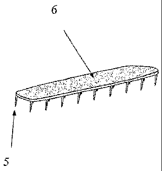
A schematic illustration of one embodiment of= the
scaffold of the present invention is shown in Figure 8a.
The body of the scaffold (6) may comprise nitinol (or a
similar material). The body may comprise bioerodible
material. Engaging members (5) may be attached to the body
of the scaffold. The engaging members may be tines, or
prongs to be driven into the cartilage. The scaffold may be
bent into shape or may be pre-programmed to a specific
deg-iee or curvature (Figure 8b).
Figures 9a to 9e illustrate one self-explanatory method
of inserting the scaffold into the cartilage of an ear. The
scaffold may be mounted on the tip of the applicator (Figure
9a). The scaffold may then be deployed into the cartilage
(3).
Figure 10a shows an illustration of a cross section of
an ear before insertion of the scaffold. Figure 10b shows
the scaffold in place in the ear. The scaffold may be
designed such that it can be bent to reshape the antihelical
fold,by a desired amount, or the ear staple may be pre-
programmed to bend with a certain degree of curvature which
may be selected before insertion.
Figures lla to 11c illustrate an applicator which may
be used to insert the scaffold of the present invention into
an ear or nose. In this embodiment, the applicator (8) has
a battery pack in its handle, which may be switched on to
heat the scaffold via switch (7). A trigger may be used to
operate the anvil which drives the ear staple into the
cartilage. Figure 11b shows an enlarged illustration of the
CA 02620848 2008-02-25
WO 2007/023296
PCT/GB2006/003181
- 24 -
anvil (10). The ear staple is held towards the end of the
applicator (9). Advantageously the ear staple may be held
straight during application to the cartilage. The
applicator is then slide off allowing the ear staple to
return to its curved shape upon cooling. Figure 11c shows
heating elements (11) at the tip of the applicator (8).
Figures 12a to 12d illustrate cross sections of an ear
staple (13) being inserted into an ear to correct prominence
due to deep conchal fossa. Figure 12e shows a side view of
an ear showing the scaffold in place (15) and the incision
made in the conchal fossa to place the scaffold (14).
Figures 13a to 13d show a scaffold (Figure 13d)of the
present invention being inserted into a human nose. The skin
envelope of the nose is released (Figure 13a). The scaffold
is then inserted into the nose cartilage (Figure 13b). The
scaffold ma be secured in place by driving the engaging
members into the cartilage. The scaffold may then be
transformed into the predetermined shape (Figure 13c). In
Figure 13c, the scaffold is secured to the alar cartilages
by;driving the tines (engaging members) into the cartilage.
Once secure, the nasal cartilages preferably conform to the
shape of the scaffold reshaping the nose.
=
Figure 14 shows a preferred embodiment of an applicator
for the scaffold of =the present invention. The applicator
may comprise a handle (19), a portion (18) on which the
scaffold (not shown) is held prior to insertion by a
retaining means (17), and a protruding section (16) which
helps to position the scaffold on the applicator upon
insertion to the nose or ear. The scaffold is positioned on
CA 02620848 2008-02-25
WO 2007/023296
PCT/GB2006/003181
- 25 -
portion (18) of the applicator prior to insertion. The
portion (18) preferably holds the scaffold in the first
configuration. The applicator is then inserted into a skin
incision made in the ear or nose. Preferably only the
portion (18) is inserted into the incision. To facilitate
insertion of the applicator into the incision the applicator
may be tapered towards the distal end, preferably along the
portion (18) as shown in Figure 14. The retaining means
(17) may be a groove as shown in Figure 14 into which the
scaflold is designed to rest. The retaining means may be a
channel for releasably retaining the scaffold. The handle
(19) may be designed such that a finger may be inserted into
it. Preferably the handle is designed for insertion of the
middle finger. The index finger may then be used to steady
the applicator.
Preferably the applicator has stop means for stopping
further deployment of portion (18) into the nose or ear.
For example, the stop means may be a protruding section (16)
as shown in Figure 16.
-Preferably the applicator retains the scaffold in a
first configuration.
After the applicator has been inserted under the skin
the scaffold may be deployed into position by pushing the
scaffold from portion (18) of the applicator and removing
the applicator from the nose or ear. The scaffold may be
deployed from the applicator by means of a slider (20)
(Figure 15) which is positioned on the applicator as shown
in Figure 16. The scaffold bends into the pre-programmed
shape as it is deployed from the applicator.
CA 02620848 2008-02-25
WO 2007/023296
PCT/GB2006/003181
- 26 -
The applicator may further comprise a locator means
(21) attached to the slider (20). The locator means is
designed to help the operator to locate the position of the
centre of the scaffold when it has been inserted under the
skin. This will allow the operator to ensure that the
scaffold is located directly over the middle of the
antihelical fold. One example of a locator means is shown
in Figure 17.
Figure 18a shows the cross section of portion 18 of the
scaffold applicator. The scaffold (25) is retained on the
applicator prior to insertion in a groove (22) or channel in
portion (18) of the applicator. In this example the
scaffold has a substantially smooth surface so that
insertion of the scaffold from the applicator is
facilitated.
Figure 18b shows possible alternative to the cross
section of portion (18) of the applicator. In this
embodiment the scaffold is designed to have a roughed
su.face (23) over at least some of its body. In order to
ease application of such a scaffold (26), portion (18) may
have a further groove (24) or channel to make space for the
roughened surface (23).
Embodiment 1
In a first example of the present invention, a scaffold
is used to reshape the antihelical fold of the par with the
aim of correcting a prominent ear (see Figures la and 1b).
CA 02620848 2008-02-25
WO 2007/023296
PCT/GB2006/003181
=- 27
In this example, to change the shape of the antihelical
fold, a thin strip of nitinol metal alloy (or material with
similar properties) is inserted into the subcutaneous space
of the skin on the posterior aspect of.the ear through a
small incision or series of incisions (Figures 7a to 7c).
The scaffold of the present invention may also be
effective when placed into the subcutaneous space on the
anterior aspect of the ear. However, it may be more
advahtageous for placement at the posterior position,
because this will reduce the likelihood that the engaging
member (and any incision to insert it) may become visible
overtime.
In this example the scaffold is shaped with several
thin "spikes", "prongs" or "tines" along its length (or just
at each end) on one side of the strip (Figures 8a and 8b).
The purpose of these spikes or tines is to allow the
scaffold to be fixed securely into the cartilage of the ear.
To fix the scaffold to the cartilage, a specially
deigned applicator may be used to hold the scaffold in the
correct position in relation to the antihelical fold of the
ear (Figures la, lb and Figures 9a to 9e). Once it is in
the correct position (Figure 9a), the applicator is deployed
to drive the tines into the cartilage (Figure 9b). This
method may be sufficient to hold the scaffold securely
(Figure 9c). Alternatively, it may be necessary to cause
the tines to curve over at their tips (Figure 9d) to bind
the scaffold more closely to the cartilage.
CA 02620848 2008-02-25
WO 2007/023296 PCT/GB2006/003181
= - 28 -
Once the scaffold is secured to the cartilage it is
either bent into the desired shape by the user (causing the
antihelical fold to be formed) or it is allowed to bend into
a pre-programmed shape (Figure 10b). The latter method
allows different degrees of curvature to be pre-programmed
into the invention before insertion.
The specific degree of curvature of the antihelical
fold required to correct the prominence may be measured,
prior to design of the scaffold. The scaffold may then be
designed to specfic measurements. The results of this
method of correction would be highly predictable and
reproducible compared with conventional techniques.
A possible applicator used to insert the invention is
shown in (Figures 11a to 11c). The applicator may be
electrically driven. This allows the shape pre-programmed
into the nitinol metal alloy to be activated on command.
The pre-programmed shapes could include, for example, a
shape where the tines are either straight or curved. The
ability to control the shape of the tines would facilitate
reiioval of the invention from the ear. This might be
necessary to allow the position of the invention to be
adjusted infinitely to produce the desired effect and would
remove any concern about the learning curve required to
produce a particular outcome.
It is anticipated that a maximum of three and a minimum
of one of the scaffolds may be required to produce the
desired curvature of the antihelical fold (Figures 7a to
7c). Once inserted, the inventions would be left in place
CA 02620848 2008-02-25
WO 2007/023296
PCT/GB2006/003181
- 29 -
permanently but could be removed at a later date if problems
were to develop.
Embodiment 2
In a second embodiment of the present invention, a
scaffold is used to correct deep conchal fossa (see Figures
3a, 3b and Figures 12a to 12e).
An incision is made in the conchal fossa to facilitate
inSertion of the staple (Figure 12b and 12e). A separate
incision is made behind the ear to allow the soft tissues to
be repositioned (Figure 12b). The ear is pushed back
alongside the head by the desired amount (Figure 12b). A
staple is inserted through the anterior incision which holds
the ear in the desired position (Figure 12c and 12e). = The
engaging members, for example, the tines or spikes will then
be made to curve over holding the staple in the correct
position (Figure 12d) as with the invention for reshaping
the antihelical fold.
Embodiment 3
In the third embodiment of the present invention a
scaffold is used to correct a deformed nose (see Figures 13a
to d).
The skin envelope is released from the nose to allow
reshaping of the deformed nasal cartilage. The nose
scaffold used to correct the deformity, in this example,
comprises two bent body portions. Each portion comprises a
substantially straight part, and a curved part. The curved
part comprises engaging members which may be used to engage
in the cartilage of the nose.
CA 02620848 2008-02-25
WO 2007/023296
PCT/GB2006/003181
- 30 -
The scaffold is inserted into the cartilage of the
nasal cavity. The skin envelope is then draped over the new
cartilage scaffold.
The scaffold is then secured to the alar cartilage by
driving the engaging members into the cartilage. The
engaging members are than heated (or they may be cooled in
other embodiments of the present invention) to cause the
engaging members to curve into the alar cartilage. In other
embodiments of the present invention, the engaging members
need to curve upon transition to a second pre-programmed
configuration.
Once the scaffold is in place. The nasal cartilage may
conform to the new scaffold shape, giving the nose a new
shape.