Note: Descriptions are shown in the official language in which they were submitted.
CA 02622850 2008-03-17
WO 2007/035647 PCT/US2006/036314
-1-
EMBRYO TRANSFER USING
TRANSVAGINAL ULTRASOUND TRANSDUCER
RELATED APPLICATIONS
[0001] This application is a continuation-in-part of U.S. Pat. Appl.
10/264,651, filed October 4, 2002, presently pending, which is a continuation-
in-
part of U.S. Patent Application Serial No. 09/669,315, now U.S. Pat. No.
6,527,752, and also claims the benefit of the filing date under 35 U.S.C.
119(e),
of U.S. Provisional Application 60/156,049, filed September 24, 1999, which is
hereby incorporated by reference in its entirety. Each of these applications
and
patents is hereby incorporated by reference in its entirety, as though each
document were reproduced in the text below.
FIELD OF THE INVENTION
[0002] The technical field of the invention is that of assisted reproductive
technology, involving human in vitro fertilization (IVF) and embryo transfer
(ET).
BACKGROUND
[0003] Human In Vitro Fertilization (IVF) and Embryo Transfer (ET), first
successfully performed in 1978, has become a widely practiced procedure to
treat
infertile couples who have failed with more conventional methods of therapy
such
as superovulation and intrauterine insemination. The most common indications
for
IVF and related procedures, such as Gamete In Vitro Fertilization or Gamete
Intra-
Fallopian Transfer (GIFT) which includes women having blocked or damaged
fallopian tubes, and includes low sperm and/or egg quality. Related factors
include age of the female, and the degree of endometrial receptivity. The
procedure may also be used in cases of severe male factor where direct
(intracytoplasmic) injection of sperm is an option.
[0004] The IVF/ET procedure typically involves the hormonal stimulation of
the female to first suppress her ability to ovulate on her own, then stimulate
development of follicles in the ovaries with a fertility medication. The
mature eggs
CA 02622850 2008-03-17
WO 2007/035647 PCT/US2006/036314
-2-
are removed from the ovary transvaginally using a needle, preferably guided
under
ultrasound. Following harvesting of the eggs, the eggs are identified and
sorted
with regard to maturity, and then placed with a sperm sample from the male.
Approximately 24 hours after fertilization, the eggs are examined to confirm
fertilization, which occurs in approximately 659o to 85% of the eggs
harvested.
[0005] After a short development period, the embryos are transferred, along
with a volume of fluid, to the uterus using a delivery catheter. The delivery
catheter is made of a soft plastic material to avoid damage to the
endometrium.
There are many potential difficulties in achieving a successful implantation.
Because of the soft nature of the standard delivery catheter, in a number of
cases,
the tip of the catheter may bend back on itself or curve away from the fundus
of
the uterus. The tip may also accidentally pass between the layer of the
endometrium and myometrium. Conversely, a stiffer catheter increases the risk
of
trauma to the uterus or cervix, with the latter possibly leading to the
release of
prostaglandins and expulsion of the eggs from the endometrium.
[0006] One particular difficulty in achieving successful implantation is the
difficulty the surgeon has in visualizing the uterus and the endometrium into
which the embryo is implanted. The embryo is desirably transferred onto the
rich
lining of the uterus (endometrium) without disturbing the lining or causing
trauma.
As noted above, transfer catheters are typically made from very soft material
to
minimize trauma. However, the endometrium is very fragile and can be easily
disturbed. In addition, transfer catheters may be marked with an echogenic or
radiopaque band or feature on the distal tip. This feature allows the
physician to
visualize the tip when used with an ultrasound probe on the patient's abdomen.
However, most surgeons have difficulty visualizing both the uterus and the tip
of
the catheter using ultrasound with this technique.
[0007] Several unsuccessful attempts have been made to improve success
rates. U.S. Pat. No. 6,165,165 uses a guiding catheter and an implant
catheter, the
implant catheter made from materials of two different durometers, so that the
stiffness of the catheter decreases from the proximal end to the distal end of
the
catheter. The resulting catheter may be easier to guide, but is still subject
to
CA 02622850 2008-03-17
WO 2007/035647 PCT/US2006/036314
-3-
interference from mucus. WIPO International Patent Applications W099/37348
and WO01/74417 attempt to solve the problem with an end cap on a guiding
catheter that swings open to allow the transfer catheter to pass through an
opening
and transfer the embryos. Alternatively, the transfer catheter may have a side
port
rather than an axial port on the distal end, so the side port will avoid
interference
from mucus. These embodiments are still subject to interference from mucus.
[0008] One way to increase the likelihood of success is to tailor the
catheters
used to the person undergoing the treatment, i.e., by using different lengths
of
catheter. These attempts to tailor the catheters have led to a proliferation
of
lengths of catheters, especially in guide catheters. Even with overnight
delivery of
the desired resources, this results in the need for hospitals and clinics to
inventory
more catheters and more sets of catheters than is desirable. What is needed is
a
catheter system and a better technique that can increase the likelihood of
successful embryo implantation patients desiring this procedure by better
placement of the embryos that are transferred.
BRIEF SUMMARY
[0009] The foregoing problems are solved and a technical advance is achieved
in an illustrative transfer or delivery catheter which includes ultrasonically
reflective components or features to enhance its visibility under
transabdominal or
transvaginal ultrasound guidance, during embryo transfer, for example. The
present invention helps to increase the likelihood of successful implantation
by
using a cervical stop to limit the penetration of a transfer catheter into a
woman.
One aspect of the invention is a transfer system for cellular material, the
transfer
system comprising a distal end detectably different from adjacent portions of
the
catheter. The transfer catheter includes a guide catheter for holding and
guiding
the transfer catheter, the guide catheter further comprising a movable
cervical stop.
The transfer system also includes a locking mechanism for fixing the position
of
the transfer catheter with respect to the guide catheter, wherein the transfer
system
is suitable for guidance using a transvaginal ultrasound tecllnique
CA 02622850 2008-03-17
WO 2007/035647 PCT/US2006/036314
-4-
[0010] Another aspect of the invention is a catheter transfer system. The
transfer catheter system includes a soft inner transfer catheter comprising a
connector for fluid transfer on a proximal end and an echogenic or radiopaque
marker on a distal end. The catheter transfer system also includes an outer
guide
catheter comprising a cervical stop and a series of spaced marks on a distal
end of
the outer guide catheter, and a locking mechanism for fixing a position of the
inner
and outer catheters with respect to one another.
[0011] Another aspect of the invention is a method of transferring an embryo.
The method includes steps of adjusting a cervical stop on a catheter transfer
system, and placing the catheter transfer system near an opening of a cervix.
The
method also includes observing at least one echogenic or radiopaque feature of
the
catheter transfer system using an ultrasound transducer placed near a vagina,
and
also optionally using at least one echogenic or radiopaque feature of the
catheter
transfer system using an ultrasound transducer placed near an abdomen. The
method concludes with steps of advancing a transfer catheter to a desired
position
in a uterus and transferring the embryo into the uterus.
[0012] Another aspect of the invention is a method of transferring cellular
material. The method comprises placing at least one catheter near an opening
of a
cervix, and observing a position of the at least one catheter using an
ultrasound
transducer placed in or near a vagina, and optionally using an ultrasound
transducer placed near an abdomen. The method includes steps of adjusting a
cervical stop on the at least one catheter, and advancing the at least one
catheter
into the cervix and uterus until the stop is reached. The method concludes
with
steps of observing the position of a distal end of the at least one catheter
using at
least one ultrasound transducer and transferring the cellular material.
[0013] There are many aspects and embodiments of the invention, some of
which are described below in a specification and drawings which are meant to
be
illustrative and descriptive, rather than limiting.
CA 02622850 2008-03-17
WO 2007/035647 PCT/US2006/036314
-5-
BRIEF DESCRIPTION OF THE DRAWINGS
[0014] Fig. 1-3 depict a first embodiment of a catheter transfer system
according to the present invention;
[0015] Figs. 4-5 depict a second embodiment of a transfer catheter system;
[0016] Figs. 6-7 depict a third embodiment of a transfer catheter system; and
[0017] Figs. 8-9 are flowcharts illustrating methods of practicing an improved
method of embryo transfer.
DETAILED DESCRIPTION OF THE DRAWINGS AND THE
PRESENTLY PREFERRED EMBODIMENTS
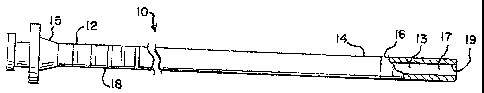
[0018] Figs. 1-3 depict a cellular material transfer catheter system that
comprises three catheters, an inner transfer catheter 10, a guide catheter 20,
and an
outer protective sheath 30. The transfer catheter 10 extends somewhat longer
than the guide catheter 20, and the protective outer sheath 30. The inner
catheter,
transfer catheter 10, includes a passageway 13 of sufficient diameter to hold
and
deliver cellular material, such as early embryos, gametes (oocyte or sperm),
blastocysts, or zygotes that are to be transferred from in vitro culture for
in vivo
implantation and/or fertilization.
[0019] The cellular material or embryo transfer catheter 10 includes a
proximal
portion 12 and a distal portion 14. The proximal portion may include a hub 15
for
interfacing with a syringe for implanting cellular material. The catheter may
also
include an echogenic tip 16, preferably made of stainless steel, for detecting
the
distal end via ultrasound. Echogenic tip 16 has an ultrasound reflectivity
very
different from, and preferably greater than, adjacent portions of embryo
transfer
catheter 10. The catheter may also have markings 18 at the proximal or distal
end
indicating a position of the catheter to implantation personnel. The catheter
itself
is made of relatively soft material, such as polyethylene. Other materials
that may
be used include urethane, polyolefin, poly-octene-ethylene, polyamides,
fluoropolymers including but not limited to polytetrafluoroethylene, and
silicone.
[0020] The diameter of the passageway and volume of the fluid and material
contained therein is preferably minimized to a diameter of no greater than
0.025",
CA 02622850 2008-03-17
WO 2007/035647 PCT/US2006/036314
-6-
preferably less than 0.023", and most preferably between 0.018" and 0.021".
The
transfer volume is no greater than 30 l, more preferably 20 l or less, and
most
preferably between 5 and 15 l. Clinical experience with this catheter, for
IVF/ET
having a 0.020" diameter with a volume of approximately 10 l, indicates an
unexpected increase in pregnancy rates, possibly due to the reduced amount of
fluid delivered with the embryos. The reduced transfer volume ostensibly
lessens
the tendency of embryos to migrate to another section of the uterus, for
instance,
into the fallopian tubes. By increasing the implantation rate, fewer embryos
may
be needed, thereby reducing the number of unwanted multiple pregnancies and
further risks.
[0021] Fig. 2 depicts a guide catheter 20 used coaxially with transfer
catheter
10. The guide catheter 20 is of relatively simple construction, and may
comprise a
proximal portion 22 and a distal portion 24, and distal end 26. The distal end
26 is
preferably open rather than closed. The catheter 20 also comprises a hub 25
for
interfacing with the embryo transfer catheter 10, and possibly the protective
sheath
30. The guide catheter may also comprise markings 28 at the distal portion 24
to
guide delivery personnel. The guide catheter also includes cervical stop 29.
The
guide catheter is preferably somewhat stiffer than the embryo transfer
catheter.
Materials suitable for the guide catheter are many, so long as the guide
catheter is
able to hold its shape without drooping or sag during the implantation
procedure.
Materials used include fluoropolymers such as polytetrafluoroethylene (PTFE),
although other materials, as mentioned above, may also be used.
[0022] Cervical stop 29 is desirably made from a soft material, such as
silicone
or urethane. Stop 29 is preferably large enough so that it cannot enter a
cervix. A
diameter of stop 29 is preferably in the range of about 5-15 mm. The width is
preferably about 1-3 mm, sufficiently wide that the stop cannot be bent and
deformed easily.
[0023] The outer protective sheath 30 of Fig. 3 is also of relatively simple
construction. It has a proximal end 32, a distal end portion 34 with a distal
end
portions 36 and may have a hub or interface 35. Distal portions 36 of the
protective sheath pull apart and draw mucus and blood away as tlie guide
catheter
CA 02622850 2008-03-17
WO 2007/035647 PCT/US2006/036314
-7-
emerges from the protective sheath. The sheath may be made with markings 38
for guidance of delivery personnel. The protective sheath is also relatively
stiff
compared to the embryo transfer catheter. It is important that the guide
catheter
and the protective sheath be dimensionally stable (do not sag) so that
operating
personnel may control the exact position of the catheter and the sheath during
implantation procedures. The inner transfer catheter should be relatively soft
so as
to avoid any damage to delicate tissues in the uterus.
[0024] The transfer catheter, the guide catheter and the protective sheath
catheter are used to implant an embryo into a uterus of a woman. As discussed
above, one problem with such implantations is fouling of the distal end of the
transfer catheter. The mucousal nature of the cervix, and the presence of
mucus
and blood, makes the problem an inherent one for any procedure in this area of
the
body. The present invention solves the problem in the following manner. The
three catheters are advanced as a unit through the vagina, through the cervix,
and
positioned at the internal cervical ostium. The echogenic tip and the markings
on
one or more of the catheters assist in this operation.
[0025] The protective sheath, with the guide catheter still inside the
protective
sheath, is then retracted to expose the tip of the guide catheter. The distal
end of
the protective sheath is closed and is impervious to the fouling substances in
the
cervix, but the protective sheath is also scored or weakened so that the guide
catheter is easily advanced through the scored or weakened end portion of the
protective sheath. The protective sheath is designed to snap onto the guide
catheter when completely retracted. The protective sheath at this point may be
covered with mucus or blood or other fouling substances. In practice, these
substances cling to the sheath while the guide catheter advances relatively
free of
the mucus and blood. It is not necessary to retract the protective sheath a
great
distance; about 1 to 2 cm is sufficient to clear the guide catheter and pull
mucus
and blood away from the guide catheter tip.
[0026] While most of the mucus and blood are retained on the protective
sheath, a small amount may cling to the distal (protruding) end of the guide
catheter. In practice, this small amount tends to cling to the sides of the
guide
CA 02622850 2008-03-17
WO 2007/035647 PCT/US2006/036314
-8-
catheter, rather than the area of the central lumen of the guide catheter.
Thus, by
extending the guide catheter through the protective sheath, a passageway that
is
free of fouling substances, such as mucus and blood, is cleared through the
central
lumen of the guide catheter. All that remains is to advance the transfer
catheter
through the guide catheter, and to implant the embryo or cellular material. As
stated above, the transfer catheter preferably has an echogenic tip to guide
operating personnel as to its exact position and to complete the transfer
procedure
for the embryo or other cellular material. It is preferable to use a syringe
and to
expel the embryo into the uterus by means of fluid pressure.
[0027] Because the delivery catheter is preferably made of a softer (lower
durometer) polymer the surface energy density is usually higher, making the
embryo more likely to adhere to the inner luminal surface. This is especially
critical with a small lumen diameter, since with a typical embryo having a
diameter of about 120 micrometers and a blastocyst having a diameter of about
260 micrometers, there is an increased likelihood of problems in delivery.
Luminal surface treatments may help reduce friction for the smooth expulsion
of
oocytes and embryos. Ion beam bombardment is a well-known technique for
reducing surface energy density of polymers. Polishing and surface coatings
can
also offer improvement in friction coefficients for otherwise "sticky"
polymers.
The luminal surface 19 of the passageway 13 of the distal portion 14 of the
delivery catheter is coated with lubricious material 17, such as parylene, to
reduce
surface energy density. Paralyne coatings may be applied by in-house systems
or
by vendors, such as Specialty Coating Systems, Indianapolis,IN, or Parylene
Coating Services, Katy TX, among other vendors. Other coatings, such as PTFE,
plasma or corona treatments, may also be used.
[0028] In one illustrative embodiment, the protective sheath has an outer
diameter of about 6.8 Fr (about 2.27 mm) and has an overall length of about 11
or
16 cm. The guide catheter has an outer diameter of 4.7 Fr (1.57 mm) and an
overall length of about 12 or 17 cm. The inner catheter diameter is about
0.483
mm with a length of approximately 19 or 24 cm. The delivery or transfer
catheter
CA 02622850 2008-03-17
WO 2007/035647 PCT/US2006/036314
-9-
extends approximately 5 cm beyond the tip of the guiding catheter, and the
guide
catheter extends about 1 to 2 cm beyond the tip of the protective sheath
catheter.
[0029] As mentioned above, optional graduated markings 18, 28 can be placed
about the proximal portion 12 of the delivery catheter 10 or the distal
portion 24 of
the guiding catheter 20 to determine the depth of penetration of the guide
catheter
into the uterus or the amount of delivery catheter 10 to be exposed beyond the
distal tip 26 of the guiding catheter 20. Additional graduated markings may
also
be placed on the guide sheath if desired. The physician or medical
professional
may use these marks in conjunction with ultrasonic imaging techniques in order
to
visualize the position of the transfer catheter tip and the patient's uterus.
[0030] In addition to the delivery catheter embodiment depicted in Fig. 1, the
transfer catheter can be made with a stiffened proximal component. Fig. 4
depicts
an embryo transfer catheter 40 having a stiffening or reinforcing portion 47
in its
proximal portion 42. The embryo transfer catheter 40 also includes a central
lumen 43 and a distal portion 44, preferably with an echogenic tip 46. The
echogenic tip may be made of stainless steel, or may also take the form of
particles
embedded into the outer surface of the catheter. It has been found that
spherically-
shaped metallic particles, or hemispherically-shaped voids or cavities are
better for
the resulting ultrasonic images. The particles are preferably incorporated
into the
desired location of the embryo transfer catheter, or possibly into the guide
catheter, by molding them into the catheter.
[0031] The echogenic tip and the markings on one or more of the catheters
assist in this operation. As stated above, the echogenic tip may be made of
stainless steel, or may also take the form of particles embedded into the
outer
surface of the catheter. It has been found that spherically-shaped metallic
particles
are better for the resulting ultrasonic images. The particles are preferably
incorporated into the desired location of the embryo transfer catheter, or
possibly
into the guide catheter, by molding them into the catheter. If a ring of
stainless
steel or other suitable material is used, it may be made echogenic by
machining or
otherwise placing on the surface grooves, bars, lines, bands, dimples, or
other
CA 02622850 2008-03-17
WO 2007/035647 PCT/US2006/036314
-10-
patterns which cause reflection, scattering and diffraction of ultrasound or
other
energy used to guide the surgeon in the placement of the catheter in the
uterus.
[0032] The proximal portion 42 may also include graduated markings 48 and
an interface 45. Reinforcing member 47 may be a stainless steel tube that is
bonded to the embryo catheter, preferably by heat or by an adhesive. However,
the fit between the reinforcing member and the delivery catheter is typically
sufficient that bonding is not required. The reinforcing member may be a
cannula
on the inside or on the outside of the transfer catheter. An example of a
stiffened
embryo transfer catheter is polyethylene tubing having a central lumen of
0.019 in
(about 0.483 mm) diameter with a 23GXTW stainless steel cannula. An outer
cannula, with polyethylene tubing on the inside of the cannula, may also be
used.
[0033] The transfer catheter and the guide catheter are used to implant an
embryo or other cellular material into a uterus of a woman. As discussed
above,
one problem with such implantations is the proliferation of sizes, especially
of
guide catheters. In one line of embryo transfer catheters, the lengths of
transfer
catheter may range from about 18.5 cm to about 23.5 cm, while the guiding
catheters may range from 12 cm to 17 cm.
[0034] Transfer catheter 40 with proximal portion 42 also includes a male snap
on or snap fit feature 41. This feature is a protrusion on an external surface
of
catheter 40. Snap fit feature 41 has an edge 41a facing the proximal
direction, so
that edge 41a may interface with a female snap fit or snap on feature on a
mating
part, such as guide catheter 50 in Fig. 5. Using the snap fit or snap on
features,
guide catheter 50 may be snap fit over delivery catheter 40. Guide catheter 50
includes a hub 55 at its proximal end, a female snap fit or snap on feature
59, and a
relatively soft uterine stop 52 with a short hub 52a. Hub 52a provides a
larger
interface for stop 52 with catheter 50, holding stop 52 more firmly in place
while
the catheters are being advanced through the patient's body.
[0035] Catheter 50 also has a central lumen 56 and may have marking bands
58 preferably at distal end 54. Catheter 50 has one or more ribs 53 and a
reinforcing band 57 which may include connecting hub 55 around the proximal
CA 02622850 2008-03-17
WO 2007/035647 PCT/US2006/036314
-11-
end. The band may be made of any desired, relatively stiffer material suitable
for
the application, such as PTFE or polyolefin.
[0036] Snap on feature 59 includes a space or void 59a for receiving male snap
on feature 41 and an edge 59b for mating and interfering with edge 41a of the
male snap on feature. The edges form an interference that prevents axial
movement of the two components of which the edges are a part in a direction
opposed to the direction that caused the engagement. That is, once catheter 40
is
placed inside catheter 50, the snap fit features tend to prevent the removal
of
catheter 40 from catheter 50. Catheter 50 may also have a male snap on feature
51
for assembling a protective sheath to catheter 50. A protective sheath may
have a
mating female snap on feature to accommodate catheter 50
[0037] The catheters described above are preferably used with imaging
techniques that allow a doctor or medical professional to visualize the
placement
of the catheter and the embryo. It is well known that ultrasonic images may be
made through the abdomen, i.e., placing an ultrasound transducer on the
abdomen
of a patient to visualize the internal organs. However, ultrasonic detection
using
an abdominal technique is usually less than clear, and often not helpful in
locating
the uterine opening or the precise place in the uterine endometrium at which
placement is desired.
[0038] It has been discovered that ultrasonic imaging using a vaginal
technique
may be superior to the abdominal techniques used to date. The physician places
an ultrasonic transducer into the vagina, and observes both the uterus and the
catheters. As noted above, the transfer catheter desirably has an echogenic
tip, or
a radiopaque tip, allowing for easier observation with a suitable imaging
technique. Using this technique, the physician can then estimate the distance
from
the cervical os or opening, to the desired location for implantation on the
back
wall of the uterine endometrium. The physician then calculates the total
distance
of advance desired into the uterus, and allocates a portion of this distance
as the
distance between the cervical stop and the distal tip of the guide catheter;
the
remainder is the distance the distal tip of the transfer catheter is advanced
beyond
the distal tip of the guide catheter. The physician can then adjust the
uterine stop
CA 02622850 2008-03-17
WO 2007/035647 PCT/US2006/036314
-12-
on the guide catheter, limiting the travel of the guide catheter into the
uterus. A
transfer catheter with marks on its proximal end can assist in this process,
since
the physician then knows how far the transfer catheter should be advanced and
uses these marks, and its echogenic tip, to achieve the desired advance.
[0039] Figs. 6-7 depict alternative embodiments of the present invention
having ultrasonically reflective components or features to enhance visibility
under
transabdominal or transvaginal ultrasound guidance during embryo transfer.
Transfer catheter systems as described above are useful in carrying out this
technique. The transfer catheter systems 60, 70 in Figs. 6-7 may also be used.
In
Fig. 6, a transfer catheter 61 includes a length of soft plastic or
elastomeric tubing,
preferably in the range of 80-85 Shore A durometer. The band 62 on the distal
tip
of transfer catheter 60 is preferably echogenic, but may instead be
radiopaque.
There are markings, preferably bands 63 spaced at about 1 cm intervals, near
the
proximal end of transfer catheter 61. Other distances may be used. There is
also a
fluid connection 64 at the proximal end, such as a female Luer lock adapter
(FLLA) for connecting to a source of transfer fluid and embryos. An outer
guide
catheter 65 is used for guiding the inner transfer catheter to the desired
location in
the patient. The outer guide catheter also includes a checkflow fitting 66
with a
silicone septum 67 for admitting and helping to hold the transfer catheter 61.
A
cervical positioner or stop 68 may also be placed on outer guide catheter 65
to aid
the physician in positioning the catheters. Spaced marks 69 may also be used
to
help in positioning the catheters. The marks may be placed at any desired
interval.
1 cm intervals are presently preferred, but other intervals may be used.
[0040] Another transfer catheter system 70 is depicted in Fig. 7. In this
embodiment, inner transfer catheter 71 also includes an echogenic tip 72 at
its
distal end, and a series of spaced markings at its proximal end (not shown). A
fluid connector, such as FLLA 74 is attached at the proximal end for accepting
medium and embryos to be transferred. Outer guide catheter 75 includes a
inovable stop or positioner 78 for convenience by the physician. The outer
catheter also includes spaced marlcs 79, preferably at 1 cm intervals. An
adapter
76 and a Tuohy-Borst adapter 77 may be placed on guide catheter 75 to lock
inner
CA 02622850 2008-03-17
WO 2007/035647 PCT/US2006/036314
-13-
catheter 71 in place when the physician is advancing the catheters together
through
the patient, or otherwise during the procedure when convenient. The Tuohy-
Borst
adapter works by compressing the outside of the inner catheter sufficiently
that is
cannot be moved axially with respect to the outer catheter, and is locked in
place.
[0041] Figs. 8-9 are flowcharts depicting methods of using a transvaginal
ultrasound technique for implantation of an embryo or cellular matter. Fig. 8
depicts a method in which a physician or health-care professional places an
ultrasound transducer in the vagina to estimate the distance 81 between the
cervical os and the endometrium. This is approximately the total distance the
transfer catheter will desirably advance beyond the cervical opening.
[0042] The physician will want to advance the guide catheter for a portion of
this distance to protect the transfer catheter from mucus and other material
that
could foul the distal tip of the transfer catheter. The physician then sets
the
cervical stop 82 on the transfer catheter to limit its travel into the uterus,
and then
calculates the distance remaining for the transfer catheter to travel. The two
catheters, or three if a protective sheath is used, are then locked together
using the
connectors described above, and the catheters are advanced through the vagina
to
the cervical opening 83. The physician then unlocks the connectors, allowing
the
transfer catheter to move axially with respect to the guide catheter, and
advances
the transfer catheter the remaining desired amount into the uterus 84. The
cellular
material or embryos may then be transferred into the uterus.
[0043] Another technique is described in Fig. 9, in which the catheters are
first
advanced in order to aid in the step of estimating the distance required for
travel
into the uterus. A first step is to place the catheter system into the vagina
and to
advance the system to the cervix 91. This is preferably accomplished using
ultrasound imaging. The echogenic tip previously described may assist in
visualizing the distance from the cervical opening to the desired area, the
endometrium, by using an ultrasonic transducer placed into the vagina 92. Once
the distance is estimated, the catheters may be removed and the cervical stop
set in
place on the guide catheter using the markings on the distal end of the guide
CA 02622850 2008-03-17
WO 2007/035647 PCT/US2006/036314
-14-
catheter. The physician also calculates the distance the transfer catheter
will have
to be advanced for the most desirable implantation.
[0044] The catheters are then connected or locked together, and passed through
the vagina and through the cervical opening 94. The transfer catheter is
advanced
through the guide catheter 95 by the desired amount, preferably using the
spaced
marks on the proximal end of the transfer catheter. The embryos or cellular
material is then transferred 96 from the transfer catheter to the uterus.
Other
techniques of imaging, estimating, and advancing may be used.
[0045] Embodiments of the present invention may be made from one or more
of the niaterials listed above, and may be used for any of the procedures
described
herein. While the cervical stop is preferably made from a soft urethane or
silicone
material, it may be made from any other medically-acceptable elastomer, such
as
nitrile, or from a medically-acceptable plastic, such as polyethylene or
polypropylene.
The catheters may also be made from alternative materials, although the
transfer
catheter is preferably made from a very soft plastic or elastomer, in a Shore
A
hardness from about 80-85. Thermoplastic olefin elastomers may be useful
applied
to transfer catheter applications. Several grades of polyolefin from DuPont
Dow
Elastomers may be useful for this purpose, including olefins made from blends
of
ethylene and octene. The soft grades are preferred, such as Engage 8100 and
8480,
especially preferred is 8003, which has a slightly higher density than 8100
and 8480.
[0046] It is possible to obtain a number of advantages of the present
invention
by using methods other than those specifically described above. For instance,
one
may place a transfer catheter or other device with an echogenic feature near
the
cervical os, and observe via abdominal ultrasound or fluoroscopy the distance
from
the cervical opening to the desired location for implantation within the
uterus.
Implantation may then be accomplished using the catheter system and cervical
stop
described above. While it is believed that this method is inferior to one
using vaginal
ultrasound, the use of the cervical stop would be an improvement over present
methods that do not use a cervical stop.
[0047] It is intended that the foregoing detailed description be regarded as
illustrative rather than limiting, and that it be understood that it is the
following
CA 02622850 2008-03-17
WO 2007/035647 PCT/US2006/036314
-15-
claims, including all equivalents, that are intended to define the spirit and
scope of
this invention.