Note: Descriptions are shown in the official language in which they were submitted.
CA 02623268 2014-05-16
WO 2007/035806
PCT/US2006/036605
MELTING CURVE ANALYSIS WITH EXPONENTIAL BACKGROUND
SUBTRACTION
CROSS REFERENCE TO RELATED APPLICATION
This application claims the benefit of U.S. Provisional Patent Application No.
60/719,250, entitled "Removal of Background from the Melting Curve of a Double
Stranded Nucleotide" filed on September 20, 2005
FIELD OF THE INVENTION
Generally the present invention relates to nucleic acid melting curve
analysis.
More specifically various embodiments of the present invention relate to
methods and
systems for analyzing the melting profiles of double stranded nucleic acids by
removing
the background fluorescence signals.
BACKGROUND OF THE INVENTION
Methods for analyzing DNA sequence variation can be divided into two general
categories: 1) genotyping for known sequence variants and 2) scanning for
unknown
variants. There are many methods for genotyping known sequence variants, and
single
step, homogeneous, closed tube methods that use fluorescent probes are
available (Lay M
J, et al., Clin. Chem 1997;43:2262-7). In contrast, most scanning techniques
for unknown
variants require gel electrophoresis or column separation after PCR. These
include single-
strand conformation polymorphism (Orita 0, et al., Proc Natl Acad Sci USA
1989;
86:2766-70), heteroduplex migration (Nataraj A J, et al., Electrophoresis
1999;20:1177-
85), denaturing gradient gel electrophoresis (Abrams E S, et al., Genomics
1990;7:463-
75), temperature gradient gel electrophoresis (Wartell R M, et al., J
Chromatogr A
1998;806:169-85), enzyme or chemical cleavage methods (Taylor G R, et al.,
Genet Anal
1999;14:181-6), as well as DNA sequencing. Identifying new mutations by
sequencing
also requires multiple steps after PCR, namely cycle sequencing and gel
electrophoresis.
Denaturing high-performance liquid chromatography (Xiao W, et al., Hum Mutat
2001;17:439-74) involves injecting the PCR product into a column.
Single nucleotide polymorphisms (SNPs) are by far the most common genetic
variations observed in man and other species. In these polymorphisms, only a
single base
varies between individuals. The alteration may cause an amino acid change in a
protein,
1
CA 02623268 2014-05-16
WO 2007/035806
PCT/US2006/036605
alter rates of transcription, affect mRNA spicing, or have no apparent effect
on cellular
processes. Sometimes when the change is silent (e.g., when the amino acid it
codes for
does not change), SNP genotyping may still be valuable if the alteration is
linked to
(associated with) a unique phenotype caused by another genetic alteration.
There are many methods for genotyping SNPs. Most use PCR or other
amplification techniques to amplify the template of interest. Contemporaneous
or
subsequent analytical techniques may be employed, including gel
electrophoresis, mass
spectrometry, and fluorescence. Fluorescence techniques that are homogeneous
and do
not require the addition of reagents after commencement of amplification or
physical
sampling of the reactions for analysis are attractive. Exemplary homogeneous
techniques
use oligonucleotide primers to locate the region of interest and fluorescent
labels or dyes
for signal generation. Various PCR-based methods are completely closed-tubed,
using a
thermostable enzyme that is stable to DNA denaturation temperature, so that
after heating
begins, no additions are necessary.
Several closed-tube, homogeneous, fluorescent PCR methods are available to
genotype SNPs. These include systems that use FRET oligonucleotide probes with
two
interacting chromophores (adjacent hybridization probes, TaqMae probes,
Molecular
Beacons, Scorpions), single oligonucleotide probes with only one fluorophore
(G-
quenching probes, Crockett, A. 0. and C. T. Wittwer, Anal. Biochem.
2001;290:89-97
and SimpleProbes2), Idaho Technology), and techniques that use a dsDNA dye
instead of
covalent, fluorescently-labeled oligonucleotide probes.
PCR methods that monitor DNA melting with dsDNA fluorescent dyes have
become popular in conjunction with real-time PCR. Because PCR produces enough
DNA
for fluorescent melting analysis, both amplification and analysis can be
performed in the
same tube, providing a homogeneous, closed-tube system that requires no
processing or
separation steps. dsDNA dyes are commonly used to identify products by their
melting
temperature, or Tm.
The power of DNA melting analysis depends on its resolution. Studies with UV
absorbance often required hours to collect high-resolution data at rates of
0.1-1.0 C/min
to ensure equilibrium. In contrast, fluorescent melting analysis is often
acquired at 0.1-
1.0 C/sec and resolution is limited to 2-4 points/ C. With recent advances in
electronics
(e.g., 24-bit A-to-D converters), high-resolution melting can be performed
rapidly with
10-100 times the data density (50-100 points/ C) of conventional real-time PCR
instruments, as recently demonstrated for probe and PCR product melting.
Furthermore.
2
CA 02623268 2014-05-16
WO 2007/035806
PCT/US2006/036605
saturating DNA dyes, such as LCGreen' Plus (Idaho Technology, Salt Lake City,
UT),
that maximize detection of mismatched duplexes (heteroduplexes) are now
available (see,
e.g. U.S. Patent Publication Nos. 2005/0233335 and 2006/0019253):
These two developments dramatically increase the power
of fluorescence-based DNA melting for robust identification of single-base
changes
within PCR products.
High-resolution melting analysis for gene scanning relies primarily on the
shape
of the melting transition of the PCR products. An available method for
screening for
heterozygous single nucleotide polymorphisms (SNPs) within products up to
1,000 bp has
a sensitivity and specificity of 97% and 99%, respectively. In many cases,
high-resolution
analysis of the melting transition also allows genotyping without probes. Even
greater
specificity for variant discrimination over a smaller region can be obtained
by using
unlabeled probes. Specific genotypes are inferred by correlating sequence
alterations
under the probe to changes in the probe Tm. With the recent advances with dyes
and
instrumentation, high-resolution gene scanning and genotyping with unlabeled
probes can
optionally be done simultaneously in the same reaction. Both PCR product and
probe
melting transitions may be observed in the presence of a saturating DNA dye.
In addition
to screening for any sequence variant between the primers in the PCR product,
common
polymorphisms and mutations can be genotyped. Furthermore, unbiased,
hierarchal
clustering can accurately group the melting curves into genotypes. One, two,
or even
more unlabeled probes can be used in a single PCR.
In simultaneous genotyping and scanning, product melting analysis detects
sequence variants anywhere between two primers, while probe melting analysis
identifies
variants under a probe. If a sequence variant is between the primers and under
a probe,
both the presence of a variant and its genotype are obtained. If product
melting indicates a
variant but the probe does not, then the variation likely occurs between the
primers but
not under the probe, and further analysis for genotyping is necessary. Probes
can be
placed at sites of common sequence variation so that in most cases, if product
scanning is
positive, the probes will identify the sequence variants, greatly reducing the
need for
sequencing. With one probe, the genotype of an SNP can be established by both
PCR
product and probe melting. With two probes, two separate regions of the
sequence can be
interrogated for genotype and the rest of the PCR product scanned for rare
sequence
variants. Multiple probes can be used if they differ in melting temperature
and if each
allele presents a unique pattern of probe and/or product meltina.
3
CA 02623268 2014-05-16
WO 2007/035806
PCT/US2006/036605
In one illustrative example, a population is screened for cystic fibrosis
mutations.
Since only 3.8% of Caucasians are cystic fibrosis carriers, one would expect
96.2% of
randomly screened individuals to be negative by complete (exon and splice
site)
sequencing. With 27 exons, the percentage of sequencing runs expected to be
positive is
less than 0.14%. That is, only about 1 in a 1000 sequencing runs would be
useful. This is
why sequencing is not recommended for cystic fibrosis screening. Instead, a
selected
mutation panel is usually performed that detects 83.7% of cystic fibrosis
alleles.
Alternatively, consider simultaneous scanning and genotyping for cystic
fibrosis
screening by high-resolution melting. If the amplicon length is kept under 400
bp, the
sensitivity of high-resolution scanning approaches 100.0%. If common mutations
and
polymorphisms are analyzed with unlabeled probes in the same reaction, then
about 80%
of mutations will also be genotyped. Compared to screening by de novo
sequencing, the
sequencing burden can be reduced by 99.97%.
Closed-tube genotyping methods that use melting analysis have the capacity to
scan for unexpected variants. Melting methods also use less complex and fewer
probes
than allele specific methods that require one probe for each allele analyzed.
Allele
discrimination by Tm or curve shape is an interesting option to fluorescent
color. Dyes
that generically stain double-stranded DNA are attractive for simplicity and
cost.
Although the reliability of genotyping by amplicon melting is controversial, a
recent
study found that 21 out of 21 heteroduplex pairs tested were distinguishable
by high-
resolution melting of small amplicons (Graham R, Liew M, Meadows C, Lyon E,
Wittwer CT. Distinguishing different DNA heterozygotes by high-resolution
melting. Clin
Chem 2005;51).
Although common sequence variants can usually be genotyped with one or two
unlabeled probes in the same reaction, more than two probes and/or sequential
reactions
can also be used. For example, multiple overlapped probes can locate
unexpected rare
variants to within the region covered by one probe. Additional probes can be
designed to
identify the exact position and sequence of the variation. However, DNA
sequencing is a
more direct approach for identifying new, previously unknown variations,
particularly
when the amplified region is highly variable. Nevertheless, in the vast
majority of genetic
analysis, the amplified wild type sequence is known and potential common
variants are
limited. In these cases, scanning and genotyping can be performed in one step
by DNA
melting with simple oligonucleotides. No fluorescent probes or separations are
required,
and both amplification (15 min) and melting analysis (1-2 min) can he ranid.
4
CA 02623268 2014-05-16
WO 2007/035806
PCT/US2006/036605
As discussed above, simultaneous genotyping and scanning, as well as other
genotyping techniques that employ melting analysis have been promising areas
of
research. However, the melting curve analysis prior to high-resolution
capabilities
provided a lack of specificity and accuracy. With the advent of high-
resolution melting
curve analysis, background fluorescence noise can interfere with the use of
melting
curves to accurately genotype SNPs, detect sequence variations, and detect
mutations.
Depending on the amplicon, previous background fluorescence removal techniques
have
led to some erroneous calls. By example, the baseline technique uses linear
extrapolation
as a method for normalizing melting curves and removing background
fluorescence. This
technique works well with labeled probes. However, this and other previous
normalization techniques have not worked as well with unlabeled probes (Zhou
L, Myers
AN, Vandersteen JG, Wang L, Wittwer CT. Closed-Tube Genotyping with Unlabeled
Oligonucleotide Probes and a Saturating DNA Dye. Clin Chem. 2004;50:1328-35)
multiplex small amplicon melting (Liew M, Nelson L, Margraf R, Mitchell S,
Erali M,
Mao R, Lyon E, Wittwer CT. Genotyping of human platelet antigens 1-6 and 15 by
high-
resolution amplicon melting and conventional hybridization probes. J Mol Diag,
2006;8:97-104) and combined amplicon and unlabeled probe melting (Thou L, Wang
L,
Palais R, Pryor R, Wittwer CT. High-resolution melting analysis for
simultaneous
mutation scanning and genotyping in solution. Clin Chem 2005;51:1770-7 ),
nor do they work as well for small amplicons. At least in
part, this is because unlabeled probe and small amplicon melting methods often
require
background subtraction at lower temperatures (40-80 C) then is usual for
standard
amplicon melting at 80-95 C. At these lower temperatures, the low temperature
baseline
is not linear, but a curve with rapidly increasing fluorescence at low
temperatures. When
linear extrapolation is used, the lines can intersect before the melting
transition is
complete, and when this occurs the previous techniques do not provide the most
accurate
means for melting curve analysis, in part due to their mathematical reliance
on absolute
fluorescence.
It would be advantageous for a system and method to genotype SNPs, detect
sequence variations, and/or detect mutations with high accuracy in double
stranded
nucleic acids through use of high resolution melting profile techniques. It
would be
further advantageous if the background fluorescence could be automatically and
accurately separated from a double stranded nucleic acid sample melting
profile. It would
CA 02623268 2014-05-16
WO 2007/035806
PCT/US2006/036605
be a further advantage if the system and method performed accurate melting
curve
analysis for small and large amplicons, as well as with unlabeled probes.
SUMMARY OF THE INVENTION
In one aspect of the invention a method for analyzing the melting profile of a
nucleic acid sample is provided. The method includes measuring the
fluorescence of a
sample as a function of temperature to produce a raw melting curve, where the
sample has
a nucleic acid and a molecule that binds the nucleic acid to form a
fluorescently
detectable complex. The raw melting curve includes a background fluorescence
signal
and a nucleic acid sample signal. The method also includes separating the
background
signal from the nucleic acid sample signal by use of an exponential algorithm,
thereby
generating a corrected melting curve. The corrected melting curve includes the
nucleic
acid sample signal.
In another aspect of the invention a system is provided for analyzing a
nucleic
acid sample. The system includes a high resolution melting instrument for
heating a
fluorescently detectable complex while monitoring its fluorescence. The
complex
includes a nucleic acid and a fluorescent species and the melting instrument
is adapted to
measure and to record sample temperature and sample fluorescence to determine
sample
fluorescence as a function of sample temperature to produce a melting profile.
The
melting profile includes a background fluorescence signal and sample
fluorescence
signal. The system also includes a central processing unit (CPU) for
performing
computer executable instructions and a memory storage device for storing
computer
executable instructions. When the instructions are executed by the CPU they
cause the
CPU to perform a process for analyzing a nucleic acid for sequence variations.
The
process includes separating the background fluorescence signal from the melt
profile by
means of an exponential algorithm to generate a corrected melting curve. The
corrected
melting curve includes the sample signal.
In yet another aspect of the invention a method of analyzing a data set is
provided.
The data set includes a signal function that is a sum of an exponential
background signal
equation and a sample signal, including identification of first and second
slope values
from two regions of the signal function, where all change in signal the two
regions is due
to the exponential background signal. Calculating the exponential background
signal
equation using the slope values, and subtracting the exponential background
signal
equation from the signal function to identify the sample sipnal iR alRo
innInded.
6
CA 02623268 2014-05-16
WO 2007/035806
PCT/US2006/036605
In yet another aspect of the invention a method of analyzing a plurality of
melting
plots is provided. The method includes subjecting each of the melting plots to
an
algorithm selected from the group consisting of exponential background
subtraction,
curve overlay function, difference plot function, and the clustering function.
Additional features of the present invention will become apparent to those
skilled
in the art upon consideration of the following detailed description of
preferred
embodiments exemplifying the best mode of carrying out the invention as
presently
perceived.
BRIEF DESCRIPTION OF THE DRAWINGS
Figures 1A-B are high resolution melting curves of a Factor V Leiden gene
target
interrogated with an unlabeled probe. Figure 1A displays the original melting
curves.
Figure 1B shows genotyping after exponential background subtraction.
Figure 2A shows original melting curves of the Factor V Leiden gene target
(top)
and the melting curves after exponential background subtraction and the
improved curve
overlay function (bottom). Figure 213 shows the failed attempt of a previous
curve
overlay function on the original melting curve of Figure 2A,
Figures 3A-C show analyzed melting curves of the Hepatic Lipase gene. Figure
3A shows a raw melting curve (top panel) and a failed attempt at genotyping
the sample
by the previous genotyping function (bottom panel). Figure 3B shows a raw
melting
curve (top panel) and a successful result of genotyping the sample by the
novel clustering
function (bottom panel). Figure 3C provides the melting curve of Figure 3B
(bottom
panel) along side the 96 well reaction plate.
Figures 4A-F shows unlabeled probe and whole amplicon genotyping of the
hemochomatosis gene target. Figure 4A is a high resolution original melting
curve.
Figure 4B is a negative derivative plot of the melting curve on Figure 4A.
Figure 4C is
the result of exponential background subtraction of the melting curve of
Figure 4B.
Figure 4D shows the original melting curve, and Figure 4E shows a failed
linear
baseline normalization of the data in Figure 4D. Figure 4F shows genotyping of
the
hemochomatosis gene after exponential background subtraction.
7
CA 02623268 2014-05-16
WO 2007/035806
PCT/US2006/036605
Figures 5A-D show high resolution melting curves of the Factor V Leiden locus
and unbiased hierarchal clustering after various functions. Figure SA shows
the orginal
melting curve data. Figure 5B shows the negative derivative of the original
melting data.
Figure 5C shows a linear baseline subtraction performed on the data of Figure
5B.
Figure 5D shows the exponential background subtraction function performed on
the data
of Figure 5A, followed by normalization and plotting as the negative
derivative.
Figures 6A-D show high resolution melting curves of the Factor V Leiden locus
with unlabeled probe genotyping. Figure 6A shows the original melting curve
data.
Figure 6B shows the result of a negative derivative plot on the data of Figure
6A.
Figure 6C shows a negative derivative plot of the probe region after
exponential
background subtraction using slopes from regions indicated on the data of
Figure 6B.
Figure 6D shows the clustering of 3 genotypes performed by the clustering
function on
the data of Figure 6C.
Figures 7A-D show scanning and genotyping data of exon 11 of the cystic
fibrosis transconductance regulator (CFTR) gene. Figure 7A shows the variant
sequences analyzed under the unlabeled probes. Figure 7B shows the normalized
melting curves after exponential background subtraction. Figure 7C shows the
negative
derivative plot of the probe region. Figure 7D shows a difference plot of the
PCR
product melting transition.
Figures 8A-E show high resolution melting curves of exon 10 of the CFTR gene.
Figure 8A shows the variant sequences under the probes. Figure 8B shows a
normalized
melting curve after exponential background subtraction. Figure 8C shows the
negative
derivative plot performed on the data of Figure 8B. Figure 8D shows a
difference plot
of the PCR product melting transition.
Figures 9A shows the original melting curves for a I3-globin amplicon
including
the HbS and HbC SNP loci. Figure 9B shows the melting curve data after
exponential
background subtraction. Figure 10C shows the clustered genotypes of the
normalized
melting curves of Figure 9B.
Figures 10A-C show high resolution melting curves of 60 sample wild types at
the Factor V Leiden locus. Figure 10A is a normalized melting curve after
exponential
background subtraction. Figure 10B shows the difference plot after the
previous vertical
8
CA 02623268 2014-05-16
WO 2007/035806
PCT/US2006/036605
difference plot technique. Figures 10C shows the difference plot after the
orthogonal
difference plot technique.
Figure 11 is a block diagram of an illustrative example of the nucleic
analyzing
system.
DETAILED DESCRIPTION OF THE PREFERRED EMBODIMENTS
Referring to Figure 1, a melting curve profile is shown before (Fig. 1A) and
after
(Fig. 1B) the exponential background subtraction (EBS) normalization is
performed.
EBS is a method for normalizing raw melting curve data and provides a better
data set for
analysis and genotyping. Performing EBS on a derivative or raw melting curve
results in
a corrected melting curve better suited for detailed analysis. Raw melting
curves (Fig.
1A) often plot fluorescence values as a function of temperature.
In one example, exponential background subtraction is calculated by fitting
the
slope of the raw melting curve at two temperatures, TL and T. The raw melting
curve is
represented by the Equation Set (1), below, where F(T) represents the raw
melting curve,
M(T) represents the nucleic acid sample signal, and where B(T) represents the
background signal. TL and TR are obtained from points away from the melting
transition
temperatures of the sample signal where the melting transition does not
significantly
affect the slope, therefore the slope (M'(T)) of the signal (M(T)) is
essentially zero and
effectively vanishes exponentially. This is reflected in Equation Set (2).
Equation Set (1) F(T) = M(T) B(T)
Equation Set (2) FITL) = IY(TT.,) and F(TR) = B'(T).
An exponential model is fit for Equation 3, where the form of the exponential
is
shifted to TL for numerical stability to these two values: B'(T) = aCe4T-Tri
at T = TR, TL.
At T TL, this gives aC =KT') and at TR this gives aCeacri'll) = Bi(TR).
Equation Set (3) B(T) = CeaCr'11)
B'(T) aCe1) at T = TR, TL
aC'B'(TL)atT'TL
aCeaCrit-TD = BscrR) at T = TR
9
CA 02623268 2014-05-16
WO 2007/035806
PCT/US2006/036605
It is understood that TL and TR have been measured in generating the raw
melting curve,
and therefore these values are used to obtain the parameters (a) and (C), as
shown in
Equation Set (4).
Equation Set (4) e4TR-11) = B'(TR)/131(TL), so that
a = ln(Bi(TR)/131(Ti.))
(TR -
C = B'(TL)/a
M(T) F(T) - CeaCT:11) (Background removed)
Because the slopes of TL and TR are used to determine the exponential
background rather
than the fluorescent values, the background subtraction may be calculated
without
reference to the amount of signal present, which may vary due to amount of
materials
present or simply sample-to-sample variation.
The signal M(f) may optionally be normalized, illustratively to the range 0¨
100
by applying the linear shift and resealing according to Equation Set (5) on
the interval of
interest.
Equation Set (5) M(T) = 100(M(T) - m)/(M - m),
where m = min{M(T)} and M = max{M(T)}
Alternatively, in another example, an exponential can be fit to the background
fluorescence of a derivative melting curve. The background is removed by
fitting the
height of a numerically computed derivative curve with an exponential, then
subtracting
the background from the raw melting curve. Since the derivative of an
exponential is an
exponential with the same decay rate, exponential background subtraction is
applied in
the present embodiment to the derivative curve by subtracting an exponential
fit of the
values at the temperatures of interest. In this method, one may use the value
(height) of
the collective derivative curve at the two temperatures Tr, and TR and fit
these values to
the corresponding model for B'(T) = Deq".) where D corresponds to aC from the
derivation above. In this situation, the parameters D and a are solved as
follows. At
T TL, this gives D = FATL) so there is no need to solve for the parameter
D; it appears
directly as a measurement. At TR this gives DeaCrR:r =131(TR). Dividing the
second
CA 02623268 2014-05-16
WO 2007/035806
PCT/US2006/036605
equation by the first gives e'Crit-TL) = B1(TR)/131(TL) so that a =
In(l31(TR)/B'(TL))/(TR -
consistent with the method above (the exponential decay rates of an
exponential and its
derivative are the same) though now the values of B' are determined from the
height of the
numerical derivative of the measured data instead of the fitting the slope of
the measured
data. Finally, the derivative of the signal with the background derivative
removed by
subtraction is obtained: M'(T) = F'(T) - De(1.41) with the parameters D and a
determined as
above. As above, the derivative signal M'(T) may optionally be normalized,
illustratively to
the range 0 - 100 by applying the linear shift and resealing Mi(T) =
100(1\f(T) - m)/(M - m)
where m = min{Mr(T)} and M = max{Ms(T)} on the interval of interest,
respectively.
In Fig. 1, illustrative melting curves are shown for various genotypes in a
model
system of the Factor V Leiden gene using an unlabeled probe. Transitions for
melting of
both the unlabeled probe and the amplicon are visible. In this illustrative
example, line
pairs 1 (lines 1, 2) and 2 (lines 3, 4), as shown in Fig. 1A, represent
respective cursor
pairs. Each cursor pair provides a region outside of the melt transition for
extracting
temperature intervals of the raw melting curve data that are used for
determining FITL)
and F(TR) in EBS analysis. Each of the two regions is selected so that they
are small
enough that the slope does not change significantly in the region, but wide
enough to
provide an accurate sample of the local slope. However, since the background
is an
exponential, the slope will not be constant if the region is widened too much.
In one
example, automatic initial positioning algorithms may be provided in the
software. For
example, background regions may be identified where the exponential
differential
equation is satisfied, y'= Cy where C is constant. If desired, the software
may permit the
user to adjust the cursors to try to improve the results. Alternatively, the
software may
permit the user to set the cursors to specific areas, if the melting
transition regions are
known. Other methods of setting the regions are possible.
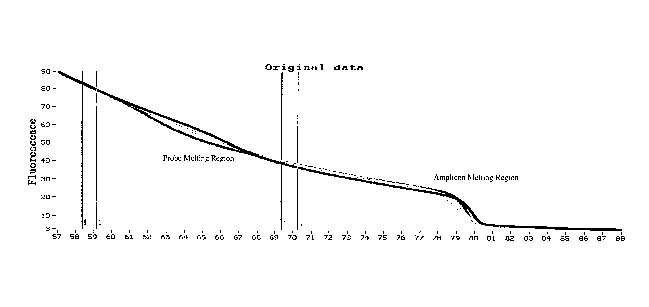
In the present example, melting curves were generated from data taken from the
HR-1 melting instrument. A slope value 11 is generated from cursor pair 1 and
TR is
generated from cursor pair 2. In this example, the cursor pairs are
respectively located at
approximately 76 C and 85 C, but it is understood that the placement of the
cursor pairs
will vary depending upon the melting transitions of the nucleic acid(s)
present in the
sample. Cursor pair 1 and 2 bracket the probe melting transition of the sample
nucleic
acid, but cursor pair 2 is also located before the amplicon melting transition
region. After
the exponential fit is performed with the slopes generated from the cursor
pairs the
11
CA 02623268 2014-05-16
WO 2007/035806
PCT/US2006/036605
exponential background subtraction is performed. Fig. 1B represents the probe
melting
region after the background has been subtracted, using the EBS equations and
normalization discussed above. Exponential background subtraction for the
amplicon
melting region using the same or different cursor pairs can be performed (not
shown) with
the same exponential found for the probe melting region.
While illustratively the cursor pairs are placed on either side of at least
one melt
transition, it is understood that the position of the cursor pairs may vary in
the practice of
this invention. For example, in an alternative embodiment, slope values (II
and TR) are
both obtained from points before the probe melting region. In yet another
alternative
embodiment, slope values (Ti, and TR) are both obtained from points after the
probe
melting region but before the amplicon melting region. In yet another
alternative
embodiment, slope values (TL and TR) are both obtained from points after the
amplicon
melting region. In yet another alternative embodiment, slope values (TL and
TR) are
obtained from points before the probe melting region and after the amplicon
melting
region. In another alternative embodiment, the slope values (TT., and TR) are
obtained at
any two points on the raw melting curve where neither the probe nor the
amplicon are
melting. Although the illustrative cursor pairs are spaced apart from each
other, it is
possible to do exponential background subtraction with two slope values (Ti,
and TR) that
are close together. Such may be useful for very crowded melting curves with
limited
non-melting regions.
Exponential background removal identifies the sample melting curve signal
independent of user choice of where the user decides to fit the background, as
long as
and TR are outside the melting range. In general, background noise includes
background
fluorescence signals and alternate non-nucleic acid melting signals, both of
which
interfere with the analysis of the sample data. By example, when using
unlabeled probes
and multiplex amplicons at lower temperatures (less than 80 C), background
noise at low
temperatures has occasionally previously prevented the identification of
sample signal
curves.
Prior art methods for background subtraction may fail as the background
signals
and the sample signals approach an equal amplitude. Even where the sample
signal is
significantly higher than the background, the exponential background
subtraction has
been found to be more consistent and accurate.
As an example, the exponential background subtraction provides enhanced
accuracy and specificity when distinguishing the melting curves of samples. A
recent
12
CA 02623268 2014-05-16
WO 2007/035806
PCT/US2006/036605
study found 100% accuracy in distinguishing between a normal wild type sample
and a
homozygous mutant sample where the amplicons were approximately 40 base pairs
in
length. Prior to the use of exponential background subtraction, it was
theoretically
understood that a fraction of small amplicons of a normal wild type sample and
their
homologous mutant sample would have identical curves, particularly if the GC
content
remained the same between the two amplicons. The use of higher resolution
melting and
exponential background removal is therefore attributed to extremely high
accuracy of
genotyping and mutation scanning of double stranded nucleic acid samples.
High resolution melting analysis is useful to obtain viable results for
identifying
the efficacy of background removal and genotyping. Melting analysis may be
performed
on a variety of melting instruments, including the high-resolution melting
instruments
HRlTM (a capillary-based melter) and LightScanner (a plate-based melter)
(both Idaho
Technology, Salt Lake City, Utah). However, it is understood that melting
curve analysis
may be performed in the absence of amplification, particularly on highly
uniform nucleic
acid samples. In one illustrative melting protocol using the HR-1, the samples
were first
amplified in the LightCycler (Roche Diagnostics Corp, Indianapolis, TN) (or
in the
RapidCycler (Idaho Technology, Salt Lake City, Utah)), then heated
momentarily in the
LightCycler to 94 C and rapidly cooled (program setting of ¨20 Cis) to 40
C. The
LightCycler capillaries can be transferred one at a time to the high-
resolution instrument
and heated, illustratively at 0.3 C/s. The HR-1 is a single sample instrument
that
surrounds one LightCycler capillary with an aluminum cylinder. The system is
heated by
Joule heating through a coil wound around the outside of the cylinder.
Approximately 50
data points may acquired for every C. The LightScanner is a plate-based
system that
provides high resolution melting on 96- or 384-well microtiter plates. PCR may
be
performed in any compatible plate-based thermal cycler.
In some cases it is advantageous not to denature the product after PCR before
melting curve acquisition. For example, when the goal is to type the number of
repeat
sequences (e.g. STRs, VNTRs), amplification may be stopped at the extension
step during
the exponential phase of the reaction before plateau, and then melting
analysis is
performed. This way, homoduplex extension products can be analyzed. In repeat
typing,
homoduplex products can be more informative than heteroduplex products,
especially
since many different heteroduplex products may form from different alignment
of the
repeats. In some cases, it may be helpful to obtain both a homoduplex melting
curve
(without prior denaturation) and a heteroduplex melting curve (with
denaturation and the
13
CA 02623268 2014-05-16
WO 2007/035806
PCT/U52006/036605
formation of all possible duplex combinations). The difference between these
two melting
curves gives a measure of the extent of heteroduplexes that can be formed,
using the same
sample as the "homoduplex control".
Previous background subtraction techniques often included numerical
differentiation as an element. Numerical differentiation of raw data involves
artificial
fitting and smoothing, which can affect the efficacy of the data. Common
numerical
differentiation techniques include negative derivative curves and integrated
derivative
curves. The present embodiment does not require the raw data to be numerically
differentiated. This is an advantage over previous background subtraction
techniques.
The conversion of raw data to derivative curves often involves the
amplification of
background noise and artificial smoothing of significant features of the
melting data. The
present embodiment is capable of distinguishing subtle but molecularly
significant
differences in melting data, which is an advantage over previous techniques
that involved
derivative curve analysis.
While the above exponential background subtraction method is used hi reference
to nucfeie acid melting curves, it is understood that this method may be
applied to a
variety of data sets, including other biological data sets, having exponential
background
noise, and is particularly suited where background subtraction without using
the value of
the signal for calculation is important.
Curve Overlay Function
An alternative embodiment includes a curve overlay function for use in melting
curve analysis. Previous methods of curve overlay, or temperature shifting,
include the
steps of selecting a fluorescence interval, usually at low fluorescence (e.g.
5-15%) of the
normalized melting curve, fitting a second degree polynomial to all points
within the
interval for each curve, and then shifting each curve to best overlay the
plots in this
interval. Curve overlay corrects any minor inter-run temperature variation and
increases
the ability to distinguish heterozygotes from homozygotes. However, the
previous
method failed when fewer than three (3) points were in the interval. Absent
the three data
points, the previous method could not provide a means for automatic and
accurate curve
overlay, as the known mathematical overlay methods could not be applied with
high
accuracy. Known methods of mathematical overlay include the least distance
method, the
lowest average of absolute distance values method, and the least squares
method.
14
CA 02623268 2014-05-16
WO 2007/035806
PCT/US2006/036605
The present embodiment analyzes the normalized melting curves by overlaying
them between a lower dependent variable value (n) and a higher dependent
variable
value(m). This is performed by extracting all numerical (x,y) values in the
interval
provided that all (y) values continue to decrease, or all (y) values continue
to increase.
Instead of fitting (y) as a function of (x) and finding the best overlay of
such fits, the
ordered pairs are reversed, thereby making (x) a function of (y). An example
of the
optimal least squares fit of one horizontally shifted function is shown in
Equation Set 6,
xi(y)-I-c to another, x2(y) is obtained by finding the constant c which makes
the mean
difference of (x1(y)+c)-x2(y) equal to zero.
Equation Set (6) min_c lb ((f(z) + c) - g(z))2 dz = f g(z) - f(z) dz
The value x2(y) is obtained by finding the constant (c), thereby making the
mean
difference of (xi (y)+c)-x2(y) equal to zero. The value (z) represents a
variable of
integration. The value (z) in Equation Set 6 represents either value (x) or
value (y), which
is in part due to the independent variable being the original (y) and equal to
the
fluorescence value. The functions f(z) and g(z) represent sections between two
normalized fluorescence values (low and high) that are chosen for overlay of
two
normalized melting curves, where temperature is plotted as a function of
fluorescence.
The value (dz) is a normalized measure on a normalized fluorescence interval.
In order to scan for heterozygotes within a PCR product, the shape is more
important than the absolute temperature or Tm. Heterozygotes produce
heteroduplexes
that melt at lower temperatures and distort the shape of an overall melting
curve. Shape
differences are more efficient to use when identifying different genotypes
than
absolute Tms because temperature variation can be caused by minor sample
differences
and instrument variability. Possible sample differences include, but are not
limited to,
variances in ionic strength and the occurrence of evaporation during
processing
procedures. Instrument variability is also possible with respect to relative
and exact well
positions on the sample plates.
As discussed above, previous methods for comparing curve shape included the
overlay of curves by shifting them along the temperature axis, implemented by
fitting a
second degree polynomial to a small fluorescence interval of each curve. An
arbitrary
standard curve was then chosen, and the remaining curves were shifted to
overlay the
CA 02623268 2014-05-16
WO 2007/035806
PCT/US2006/036605
standard curve over this region. However, when the data density and
fluorescence
interval are small, the previous method is prone to failure, as shown in
Figure 2B. The
top panel (Figure 2B) includes melting curves for a PCR fragment of Factor V
Leiden
from 96 genomic DNA samples, having been normalized by the exponential
background
subtraction method. Three distinct genotype clusters are visible within Figure
2B. Also
shown in the top panel are fluorescence interval markers F1, F2 over which the
overlay is
attempted. The first Interval marker Fl is approximately at 10% fluorescence
and the
second interval marker F2 is approximately at 15% fluorescence. A magnified
plot of
actual data points from 80 to 82 C contained' within the selected
fluorescence interval is
shown in the middle panel. Various curves show that fewer than three points
are included
within this interval, which makes a quadratic fit impossible and ultimately
leads to failure
of the previous curve overlay method, as shown in the bottom panel.
The present embodiment utilizes the algorithm of Equation Set (6) (Figure 2A)
with the same data set. The present embodiment accurately and successfully
analyzes the
data, and in fact needs only one data point from each of the respective curves
to do so,
thereby providing a novel and improved method of analysis. In the top panel of
Figure
2A, raw data are shown along with vertical cursors (See Figure lA and
description above)
that define the regions for slope estimation of the exponential background.
Provided in
the bottom panel (Figure 2A) is the normalized background subtracted curves
that have
been successfully temperature shifted so that all curves are overlaid within
the 10-15%
fluorescence region.
The present embodiment is an advantageous method over previous methods for
various reasons, including that only one (1) data point is required for
validity and
accuracy of the curve overlay function. The present embodiment is furthermore
advantageous as it has rigorous optitnality for least-squares fitting. It is
contemplated that
the present embodiment of the curve overlay function can be represented in
numerous
mathematical representations other than described herein, and Equation Set (6)
is
considered one of such examples.
Difference Plot Function
Another alternative embodiment includes an improved difference plot function
for
analyzing differences between nucleic acid sample melting curves. Previous
methods
subtracted all curves from an arbitrary reference curve or an average of
reference curves.
The result of the subtraction was purely the vertical distance between curves
at each
16
CA 02623268 2014-05-16
WO 2007/035806
PCT/U52006/036605
temperature point. These difference plots visually magnified the difference
between
curves so they could be more easily viewed. However, the vertical distance
between
curves does not accurately portray the shortest distance between curves. This
is
especially the case when the fluorescence value of the melting curves drop at
a significant
rate or negative slope. This shortcoming presented an artifact known as a
"variation
bubble", which is often clearly visible where the decrease in fluorescence is
maximal.
A novel improvement in the present difference plot function is to weight the
differences between curves according to the slope of the curves. This provides
a better
distance metric between curves so that common and distinct genotypes can be
correctly
and automatically identified. Weighting balances the effect of slope on the
difference
measure between curves. When weighting is not performed, standard vertical
differencing overemphasizes the difference between curves when the slope is
steep,
enlarging the spread of melting curves within the same genotype. The present
method
provides a practical computable approximation to the orthogonal difference
between
melting curves, which thus better represents variations in the distance
between curves.
The present embodiment provides a novel method for analysis of melting curve
data. The analyzed melting curves may be normalized by various known methods,
but
the novel exponential background subtraction described herein is preferable.
Instead of
the vertical distance at each temperature, an orthogonal distance between
curves is
advantageous. The present embodiment obtains the least distance between
curves, which
is the least distance between curves at each point. When the fluorescence is
dropping
rapidly, instead of using the vertical distance between lines, a metric
orthogonal to the
slope of curves is more useful and accurate to assess differences between
curves. When
measuring orthogonal distances between two curves, the results are often
different
depending on which curve is used as the reference. The present embodiment
provides a
method to estimate the orthogonal distance between two curves.
To compensate for exaggerated emphasis upon the melting regions of two melting
curves being compared and consequent de-emphasis upon regions surrounding
their
primary melts when using simple vertical differencing, f1(T)-f2(T), an
approximation of
the orthogonal distance between curves can be found by using Equation Set (7).
Equation Set (7) f1(T)42(T) = max{ q(1+ f1'(T)2), f2'(T)2) 1
17
CA 02623268 2014-05-16
WO 2007/035806
PCT/US2006/036605
Equation Set 7 is an example of a method for taking the melting regions of
both melting
curves into account symmetrically. The equation further generalizes the
orthogonal
distance from the origin to the line (y=b-mx), where the vertical distance
from the origin
is (b), but where the orthogonal distance from the origin is (b/4(1+m2)). The
result of the
present embodiment is a measurement which reflects sequence dependent
variations in
the melting curves more sensitively and evenly throughout the range of
measurement,
while being suitable for automated and dynamic use within a computer system.
An
example of difference functions are provided in Figures 10A-C where both the
previous
vertical (Fig 10B) and the novel orthogonal (Fig 10C) differences are plotted
using
normalized melting curves that have had their background exponentially
subtracted.
Vertical difference plots show a "variation bubble" around the region of
steepest slope,
even though all samples are wild types. The variation bubble is eliminated
when
orthogonal differencing is employed.
Weighting balances the effect of slope on the difference measure between
curves.
When weighting is not performed, standard vertical differencing overemphasizes
the
difference between curves when the slope is steep, enlarging the spread of
melting curves
within the same genotype. The present invention provides a practically
computable
approximation to the orthogonal difference between melting curves which thus
better
represents variations in the distance between curves.
Genotype Clustering Function
= Yet another alternative embodiment includes a genotype clustering
function with
automatic determination of the number of clusters. Specifically, unbiased
hierarchal
clustering was used in contrast to previous methods that clustered genotypes
based on
learned data sets and arbitrary cutoffs. Since the genotypes are usually
unknown before
analysis, prior data sets do not satisfactorily predict the appropriate
grouping of future
data sets. Such learned data set methods are often unable to provide an
accurate and
automated means for clustering, particularly for unknown genotypes. In
contrast,
unbiased hierarchal clustering requires no prior learning or established
cutoffs and
robustly adjusts to the quality and resolution of the available melting data.
The novel unbiased hierarchal clustering method is based on a distance metric
between melting curves. The distance metric is selected from: 1) the maximal
distance
between curves, 2) the average of the absolute value of the distance between
curves at all
18
CA 02623268 2014-05-16
WO 2007/035806
PCT/US2006/036605
temperature points, and 3) the average of the root mean square of the distance
between
curves at all temperature points. Preferably, the distance metric is derived
after ED S,
normalization, optional curve overlay, and is the novel orthogonal distance
metric
described in the previous section. Sequential, unbiased, hierarchal clustering
is then
performed as is standard in the art.
Automatic determination of the most likely number of clusters is a novel
aspect of
the present disclosure. Specifically, the likelihood for each level of
clustering (number of
clusters) is determined by considering the ratio of distances between cluster
levels.
During hierarchal clustering, two subc lusters C1 and C2 are joined into a
larger cluster by
considering the weighted average of all points in each subcluster (the
quantity minimized
to determine which subclusters to merge at each stage). Instead of using the
weighted
averages to calculate the distance ratios, the minimum distance between curves
in distinct
clusters is used as a more accurate measure of the separation between clusters
of data and
their subclusters. This compensates for the naturally growing distances
between the
weighted average curves representing hierarchical sub-clusters formed during
the
agglomerative clustering process by keeping the distance measure associated
with sub-
clusters limited to the distance between the nearest-neighboring curves in
each sub-
cluster, rather than the distance between the most recently joined weighted
averages. The
present embodiment accurately determines which cluster level is the most
likely by
assessing the ratio between two adjacent cluster level distances. This ratio
provides a
more accurate and stable likelihood assessment of the clustering level (number
of
clusters), accurately separating fme scale from large scale phenomena. The
largest ratio
defines the most likely number of clusters, the next largest ratio defines the
second most
likely number of clusters, etc. In genotyping applications where there are
sufficient
curves for multiple representations of the main genotypes, this method
provides a robust
criterion for identifying the genotyping level in hierarchical clustering
intrinsically.
A mathematical representation of the clustering process is shown at Equation
Set
(8), where II frf2 It represents the distance metric used in the ratio that
orders the cluster
levels (number of clusters) by likelihood. The difference measurement (II fi-
f2 II) indicates
the measure of distance between two normalized melting curves and is selected
from a
group comprising: the mean absolute separation, the mean squared separation,
and the
maximum separation at a chosen temperature. It is contemplated that various
other
known methods for measuring distances between two melting curves may be used
with
respect to Equation Set 8.
19
CA 02623268 2014-05-16
WO 2007/035806
PCT/US2006/036605
Equation Set (8) min_Ifi a CI, f2 a C2) It fi-f2
At a point when subclusters C1 and C2 have been joined the value (fp
represents a
melting curve associated with subcluster C1, and the value (f2) is a melting
curve
associated with subcluster C2, The value (min_ffi a C1, f2 a C2)) represents
the smallest
value of the distance among all pairs of melting curves, where a first value
is taken from a
first subcluster, the first subcluster having been joined, and a second value
being taken
from a second subcluster.
The present embodiment is a new method for measuring the distance between
cluster levels to determine the likelihood of a particular level (number of
clusters). The
new distance measure is the minimal distance between any two members, as long
as each
is from a different cluster. In order to determine the likelihood of any
cluster level (i.e. 3
vs. 4 vs. 5 clusters), the ratio of the distance to the next cluster is
divided by the distance
to the previous cluster. The above method provides the correct classification
of
genotypes when parameters such as cursor locations are varied. When the
weighted
averages are used rather than the distance between the nearest-neighboring
curves in each
sub-cluster to determine the ratio, the choice of the most likely number of
clusters is
much less stable. Furthermore, the present embodiment can be automatically
executed
with high accuracy.
Figures 3A-C demonstrate the ability of the novel clustering function to
assign the
correct number of genotype clusters to a multi-sample melting curve. Six
separate
genotypes of human genomic DNA of the hepatic lipase gene were amplified
(BioRad
iCycler) using 10 pl reaction volumes in a 96 well plate. The IX LightScanner
Master
Mix (Idaho Technology, Salt Lake City, UT) was used. The samples were heated
from
75 C to 94 C at 0.1 C/second in the LightScanner melting instrument.
Figure 3A shows a screen shot of the raw melting curve (top panel) for the
hepatic
lipase gene amplification and melting transition. The bottom panel shows the
result of
the previous clustering function along with a drop-down menu indicating that
the
previous clustering function incorrectly indicates the presence of only three
distinct
genotype clusters in the samples, which are represented as three separate
colored line
clusters.
Figure 3B shows a screen shot of the raw melting curve (top panel) for the
hepatic
lipase gene amplification and melting transition. The bottom panel shows the
result of
CA 02623268 2014-05-16
WO 2007/035806
PCT/US2006/036605
the present embodiment clustering function along with a drop-down menu
indicating that
the novel clustering function correctly indicates the presence of six distinct
genotype
clusters in the samples, which are represented as six separate colored line
clusters. Figure
3C demonstrates that the user can identify the samples by genotype by their
positioning
on the 96 well reaction plate shown along side the fluorescence vs.
temperature plot of
Figure 3B.
It is understood that melting plots may be analyzed using one or more of the
algorithms of exponential background subtraction, curve overlay function,
difference plot
function, and the clustering function, and that each of these methods may be
used alone or
in any combination and may be used in combination with other methods of
analyzing
melting plots.
Example 1- Hemochromatosis (HFE) Mutation and Polymorphism Genotyping
Hemochromotosis gene mutations and polymorphisms are known to interfere with
normal iron metabolism in Humans. In this illustrative example, detection and
identification of polymorphism and mutation genotypes through melting curve
analysis is
more accurate with exponential background subtraction then baseline background
subtraction. Analysis was performed with small amplicons (78 bp and 40 bp) in
order to
increase the Tm difference between different homozygote samples. Unlabeled
probes are
utilized for genotyping SNPs that otherwise could not be easily genotyped by
amplicon
melting.
Human genomic DNA representing distinct hernochromatosis genotypes can be
amplified by various PCR instruments. An exemplary method includes 10 id
reaction
volumes with a Roche LightCycler 2Ø Following PCR amplification, the FIFE
samples
are heated in an HR-1 (Idaho Technology) melting instrument for approximately
115
seconds. The resulting florescence versus temperature plot is depicted as
Figure 4A. The
derivative melting curves are shown in Figure 4B. In this illustrative
example,
exponential background subtraction is performed on the derivative melting
curves, and
the normalized derivative melting curves are displayed as Figure 4C. Compared
to the
exponential background normalized melting curve (Figure 4C) the baseline
technique for
normalizing melting curves is clearly deficient. As shown in Figures 4D-4E,
the baseline
technique applied to a combined probe plus amplicon melting curve (Figure 4D)
results in
a absence of useable data (Figure 4E), even though the baseline and
exponential
background subtraction techniques used the same melting alirvs data set Mauve
4R 4n1
21
CA 02623268 2014-05-16
WO 2007/035806
PCT/US2006/036605
Genotype identification of Figure 4C for the C282Y homozygous and
heterozygous,
H63D heterozygous and homozygous and wild type samples is shown in Figure 4F.
Example 2- Factor V Leiden (384 well plate) Derivative Clustering Combined
Probe
and Amplicon
Factor V Leiden is the most common hereditary blood coagulation disorder in
the
United States. It is present in approximately 5% of the Caucasian population
and
approximately 1.2% of the African American population. Factor V Leiden as a
gene
target is important for the detection of SNPs that are linked to coagulation
disorder
disposition.
Human Factor V Leiden genomic DNA samples representing different Factor V
Leiden genotypes were amplified in an AI31 9700 with 10 al reaction volume and
12 al
oil overlay. The assay included a Factor V Leiden amplicon of approximately
100bp
having a sequence of
5'-CTGAAAGGTTACTTCAAGGACAAAATACCTGTATTCCTCGCCTGTCCAGGG
ATCTGCTCTTACAGATTAGAAGTAGTCCTATTAGCCCAGAGGCGATGTC-3'
(SEQ. ID NO:1), which was amplified by forward primer
5'-CTGAAAGGITACTTCAAGGAC-'3 (SEQ. ID NO:2) and reverse primer
5'-GACATCGCCTCTGGG-3 (SEQ. ID NO:3). The assay also included an unlabeled
probe 3'-TGGACATAAGGAGCGGACAGGT-5' (SEQ. ID NO:4) that is configured to
hybridize to the forward strand of the amplicon, as indicated by the
underline. The
resultant PCR samples were heated from 58 C to 88 C at 0.1 C/s in a
LightScanner
melting instrument using a 384 well reaction plate. The total melting
procedure required
approximately 5 minutes for completion.
The LightScanner melting instrument measured and recorded the fluorescence as
a
function of temperature. The raw melting curve result of the procedure is
shown in
Figure 5A. The negative derivative of the melting curve of Figure 5A was
calculated and
is shown in Figure 5B. A representative diagram of the 384 well plate is shown
in Figure
5B along with the clustering of the genotypes. Background removal has not been
performed, and the clustering performed did not correctly genotype several of
the samples
tested (compare 384 well plate to that in Fig. 5D).
A linear correction function was performed on the derivative plot of Figure 5B
and is shown in Figure 5C. A representative diagram of the 384 well plate is
shown in
Figure 5C along with the clustering of the genotypes. It is nle.nr frnm the
184 well bite
22
CA 02623268 2014-05-16
WO 2007/035806
PCT/US2006/036605
that the clustering performed after the linear correction did not correctly
genotype several
of the samples tested. The linear correction performed on the derivative
melting curve
data includes deleting the end to end slope by subtracting a linear function.
Figure 5D represents the derivative melting curve data of Figure 5B after
exponential background subtraction has been performed on the data set. Only
after the
exponential background subtraction is the correct clustering of genotypes
obtained. The
accuracy of the genotype clustering is visible within the 384 well plate
(Figure 5D),
where the letters "S-N-P" are spelled out indicating genotypes by plate
position, and the
proper identification is visible for both the amplicon and probe region of the
Factor V
Leiden gene target.
Example 3 - Factor V Leiden (96 well plate) Derivative Clustering of Probe
Only
Human Factor V Leiden genomic DNA samples representing different Factor V
Leiden genotypes were amplified in an ABI 9700 with 10 I reaction volume and
12 I
oil overlay. The assay included a Factor V Leiden amplicon and an unlabeled
probe, as
described above in Example 2, in the presence of lx LCGreen Plus .. The
resultant
PCR samples were heated from 58 C to 88 C at 0.1 C/s in a LightScanner
melting
instrument using a 96 well reaction plate. The total melting procedure
required
approximately 5 minutes for completion.
The LightScanner melting instrument measured and recorded the fluorescence as
a
function of temperature. The raw melting curve result of the procedure is
shown in
Figure 6A. A negative derivative plot as a function of temperature was
performed from
the melting curve data of Figure 6A and is shown in Figure 6B. Two sets of
vertical
cursor lines are present at approximately 59 C and 60 C, and 71 C and 72 C
respectively. The exponential background subtraction technique slopes are
generated
using these cursor lines. Following the automatic calculation and fitting, the
exponential
background is subtracted from the raw negative derivative plot. The result of
the
exponential background subtraction specific to the probe region is shown in
Figure 6C.
Though the vertical slope cursors are located on either side of the probe
region, the
exponential used for the probe region may also be used for the amplicon
region. In fact,
the absolute location of where the slopes are obtained is not determinative of
the
exponential, so long as the slopes are found at positions where no melting of
the sample
occurs. The probe region data (Figure 6C) is automatically clustered by the
genotype
23
CA 02623268 2014-05-16
WO 2007/035806
PCT/US2006/036605
clustering function. The nucleic acid samples from the 96 well plate are
clustered into
three genotypes and shown in Figure 6D.
Regarding Examples 4 and 5 the following procedures and instruments were used.
Human genomic DNA of known Factor V Leiden genotype samples and heterozygous
genomic DNA samples with selected cystic fibrosis mutations were used for
analysis.
Predicted probe Tms were lower than observed Tms, perhaps because of dye
stabilization. The melting temperature of different probe/allele duplexes was
adjusted by
probe length, mismatch position, and probe dU vs dT content. Extension of
unlabeled
probes during PCR was prevented by incorporating a 3'-phosphate during
synthesis.
Alternatively, other 3'-blocking mechanisms may be employed, illustratively by
providing an additional two base mismatch to the 3'-end of the probe. When a
5'-
exonuclease negative polymerase is used, probes should be designed to melt
lower than
the PCR extension temperature.
Primer asymmetry, illustratively at ratios of 1:5 to 1:10, may be used to
produce
sufficient double stranded product for amplicon melting and enough single
stranded
product for probe annealing. PCR for Factor V performed in 384-well format
used 5 I
volumes, and included 20 ng of genomic DNA in 50 mM Tris, pH 8.3 with 3 mM
MgC12,
0,2 mM each dN'I?, 500 Itg/m1 BSA, 1X LCGreene PLUS (Idaho Technology), 0.2 U
KlenTaqlTm (AB Peptides), and 70 ng TaqStartTm antibody (Clontech). PCR was
performed in a 9700 thermal cycler (ABI) with an initial denaturation at 94 C
for 10 s,
followed by 50 cycles of 94 C for 5 s, 57 C for 2 s, and 72 C for 2 s. After
PCR, the
samples were heated to 94 C for 1 s and then cooled to 10 C before melting.
PCR for amplification of CFTR exons 10 and 11 was performed in 10 1 volumes
and included 50 ng of genomic DNA in 50 mM Tris, pH 8.3 with 2 mM MgC12, 0.2
mM
each dNTP, 500 ng/ml BSA, 1X LCGreen I (Idaho Technology) and 0.4 U Taq
polymerase (Roche), The PCR was performed in capillaries on a LightCycler
(Roche)
with an initial denaturation of 95 C for 10 seconds followed by 45 cycles of
95 C for 1 s,
54 C for 0 s, and 72 C for 10 s. After amplification, the samples were heated
to 95 C for
0 s and rapidly cooled to 40 C before melting.
When the nucleic acid samples were amplified on 384-well plates, melting
acquisition was performed on a prototype version of the LightScanner (Idaho
Technology). The standard 470 nm light-emitting diodes were replaced with 450
nm
light-emitting diodes (Bright-LED Optoelectronics). In addition, the optical
filters were
changed to 425-475 run excitation and 485 nm long-pass emiRcion filters (Omega
24
=
CA 02623268 2014-05-16
WO 2007/035806
PCT/US2006/036605
Optical). The plate was heated from 55 to 88 C at 0.1 C/s with a 300 ms frame
interval,
15 ms exposure and 100% LED power, resulting in about 25 points/ C.
Melting of CFTR exons was performed on the HR-1 high-resolution melting
instrument (Idaho Technology) with 24-bit acquisition of temperature and
fluorescence.
After PCR on the LightCycler, each capillary was transferred to the int-1 and
melted
from 50 C to 90 C with a slope of 0.3 C/s, resulting in 65 points/ C.
Melting curves can be analyzed on any suitable software known in the art. An
exemplary software package for implementing the melting analysis methods of
the
various embodiments of the present invention is LabVIEW (National
Instruments).
Normalization and background subtraction was first performed by fitting an
exponential
to the background surrounding the melting transitions of interest. Derivative
plots of
probe melting transitions were obtained by Salvitsky-Golay polynomial
estimation.
Melting curves of PCR products were compared on difference plots of
temperature-
overlaid, normalized melting curves. The normalized melting curves were
temperature-
overlaid (to eliminate slight temperature errors between wells or runs) by
selecting a
fluorescence range (illustratively low fluorescence/high temperature,
typically 5-10%
fluorescence) and shifting each curve along the X-axis to best overlay a
standard sample
within this range. Difference plots of temperature-overlaid, normalized curves
were
obtained by taking the fluorescence difference of each curve from the average
wild type
curve at all temperature points. These analytical methods have been previously
applied to
mutation scanning and HLA matching.
Agglomerative, unbiased hierarchical clustering of melting curve data was
performed by previous methods, custom programmed in LabVIEW. The distance
between
curves was taken as the average absolute value of the fluorescence difference
between
curves over all temperature acquisitions. The number of groups was
automatically
identified by selecting the largest ratio of distances between consecutive
cluster levels.
The clustering methods represent less accurate means for clustering genotypes
than the
novel clustering methods described herein (Figures 9A-D).
Example 4- CFTR Scanning and Genotyping
Various exons of the cystic fibrosis transconductance regulator (CFTR) gene
have
been chosen to demonstrate simultaneous scanning and genotyping of multiple
variants.
Three SNPs in two regions of exon 11 of the CFTR gene were analyzed with two
unlabeled probes, sequences in part TCTTGGAGAA (SEO. ID NO:51 and
=
CA 02623268 2014-05-16
WO 2007/035806
PCT/US2006/036605
AGGTCAACGA (SEQ. ID NO:6). Two of the mutations were only six bases apart,
allowing one of the probes to cover both mutations (Fig. 7A). Five replicates
of each
genotype were amplified and analyzed. The normalized melting curves after EBS
(Fig.
7B) show regions of probe melting (56-74 C) and PCR product melting (80-83 C).
On
casual observation, it is not clear from the normalized melting curve what
information can
be extracted. However, when the probe region is displayed as a derivative plot
(Fig. 7C),
the melting transitions of all common alleles under both probes are apparent.
Both
unlabeled probes were matched to the wild type sequence, but one of the probes
was
made shorter and contained dU instead of dT to decrease its melting
temperature. The
more stable probe covered a single SNP, resulting in two alleles being
separated by Tm,
both being more stable than all alleles of the less stable probe. The less
stable probe
covered two SNPs, resulting in three peaks for common genotypes. The specific
mismatch and its position within the probe affect duplex stability, allowing
probe design
that distinguishes multiple alleles. A difference plot of the PCR product
melting transition
is shown in Fig 7D. The heterozygous, wild type, and homozygous mutant samples
are
clearly different. However, it is difficult to distinguish between different
heterozygotes by
PCR product melting alone. Unbiased hierarchal clustering grouped all
heterozygotes
together (data not shown). The three heterozygotes are all in the same SNP
class (12),
resulting in the same heteroduplex mismatches (C:A and T:G) and homoduplex
matches
(C:G and A:T). Although predicted stabilities of all three heterozygotes using
nearest
neighbor thermodynamics (13, 14) are not identical, definitive genotyping
required the
use of probes. The strength of product melting is to easily identify the
presence of
heterozygotes, while unlabeled probes further discriminate between
heterozygotes and
more easily identify homozygous variants. As illustrated with this example,
combining
both genotyping and scanning results in the display of both amplicon (PCR
product) and
unlabeled probe melting transitions.
Example 5- CFTR Genotyping
Three SNPs and two deletions within exon 10 of the CFTR gene were also
analyzed with two unlabeled probes. The probe with the higher Tm, sequence in
part
'ITCTCAGTTT (SEQ, ID NO:7), covered a single SNP, while the probe with the
lower
Tm, sequence in part TATCATCMG (SEQ. ID NO:8), covered two SNPs and two
deletions (Fig. 8A). The normalized melting curves after EBS (Fig. 8B) show
regions of
low temperature probe (56-67 C), high temperature probe (67-75 C and Pell nil-
Aunt
26
CA 02623268 2014-05-16
WO 2007/035806
PCT/US2006/036605
(80-83 C) melting. When the probe regions are displayed as a derivative plot
(Fig. 8C),
all five heterozygous genotypes follow unique paths that distinguish them from
wild type
and each other. Four of the heterozygotes show resolved peaks, while one is
identified by
a broad peak resulting from a relatively stable mismatch (an A:G mismatch near
one end
of the probe in an AT-rich region). Allele discrimination does not require a
unique Tm for
each allele, only that the curves are different in some region of the melting
transition. A
difference plot of the PCR product melting transition is shown in Fig. 8D. The
double
heterozygote shows the greatest deviation from wild type because two
mismatches are
present within the PCR product. The four single heterozygotes are all easily
distinguishable from wild type. In contrast to exon 11, all heterozygotes
could be
genotyped by either PCR product or probe melting. Consideration of both
regions often
provides independent confirmation of genotype.
Example 6¨ Whole Amplicon Genotyping of fl globin
13 globin presents a gene target with known SNPs that are important for the
analysis of hemoglobinpathies, most notable are the HbC and HbS mutations.
Human
genomic DNA samples of different f3 globin genotypes were amplified in a Roche
LightCycler using 10 ul reaction volumes. lx LCGreen from Idaho Technology
was
used in PCR to amplify a 45 bp amplicon:
5'-CCATGGTGCACCTGACTCCTGAGGAGAAGTCTGCCGTTACTGCCC-3' (SEQ.
ID NO:9). The PCR product samples were heated from about 72 C to about 88 C
at 0.3
C/s in the HR-1 melting instrument. The time required for melting is about 60
seconds.
The HR-1 melting instrument measured and recorded the fluorescence of the
samples as a function of temperature. The raw melting curve for the whole (3
globin
amplicon is shown in Figure 9A. The raw melting curve for the whole (3 globin
amplicon
is then normalized by the exponential background subtraction method described
herein,
which results in a melting curve that is the relative fluorescence of the
samples as a
function of temperature in Figure 9B. The genotype clustering function is
performed on
the normalized melting curves from Figure 9B. Figure 9C shows the clustered
genotypes
of the normalized melting curves from Figure 9B.
Example 7- Factor V Leiden Wild type Difference Plot Analysis
Human Factor V Leiden genomic DNA samples representing 60 different Factor
V Leiden wild types were amplified in an ABI 9700 with 1(liil
rpariinn vAliTniP and 19 ill
. 27
CA 02623268 2014-05-16
WO 2007/035806
PCT/US2006/036605
oil overlay. The assay included a Factor V Leiden amplicon and an unlabeled
probe, as
described above in Example 2, in the presence of lx LCGreen Plus . The
resultant PCR
samples were heated from 58 C to 88 C at 0.1 C/s in a LightScanner melting
instrument using a 96 well reaction plate. The total melting procedure
required
approximately 5 minutes for completion.
The LightScanner melting instrument measured and recorded the fluorescence as
a
fraction temperature. Execution of various difference plot functions provides
a
fluorescence difference value as a function of temperature. The melting curves
for 60
Factor V Leiden wild types is shown in Figure 10A after performing the
exponential
background subtraction function. The normalized melting curve appears as one
thick
black curve, which is characteristic of the substantial similarities of the 60
wild type
samples.
Figure 10B represents the graphical result of the previous vertical difference
plot
technique, which is plot as the vertical fluorescence difference between the
60 wild type
samples as a function of temperature. This previous vertical difference plot
technique
presents an interesting artifact when the melting curves have a sufficiently
steep slope.
The artifact manifests as a "bubble" approximately between 80,25 C and 81.5 C
(Figure
10B), which is the steepest portion of the raw data melting curve slope.
The artifact of the vertical difference plot represents the artificial
amplification of
small differences between the 60 wild type samples. Figure IOC represents the
results of
the orthogonal difference plot function. As the difference plot function is
designed to
accurately measure the difference between the samples, the close proximity of
the
fluorescence difference plotted as a function of temperature is indicative of
the significant
similarity among the 60 wild type samples (Figure 10C). The difference
fluorescence vs.
temperature plot of the 60 wild types appears as a single thick line
approximately
centered upon zero fluorescence difference.
Referring to Figure 11, it is contemplated that the embodiments of the present
invention can be implemented on a computer system 100. The computer system 100
comprises a nucleic acid melting instrument 102, a memory storage device 104
and a
central processing unit (CPU) 106. The memory storage device 104 and the CPU
106
may be integral (not shown) within the housing of the melting instrument 102
or may be
provided externally (Figure 11), illustratively as a laptop or desktop
computer. The
system 100 alternatively can include a graphical user interface (GUI) 108 for
viewing,
manipulating, and analyzing the high resolution melting curve The licer 110
may
28
CA 02623268 2014-05-16
WO 2007/035806
PCT/US2006/036605
interface directly with the melting instrument, GUI, or any other aspect of
the system 100.
The memory storage device 104 includes computer readable code that is the
exact
embodiment or equivalent to the embodiments of the invention described herein.
The
computer readable code includes instructions for performing the methods
described
herein and is executed by any suitable central processing device known in the
art.
Software is available with most melting instruments and often allows
visualization of
probe and product melting transitions as derivative peaks, usually by
Salvitsky-Golay
polymonial estimation of the slope at each point. Various central processing
devices
execute different coding languages, and coding languages are often distinct in
their
structure and execution.
The scope of the claims should not be limited by the preferred embodiments
set forth in the examples, but should be given the broadest interpretation
consistent
with the description as a whole.
29