Note: Descriptions are shown in the official language in which they were submitted.
CA 02623948 2008-03-26
WO 2007/038715 PCT/US2006/037978
-1-
TRANSGASTRIC SURGICAL DEVICES AND PROCEDURES
FIELD OF THE INVENTION
The present invention relates to the field of access devices and procedures
for use
in performing surgery in the peritoneal cavity.
BACKGROUND OF THE INVENTION
Surgery in the abdominal cavity is typically performed using open surgical
techniques or laparoscopic procedures. Each of these procedures requires
incisions
through the skin and underlying muscle and peritoneal tissue, and thus results
in the
potential for post-surgical scarring and/or hernias.
Systems and techniques in which access to the abdominal cavity is gained
through
a natural orifice are advantageous in that incisions through the skin and
underlying
muscle and peritoneal tissue may be avoided. Use of such systems can provide
access to
the peritoneal cavity using an access device inserted into the esophagus,
stomach or
intestine (via, for example, the mouth or rectum). Instruments are then
advanced through
the access device into the peritoneal cavity via an incision in the wall of
the esophagus,
stomach or intestine. Other forms of natural orifice access, such as vaginal
access, may
similarly be used.
BRIEF DESCRIPTION OF THE DRAWINGS
Fig. 1 is a side elevation view showing one embodiment of a surgical access
cannula.
Fig. 2 is a cross-sectional top view taken along the plane designated 2-2 in
Fig. 1.
Fig. 3 is a perspective view of the instrument/scope port of the cannula of
Fig. 1.
Fig. 4 is a perspective view of the distal portion of the cannula of Fig. 1,
including
the valve and anchors.
Fig. 5A is a side elevation view of the distal portion of the cannula of Fig.
1.
Fig. 5B is a view similar to the view of Fig. 5A showing detachable anchoring
elements on the distal end of the cannula.
Fig. 6 is a perspective view showing alternate anchorss suitable for use on
the
cannula of Fig. 1.
CA 02623948 2008-03-26
WO 2007/038715 PCT/US2006/037978
-2-
Fig. 7 is a perspective view of the seals of Fig. 6 mounted on the cannula.
Fig. 8A is a cross-sectional side view of the distal end of an access cannula
showing an alternative anchor design. Fig. 8B is a side elevation view of the
anchor of
Fig. 8A in the expanded position.
Figs. 9A and 9B are cross-sectional side views of the distal end of an access
cannula showing another alternative anchor design.
Fig. 10A is a side elevation view of the distal end of an access cannula
showing
yet another anchor design. Fig. 10B is a cross-sectional side view of the
distal end shown
in Fig. 10A, showing the anchor in the expanded position.
Fig. 11A is a cross-sectional side view of the distal end of an access cannula
showing another anchor design. Fig. 11B is a side elevation view of the anchor
of Fig.
11A in the expanded position.
Fig. 12A is a side elevation view of a distal end of a cannula having a
tapered
obturator tip and a threaded anchor. Fig. 12B is a similar view showing a
threaded anchor
only on the cannula shaft.
Figs. 13A through 13H are a sequence of drawings illustrating one method of
placing the access cannula of Fig. 1.
Figs. 14A through 14C are a sequence of schematic drawings illustrating an
alternative placement method for the cannula of Fig. 1 and its use to perform
surgery in
the abdominal cavity.
Figs. 15 is an exploded side elevation view of an access system in which the
access cannula and septum are shown in cross-section.
Fig. 16 is a partial cross-sectional side view showing the cannula and
obturator tip
of Fig. 15 assembled for use.
Figs. 17A through 17K are a sequence of side views showing use of the access
system of Fig. 16. In Figs. 17A, 17B, 17D, 17F, 17H and 17J the cannula is
shown in
cross-section. In Figs. 17C, 17E, 17G, 17K, 17K the cannula is shown in cross-
section
and the stomach wall is not visible.
Figs. 18 and 19 are views similar to Fig. 17F showing alternative balloon
dilator
configurations.
Figs. 20A through 20B are a sequence of perspective drawings illustrating use
of
an alternative access system.
CA 02623948 2008-03-26
WO 2007/038715 PCT/US2006/037978
-3-
Fig. 21A is a cross-sectional side view showing an alternative embodiment of
an
access system. Figs. 21B, 22A and 22B illustrate use of the system of Fig.
21A.
Fig. 23 is a cross-sectional side view of an alternative access system.
Fig. 24 is a perspective view of yet another access system.
Fig. 25A is a front plan view of a first embodiment of a closure device.
Fig. 25B is a side elevation view of the closure device of Fig. 25A.
Fig. 25C is a perspective view of the closure device of Fig. 25A.
Fig. 25D is a top view of the closure device of Fig. 25A.
Figs. 25E and 25F are a top view and a side elevation view of the closure
device
of Fig. 25A after each wing has been folded in preparation for insertion of
the closure
device into a delivery tube.
Fig. 25G is similar to Fig. 25F and shows the closure device following a
second
folding step.
Fig. 26 is a perspective view showing the closure device of Fig. 25A in a
folded
configuration and positioned next to a deployment system for use is placing
the closure
device in an abdominal wall incision.
Figs. 27 through 33 are a sequence of perspective drawings illustrating
deployment of the closure device of Fig. 25A using the Fig. 26 system. Figs.
34 and 35
are side elevation views of an alternative embodiment of a surgical access
cannula, in
which use of the cannula is illustrated.
Fig. 36 is a scheinatic drawing illustrating use of the cannula of Fig. 1 in
performing surgery on a portion of a bowel.
Fig. 37A is a side elevation view illustrating components of a system used to
facilitate visual inspection of an intestine. Fig. 37B illustrates the
arrangement of the
components of the Fig. 37A system during use.
Figs. 38 - 42 are a sequence of schematic drawings illustrating use of the
intralumenal inspection system of Fig. 12A in the intestine of a human
patient.
DETAILED DESCRIPTION OF TBE DRAWINGS
Generally speaking, the present application describes embodiments of surgical
access cannulas and access systeins for use in gaining access to a body cavity
of a patient
via a natural orifice. The cannula is configured such that its distal end may
be advanced
through a natural orifice (e.g. mouth, rectum, vaginal opening) into a hollow
organ
CA 02623948 2008-03-26
WO 2007/038715 PCT/US2006/037978
-4-
(esophagus, stomach, intestine, vagina or uterus). Once the cannula is
positioned in the
hollow organ, instruments passed through the cannula are used to form an
incision in the
wall of the hollow organ. Elements of the cannula create sealed access through
the
incision, permitting preferably sterile passage of instruments into the
peritoneal cavity.
The application also describes a system allowing intralumenal inspection of a
patient's
intestine using transoral access. This system may be used in procedures
utilizing the
disclosed access cannula, as well as in separate procedures.
The disclosed devices, systems and methods are described with respect to
transgastric access to the peritoneal cavity. This is by way of example only,
as the
disclosed embodiments are equally suitable for other natural orifice
procedures.
Procedures within the body that can be performed using natural orifice access
include but are not limited appendectomy, cholecystectomy, hysterectomy,
oopherectomy, and treatment of the intestine and prostate.
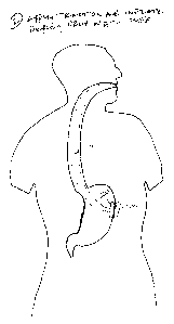
Referring to Fig. 1, one embodiment of a transgastric access device includes
an
elongate cannula 10 having at least one working lumen 14 extending the length
of the
cannula to a distal port 12. An instrument port 16 is formed at the proximal
end of the
lumen, and a valve 18 is positioned to seal the distal portion of the lumen. A
pair of
sealing elements 20a, 20b are positioned on the exterior of the cannula 10,
near the distal
port 12. As discussed in connection with Figs. 4 and 6, the sealing elements
may
comprise inflatable balloons or other elements capable of anchoring the
cannula within an
incision formed in a stomach wall and preferably forming a seal between the
cannula and
the incision.
In one embodiment, the working lumen 14 may be a single lumen of a size
appropriate for receiving instruments needed for the procedure, as shown in
Fig. 2.
Alternate embodiments may include two or more lumens.
Fig. 3 illustrates the proximal portion of the system, which during use is
positioned with the instrument port 16 in the mouth or outside of the mouth
with the
cannula 10 extending down the esophagus to the stomach. A light source lumen
22
extends the length of the cannula. The light source lumen includes fiber optic
elements
coupled to a fiber optic lighting system or other suitable lighting source
(not shown) so as
to permit illumination of the procedure to be carried out at the distal end of
the cannula
10. If the anchoring elements 20a, 20b (Fig. 1) are inflatable, inflation
ports 23 (Figs. 2
and 3) provide a conduit for delivery of inflation fluid or gas into the
balloons using an
CA 02623948 2008-03-26
WO 2007/038715 PCT/US2006/037978
-5-
inflation device such as a syringe (not shown) or other suitable inflation
system. If a
deflectable/steerable cannula is desired, pullwires 25 (Fig. 2) extend through
corresponding pullwire lumens in the cannula 10 and are anchored within the
cannula's
distal region.
Referring to Fig. 4, valve 18 may be positioned within the cannula 10, near
the
distal port 12 as shown, or in a more proximal portion of the cannula 10. The
valve 18
may take the form of a duck bill valve as shown, or any other type of valve
suitable for
sealing the distal portion of the lumen 14 in the absence of an instrument
through the
lumen. The valve 18 can thus prevent movement of fluids and/or gases into the
lumen
during passage of the distal port 12 through the stomach and into the
peritoneal cavity.
The valve may additionally be configured such that it will seal against
instruments passed
through the valve 18, thus preventing movement of fluids and gases around
instruments
extending through the valve 18 and preventing loss of insufflation pressure
from the
peritoneal cavity is insufflation is used. In alternative embodiments, a
separate valve or
seal may be mounted within the lumen 14 for use in forming a seal around the
periphery
of instruments passed through the lumen 14. Valves and seals useful for these
purposes
include those of the type used in trocars commonly used in laparoscopic
surgical
procedures.
Anchoring elements 20a, 20b may be inflatable annular cuffs as shown in Fig.
5.
Each such anchoring element is fluidly coupled to a corresponding one of the
inflation
ports 23 (Fig. 2), so that the anchoring elements 20a, 20b may be separately
inflated.
Anchoring elements 20a, 20b are formed of a durable polymeric material, and
are spaced
from one another along the length of the cannula 10 so as to allow them to be
positioned
on opposite sides of a portion of stomach wall.
In an alternative embodiment, the anchoring elements 20a, 20b are detachable
from the cannula 10 so that they inight be left in place against the stomach
wall to
continue to seal the incision formed in the stomach wall. For example, as
shown in Fig.
513, the distal end of the cannula may be sealed using a closure pin 21 or
other device
positioned within the lumen of the cannula, and a distal portion of the
cannula 10 (where
the anchoring elements are positioned) may be detachable from the remainder of
the
cannula 10. According to this alternative embodiment, the portions of the
cannula that
are to remain within the body may be formed of bioerodible material that will
passively
degrade at some point after the incision in the stomach wall has healed or
actively
CA 02623948 2008-03-26
WO 2007/038715 PCT/US2006/037978
-6-
degrade once exposed to heat, light, electrical energy or certain chemical
agents.
Detachable anchoring elements might also include have drug delivery capability
via a
coating matrix impregnated with one or more pharmaceutical agents, including
therapeutic agents and/or agents selected to promote healing of the incision
or ingrowth
of tissue onto the anchoring elements.
Figs. 6 and 7 illustrate an access cannula using alternative anchoring
elements
20c, 20d, each of which includes a frame member 30 that may include a central
ring 32
mounted to the cannula 10 (Fig. 7), and radial members 34 extending from the
ring 32.
The fraine members 30 may be formed of a shape memory material such as nitinol
or
shape memory polymer, or other material that allow the anchoring elements 20c,
20d to
be compressed into to a delivery sheath 38 (Fig. 7) but that will allow the
anchoring
elements 20c, 20d to spring to their expanded position once released from the
delivery
sheath 38. A polymeric disk 36 is mounted to the frame member 30.
Other anchoring systems are illustrated in Figs. 8A through 12B. The
illustrated
systems may provide only distal anchoring (i.e. an anchor against the exterior
of the
stomach wall) to prevent the cannula 10 from pulling out of the incision in
the stomach
wall, or they may provide both proximal and distal anchoring similar to that
provided by
balloons 20a, 20b of Fig. 1 to also prevent inadvertent advancement of the
cannula further
into the peritoneum. Preferred anchoring systems will also seal the periphery
of the
incision to prevent material from within the stomach from contaminating the
sterile
peritoneal cavity, however as an alternative the portion of the cannula that
seats within
the incision may have a compliant exterior surface that itself forms a seal
with the
incision.
Referring to Fig. 8A, the cannula 10 may have a distal portion having a
tubular
length of braid 29 overlaying a shaft 31. Braid 29 is shaped such that at
least a portion of
it will expand outwardly to form anchors 20e, 20f as shown in Fig. 8B when
shaft 31 is
withdrawn relative to the braid 29.
In the Fig. 9A embodiment, the distal portion of the cannula 10 includes a
hinged
annular collar 33 that self-expands or is actively pivoted to the radially
extended position
shown in Fig. 9B. The Fig. 10A embodiment includes longitudinal strips 35 cut
into the
distal portion of the cannula 10. Strips 25 bow outwardly as shown in Fig. lOB
when the
distal end of the cannula is longitudinally compressed. Compressive forces can
be
applied in a number of ways, such as by applying tension to pullwires
connected to the
CA 02623948 2008-03-26
WO 2007/038715 PCT/US2006/037978
-7-
distal end of the cannula while pushing against the proximal end of the
cannula, or by
pushing against the cannula while supporting the distal end of the cannula
using an
instrument passed through the lumen of the cannula. Circumferential folds
lines or
weakened regions 27 may be formed in the strips such that the strips will
crease at
selected locations.
In another alternative anchoring system shown in Figs. 11A and 11B, the distal
end of the access cannula 10 may have a braided distal end that can be made to
self-
expand (e.g. upon withdrawal of a sheath 39) to a flared "trumpet"
configuration (Fig.
11B) outside the stomach wall. The cannula may optionally include a
corresponding lip
(which may be pre-formed or self expandable) spaced from the distal end and
positionable inside the stomach wall, such that the wall is retained between
the flare and
the lip.
In another embodiment shown in Fig, 12A, cannula 10 includes a tapered tip 41
having helical ribs 43 or threads on the cannula shaft and the tip 41, or only
on the shaft
as in the Fig. 12B embodiment. These embodiments allow simultaneous
advancement of
the cannula through an incision, dilation of the incision, and anchoring of
the cannula
within the incision. Tip 41 may be retractable to open the cannula, following
anchoring,
for passage of instruments. Other retractable tips are described below.
The access cannula 10 may be a flexible tube formed of polymeric material
(e.g.
polyurethane). The cannula 10 may be highly compliant for introduction into
the body,
allowing the cannula to be partially or fully collapsed for delivery into the
stomach. The
cannula's properties can be tailored for optimal radial strength, compliance
and bending
radius. A compliant cannula may be supported during or after passage into the
stomach
by a secondary structure such as the access system (e.g. obturators of the
type discussed
below) or by other instruments inserted into the cannula.
Materials useful for the cannula include ePTFE, woven materials such as
polyester, polyurethane, composite materials (e.g. lycra with polyester) as
well as others.
A lubricious material such as ePTFE will provide a lubricious surface for ease
of delivery
through the esophagus and passage of instruments through the cannula. In some
embodiments, all or a portion of the cannula may include microporous regions
having a
pore size that allows therapeutic or antiseptic solutions to be administered
to the
surrounding area while preventing flow of contaminants into the cannula. For
example, a
solution may be directed under pressure through the cannula, causing the
solution to pass
CA 02623948 2008-03-26
WO 2007/038715 PCT/US2006/037978
-8-
ttirough the pores in the walls of the cannula. Alternative cannula
embodiments may be
reinforced using various materials. Reinforcements may be continuous,
variable, or site
specific along the length of the cannula.
The cannula may be a polymeric material reinforced with an internal, external,
or
embedded spiral wrapped coil (e.g. flat or round wire of stainless steel,
nitinol or suitable
alternatives, monofilament of polyester, nylon etc, or other material). The
spiral wrap
reinforcement provides radial strength allowing for an improved bend radius. A
tightly
wound (e.g. closed) coil improves the axial stiffness of the cannula, which
may improve
column strength for advancing the cannula, actuating anchoring systems, or
improving
advancement of instruments through the cannula.
In other embodiments, an internal, external or embedded braided structure may
be
on or in the walls of the cannula to improve radial strength, column strength,
and
torsional stiffness. Braid structures may be additionally be used to make the
cannula
compressible to a reduced diameter (such as through the application of
longitudinal
tension on the braid) or expandable (through longitudinally compression of the
braid.
Expandable braid features may be used to anchor the cannula within an incision
as
discussed above. Exposed braid on the exterior of the cannula may provide
additional
traction for anchoring.
A method for using the access cannula 10 includes passing the distal end 12 of
access cannula 10 into the mouth of a patient, through the esophagus E, and
into the
stomach S (or, in alternative embodiments, into the intestine via the rectum,
or through
the vagina for access through the vaginal ceiling or the uterus). Referring to
Figs. 13A
and 13B, with the cannula 10 preferably in contact with the wall W to be
penetrated, an
incision I or perforation is formed in the wall W using an instrument such as
a needle 50
passed through the cannula 10.
Once an incision is made using the needle, it may be necessary to pass a
dilator
through the incision to expand the incision I. In the embodiment shown, needle
50
extends from the distal end of a dilator 52, which is pushed through the
incision I to
expand the incision as shown in Figs. 13C and 13D. In an alternative
embodiment
discussed below, the needle may be protected within the lumen of the dilator
as it is
advanced through the access cannula, and then advanced from the dilator to
form the
incision I. Small knife edges (not shown) may extend from the surface of the
dilator to
allow the incision to be expanded by cutting, thus minimizing trauma to the
wall. In
CA 02623948 2008-03-26
WO 2007/038715 PCT/US2006/037978
-9-
other alternatives, the dilator may have an expandable portion incorporating
inflatable
balloons, expandable shape-memory braid sections, or other expandable features
that may
be positioned within the incision I and then expanded to increase the size of
the incision.
The dilator may further incorporate an endoscope to give the practitioner
visual feedback
as s/he forms the incision and anchors the access cannula.
The distal end 12 of the cannula 10 is advanced into the incision I, and
proximal
anchoring element 20b on the cannula is inflated as shown in Fig. 13E. Next,
the distal
end 12 of the cannula 10 is passed fully through the incision I as shown in
Fig. 13F, such
that distal anchoring element 20a (which at this point is uninflated) on the
cannula is
positioned outside of the stomach and proximal most anchoring element 20b on
the
cannula remains inside the stomach, preferably in contact with wall W. The
dilator 52
and needle 50 are withdrawn from the body as illustrated in Fig. 13G.
Inflation fluid is
delivered to inflate the distal anchoring element 20a as shown in Fig. 13H,
causing the
wall W to be engaged between the anchoring elements 20a, 20b, and further
causing the
anchoring elements 20a, 20b to seal the incision I against passage of fluids
and/or gases.
Once anchored in place, the access cannula provides sterile access to the
peritoneal
cavity. Instruments to be used to perforxn a procedure within the peritoneal
cavity are
thus passed into the proximal end of the access cannula which remains outside
the body,
and advanced through the cannula into the peritoneal cavity.
In an alternative method for placing the access cannula of Fig. 1, the distal
portion
of the cannula 10 is passed through the incision I, such that the distal most
anchoring
element 20a is positioned outside of the stomach and the proximal most
anchoring
element 20b remains inside the stomach. Inflation fluid is delivered to
inflate the distal
anchoring element 20a as shown in Fig. 14A. If the embodiment of Fig. 7 is
instead used,
the cannula 10 is introduced into the stomach while disposed inside the sheath
38, with
the anchoring elements 20c, 20d in a compressed orientation inside the sheath
38. The
sheath 38 (with the cannula 10 inside it) is passed through the perforation P.
The cannula
10 is advanced slightly in a distal direction to release the distal most
anchoring element
20c from the distal end of the sheath, causing the anchoring element 20c to
expand.
Referring to Fig. 14B, once the distal anchoring element 20a has been
inflated,
traction is applied to the cannula 10 to draw the distal anchoring element 20a
into firm
contact with the stomach wall. Next, inflation fluid is delivered to inflate
the proximal
anchoring element 20b, causing the stomach wall to be engaged between the
anchoring
CA 02623948 2008-03-26
WO 2007/038715 PCT/US2006/037978
-10-
elements 20a, 20b, and further causing the anchoring elements 20a, 20b to seal
the
perforation P against passage of fluids and/or gases. If the Fig. 7 embodiment
is used,
deployment of the proximal anchoring element 20d of the Fig. 7 embodiment is
achieved
by witlidrawing the sheath 38 proximally to release the anchoring element 20d,
thus
causing the stomach wall to be engaged between the anchoring elements 20c,
20d.
Finally, referring to Fig. 10, a procedural cannula 40 is passed through the
cannula 10. Procedural cannula 40 preferably includes a valve 42 sealing its
distal end
against passage of fluids. Valve 42 may be a duckbill type valve as described
above,
and/or one which will seal around instruments passed through it, each of which
is
commonly found in laparoscopic trocars. Instruments 44 needed to perform the
desired
procedure within the peritoneal cavity (e.g. forceps, electrosurgical tools,
snares, cutters,
endoscopes, staplers etc.) are passed through the access cannula 40 and used
to carry out
the procedure. Once the procedure has been completed, the procedural cannula
40 and
instruments are removed, anchoring elements 20a, 20b are deflated (or, in the
case of
anchoring elements 20c, 20d of Fig. 7, withdrawn into sheath 38), and the
cannula 10 is
removed from the body.
Ease of passage of the cannula 10 through the esophagus (or intestine) may be
enhanced through the use of an access system employing an obturator. One
access
system comprising an access cannula 10 and obturator 200 is shown in Fig. 15.
Obturator
200 includes an elongate tubular shaft 202 that extends through the cannula 10
out of the
patient, and a tip 204 on the distal end of the obturator. A passage or lumen
203 extends
through the shaft 202 and the tip 204. Tip 204 preferably includes a proximal
portion 206
that flares outwardly from the shaft 202, and a tapered distal portion 208.
The shaft 202
is preferably formed of braided tubing or other materials that give sufficient
column
strength, a desired bend radius, torsional stiffness for movement through the
target region
of the body (e.g. esophagus, intestine). Suitable examples include those
listed with
respect to reinforced cannula designs.
Tip 204 is divided into a number of circumferentially spaced spring elements
205.
Fig. 15 illustrates that the cannula 10 may include a beveled distal edge 210
on its interior
lumen, such that when the obturator 200 is disposed within the cannula 10 as
shown in
Fig. 16, the flared proximal portion 206 of the tip is adjacent to the beveled
edge 210 of
the cannula 10. A locking element 212 (Fig. 17B) positioned within the lumen
203 of the
obturator 200 urges the spring elements 205 outwardly into contact with the
beveled
CA 02623948 2008-03-26
WO 2007/038715 PCT/US2006/037978
-11-
edge 210 so as to prevent the obturator 200 from moving in a proximal
direction within
the cannula. The locking element 212 is shown as a tube, but it may be any
other feature
that will lock the obturator in its distal position.
A dilation balloon catheter 220 is advanceable through the cannula 10 and
obturator 200. A needle 218 is extendable through a lumen in the balloon
catheter 220,
or it may be an extendable and retractable component of the balloon catheter
220.
The obturator system of Fig. 16 allows the access cannula to be aseptically
positioned within a stomach wall incision. As shown, a transparent septum 214
covers
the obturator and is sealed around the circumference of the cannula. The
septum 214
seals the distal ends of the obturator and cannula so as to maintain a sterile
environment
within the cannula allowing clean passage of instruments into the peritoneal
space. The
transparent material of the septum allows visualization of structures outside
the distal end
of the obturator 200 and cannula 210 using endoscope 216. Septum 214 is
preferably
coupled to the obturator tip 202.
According to one method of placing the cannula 10 using the access system of
Fig. 16, the system is advanced through the esophagus and into contact or
close
proximity with the stomach wall W under visualization using endoscope 16 (Fig.
17A).
Needle 218 is advanced through the cannula and out the distal end of the
obturator,
perforating both the septum 214 (see Figs. 15 and 16) and the stomach wall W.
(Figs.
17B and 17C). If insufflation is needed for visualization within the
peritoneal cavity, the
cavity may be insufflated using gas directed through the needle 218.
Balloon dilator 220 is advanced through the incision I (Figs. 17D) and the
locking
element 212 is retracted (Fig. 17E). A stream 221 of sterile saline or other
substance (e.g.
antiseptic) may be directed through the cannula 10 to the stomach wall or
incision during
any part of the procedure.
The obturator tip 204 is retracted as shown in Figs. 17F and 17G by sliding
the
shaft 202 of the obturator in a proximal direction. Retraction of the
obturator tip 204 also
retracts the septum 214 as shown. The balloon 220 is expanded to dilate the
incision I.
Figs. 17 H-17I. The beveled edge of the cannula and expansion of the balloon
create an
isodiametric fit with the stomach wall surrounding the incision, facilitating
advancement
of the cannula through the incision. In an alternative embodiment shown in
Fig. 18, the
proximal portion of the balloon may include a proximal taper 222 to facilitate
advancement of the cannula by orienting the edges of the incision towards the
cannula 10.
CA 02623948 2008-03-26
WO 2007/038715 PCT/US2006/037978
-12-
Fig. 19 illustrates that the dilation balloon 220 may include an outer annular
balloon 224
that expands in a proximal direction, driving tissue surrounding the incision
over the
edges of the cannula 10. Once the incision I has been dilated, the cannula 10
is advanced
through the incision and the anchoring balloons 20a, 20b are expanded as
discussed
above. Fig. 17K.
In a slight modification to the method described in connection with Figs. 17 A-
17I, the obturator and septum may be retracted prior to penetration using the
needle 218
so as to create suction against the stomach wall, thus provided counter-
traction for the
advancement of the needle. In either case, suction may be applied through the
obturator
or access cannula to engage the stomach wall for penetration.
Fig. 20A shows an alternative access system for use in aseptically positioning
the
access cannula 10. The Fig. 20A system, which is similar to the Fig. 16
system, includes
cannula 10, obturator 200, a balloon dilator 220 having a retractable needle
tip 218, and a
septum 214a. In this embodiment, the obturator and septum are independent
structures.
The tip of the septum 214a includes an o-ring 230 having notches 232. The
center of the
o-ring is covered by the septum to seal the distal end of the cannula and
obturator.
During use of the Fig. 20A embodiment, needle 218 and balloon dilator 220 are
advanced
through the o-ring 230, penetrating the septum 214a and the stomach wall W as
shown in
Fig. 20C. Expansion of balloon dilator 220 raptures the o-ring 230 and the
septum as
shown in Fig. 20D.
Another alternative embodiment shown in Figs. 21A through 22B is similar to
the
Fig. 20A embodiment in that the balloon dilator 220 is used to rupture the
septum 214b.
Referring to Fig. 21B, after the obturator 202 is retracted, the septum 214b
is pressurized
and stretched to a tensioned state using sterile saline. When the septum 214b
is
penetrated and ruptured using the balloon dilator, the ruptured septum gathers
on the
exterior of the cannula 10, forming a stop 234 to prevent inadvertent
advancement of the
cannula 10 further into the stomach, and additionally forming a seal around
the incision.
0-ring 230a may be sufficiently large that it will not rupture in response to
expansion of
the dilator, but will instead retract towards the exterior surface of the
cannula when the
septum is ruptured.
As illustrated in Fig. 23, an alternative obturator 236 includes a tapered tip
238 on
a braided shaft 240. A lumen 242 in the shaft 240 and tip 238 is fluidly
coupled to a
duckbill valve 244, which remains closed except when the needle and balloon
dilator are
CA 02623948 2008-03-26
WO 2007/038715 PCT/US2006/037978
-13-
passed through it. An o-ring sea1246 seals the obturator against the interior
surface of the
cannula 10.
Fig. 24 illustrates a dilator that may be used with any of the disclosed
embodiments. Dilator 248 includes a tip having an off-set taper. A transparent
window
250 is positioned to allow viewing of the target tissue using an endoscope
although the
entire dilator tip may also be transparent. Flush ports 252 are positioned to
direct a sterile
saline solution or an antiseptic agent into contact with the stomach wall
before and/or
during penetration of the wall. A needle sheath 254 having a safety needle
extendable
from it is used to penetrate the stomach wall.
As discussed earlier, the anchors described above may be left behind to close
the
incision formed in the stomach wall or the wall of another body cavity. Figs.
25A - 25C
show other closure devices that may be endoscopically implanted to close the
incision
formed in the stomach wall or other body wall. For simplicity, any type of
opening
formed in the body wall (including but not limited to the dilated needle
punctures
described above) will be referred to as an incision. In general, the closure
devices
comprise a pair of expandable portions, one of which is positioned inside the
stomach and
the other of which is positioned on the stomach exterior. A connecting feature
extends
between the expandable portions and is generally positioned extending through
the
incision. The closure devices seal the incision preventing passage of fluids
or material
from stomach into the peritoneal cavity. They are preferably
bioabsorbable/bioerodible
implants, but may instead be permanent implants.
Figs 25A - 25C illustrate one exemplary embodiment of a closure device 310,
which includes a pair of wings 312a, 312b and a connecting element 314 of any
of a
number of shapes extending between the wings. Wings 312a, 312b are shown as
having
an oval shape, although other shapes including, but not limited to, elliptical
or circular
shapes may be used. In the first embodiment, the connecting element 314 is an
elongate
rib proportioned so that it may be positioned within an incision in the
stomach. While not
mandatory, the elongate shape of the rib is particularly suitable for a
closure device used
to close an elongate cut or tear in the tissue. The dimensions for the closure
device are
selected such that the spacing between the wings is sufficient to seal the
incision without
imparting excessive compressive forces on the stomach wall tissue. In one
embodiment,
the separation between the opposed surfaces of the wings is in the range of
0.06 - 0.1
inches.
CA 02623948 2008-03-26
WO 2007/038715 PCT/US2006/037978
-14-
The materials for the wings and rib are preferably materials that will
bioerode,
degrade or absorb after a period of time calculated to allow healing of the
incision.
Preferred materials include but are not limited to bioerodible elastomers or
biorubbers
such as those formed using sebacic acid materials. Mesh, braid or woven
materials
formed using absorbable suture material may also be used. If mesh, braid or
woven
components are used for sealing components (e.g. one or both of the wings),
they are
desirably of sufficiently tight construction to prevent fluid passage through
them, or they
are sealed against fluid passage using bioabsorbable adhesives or other
structures. The
closure devices may be constructed with various combinations of materials. As
one
example, a device may have bioabsorbable polymer wings and a bioabsorbable
mesh
connector element. Additionally, each feature may have combinations of
materials - such
as a biopolymer reinforced by an embedded absorbable mesh structure. The
materials
may be coated or impregnated using sclerosing agents or other materials that
will promote
healing of the stomach wall tissue.
Ribs 314 may be provided with pores, openings or other features through which
tissue may grow as the stomach tissue heals. In the Fig.25A-25C embodiment,
such
features are in the form of slots 316.
The closure device 310 is constructed so it may be folded for insertion into a
tube
for deployment. Various folding arrangements may be used. One example is shown
in
Figs. 25D - 25F. Fig. 25D is a top view of the closure device prior to
folding. As
indicated by arrows, each wing 312a, 312b is first folded onto itself along
its longitudinal
axis, configuring the device 10 as shown in the top view of Fig. 25E and the
side view of
Fig. 25F. Next, with reference to Fig. 25F, the upper portion of the device
310 is folded
across the horizontal axis A so that each wing 312a, 312b is again folded over
on itself,
placing the device 310 into the configuration shown in Fig. 25G.
Fig. 26 illustrates a deployment system 318 of a type that may be used for
implanting the closure device 310. System 318 includes a delivery cannula 320,
a grasper
322 extending through cannula 320, a outer sheatli 324, an endoscope 326 and
an
intermediate sheath 328. Use of the system 318 will next be described.
In preparation for deployment, the closure device 310 is folded as described
above, and the wing 312b to be deployed in the stomach interior is engaged in
its folded
state by grasper 322. The grasper 322 and a portion of the device 310
(including wing
312b) is withdrawn into the delivery cannula 320, leaving wing 312a positioned
outside
CA 02623948 2008-03-26
WO 2007/038715 PCT/US2006/037978
-15-
the distal opening of the delivery cannula 320. The delivery cannula 320 and
the folded
closure device 310 are positioned within the intermediate sheath 328 so as to
maintain the
folded configuration of the device 310. The intermediate sheath 328 and
endoscope are
positioned within the outer sheath 324 as shown in Fig. 27.
The distal end of the outer sheath 324 is passed through the mouth and
esophagus
and into the stomach. As shown in Fig. 28, the intermediate sheath 328 is
advanced out
of the outer sheath 324 and through the incision (not shown) under
visualization using the
endoscope 326. At this stage the device 310 is within the intermediate sheath
328, along
with the grasper 322 and delivery cannula 320, neither of which is visible in
Fig. 28.
Referring to Fig. 29, the intermediate sheath 328 is next withdrawn, exposing
the wing
312a of the device 310, causing the wing to expand on the exterior of the
stomach to the
position shown in Fig. 29. The delivery cannula 320 is withdrawn as shown in
Fig. 30,
but the wing 312b remains folded because it remains within the jaws of the
grasper 322.
Traction is applied to the grasper to pull the external wing 312a into contact
with the
stomach wall. The grasper 322 is then actuated to release the wing 312b,
causing it to
expand in the stomach interior (Fig. 32), leaving the device positioned within
the incision
as shown in Fig. 33. One or both of the wings 312a, 312b forms a seal with the
stomach
wall to prevent leakage of stomach contents into the peritoneal space. As the
incision
heals, tissue grows through the slots 316. Over time, the device degrades or
absorbs
within the body.
In the system for deploying the closure devices, the delivery cannula 320 may
be
the access cannula 10 of Fig. 1 or a separate cannula. If the closure device
is deployed
while the access cannula 10 is in place, the anchoring elements 20a, 20b will
be deflated
at appropriate times to make way for the wings of the closure device.
Figs. 34 and 35 shown an alternative embodiment of an access cannula 400,
which
includes an inner cannula section that remains in a sterile environment until
it is passed
through the deployed anchors 20a, 20b and into the peritoneal cavity.
Specifically,
cannula 400 includes a tubular proximal section 402 having a lumen 404, and a
distal
section 406 that is longitudinally compressible from the elongated position
shown in Fig.
34 to the compressed position shown in Fig. 35. An inner cannula 408 extends
longitudinally from the proximal section 402 and includes a lumen 410 in
communication
with lumen 404 of the proximal section 404. When the cannula distal section
406 is in
the elongated position, the inner cannula 408 is fully within the distal
section 406,
CA 02623948 2008-03-26
WO 2007/038715 PCT/US2006/037978
-16-
allowing sterility of the inner cannula 408 during movement of the cannula 400
through
the mouth, esophagus and stomach. After the anchors 20a, 20b are deployed as
described
above, the distal section 406 is compressed by axially loading the cannula 400
in the
direction of the arrow A in Fig. 35. Compression of the distal section causes
inner
cannula 408 to exit the distal section 406 (via valve 418 if one is provided
as in Fig. 4)
and to protrude into the peritoneal cavity, allowing sterile access to the
peritoneal cavity
via lumens 404 and 410.
Referring to Fig. 36, the access cannula 10 (or cannula 100) may be used for
introduction of instruments used to perform surgery on the bowel B, such as
bowel
resection to remove a diseased portion of the bowel. As shown, an intraluminal
endoscope 46 is passed transorally into the stomach and into the intestine,
allowing the
surgeon to identify diseased or injured sections of the bowel. A grasper 48
passed into
the peritoneal cavity via access cannula 10 may be used to manipulate the
bowel into a
desired position for treatment, and/or it may be used to pull a target region
of the bowel
over the intraluminal endoscope 46 for inspection. An endoscopic stapler 50
introduced
through the access cannula 10 can be used to resect and/or staple a portion of
the bowel,
and a camera 52 may be used for visualization of the procedure. Instruments
(e.g.
staplers, endoscopes, and/or others) may also be introduced through one or
more
laparoscopic ports providing access to the surgical cavity.
As discussed in connection with Fig.36, if it is desired to inspect the bowel
using a
transorally introduced endoscope, manipulation of the bowel may be necessary
in order to
bring portions of the bowel into the viewing range of the endoscope. Fig. 37A
illustrates
a system 60 that allows for such manipulation and inspection from within the
bowel. As
shown, system 60 includes a pair of flexible elongate tubular members 62a,
62b, each of
which includes an inflatable balloon 64a, 64b on its distal end. Balloons 64a,
64b are
constructed of a size and material that will allow them to engage the interior
wall of the
intestine when they are inflated from inside the intestine. The exterior
surfaces of the
balloons 64a, 64b may include surface features (for example, textures, ridges,
barbs, or
fish scale type structures) that facilitate engagement of the intestinal wall.
Inflation ports 66a, 66b are provided for inflating the balloons using a
syringe 68
or other inflation device. Guide wires 70a, 70b may also extend through lumens
in the
tubular members. As shown in Fig. 37B, the tubular members 62a, 62b and
endoscope 72
are arranged such that the endoscope 72 extends through the lumen of the
tubular member
CA 02623948 2008-03-26
WO 2007/038715 PCT/US2006/037978
-17-
62b, and the tubular member 62b extends through the lumen of the tubular
member 62a.
The system may include one or more elements (not shown) for locking the
positions of
the tubular members 62a, 62b (and/or the endoscope 72) relative to one
another.
Figs. 38 through 42 illustrate use of the bowel manipulation device of Fig.
37A.
First, the components are arranged as shown in Fig. 37B, but with the balloons
64a, 64b
in their deflated state. The assembled components are introduced into the
intestine via the
esophagus and stomach. Once the system is within the intestine, balloon 64b is
inflated
as shown in Fig. 39. However, before the tubular member 62a is advanced to the
position
shown in Fig. 39, endoscope 72 is advanced out of the tubular members and used
to
inspect the section of intestine 80.
Next, tubular member 62a is advanced further to a more distal region of the
intestine (Fig. 39), and then balloon 64a is inflated as shown in Fig. 40.
With both
balloons inflated, tubular member 62a is retracted in a proximal direction as
indicated by
an arrow in Fig. 40, causing balloon 64a to carry a section of the intestine
in a proximal
direction, thereby compressing the previously inspected section of bowel 80
and thus
causing a distally adjacent section of bowe182 to be presented within the
viewing range
of scope 72. See Fig. 41. Once section 82 is inspected, balloon 64b is
deflated and
tubular member 62b is advanced to move balloon 64b into position adjacent to
balloon
64a as shown in Fig. 42. Repositioned balloon 64b retains the previously
retracted bowel
section 82 in its retracted state, thus allowing repositioning of balloon 64a
without
releasing retracted section 80. The scope 72 is advanced distally to a new
position, and
then balloon 64a is then deflated, advanced distally, reinflated and then
retracted towards
balloon 64b, thus retracting bowel section 82 while presenting another section
of the
intestine within view of the scope 72. The method is repeated as required to
permit
viewing of as much of the intestine as needed.
While certain embodiments have been described above, it should be understood
that these embodiments are presented by way of example, and not limitation. It
will be
apparent to persons skilled in the relevant art that various changes in form
and detail may
be made therein without departing from the spirit and scope of the invention.
This is
especially true in light of technology and terms within the relevant art(s)
that may be later
developed. Moreover, various features of the disclosed embodiments may be
combined
with one other or with additional features to create additional embodiments
falling within
the scope of the present invention.
CA 02623948 2008-03-26
WO 2007/038715 PCT/US2006/037978
-18-
Any and all patents, patent applications and printed publications referred to
above,
including those relied upon for purposes of priority, are incorporated by
reference.