Note: Descriptions are shown in the official language in which they were submitted.
DEMANDE OU BREVET VOLUMINEUX
LA PRESENTE PARTIE DE CETTE DEMANDE OU CE BREVET COMPREND
PLUS D'UN TOME.
CECI EST LE TOME 1 DE 2
CONTENANT LES PAGES 1 A 115
NOTE : Pour les tomes additionels, veuillez contacter le Bureau canadien des
brevets
JUMBO APPLICATIONS/PATENTS
THIS SECTION OF THE APPLICATION/PATENT CONTAINS MORE THAN ONE
VOLUME
THIS IS VOLUME 1 OF 2
CONTAININGPAGES 1 TO 115
NOTE: For additional volumes, please contact the Canadian Patent Office
NOM DU FICHIER / FILE NAME:
NOTE POUR LE TOME / VOLUME NOTE:
CA 02624535 2008-03-28
WO 2007/036945 PCT/IL2006/001155
HEPATOCYTE GROWTH FACTOR RECEPTOR SPLICE VARIANTS AND
METHODS OF USING SAME
FIELD OF THE INVENTION
The present invention relates to hepatocyte growth factor receptor splice
variant
polypeptides, and polynucleotides encoding same, vectors and host cells
comprising same and
more particularly, to therapeutic and diagnostic compositions and methods
utilizing same.
BACKGROUND OF THE INVENTION
The protein product of c-Met oncogene is the tyrosine kinase receptor for
hepatocyte
growth factor (HGF) also known as scatter factor (SF). HGF and its receptor c-
Met are widely
expressed in a variety of tissues, and their expression is normally confined
to cells of
mesenchymal and epithelial origin, respectively. The HGF-Met pathway is
involved in a wide
range of biological effects, including cell proliferation and survival, cell
adhesion, cell migration
and invasion, morphogenic differentiation, organization of tubular structures
and angiogenesis.
Such paracrine signaling is vital to normal embryogenic development, wound
healing and tissue
maintenance and regeneration (reviewed in Christensen et al, 2005, Cancer
Letters 225: 1-26).
While HGF-Met signaling plays a key role during normal development,
inappropriate
activation of this signaling pathway has been implicated in tumor development
and progression.
Aberrant c-Met signaling has been described in a variety of human cancers,
including solid
tuniors and hematologic malignancies. Met activation may be involved in
different stages of
tumor progression, such as tumor cell proliferation and survival in primary
tumors, induction of
angiogenesis, stimulation of cell motility to form micrometastases, induction
of invasive
phenotype, and regaining the proliferation phenotype to form overt metastases
(Birchmeier et al
2003, Nat. Rev. Mol. Cell Biol. 4: 915-925).
Several mechanisms cause dysregulation of the HGF-Met pathway in tumor cells,
such as
overexpression of c-Met and/or HGF, constitutive kinase activation of c-Met in
the presence or
absence of gene amplification, activating mutations of c-Met, and autocrine
activation of c-Met
by HGF. c-Met is expressed in most carcinomas, but the degree of expression
varies among
distinct tumor types. High expression is detected in renal and colorectal
carcinomas, and lung
adenocarcinomas. Overexpression of ligand and/or receptor correlates with high
tumor grade and
poor prognosis. c-Met mutations have been reported in several types of tumors,
such as
hereditary and sporadic human papillary renal carcinomas, as well as ovarian
cancer, childhood
hepatocellular carcinoma, head and neck squamous cell carcinomas, gastric and
lung cancers
1
CA 02624535 2008-03-28
WO 2007/036945 PCT/IL2006/001155
(reviewed in Maulik et al, 2002. Cytokine & Growth Factor Rev. 13: 41-59; Ma
et al, 2003.
Cancer and Metastasis Rev. 22: 309-325).
The HGF-Met pathway is involved in cell scattering. HGF was discovered as a
secretory
product of fibroblasts and smooth muscle cells that induces dissociation and
motility of epithelial
cells. It is able to induce cell dissociation and mutual repulsion in a
similar manner to
semaphorins. HGF-Met signaling is also involved in cell motility. The key
events regulating
cell motility are polymerization of actin, formation of actin stress fibers,
and focal adhesion
formation. HGF has been shown to induce branching morphogenesis of kidney,
mammary and
bile ductular cells. In response to HGF, Met-expressing cells form branches in
three-dimensional
matrigel or tubule-like structures in collagen gels. This process is mediated
through changes in
cell shape, asymmetric polarization of the cells in the direction of
branching, branch elongation,
cell-cell contact, cell-ECM communication, ECM remodeling, controlled
proteolysis and cell
motility (Zhang et al. 2003. J. Cell. Biochem., 88:408-417; Ma et al, 2003.
ibid). HGF acts as a
potent angiogenic factor. HGF stimulation of vascular endothelial cells
promotes migration,
proliferation, protease production, invasion, and organization into capillary-
like tubes. HGF can
also promote the expression of angiogenic factors by tumor cells (Ma et al,
2003. ibici).
HGF-Met signaling has been strongly implicated in the promotion of the
invasive/metastatic tumor phenotype. An HGF-stimulated pathway involving
MAPKl/2
signaling is important in the up-regulation of expression of the serine
protease urokinase (uPA)
and its receptor (uPAR), resulting in an increase of uPA on the cell surface.
Certain components
of the ECM can be directly degraded by uPA, and more importantly, uPA cleaves
plasminogen
into the broader-specificity protease plasmin, which is able to efficiently
degrade several ECM
and basement membrane (BM) components. Plasmin also activates
metalloproteinases, which
have potent ECM/BM degrading abilities. HGF has been reported to promote
attachment of
tumor cells to endothelium, an important step in the metastatic cascade. This
activity may be
mediated by HGF induced up-regulation of CD44 expression on endothelium cells,
and integrin
expression on tumor cells.
The human Met gene, which includes 21 exons, is located on chromosome 7 band
7q21-
q31 and spans more than 1201cb in length. The primary Met transcript produces
a 150kDa
polypeptide (1390 amino acids) that is partially glycosylated to produce a
170kDa precursor
protein. This 170kDa precursor is further glycosylated and then cleaved into a
50kDa a-chain
and a 140kDa (3-chain which are disulfide-linked. The a-subunit of the mature
Met heterodimer
is highly glycosylated and is entirely extracellular, while the (3-subunit
contains a large
extracellular region, a membrane spanning segment, and an intracellular
tyrosine kinase domain
(Ma et al, 2003. ibid).
2
CA 02624535 2008-03-28
WO 2007/036945 PCT/IL2006/001155
Met is the prototypic member of a subfamily of heterodimeric receptor tyrosine
kinases
which include Met, Ron, and Sea. Members of the Met receptor subfamily have
been shown to
share homology with semaphorins and semaphorin receptors (plexin), which play
a role in cell
scattering (Reviewed in Trusolino et al. 1998, FASEB J. 12: 1267-1280). All
semaphorins
contain a conserved 500 amino acid extracellular domain (Sema domain), which
spans the
cysteine-rich Met related sequence (MRS), containing the consensus motif C-X(5-
6)-C-X(2)-C-
X(6-8)-C-X(2)-C-X(3-5)-C. The extracellular portions of Met, Ron, and Sea
contain a region of
homology to semaphorins including the N-terminal Sema domain and the MRS.
Other domains
identified in the extracellular portion of Met are the PSI domain and the
IPT/TIG repeat domain.
The PSI domain is found in plexins, semaphorins and integrins while the IPT
repeats (also
known as TIG domains) are found within immunoglobulin, plexins and
transcription factors. The
C-terminus intracellular tyrosine kinase domain shares homology with Ron and
Sea.
The Sema domain plays a critical role in ligand binding and is also necessary
for receptor
dimerization (Kong-Beltran et al 2004, Cancer Cell, 6: 75-84; Wickramasinghe
and Kong-
Beltran, 2005, Cell Cycle, 4: 683-685). Treatment of Met-overexpressing tumor
cells with a
recombinant Sema protein construct (rSema, which contains also the PSI domain)
inhibits both
ligand dependent and independent activation of Met-mediated signal
transduction, cell motility
and migration, in a mamier similar to the antagonisitic anti-Met Fab 5D5 (Kong-
Beltran et al
2004. ibid). Decoy Met (the entire extracellular domain of Met, produced as a
truncated soluble
receptor) interferes with HGF binding to Met, and with receptor dimerization.
Similarly, a
chimeric soluble protein containing the extracellular domain of Met fused to
the constant region
of IgG Izeavy chain, binds HGF with an affinity similar to that of the
authentic, membrane-
associated receptor, and inhibits the binding of HGF to Met, expressed on A549
cells (Mark, et
al., 1992, J Biol Chem. 267:26166-26171). Local or systemic delivery of decoy
Met in mice, by
lentiviral vector technology, inhibits tumor cell proliferation and survival
in a variety of human
xenografts, impairs tumor angiogenesis, suppresses or prevents the formation
of spontaneous
metastases, and synergizes with radiotherapy in inducing tumor regression
(Michieli et al, 2004,
Cancer Cell 6: 61-73). These data suggest that the extracellular domain of Met
may not only
represent a novel anticancer therapeutic target, but also acts as a
biotherapeutic itself (reviewed
in Zhang et al 2004, Cancer Cell 6: 5-6).
Various inhibitory strategies have been employed to therapeutically target the
HGF-Met
pathway (reviewed in Christensen et al, 2005, Cancer Letters 225: 1-26), and
several candidates
are under development. Tliree main approaches have been employed for selective
anticancer
drug development: antagonism of HGF/Met interaction, inhibition of tyrosine
kinase catalytic
activity of Met, and blockade of intracellular Met/effectors interactions.
Among the current
3
CA 02624535 2008-03-28
WO 2007/036945 PCT/IL2006/001155
developments are a humanized anti-HGF mAb AMG-102 (Amgen); NK4, a proteolytic
cleavage
fragment of HGF that acts as a competitive HGF antagonist (Kringle Pharma);
and small
molecule inhibitors of the c-Met receptor, such as XL880 (Exelixis), ARQ 197
(Arqule),
SU11274, PHA665752, PF-02341066 of Pfizer; a series of small molecules of
Methylgene, and
others.
US Patent Application Publication No. 2004/0248157, assigned to the applicant
of the
present invention discloses polynucleotides and their respective encoded
polypeptides. One of
several transcripts disclosed therein is a Met-934 variant (denoted herein SEQ
ID NO: 2 and
SEQ ID NO:38, for mRNA and protein sequences, respectively), which results
from alternative
splicing of the c-Met gene, thereby causing an extension of exon 12 (the last
exon before the
transmembrane region encoding exon) leading to an insertion of a stop codon
and the generation
of a truncated Met protein which terminates just before the transmembrane
domain. Met splice
variant has an open reading frame (ORF) of 934 amino acids including 910 amino
acids of the
wild-type (w.t.) Met protein and a unique sequence of 24 amino acids at the C-
terminus of the
protein. It contains nearly the complete extracellular portion of Met (910
amino acids of 933 of
the w.t. protein) and therefore comprises all its structural domains (the
Sema, PSI and TIG
domains). Met-934 is predicted to be a secreted protein since it retains the
original N-terminal
signal peptide (amino acids 1-24) and lacks the transmembrane domain (amino
acids 933-955 of
the w.t.). The Met-934 secreted isoform was suggested to function as an
antagonist (i.e.,
inhibitor) of Met-HGF interaction by competing with the membrane-bound
receptor for the
ligand-HGF. Met-934 splice variant was suggested to be useful in the treatment
and/or diagnosis
of cancers such as, hereditary and sporadic papillary renal carcinoma, breast
cancer, ovarian
cancer, childhood hepatocellular carcinoma, metastatic head and neck squamous
cell
carcinomas, lung cancer (e.g., non-small cell lung cancer, small cell lung
cancer), prostate
cancer, pancreatic cancer, gastric cancer and other diseases such as diabetic
retinopathy.
WO 05/071059 and US Patent Application No. 11/043,591 assigned to the
applicant of
the present invention disclose polynucleotides and their respective encoded
polypeptides. One
among the hundreds of polynucleotide transcripts disclosed therein is
HSU08818_orig trans_9 drop_nodes_28 new num 15 tr0 rl_1_gPRT (denoted herein
SEQ
ID NO:48) which encodes an amino acid sequence termed hereinafter Met-885 (SEQ
ID NO:66).
This splice isoform was generated through exon skipping and it contains the
first 11 exons of the
c-Met gene, skips the 12th exon and enters the intron following the 12th exon,
leading to an
insertion of a stop codon and the generation of a truncated Met protein which
terminates just
before the transmembrane domain. The derived protein contains 885 amino acids,
that includes
861 amino acids of the wild-type and a unique sequence of 24 intron-derived
amino acids at the
4
CA 02624535 2008-03-28
WO 2007/036945 PCT/IL2006/001155
C-terminus of the protein. The Met-885 (SEQ ID NO:66) secreted isoform was
suggested to be
useful for treatment of Papillary Renal Carcinoma, head and neck cancers and
other cancers.
WO 2005/113596 assigned to Receptor Biologix Inc, discloses several in silico
predicted
polypeptides that are isoforms of cell surface receptors, including, inter
alia, Met receptor,
wherein each polypeptide comprises at least one domain of the receptor,
operatively linked to at
least one amino acid encoded by an intron of a relevnt gene; and the
polypeptide lacks a
transmembrane domain, protein kinase domain and at least one additional domain
compared to
the wt receptor, whereby the membrane localization and protein kinase activity
of the
polypeptide is reduced or abolished compared to the receptor. It is fixrther
speculated that these
isoforms may be useful in treating or preventing metastatic cancer, inhibiting
angiogenesis,
treating lung cancer, malignant peripheral nerve sheath tumors, colon cancer,
gastric cancer,
cutaneous malignant melanoma and prevention of malaria. WO 2005/113596
mentions that the
Met isoforms might be provided in pharmaceutical compositions as conjugates
between the
isoform and another agent, including coupling to an Fc fragment of an antibody
that binds to a
specific cell surface marker to induce killer T cell activity in neturophils,
natural killer cells and
macrophages. However, no guidance is provided for production of any
conjugates, nor are there
any examples for actual biological activities of said Met isoforms.
US Patent No. 5,571,509 assigned to Farmitalia Carlo Erba S.R.L., discloses a
carboxy-
terminal truncated form of the c-Met oncogene. The truncated form results in a
beta chain of the
receptor, which is 75 to 85 kDa long that acts as an antagonist of the HGF
receptor. US
5,571,509 reveals that this soluble Met protein is released in the culture
medium by proteolytic
cleavage of the membrane-bound Met proteins. However, these proteolytic
fragments are not
novel splice variants of cMet.
US Patent Application Publication No. 2005/0233960 assigned to GENETECH, INC.
discloses c-Met antagonists for modulating the HGF/c-met signaling pathway.
The c-Met
antagonists of US 2005/0233960 are particularly peptides comprising at least a
portion of c-Met
Sema domain or variant thereof.
There is an unnlet need to develop therapies which target the HGF-Met pathway
and Met
signaling via Met receptor tyrosine kinase, and which inhibit Met receptor
action and/or its
physiological effects.
SUMMARY OF THE INVENTION
The present invention provides splice variants of the Met receptor tyrosine
kinase,
derivatives thereof and vectors encoding same. Specifically, the present
invention provides
soluble Met receptor splice variants or derivatives thereof having inhibitory
effects on Met
5
CA 02624535 2008-03-28
WO 2007/036945 PCT/IL2006/001155
tyrosine kinase activity. The invention further provides pharmaceutical
compositions, fusion
proteins and host cells comprising said splice variants and vector encoding
said splice variants.
In addition, the present invention provides methods of treating, preventing
and diagnosing
cancers and non-cancerous proliferative disorders reliant on Met signaling,
using said splice
variants.
The Met variant products (splice variants) of the present invention are devoid
of
transmembrane and intracellular domains while retaining the extracellular
region of Met (i.e.,
HGF binding site). Without wishing to be bound by a single theory, these
splice variants are
likely to compete for HGF binding to the membrane bound Met receptor and as a
consequence
may block Met activation and the signaling pathway. Alternatively, Met soluble
splice variants
can interfere with constitutive Met signaling in cancer cells, in an HGF-
independent manner.
Therefore, Met splice variants of the present invention can serve as
antagonists (i.e., inhibitors)
of HGF dependent or independent Met signaling.
According to a first aspect the present invention provides an isolated
polynucleotide
encoding Met splice variant protein comprising an amino acid sequence as set
forth in any one of
SEQ ID NO:36 (Met588 protein) and SEQ ID NO:37 (Met877 protein).
According to one embodiment, the present invention provides an isolated
polynucleotide
encoding Met splice variant protein having a nucleic acid sequence as set
forth in any one of
SEQ ID NO:1 (Met588) and SEQ ID NO:3 (Met877).
According to another embodiment, the isolated polynucleotide further comprises
an Fc
fragment coding sequence wherein the expression of the polynucleotide leads to
the formation of
a fusion protein with an Fc fragment.
According to yet another embodiment, the isolated polynucleotide comprising
the Fc
fragment encodes a MET splice variant fusion protein comprising an amino acid
sequence as set
forth in SEQ ID NO:79 (Met877-Fc protein).
According to yet a further embodiment, the isolated polynucleotide comprising
the Fc
fragment coding sequence comprises a nucleic acid sequence as set forth in SEQ
ID NO:78
(Met877-Fc).
According to yet another embodiment, the isolated polynucleotide further
comprises a tag
coding sequence wherein the expression of the polynucleotide leads to the
formation of a fusion
protein with a tag.
According to one embodiment, the isolated polynucleotide comprising a tag
sequence
encodes a MET splice variant fusion protein comprising an amino acid sequence
as set forth in
SEQ ID NO:47 (Met877-His-tag protein).
6
CA 02624535 2008-03-28
WO 2007/036945 PCT/IL2006/001155
According to another embodiment, the isolated polynucleotide comprising a tag
coding
sequence comprises a nucleic acid sequence as set forth in SEQ ID NO:46
(Met877-His tag).
According to another aspect, the present invention provides an isolated Met
splice variant
polypeptide having an amino acid sequence as set forth in any one of SEQ ID
NOS:36 (Met588
protein) or 37 (Met877 protein).
According to one embodiment, the isolated polypeptide further comprises an Fc
fragment
contiguously joined thereto. According to another embodiment, the isolated
polypeptide further
comprises a tag contiguously joined thereto.
According to another embodiment, the isolated Met splice variant comprising an
Fc
fragment is having an amino acid sequence as set forth in SEQ ID NO:79
(Met877Fc protein).
According to yet another embodiment, the isolated Met splice variant
comprising a tag is
having an amino acid sequence as set forth in SEQ ID NO:47 (Met877-His-tag
protein).
According to yet another aspect, the present invention provides an isolated
polynucleotide encoding Met splice variant tagged protein comprising a first
nucleic acid
sequence encoding a Met splice variant having an amino acid sequence as set
forth in any one of
SEQ ID NO:66 (Met885 protein) and SEQ ID NO:38 (Met934 protein) and a second
nucleic
acid sequence encoding a tag sequence.
According to one embodiment, the polynucleotide encoding Met splice variant
tagged
protein, wherein the protein comprises a sequence as set forth in SEQ ID NO:75
(Met885-His-
tag protein).
According to other embodiments, the polynucleotide encoding Met splice variant
tagged
protein comprises a nucleic acid sequence as set forth in SEQ ID NO:74 (Met885-
His-tag).
According to yet another aspect, the present invention provides an isolated
polynucleotide encoding a Met splice variant fusion protein comprising a first
nucleic acids
sequence encoding a Met splice variant having an amino acid sequence as set
forth in any one of
SEQ ID NO:66 (Met885 protein) and SEQ ID NO:38 (Met934 protein) and a second
nucleic
acid sequence encoding an Fc fragment.
According to one embodiment, the isolated polynucleotide encodes a fusion
protein
comprising an amino acid sequence as set forth in any of SEQ ID NO:77 (Met885-
Fc protein)
and SEQ ID NO:68 (Met934-Fc protein). According to another embodiment, the
isolated
polynucleotide comprising an Fc fragment coding sequence is having the nucleic
acid sequence
as set forth in any of SEQ ID NO:76 (Met885-Fc) and SEQ ID NO:67 (Met934-Fc).
According to a further aspect, the present invention provides an isolated Met
splice
variant tagged protein comprising a first fragment having an amino acid
sequence as set forth in
7
CA 02624535 2008-03-28
WO 2007/036945 PCT/IL2006/001155
any one of SEQ ID NO:66 (Met885 protein) and SEQ ID NO:38 (Met934 protein) and
a second
fragment contiguously joined thereto, wherein the second fragment is a tag.
According to one embodiment, the tagged protein comprises an amino acid as set
forth in
SEQ ID NO:75 (Met885- His-tag protein).
According to yet another aspect, the present invention provides isolated Met
splice
variant fusion protein comprising a first fragment having an amino acid
sequence as set forth in
any one of SEQ ID NO:66 (Met885 protein) and SEQ ID NO:38 (Met934 protein) and
a second
fragment contiguously joined thereto, wherein the second fragment is an Fc
fragment.
According to one embodiment, the isolated Met splice variant having an Fc
fragment
coding sequence contiguously joined thereto comprises an amino acid sequence
as set forth in
any one of SEQ ID NO:77 (Met885-Fc protein) and SEQ ID NO:68 (Met934-Fc
protein).
According to alternative embodiments, the present invention further provides
derivatives
of the Met receptor tyrosine kinase variants and modified Met receptor
tyrosine kinase variants.
According to some embodiments the derivatives are obtained by glycosylation
and/or
phosphorylation and/or chemical modifications. According to other embodiments,
the
derivatives are fusion proteins. According to certain embodiments the modified
splice variants
are fused to an Fc fragment of Ig. According to certain embodiments the
modified Met receptor
tyrosine kinase variants are obtained by addition of C-terminal His/StrepII
tag.
According to certain embodiments, the protein variants of the present
invention can be
modified to form synthetically modified variants.
Advantageously, the protein variants of the present invention comprise
modifications that
enhance their inhibitory and/or therapeutic effect including, e.g., enhanced
affinity, improved
pharmacokinetics properties (such as half life, stability, clearance rate),
and reduced toxicity to
the subject. Such modifications include, but are not limited to, modifications
involving
glycosylation, pegylation, substitution with non-naturally occurring but
functionally equivalent
amino acid and linking groups.
According to additional aspects, the present invention provides vectors,
cells, liposomes
and compositions comprising the isolated nucleic acids of this invention.
According to further aspects, the present invention provides pharmaceutical
compositions
comprising the novel splice variant polypeptides of this invention.
According to yet additional aspects, the present invention provides
pharmaceutical
compositions comprising the novel splice variant polynucleotides of this
invention.
According to yet other aspects, the present invention provides pharmaceutical
compositions coinprising an expression vector, wherein the expression vector
contains the
8
CA 02624535 2008-03-28
WO 2007/036945 PCT/IL2006/001155
nucleic acid sequence encoding Met variant of the present invention. According
to still further
aspects the present invention provides pharmaceutical compositions comprising
host cells
containing the expression vectors of the invention.
According to yet another aspect, the present invention provides a method for
treating a
Met-related disease, comprising administering an agent selected from: Met
variant therapeutic
protein, variant peptide, nucleic acid sequence encoding Met variant of the
present invention,
expression vector containing the nucleic acid sequence encoding Met variant of
the present
invention or host cells containing the expression vector as above, to a
subject in need of
treatment thereof.
According to certain embodiment, Met-related diseases including, but not
limited to,
diseases wherein Met receptor tyrosine kinase is involved in the etiology or
pathogenesis of the
disease process, as will be explained in detail hereinbelow. Optionally, the
transcripts of novel
Met variants of the present invention are useful as therapeutic agents for
treatment of Met-related
diseases.
In particular, Met-related diseases include, but are not limited to, disorders
or conditions
that would benefit from treatment with a molecule or method of the invention.
These include
chronic and acute disorders or diseases, such as pathological conditions which
predispose to the
disorder in question. Non-limiting examples of the disorders to be treated
herein include
malignant and benign tumors; lymphoid malignancies; neuronal, glial,
astrocytal, hypothalamic
and other glandular, macrophagal, epithelial, stromal and blastocoelic
disorders; and
angiogenesis-related disorders.
Examples of cancer include but are not limited to, carcinoma, lymphoma,
leukemia,
sarcoma and blastoma. According to certain preferred embodiments, the methods
of the present
invention are useful in treating primary and metastatic cancer such as breast
cancer, colon
cancer, colorectal cancer, gastrointestinal tumors, esophageal cancer,
cervical cancer, ovarian
cancer, endometrial or uterine carcinoma, vulval cancer, liver cancer,
hepatocellular cancer,
bladder cancer, kidney cancer, hereditary and sporadic papillary renal cell
carcinoma, pancreatic
cancer, various types of head and neck cancer, lung cancer (e.g., non-small
cell lung cancer,
small cell lung cancer, squamous cell carcinoma, lung adenocarcinoma),
prostate cancer, thyroid
cancer, brain tumors, glioblastoma, glioma, malignant peripheral nerve sheath
tumors, cancer of
the peritoneum, cutaneous malignant melanoma, and salivary gland carcinoma.
Met-related diseases also consist of diseases in which anti-angiogenic
activity plays a
favorable role, including but not limited to, diseases having abnormal quality
and/or quantity of
vascularization as a characteristic feature. Dysregulation of angiogenesis can
lead to many
9
CA 02624535 2008-03-28
WO 2007/036945 PCT/IL2006/001155
disorders that can be treated by compositions and methods of the invention.
These disorders
include both non-neoplastic and neoplastic conditions. Neoplastic include but
are not limited to
the type of primary and metastatic cancers described above. Non-neoplastic
disorders include but
are not limited to inflammatory and autoimmune disorders, such as aberrant
hypertrophy,
arthritis, psoriasis, sarcoidosis, scleroderma, atherosclerosis, synovitis,
dermatitis, Crohn's
disease, ulcerative colitis, inflammatory bowel disease, respiratory distress
syndrome, uveitis,
meningitis, encephalitis, Sjorgen's syndrome, systemic lupus erythematosus,
diabetes mellitus,
multiple sclerosis, juvenile onset diabetes; allergic conditions, eczema and
asthma; proliferative
retinopathies, including but not limited to diabetic retinopathy, retinopathy
of prematurity,
retrolental fibroplasia, neovascular glaucoma, age-related macular
degeneration, diabetic
macular edema, comal neovascularization, corneal graft neovascularization
and/or rejection,
ocular neovascular disease; and various other disorders in which anti-
angiogenic activity plays a
favorable role including but not limited to vascular restenosis, arteriovenous
malformations,
meningioma, hemangioma, angiofibroma, thyroid hyperplasia, hypercicatrization
in wound
healing, hypertrophic scars.
The compositions and methods of the present invention can be further employed
in
combination with surgery or cytotoxic agents, or other anti-cancer agents,
such as chemotherapy
or radiotherapy and/or in combination with anti-angiogenesis drugs.
Additionally or alternatively, Met receptor tyrosine kinase variants according
to the
present invention may be useful for diagnosis of diseases wherein Met receptor
tyrosine kinase is
involved in the etiology or pathogenesis of the disease process, and/or
disease in which Met
expression is altered as compared to the normal level, as will be explained in
detail hereinbelow.
Furthermore, the novel variants may be useful for diagnosis of any disease or
condition where
Met receptor tyrosine kinase is known to serve as a diagnostic or prognostic
marker.
Examples of diseases where the novel variants may be useful for diagnosis
include, but
are not limited to, cancer, such as hereditary and sporadic papillary renal
carcinoma, breast
cancer, ovarian cancer, childhood hepatocellular carcinoma, metastatic head
and neck squamous
cell carcinomas, lung cancer (e.g., non-small cell lung cancer, small cell
lung cancer), prostate
cancer, pancreatic cancer and gastric cancer, diabetic retinopathy,
regenerative processes such as
wound healing and conditions, which require enhanced angiogenesis such as
atherosclerotic
diseases, ischemic conditions and diabetes, and diseases of the liver such as
hepatic cirrhosis and
hepatic dysfunction.
According to yet another aspect, the present invention provides a kit for
detecting a
variant-detectable disease, comprising a kit detecting specific expression of
a splice variant
according to any of the above embodiments.
CA 02624535 2008-03-28
WO 2007/036945 PCT/IL2006/001155
These and additional features of the invention will be better understood in
conjunction
with the figures description, examples and claims which follow.
BRIEF DESCRIPTION OF DRAWINGS
Figures 1A-E demonstrate amino acid sequence comparison between the Met
variants of
the invention and the known Met receptor protein kinase. Figure 1A
demonstrates the
comparison between Met-877 variant of the invention (SEQ ID NO:37) and the
known Met
receptor protein kinase (SEQ ID NO:34). Figure 1 B demonstrates the comparison
between Met-
934 variant of the invention (SEQ ID NO:38) and the known Met receptor protein
kinase (SEQ
ID NO:34). Figure 1 C dernonstrates the comparison between Met-885 variant of
the invention
(SEQ ID NO:66) and the known Met receptor protein kinase (SEQ ID NO:34).
Figure 1D
demonstrates the comparison between Met-588 variant of the invention (SEQ ID
NO:36) and the
known Met receptor protein kinase MET HUMAN (SEQ ID NO:34). Figure 1E
demonstrates
the comparison between Met-588 variant of the invention (SEQ ID NO:36) and the
known Met
receptor protein kinase MET HUMAN V1 (SEQ ID NO:35).
Figures 2A-D demonstrates amino acid sequence comparison between the Met
variants
of the invention and a Met variant previously disclosed by Receptor Biologix
Inc. (RB). The
unique amino acids are marked in bold. Figure 2A demonstrates the comparison
between Met-
877 variant of the invention (SEQ ID NO:37) and the RB Met variant (SEQ ID
NO:40). Figure
2B demonstrates the comparison between Met-885 variant of the invention (SEQ
ID NO:66) and
the RB Met variant (SEQ ID NO:40). Figure 2C demonstrates the comparison
between Met-934
variant of the invention (SEQ ID NO:38) and the RB Met variant (SEQ ID NO:40).
Figure 2D
demonstrates the comparison between Met-588 variant of the invention (SEQ ID
NO:36) and the
RB Met variant (SEQ ID NO:40).
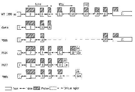
Figure 3 shows schematic mRNA and protein structure of Met. "WT 1390aa"
represents
the known Met receptor protein kinase (SEQ ID NO:34). "rSEMA" represents the
recombinant
SEMA domain of the Met extracellular region (Kong-Beltran et al., 2004, Cancer
Cell 6, 75-84),
SEQ ID NO:39. "P588" represents the Met-588 variant of the present invention
(SEQ ID NO: 1
and 36, for mRNA and protein, respectively). "P934" represents the Met-934
variant previously
disclosed in US Patent Application No. 10/764,833 publication No. 2004/0248157
assigned to
the applicant of the present invention (SEQ ID NO:2 and 38, for mRNA and
protein,
respectively). "P877" represents the Met-877 variant of the present invention
(SEQ ID NO:3
and 37, for mRNA and protein, respectively). "P885" represents the Met-885
variant previously
disclosed in WO 05/071059 and US Patent Application No.11/043,591 assigned to
the applicant
11
CA 02624535 2008-03-28
WO 2007/036945 PCT/IL2006/001155
of the present invention (SEQ ID NO:48 and 66, for mRNA and protein,
respectively). Exons are
represented by white boxes, while introns are represented by two headed
arrows. Dotted lines
between exons mean that all exons between them are present with no changes.
Proteins are
shown in boxes with upper right to lower left fill. The unique regions are
represented by white
boxes with dashed frame. SEMA domain, transmembrane domain (TM), and
immunoglobulin-
plexin-transcription factor domain (IPT) are identified accordingly.
Figure 4 is a histogram showing cancer and cell-line vs. normal tissue
expression for
Cluster HSU08818, demonstrating overexpression in a mixture of malignant
tumors from
different tissues and gastric carcinoma.
Figure 5A shows the Met-934-Fc sequence that was codon optimized to boost
protein
expression in mammalian system (SEQ ID NO:67). The bold part of the nucleotide
sequence
shows the relevant ORF (open reading frame) including the tag sequence.
Figure 5B shows the optimized Met-934-Fc protein sequence (SEQ ID NO:68). The
bold
part of the sequence is the Fc tag.
Figure 6 shows the Western blot result, demonstrating stable Met-934-Fc
expression
using anti IgG antibodies.
Figure 7A shows the optimized nucleotide sequences of Met885 StrepHis (SEQ ID
NO:74). The bold part of the nucleotide sequence shows the relevant ORF (open
reading frame)
including the tag sequence. The Strep-His tag is underlined.
Figure 7B shows the optimized protein sequences of Met885 StrepHis (SEQ ID
NO:75).
The Strep-His tag is underlined.
Figure 8A shows the optimized nucleotide sequences of Met-885-Fc (SEQ ID
NO:76).
The bold part of the nucleotide sequence shows the relevant ORF (open reading
frame) including
the tag sequence. The Fc-tag is underlined.
Figure 8B shows the optimized Met-885-Fc protein sequence (SEQ ID NO:77). The
Fc-
tag is underlined.
Figure 9 shows Western blot results, demonstrating stable Met885-Fc (SEQ ID
NO:77)
expression using anti IgG (lane 1). 100ng of Fe control is shown in lane 4.
Figure 10 shows Western blot results, demonstrating stable Met885_StrepHis
(SEQ ID
NO:75) expression using anti His (lane 7). Molecular weight marker (Rainbow
AMERSHAM
RPN800) is shown in lane 1.
Figure 11 shows the RT-PCR results of Met-877 (SEQ ID NO:3) variant. The
various
lanes show RT-PCR products on cDNA prepared from RNA extracted from the
following
sources: lanes 1-3 colon cell lines, as follows: lane 1-caco; lane 2-CG22 from
Ichilov; lane 3-
(CG224); lane 4 lung cell line H1299; lane 5 ovary cell line ES2, lane 6
breast cell line MCF7;
12
CA 02624535 2008-03-28
WO 2007/036945 PCT/IL2006/001155
lane 7 lung tissue A609163, Biochain; lanes 8-9 breast tissues A605151 and
A609221, Biochain,
respectively; lane 10 - 293 cell line.
Figure 12 shows the Met-877 (SEQ ID NO:45) PCR product sequence. The sequences
of
the primers used for the RT-PCR in Figure, are shown in bold.
Figure 13A shows the Met-877 (SEQ ID NO:46) sequence that was codon optimized
to
boost protein expression in mammalian system. The bold part of the nucleotide
sequence shows
the relevant ORF (open reading frame) including the tag sequence.
Figure 13B shows the optimized Met-877 His tag (SEQ ID NO:47) amino acid
sequence.
In bold there is the Strep tag, following the amino acid Pro (Strep II tag:
WSHPQFEK); and His
tag (8 His residues- HHHHHHHH) sequences which are separated by a linker of
two amino
acids (Thr-Gly). The 8 His tag is followed by Gly-GIy-GIn.
Figure 14 shows a schematic diagram of the pIRESpuro3 construct containing the
Met-
877 DNA fragment.
Figure 15 shows a Western Blot analysis, demonstrating the expression of the
cloned
Met-877 (SEQ ID NO:47) protein. Lane 1 represent molecular weight marker.
Figure 16 demonstrates the analysis of the purified Met-877 His tag (SEQ ID
NO:47)
protein by SDS-PAGE stained by Coomassie (lane 6). Lane 1 represent molecular
weight
marker. Lanes 2-5 represent BSA in different concentrations for quantity
reference.
Figure 17 demonstrates the analysis of the purified Met-877 His tag (SEQ ID
NO:47)
protein by the Bioanalyzer (Agilent).
Figure 18A shows the optimized nucleotide sequences of Met-877-Fc (SEQ ID
NO:78).
The bold part of the nucleotide sequence shows the relevant ORF (open reading
frame) including
the tag sequence. The Fc-tag is underlined.
Figure 18B shows the optimized protein sequence of Met-877-Fc (SEQ ID NO: 79).
The
bold part of the sequence represents the Fc tag.
Figure 19 demonstrate the COOMASSIE staining results of SDS-PAGE gel of Met-Fc
variants. Figure 19A demonstrates the SDS-PAGE results of Met-885-Fc (SEQ ID
NO:77);
Figure 19B demonstrates SDS-PAGE results of Met-934 Fc (SEQ ID NO:68); Figure
19C
demonstrates SDS-PAGE results of Met877-Fc (SEQ ID NO:79).
Figure 20 shows immunoprecipitation and immunoblotting results, demonstrating
HGF
induction of Met phosphorylation in two different cell lines, MDA-231 and
A549, using HGF
from two different commercial sources (R&D and Calbiochem). The results
demonstrate the
calibration of minimal HGF concentration required to induce Met
phosphorylation.
13
CA 02624535 2008-03-28
WO 2007/036945 PCT/IL2006/001155
Figure 21 shows HGF induction (20ng/ml, Calbiochem) of Met phosphorylation in
different human cell lines: A431, A549, MDA-MB-231 and MDA-MB-435S. NCI-H441
cells
show constitutive Met phosphorylation.
Figures 22A-22B demonstrate the influence of Met-877 on HGF induced Met
phosphorylation, using A431 (epidermoid carcinoma) or A549 (non-small cell
lung carcinoma)
cells treated with 10 ng/ml HGF (R&D) for 10 min, in the presence or absence
of 100 g/ml
Met-877. UT= untreated cells. Immunoprecipitation of Met was followed by
immunoblotting
with anti-Ptyr Ab. After stripping, the same membrane was immunoblotted with
anti-Met Ab.
Figure 22A shows the autoradiograms, Figure 22B demonstrates the densitometry
results of the
scanned autoradiograms. The level of P-tyr on Met upon HGF-induction was
defined as 100%.
Figures 22C-22D demonstrate the influence of Met-877 on HGF induced Met
phosphorylation,
using NCI-H441 cells (non-small cell lung carcinoma) cells, treated with 10
ng/ml HGF
(Calbiochem), in the presence or absence of 100 g/ml CgenM3-877. UT=
untreated cells. Cells
were also exposed to the appropriate Mock preparation in the presence of HGF.
Immunoprecipitation of Met was followed by immunoblotting with anti-Ptyr Ab.
After stripping,
the same membrane was tested again with anti-Met Ab. Figure 22C shows the
autoradiogram,
Figure 22D demonstrates the densitometry results of the scanned autoradiogram.
Figures 23A-23D demonstrate the influence of Met-877-Fc, -885-Fc and 934-Fc
(SEQ ID
NOS:79, 77 and 68, respectively) on HGF-induced phosphorylation of specific
Met tyrosine
residues (Y1230, 1234 and 1235) using an antibody that recognizes Met when it
is
phosphorylated at these residues. A549 (non-small cell lung carcinoma) or MDA-
MB-231
(breast carcinoma) cells (in Figs. 23A-B or 23C-D, respectively) were treated
with 10 ng/ml
HGF for 10 min, in the presence or absence of various concentrations of Met
variants. Lysates of
treated cells were immunoblotted with an anti-pY1230/4/5 specific Ab. After
stripping, the same
membrane was immunoblotted with anti-Met Ab. Densitometry was carried out on
the scanned
autoradiograms and levels of phosphorylated Met were normalized to levels of
Met expression.
The level of pY1230/4/5 on Met upon HGF-induction was defined as 1Ø The
histogranis show
the relative levels of Met phosphorylation following the various inhibitory
treatments.
Figure 24 presents the results of a representative scattering assay using MDCK
II cells,
demonstrating that Met-871-Fc (SEQ ID NO:79) and Met-885-Fc (SEQ ID NO:77)
strongly
inhibit HGF-induced scattering, while a mock Fe preparation has no effect.
Figures 25A-25G
present the influence of Met-variants on HGF-induced invasion of DA3 cells.
Figure 25A and
25B show the plate layout and the scanned filter of a representative
experiment. Figures 25C
and 25D show the results of two separate experiments carried out with Met-877,
at doses of 10-
100 g/ml.
14
CA 02624535 2008-03-28
WO 2007/036945 PCT/IL2006/001155
Figures 25E-25G show results of three separate experiments carried out with
different
batches and various doses (10, 30 and 100 g/ml) of Met variants, and
respective Mock
preparations. The following batches of Met-877 were used: 877Br2B-Fr2, 877Bt2,
and
877Br4A. Other proteins tested were Met-877-Fc (SEQ ID NO:79), Met-934-Fc (SEQ
ID
NO:68) and Met-885-Fc (SEQ ID NO:77). Shown in each graph is the relative
level of DA3
migration obtained in response to different doses of Met-variants or Mock
preparations, where
migration in response to 100 ng/ml HGF and absence of inhibitors is defined as
100%.
Figures 26A-26D show the influence of Met-variants on HGF-induced urokinase
upregulation in MDCK II cells. Urokinase activity is evaluated indirectly by
measuring plasmin
activity, upon addition of plasminogen (a substrate of urokinase which is
converted into plasmin)
and a specific plasmin chromophore. Figure 26A shows the calibration of the
assay with various
doses of HGF. The Met variants were subsequently tested at an HGF
concentration of lOng/ml.
Figure 26B shows the effect of Met-877-Fc (SEQ ID NO:79) on HGF-induced
urokinase
upregulation, indicating a strong inhibition at doses higher than 10nM. Figure
26C shows that
similar results were obtained in a separate experiment, and also with Met-885-
Fc (SEQ ID
NO:77) and Met-934-Fc (SEQ ID NO:68). Figure 26D indicates similar inhibitory
activity
among these variants.
Figures 27A-27F show the influence of Met variants on HGF-induced cell
proliferation
of two cell lines: H441 (non-small cell lung cancer) and AsPC-1 (human
pancreatic carcinoma).
Figure 27A shows the effect of Met-877-Fc (SEQ ID NO: 79) on proliferation of
H441 upon
induction by 10 ng/ml HGF. Figure 27B and 27C depict more clearly the level of
inhibition by
Met-877-Fc (SEQ ID NO:79) and Met-885-Fc (SEQ ID NO:77), respectively. In
these figures,
the induction of proliferation by 10 ng/ml HGF is defined as 1.0, and shown
are the levels of the
inhibition of this induction exerted by various doses of Met-variants. Figure
27D shows the
effect of Met-877-Fc (SEQ ID NO:79) on the proliferation of AsPC-1 cells (as
measured by
BrdU incorporation), upon induction with various doses of HGF, while Figure
27E indicates the
levels of inhibition of the induction of proliferation when HGF was used at 10
ng/ml. Figure
27F shows the results of a proliferation assay, similar to the one depicted in
Figure 27D, but
measured by MTT.
DESCRIPTION OF THE PREFERRED EMBODIMENTS
The present invention provides hepatocyte growth factor receptor (MET HUMAN)
variants, which may optionally be used for therapeutic applications and/or as
diagnostic markers.
Preferably, but without wishing to be limited, these therapeutic protein
variants are
inhibitory peptides antagonistic to the activity of Met receptor protein
kinase and as such are
CA 02624535 2008-03-28
WO 2007/036945 PCT/IL2006/001155
useful as therapeutic proteins or peptides for diseases in which Met receptor
protein kinase is
involved either in the etiology or pathogenesis of the disease or disorder.
According to a currently preferred embodiment the Met variant of the
invention, denoted
Met-877 (SEQ ID NO:3) represents a splice variant that is encoded by exons 1-
11 of the Met
receptor protein kinase gene with the addition of unique nucleic acid
sequence, as depicted in
SEQ ID NO:82, refered as "exon 11 a" in Figure 3. It should be noted that
inclusion of exon 11 a
encodes a polypeptide containing amino acids 1-861 of the wild type or native
Met (SEQ ID
NO:35) with 16 additional unique amino acids residues, as set fourth in SEQ ID
NO:83, and the
remainder of the polypeptide is terminated. This embodiment is represented
herein by SEQ ID
NO:37. Thus, the mature secretory variant Met-877 will have 877 amino acid
residues in total,
and is represented herein by SEQ ID NO:37.
According to another currently preferred embodiment the Met variant of the
invention,
denoted Met-588 (SEQ ID NO:1) represents a splice variant that is encoded by
exons 1-3, 20 and
21 of the Met receptor protein kinase gene, generating a polypeptide
containing amino acids 1-
464 and 1267-1390 of the wild type or native Met (SEQ ID NO: 35) generating a
unique
junction between amino acid residues 464 and 1267. This embodiment is
represented herein by
SEQ ID NO:36. Thus, the mature secretory variant Met-588 will have 588 amino
acid residues
in total, and is represented herein by SEQ ID NO:36.
According to another currently preferred embodiment the Met variant of the
invention,
denoted Met-885 (SEQ ID NO:48) represents a splice variant that is encoded by
exons 1-11 of
the Met receptor protein kinase gene with the addition of unique nucleic acid
sequence as set
forthin SEQ ID NO:80, referred to as exon 12a in Figure 3. It should be noted
that inclusion of
exon 12a encodes a polypeptide containing amino acids 1-861 of the wild type
or native Met
(SEQ ID NO:35) with 24 additional unique amino acids residues as set fourth in
SEQ ID NO:81,
and the remainder of the polypeptide is terminated. This embodiment is
represented herein by
SEQ ID NO:66. Thus, the mature secretory variant Met-885 will have 885 amino
acid residues
in total, and is represented herein by SEQ ID NO:66.
According to another aspect, the present invention provides an isolated
nucleic acid
molecule encoding for a splice variant according to the present invention,
having a nucleotide
sequence as set forth in any one of SEQ ID NOS: 1 and 3 (for Met588 and
Met877,
respectively); SEQ ID NOS: 67, 76, and 78 (for Met-934-Fc, Met885-Fc and Met
877-Fc,
respectively); SEQ ID NOS: 74 and 46 (for Met885-tag and Met877-tag,
respectively) or a
sequence complementary thereto.
The variant polypeptides and polynucleotides encoding same are useful for the
diagnosis
and treatment of a wide range of Met-related diseases, in which Met activity
and/or expression
16
CA 02624535 2008-03-28
WO 2007/036945 PCT/IL2006/001155
contribute to disease onset and/or progression, such that treating the disease
may involve
blocking Met activity and/or expression. Met-related diseases include, but are
not limited to, all
disorders or conditions that would benefit from treatment with a
substance/molecule or method
of the invention. These include chronic and acute disorders or diseases,
including pathological
conditions which predispose to the disorder in question. Non-limiting examples
of the disorders
to be treated herein include malignant and benign tumors; non-leukemias and
lymphoid
malignancies; neuronal, glial, astrocytal, hypothalamic and other glandular,
macrophagal,
epithelial, stromal and blastocoelic disorders; and angiogenesis-related
disorders.
The term "Tumor", as used herein, refers to all neoplastic cell growth and
proliferation,
whether malignant or benign, and all pre-cancerous and cancerous cells and
tissues. Examples of
cancer include but are not limited to, carcinoma, lymphoma, leukemia, sarcoma
and blastoma.
While the terms "Tumor" or "Cancer" as used herein is not limited to any one
specific form of
the disease, it is believed that the methods will be particularly effective
for cancers which are
found to be acconipanies by increased levels of HGF, or over expression or
other activation of
the Met receptor. Examples of such cancers include primary and metastatic
cancer such as breast
cancer, colon cancer, colorectal cancer, gastrointestinal tumors, esophageal
cancer, cervical
cancer, ovarian cancer, endometrial or uterine carcinoma, vulval cancer, liver
cancer,
hepatocellular cancer, bladder cancer, kidney cancer, hereditary and sporadic
papillary renal cell
carcinoma, pancreatic cancer, various types of head and neck cancer, lung
cancer (e.g., non-
small cell lung cancer, small cell lung cancer, squamous cell carcinoma, lung
adenocarcinoma),
prostate cancer, thyroid cancer, brain tumors, glioblastoma, glioma, malignant
peripheral nerve
sheath tumors, cancer of the peritoneum, cutaneous malignant melanoma, and
salivary gland
carcinoma.
Met-related diseases also consist of diseases in which anti-angiogenic
activity plays a
favorable role, including but not limited to, diseases having abnormal quality
and/or quantity of
vascularization as a characteristic feature. Dysregulation of angiogenesis can
lead to many
disorders that can be treated by compositions and methods of the invention.
These disorders
include both non-neoplastic and neoplastic conditions. Neoplastics include but
are not limited to
the type of primary and metastatic cancers described above. Non-neoplastic
disorders include but
are not limited to inflammatory and autoimmune disorders, such as aberrant
hyperthrophy,
arthritis, psoriasis, sarcoidosis, scleroderma, sclerosis, atherosclerosis,
synovitis, dermatitis,
Crohn's disease, ulcerative colitis, inflammatory bowel disease, respiratory
distress syndrome,
uveitis, meningitis, encephalitis, Sjorgen's syndrome, systemic lupus
erythematosus, diabetes
mellitus, multiple sclerosis, juvenile onset diabetes; allergic conditions
such as eczema and
asthma; proliferative retinopathies, including but not limited to diabetic
retinopathy, retinopathy
17
CA 02624535 2008-03-28
WO 2007/036945 PCT/IL2006/001155
of prematurity, retrolental fibroplasia, neovascular glaucoma, age-related
macular degeneration,
diabetic macular edema, cornal neovascularization, corneal graft
neovascularization and/or
rejection, ocular neovascular disease; and various other disorders in which
anti-angiogenic
activity plays a favorable role including but not limited to vascular
restenosis, arteriovenous
malformations, meningioma, hemangioma, angiofibroma, thyroid hyperplasia,
hypercicatrization
in wound healing, hyperthrophic scars.
The compositions and methods of the present invention can be further employed
in
combination with surgery or cytotoxic agents, or other anti-cancer agents,
such as chemotherapy
or radiotherapy and/or in combination with anti-angiogenesis drugs.
The present invention is of novel hepatocyte growth factor receptor (MET
HUMAN)
variant polypeptides and polynucleotides encoding same, which can be used for
the diagnosis of
a wide range of diseases wherein Met receptor tyrosine kinase is involved in
the etiology or
pathogenesis of the disease process, and/or disease in which Met expression is
altered as
compared to the normal level, as will be explained in detail hereinbelow.
Furthermore, the novel
variants may be useful for diagnosis of any disease or condition where Met
receptor tyrosine
kinase is known to serve as a diagnostic or prognostic marker.
Examples of diseases where the novel variants may be useful for diagnosis,
include, but
are not limited to, regenerative processes such as wound healing and
conditions, which require
enhanced angiogenesis such as atherosclerotic diseases, ischemic conditions
and diabetes, and
diseases of the liver such as hepatic cirrhosis and hepatic dysfiuiction.
According to still other preferred embodiments, the present invention
optionally and
preferably encompasses any amino acid sequence or fragment thereof encoded by
a nucleic acid
sequence corresponding to a splice variant protein as described herein,
including any
oligopeptide or peptide relating to such an amino acid sequence or fragment,
including but not
limited to the unique amino acid sequences of these proteins that are depicted
as tails, heads,
insertions, edges or bridges. The present invention also optionally
encompasses antibodies
capable of recognizing, and/or being elicited by, such oligopeptides or
peptides.
The present invention also optionally and preferably encompasses any nucleic
acid
sequence or fragment thereof, or amino acid sequence or fragment thereof,
corresponding to a
splice variant of the present invention as described above, optionally for any
application.
In another embodiment, the present invention relates to bridges, tails, heads
and/or
insertions, and/or analogs, homologs and derivatives of such peptides. Such
bridges, tails, heads
and/or insertions are described in greater detail below with regard to the
Examples.
As used herein a "tail" refers to a peptide sequence at the end of an amino
acid sequence
that is unique to a splice variant according to the present invention.
Therefore, a splice variant
18
CA 02624535 2008-03-28
WO 2007/036945 PCT/IL2006/001155
having such a tail may optionally be considered as a chimera, in that at least
a first portion of the
splice variant is typically highly homologous (often 100% identical) to a
portion of the
corresponding known protein, while at least a second portion of the variant
comprises the tail.
As used herein a "head" refers to a peptide sequence at the beginning of an
amino acid
sequence that is unique to a splice variant according to the present
invention. Therefore, a splice
variant having such a head may optionally be considered as a chimera, in that
at least a first
portion of the splice variant comprises the head, while at least a second
portion is typically
highly homologous (often 100% identical) to a portion of the corresponding
known protein.
As used herein "an edge portion" refers to a connection between two portions
of a splice
variant according to the present invention that were not joined in the wild
type or known protein.
An edge may optionally arise due to a join between the above "known protein"
portion of a
variant and the tail, for example, and/or may occur if an internal portion of
the wild type
sequence is no longer present, such that two portions of the sequence are now
contiguous in the
splice variant that were not contiguous in the known protein. A "bridge" may
optionally be an
edge portion as described above, but may also include a join between a head
and a "known
protein" portion of a variant, or a join between a tail and a "known protein"
portion of a variant,
or a join between an insertion and a "known protein" portion of a variant.
As used herein the phrase "known protein" refers to a known database provided
sequence
of a specific protein, including, but not limited to, SwissProt
(http://ca.expasy.org/), National
Center of Biotechnology Information (NCBI) (http://www.ncbi.nlm.nih.gov/), PIR
(http://pir.georgetown.edu/), A Database of Human Unidentified Gene-Encoded
Large Proteins
[HUGE <http://www.kazusa.or.jp/huge>], Nuclear Protein Database
[NPDhttp://npd.hgu.mrc.ac.uk], human mitochondrial protein database
(http://bioinfo.nist.gov:8080/examples/servlets/index.html), and University
Protein Resource
(UniProt) (http://www.expasy.uniprot.org/).
In another embodiment, this invention provides antibodies specifically
recognizing the
splice variants and polypeptide fragments thereof of this invention.
Preferably such antibodies
differentially recognize splice variants of the present invention but do not
recognize a
corresponding known protein (such known proteins are discussed with regard to
their splice
variants in the Examples below).
In another embodiment, this invention provides an isolated nucleic acid
molecule
encoding for a splice variant according to the present invention, having a
nucleotide sequence as
set forth in any one of the sequences listed herein, or a sequence
complementary thereto. In
another embodiment, this invention provides an isolated nucleic acid molecule,
having a
nucleotide sequence as set forth in any one of the sequences listed herein, or
a sequence
19
CA 02624535 2008-03-28
WO 2007/036945 PCT/IL2006/001155
complementary thereto. In another embodiment, this invention provides an
oligonucleotide of at
least about 12 nucleotides, specifically hybridizable with the nucleic acid
molecules of this
invention. In another embodiment, this invention provides vectors, cells,
liposomes and
compositions comprising the isolated nucleic acids of this invention.
Description of the methodology undertaken to uncover the biomolecular
sequences of the present
invention
Human ESTs and cDNAs were obtained from GenBank versions 145 (December 23,
2004 -
ftp://ftp.ncbi.nih.gov/genbank/release.notes/gb145136.release.notes) and NCBI
genome
assembly of August 26, 2005 (Build 35). Novel splice variants were predicted
using the LEADS
clustering and assembly system as described in US Patent No: 6,625,545, U.S.
Patent
Application No. 10/426,002, both of which are hereby incorporated by reference
as if fully set
forth herein. Briefly, the software cleans the expressed sequences from
repeats, vectors and
immunoglobulins. It then aligns the expressed sequences to the genome taking
alternative
splicing into account and clusters overlapping expressed sequences into
"clusters" that represent
genes or partial genes.
These were annotated using the GeneCarta (Compugen, Tel-Aviv, Israel)
platform. The
GeneCarta platform includes a rich pool of annotations, sequence information
(particularly of
spliced sequences), chromosomal information, alignments, and additional
information such as
SNPs, gene ontology terms, expression profiles, functional analyses, detailed
domain structures,
known and predicted proteins and detailed homology reports.
Brief description of the methodology used to obtain annotative sequence
information is
summarized infra (for detailed description see US Patent Application No.
10/426,002, published
as US20040101876).
The ontological annotation approach - An ontology refers to the body of
knowledge in a
specific knowledge domain or discipline such as molecular biology,
microbiology, immunology,
virology, plant sciences, pharmaceutical chemistry, medicine, neurology,
endocrinology,
genetics, ecology, genomics, proteomics, cheminformatics, pharmacogenomics,
bioinformatics,
computer sciences, statistics, mathematics, chemistry, physics and artificial
intelligence.
An ontology includes domain-specific concepts - referred to, herein, as sub-
ontologies.
A sub-ontology may be classified into smaller and narrower categories. The
ontological
annotation approach is effected as follows.
CA 02624535 2008-03-28
WO 2007/036945 PCT/IL2006/001155
First, biomolecular (i.e., polynucleotide or polypeptide) sequences are
computationally
clustered according to a progressive homology range, thereby generating a
plurality of clusters
each being of a predetermined homology of the homology range.
Progressive homology is used to identify meaningful homologies aniong
biomolecular
sequences and to thereby assign new ontological annotations to sequences,
which share requisite
levels of homologies. Essentially, a biomolecular sequence is assigned to a
specific cluster if
displays a predetennined homology to at least one member of the cluster (i.e.,
single linkage). A
"progressive homology range" refers to a range of homology thresholds, which
progress via
predetermined increments from a low homology level (e.g. 35 %) to a high
homology level (e.g.
99%).
Following generation of clusters, one or more ontologies are assigned to each
cluster.
Ontologies are derived from an aimotation preassociated with at least one
biomolecular sequence
of each cluster; and/or generated by analyzing (e.g., text-mining) at least
one biomolecular
sequence of each cluster thereby annotating biomolecular sequences.
The hierarchical annotation approach - "Hierarchical annotation" refers to any
ontology
and subontology, which can be hierarchically ordered, such as, a tissue
expression hierarchy, a
developmental expression hierarchy, a pathological expression hierarchy, a
cellular expression
hierarchy, an intracellular expression hierarchy, a taxonomical hierarchy, a
functional hierarchy
and so forth.
The hierarchical annotation approach is effected as follows. First, a
dendrogram
representing the hierarchy of interest is computationally constructed. A
"dendrogram" refers to a
branching diagram containing multiple nodes and representing a hierarchy of
categories based on
degree of similarity or number of shared characteristics.
Each of the multiple nodes of the dendrogram is annotated by at least one
keyword
describing the node, and enabling literature and database text mining, such as
by using publicly
available text mining software. A list of keywords can be obtained from the GO
Consortium
(www.geneontlogy.org). However, measures are taken to include as many
keywords, and to
include keywords which might be out of date. For example, for tissue
annotation, a hierarchy is
built using all available tissue/libraries sources available in the GenBank,
while considering the
following parameters: ignoring GenBank synonyms, building anatomical
hierarchies, enabling
flexible distinction between tissue types (normal versus pathology) and tissue
classification
levels (organs, systems, cell types, etc.).
In a second step, each of the biomolecular sequences is assigned to at least
one specific
node of the dendrograni.
21
CA 02624535 2008-03-28
WO 2007/036945 PCT/IL2006/001155
The biomolecular sequences can be annotated biomolecular sequences,
unannotated
biomolecular sequences or partially annotated biomolecular sequences.
Annotated biomolecular sequences can be retrieved from pre-existing annotated
databases as described hereinabove.
For example, in GenBank, relevant annotational information is provided in the
definition
and keyword fields. In this case, classification of the annotated biomolecular
sequences to the
dendrogram nodes is directly effected. A search for suitable annotated
biomolecular sequences
is performed using a set of keywords which are designed to classify the
biomolecular sequences
to the hierarchy (i.e., same keywords that populate the dendrogram).
In cases where the biomolecular sequences are unannotated or partially
annotated,
extraction of additional annotational information is effected prior to
classification to dendrogram
nodes. This can be effected by sequence alignment, as described hereinabove.
Alternatively,
annotational information can be predicted from structural studies. Where
needed, nucleic acid
sequences can be transformed to amino acid sequences to thereby enable more
accurate
annotational prediction.
Finally, each of the assigned biomolecular sequences is recursively classified
to nodes
hierarchically higher than the specific nodes, such that the root node of the
dendrogram
encompasses the full biomolecular sequence set, which can be classified
according to a certain
hierarchy, while the offspring of any node represent a partitioning of the
parent set.
For example, a biomolecular sequence found to be specifically expressed in
"rhabdomyosarcoma", will be classified also to a higher hierarchy level, which
is "sarcoma", and
then to "Mesenchynlal cell tumors" and finally to a highest hierarchy level
"Tumor". In another
example, a sequence found to be differentially expressed in endometrium cells,
will be classified
also to a higher hierarchy level, which is "uterus", and then to "women
genital system" and to
"genital system" and finally to a highest hierarchy level "genitourinary
system". The retrieval
can be performed according to each one of the requested levels.
Annotating gene expression according to relative abundance - Spatial and
temporal gene
annotations are also assigned by comparing relative abundance in libraries of
different origins.
This approach can be used to find genes, which are differentially expressed in
tissues,
pathologies and different developmental stages. In principal, the presentation
of a contigue in at
least two tissues of interest is determined and significant over or under
representation of the
contigue in one of the at least two tissues is assessed to identify
differential expression.
Significant over or under representation is analyzed by statistical pairing.
Annotating spatial and temporal expression can also be effected on splice
variants. This
is effected as follows. First, a contigue which includes exonal sequence
presentation of the at
22
CA 02624535 2008-03-28
WO 2007/036945 PCT/IL2006/001155
least two splice variants of the gene of interest is obtained. This contigue
is assembled from a
plurality of expressed sequences. Then, at least one contigue sequence region,
unique to a
portion (i.e., at least one and not all) of the at least two splice variants
of the gene of interest, is
identified. Identification of such unique sequence region is effected using
computer alignment
software. Finally, the number of the plurality of expressed sequences in the
tissue having the at
least one contigue sequence region is compared with the number of the
plurality of expressed
sequences not-having the at least one contigue sequence region, to thereby
compare the
expression level of the at least two splice variants of the gene of interest
in the tissue.
Data concern.i.ng therapies, indications and possible pharmacological
activities of the
polypeptides of the present invention was obtained from PharmaProject (PJB
Publications Ltd
2003 http://www.pjbpubs.com/cros.asp?pageid=340) and public databases,
including LocusLink
(http://www.genelynx.org/cgi-bin/resource?res=locuslink) and Swissprot
(http://www.ebi.ac.uk/swissprot/index.html). Functional structural analysis of
the polypeptides
of the present invention was effected using Interpro domain analysis software
(Interpro default
parameters, the analyses that were run are HMMPfam, HMMSmart, ProfileScan,
FprintScan,
and BlastProdom). Subcellular localization was analyzed using ProLoc software
(Einat
Hazkani-Covo, Erez Y. Levanon, Galit Rotman, Dan Graur, Amit Novik. Evolution
of
multicellularity in metazoa: comparative analysis of the subcellular
localization of proteins in
Saccharomyces, Drosophila and Caenorhabditis. Cell Biology International
(2004;28(3):171-8).
Prediction of cellular localization
Information given in the text with regard to cellular localization was
determined
according to four different software programs: (i) tmlunm (from Center for
Biological Sequence
Analysis, Technical University of Denmark DTU,
http://www.cbs.dtu.dk/services/TMHMM/TMHMM2.0b.guide.php) or (ii) tmpred (from
EMBnet, maintained by the ISREC Bionformatics group and the LICR Information
Technology
Office, Ludwig Institute for Cancer Research, Swiss Institute of
Bioinfoi7natics,
http://www.ch.embnet.org/software/TMPRED-form.html) for transmembrane region
prediction;
(iii) signalp hmm and (iv) signalp_nn (both from Center for Biological
Sequence Analysis,
Technical University of Denmark DTU,
http://www.cbs.dtu.dk/services/SignalP/background/prediction.php) for signal
peptide
prediction. The terms "signalp_hmm" and "signalp nn" refer to two modes of
operation for the
program SignalP: hmm refers to Hidden Markov Model, while nn refers to neural
networks.
Localization was also determined through manual inspection of known protein
localization
and/or gene structure, and the use of heuristics by the individual inventor.
In some cases for the
23
CA 02624535 2008-03-28
WO 2007/036945 PCT/IL2006/001155
manual inspection of cellular localization prediction, inventors used the
ProLoc computational
platform [Einat Hazkani-Covo, Erez Levanon, Galit Rotman, Dan Graur and Amit
Novik; (2004)
Evolution of multicellularity in metazoa: comparative analysis of the
subcellular localization of
proteins in Saccharomyces, Drosophila and Caenorhabditis. Cell Biology
International
2004;28(3):171-8.], which predicts protein localization based on various
parameters including,
protein domains (e.g., prediction of trans-membranous regions and localization
thereof within
the protein), pI, protein length, amino acid composition, homology to pre-
annotated proteins,
recognition of sequence patterns which direct the protein to a certain
organelle (such as, nuclear
localization signal, NLS, mitochondria localization signal), signal peptide
and anchor modeling
and using unique domains from Pfam that are specific to a single compartment.
Single nucleotide pol mo hrp isms
Information is given in the text with regard to SNPs (single nucleotide
polymorphisms).
A description of the abbreviations is as follows. "T - > C", for example,
means that the SNP
results in a change at the position given in the table from T to C. Similarly,
"M - > Q", for
example, means that the SNP has caused a change in the corresponding amino
acid sequence,
from methionine (M) to glutamine (Q). If, in place of a letter at the right
hand side for the
nucleotide sequence SNP, there is a space, it indicates that a frameshift has
occurred. A
frameshift may also be indicated with a hyphen (-). A stop codon is indicated
with an asterisk at
the right hand side (*). As part of the description of an SNP, a comment may
be found in
parentheses after the above description of the SNP itself. This comment may
include an FTId,
which is an identifier to a SwissProt entry that was created with the
indicated SNP. An FTId is a
unique and stable feature identifier, which allows construction of links
directly from position-
specific annotation in the feature table to specialized protein-related
databases. The FTId is
always the last component of a feature in the description field, as follows:
FTId=XXX number,
in which XXX is the 3-letter code for the specific feature key, separated by
an underscore from a
6-digit number. In the table of the amino acid mutations of the wild type
proteins of the selected
splice variants of the invention, the header of the first column is "SNP
position(s) on amino acid
sequence", representing a position of a known mutation on amino acid sequence.
For each given
SNP, it was determined whether it was previously known by using dbSNP build
122 from NCBI,
released on August 13, 2004.
Information given in the text with regard to the Homology to the wild type was
determined by Smith-Waterman version 5.1.2
Using Special (non default) parameters as follows:
24
CA 02624535 2008-03-28
WO 2007/036945 PCT/IL2006/001155
-model=sw.model
-GAPEXT=O
-GAPOP=100.0
-MATRIX=blosuml00
Unless defined otherwise, all technical and scientific terms used herein have
the meaning
commonly understood by a person skilled in the art to which this invention
belongs. The
following references provide one of skill with a general definition of many of
the terms used in
this invention: Singleton et al., Dictionary of Microbiology and Molecular
Biology (2nd ed.
1994); The Cambridge Dictionary of Science and Technology (Walker ed., 1988);
The Glossary
of Genetics, 5th Ed., R. Rieger et al. (eds.), Springer Verlag (1991); and
Hale & Marham, The
Harper Collins Dictionary of Biology (1991). All of these are hereby
incorporated by reference
as if fully set forth herein.
As used herein, the following terms have the meanings ascribed to them unless
specified
otherwise.
Terms and definitions
As used herein the phrase "disease" includes any type of pathology and/or
damage,
including both chronic and acute damage, as well as a progress from acute to
chronic damage.
The term "biologically active", as used herein, refers to a protein having
structural,
regulatory, or biochemical functions of a naturally occurring molecule.
Likewise,
"immunologically active" refers to the capability of the natural, recombinant,
or synthetic ligand,
or any oligopeptide thereof, to induce a specific immune response in
appropriate animals or cells
and to bind with specific antibodies.
The term "modulate", as used herein, refers to a change in the activity of at
least one
receptor-mediated activity. For example, modulation may cause an increase or a
decrease in
protein activity, binding characteristics, or any other biological, functional
or immunological
properties of a ligand.
Nucleic acids
A "nucleic acid fragment" or an "oligonucleotide" or a "polynucleotide" are
used herein
interchangeably to refer to a polymer of nucleic acid residues. A
polynucleotide sequence of the
present invention refers to a single or double stranded nucleic acid sequences
which is isolated
and provided in the form of an RNA sequence, a complementary polynucleotide
sequence
(cDNA), a genomic polynucleotide sequence and/or a composite polynucleotide
sequences (e.g.,
a combination of the above).
CA 02624535 2008-03-28
WO 2007/036945 PCT/IL2006/001155
As used herein the phrase "complementary polynucleotide sequence" refers to a
sequence,
which results from reverse transcription of messenger RNA using a reverse
transcriptase or any
other RNA dependent DNA polymerase. Such a sequence can be subsequently
amplified in vivo
or in vitro using a DNA dependent DNA polymerase.
As used herein the phrase "genomic polynucleotide sequence" refers to a
sequence
derived (isolated) from a chromosome and thus it represents a contiguous
portion of a
chromosome.
As used herein the phrase "composite polynucleotide sequence" refers to a
sequence,
which is composed of genomic and cDNA sequences. A composite sequence can
include some
exonal sequences required to encode the polypeptide of the present invention,
as well as some
intronic sequences interposing therebetween. The intronic sequences can be of
any source,
including of other genes, and typically will include conserved splicing signal
sequences. Such
intronic sequences may further include cis acting expression regulatory
elements.
Thus, the present invention encompasses nucleic acid sequences described
hereinabove;
fragments thereof, sequences hybridizable therewith, sequences homologous
thereto [e.g., at least
90%, at least 95 % or more identical to the nucleic acid sequences set forth
herein], sequences
encoding similar polypeptides with different codon usage, altered sequences
characterized by
mutations, such as deletion, insertion or substitution of one or more
nucleotides, either naturally
occurring or man induced, either randomly or in a targeted fashion. The
present invention also
encompasses homologous nucleic acid sequences (i.e., which form a part of a
polynucleotide
sequence of the present invention), which include sequence regions unique to
the polynucleotides
of the present invention.
In cases where the polynucleotide sequences of the present invention encode
previously
unidentified polypeptides, the present invention also encompasses novel
polypeptides or portions
thereof, which are encoded by the isolated polynucleotide and respective
nucleic acid fragments
thereof described hereinabove.
Thus, the present invention also encompasses polypeptides encoded by the
polynucleotide
sequences of the present invention. The present invention also encompasses
homologues of these
polypeptides, such homologues can be at least 90 %, at least 95 % or more
homologous to the
amino acid sequences set forth below, as can be determined using BlastP
software of the National
Center of Biotechnology Information (NCBI) using default parameters. Finally,
the present
invention also encompasses fragments of the above described polypeptides and
polypeptides
having mutations, such as deletions, insertions or substitutions of one or
more amino acids, either
naturally occurring or man induced, either randomly or in a targeted fashion.
26
CA 02624535 2008-03-28
WO 2007/036945 PCT/IL2006/001155
As mentioned hereinabove, biomolecular sequences uncovered using the
methodology of
the present invention can be efficiently utilized as tissue or pathological
markers and as putative
drugs or drug targets for treating or preventing a disease.
Oligonucleotides designed for carrying out the methods of the present
invention for any
of the sequences provided herein (designed as described above) can be
generated according to
any oligonucleotide synthesis method known in the art such as enzymatic
synthesis or solid
phase synthesis. Equipment and reagents for executing solid-phase synthesis
are commercially
available from, for example, Applied Biosystems. Any other means for such
synthesis may also
be employed; the actual synthesis of the oligonucleotides is well within the
capabilities of one
skilled in the art.
Oligonucleotides used according to this aspect of the present invention are
those having a
length selected from a range of about 10 to about 200 bases preferably about
15 to about 150
bases, more preferably about 20 to about 100 bases, most preferably about 20
to about 50 bases.
The oligonucleotides of the present invention may comprise heterocyclic
nucleosides
consisting of purines and the pyrimidines bases, bonded in a 3' to 5'
phosphodiester linkage.
Preferable oligonucleotides are those modified in either backbone,
intemucleoside
linkages or bases, as is broadly described hereinunder. Such modifications can
oftentimes
facilitate oligonucleotide uptake and resistivity to intracellular conditions.
Specific examples of preferred oligonucleotides useful according to this
aspect of the
present invention include oligonucleotides containing modified backbones or
non-natural
internucleoside linkages. Oligonucleotides having modified backbones include
those that retain
a phosphorus atom in the backbone, as disclosed in U.S. Patent Nos: 4,469,863;
4,476,301;
5,023,243; 5,177,196; 5,188,897; 5,264,423; 5,276,019; 5,278,302; 5,286,717;
5,321,131;
5,399,676; 5,405,939; 5,453,496; 5,455,233; 5,466, 677; 5,476,925; 5,519,126;
5,536,821;
5,541,306; 5,550,111; 5,563,253; 5,571,799; 5,587,361; and 5,625,050.
Preferred modified oligonucleotide backbones include, for example,
phosphorothioates,
chiral phosphorothioates, phosphorodithioates, phosphotriesters, aminoalkyl
phosphotriesters,
methyl and other alkyl phosphonates including 3'-alkylene phosphonates and
chiral
phosphonates, phosphinates, phosphorainidates including 3'-amino
phosphoramidate and
aminoalkylphosphoramidates, thionophosphoramidates, thionoalkylphosphonates,
thionoalkylphosphotriesters, and boranophospliates having normal 3'-5'
linkages, 2'-5' linked
analogs of these, and those having inverted polarity wherein the adjacent
pairs of nucleoside
units are linked 3'-5' to 5'-3' or 2'-5' to 5'-2'. Various salts, mixed salts
and free acid forms can
also be used.
27
CA 02624535 2008-03-28
WO 2007/036945 PCT/IL2006/001155
Alternatively, modified oligonucleotide backbones that do not include a
phosphorus atom
therein have backbones that are formed by short chain alkyl or cycloalkyl
internucleoside
linkages, mixed heteroatom and alkyl or cycloalkyl intemucleoside linkages, or
one or more
short chain heteroatomic or heterocyclic internucleoside linkages. These
include those having
morpholino linkages (formed in part from the sugar portion of a nucleoside);
siloxane
backbones; sulfide, sulfoxide and sulfone backbones; formacetyl and
thioformacetyl backbones;
methylene formacetyl and thioformacetyl backbones; alkene containing
backbones; sulfaniate
backbones; methyleneimino and methylenehydrazino backbones; sulfonate and
sulfonamide
backbones; amide backbones; and others having mixed N, 0, S and CH2 component
parts, as
disclosed in U.S. Pat. Nos. 5,034,506; 5,166,315; 5,185,444; 5,214,134;
5,216,141; 5,235,033;
5,264,562; 5,264,564; 5,405,938; 5,434,257; 5,466,677; 5,470,967; 5,489,677;
5,541,307;
5,561,225; 5,596,086; 5,602,240; 5,610,289; 5,602,240; 5,608,046; 5,610,289;
5,618,704; 5,623,
070; 5,663,312; 5,633,360; 5,677,437; and 5,677,439.
Other oligonucleotides which can be used according to the present invention,
are those
modified in both sugar and the internucleoside linkage, i.e., the backbone, of
the nucleotide units
are replaced with novel groups. The base units are maintained for
complementation with the
appropriate polynucleotide target. An example for such an oligonucleotide
mimetic, includes
peptide nucleic acid (PNA). A PNA oligonucleotide refers to an oligonucleotide
where the
sugar-backbone is replaced with an amide containing backbone, in particular an
aminoethylglycine backbone. The bases are retained and are bound directly or
indirectly to aza
nitrogen atoms of the amide portion of the backbone. United States patents
that teach the
preparation of PNA compounds include, but are not limited to, U.S. Patent Nos.
5,539,082;
5,714,331; and 5,719,262, each of which is herein incorporated by reference.
Other backbone
modifications, which can be used in the present invention, are disclosed in
U.S. Patent No:
6,303,374.
Oligonucleotides of the present invention may also include base modifications
or
substitutions. As used herein, "unmodified" or "natural" bases include the
purine bases adenine
(A) and guanine (G), and the pyrimidine bases thymine (T), cytosine (C) and
uracil (U).
Modified bases include but are not limited to other synthetic and natural
bases such as 5-
methylcytosine (5-me-C), 5-hydroxymethyl cytosine, xanthine, hypoxanthine, 2-
aminoadenine,
6-methyl and other alkyl derivatives of adenine and guanine, 2-propyl and
other alkyl derivatives
of adenine and guanine, 2-thiouracil, 2-thiothymine and 2-thiocytosine, 5-
halouracil and
cytosine, 5-propynyl uracil and cytosine, 6-azo uracil, cytosine and thymine,
5-uracil
(pseudouracil), 4-thiouracil, 8-halo, 8-amino, 8-thiol, 8-thioalkyl, 8-
hydroxyl and other 8-
substituted adenines and guanines, 5-halo particularly 5-bromo, 5-
trifluoromethyl and other 5-
28
CA 02624535 2008-03-28
WO 2007/036945 PCT/IL2006/001155
substituted uracils and cytosines, 7-methylguanine and 7-methyladenine, 8-
azaguanine and 8-
azaadenine, 7-deazaguanine and 7-deazaadenine and 3-deazaguanine and 3-
deazaadenine.
Further bases include those disclosed in U.S. Pat. No: 3,687,808, those
disclosed in The Concise
Encyclopedia Of Polymer Science and Engineering, pages 858-859, Kroschwitz, J.
I., ed. John
Wiley & Sons, 1990, those disclosed by Englisch et al., Angewandte Chemie,
International
Edition, 1991, 30, 613, and those disclosed by Sanghvi, Y. S., Chapter 15,
Antisense Research
and Applications, pages 289-302, Crooke, S. T. and Lebleu, B., ed., CRC Press,
1993. Such
bases are particularly useful for increasing the binding affinity of the
oligomeric compounds of
the invention. These include 5-substituted pyrimidines, 6-azapyrimidines and N-
2, N-6 and 0-6
substituted purines, including 2-aminopropyladenine, 5-propynyluracil and 5-
propynylcytosine.
5-methylcytosine substitutions have been shown to increase nucleic acid duplex
stability by 0.6-
1.2 C. [Sanghvi YS et al. (1993) Antisense Research and Applications, CRC
Press, Boca Raton
276-278] and are presently preferred base substitutions, even more
particularly when combined
with 2'-O-methoxyethyl sugar lnodifications.
Another modification of the oligonucleotides of the invention involves
chemically
linking to the oligonucleotide one or more moieties or conjugates, which
enhance the activity,
cellular distribution or cellular uptake of the oligonucleotide. Such moieties
include but are not
limited to lipid moieties such as a cholesterol moiety, cholic acid, a
thioether, e.g., hexyl-S-
tritylthiol, a thiocholesterol, an aliphatic chain, e.g., dodecandiol or
undecyl residues, a
phospholipid, e.g., di-hexadecyl-rac-glycerol or triethylammonium 1,2-di-O-
hexadecyl-rac-
glycero-3-H-phosphonate, a polyamine or a polyethylene glycol chain, or
adamantane acetic
acid, a palmityl moiety, or an octadecylamine or hexylamino-carbonyl-
oxycholesterol moiety, as
disclosed in U.S. Pat. No: 6,303,374.
It is not necessary for all positions in a given oligonucleotide molecule to
be uniformly
modified, and in fact more than one of the aforementioned modifications may be
incorporated in
a single compound or even at a single nucleoside within an oligonucleotide.
Antibodies:
"Antibody" refers to a polypeptide ligand substantially encoded by an
immunoglobulin
gene or immunoglobulin genes, or fragments thereof, which specifically binds
and recognizes an
epitope (e.g., an antigen). The recognized immunoglobulin genes include the
kappa and lambda
light chain constant region genes, the alplia, gamma, delta, epsilon and mu
heavy chain constant
region genes, and the myriad-immunoglobulin variable region genes. Antibodies
exist, e.g., as
intact immunoglobulins or as a number of well characterized fragments produced
by digestion
with various peptidases. This includes, e.g., Fab' and F(ab)'2 fragments. The
term "antibody," as
29
CA 02624535 2008-03-28
WO 2007/036945 PCT/IL2006/001155
used herein, also includes antibody fragments either produced by the
modification of whole
antibodies or those synthesized de novo using recombinant DNA methodologies.
It also includes
polyclonal antibodies, monoclonal antibodies, chimeric antibodies, humanized
antibodies, or
single chain antibodies. "Fc" portion of an antibody refers to that portion of
an immunoglobulin
heavy chain that comprises one or more heavy chain constant region domains,
CHl, CH2 and
CH3, but does not include the heavy chain variable region.
' The functional fragments of antibodies, such as Fab, F(ab')2, and Fv that
are capable of
binding to macrophages, are described as follows: (1) Fab, the fragment which
contains a
monovalent antigen-binding fragment of an antibody molecule, can be produced
by digestion of
whole antibody with the enzyme papain to yield an intact light chain and a
portion of one heavy
chain; (2) Fab', the fragment of an antibody molecule that can be obtained by
treating whole
antibody with pepsin, followed by reduction, to yield an intact light chain
and a portion of the
heavy chain; two Fab' fragments are obtained per antibody molecule; (3)
(Fab')2, the fragment of
the antibody that can be obtained by treating whole antibody with the enzyme
pepsin without
subsequent reduction; F(ab')2 is a dimer of two Fab' fragments held together
by two disulfide
bonds; (4) Fv, defined as a genetically engineered fragment containing the
variable region of the
light chain and the variable region of the heavy chain expressed as two
chains; and (5) Single
chain antibody ("SCA"), a genetically engineered molecule containing the
variable region of the
light chain and the variable region of the heavy chain, linked by a suitable
polypeptide linker as a
genetically fused single chain molecule.
Methods of producing polyclonal and monoclonal antibodies as well as fragments
thereof
are well known in the art (See for example, Harlow and Lane, Antibodies: A
Laboratory Manual,
Cold Spring Harbor Laboratory, New York, 1988, incorporated herein by
reference).
Antibody fragments according to the present invention can be prepared by
proteolytic
hydrolysis of the antibody or by expression in E. coli or mammalian cells
(e.g. Chinese hamster
ovary cell culture or other protein expression systems) of DNA encoding the
fragment. Antibody
fragments can be obtained by pepsin or papain digestion of whole antibodies by
conventional
methods. For example, antibody fragments can be produced by enzymatic cleavage
of antibodies
with pepsin to provide a 5S fragment denoted F(ab')2. This fragment can be
fixrther cleaved
using a thiol reducing agent, and optionally a blocking group for the
sulfhydryl groups resulting
from cleavage of disulfide linkages, to produce 3.5S Fab' monovalent
fragments. Alternatively,
an enzymatic cleavage using pepsin produces two monovalent Fab' fragments and
an Fc
fragment directly. These methods are described, for example, by Goldenberg,
U.S. Patent Nos.
4,036,945 and 4,331,647, and references contained therein, which patents are
hereby
incorporated by reference in their entirety. See also Porter, R. R. (1959.
Biochem. T. 73:119-
CA 02624535 2008-03-28
WO 2007/036945 PCT/IL2006/001155
126). Other methods of cleaving antibodies, such as separation of heavy chains
to form
monovalent light-heavy chain fragments, further cleavage of fragments, or
other enzymatic,
chemical, or genetic techniques may also be used, so long as the fragments
bind to the antigen
that is recognized by the intact antibody.
Fv fragments comprise an association of VH and VL chains. This association may
be
noncovalent, as described in Inbar et al. (1972. Proc. Nat'l Acad. Sci. USA
69:2659-62).
Alternatively, the variable chains can be linked by an intermolecular
disulfide bond or cross-
linked by chemicals such as glutaraldehyde. Preferably, the Fv fragments
comprise VH and VL
chains connected by a peptide linker. These single-chain antigen binding
proteins (sFv) are
prepared by constructing a structural gene comprising DNA sequences encoding
the VH and VL
domains connected by an oligonucleotide. The structural gene is inserted into
an expression
vector, which is subsequently introduced into a host cell such as E. coli. The
recombinant host
cells synthesize a single polypeptide chain with a linker peptide bridging the
two V domains.
Methods for producing sFvs are described, for example, by Whitlow and Filpula
1991. Methods
2:97-105; Bird et al., 1988. Science 242:423-426; Pack et al., 1993.
Bio/Technology 11:1271-77;
and U.S. Patent No. 4,946,778, which is hereby incorporated by reference in
its entirety.
Another form of an antibody fragment is a peptide coding for a single
complementarity-
determining region (CDR). CDR peptides ("minimal recognition units") can be
obtained by
constructing genes encoding the CDR of an antibody of interest. Such genes are
prepared, for
example, by using the polymerase chain reaction to synthesize the variable
region from RNA of
antibody-producing cells. See, for example, Larrick and Fry (1991. Methods,
2:106-10).
Humanized fomis of non-human (e.g., murine) antibodies are chimeric molecules
of
immunoglobulins, immunoglobulin chains or fragments thereof (such as Fv, Fab,
Fab', F(ab') or
other antigen-binding subsequences of antibodies) which contain minimal
sequence derived from
non-human immunoglobulin. Humanized antibodies include human immunoglobulins
(recipient
antibody) in which residues from a complementary determining region (CDR) of
the recipient
are replaced by residues from a CDR of a non-human species (donor antibody)
such as mouse,
rat or rabbit having the desired specificity, affinity and capacity. In some
instances, Fv
framework residues of the human immunoglobulin are replaced by corresponding
non-human
residues. Humanized antibodies may also coniprise residues which are found
neither in the
recipient antibody nor in the imported CDR or framework sequences. In general,
the humanized
antibody will coniprise substantially all of at least one, and typically two,
variable domains, in
which all or substantially all of the CDR regions correspond to those of a non-
human
immunoglobulin and all or substantially all of the FR regions are those of a
human
immunoglobulin consensus sequence. The humanized antibody optimally also will
comprise at
31
CA 02624535 2008-03-28
WO 2007/036945 PCT/IL2006/001155
least a portion of an immunoglobulin constant region (Fe), typically that of a
human
immunoglobulin [Jones et al., Nature, 321:522-525 (1986); Riechmann et al.,
Nature, 332:323-
329 (1988); and Presta, Curr. Op. Struct. Biol., 2:593-596 (1992)].
Methods for humanizing non-human antibodies are well known in the art.
Generally, a
humanized antibody has one or more amino acid residues introduced into it from
a source which
is non-human. These non-human amino acid residues are often referred to as
import residues,
which are typically taken from an import variable domain. Humanization can be
essentially
performed following the method of Winter and co-workers [Jones et al., Nature,
321:522-525
(1986); Ri.echmann et al., Nature 332:323-327 (1988); Verhoeyen et al.,
Science, 239:1534-1536
(1988)], by substituting rodent CDRs or CDR sequences for the corresponding
sequences of a
human antibody. Accordingly, such humanized antibodies are chimeric antibodies
(U.S. Patent
No. 4,816,567), wherein substantially less than an intact human variable
domain has been
substituted by the corresponding sequence from a non-human species. In
practice, humanized
antibodies are typically human antibodies in which some CDR residues and
possibly some FR
residues are substituted by residues from analogous sites in rodent
antibodies.
Human antibodies can also be produced using various techniques known in the
art,
including phage display libraries [Hoogenboom and Winter, J. Mol. Biol.,
227:381 (1991);
Marks et al., J. Mol. Biol., 222:581 (1991)]. The techniques of Cole et al.
and Boerner et al. are
also available for the preparation of human monoclonal antibodies (Cole et
al., Monoclonal
Antibodies and Cancer Therapy, Alan R. Liss, p. 77 (1985) and Boemer et al.,
J. Immunol.,
147(1):86-95 (1991)]. Similarly, human antibodies can be made by introduction
of human
immunoglobulin loci into transgenic animals, e.g., mice in which the
endogenous
immunoglobulin genes have been partially or completely inactivated. Upon
challenge, human
antibody production is observed, which closely resembles that seen in humans
in all respects,
including gene rearrangement, assembly, and antibody repertoire. This approach
is described, for
example, in U.S. Pat. Nos. 5,545,807; 5,545,806; 5,569,825; 5,625,126;
5,633,425; 5,661,016,
and in the following scientific publications: Marks et al., Bio/Technology
10:779-783 (1992);
Lonberg et al., Nature 368: 856-859 (1994); Morrison, Nature 368 812-13
(1994); Fishwild et
al., Nature Biotechnology 14, 845-51 (1996); Neuberger, Nature Biotechnology
14: 826 (1996);
and Lonberg and Huszar, Intern. Rev. Immunol. 13, 65-93 (1995).
Monoclonal antibody development may optionally be performed according to any
method that is known in the art. The methods described in WO 2005/072049 are
expressly
incorporated by reference as if fully set forth herein.
32
CA 02624535 2008-03-28
WO 2007/036945 PCT/IL2006/001155
Olig;onucleotides
Oligonucleotides according to the present invention may optionally be used as
molecular
probes as described herein. Such probes are useful for hybridization assays,
and also for NAT
assays (as primers, for example).
Thus, the present invention encompasses nucleic acid sequences described
hereinabove;
fragments thereof, sequences hybridizable therewith, sequences homologous
thereto, sequences
encoding similar polypeptides with different codon usage, altered sequences
characterized by
mutations, such as deletion, insertion or substitution of one or more
nucleotides, either naturally
occurring or man induced, either randomly or in a targeted fashion.
Typically, detection of a nucleic acid of interest in a biological sample is
effected by
hybridization-based assays using an oligonucleotide probe.
The term "oligonucleotide" refers to a single stranded or double stranded
oligomer or
polymer of ribonucleic acid (RNA) or deoxyribonucleic acid (DNA) or mimetics
thereof. This
term includes oligonucleotides composed of naturally-occurring bases, sugars
and covalent
internucleoside linkages (e.g., backbone) as well as oligonucleotides having
non-naturally-
occurring portions which function similarly to respective naturally-occurring
portions. An
exainple of an oligonucleotide probe which can be utilized by the present
invention is a single
stranded polynucleotide which includes a sequence complementary to the unique
sequence region
of any variant according to the present invention, including but not limited
to a nucleotide
sequence coding for an amino sequence of a bridge, tail, head and/or insertion
according to the
present invention, and/or the equivalent portions of any nucleotide sequence
given herein
(including but not limited to a nucleotide sequence of a node, segment or
amplicon described
herein).
Alternatively, an oligonucleotide probe of the present invention can be
designed to
hybridize with a nucleic acid sequence encompassed by any of the above nucleic
acid sequences,
particularly the portions specified above, including but not limited to a
nucleotide sequence
coding for an amino sequence of a bridge, tail, head and/or insertion
according to the present
invention, and/or the equivalent portions of any nucleotide sequence given
herein (including but
not limited to a nucleotide sequence of a node, segment or amplicon described
herein).
Oligonucleotides designed according to the teachings of the present invention
can be
generated according to any oligonucleotide synthesis method known in the art
such as enzyinatic
synthesis or solid phase synthesis. Equipment and reagents for executing solid-
phase synthesis
are commercially available from, for example, Applied Biosystems. Any other
means for such
synthesis may also be employed; the actual synthesis of the oligonucleotides
is well within the
capabilities of one skilled in the art and can be accomplished via established
methodologies as
33
CA 02624535 2008-03-28
WO 2007/036945 PCT/IL2006/001155
detailed in, for example, "Molecular Cloning: A laboratory Manual" Sambrook et
al., (1989);
"Current Protocols in Molecular Biology" Volumes I-III Ausubel, R. M., ed.
(1994); Ausubel et
al., "Current Protocols in Molecular Biology", John Wiley and Sons, Baltimore,
Maryland
(1989); Perbal, "A Practical Guide to Molecular Cloning", John Wiley & Sons,
New York (1988)
and "Oligonucleotide Synthesis" Gait, M. J., ed. (1984) utilizing solid phase
chemistry, e.g.
cyanoethyl phosphoramidite followed by deprotection, desalting and
purification by for example,
an automated trityl-on method or HPLC.
Oligonucleotides of the present invention may also include base modifications
or
substitutions. As used herein, "unmodified" or "natural" bases include the
purine bases adenine
(A) and guanine (G), and the pyrimidine bases thymine (T), cytosine (C) and
uracil (U).
Modified bases include but are not limited to other synthetic and natural
bases such as 5-
methylcytosine (5-me-C), 5-hydroxymethyl cytosine, xanthine, hypoxanthine, 2-
aminoadenine,
6-methyl and other alkyl derivatives of adenine and guanine, 2-propyl and
other alkyl derivatives
of adenine and guanine, 2-thiouracil, 2-thiothymine and 2-thiocytosine, 5-
halouracil and cytosine,
5-propynyl uracil and cytosine, 6-azo uracil, cytosine and thymine, 5-uracil
(pseudouracil), 4-
thiouracil, 8-halo, 8-amino, 8-thiol, 8-thioalkyl, 8-hydroxyl and other 8-
substituted adenines and
guanines, 5-halo particularly 5-bromo, 5-trifluoromethyl and other 5-
substituted uracils and
cytosines, 7-methylguanine and 7-methyladenine, 8-azaguanine and 8-azaadenine,
7-
deazaguanine and 7-deazaadenine and 3-deazaguanine and 3-deazaadenine. Further
bases
include those disclosed in U.S. Pat. No: 3,687,808, those disclosed in The
Concise Encyclopedia
Of Polymer Science And Engineering, pages 858-859, Kroschwitz, J. I., ed. John
Wiley & Sons,
1990, those disclosed by Englisch et al., Angewandte Chemie, International
Edition, 1991, 30,
613, and those disclosed by Sanghvi, Y. S., Chapter 15, Antisense Research and
Applications,
pages 289-302, Crooke, S. T. and Lebleu, B. , ed., CRC Press, 1993. Such bases
are particularly
useful for increasing the binding affinity of the oligomeric compounds of the
invention. These
include 5-substituted pyrimidines, 6-azapyrimidines and N-2, N-6 and 0-6
substituted purines,
including 2-aminopropyladenine, 5-propynyluracil and 5-propynylcytosine. 5-
methylcytosine
substitutions have been shown to increase nucleic acid duplex stability by 0.6-
1.2 C. [Sanghvi
YS et al. (1993) Antisense Research and Applications, CRC Press, Boca Raton
276-278] and are
presently preferred base substitutions, even more particularly when combined
with 2'-O-
methoxyethyl sugar modifications.
It will be appreciated that oligonucleotides of the present invention may
include further
modifications which increase bioavailability, therapeutic efficacy and reduce
cytotoxicity. Such
modifications are described in Younes (2002) Current Pharmaceutical Design
8:1451-1466.
34
CA 02624535 2008-03-28
WO 2007/036945 PCT/IL2006/001155
The isolated polynucleotides of the present invention can optionally be
detected (and
optionally quantified) by using hybridization assays. Thus, the isolated
polynucleotides of the
present invention are preferably hybridizable with any of the above described
nucleic acid
sequences under moderate to stringent hybridization conditions.
Moderate to stringent hybridization conditions are characterized by a
hybridization
solution such as containing 10 % dextran sulfate, 1 M NaC1, 1% SDS and 5 x 10⁶
cpm 32P
labeled probe, at 65 C, with a final wash solution of 0.2 x SSC and 0.1 % SDS
and final wash at
65 C and whereas moderate hybridization is effected using a hybridization
solution containing 10
% dextran sulfate, 1 M NaCI, 1% SDS and 5 x 10⁶ cpm 32P labeled probe, at 65
C, with a final
wash solution of 1 x SSC and 0.1 % SDS and final wash at 50 C.
Hybridization based assays which allow the detection of the biomarkers of the
present
invention (i.e., DNA or RNA) in a biological sample rely on the use of
oligonucleotides which
can be 10, 15, 20, or 30 to 100 nucleotides long, preferably from 10 to 50,
and more preferably
from 40 to 50 nucleotides.
Hybridization of short nucleic acids (below 200 bp in length, e.g. 17-40 bp in
length) can
be effected using the following exemplary hybridization protocols which can be
modified
according to the desired stringency; (i) hybridization solution of 6 x SSC and
1% SDS or 3 M
TMACI, 0.01 M sodium phosphate (pH 6.8), 1 mM EDTA (pH 7.6), 0.5 % SDS, 100
g/ml
denatured salmon sperm DNA and 0.1 % nonfat dried milk, hybridization
temperature of 1- 1.5
C below the Tm, final wash solution of 3 M TMACI, 0.01 M sodium phosphate (pH
6.8), 1 mM
EDTA (pH 7.6), 0.5 % SDS at 1 - 1.5 C below the Tm; (ii) hybridization
solution of 6 x SSC
and 0.1 % SDS or 3 M TMACI, 0.01 M sodium phosphate (pH 6.8), 1 mM EDTA (pH
7.6), 0.5
% SDS, 100 g/ml denatured salmon sperm DNA and 0.1 % nonfat dried milk,
hybridization
temperature of 2 - 2.5 C below the Tm, final wash solution of 3 M TMACI, 0.01
M sodium
phosphate (pH 6.8), 1 mM EDTA (pH 7.6), 0.5 % SDS at 1 - 1.5 C below the Tm,
final wash
solution of 6 x SSC, and final wash at 22 C; (iii) hybridization solution of
6 x SSC and 1% SDS
or 3 M TMACI, 0.01 M sodium phosphate (pH 6.8), 1 mM EDTA (pH 7.6), 0.5 % SDS,
100
g/ml denatured salmon sperm DNA and 0.1 % nonfat dried milk, hybridization
temperature.
The detection of hybrid duplexes can be carried out by a number of methods.
Typically,
hybridization duplexes are separated from unhybridized nucleic acids and the
labels bound to the
duplexes are then detected. Such labels refer to radioactive, fluorescent,
biological or enzymatic
tags or labels of standard use in the art. A label can be conjugated to either
the oligonucleotide
probes or the nucleic acids derived from the biological sample (target).
CA 02624535 2008-03-28
WO 2007/036945 PCT/IL2006/001155
For example, oligonucleotides of the present invention can be labeled
subsequent to
synthesis, by incorporating biotinylated dNTPs or rNTP, or some similar means
(e.g., photo-
cross-linking a psoralen derivative of biotin to RNAs), followed by addition
of labeled
streptavidin (e.g., phycoerythrin-conjugated streptavidin) or the equivalent.
Alternatively, when
fluorescently-labeled oligonucleotide probes are used, fluorescein, lissamine,
phycoerythrin,
rhodamine (Perkin Elmer Cetus), Cy2, Cy3, Cy3.5, Cy5, Cy5.5, Cy7, FluorX
(Amersham) and
others [e.g., Kricka et al. (1992), Academic Press San Diego, California] can
be attached to the
oligonucleotides.
Traditional hybridization assays include PCR, RT-PCR, Real-time PCR, RNase
protection, in-situ hybridization, primer extension, Southern blots (DNA
detection), dot or slot
blots (DNA, RNA), and Northern blots (RNA detection) (NAT type assays are
described in
greater detail below). More recently, PNAs have been described (Nielsen et al.
1999, Current
Opin. Biotechnol. 10:71-75). Other detection methods include kits containing
probes on a
dipstick setup and the like.
Although the present invention is not specifically dependent on the use of a
label for the
detection of a particular nucleic acid sequence, such a label might be
beneficial, by increasing the
sensitivity of the detection.
Furthermore, it enables automation. Probes can be labeled according to
numerous well
known methods (Sambrook et al., 1989, supra). Non-limiting examples of
radioactive labels
include 3H, 14C, 32P, and 35S. Non-limiting examples of detectable markers
include ligands,
fluorophores, chemiluminescent agents, enzymes, and antibodies. Other
detectable markers for
use with probes, which can enable an increase in sensitivity of the method of
the invention,
include biotin and radio-nucleotides. It will become evident to the person of
ordinary skill that
the choice of a particular label dictates the manner in which it is bound to
the probe.
As commonly known, radioactive nucleotides can be incorporated into probes of
the
invention by several methods. Non-limiting examples thereof include kinasing
the 5' ends of the
probes using gamma ATP and polynucleotide kinase, using the Klenow fragment of
Pol I of E
coli in the presence of radioactive dNTP (i.e. uniformly labeled DNA probe
using random
oligonucleotide primers in low-melt gels), using the SP6/T7 system to
transcribe a DNA segment
in the presence of one or more radioactive NTP, and the like.
Those skilled in the art will appreciate that wash steps may be employed to
wash away
excess target DNA or probe as well as unbound conjugate. Further, standard
heterogeneous assay
formats are suitable for detecting the hybrids using the labels present on the
oligonucleotide
primers and probes.
36
CA 02624535 2008-03-28
WO 2007/036945 PCT/IL2006/001155
It will be appreciated that a variety of controls may be usefully employed to
improve
accuracy of hybridization assays. For instance, samples may be hybridized to
an irrelevant probe
and treated with RNAse A prior to hybridization, to assess false
hybridization.
Probes of the invention can be utilized with naturally occurring sugar-
phosphate
backbones as well as modified backbones including phosphorothioates,
dithionates, alkyl
phosphonates and a-nucleotides and the like. Modified sugar-phosphate
backbones are generally
taught by Miller, 1988, Ann. Reports Med. Chem. 23:295 and Moran et al., 1987,
Nucleic acid
molecule. Acids Res., 14:5019. Probes of the invention can be constructed of
either ribonucleic
acid (RNA) or deoxyribonucleic acid (DNA), and preferably of DNA.
Detection (and optionally quantification) of a nucleic acid of interest in a
biological
sample may also optionally be effected by NAT-based assays, which involve
nucleic acid
amplification technology, such as PCR for example (or variations thereof such
as real-time PCR
for example).
Amplification of a selected, or target, nucleic acid sequence may be carried
out by a
number of suitable methods. See generally Kwoh et al., 1990, Am. Biotechnol.
Lab. 8:14
Numerous amplification techniques have been described and can be readily
adapted to suit
particular needs of a person of ordinary skill. Non-limiting examples of
amplification techniques
include polymerase chain reaction (PCR), ligase chain reaction (LCR), strand
displacement
amplification (SDA), transcription-based amplification, the q3 replicase
system and NASBA
(Kwoh et al., 1989, Proc. Natl. Acad. Sci. USA 86, 1173-1177; Lizardi et al.,
1988,
BioTechnology 6:1197-1202; Malek et al., 1994, Methods Mol. Biol., 28:253-260;
and
Sambrook et al., 1989, supra).
Polymerase chain reaction (PCR) is carried out in accordance with known
techniques, as
described for example, in U.S. Patent Nos. 4,683,195; 47683,202; 4,800,159;
and 4,965,188 (the
disclosures of all three U.S. patents are incorporated herein by reference).
In general, PCR
involves a treatment of a nucleic acid sample (e.g., in the presence of a heat
stable DNA
polymerase) under hybridizing conditions, with one oligonucleotide primer for
each strand of the
specific sequence to be detected. An extension product of each primer, which
is synthesized is
complementary to each of the two nucleic acid strands, witli the primers
sufficiently
complementary to each strand of the specific sequence to hybridize therewith.
The extension
product synthesized from each primer can also serve as a template for further
synthesis of
extension products using the same primers. Following a sufficient number of
rounds of synthesis
of extension products, the sample is analyzed to assess whether the sequence
or sequences to be
detected are present. Detection of the ainplified sequence may be carried out
by visualization
following EtBr staining of the DNA following gel electrophoresis, or using a
detectable label in
37
CA 02624535 2008-03-28
WO 2007/036945 PCT/IL2006/001155
accordance with known techniques, and the like. For a review of PCR
techniques, see PCR
Protocols, A Guide to Methods and Amplifications, Michael et al. Eds, Acad.
Press, 1990.
As used herein, a "primer" defines an oligonucleotide which is capable of
annealing to a
target sequence, thereby creating a double stranded region which can serve as
an initiation point
for DNA synthesis under suitable conditions.
Ligase chain reaction (LCR) is carried out in accordance with known techniques
(Weiss,
1991, Science 254:1292). Adaptation of the protocol to meet the desired needs
can be carried out
by a person of ordinary skill. Strand displacement amplification (SDA) is also
carried out in
accordance with known techniques or adaptations thereof to meet the 1 5
particular needs
(Walker et al., 1992, Proc. Natl. Acad. Sci. USA 89:392-396; and ibid., 1992,
Nucleic Acids
Res. 20:1691-1696).
The terminology "amplification pair" refers herein to a pair of
oligonucleotides (oligos)
of the present invention, which are selected to be used together in amplifying
a selected nucleic
acid sequence by one of a number of types of amplification processes,
preferably a polymerase
chain reaction. Other types of amplification processes include ligase chain
reaction, strand
displacement amplification, or nucleic acid sequence-based amplification, as
explained in greater
detail below. As commonly known in the art, the oligos are designed to bind to
a complementary
sequence under selected conditions.
In one particular embodiment, amplification of a nucleic acid sample from a
patient is
amplified under conditions which favor the amplification of the most abundant
differentially
expressed nucleic acid. In one preferred embodiment, RT-PCR is carried out on
an mRNA
sample from a patient under conditions which favor the amplification of the
most abundant
mRNA. In another preferred embodiment, the amplification of the differentially
expressed
nucleic acids is carried out simultaneously. Of course, it will be realized by
a person skilled in
the art that such methods could be adapted for the detection of differentially
expressed proteins
instead of differentially expressed nucleic acid sequences.
The nucleic acid (i.e. DNA or RNA) for practicing the present invention may be
obtained
according to well known methods.
Oligonucleotide primers of the present invention may be of any suitable
length,
depending on the particular assay format and the particular needs and targeted
genomes
employed. In general, the oligonucleotide primers are at least 12 nucleotides
in length, preferably
between 15 and 24 molecules, and they may be adapted to be especially suited
to a chosen
nucleic acid amplification system. As commonly known in the art, the
oligonucleotide primers
can be designed by taking into consideration the melting point of
hybridization thereof with its
targeted sequence (see below and in Sambrook et al., 1989, Molecular Cloning -
A Laboratory
38
CA 02624535 2008-03-28
WO 2007/036945 PCT/IL2006/001155
Manual, 2nd Edition, CSH Laboratories; Ausubel et al., 1989, in Current
Protocols in Molecular
Biology, John Wiley & Sons Inc., N.Y.).
It will be appreciated that antisense oligonucleotides may be employed to
quantify
expression of a splice isoform of interest. Such detection is effected at the
pre-mRNA level.
Essentially the ability to quantitate transcription from a splice site of
interest can be effected
based on splice site accessibility. Oligonucleotides may compete with splicing
factors for the
splice site sequences. Thus, low activity of the antisense oligonucleotide is
indicative of splicing
activity [see Sazani and Kole (2003), supra].
Polymerase chain reaction (PCR)-based methods may be used to identify the
presence of
mRNA of the markers of the present invention. For PCR-based methods a pair of
oligonucleotides is used, which is specifically hybridizable with the
polynucleotide sequences
described hereinabove in an opposite orientation so as to direct exponential
amplification of a
portion thereof (including the hereinabove described sequence alteration) in a
nucleic acid
amplification reaction. For exaniple, oligonucleotide pairs of primers
specifically hybridizable
with nucleic acid sequences according to the present invention are described
in greater detail with
regard to the Examples below.
The polymerase chain reaction and other nucleic acid amplification reactions
are well
known in the art (various non-limiting examples of these reactions are
described in greater detail
below). The pair of oligonucleotides according to this aspect of the present
invention are
preferably selected to have compatible melting temperatures (Tm), e.g.,
melting temperatures
which differ by less than that 7 C, preferably less than 5 C, more preferably
less than 4 C,
most preferably less than 3 C, ideally between 3 C and 0 C.
Hybridization to oligonucleotide arrays may be also used to determine
expression of the
biomarkers of the present invention (hybridization itself is described above).
Such screening has
been undertaken in the BRCA1 gene and in the protease gene of HIV-1 virus [see
Hacia et al.,
(1996) Nat Genet 1996;14(4):441-447; Shoemaker et al., (1996) Nat Genet
1996;14(4):450-456;
Kozal et al., (1996) Nat Med 1996;2(7):753-759]. Optionally and preferably,
such hybridization
is combined with amplification as described herein.
The nucleic acid sample which includes the candidate region to be analyzed is
preferably
isolated, amplified and labeled with a reporter group. This reporter group can
be a fluorescent
group such as phycoerythrin. The labeled nucleic acid is then incubated with
the probes
immobilized on the chip using a fluidics station. For example, Manz et al.
(1993) Adv in
Chromatogr. 1993; 33:1-66 describe the fabrication of fluidics devices and
particularly
microcapillary devices, in silicon and glass substrates.
39
CA 02624535 2008-03-28
WO 2007/036945 PCT/IL2006/001155
Once the reaction is completed, the chip is inserted into a scanner and
patterns of
hybridization are detected. The hybridization data is collected, as a signal
emitted from the
reporter groups already incorporated into the nucleic acid, which is now bound
to the probes
attached to the chip. Since the sequence and position of each probe
immobilized on the chip is
known, the identity of the nucleic acid hybridized to a given probe can be
determined.
It will be appreciated that when utilized along with automated equipment, the
above
described detection methods can be used to screen multiple samples for
ferretin light chain
variant detectable disease both rapidly and easily.
According to various preferred embodiments of the methods of the present
invention,
determining the presence and/or level of any specific nucleic or amino acid in
a biological sample
obtained from, for example, a patient is effected by any one of a variety of
methods including, but
not limited to, a signal amplification method, a direct detection method and
detection of at least
one sequence change.
The signal amplification methods according to various preferred embodiments of
the
present invention may amplify, for example, a DNA molecule or an RNA molecule.
Signal
amplification methods which might be used as part of the present invention
include, but are not
limited to PCR, LCR (LAR), Self-Sustained Synthetic Reaction (3SR/NASBA) or a
Q-Beta (Q(3)
Replicase reaction.
Peptides
The terms "polypeptide," "peptide" and "protein" are used interchangeably
herein to refer
to a polymer of amino acid residues. The terms apply to amino acid polymers in
which one or
more amino acid residue is an analog or mimetic of a corresponding naturally
occurring amino
acid, as well as to naturally occurring amino acid polymers. Polypeptides can
be modified, e.g.,
by the addition of carbohydrate residues to form glycoproteins. The terms
"polypeptide,"
"peptide" and "protein" include glycoproteins, as well as non-glycoproteins.
Polypeptide products can be biochemically synthesized such as by employing
standard
solid phase techniques. Such methods include exclusive solid phase synthesis,
partial solid phase
synthesis methods, fragment condensation, classical solution synthesis. These
methods are
preferably used when the peptide is relatively short (i.e., 10 kDa) and/or
when it cannot be
produced by recombinant techniques (i.e., not encoded by a nucleic acid
sequence) and therefore
involves different chemistry.
Solid phase polypeptide synthesis procedures are well known in the art and
further
described by John Morrow Stewart and Janis Dillaha Young, Solid Phase Peptide
Syntheses (2nd
Ed., Pierce Chemical Company, 1984).
CA 02624535 2008-03-28
WO 2007/036945 PCT/IL2006/001155
Synthetic polypeptides can be purified by preparative high performance liquid
chromatography [Creighton T. (1983) Proteins, structures and molecular
principles. WH Freeman
and Co. N.Y.] and the composition of which can be confirmed via amino acid
sequencing.
In cases where large amounts of a polypeptide are desired, it can be generated
using
recombinant techniques such as described by Bitter et al., (1987) Methods in
Enzymol. 153:516-
544, Studier et al. (1990) Methods in Enzymol. 185:60-89, Brisson et al.
(1984) Nature 310:511-
514, Takamatsu et al. (1987) EMBO J. 6:307-311, Coruzzi et al. (1984) EMBO J.
3:1671-1680
and Brogli et al., (1984) Science 224:838-843, Gurley et al. (1986) Mol. Cell.
Biol. 6:559-565
and Weissbach & Weissbach, 1988, Methods for Plant Molecular Biology, Academic
Press, NY,
Section VIII, pp 421-463.
It will be appreciated that peptides identified according to the teachings of
the present
invention may be degradation products, synthetic peptides or recombinant
peptides as well as
peptidomimetics, typically, synthetic peptides and peptoids and semipeptoids
which are peptide
analogs, which may have, for example, modifications rendering the peptides
more stable while in
a body or more capable of penetrating into cells. Such modifications include,
but are not limited
to N terminus modification, C terminus modification, peptide bond
modification, including, but
not limited to, CH2-NH, CH2-S, CH2-S=O, O=C-NH, CH2-O, CH2-CH2, S=C-NH, CH=CH
or
CF=CH, backbone modifications, and residue modification. Methods for preparing
peptidomimetic compounds are well known in the art and are specified, for
example, in
Quantitative Drug Design, C.A. Ramsden Gd., Chapter 17.2, F. Choplin Pergamon
Press (1992),
which is incorporated by reference as if fully set forth herein. Further
details in this respect are
provided hereinunder.
Peptide bonds (-CO-NH-) within the peptide may be substituted, for example, by
N-
methylated bonds (-N(CH3)-CO-), ester bonds (-C(R)H-C-O-O-C(R)-N-),
ketomethylen bonds (-
CO-CH2-), a-aza bonds (-NH-N(R)-CO-), wherein R is any alkyl, e.g., methyl,
carba bonds (-
CH2-NH-), hydroxyethylene bonds (-CH(OH)-CH2-), thioamide bonds (-CS-NH-),
olefinic
double bonds (-CH=CH-), retro amide bonds (-NH-CO-), peptide derivatives (-
N(R)-CH2-CO-),
wherein R is the "normal" side chain, naturally presented on the carbon atom.
These modifications can occur at any of the bonds along the peptide chain and
even at
several (2-3) at the same time.
Natural aromatic amino acids, Trp, Tyr and Phe, may be substituted by
synthetic non-
natural acid such as Phenylglycine, TIC, naphthylelanine (Nol), ring-
methylated derivatives of
Phe, halogenated derivatives of Phe or o-methyl-Tyr.
41
CA 02624535 2008-03-28
WO 2007/036945 PCT/IL2006/001155
In addition to the above, the peptides of the present invention may also
include one or
more modified amino acids or one or more non-amino acid monomers (e.g. fatty
acids, complex
carbohydrates etc).
As used herein in the specification and in the claims section below the term
"amino acid"
or "amino acids" is understood to include the 20 naturally occurring amino
acids; those amino
acids often modified post-translationally in vivo, including, for example,
hydroxyproline,
phosphoserine and phosphothreonine; and other unusual amino acids including,
but not limited
to, 2-aminoadipic acid, hydroxylysine, isodesmosine, nor-valine, nor-leucine
and ornithine.
Furthermore, the term "amino acid" includes both D- and L-amino acids.
Since the peptides of the present invention are preferably utilized in
therapeutics which
require the peptides to be in soluble form, the peptides of the present
invention preferably include
one or more non-natural or natural polar amino acids, including but not
limited to serine and
threonine which are capable of increasing peptide solubility due to their
hydroxyl-containing side
chain.
The peptides of the present invention are preferably utilized in a linear
form, although it
will be appreciated that in cases where cyclization does not severely
interfere with peptide
characteristics, cyclic forms of the peptide can also be utilized.
The peptides of the present invention can be biochemically synthesized such as
by using
standard solid phase techniques. These methods include exclusive solid phase
synthesis, partial
solid phase synthesis methods, fragment condensation, classical solution
synthesis. These
methods are preferably used when the peptide is relatively short (i.e., 10
kDa) and/or when it
cannot be produced by recombinant teclmiques (i.e., not encoded by a nucleic
acid sequence) and
therefore involves different chemistry.
Solid phase peptide synthesis procedures are well known in the art and further
described
by John Morrow Stewart and Janis Dillaha Young, Solid Phase Peptide Syntheses
(2nd Ed.,
Pierce Chemical Company, 1984).
Synthetic peptides can be purified by preparative high performance liquid
chromatography [Creighton T. (1983) Proteins, structures and molecular
principles. WH Freeman
and Co. N.Y.] and the composition of which can be confirmed via amino acid
sequencing.
In cases where large amounts of the peptides of the present invention are
desired, the
peptides of the present invention can be generated using recombinant
techniques such as
described by Bitter et al., (1987) Methods in Enzymol. 153:516-544, Studier et
al. (1990)
Methods in Enzymol. 185:60-89, Brisson et al. (1984) Nature 310:511-514,
Takamatsu et al.
(1987) EMBO J. 6:307-311, Coruzzi et al. (1984) EMBO J. 3:1671-1680 and Brogli
et al.,
(1984) Science 224:838-843, Gurley et al. (1986) Mol. Cell. Biol. 6:559-565
and Weissbach &
42
CA 02624535 2008-03-28
WO 2007/036945 PCT/IL2006/001155
Weissbach, 1988, Methods for Plant Molecular Biology, Academic Press, NY,
Section VIII, pp
421-463.
Expression systems
To enable cellular expression of the polynucleotides of the present invention,
a nucleic
acid construct according to the present invention may be used, which includes
at least a coding
region of one of the above nucleic acid sequences, and further includes at
least one cis acting
regulatory element. As used herein, the phrase "cis acting regulatory element"
refers to a
polynucleotide sequence, preferably a promoter, which binds a trans acting
regulator and
regulates the transcription of a coding sequence located downstream thereto.
Any suitable promoter sequence can be used by the nucleic acid construct of
the present
invention.
Preferably, the promoter utilized by the nucleic acid construct of the present
invention is
active in the specific cell population transformed. Examples of cell type-
specific and/or tissue-
specific promoters include promoters such as albumin that is liver specific
[Pinkert et al., (1987)
Genes Dev. 1:268-277], lymphoid specific promoters [Calame et al., (1988) Adv.
Immunol.
43:235-275]; in particular promoters of T-cell receptors [Winoto et al.,
(1989) EMBO J. 8:729-
733] and immunoglobulins; [Banerji et al. (1983) Cell 33729-740], neuron-
specific promoters
such as the neurofilament promoter [Byrne et al. (1989) Proc. Natl. Acad. Sci.
USA 86:5473-
5477], pancreas-specific promoters [Edlunch et al. (1985) Science 230:912-916]
or mammary
gland-specific promoters such as the milk whey promoter (U.S. Pat. No.
4,873,316 and
European Application Publication No. 264,166). The nucleic acid construct of
the present
invention can further include an enhancer, which can be adjacent or distant to
the promoter
sequence and can function in up regulating the transcription therefrom.
The nucleic acid construct of the present invention preferably further
includes an
appropriate selectable marker and/or an origin of replication. Preferably, the
nucleic acid
construct utilized is a shuttle vector, which can propagate both in E. coli
(wherein the construct
comprises an appropriate selectable marker and origin of replication) and be
compatible for
propagation in cells, or integration in a gene and a tissue of choice. The
construct according to
the present invention can be, for example, a plasmid, a bacmid, a phagemid, a
cosmid, a phage, a
virus or an artificial chromosome.
Examples of suitable constructs include, but are not limited to, pcDNA3,
pcDNA3.1 (+/-
), pGL3, PzeoSV2 (+/-), pDisplay, pEF/myc/cyto, pCMV/myc/cyto each of which is
commercially available from Invitrogen Co. (www.invitrogen.com). Examples of
retroviral
vector and packaging systems are those sold by Clontech, San Diego, Calif.,
including Retro-X
43
CA 02624535 2008-03-28
WO 2007/036945 PCT/IL2006/001155
vectors pLNCX and pLXSN, which permit cloning into multiple cloning sites and
the transgene
is transcribed from CMV promoter. Vectors derived from Mo-MuLV are also
included such as
pBabe, where the transgene will be transcribed from the 5'LTR promoter.
Currently preferred in vivo nucleic acid transfer techniques include
transfection with
viral or non-viral constructs, such as adenovirus, lentivirus, Herpes simplex
I virus, or adeno-
associated virus (AAV) and lipid-based systems. Useful lipids for lipid-
mediated transfer of the
gene are, for example, DOTMA, DOPE, and DC-Chol [Tonkinson et al., Cancer
Investigation,
14(1): 54-65 (1996)]. The most preferred constructs for use in gene therapy
are viruses, most
preferably adenoviruses, AAV, lentiviruses, or retroviruses. A viral construct
such as a
retroviral construct includes at least one transcriptional promoter/enhancer
or locus-defining
element(s), or other elements that control gene expression by other means such
as alternate
splicing, nuclear RNA export, or post-translational modification of messenger.
Such vector
constructs also include a packaging signal, long terminal repeats (LTRs) or
portions thereof, and
positive and negative strand primer binding sites appropriate to the virus
used, unless it is
already present in the viral construct. In addition, such a construct
typically includes a signal
sequence for secretion of the peptide from a host cell in which it is placed.
Preferably the signal
sequence for this purpose is a mammalian signal sequence or the signal
sequence of the
polypeptide variants of the present invention. Optionally, the construct may
also include a signal
that directs polyadenylation, as well as one or more restriction sites and a
translation termination
sequence. By way of example, such constructs will typically include a 5' LTR,
a tRNA binding
site, a packaging signal, an origin of second-strand DNA synthesis, and a 3'
LTR or a portion
thereof. Other vectors can be used that are non-viral, such as cationic
lipids, polylysine, and
dendrimers.
Variant Recombinant Expression Vectors and Host Cells
Another aspect of the invention pertains to vectors, preferably expression
vectors,
containing a nucleic acid encoding a variant protein, or derivatives,
fragments, analogs or
homologs thereof. As used herein, the term "vector" refers to a nucleic acid
molecule capable of
transporting another nucleic acid to which it has been linked. One type of
vector is a "plasmid",
which refers to a circular double stranded DNA loop into which additional DNA
segments can
be ligated. Another type of vector is a viral vector, wherein additional DNA
segments can be
ligated into the viral genome. Certain vectors are capable of autonomous
replication in a host
cell into which they are introduced (e.g., bacterial vectors having a
bacterial origin of replication
and episomal mammalian vectors). Other vectors (e.g., non-episomal mammalian
vectors) are
integrated into the genome of a host cell upon introduction into the host
cell, and thereby are
44
CA 02624535 2008-03-28
WO 2007/036945 PCT/IL2006/001155
replicated along with the host genome. Moreover, certain vectors are capable
of directing the
expression of genes to which they are operatively-linked. Such vectors are
referred to herein as
"expression vectors". In general, expression vectors of utility in recombinant
DNA techniques
are often in the form of plasmids. In the present specification, "plasmid" and
"vector" can be
used interchangeably as the plasmid is the most commonly used form of vector.
However, the
invention is intended to include such other forms of expression vectors, such
as viral vectors
(e.g., replication defective retroviruses, adenoviruses and adeno-associated
viruses), which serve
equivalent functions.
The recombinant expression vectors of the invention comprise a nucleic acid of
the
invention in a form suitable for expression of the nucleic acid in a host
cell, which means that
the recombinant expression vectors include one or more regulatory sequences,
selected on the
basis of the host cells to be used for expression, that is operatively-linked
to the nucleic acid
sequence to be expressed. Within a recombinant expression vector, "operably-
linked" is
intended to mean that the nucleotide sequence of interest is linked to the
regulatory sequence(s)
in a manner that allows for expression of the nucleotide sequence (e.g., in an
in vitro
transcription/translation system or in a host cell when the vector is
introduced into the host cell).
The term "regulatory sequence" is intended to includes promoters, enhancers
and other
expression control elements (e.g., polyadenylation signals). Such regulatory
sequences are
described, for example, in Goeddel, Gene Expression Technology: Metliods in
Enzymology 185,
Academic Press, San Diego, Calif. (1990). Regulatory sequences include those
that direct
constitutive expression of a nucleotide sequence in many types of host cell
and those that direct
expression of the nucleotide sequence only in certain host cells (e.g., tissue-
specific regulatory
sequences). It will be appreciated by those skilled in the art that the design
of the expression
vector can depend on such factors as the choice of the host cell to be
transformed, the level of
expression of protein desired, etc. The expression vectors of the invention
can be introduced
into host cells to thereby produce proteins or peptides, including fusion
proteins or peptides,
encoded by nucleic acids as described herein (e.g., variant proteins, mutant
forms of variant
proteins, fusion proteins, etc.).
The recombinant expression vectors of the invention can be designed for
production of
variant proteins in prokaryotic or eukaryotic cells. For example, variant
proteins can be
expressed in bacterial cells such as Escherichia coli, insect cells (using
baculovirus expression
vectors) yeast cells or mammalian cells. Suitable host cells are discussed
further in Goeddel,
Gene Expression Technology: Methods in Enzymology 185, Academic Press, San
Diego, Calif.
(1990). Alternatively, the recombinant expression vector can be transcribed
and translated in
vitro, for example using T7 promoter regulatory sequences and T7 polymerase.
CA 02624535 2008-03-28
WO 2007/036945 PCT/IL2006/001155
Expression of proteins in prokaryotes is most often carried out in Escherichia
coli with
vectors containing constitutive or inducible promoters directing the
expression of either fusion
or non-fusion proteins. Fusion vectors add a number of amino acids to a
protein encoded
therein, to the amino or C terminus of the recombinant protein. Such fusion
vectors typically
serve three purposes: (i) to increase expression of recombinant protein; (ii)
to increase the
solubility of the recombinant protein; and (iii) to aid in the purification of
the recombinant
protein by acting as a ligand in affinity purification. Often, in fusion
expression vectors, a
proteolytic cleavage site is introduced at the junction of the fusion moiety
and the recombinant
protein to enable separation of the recombinant protein from the fusion moiety
subsequent to
purification of the fusion protein. Such enzymes, and their cognate
recognition sequences,
include Factor Xa, thrombin, PreScission, TEV and enterokinase. Typical fusion
expression
vectors include pGEX (Pharmacia Biotech Inc; Smith and Johnson, 1988. Gene 67:
31-40),
pMAL (New England Biolabs, Beverly, Mass.) and pRIT5 (Pharmacia, Piscataway,
N.J.) that
fuse glutathione S-transferase (GST), maltose E binding protein, or protein A,
respectively, to
the target recombinant protein.
Examples of suitable inducible non-fusion E. coli expression vectors include
pTrc
(Amrann et al., (1988) Gene 69:301-315) and pET lld (Studier et al., Gene
Expression
Technology: Methods in Enzymology 185, Academic Press, San Diego, Calif.
(1990) 60-89)-
not accurate, pET11a-d have N terminal T7 tag.
. One strategy to maximize recombinant protein expression in E. coli is to
express the
protein in a host bacterium with an impaired capacity to proteolytically
cleave the recombinant
protein. See, e.g., Gottesman, Gene Expression Technology: Methods in
Enzymology 185,
Academic Press, San Diego, Calif. (1990) 119-128. Another strategy is to alter
the nucleic acid
sequence of the nucleic acid to be inserted into an expression vector so that
the individual
codons for each amino acid are those preferentially utilized in E. coli (see,
e.g., Wada, et al.,
1992. Nucl. Acids Res. 20: 2111-2118). Such alteration of nucleic acid
sequences of the
invention can be carried out by standard DNA synthesis techniques. Another
strategy to solve
codon bias is by using BL21-codon plus bacterial strains (Invitrogen) or
Rosetta bacterial strain
(Novagen), these strains contain extra copies of rare E.coli tRNA genes.
In another embodiment, the expression vector encoding for the variant protein
is a yeast
expression vector. Examples of vectors for expression in yeast Saccharomyces
cerivisae include
pYepSecl (Baldari, et al., 1987. EMBO J. 6: 229-234), pMFa (Kurjan and
Herskowitz, 1982.
Cell 30: 933-943), pJRY88 (Schultz et al., 1987. Gene 54: 113-123), pYES2
(Invitrogen
Corporation, San Diego, Calif.), and picZ (InVitrogen Corp, San Diego,
Calif.).
46
CA 02624535 2008-03-28
WO 2007/036945 PCT/IL2006/001155
Alternatively, variant protein can be produced in insect cells using
baculovirus
expression vectors. Baculovirus vectors available for expression of proteins
in cultured insect
cells (e.g., SF9 cells) include the pAc series (Smith, et al., 1983. Mol.
Cell. Biol. 3: 2156-2165)
and the pVL series (Lucklow and Summers, 1989. Virology 170: 31-39).
In yet another embodiment, a nucleic acid of the invention is expressed in
mammalian
cells using a mammalian expression vector. Examples of mammalian expression
vectors include
pCDM8 (Seed, 1987. Nature 329: 840) and pMT2PC (Kaufman, et al., 1987. EMBO J.
6:
187-195), pIRESpuro (Clontech), pUB6 (Invitrogen), pCEP4 (Invitrogen) pREP4
(Invitrogen),
pcDNA3 (Invitrogen). When used in mammalian cells, the expression vector's
control functions
are often provided by viral regulatory elements. For example, commonly used
promoters are
derived from polyoma, adenovirus 2, cytomegalovirus, Rous Sarcoma Virus, and
simian virus
40. For other suitable expression systems for both prokaryotic and eukaryotic
cells see, e.g.,
Chapters 16 and 17 of Sambrook, et al., Molecular Cloning: A Laboratory
Manual. 2nd ed.,
Cold Spring Harbor Laboratory, Cold Spring Harbor Laboratory Press, Cold
Spring Harbor,
N.Y., 1989.
In another embodiment, the recombinant mammalian expression vector is capable
of
directing expression of the nucleic acid preferentially in a particular cell
type (e.g.,
tissue-specific regulatory elements are used to express the nucleic acid).
Tissue-specific
regulatory elements are known in the art. Non-limiting examples of suitable
tissue-specific
promoters include the albumin promoter (liver-specific; Pinkert, et al., 1987.
Genes Dev. 1:
268-277), lymphoid-specific promoters (Calame and Eaton, 1988. Adv. Immunol.
43: 235-275),
in particular promoters of T cell receptors (Winoto and Baltimore, 1989. EMBO
J. 8: 729-733)
and immunoglobulins (Banerji, et al., 1983. Cell 33: 729-740; Queen and
Baltimore, 1983. Cell
33: 741-748), neuron-specific promoters (e.g., the neurofilament promoter;
Byrne and Ruddle,
1989. Proc. Natl. Acad. Sci. USA 86: 5473-5477), pancreas-specific promoters
(Edlund, et al.,
1985. Science 230: 912-916), and mammary gland-specific promoters (e.g., milk
whey
promoter; U.S. Pat. No. 4,873,316 and European Application Publication No.
264,166).
Developmentally-regulated promoters are also encompassed, e.g., the murine hox
promoters
(Kessel and Gruss, 1990. Science 249: 374-379) and the alpha-fetoprotein
promoter (Campes
and Tilghman, 1989. Genes Dev. 3: 537-546).
The invention further provides a recombinant expression vector comprising a
DNA
molecule of the invention cloned into the expression vector in an antisense
orientation. That is,
the DNA molecule is operatively-linked to a regulatory sequence in a manner
that allows for
expression (by transcription of the DNA molecule) of an RNA molecule that is
antisense to
mRNA encoding for variant protein. Regulatory sequences operatively linked to
a nucleic acid
47
CA 02624535 2008-03-28
WO 2007/036945 PCT/IL2006/001155
cloned in the antisense orientation can be chosen that direct the continuous
expression of the
antisense RNA molecule in a variety of cell types, for instance viral
promoters and/or enhancers,
or regulatory sequences can be chosen that direct constitutive, tissue
specific or cell type specific
expression of antisense RNA. The antisense expression vector can be in the
form of a
recombinant plasmid, phagemid or attenuated virus in which antisense nucleic
acids are
produced under the control of a high efficiency regulatory region, the
activity of which can be
determined by the cell type into which the vector is introduced. For a
discussion of the
regulation of gene expression using antisense genes see, e.g., Weintraub, et
al., "Antisense RNA
as a molecular tool for genetic analysis," Reviews-Trends in Genetics, Vol.
1(1) 1986.
Another aspect of the invention pertains to host cells into which a
recombinant
expression vector of the invention has been introduced. The terms "host cell"
and "recombinant
host cell" are used interchangeably herein. It is understood that such terms
refer not only to the
particular subject cell but also to the progeny or potential progeny of such a
cell. Because
certain modifications may occur in succeeding generations due to either
mutation or
environmental influences, such progeny may not, in fact, be identical to the
parent cell, but are
still included within the scope of the term as used herein.
A host cell can be any prokaryotic or eukaryotic cell. For example, variant
protein can be
produced in bacterial cells such as E. coli, insect cells, yeast, plant or
mammalian cells (such as
Chinese hamster ovary cells (CHO) or COS or 293 cells). Other suitable host
cells are known to
those skilled in the art.
Vector DNA can be introduced into prokaryotic or eukaryotic cells via
conventional
transformation or transfection techniques. As used herein, the terms
"transformation" and
"transfection" are intended to refer to a variety of art-recognized techniques
for introducing
foreign nucleic acid (e.g., DNA) into a host cell, including calcium phosphate
or calcium
chloride co-precipitation, DEAE-dextran-mediated transfection, lipofection, or
electroporation.
Suitable methods for transforming or transfecting host cells can be found in
Sambrook, et al.
(Molecular Cloning: A Laboratory Manual. 2nd ed., Cold Spring Harbor
Laboratory, Cold
Spring Harbor Laboratory Press, Cold Spring Harbor, N.Y., 1989), and other
laboratory
manuals.
For stable transfection of mammalian cells, it is known that, depending upon
the expression
vector and transfection technique used, only a small fraction of cells may
integrate the foreign
DNA into their genome. In order to identify and select these integrants, a
gene that encodes a
selectable marker (e.g., resistance to antibiotics) is generally introduced
into the host cells along
with the gene of interest. Various selectable markers include those that
confer resistance to
drugs, such as G418, hygromycin, puromycin, blasticidin and methotrexate.
Nucleic acids
48
CA 02624535 2008-03-28
WO 2007/036945 PCT/IL2006/001155
encoding a selectable marker can be introduced into a host cell on the same
vector as that
encoding variant protein or can be introduced on a separate vector. Cells
stably transfected with
the introduced nucleic acid can be identified by drug selection (e.g., cells
that have incorporated
the selectable marker gene will survive, while the other cells die).
A host cell of the invention, such as a prokaryotic or eukaryotic host cell in
culture, can be used
to produce (i.e., express) variant protein. Accordingly, the invention further
provides methods
for producing variant protein using the host cells of the invention. In one
embodiment, the
method comprises culturing the host cell of the present invention (into which
a recombinant
expression vector encoding variant protein has been introduced) in a suitable
medium such that
variant protein is produced. In another embodiment, the method further
comprises isolating
variant protein from the medium or the host cell.
For efficient production of the protein, it is preferable to place the
nucleotide sequences
encoding the variant protein under the control of expression control sequences
optimized for
expression in a desired host. For example, the sequences may include optimized
transcriptional
and/or translational regulatory sequences (such as altered Kozak sequences).
Protein modifications
Fusion Proteins
A fusion protein may be prepared from a variant protein according to the
present
invention by fusion with a portion of an immunoglobulin comprising a constant
region of an
immunoglobulin. More preferably, the portion of the immunoglobulin comprises a
heavy chain
constant region which is optionally and more preferably a human heavy chain
constant region.
The heavy chain constant region is most preferably an IgG heavy chain constant
region, and
optionally and most preferably is an Fc chain, most preferably an IgG Fc
fragment that
comprises CH2 and CH3 domains. Although any IgG subtype may optionally be
used, the IgGl
subtype is preferred. The Fc chain may optionally be a known or "wild type" Fc
chain, or
alternatively may be mutated. Non-limiting, illustrative, exemplary types of
mutations are
described in US Patent Application No. 20060034852, published on February 16
2006, hereby
incorporated by reference as if fully set forth herein. The term "Fc chain"
also optionally
comprises any type of Fc fragment.
One reason for adding the Fc fragment is to increase the in vivo half-life of
the
therapeutic protein.
Several of the specific amino acid residues that are important for antibody
constant
region-mediated activity in the IgG subclass have been identified. Inclusion,
substitution or
exclusion of these specific amino acids therefore allows for inclusion or
exclusion of specific
49
CA 02624535 2008-03-28
WO 2007/036945 PCT/IL2006/001155
immunoglobulin constant region-mediated activity. Furthermore, specific
changes may result in
aglycosylation for example and/or other desired changes to the Fc chain. At
least some changes
may optionally be made to block a function of Fc which is considered to be
undesirable, such as
an undesirable immune system effect, as described in greater detail below.
Non-limiting, illustrative examples of mutations to Fc which may be made to
modulate
the activity of the fusion protein include the following changes (given with
regard to the Fc
sequence nomenclature as given by Kabat, from Kabat EA et al: Sequences of
Proteins of
Immunological Interest. US Department of Health and Human Services, NIH,
1991): 220C -> S;
233-238 ELLGGP - > EAEGAP; 265D -> A, preferably in combination with 434N ->
A; 297N
- > A (for example to block N-glycosylation); 318-322 EYKCK -> AYACA; 330-
331AP ->
SS; or a combination thereof (see for example M. Clark, "Chemical Inununol and
Antibody
Engineering", pp 1-31 for a description of these mutations and their effect).
The construct for
the Fc chain which features the above changes optionally and preferably
comprises a
combination of the hinge region with the CH2 and CH3 domains.
The above mutations may optionally be implemented to enhance desired
properties or
alternatively to block non-desired properties. For example, aglycosylation of
antibodies was
shown to maintain the desired binding functionality while blocking depletion
of T-cells or
triggering cytokine release, which may optionally be undesired functions (see
M. Clark,
"Chemical Immunol and Antibody Engineering", pp 1-31). Substitution of
331proline for serine
may block the ability to activate complement, which may optionally be
considered an undesired
function (see M. Clark, "Chemical Immunol and Antibody Engineering", pp 1-31).
Changing
330alanine to serine in combination with this change may also enhance the
desired effect of
blocking the ability to activate complement.
Residues 235 and 237 were shown to be involved in antibody-dependent cell-
mediated
cytotoxicity (ADCC), such that changing the block of residues from 233-238 as
described may
also block such activity if ADCC is considered to be an undesirable function.
Residue 220 is normally a cysteine for Fc from IgGl, which is the site at
which the heavy
chain forms a covalent linkage with the light chain. Optionally, this residue
may be changed to a
serine, to avoid any type of covalent linkage (see M. Clark, "Chemical Immunol
and Antibody
Engineering", pp 1-3 1).
The above changes to residues 265 and 434 may optionally be implemented to
reduce or
block binding to the Fc receptor, which may optionally block undesired
functionality of Fc
related to its immune system functions (see "Binding site on Human IgGl for Fc
Receptors",
Shields et al, vol 276, pp 6591-6604, 2001).
CA 02624535 2008-03-28
WO 2007/036945 PCT/IL2006/001155
The above changes are intended as illustrations only of optional changes and
are not
meant to be limiting in any way. Furthermore, the above explanation is
provided for descriptive
purposes only, without wishing to be bound by a single hypothesis.
Addition of groups
If a variant according to the present invention is a linear molecule, it is
possible to place
various functional groups at various points on the linear molecule which are
susceptible to or
suitable for chemical modification. Functional groups can be added to the
termini of linear forms of
the variant. In some embodiments, the functional groups improve the activity
of the variant with
regard to one or more characteristics, including but not limited to,
improvement in stability,
penetration (through cellular membranes and/or tissue barriers), tissue
localization, efficacy,
decreased clearance, decreased toxicity, improved selectivity, improved
resistance to expulsion by
cellular pumps, and the like. For convenience sake and without wishing to be
limiting, the free N-
terminus of one of the sequences contained in the compositions of the
invention will be termed as
the N-terminus of the composition, and the free C-terminal of the sequence
will be considered as the
C-terminus of the composition. Either the C-terminus or the N-terminus of the
sequences, or both,
can be linked to a carboxylic acid functional groups or an amine functional
group, respectively.
Non-limiting examples of suitable functional groups are described in Green and
Wuts,
"Protecting Groups in Organic Synthesis", John Wiley and Sons, Chapters 5 and
7, 1991, the
teachings of which are incorporated herein by reference. Preferred protecting
groups are those that
facilitate transport of the active ingredient attached thereto into a cell,
for example, by reducing the
hydrophilicity and increasing the lipophilicity of the active ingredient,
these being an example for "a
moiety for transport across cellular membranes".
These moieties can optionally and preferably be cleaved in vivo, either by
hydrolysis or
enzymatically, inside the cell. (Ditter et al., J. Pharm. Sci. 57:783 (1968);
Ditter et al., J. Pharm.
Sci. 57:828 (1968); Ditter et al., J. Pharm. Sci. 58:557 (1969); King et al.,
Biochemistry 26:2294
(1987); Lindberg et al., Drug Metabolism and Disposition 17:311 (1989); and
Tunek et al.,
Biochem. Pharm. 37:3867 (1988), Anderson et al., Arch. Biochem. Biophys.
239:538 (1985) and
Singhal et al., FASEB J. 1:220 (1987)). Hydroxyl protecting groups include
esters, carbonates and
carbamate protecting groups. Amine protecting groups include alkoxy and
aryloxy carbonyl
groups, as described above for N-terminal protecting groups. Carboxylic acid
protecting groups
include aliphatic, benzylic and aryl esters, as described above for C-terminal
protecting groups. In
one embodiment, the carboxylic acid group in the side chain of one or more
glutamic acid or
aspartic acid residue in a composition of the present invention is protected,
preferably with a
methyl, ethyl, benzyl or substituted benzyl ester, more preferably as a benzyl
ester.
51
CA 02624535 2008-03-28
WO 2007/036945 PCT/IL2006/001155
Non-limiting, illustrative examples of N-terminal protecting groups include
acyl groups
(-CO-R1) and alkoxy carbonyl or aryloxy carbonyl groups (-CO-O-Rl), wherein Rl
is an aliphatic,
substituted aliphatic, benzyl, substituted benzyl, aromatic or a substituted
aromatic group. Specific
examples of acyl groups include but are not limited to acetyl, (ethyl)-CO-, n-
propyl-CO-,
iso-propyl-CO-, n-butyl-CO-, sec-butyl-CO-, t-butyl-CO-, hexyl, lauroyl,
pahnitoyl, myristoyl,
stearyl, oleoyl phenyl-CO-, substituted phenyl-CO-, benzyl-CO- and
(substituted benzyl)-CO-.
Examples of alkoxy carbonyl and aryloxy carbonyl groups include CH3-O-CO-,
(ethyl)-O-CO-,
n-propyl-O-CO-, iso-propyl-O-CO-, n-butyl-O-CO-, sec-butyl-O-CO-, t-butyl-O-CO-
, phenyl-O-
CO-, substituted phenyl-O-CO- and benzyl-O-CO-, (substituted benzyl)- O-CO-,
Adamantan,
naphtalen, myristoleyl, toluen, biphenyl, cinnamoyl, nitrobenzoy, toluoyl,
furoyl, benzoyl,
cyclohexane, norbornane, or Z-caproic. In order to facilitate the N-acylation,
one to four glycine
residues can be present in the N-terminus of the molecule.
The carboxyl group at the C-terminus of the compound can be protected, for
example, by a
group including but not limited to an amide (i.e., the hydroxyl group at the C-
terminus is replaced
with -NH 2, -NHR2 and -NR2R3) or ester (i.e. the hydroxyl group at the C-
terminus is replaced
with -OR2). R2 and R3 are optionally independently an aliphatic, substituted
aliphatic, benzyl,
substituted benzyl, aryl or a substituted aryl group. Tn addition, taken
together with the nitrogen
atom, R2 and R3 can optionally fornl a C4 to C8 heterocyclic ring with from
about 0-2 additional
heteroatoms such as nitrogen, oxygen or sulfur. Non-limiting suitable examples
of suitable
heterocyclic rings include piperidinyl, pyrrolidinyl, morpholino,
thiomorpholino or piperazinyl.
Examples of C-terminal protecting groups include but are not limited to -NH2,
NHCH3, -N(CH3)2
, -NH(ethyl), N(ethyl)2, N(methyl) (ethyl), -NH(benzyl), -N(C1-C4
alkyl)(benzyl), -NH(phenyl),
-N(C1-C4 alkyl) (phenyl), -OCH3, -O-(ethyl), -O-(n-propyl), -O-(n-butyl), -O-
(iso-propyl),
-O-(sec- butyl), -O-(t-butyl), -O-benzyl and -0-phenyl.
Substitution by Peptidomimetic moieties
A "peptidomimetic organic moiety" can optionally be substituted for amino acid
residues in
the composition of this invention botli as conservative and as non-
conservative substitutions. These
moieties are also termed "non-natural amino acids" and may optionally replace
amino acid residues,
amino acids or act as spacer groups within the peptides in lieu of deleted
amino acids. The
peptidomimetic organic moieties optionally and preferably have steric,
electronic or configurational
properties similar to the replaced amino acid and such peptidomimetics are
used to replace amino
acids in the essential positions, and are considered conservative
substitutions. However such
similarities are not necessarily required. According to preferred embodiments
of the present
52
CA 02624535 2008-03-28
WO 2007/036945 PCT/IL2006/001155
invention, one or more peptidomimetics are selected such that the composition
at least substantially
retains its physiological activity as compared to the native variant protein
according to the present
invention.
Peptidomimetics may optionally be used to inhibit degradation of the peptides
by enzymatic
or other degradative processes. The peptidomimetics can optionally and
preferably be produced by
organic synthetic techniques. Non-limiting examples of suitable
peptidomimetics include D amino
acids of the corresponding L amino acids, tetrazol (Zabrocki et al., J. Am.
Chem. Soc.
110:5875-5880 (1988)); isosteres of amide bonds (Jones et al., Tetrahedron
Lett. 29:3853-3856
(1988)); LL-3-amino-2-propenidone-6-carboxylic acid (LL-Acp) (Kemp et al., J.
Org. Chem.
50:5834-5838 (1985)). Similar analogs are shown in Kemp et al., Tetrahedron
Lett. 29:5081-5082
(1988) as well as Kemp et al., Tetrahedron Lett. 29:5057-5060 (1988), Kemp et
al., Tetrahedron
Lett. 29:4935-4938 (1988) and Kemp et al., J. Org. Chem. 54:109-115 (1987).
Other suitable but
exemplary peptidomimetics are shown in Nagai and Sato, Tetrahedron Lett.
26:647-650 (1985); Di
Maio et al., J. Chem. Soc. Perkin Trans., 1687 (1985); Kahn et al.,
Tetrahedron Lett. 30:2317
(1989); Olson et al., J. Am. Chem. Soc. 112:323-333 (1990); Garvey et al., J.
Org. Chein. 56:436
(1990). Further suitable exemplary peptidomimetics include hydroxy-
1,2,3,4-tetrahydroisoquinoline- 3-carboxylate (Miyake et al., J. Takeda Res.
Labs 43:53-76 (1989));
1,2,3,4-tetrahydro- isoquinoline-3-carboxylate (Kazmierslci et al., J. Am.
Chem. Soc.
133:2275-2283 (1991)); histidine isoquinolone carboxylic acid (HIC) (Zechel et
a1., Int. J. Pep.
Protein Res. 43 (1991)); (2S, 3S)-methyl-phenylalanine, (2S, 3R)-methyl-
phenylalanine, (2R,
3S)-methyl- phenylalanine and (2R, 3R)-methyl-phenylalanine (Kazmierski and
Hruby,
Tetrahedron Lett. (1991)).
Exemplary, illustrative but non-limiting non-natural amino acids include beta-
amino acids
(beta3 and beta2), homo-amino acids, cyclic amino acids, aromatic amino acids,
Pro and Pyr
derivatives, 3-substituted Alanine derivatives, Glycine derivatives, ring-
substituted Phe and Tyr
Derivatives, linear core amino acids or diamino acids. They are available from
a variety of
suppliers, such as Sigma-Aldrich (USA) for example.
Chemical Modifications
In the present invention any part of a variant protein may optionally be
chemically modified,
i.e. changed by addition of functional groups. For example the side amino acid
residues appearing
in the native sequence may optionally be modified, although as described below
alternatively other
part(s) of the protein may optionally be modified, in addition to or in place
of the side amino acid
residues. The modification may optionally be performed during synthesis of the
molecule if a
chemical synthetic process is followed, for example by adding a chemically
modified amino acid.
53
CA 02624535 2008-03-28
WO 2007/036945 PCT/IL2006/001155
However, chemical modification of an amino acid when it is already present in
the molecule ("in
situ" modification) is also possible.
The amino acid of any of the sequence regions of the molecule can optionally
be
modified according to any one of the following exemplary types of modification
(in the peptide
conceptually viewed as "chemically modified"). Non-limiting exemplary types of
modification
include carboxymethylation, acylation, phosphorylation, glycosylation or fatty
acylation. Ether
bonds can optionally be used to join the serine or threonine hydroxyl to the
hydroxyl of a sugar.
Amide bonds can optionally be used to join the glutamate or aspartate carboxyl
groups to an
amino group on a sugar (Garg and Jeanloz, Advances in Carbohydrate Chemistry
and
Biochemistry, Vol. 43, Academic Press (1985); Kunz, Ang. Chem. Int. Ed.
English 26:294-308
(1987)). Acetal and ketal bonds can also optionally be formed between amino
acids and
carbohydrates. Fatty acid acyl derivatives can optionally be made, for
example, by acylation of a
free amino group (e.g., lysine) (Toth et al., Peptides: Chemistry, Structure
and Biology, Rivier
and Marshal, eds., ESCOM Publ., Leiden, 1078-1079 (1990)).
As used herein the term "chemical modification", when referring to a protein
or peptide
according to the present invention, refers to a protein or peptide where at
least one of its amino
acid residues is modified either by natural processes, such as processing or
other post-
translational modifications, or by chemical modification techniques which are
well known in the
art. Examples of the numerous known modifications typically include, but are
not limited to:
acetylation, acylation, amidation, ADP-ribosylation, glycosylation, GPI anchor
formation,
covalent attachment of a lipid or lipid derivative, methylation,
myristylation, pegylation,
prenylation, phosphorylation, ubiquitination, or any similar process.
Other types of modifications optionally include the addition of a cycloalkane
moiety to a
biological molecule, such as a protein, as described in PCT Application No. WO
2006/050262,
hereby incorporated by reference as if fully set forth herein. These moieties
are designed for use
with biomolecules and may optionally be used to impart various properties to
proteins.
Furthermore, optionally any point on a protein may be modified. For example,
pegylation of a glycosylation moiety on a protein may optionally be performed,
as described in
PCT Application No. WO 2006/050247, hereby incorporated by reference as if
fully set forth
herein. One or more polyethylene glycol (PEG) groups may optionally be added
to 0-linked
and/or N-linked glycosylation. The PEG group may optionally be branched or
linear. Optionally
any type of water-soluble polymer may be attached to a glycosylation site on a
protein through a
glycosyl linker.
54
CA 02624535 2008-03-28
WO 2007/036945 PCT/IL2006/001155
Altered Glycosylation
Variant proteins of the invention may be modified to have an altered
glycosylation
pattern (i.e., altered from the original or native glycosylation pattern). As
used herein, "altered"
means having one or more carbohydrate moieties deleted, and/or having at least
one
glycosylation site added to the original protein.
Glycosylation of proteins is typically either N-linked or 0-linked. N-linked
refers to the
attachment of the carbohydrate moiety to the side chain of an asparagine
residue. The tripeptide
sequences, asparagine-X-serine and asparagine-X-threonine, where X is any
amino acid except
proline, are the recognition sequences for enzymatic attachment of the
carbohydrate moiety to
the asparagine side chain. Thus, the presence of either of these tripeptide
sequences in a
polypeptide creates a potential glycosylation site. 0-linked glycosylation
refers to the attachment
of one of the sugars N-acetylgalactosamine, galactose, or xylose to a
hydroxyamino acid, most
commonly serine or threonine, although 5-hydroxyproline or 5-hydroxylysine may
also be used.
Addition of glycosylation sites to variant proteins of the invention is
conveniently
accomplished by altering the amino acid sequence of the protein such that it
contains one or
more of the above-described tripeptide sequences (for N-linked glycosylation
sites). The
alteration may also be made by the addition of, or substitution by, one or
more serine or
threonine residues in the sequence of the original protein (for 0-linked
glycosylation sites). The
protein's amino acid sequence may also be altered by introducing changes at
the DNA level.
Another means of increasing the number of carbohydrate moieties on proteins is
by
chemical or enzymatic coupling of glycosides to the amino acid residues of the
protein.
Depending on the coupling mode used, the sugars may be attached to (a)
arginine and histidine,
(b) free carboxyl groups, (e) free sulfhydryl groups such as those of
cysteine, (d) free hydroxyl
groups such as those of serine, threonine, or hydroxyproline, (e) aromatic
residues such as those
of phenylalanine, tyrosine, or tryptophan, or (f) the arnide group of
glutamine. These methods
are described in WO 87/05330, and in Aplin and Wriston, CRC Crit. Rev.
Biochem., 22: 259-
306 (1981).
Removal of any carbohydrate moieties present on variant proteins of the
invention may
be accomplished chemically or enzymatically. Chemical deglycosylation requires
exposure of
the protein to trifluoromethanesulfonic acid, or an equivalent compound. This
treatment results
in the cleavage of most or all sugars except the linking sugar (N-
acetylglucosamine or N-
acetylgalactosamine), leaving the amino acid sequence intact. Chemical
deglycosylation is
described by Hakimuddin et al., Arch. Biochem. Biophys., 259: 52 (1987); and
Edge et al., Anal.
Biochem., 118: 131 (1981). Enzymatic cleavage of carbohydrate moieties on
proteins can be
CA 02624535 2008-03-28
WO 2007/036945 PCT/IL2006/001155
achieved by the use of a variety of endo- and exo-glycosidases as described by
Thotakura et al.,
Meth. Enzymol., 138: 350 (1987).
Methods of Treatment
As mentioned hereinabove the novel therapeutic protein variants of the present
invention
and compositions derived therefrom (i.e., peptides, oligonucleotides) can be
used to treat cluster,
variant or protein-related diseases, disorders or conditions.
Thus, according to an additional aspect of the present invention there is
provided a
method of treating cluster, variant or protein-related disease, disorder or
condition in a subject.
The subject according to the present invention is a mammal, preferably a human
which is
diagnosed with one of the disease, disorder or conditions described
hereinabove, or alternatively
is predisposed to at least one type of the cluster, variant or protein-related
disease, disorder or
conditions described hereinabove.
As used herein the term "treating" refers to preventing, curing, reversing,
attenuating,
alleviating, minimizing, suppressing or halting the deleterious effects of the
above-described
diseases, disorders or conditions.
Treating, according to the present invention, can be effected by specifically
upregulating
the expression of at least one of the polypeptides of the present invention in
the subject.
Optionally, upregulation may be effected by administering to the subject at
least one of
the polypeptides of the present invention (e.g., recombinant or synthetic) or
an active portion
thereof, as described herein. However, since the bioavailability of large
polypeptides may
potentially be relatively small due to high degradation rate and low
penetration rate,
administration of polypeptides is optionally confined to small peptide
fragments (e.g., about 100
amino acids). The polypeptide or peptide may optionally be administered in as
part of a
pharmaceutical composition, described in more detail below.
It will be appreciated that treatment of the above-described diseases
according to the
present invention may be combined with other treatment methods known in the
art (i.e.,
conibination therapy). Thus, treatment of malignancies using the agents of the
present invention
may be combined with, for example, radiation therapy, antibody therapy and/or
chemotherapy.
Alternatively or additionally, an upregulating method may optionally be
effected by
specifically upregulating the amount (optionally expression) in the subject of
at least one of the
polypeptides of the present invention or active portions thereof.
As is mentioned hereinabove and in the Examples section which follows, the
biomolecular sequences of this aspect of the present invention may be used as
valuable
therapeutic tools in the treatment of diseases, disorders or conditions in
which altered activity or
56
CA 02624535 2008-03-28
WO 2007/036945 PCT/IL2006/001155
expression of the wild-type gene product (known protein) is known to
contribute to disease,
disorder or condition onset or progression. For example, in case a disease is
caused by
overexpression of a membrane bound-receptor, a soluble variant thereof may be
used as an
antagonist which competes with the receptor for binding the ligand, to thereby
terminate
signaling from the receptor. Examples of such diseases are listed in the
Examples section which
follows.
Pharmaceutical Compositions and Delivery Thereof
The present invention features a pharmaceutical composition comprising a
therapeutically effective amount of a therapeutic agent according to the
present invention, which
is preferably a therapeutic protein variant as described herein. Optionally
and alternatively, the
therapeutic agent could be an antibody or an oligonucleotide that specifically
recognizes and
binds to the therapeutic protein variant, but not to the corresponding full
length known protein.
According to the present invention the therapeutic agent could be any one of
novel Met
receptor protein tyrosine kinase variant polypeptides and polynucleotides of
the present
invention. Optionally and alternatively, the therapeutic agent could be an
antibody or an
oligonucleotide that specifically recognizes and binds to the novel Met
receptor protein tyrosine
kinase variant polypeptides and polynucleotides of the present invention.
According to the present invention the therapeutic agent could be used for the
treatment
or prevention of a wide range of diseases, as described in greater detail
below.
Alternatively, the pharmaceutical composition of the present invention
includes a
therapeutically effective amount of at least an active portion of a
therapeutic protein variant
polypeptide.
The pharmaceutical composition according to the present invention is
preferably used for
the treatment of cluster-related (variant-related) diseases, which includes
but is not limited to
diseases wherein Met receptor protein tyrosine kinase is involved in the
etiology or pathogenesis
of the disease process as described herein.
"Treatment" refers to both therapeutic treatment and prophylactic or
preventative
measures. Those in need of treatment include those already with the disorder
as well as those in
which the disorder is to be prevented. Hence, the mammal to be treated herein
may have been
diagnosed as having the disorder or may be predisposed or susceptible to the
disorder.
"Mammal" for purposes of treatment refers to any animal classified as a
mammal, including
humans, domestic and farm animals, and zoo, sports, or pet animals, such as
dogs, horses, cats,
cows, etc. Preferably, the mammal is human.
57
CA 02624535 2008-03-28
WO 2007/036945 PCT/IL2006/001155
A "disorder" is any condition that would benefit from treatment with the agent
according
to the present invention. This includes chronic and acute disorders or
diseases including those
pathological conditions which predispose the mammal to the disorder in
question.
The term "therapeutically effective amount" refers to an amount of agent
according to the
present invention that is effective to treat a disease or disorder in a
mammal.
The therapeutic agents of the present invention can be provided to the subject
per se, or as
part of a pharmaceutical composition where they are mixed with a
pharmaceutically acceptable
carrier.
As used herein a"pharmaceutical composition" refers to a preparation of one or
more of
the active ingredients described herein with other chemical components such as
physiologically
suitable carriers and excipients. The purpose of a pharmaceutical composition
is to facilitate
administration of a compound to an organism.
Herein the term "active ingredient" refers to the preparation accountable for
the biological
effect.
Hereinafter, the phrases "physiologically acceptable carrier" and
"pharmaceutically
acceptable carrier" which may be interchangeably used refer to a carrier or a
diluent that does not
cause significant irritation to an organism and does not abrogate the
biological activity and
properties of the administered compound. An adjuvant is included under these
phrases. One of
the ingredients included in the pharmaceutically acceptable carrier can be for
example
polyethylene glycol (PEG), a biocompatible polymer with a wide range of
solubility in both
organic and aqueous media (Mutter et al. (1979).
Herein the term "excipient" refers to an inert substance added to a
pharmaceutical
composition to further facilitate administration of an active ingredient.
Examples, without
limitation, of excipients include calcium carbonate, calcium phosphate,
various sugars and types
of starch, cellulose derivatives, gelatin, vegetable oils and polyethylene
glycols.
Techniques for formulation and administration of drugs may be found in
"Remington's
Pharmaceutical Sciences," Mack Publishing Co., Easton, PA, latest edition,
which is incorporated
herein by reference.
Suitable routes of administration may, for example, include oral, rectal,
transmucosal,
especially transnasal, intestinal or parenteral delivery, including
intramuscular, subcutaneous and
intramedullary injections as well as intrathecal, direct intraventricular,
intravenous,
intraperitoneal, intranasal, or intraocular injections. Alternately, one may
administer a preparation
in a local rather than systemic manner, for example, via injection of the
preparation directly into a
specific region of a patient's body.
58
CA 02624535 2008-03-28
WO 2007/036945 PCT/IL2006/001155
Pharmaceutical compositions of the present invention may be manufactured by
processes
well known in the art, e.g., by means of conventional mixing, dissolving,
granulating, dragee-
making, levigating, emulsifying, encapsulating, entrapping or lyophilizing
processes.
Pharmaceutical compositions for use in accordance with the present invention
may be
formulated in conventional manner using one or more physiologically acceptable
carriers
comprising excipients and auxiliaries, which facilitate processing of the
active ingredients into
preparations which, can be used pharmaceutically. Proper formulation is
dependent upon the
route of administration chosen.
For injection, the active ingredients of the invention may be formulated in
aqueous
solutions, preferably in physiologically compatible buffers such as Hank's
solution, Ringer's
solution, or physiological salt buffer. For transmucosal administration,
penetrants appropriate to
the barrier to be permeated are used in the formulation. Such penetrants are
generally known in
the art.
For oral administration, the compounds can be formulated readily by combining
the active
compounds with pharmaceutically acceptable carriers well known in the art.
Such carriers enable
the compounds of the invention to be formulated as tablets, pills, dragees,
capsules, liquids, gels,
syrups, slurries, suspensions, and the like, for oral ingestion by a patient.
Pharmacological
preparations for oral use can be made using a solid excipient, optionally
grinding the resulting
mixture, and processing the mixture of granules, after adding suitable
auxiliaries if desired, to
obtain tablets or dragee cores. Suitable excipients are, in particular,
fillers such as sugars,
including lactose, sucrose, mannitol, or sorbitol; cellulose preparations such
as, for example,
maize starch, wheat starch, rice starch, potato starch, gelatin, gum
tragacanth, methyl cellulose,
hydroxypropylmethyl-cellulose, sodium carbomethylcellulose; and/or
physiologically acceptable
polymers such as polyvinylpyrrolidone (PVP). If desired, disintegrating agents
may be added,
such as cross-linked polyvinyl pyrrolidone, agar, or alginic acid or a salt
thereof such as sodium
alginate.
Dragee cores are provided with suitable coatings. For this purpose,
concentrated sugar
solutions may be used which may optionally contain gum arabic, talc, polyvinyl
pyrrolidone,
carbopol gel, polyethylene glycol, titanium dioxide, lacquer solutions and
suitable organic
solvents or solvent mixtures. Dyestuffs or pigments may be added to the
tablets or dragee
coatings for identification or to characterize different combinations of
active compound doses.
Pharmaceutical compositions, which can be used orally, include push-fit
capsules made of
gelatin as well as soft, sealed capsules made of gelatin and a plasticizer,
such as glycerol or
sorbitol. The push-fit capsules may contain the active ingredients in
admixture with filler such as
lactose, binders such as starches, lubricants such as talc or magnesium
stearate and, optionally,
59
CA 02624535 2008-03-28
WO 2007/036945 PCT/IL2006/001155
stabilizers. In soft capsules, the active ingredients may be dissolved or
suspended in suitable
liquids, such as fatty oils, liquid paraffin, or liquid polyethylene glycols.
In addition, stabilizers
may be added. All formulations for oral administration should be in dosages
suitable for the
chosen route of administration.
For buccal administration, the compositions may take the form of tablets or
lozenges
formulated in conventional manner.
For administration by nasal inhalation, the active ingredients for use
according to the
present invention are conveniently delivered in the form of an aerosol spray
presentation from a
pressurized pack or a nebulizer with the use of a suitable propellant, e.g.,
dichlorodifluoromethane, trichlorofluoromethane, dichloro-tetrafluoroethane or
carbon dioxide.
In the case of a pressurized aerosol, the dosage unit may be determined by
providing a valve to
deliver a metered amount. Capsules and cartridges of, e.g., gelatin for use in
a dispenser may be
formulated containing a powder mix of the compound and a suitable powder base
such as lactose
or starch.
The preparations described herein may be formulated for parenteral
administration, e.g.,
by bolus injection or continuous infusion. Formulations for injection may be
presented in unit
dosage form, e.g., in ampoules or in multidose containers with optionally, an
added preservative.
The compositions may be suspensions, solutions or emulsions in oily or aqueous
vehicles, and
may contain formulatory agents such as suspending, stabilizing and/or
dispersing agents.
Pharmaceutical coixipositions for parenteral administration include aqueous
solutions of
the active preparation in water-soluble form. Additionally, suspensions of the
active ingredients
may be prepared as appropriate oily or water based injection suspensions.
Suitable lipophilic
solvents or vehicles include fatty oils such as sesame oil, or synthetic fatty
acids esters such as
ethyl oleate, triglycerides or liposomes. Aqueous injection suspensions may
contain substances,
which increase the viscosity of the suspension, such as sodium carboxymethyl
cellulose, sorbitol
or dextran. Optionally, the suspension may also contain suitable stabilizers
or agents which
increase the solubility of the active ingredients to allow for the preparation
of highly concentrated
solutions.
Alternatively, the active ingredient may be in powder form for constitution
with a suitable
vehicle, e.g., sterile, pyrogen-free water based solution, before use.
The preparation of the present invention may also be formulated in rectal
compositions
such as suppositories or retention enemas, using, e.g., conventional
suppository bases such as
cocoa butter or other glycerides.
Pharmaceutical compositions suitable for use in context of the present
invention include
compositions wherein the active ingredients are contained in an amount
effective to achieve the
CA 02624535 2008-03-28
WO 2007/036945 PCT/IL2006/001155
intended purpose. More specifically, a therapeutically effective amount means
an amount of
active ingredients effective to prevent, alleviate or ameliorate symptoms of
disease or prolong the
survival of the subject being treated.
Determination of a therapeutically effective amount is well within the
capability of those
skilled in the art.
For any preparation used in the methods of the invention, the therapeutically
effective
amount or dose can be estimated initially from in vitro assays. For example, a
dose can be
formulated in animal models and such information can be used to more
accurately determine
useful doses in humans.
Toxicity and therapeutic efficacy of the active ingredients described herein
can be
determined by standard pharmaceutical procedures in vitro, in cell cultures or
experimental
animals. The data obtained from these in vitro and cell culture assays and
animal studies can be
used in formulating a range of dosage for use in human. The dosage may vary
depending upon
the dosage form employed and the route of administration utilized. The exact
formulation, route
of administration and dosage can be chosen by the individual physician in view
of the patient's
condition. (See e.g., Fingl, et al., 1975, in "The Pharmacological Basis of
Therapeutics", Ch. 1
p.1).
Depending on the severity and responsiveness of the condition to be treated,
dosing can
be of a single or a plurality of administrations, with course of treatment
lasting from several days
to several weeks or until cure is effected or diminution of the disease state
is achieved.
The amount of a composition to be administered will, of course, be dependent
on the
subject being treated, the severity of the affliction, the manner of
administration, the judgment of
the prescribing physician, etc.
Compositions including the preparation of the present invention formulated in
a
compatible phannaceutical carrier may also be prepared, placed in an
appropriate container, and
labeled for treatment of an indicated condition.
Pharmaceutical compositions of the present invention may, if desired, be
presented in a
pack or dispenser device, such as an FDA approved kit, which may contain one
or more unit
dosage forms containing the active ingredient. The pack may, for example,
comprise metal or
plastic foil, such as a blister pack. The pack or dispenser device may be
accompanied by
instructions for administration. The pack or dispenser may also be
accommodated by a notice
associated with the container in a form prescribed by a governmental agency
regulating the
manufacture, use or sale of pharmaceuticals, which notice is reflective of
approval by the agency
of the form of the compositions or human or veterinary administration. Such
notice, for example,
61
CA 02624535 2008-03-28
WO 2007/036945 PCT/IL2006/001155
may be of labeling approved by the U.S. Food and Drug Administration for
prescription drugs or
of an approved product insert.
Met-variants of the present invention can be used as carriers or targetors of
cytotoxic
drugs, and can be useful as anticancer therapeutic and/or diagnostic agents.
Thus, according to
an optional embodiment of the present invention, the variants of the present
invention can
optionally be conjugated to a bioactive moiety, preferably selected from the
group consisting of
but not limited to a cytotoxic compound, a cytostatic compound, an antisense
coinpound, an anti-
viral agent, a specific antibody, an imaging agent and a biodegradable
carrier.
Diagnostic Methods
The term "marker" in the context of the present invention refers to a nucleic
acid
fragment, a peptide, or a polypeptide, which is differentially present in a
sample taken from
patients having or predisposed to a Met-related disease, disorder or condition
as compared to a
comparable sample taken from subjects who do not have a such a disease,
disorder or condition.
According to the present invention the marker could be any one of novel Met
variant
polypeptides and polynucleotides of the present invention. Optionally and
alternatively, the
marker could be an antibody or an oligonucleotide that specifically recognizes
and binds to the
novel Met variant polypeptides and polynucleotides of the present invention.
According to the present invention the marker could be used for the diagnosis,
prognosis, prediction, screening, early diagnosis, determination of
progression, therapy selection
and treatment monitoring of a wide range of diseases, as described in greater
detail below.
Typically the level of the marker in a biological sample obtained from the
subject is
different (i.e., increased or decreased) from the level of the same variant in
a similar sample
obtained from a healthy individual.
In another embodiment, this invention provides antibodies specifically
recognizing the
splice variants and polypeptide fragments thereof of this invention.
Preferably such antibodies
differentially recognize splice variants of the present invention but do not
recognize a
corresponding known protein (such known proteins are discussed with regard to
their splice
variants in the Examples below).
In another embodiment, this invention provides a method for detecting a splice
variant
according to the present invention in a biological sample, comprising:
contacting a biological
sample with an antibody specifically recognizing a splice variant according to
the present
invention under conditions whereby the antibody specifically interacts with
the splice variant in
the biological sample but do not recognize known corresponding proteins
(wherein the known
protein is discussed with regard to its splice variant(s) in the Examples
below), and detecting the
62
CA 02624535 2008-03-28
WO 2007/036945 PCT/IL2006/001155
interaction; wherein the presence of an interaction correlates with the
presence of a splice variant
in the biological sample.
In another embodiment, this invention provides a method for detecting a splice
variant
nucleic acid sequences in a biological sample, comprising: hybridizing the
isolated nucleic acid
molecules or oligonucleotide fragments of at least about a minimum length to a
nucleic acid
material of a biological sample and detecting a hybridization complex; wherein
the presence of a
hybridization complex correlates with the presence of a splice variant nucleic
acid sequence in
the biological sample.
According to the present invention, any known in the art method could be used
for the
diagnosis, prognosis, prediction, screening, early diagnosis, determination of
progression,
therapy selection and treatment monitoring of a wide range of diseases. Suchh
method can be
selected from the group consisting of but not limited to: immunoassays,
immunohistochemical
analysis, radioimmunoassay, radioimaging methods, Western blot analysis,
ELISA, or nucleic
acid based technologies (eg., PCR, RT-PCR, in situ PCR, LCR, LAR, 3SR/NASBA,
CPR,
Branched DNA, RFLPs, ASO, Denaturing/Temperature Gradient Gel Electrophoresis
(DGGE/TGGE), SSCP, Dideoxy fingerprinting (ddF), Reverse dot blot).
Additional objects, advantages, and novel features of the present invention
will become
apparent to one ordinarily skilled in the art upon examination of the
following examples, which
are not intended to be limiting. Additionally, each of the various
einbodiments and aspects of
the present invention as delineated hereinabove and as claimed in the claims
section below finds
experimental support in the following examples.
EXAMPLES
Reference is now made to the following examples, which together with the above
description, illustrate the invention in a non limiting fashion.
EXAMPLE 1
DESCRIPTION FOR MET CLUSTERS HSU08818 and Z40018
Cluster HSU08818 features 3 transcripts HSU08818 PEA 1_T9 (SEQ ID NO:1);
HSU08818_PEA_1_T14 (SEQ ID NO:2); HSU08818 PEA 1 T15 (SEQ ID NO:3) and 30
segments of interest, the names for which are given in Table 1. The selected
protein variants are
given in Table 2.
Cluster Z40018 features I transcript Z40018_1_T15 (SEQ ID NO:48), encoding the
selected protein Z40018_1P17 (SEQ ID NO:66) , and 15 segments of interest, the
names for
which are given in Table 3.
63
CA 02624535 2008-03-28
WO 2007/036945 PCT/IL2006/001155
These sequences are variants of the known protein Hepatocyte growth factor
receptor
precursor (SEQ ID NO:34) (SwissProt accession identifier MET HUMAN; known also
according to the synonyms EC 2.7.1.112; Met proto- oncogene tyrosine kinase; c-
met; HGF
receptor; HGF-SF receptor, Met receptor protein tyrosine kinase), referred to
herein as the
previously known protein.
Table 1- Segnaents of interest
Segment Name
HSU08818 PEA 1 node 0(SEQ ID NO:4)
HSU08818 PEA_1 node 4(SEQ ID NO:5)
HSU08818 PEA 1 node_11 (SEQ ID NO:6)
HSU08818 PEA 1 node 13 (SEQ ID NO:7)
HSU08818 PEA 1 node_18 (SEQ ID NO:8)
HSU08818 PEA 1 node 22 (SEQ ID NO:9)
HSU08818 PEA 1 node 24 (SEQ ID NO: 10)
HSU08818 PEA 1 node 29 (SEQ ID NO:11)
HSU08818 PEA 1 node 32 (SEQ ID NO:12)
HSU08818 PEA 1 node 57 (SEQ ID NO:13)
HSU08818 PEA 1 node_60 (SEQ ID NO:14)
HSU08818 PEA 1 node_61 (SEQ ID NO:15)
HSU08818 PEA 1 node 62 (SEQ ID NO:16)
HSU08818 PEA 1 node_63 (SEQ ID NO:17)
HSU08818 PEA 1 node 65 (SEQ ID NO:18)
HSU08818 PEA 1 node 67 (SEQ ID NO:19)
HSU08818 PEA 1 node_15 (SEQ ID NO:20)
HSU08818 PEA 1 node 16 (SEQ ID NO:21)
HSU08818 PEA 1 node 20 (SEQ ID NO:22)
HSU08818 PEA 1 node 27 (SEQ ID NO:23)
HSU08818_PEA 1_node_30 (SEQ ID NO:24)
HSU08818_PEA 1 node 33 (SEQ ID NO:25)
HSU08818 PEA 1 node 52 (SEQ ID NO:26)
HSU08818 PEA 1 node_53 (SEQ ID NO:27)
HSU08818 PEA 1_node 54 (SEQ ID NO:28)
HSU08818 PEA 1 node 55 (SEQ ID NO:29)
HSU08818 PEA 1 node 58 (SEQ ID NO:30)
HSU08818 PEA 1 node 59 (SEQ ID NO:31)
HSU08818 PEA 1 node 64 (SEQ ID NO:32)
HSU08818 PEA 1 node 66 (SEQ ID NO:33)
64
CA 02624535 2008-03-28
WO 2007/036945 PCT/IL2006/001155
Table 2 - Proteins of interest
Protein Name Corresponding Transcript
HSU08818_PEA 1_P8 (Met588, SEQ ID HSU08818_PEA 1_T9 (SEQ ID NO:1)
NO:36)
HSU08818_PEA 1 P12 (Met877, SEQ ID HSU08818_PEA 1_T15 (SEQ ID NO:3)
NO:37)
HSU08818_PEA_1_P16 (Met934, SEQ ID HSU08818_PEA 1_T14 (SEQ ID NO:2)
NO:38)
Table 3 - Segnaents of interest
SegmentName
,... ..
Z40018_1_N6 (SEQ ID NO:49)
Z40018_l N13 (SEQ ID NO:50)
Z40018_1 N15 (SEQ ID NO:51)
Z40018_1 N20 (SEQ ID NO:52)
Z40018_1 N24 (SEQ ID NO:53)
Z40018_1 N26 (SEQ ID NO:54)
Z40018_1 N31 (SEQ ID NO:55)
Z40018_1 NO (SEQ ID NO:56)
Z40018_1 Nl (SEQ ID NO:57)
Z40018_1 N2 (SEQ ID NO:58)
Z40018_1 N17 (SEQ ID NO:59)
Z400181N18 (SEQ ID NO:60)
Z40018_1 N22 (SEQ ID NO:61)
Z40018_1 N29 (SEQ ID NO:62)
Z40018_1 N35 (SEQ ID NO:63)
Known polymorphisms for Met receptor protein tyrosine kinase sequence are as
shown in
Table 4.
CA 02624535 2008-03-28
WO 2007/036945 PCT/IL2006/001155
Table 4 - Amino acid mutations for Known Protein
SNP position(s) on Comment
amino acid sequence
320 A -> V. /FTId=VAR 006285.
1131 M -> T (in HPRC; germline mutation).
/FTId=VAR 006286.
1188 V -> L (in HPRC; germline mutation).
/FTId=VAR 006287.
1195 L -> V (in HPRC; somatic mutation). /FTId=VAR 006288.
1220 V -> I (in HPRC; germline mutation). /FTId=VAR 006289.
1228 D -> N (in HPRC; germline mutation).
/FTId=VAR 006290.
1228 D -> H (in HPRC; somatic mutation). /FTId=VAR 006291.
1230 Y -> C (in HPRC; germline mutation).
/FTId=VAR 006292.
1230 Y-> H (in HPRC; somatic mutation). /FTId=VAR 006293.
1250 M -> T (in HPRC; somatic lnutation). /FTId=VAR 006294.
1191 G->A
1267 W -> V
Cluster HSU08818 and/or cluster Z40018 transcripts, proteins and derived
peptides are
useful as therapeutic agents for Met-related diseases Met-related diseases
include, but are not
limited to, all disorders or conditions that would benefit from treatment with
a
substance/molecule or method of the invention. These include chronic and acute
disorders or
diseases, including pathological conditions which predispose to the disorder
in question. Non-
limiting examples of the disorders to be treated herein include malignant and
benign tumors;
non-leukemias and lymphoid malignancies; neuronal, glial, astrocytal,
hypothalamic and other
glandular, macrophagal, epithelial, stromal and blastocoelic disorders; and
angiogenesis-related
disorders.
The term "Tumor", as used herein, refers to all neoplastic cell growth and
proliferation,
whether malignant or benign, and all pre-cancerous and cancerous cells and
tissues. Examples of
cancer include but are not limited to, carcinoma, lymphoma, leukemia, sarcoma
and blastoma.
While the terms "Tumor" or "Cancer" as used herein is not limited to any one
specific fornl of
the disease, it is believed that the methods will be particularly effective
for cancers which are
found to be accompanies by increased levels of HGF, or over expression or
other activation of
the Met receptor. Examples of such cancers include primary and metastatic
cancer such as breast
66
CA 02624535 2008-03-28
WO 2007/036945 PCT/IL2006/001155
cancer, colon cancer, colorectal cancer, gastrointestinal tumors, esophageal
cancer, cervical
cancer, ovarian cancer, endometrial or uterine carcinoma, vulval cancer, liver
cancer,
hepatocellular cancer, bladder cancer, kidney cancer, hereditary and sporadic
papillary renal cell
carcinoma, pancreatic cancer, various types of head and neck cancer, lung
cancer (e.g., non-
small cell lung cancer, small cell lung cancer, squamous cell carcinoma, lung
adenocarcinoma),
prostate cancer, thyroid cancer, brain tumors, glioblastoma, glioma, malignant
peripheral nerve
sheath tumors, cancer of the peritoneum, cutaneous malignant melanoma, and
salivary gland
carcinoma.
Met-related diseases also consist of diseases in which anti-angiogenic
activity plays a
favorable role, including but not limited to, diseases having abnormal quality
and/or quantity of
vascularization as a characteristic feature. Dysregulation of angiogenesis can
lead to many
disorders that can be treated by compositions and methods of the invention.
These disorders
include both non-neoplastic and neoplastic conditions. Neoplastics include but
are not limited to
the type of primary and metastatic cancers described above. Non-neoplastic
disorders include but
are not limited to inflammatory and autoimmune disorders, such as aberrant
hyperthrophy,
arthritis, psoriasis, sarcoidosis, scleroderma, sclerosis, atherosclerosis,
synovitis, dermatitis,
Chron's disease, ulcerative colitis, inflammatory bowel disease, respiratory
distress syndrome,
uveitis, meningitis, encephalitis, Sjorgen's syndrome, systemic lupus
erythematosus, diabetes
mellitus, multiple sclerosis, juvenile onset diabetes; allergic conditions
such as eczema and
asthma; proliferative retinopathies, including but not limited to diabetic
retinopathy, retinopathy
of prematurity, retrolental fibroplasia, neovascular glaucoma, age-related
macular degeneration,
diabetic macular edema, cornal neovascularization, corneal graft
neovascularization and/or
rejection, ocular neovascular disease; and various other disorders in which
anti-angiogenic
activity plays a favorable role including but not limited to vascular
restenosis, arteriovenous
malformations, meningioma, hemangioma, angiofibroma, thyroid hyperplasia,
hypercicatrization
in wound healing, hyperthrophic scars.
The compositions and methods of the present invention can be further employed
in
combination with surgery or cytotoxic agents, or other anti-cancer agents,
such as chemotherapy
or radiotherapy and/or in combination with anti-angiogenesis drugs.
Cluster HSU08818 and/or cluster Z40018 can be used as a diagnostic marker
according to
overexpression of transcripts of this cluster in cancer. Expression of such
transcripts in normal
tissues is also given according to the previously described methods. The term
"number" in the
left hand colurnn of table 5 and the nwilbers on the y-axis of the Figure 4
refer to weighted
expression of ESTs in each category, as "parts per million" (ratio of the
expression of ESTs for a
particular cluster to the expression of all ESTs in that category, according
to parts per million).
67
CA 02624535 2008-03-28
WO 2007/036945 PCT/IL2006/001155
Overall, the following results were obtained as shown with regard to the
histograms in
Figure 4 and Table 5. P values and ratios for expression in cancerous tissues
are shown in Table
6. This cluster is overexpressed (at least at a minimum level) in the
following pathological
conditions: a mixture of malignant tumors from different tissues and gastric
carcinoma.
Table 5- Normal tissue distribution
Name of Tissue Number
bladder 41
bone 32
colon 37
epithelial 49
general 26
head and neck 0
kidney 83
liver 4
lung 48
breast 17
bone marrow 62
ovary 0
pancreas 10
prostate 120
skin 83
stomach 36
Thyroid 0
uterus 36
Table 6- P values and ratios for expression in cancerous tissue
Name of Tissue P1 P2 SP 1 R3 SP2 R4
bladder 7.6e-01 4.5e-01 6.0e-01 1.3 4.9e-01 1.4
bone 9.2e-01 2.1 e-01 1 0.5 6.5e-01 1.3
colon 4.0e-01 2.9e-01 7.8e-01 0.9 S.Oe-01 1.2
epithelial 7.0e-01 9.6e-02 7.2e-01 0.8 5.6e-02 1.2
general 4.7e-01 5.3e-03 9.2e-02 1.2 9.6e-06 1.8
head and neck 4.3e-01 2.8e-01 1 1.0 4.2e-01 1.7
68
CA 02624535 2008-03-28
WO 2007/036945 PCT/IL2006/001155
Name of Tissue P1 P2 SP1 R3 SP2 R4
kidney 7.7e-01 7.6e-01 1.9e-01 1.1 3.0e-01 1.0
liver 3.3e-01 3.4e-01 2.3e-01 3.9 1.6e-01 3.0
lung 8.6e-01 8.2e-01 7.8e-01 0.7 3.3e-01 1.0
breast 9.5e-01 6.2e-01 1 0.7 8.2e-01 0.9
bone marrow 8.6e-O1 8.5e-01 1 0.3 5.6e-01 0.9
ovary 6.2e-01 4.2e-01 6.8e-01 1.5 5.9e-01 1.6
pancreas 5.5e-01 6.8e-01 3.9e-01 1.9 5.4e-01 1.4
prostate 9.3e-01 9.3e-01 1 0.1 1 0.3
skin 6.3e-01 7.5e-01 3.2e-01 1.8 9.4e-01 0.4
stomach 5.0e-01 2.4e-02 5.0e-01 1.5 5.5e-03 3.2
Thyroid 1.8e-01 1.8e-01 6.7e-01 1.6 6.7e-01 1.6
uterus 4.le-01 4.8e-01 2.6e-01 1.4 4.4e-01 1.1
The amino acid sequence comparison between Met variants of the present
invention and
the known Hepatocyte growth factor receptor precursor is shown in Figure 1A-E.
Figure 1A
demonstrates the comparison between Met-877 variant of the invention (SEQ ID
NO: 37) and
the known Met receptor protein kinase (SEQ ID NO: 34). Figure IB demonstrates
the
comparison between Met-934 variant of the invention (SEQ ID NO: 38) and the
known Met
receptor protein kinase (SEQ ID NO: 34). Figure 1 C demonstrates the
comparison between Met-
885 variant of the invention (SEQ ID NO: 66) and the known Met receptor
protein kinase (SEQ
ID NO: 34). Figure 1D demonstrates the comparison between Met-588 variant of
the invention
(SEQ ID NO: 36) and the known Met receptor protein kinase MET HUMAN (SEQ ID
NO: 34).
Figure lE demonstrates the comparison between Met-588 variant of the invention
(SEQ ID NO:
36) and the known Met receptor protein kinase MET HUMAN V1 (SEQ ID NO: 35).
Figure 2 shows the amino acid sequence comparison between Met variants of the
present
invention and a Met variant previously disclosed by Receptor Biologix Inc.
(RB). The unique
amino acids are markad in bold. Figure 2A demonstrates the comparison between
Met-877
variant of the invention (SEQ ID NO: 37) and the RB Met variant (SEQ ID NO:
40). Figure 2B
demonstrates the comparison between Met-885 variant of the invention (SEQ ID
NO: 66) and
the RB Met variant (SEQ ID NO: 40). Figure 2C demonstrates the comparison
between Met-934
variant of the invention (SEQ ID NO: 38) and the RB Met variant (SEQ ID NO:
40). Figure 2D
demonstrates the comparison between Met-588 variant of the invention (SEQ ID
NO: 36) and
the RB Met variant (SEQ ID NO: 40).
69
CA 02624535 2008-03-28
WO 2007/036945 PCT/IL2006/001155
The comparison report between Met variants of the present invention and the
known
Hepatocyte growth factor receptor precursor is given below:
Variant protein HSU08818 PEA 1 P8 (SEQ ID NO:36) according to the present
invention is encoded by transcript HSU08818 PEA 1_T9 (SEQ ID NO:1). A brief
description
of the relationship of the variant protein according to the present invention
to the aligned protein
is as follows:
Comparison report between HSU08818 PEA 1 P8 (SEQ ID NO:36) and
MET HUMAN V1 (SEQ ID NO:35), as demonstrated in Figure 1E:
1. An isolated chimeric polypeptide encoding for HSU08818 PEA 1 P8 (SEQ ID
NO:36), comprising a first amino acid sequence being at least 90% homologous
to
MKAPAVLAPGILVLLFTLVQRSNGECKEALAKSEMNVNMKYQLPNFTAETPIQNVILHE
HHIFLGATNYIYVLNEEDLQKVAEYKTGPVLEHPDCFPCQDCS SKANLSGGV WKDNIN
MALV VDTYYDDQLIS CGS VNRGTCQRHVFPHNHTADIQSEVHCIFSPQIEEPSQCPDCV V
SALGAKVLSSVKDRFINFFVGNTINSSYFPDHPLHSISVRRLKETKDGFMFLTDQSYIDVL
PEFRDSYPIKYVHAFESNNFIYFLTVQRETLDAQTFHTRIIRFCSINSGLHSYMEMPLECIL
TEKRKKRSTKKEVFNILQAAYV SKPGAQLARQIGASLNDDILFGVFAQ SKPDSAEPMDR
SAMCAFPIKYVNDFFNKIVNKNNVRCLQHFYGPNHEHCFNRTLLRNS S GCEARRDEYRT
EFTTALQRVDLFMGQFSEVLLTSISTFIKGDLTIANLGTSEGRFMQ corresponding to amino
acids 1- 464 of MET HUMAN V1 (SEQ ID NO:35), which also corresponds to amino
acids 1
- 464 of HSU08818 PEA 1_P8 (SEQ ID NO:36), a second amino acid sequence being
at least
90% homologous to -
WSFGVLLWELMTRGAPPYPDVNTFDITVYLLQGRRLLQPEYCPDPLYEVMLKCWHPKA
EMRPSFSELVSRISAIFSTFIGEHYVHVNATYVNVKCVAPYPSLLSSEDNADDEVDTRPAS
FWETS corresponding to amino acids 1267 - 1390 of MET HUMAN V1, which also
corresponds to amino acids 465 - 588 of HSU08818 PEA 1 P8 (SEQ ID NO:36),
wherein said
first amino acid sequence and second amino acid sequence are contiguous and in
a sequential
order.
2. An isolated chimeric polypeptide encoding for an edge portion of
HSU08818_PEA 1 P8 (SEQ ID NO:36) , comprising a polypeptide having a length
"n",
wherein n is at least about 10 amino acids in length, optionally at least
about 20 amino acids in
length, preferably at least about 30 amino acids in length, more preferably at
least about 40
amino acids in length and most preferably at least about 50 amino acids in
length, wherein at
least two amino acids comprise QW, having a structure as follows: a sequence
starting from any
of amino acid numbers 464-x to 464; and ending at any of amino acid numbers
465+ ((n-2) - x),
in which x varies from 0 to n-2.
CA 02624535 2008-03-28
WO 2007/036945 PCT/IL2006/001155
Comparison report between HSU08818 PEA 1 P8 (SEQ ID NO:36) and
MET HUMAN (SEQ ID NO:34), as demonstrated in Figure 1D:
1.An isolated chimeric polypeptide encoding for HSU08818 PEA 1 P8 (SEQ ID
NO:36),
comprising a first amino acid sequence being at least 90 % homologous to
MKAPAVLAPGILVLLFTLVQRSNGECKEALAKSEMNVNMKYQLPNFTAETPIQNVILHE
HHIFLGATNYIYVLNEEDLQKVAEYKTGPVLEHPDCFPCQDCS SKANLSGGV WKDNIN
MALVVDTYYDDQLISCGSVNRGTCQRHVFPHNHTADIQSEVHCIFSPQIEEPSQCPDCVV
SALGAKVLS S VKDRFINFFVGNTINSSYFPDHPLHSISVRRLKETKDGFMFLTDQSYIDVL
PEFRDSYPIKYVHAFESNNFIYFLTVQRETLDAQTFHTRIIRFCSINSGLHSYMEMPLECIL
TEKRKKRSTKKEVFNILQAAYVSKPGAQLARQIGASLNDDILFGVFAQSKPDSAEPMDR
SAMCAFPIKYVNDFFNKIVNKNNVRCLQHFYGPNHEHCFNRTLLRNSSGCEARRDEYRT
EFTTALQRVDLFMGQFSEVLLTSISTFIKGDLTIANLGTSEGRFMQ corresponding to amino
acids 1- 464 of MET HUMAN (SEQ ID NO:34), which also corresponds to amino
acids 1-
464 of HSU08818 PEA 1 P8 (SEQ ID NO:36), a second amino acid sequence being at
least 90
% homologous to WSFGV corresponding to amino acids 1267 - 1271 of MET HUMAN
(SEQ
ID NO:34), which also corresponds to amino acids 465 - 469 of HSU08818 PEA 1
P8 (SEQ
ID NO:36) , a bridging amino acid L corresponding to aniino acid 470 of
HSU08818_PEA 1 P8 (SEQ ID NO:36), and a third amino acid sequence being at
least 90 %
homologous to -
LWELMTRGAPPYPDVNTFDITVYLLQGRRLLQPEYCPDPLYEVMLKCWHPKAEMRPSF
SELVSRISAIFSTFIGEHYVHVNATYVNVKCVAPYPSLLSSEDNADDEVDTRPASFWETS
corresponding to amino acids 1273 - 1390 of MET HUMAN (SEQ ID NO:34), which
also
corresponds to amino acids 471 - 588 of HSU08818 PEA 1 P8 (SEQ ID NO:36),
wherein said
first amino acid sequence, second amino acid sequence, bridging amino acid and
third amino
acid sequence are contiguous and in a sequential order.
2.An isolated chimeric polypeptide encoding for an edge portion of
HSU08818 PEA 1 P8 (SEQ ID NO:36), comprising a polypeptide having a length
"n", wherein
n is at least about 10 amino acids in length, optionally at least about 20
amino acids in length,
preferably at least about 30 amino acids in length, more preferably at least
about 40 amino acids
in length and most preferably at least about 50 amino acids in length, wherein
at least two amino
acids comprise QW, having a structure as follows: a sequence starting from any
of amino acid
numbers 464-x to 464; and ending at any of amino acid numbers 465+ ((n-2) -
x), in which x
varies from 0 to n-2.
71
CA 02624535 2008-03-28
WO 2007/036945 PCT/IL2006/001155
The location of the variant protein was determined according to results from a
number of
different software programs and analyses, including analyses from SignalP and
other specialized
programs. The variant protein is secreted.
Variant protein HSU08818 PEA 1 P8 (SEQ ID NO:36) also has the following non-
silent
SNPs (Single Nucleotide Polymorphisms) as listed in Table 7, (given according
to their positions
on the amino acid sequence, with the alternative amino acids listed; the last
column indicates
whether the SNP is known or not; the presence of known SNPs in variant protein
HSU08818 PEA 1 P8 (SEQ ID NO:36) sequence provides support for the deduced
sequence
of this variant protein according to the present invention).
Table 7- Amino acid mutations
SNP,pnsition(s) on amino acid .Alternative amino acid(s) Previously known SNP
sequence
230 T->A No
292 M -> V No
322 V -> A No
410 E->G No
470 L -> V No
The glycosylation sites of variant protein HSU08818 PEA 1_P8 (SEQ ID NO:36),
as
compared to the known protein Hepatocyte growth factor receptor precursor (SEQ
ID NO:34) ,
are described in Table 8 (given according to their positions on the amino acid
sequence in the
first column; the second column indicates whether the glycosylation site is
present in the variant
protein; and the last column indicates whether the position is different on
the variant protein).
Table 8 - Glycosylation site(s)
Position(s) on known amino Present in variant protein Position in variant
protein
acid sequence
635 no
879 no
405 yes 405
149 yes 149
399 yes 399
202 yes 202
607 no
106 yes 106
930 no
785 no
45 yes 45
72
CA 02624535 2008-03-28
WO 2007/036945 PCT/IL2006/001155
The phosphorylation sites of variant protein HSU08818 PEA 1 P8 (SEQ ID NO:36),
as
compared to the known protein Hepatocyte growth factor receptor precursor (SEQ
ID NO:34),
are described in Table 9 (given according to their positions on the aniiiio
acid sequence in the
first column; the second column indicates whether the phosphorylation site is
present in the
variant protein; and the last column indicates whether the position is
different on the variant
protein).
Table 9 - Phosphorylation site
Position(s) on known amino Present in variant protein Position in variant
protein
acid sequence
1235 no
Variant protein HSU08818 PEA 1 P8 (SEQ ID NO:36) is encoded by transcript
HSU08818 PEA 1 T9 (SEQ ID NO: 1) , for which the coding portion starts at
position 195 and
ends at position 1958. The transcript also has the following SNPs as listed in
Table 10 (given
according to their position on the nucleotide sequence, with the alternative
nucleic acid listed;
the last column indicates whether the SNP is known or not; the presence of
known SNPs in
variant protein HSU08818 PEA 1 P8 (SEQ ID NO:36) sequence provides support for
the
deduced sequence of this variant protein according to the present invention).
Table 10 - Nucleic acid SNPs
SNP position on nucleotide Alternative nucl'eic acid Previousl~ known SNP
sequence
2 A->G No
78 A -> T Yes
79 T -> A Yes
338 G -> A Yes
882 A -> G No
1068 A -> G No
1159 T -> C No
1423 A -> G No
1601 G -> C No
1602 C -> G No
1646 T -> C No
1805 A -> G Yes
1880 A -> G Yes
73
CA 02624535 2008-03-28
WO 2007/036945 PCT/IL2006/001155
SNP position on nucleotide Alternative nucleic acid~ Previously known SNP
sequence
1996 T -> A No
2001 A -> No
2001 A -> C No
2050 -> C No
2645 G -> A Yes
2989 A -> G No
3287 G -> A No
3389 A -> G No
3500 T -> No
4158 A -> No
Variant protein HSU08818 PEA 1 P12 (SEQ ID NO:37) according to the present
invention is encoded by transcripts HSU08818 PEA 1 T15 (SEQ ID NO:3) . A brief
description of the relationship of the variant protein according to the
present invention to aligned
known protein is as follows:
Comparison report between HSU08818 PEA 1 P12 (SEQ ID NO:37) and
MET HUMAN (SEQ ID NO:34), as demonstrated in Figure 1A:
1.An isolated chimeric polypeptide encoding for HSU08818 PEA 1 P12 (SEQ ID
NO:37) , comprising a first amino acid sequence being at least 90% homologous
to
MKAPAVLAPGILVLLFTLVQRSNGECKEALAKSEMNVNMKYQLPNFTAETPIQNVILHE
HHIFLGATNYIYVLNEEDLQKVAEYKTGPVLEHPDCFPCQDCS SKANLSGGV WKDNIN
MALVVDTYYDDQLISCGSVNRGTCQRHVFPHNHTADIQSEVHCIFSPQIEEPSQCPDCVV
SALGAKVLS SVKDRFINFFVGNTINSSYFPDHPLHSISVRRLKETKDGFMFLTDQSYIDVL
PEFRDSYPIKYVHAFESNNFIYFLTVQRETLDAQTFHTRIIRFCSINSGLHSYMEMPLECIL
TEKRKKRSTKKEVFNILQAAYVSKPGAQLARQIGASLNDDILFGVFAQSKPDSAEPMDR
SAMCAFPIKYVNDFFNKIVNKNNVRCLQHFYGPNHEHCFNRTLLRNS S GCEARRDEYRT
EFTTALQRVDLFMGQF SEVLLTSISTFIKGDLTIANLGTSEGRFMQV V V SRSGPSTPHVNF
LLDSHPVSPEVIVEHTLNQNGYTLVITGKKITKIPLNGLGCRHFQSCSQCLSAPPFVQCG
WCHDKCVRSEECLSGTWTQQICLPAIYKVFPNSAPLEGGTRLTICGWDFGFRRNNKFDL
KKTRVLLGNESCTLTLSESTMNTLKCTVGPAMNKHFNMSIIISNGHGTTQYSTFSYVDPV
ITSISPKYGPMAGGTLLTLTGNYLNSGNSRHISIGGKTCTLKSVSNSILECYTPAQTISTEF
AVKLKIDLANRETSIFSYREDPIVYEIHPTKSFISGGSTITGVGKNLNSVSVPRMVINVHEA
GRNFTVACQHRSNSEIICCTTPSLQQLNLQLPLKTKAFFMLDGILSKYFDLIYVHNPVFKP
74
CA 02624535 2008-03-28
WO 2007/036945 PCT/IL2006/001155
FEKPVMISMGNENVLEIK corresponding to amino acids 1- 861 of MET HUMAN (SEQ ID
NO:34), which also corresponds to amino acids 1- 861 of HSU08818 PEA 1 P12
(SEQ ID
NO:37) , and a second amino acid sequence being at least 70%, optionally at
least 80%,
preferably at least 85%, more preferably at least 90% and most preferably at
least 95%
homologous to a polypeptide having the sequence VRNALNTVLNHQLKLN (SEQ ID
NO:83)
corresponding to amino acids 862 - 877 of HSU0881$ PEA 1 P12 (SEQ ID NO:37),
wherein
said first amino acid sequence and second amino acid sequence are contiguous
and in a
sequential order.
2.An isolated polypeptide encoding for a tail of HSU08818_PEA 1 P12 (SEQ ID
NO:37), comprising a polypeptide being at least 70%, optionally at least about
80%, preferably
at least about 85%, more preferably at least about 90% and most preferably at
least about 95%
homologous to the sequence VRNALNTVLNHQLKLN (SEQ ID NO:83) in
HSU08818_PEA_1 P12 (SEQ ID NO:37).
The location of the variant protein was determined according to results from a
number of
different software programs and analyses, including analyses from SignalP and
other specialized
programs. The variant protein is secreted.
Variant protein HSU08818 PEA 1 P12 (SEQ ID NO:37) also has the following non-
silent SNPs (Single Nucleotide Polymorphisms) as listed in Table 11, (given
according to their
positions on the amino acid sequence, with the alternative amino acids listed;
the last column
indicates whether the SNP is known or not; the presence of known SNPs in
variant protein
HSU08818 PEA 1 P12 (SEQ ID NO:37) sequence provides support for the deduced
sequence
of this variant protein according to the present invention).
Table 11 - Amino acid mutations
SNP position'(s) on amino acid Alternative amxno acid(s) Previously known SNP
sequence
230 T -> A No
292 M -> V No
322 V -> A No
410 E -> G No
714 Q -> No
The glycosylation sites of variant protein HSU08818 PEA 1 P12 (SEQ ID NO:37),
as
compared to the known protein Hepatocyte growth factor receptor precursor (SEQ
ID NO:34),
are described in Table 12 (given according to their positions on the amino
acid sequence in the
CA 02624535 2008-03-28
WO 2007/036945 PCT/IL2006/001155
first column; the second column indicates whether the glycosylation site is
present in the variant
protein; and the last columm indicates whether the position is different on
the variant protein).
Table 12 - Glycosylation site(s)
Position(s) on known amino Present in variant protein Position in variant
protein
acid sequence
635 yes 635
879 no
405 yes 405
149 yes 149
399 yes 399
202 yes 202
607 yes 607
106 yes 106
930 no
785 yes 785
45 yes 45
The phosphorylation sites of variant protein HSU08818 PEA_1 P12 (SEQ ID
NO:37), as
compared to the known protein Hepatocyte growth factor receptor precursor (SEQ
ID NO:34),
are described in Table 13 (given according to their positions on the amino
acid sequence in the
first column; the second column indicates whether the phosphorylation site is
present in the
variant protein; and the last column indicates whether the position is
different on the variant
protein).
Table 13 - Phosphorylation site(s)
Position on known amino acid Present in variant protein Position in variant
protein
sequence
1235 no
Variant protein HSU08818 PEA 1 P12 (SEQ ID NO:37) is encoded by
HSU08818 PEA 1 T15 (SEQ ID NO:3), for which the coding portion starts at
position 195 and
ends at position 2825. The transcript also has the following SNPs as listed in
Table 14 (given
according to their position on the nucleotide sequence, with the alternative
nucleic acid listed;
the last column indicates whether the SNP is known or not; the presence of
known SNPs in
76
CA 02624535 2008-03-28
WO 2007/036945 PCT/IL2006/001155
variant protein HSU08818 PEA 1 P12 (SEQ ID NO:37) sequence provides support
for the
deduced sequence of this variant protein according to the present invention).
Table 14 - Nucleic acid SNPs
SNP position on nucleotide Alternative nucleic acid Previously known SNP
sequence
2 A->G No
78 A -> T Yes
79 T -> A Yes
338 G->A Yes
882 A -> G No
1068 A -> G No
1159 T->C No
1423 A -> G No
2138 A -> G Yes
2335 A -> No
Variant protein HSU08818 PEA 1 P16 (SEQ ID NO:38) according to the present
invention is encoded by transcripts HSU08818 PEA 1 T14 (SEQ ID NO:2). A brief
description
of the relationship of the variant protein according to the present invention
to aligned known
protein is as follows:
Comparison report between HSU08818_PEA 1 P16 (SEQ ID NO:38) and
MET HUMAN (SEQ ID NO:34), as demonstrated in figure IB:
l.An isolated chimeric polypeptide encoding for HSU08818_PEA 1_P16 (SEQ ID
NO:38), comprising a first amino acid sequence being at least 90% homologous
to
MKAPAVLAPGILVLLFTLV QRSNGECKEALAKSEMNVNMKYQLPNFTAETPIQNV ILHE
HHIFLGATNYIYVLNEEDLQKVAEYKTGPVLEHPDCFPCQDCS SKANLS GGV WKDNIN
MALVVDTYYDDQLISCGSVNRGTCQRHVFPHNHTADIQSEVHCIFSPQIEEPSQCPDCVV
SALGAKVLSSVKDRFINFFVGNTINSSYFPDHPLHSISVRRLKETKDGFMFLTDQSYIDVL
PEFRDSYPIKYVHAFESNNFIYFLTV QRETLDAQTFHTRIIRFCSINSGLHSYMEMPLECIL
TEKRKKRSTKKEVFNILQAAYVSKPGAQLARQIGASLNDDILFGVFAQSKPDSAEPMDR
SAMCAFPIKYVNDFFNKIVNKNNVRCLQHFYGPNHEHCFNRTLLRNS SGCEARRDEYRT
EFTTALQRVDLFMGQFSEVLLTSISTFIKGDLTIANLGTSEGRFMQVVVSRSGPSTPHVNF
LLDSHPVSPEVIVEHTLNQNGYTLVITGKKITKIPLNGLGCRHFQSCSQCLSAPPFVQCG
WCHDKCVRSEECLSGTWTQQICLPAIYKVFPNSAPLEGGTRLTICGWDFGFRRNNKFDL
KKTRVLLGNESCTLTLSESTMNTLKCTVGPAMNKHFNMSIIISNGHGTTQYSTFSYVDPV
77
CA 02624535 2008-03-28
WO 2007/036945 PCT/IL2006/001155
ITSISPKYGPMAGGTLLTLTGNYLNSGNSRHISIGGKTCTLKS V SNSILECYTPAQTISTEF
AVKLKIDLANRETSIFSYREDPIVYEIHPTKSFISGGSTITGVGKNLNSVSVPRMVINVHEA
GRNFTVACQHRSNSEIICCTTPSLQQLNLQLPLKTKAFFMLDGILSKYFDLIYVHNPVFKP
FEKPVMISMGNENVLEIKGNDIDPEAVKGEVLKV GNKSCENIHLHSEAVLCTVPNDLLK
LNSELNIE corresponding to amino acids 1- 910 of MET HUMAN (SEQ ID NO:34),
which
also corresponds to amino acids 1 - 910 of HSU08818 PEA 1 P16 (SEQ ID NO:38),
and a
second amino acid sequence being at least 70%, optionally at least 80%,
preferably at least 85%,
more preferably at least 90% and most preferably at least 95% homologous to a
polypeptide
having the sequence VGFLHSSHDVNKEASVIMLFSGLK (SEQ ID NO:81) corresponding to
amino acids 911 - 934 of HSU08818_PEA 1_P16 (SEQ ID NO:38), wherein said first
amino
acid sequence and second amino acid sequence are contiguous and in a
sequential order.
2.An isolated polypeptide encoding for a tail of HSU08818 PEA 1 P16 (SEQ ID
NO:38), comprising a polypeptide being at least 70%, optionally at least about
80%, preferably
at least about 85%, more preferably at least about 90% and most preferably at
least about 95%
homologous to the sequence VGFLHSSHDVNKEASVIMLFSGLK (SEQ ID NO:81) in
HSU08818_PEA 1_P16 (SEQ ID NO:38).
The location of the variant protein was determined according to results from a
number of
different software programs and analyses, including analyses from SignalP and
other specialized
programs. The variant protein is believed to be secreted.
Variant protein HSU08818 PEA 1_P16 (SEQ ID NO:38) also has the following non-
silent SNPs (Single Nucleotide Polymorphisms) as listed in Table 15, (given
according to their
positions on the amino acid sequence, with the alternative amino acids listed;
the last column
indicates whether the SNP is known or not; the presence of known SNPs in
variant protein
HSU08818 PEA 1 P16 (SEQ ID NO:38) sequence provides support for the deduced
sequence
of this variant protein according to the present invention).
Table 15 - Amino acid mutations
SNP position(s) on amino acid Alternative amino acid(s) Previously known SNP
sequence
230 T -> A No
292 M -> V No
322 V -> A No
410 E->G No
714 Q -> No
78
CA 02624535 2008-03-28
WO 2007/036945 PCT/IL2006/001155
The glycosylation sites of variant protein HSU08818 PEA 1 P16 (SEQ ID NO:38),
as
con7pared to the known protein Hepatocyte growth factor receptor precursor
(SEQ ID NO:34),
are described in Table 16 (given according to their positions on the amino
acid sequence in the
first column; the second column indicates whether the glycosylation site is
present in the variant
protein; and the last column indicates whether the position is different on
the variant protein).
Table 16 - Glycosylation site(s)
Position(s) on known amino Present in variant protein Position in variant
protein
acid sequence
635 yes 635
879 yes 879
405 yes 405
149 yes 149
399 yes 399
202 yes 202
607 yes 607
106 yes 106
930 no
785 yes 785
45 yes 45
The phosphorylation sites of variant protein HSU08818 PEA 1 P16 (SEQ ID
NO:38), as
compared to the known protein Hepatocyte growth factor receptor precursor (SEQ
ID NO:34),
are described in Table 17 (given according to their positions on the amino
acid sequence in the
first column; the second colurnn indicates whether the phosphorylation site is
present in the
variant protein; and the last column indicates whether the position is
different on the variant
protein).
Table 17 - Phosphorylation site(s)
Position(s) on known amino Present in variant protein Position in variant
protein
acid sequence
1235 no
Variant protein HSU08818 PEA 1 P16 (SEQ ID NO:38) is encoded by
HSU08818_PEA 1 T14 (SEQ ID NO:2), for which the coding portion starts at
position 195 and
ends at position 2996. The transcript also has the following SNPs as listed in
Table 18 (given
79
CA 02624535 2008-03-28
WO 2007/036945 PCT/IL2006/001155
according to their position on the nucleotide sequence, with the alternative
nucleic acid listed;
the last colunm indicates whether the SNP is known or not; the presence of
known SNPs in
variant protein HSU08818 PEA 1 P16 (SEQ ID NO:38) sequence provides support
for the
deduced sequence of this variant protein according to the present invention).
Table 18 - Nucleic acid SNPs
SNP position on nucleotide Alternative nucleic acid Previously known SNP
sequence
2 A->G No
78 A -> T Yes
79 T -> A Yes
338 G -> A Yes
882 A -> G No
1068 A -> G No
1159 T->C No
1423 A -> G No
2138 A -> G Yes
2335 A -> No
Variant protein Z40018_1 P17 (SEQ ID NO:66) according to the present invention
has an
amino acid sequence encoded by transcript Z40018_1 T15 (SEQ ID NO:48). Figure
1C shows
an alignment of Z40018_1 P17 (SEQ ID NO:66) (Met-885 (SEQ ID NO:66) to the
known
protein (Hepatocyte growth factor receptor precursor (SEQ ID NO:34). A brief
description of the
relationship of the variant protein according to the present invention to
aligned protein is as
follows:
Comparison report between Z40018_1 P17 (SEQ ID NO:66) and MET HUMAN (SEQ ID
NO:34):
A. An isolated chimeric polypeptide encoding for Z40018_1 P17 (SEQ ID NO:66),
comprising a first amino acid sequence being at least 90% homologous to
MKAPAVLAPGILVLLFTLVQRSNGECKEALAKSEMNVNMKYQLPNFTAETPIQNVILHE
HHIFLGATNYIYVLNEEDLQKVAEYKTGPVLEHPDCFPCQDCSSKANLSGGVVJKDNIN
MALVVDTYYDDQLISCGSVNRGTCQRHVFPHNHTADIQSEVHCIFSPQIEEPSQCPDCVV
SALGAKVLSSVKDRFINFFVGNTINSSYFPDHPLHSISVRRLKETKDGFMFLTDQSYIDVL
PEFRDS YPIKYVHAFESNNFIYFLTV QRETLDAQTFHTRIIRFC SINS GLHSYMEMPLECIL
TEKRKKRSTKKEVFNILQAAYV SKPGAQLARQIGASLNDDILFGVFAQ SKPDSAEPMDR
CA 02624535 2008-03-28
WO 2007/036945 PCT/IL2006/001155
S AMCAFPIKYVNDFFNKIVNKNNVRCL QHFYGPNHEHCFNRTLLRNS S GCEARRDEYRT
EFTTALQRVDLFMGQFSEVLLTSISTFIKGDLTIANLGTSEGRFMQVVVSRSGPSTPHVNF
LLDSHPVSPEVIVEHTLNQNGYTLVITGKKITKIPLNGLGCRHFQSCSQCLSAPPFVQCG
WCHDKCVRSEECLSGTWTQQICLPAIYKVFPNSAPLEGGTRLTICGWDFGFRRNNKFDL
KKTRVLLGNESCTLTLSESTMNTLKCTVGPAMNKHFNMSIIISNGHGTTQYSTFSYVDPV
ITSISPKYGPMAGGTLLTLTGNYLNSGNSRHISIGGKTCTLKSVSNSILECYTPAQTISTEF
AVKLKIDLANRETSIFSYREDPIVYEIHPTKSFISGGSTITGVGKNLNSVSVPRMVINVHEA
GRNFTVACQHRSNSEIICCTTPSLQQLNLQLPLKTKAFFMLDGILSKYFDLIYVHNPVFKP
FEKPVMISMGNENVLEIK corresponding to amino acids 1- 861 of MET HUMAN (SEQ ID
NO:34), which also corresponds to amino acids 1 - 861 of Z40018_1 P17 (SEQ ID
NO:66), and
a second amino acid sequence being at least 70%, optionally at least 80%,
preferably at least
85%, more preferably at least 90% and most preferably at least 95% homologous
to a
polypeptide having the sequence VGFLHSSHDVNKEASVIMLFSGLK (SEQ ID NO:81)
corresponding to amino acids 862 - 885 of Z40018_1 P17 (SEQ ID NO:66), wherein
said first
amino acid sequence and second amino acid sequence are contiguous and in a
sequential order.
B. An isolated polypeptide encoding for an edge portion of Z40018_1 P17 (SEQ
ID
NO:66), comprising an amino acid sequence being at least 70%, optionally at
least about 80%,
preferably at least about 85%, more preferably at least about 90% and most
preferably at least
about 95% homologous to the sequence VGFLHSSHDVNKEASVIMLFSGLK (SEQ ID NO:81)
of Z40018_1_P17 (SEQ ID NO:66).
The localization of the variant protein was determined according to results
from a number
of different software programs and analyses, including analyses from SignalP
and other
specialized programs. The variant protein is believed to be secreted.
Variant protein Z40018_1_P17 (SEQ ID NO:66) also has the following non-silent
SNPs
(Single Nucleotide Polymorphisms) as listed in Table 19, (given according to
their position(s) on
the amino acid sequence, with the alternative amino acid(s) listed; the last
column indicates
whether the SNP is known or not; the presence of known SNPs in variant protein
Z40018_1 P17
(SEQ ID NO:66) sequence provides support for the deduced sequence of this
variant protein
according to the present invention).
81
CA 02624535 2008-03-28
WO 2007/036945 PCT/IL2006/001155
Table 19 - Amino acid mutations
_ .. _.. . _. __ _ .. . _...., ._..... __W _ _. ...... .. ... .. __ ._ ,
SNP position(s) on amino Alternative amino acid(s) Previously known SNP
acid sequence
111. ., V->._.. No
230 T -> A No
292 M -> V No
322 V -> A No
410 E->G No
715 T -> No
The glycosylation sites of variant protein Z40018_1P17 (SEQ ID NO:66), as
conipared
to the known protein Hepatocyte growth factor receptor precursor (SEQ ID
NO:34), are
described in Table 20 (given according to their position(s) on the amino acid
sequence in the first
column; the second colunm indicates whether the glycosylation site is present
in the variant
protein; and the last column indicates whether the position is different on
the variant protein).
Table 20 - Glycosylation site(s)
...,... . ... _,... ., ........
Position(s) on known amino Present in variant protein? Position(s) on variant
acid sequence protein
45 Yes 45
106 Yes 106
149 Yes 149
202 Yes 202
399 Yes 399
405 Yes 405
607 Yes 607
635 Yes 635
785 Yes 785
879 No
930 No
The phosphorylation sites of variant protein Z40018_1 P 17 (SEQ ID NO:66), as
compared to the known protein, are described in Table 21 (given according to
their position(s)
on the amino acid sequence in the first column; the second column indicates
whether the
phosphorylation site is present in the variant protein; and the last column
indicates whether the
position is different on the variant protein).
82
CA 02624535 2008-03-28
WO 2007/036945 PCT/IL2006/001155
Table 21 - Phosphorylation site(s)
Position(s) on known amino Present in variant protein? Position(s) on variant
acid sequence protein
1235 No
The variant protein has the following domains, as determined by using
InterPro. The
domains are described in Table 22:
Table 22 - InterPro domain(s)
.. ........ ,
Domain description Analysis type Position(s) on protein Plexin HMMPfam 519-562
Plexin HMMSmart 519-562
Semaphorin HMMPfam 55-500
Cell surface receptor IPT HMMPfam 563-655, 657-739, 742-836
Cell surface receptor IPT HMMSmart 562-655, 656-739, 741-836
Semaphorin HMMSmart 52-496
Variant protein Z40018_1 P17 (SEQ ID NO:66) is encoded by Z40018_1_T15 (SEQ ID
NO:48), for which the coding portion starts at position 188 and ends at
position 2842. The
transcript also has the following SNPs as listed in Table 23 (given according
to their position on
the nucleotide sequence, with the alternative nucleic acid listed; the last
column indicates
whether the SNP is known or not; the presence of known SNPs in variant protein
Z40018_1 P17
(SEQ ID NO:66) sequence provides support for the deduced sequence of this
variant protein
according to the present invention).
Table 23 - Nucleic acid SNPs
,.. .. . _ ... ., . . .. ..
SNP position(s) on Alternative nucleic acid(s) PreviousYy, known SNP
nucleotide sequence
71 A->T Yes
72 T -> A Yes
331 G->A Yes
519 T -> No
875 A -> G No
1061 A -> G No
1152 T -> C No
1416 A->G No
2131 A -> G Yes
2330 A -> No
83
CA 02624535 2008-03-28
WO 2007/036945 PCT/IL2006/001155
Novel splice variants of Met encode a truncated Met, a soluble receptor, which
contains
the extracellular portion of the protein but lacks the transmembrane and
cytoplasmic domains, as
shown in Figure 3. Figure 3 shows schematic mRNA and protein structure of Met.
"WT 1390aa"
represents the known Met receptor protein kinase (SEQ ID NO:34). "rSEMA"
represents the
recombinant SEMA domain of the Met extracellular region (Kong-Beltran et al.,
2004, Cancer
Cell 6, 75-84), SEQ ID NO:39. "P588" represents the Met-588 variant of the
present invention
(SEQ ID NO: 1 and 36, for mRNA and protein, respectively). "P934" represents
the Met-934
variant previously disclosed in US Patent Application 10/764,833, published as
US
2004/0248157 assigned to the applicant of the present invention (SEQ ID NO:2
and 38, for
mRNA and protein, respectively). "P877" represents the Met-877 variant of the
present
invention (SEQ ID NO: 3 and 37, for mRNA and protein, respectively). "P885"
represents the
Met-885 variant previously disclosed in WO 05/071059 and U.S. Patent
Application No.
11/043,591 assigned to the applicant of the present invention (SEQ ID NO:48
and 66, for mRNA
and protein, respectively). Exons are represented by boxes with upper left to
lower right fill,
while introns are represented by two headed arrows. Proteins are shown in
boxes with upper
right to lower left fill. The unique regions are represented by white boxes
with dashed frame.
SEMA domain, transmembrane domain (TM), and immunoglobulin-plexin-
transcription factor
domain (IPT) are identified accordingly.
EXAMPLE 2
Met-934 Variant transcript validation, cloning, protein production and
purification.
This Example describes cloning of Met-934 variant (SEQ ID NO:2) in baculovirus
and
in mammalian expression systems. Different expression systems were used to
check expression
efficiency, amount of expressed proteins produced and also to characterize the
expressed
proteins.
Full length validation of Met-934:
mRNA from the ES2 cell line was isolated and treated with DNAse I, followed by
reverse transcription using random hexamer primer mix and SuperscriptTM.
The Met-934 variant (SEQ ID NO:2) was validated by RT-PCR amplification using
Expand High Fidelity PCR System (Roche #3300242) under the following
conditions: 2.5 l -
X10 buffer; 5 l - cDNA; 2 l - dNTPs (2.5mM each); 0.5 l -DNA polymerase; 14
l - H20;
and 0.5 l - of each primer (25 M) in a total reaction volume of 25 1;
Primers including Met-934 splice variant specific sequences are listed in
Table 24 below.
84
CA 02624535 2008-03-28
WO 2007/036945 PCT/IL2006/001155
Table 24
Primer ID: Sequence
MetT9For 5' - CTGGGCACCGAAAGATAAAC - 3'
(RT) (SEQ
ID NO:41)
MetT9UT - 5' - GTTGATGAGCCAAAACCCAC -3'
Rev (SEQ
ID NO:42)
PCR products were run in a 1% agarose gel, TAEX1 solution at 150V, and
extracted
from gel using QiaQuickTM gel extraction kit (QiagenTM).
The extracted DNA product served as a DNA template for PCR reaction entitled
for the
cloning Met-934 into mammalian expression vectors.
Cloning and expression of Met-934-Fc into mammalian expression vector:
The Met-934 was produced as an Fc-fused protein (SEQ ID NO:68). The Met-934 Fc
sequence was codon optimized (SEQ ID NO:67) to boost protein expression in
mammalian
system. The optimized gene was synthesized by GeneArt (Germany) by using their
proprietary
gene synthesis technology with the addition of DNA sequences encoding human
IgGl Fc at the
3' of the DNA fragment. The gene synthesis technology is a proprietary robust
nucleic acid
manufacturing platform that makes double stranded DNA molecules. The resultant
optimized
nucleic acid sequences (SEQ ID NO:67) is shown in Figure 5A, where the bold
part of the
nucleotide sequence shows the relevant ORF (open reading frame) including the
tag sequence,
wliile the amino acid sequence (SEQ ID NO:68) is shown in Figure 5B, where the
bold part of
the sequence is the Fc tag. This protein tag sequences was added so that the
expressed protein
can be more easily purified.
The DNA fragment was cloned into EcoRI/Notl sites (underlined portions of the
nucleotide sequence shown in Figure 5A) in pIRESpuro3 (Clontech, cat # PT3646-
5) and the
sequence was verified.
Transfection of M 934 Fc construct:
The Met-934 Fc construct was transfected into HEK-293T cells (ATCC # CRL-
11268)
as follows. One day prior to transfection, one well from a 6 well plate was
plated with 500,000
cells in 2 ml DMEM. At the day of transfection, the FuGENE 6 Transfection
Reagent (Roche,
Cat#: 1-814-443) was warmed to ambient temperature and mixed prior to use. 6
l of FuGENE
Reagent were diluted into 100 l DMEM (Dulbecco's modified Eagle's medium;
Biological
CA 02624535 2008-03-28
WO 2007/036945 PCT/IL2006/001155
Industries, Cat#: 01-055-1A). Next, 2 micrograms of construct DNA were added.
The contents
were gently mixed and incubated at room temperature (RT) for 15 minutes. 100
1 of the
complex mixture was added dropwise to the cells and swirled. The cells were
incubated
overnight at 37 C with 5% CO2. Following about 48 h, transfected cells were
split and subjected
to antibiotic selection with 5 microgram/ml puromycin. The surviving cells
were propagated for
about three weeks.
Expression analysis of Met-934 Fc:
Met-934 Fc stable pools were analyzed by Western blot analysis using anti IgG
antibodies. The supernatant of the puromycin resistant cells expressing the
Met-934 Fc
recombinant protein (SEQ ID NO:68) was collected and bound to protein A beads
as follows.
50u1 Protein A sepharose (Amersham cat# 17-5280-04) was washed twice with
water and twice
with 100mM Tris pH 7.4. The beads were centrifuged for 2 min in 5500 x g.
Next, lml of
sample was loaded on the beads, and the sample was gently shaked for 45 min.
at RT. Then, the
beads were spinned down and washed with 100mM Tris pH 7.4, and the proteins
were eluted
with 50u1 SDS sample buffer containing 100mM Citrate Phosphate pH 3.5. The
eluted proteins
were incubated for 3min, at 100 C and loaded on a 12% SDS-PAGE gel.
Following electrophoresis, proteins on the gel were transferred to
nitrocellulose
membranes for 60 min at 35V using Invitrogen's transfer buffer and X-Cell II
blot module.
Following transfer, the blots were blocked with 5% skim milk in wash buffer
(0.05% Tween-20
in PBS) for at least 60 minutes at room temperature with shaking. Following
blocking, the blots
were incubated for 60 min at room temperature with a commercially available
anti IgG HRP
antibody (SIGMA, Cat# A0170) diluted in 1/5 blocking buffer, followed by
washing with wash
buffer. Next, the blot incubated with anti IgG was immersed in ECL solution
(Enhanced
Chemiluminescence) and detection was performed according to the manufacturer's
instructions
(Amersham; Cat # RPN2209).
The Western blot result, demonstrating stable Met-934-Fc (SEQ ID NO:68)
expression
using anti IgG antibodies, is shown in Figure 6. Lane 1 represents Molecular
weight marker
(MagicMark LC5602); lane 4 represents Met-934 Fc (SEQ ID NO:68). lane 10
represents Fc
control (-100 ng).
Cloning of Met-885 variant:
Met-885 was cloned in two forms, one with a StrepHis C' terminus tag (SEQ ID
NO:74)
and the second with IgGl Fc tag (SEQ ID NO:76).
86
CA 02624535 2008-03-28
WO 2007/036945 PCT/IL2006/001155
Met885 Fc was subcloned from the codon optimized Met934 pIRESpuro clone, where
its
last 24aas were synthesized by four sequentional PCR reactions according to
the following
description:
Met934 pIRESpuro DNA was used as a template in the first PCR reaction while
the next
three PCR reactions were done using the upstream PCR product (by tooth pick)
as a template.
The following primer pairs were used:
PCR1- For (100-560) (SEQ ID NO:69) and Revl (100-586) (SEQ ID NO:70)
PCR2-- For (100-560) (SEQ ID NO:69) and Rev2 (100-587) (SEQ ID NO:71)
PCR3-- For (100-560) (SEQ ID NO:69) and Rev3 (100-588) (SEQ ID NO:72)
PCR4-- For (100-560) (SEQ ID NO:69) and Rev4 (100-562) (SEQ ID NO:73)
The PCR primer sequences are listed in table 25 below.
Table 25
Primer's name sequence
For (100-560) (SEQ ID 5'TGGACGGCATCCTGAGCAAG 3'
NO:69)
Revl (100-586) 5'GCTGCTGTGCAGAAAGCCCACCTTGATCTCCAGCACGTTCTC3'
(SEQ ID NO:70)
Rev2 (100-587) (SEQ ID 5' GGCCTCTTTGTTCACGTCGTGGCTGCTGTGCAGAAAGCCC3'
NO:71)
Rev3 (100-588) 5'GCTGAACAGCATGATCACGCTGGCCTCTTTGTTCACGTCGTGG3'
Rev4 (100-562) (SEQ ID 5' CGCTTCGAACTTCAGGCCGCTGAACAGCATGATCAC3'
NO:73)
The amplification was done using 18ng of DNA template and Platinum Pfx DNA
polymerase (Invitrogen cat#1 1708-039), under the following conditions: lul -
of each primer
(lOuM) plus 35ul - H20 were added into 5ul Amplification buffer, 5ul enhancer
solution 0.5ul
MgSO4 (50mM) lul dNTPs and lul Pfx(205u/ul) tube with a reaction program of 3
minutes at
94 C; 25 cycles of: [30 seconds at 94 C, 30 seconds at 53 C, 30 seconds at 72
C] and 10 minutes
at 72 C. At the end of each PCR amplification, products were analyzed on
agarose gels stained
with ethidium bromide and visualized with UV light. The PCR products were then
served as a
template for the next PCR reaction. The fourth PCR product was digested with
BsrGI and BstBI
and extracted from agarose gel using QiaQuickTM gel extraction kit (Qiagen,
Cat #28706). Next,
Met934 pIRESpuro DNA was digested with NheI and BsrGI and 2560 bp fragment was
87
CA 02624535 2008-03-28
WO 2007/036945 PCT/IL2006/001155
extracted from agarose gel. The two DNA fragments were then ligated into
Met934 Fc
pIRESpuro previously digested with NheI and BstBI to give the product Met885
Fc pIRESpuro.
Positive colonies were selected and sequenced by direct sequencing in order to
exclude
mutations due to the PCR reactions (Hy-Labs, Israel).
Met885 StrepHis was subcloned as follows: Met885_Fc pIRESpuro was digested
with
BmgBI and a 6868 bp fragment was extracted from agarose gel using QiaQuickTM
gel extraction
kit (Qiagen, Cat #28706), in addition, Met934 pIRESpuro was also digested with
BmgBI and a
1016 bp fragment was extracted from agarose gel and ligated to the previously
digested Met885
Fc pIRESpuro. Positive clones were selected and sequenced.
Figure 7A shows the optimized nucleotide sequences of Met885 StrepHis (SEQ ID
NO:74) and Figure 8A shows the optimized nucleotide sequences of Met885 Fc
(SEQ ID
NO:76). Figures 7B and 8B show the respective protein sequences of Met885
StrepHis (SEQ ID
NO:75) and Met885_Fc (SEQ ID NO:77). DNA sequences in bold show the relevant
ORFs
(open reading frames) including the underlined tags (StrepHis or Fc)
sequences.
Transfection of Met-885 constructs:
The Met885 constructs were transfected into HEK-293T cells (ATCC # CRL-1 1268)
as
follows. One day prior to transfection, one well from a 6 well plate was
plated with 500,000 cells
in 2 ml DMEM. At the day of transfection, the FuGENE 6 Transfection Reagent
(Roche, Cat#:
1-814-443) was warmed to ambient temperature and mixed prior to use. 6 l of
FuGENE
Reagent were diluted into 100 l DMEM (Dulbecco's modified Eagle's medium;
Biological
Industries, Cat#: 01-055-1A). Next, 2 micrograms of construct DNA were added.
The contents
were gently mixed and incubated at room temperature (RT) for 15 minutes. 100
1 of the
complex mixture was added dropwise to the cells and swirled. The cells were
incubated
overnight at 37 C with 5% COa. Following about 48 h, transfected cells were
split and subjected
to antibiotic selection with 5 microgram/ml puromycin. The surviving cells
were propagated for
about three weeks.
Expression analysis
Met-885 stable pools were analyzed by Western blot analysis using anti His and
antiiIgG
antibodies. The supernatants of the Met-885 Fc puromycin resistant cells were
collected and
were bound to protein A beads as follows: 50u1 Protein A sepharose (Amersham
cat# 17-5280-
04) was washed twice with water and twice with 100mM Tris pH 7.4. The beads
were
centrifuged for 2 min in 4000rpm. Next, lmi sample was loaded on the beads,
and gently shaked
for 45 min. at RT. Then, the beads were spinned down and washed with 100mM
Tris pH 7.4,
88
CA 02624535 2008-03-28
WO 2007/036945 PCT/IL2006/001155
and the protein was eluted with 50u1 SB containing 100mM Citrate Phosphate pH
3.5. The
eluted protein was incubated for 3min, at 100 C and loaded on a 12% SDS-PAGE.
Following
electrophoresis, proteins on the gel were transferred to nitrocellulose
membranes for 60 min at
35 V using Invitrogen's transfer buffer and X-Cell II blot module. Following
transfer, the blot
was blocked with 5% skim milk in wash buffer (0.05% Tween-20 in PBS) for at
least 60 minutes
at room temperature with shaking. Following blocking, the blot was incubated
for 60 min at
room temperature with a commercially available anti IgG HRP antibody (SIGMA,
Cat# A0170)
diluted in 1/5 blocking buffer, followed by washing with wash buffer and
incubation with the
secondary antibody Goat anti mouse HRP (Jackson, Cat# 115-035-146) diluted
1:25,000 in 1/5
blocking buffer. Next, ECL (Enhanced Chemiluminescence) detection was
performed according
to the manufacturer's instructions (Amersham; Cat # RPN2209).
The Western blot results, demonstrating stable Met885_Fc (SEQ ID NO:77)
expression
using anti IgG, is shown in Figure 9. Figure 9 demonstrates the expression of
Met885 Fc (SEQ
ID NO:77) (lane 1). 100ng of Fc control is shown in lane 4.
Binding of Met885 StrepHis (SEQ ID NO:75) to Ni-NTA beads was done as follows:
50u1 Ni-NTA agarose (Qiagen #1018244) were washed twice with water and twice
with xl
IMIDAZOLE buffer (Biologicals industries #01-914-5A) and then centrifuged for
5 min at 950 x
g. 1 ml of cell supernatant was added to the beads and the samples were gently
shaken for 45
min. at RT. Then, the samples were spun down and washed with xl IMIDAZOLE
buffer, and
were centrifuged again at 950 x g for 5min. The samples were eluted with 50ul
SDS sample
buffer incubated for 5 min. at 100 C and loaded on a 12% SDS-PAGE.
Following electrophoresis, proteins on the gel were transferred to
nitrocellulose
membrane for 60 min at 35 V using Invitrogen's transfer buffer and X-Cell II
blot module.
Following transfer, the blots were blocked with 5% skim milk in wash buffer
(0.05% Tween-20
in PBS) for at least 60 min. at room temperature with shaking. Following
blocking, the blots
were incubated for 60 min at room temperature with a commercially available
mouse anti
Histidine Tag, (Serotec, Cat# MCA1396) diluted in 1/5 blocking buffer followed
by washing
with wash buffer and incubation with the secondary antibody Goat anti Mouse
HRP, (Jackson,
Cat# 115-035-146) diluted 1:25,000 in 1/5 blocking buffer. Next, ECL (Enhanced
Chemiluminescence) detection was performed according to the manufacturer's
instructions
(Amersham; Cat # RPN2209).
The Western blot results, demonstrating stable Met885_StrepHis (SEQ ID NO:75)
expression using anti His, is shown in Figure 10. Figure 10 demonstrates the
expression of
Met885 StrepHis (SEQ ID NO:75) (lane 7). Molecular weight marker (Rainbow
AMERSHAM
RPN800) is shown in lane 1.
89
CA 02624535 2008-03-28
WO 2007/036945 PCT/IL2006/001155
EXAMPLE 3
Met-877 Variant transcript validation, cloning, protein production and
purification:
Validation of Met-877 variant transcript (SEQ IID NO:3):
Met-877 transcript (SEQ ID NO:3) was validated using a unique tail reverse
primer
(primer sequences are given in Table 26). The existence of the transcript was
checked in the
following tissues: colon, lung, ovary and breast, as demonstrated in Figure
11. Figure 11 shows
the PCR results of Met-877 variant (SEQ ID NO:45). Lanes 1-3 represent cDNA
prepared from
RNA extracted from colon cell lines, as follows: lane 1-caco; lane 2-CG22 ;
lane 3- CG224; lane
4 represents cDNA prepared from RNA extracted from lung cell line H1299; lane
5 represents
cDNA prepared from RNA extracted from ovary cell line ES2, lane 6 represents
cDNA prepared
from RNA extracted from breast cell line MCF7; lane 7 represents cDNA prepared
from RNA
extracted from lung tissue A609163, Biochain; lanes 8-9 represent cDNA
prepared from RNA
extracted from breast tissues A605151 and A609221, Biochain, respectively;
lane 10 represents
cDNA prepared from RNA extracted from 293 cell line. As demonstrated in Figure
7, the Met-
877 transcript was detected as a unique band only in cDNA prepared from RNA
extracted from
lung H1299 and ovary ES2 cell lines. The experimental method used is described
below. H1299
lung and ES2 ovary RNA was obtained from Ichilov. Total RNA samples were
treated with
DNaseI (Ambion Cat # 1906).
RT PCR:
Purified RNA (1 g) was mixed with 150 ng Random Hexamer primers (Invitrogen)
and
500 M dNTP in a total volume of 15.6 l. The mixture was incubated for 5 min
at 65 C and
then quickly chilled on ice. Thereafter, 5 l of 5X SuperscriptII first strand
buffer (Invitrogen),
2.4 l 0.1M DTT and 40 units RNasin (Promega) were added, and the mixture was
incubated for
10 min at 25 C, followed by furtlier incubation at 42 C for 2 min. Then, 1
l (200units) of
Superscriptll (Invitrogen) was added and the reaction (final volume of 25 1)
was incubated for 50
min at 42 C and then inactivated at 70 C for 15min. The resulting cDNA was
diluted 1:20 in TE
buffer (10 mM Tris pH=8, 1 mM EDTA pH=8).
The table 26 below shows primers for the reaction and PCR conditions.
Orientation for
the primers is given as F (forward) or R (reverse).
CA 02624535 2008-03-28
WO 2007/036945 PCT/IL2006/001155
Table 26
Oligonucleotide sequence Orientation Nucleotide
(ID) coordinates on
target sequence
5' CCAGCCCAAACCATTTCAAC - 3'(100-71MET F 2321-2340
n24 For (SEQ ID NO:43))
5' R 2807-2831
GCGGATCCAGCTATGAAGTCAATTAAGTTTGAG-
3' (100-72 MET877 n30 Rev (SEQ ID NO:44) )
PCR amplification and analysis:
cDNA (5u1), prepared as described above (RT PCR), was used as a template in
PCR
reactions. The amplification was done using AccuPower PCR PreMix (Bioneer,
Korea, Cat#
K2016), under the following conditions: lul - of each primer (lOuM) plus 13ul -
H20 were
added into AccuPower PCR PreMix tube with a reaction program of 5 minutes at
94 C; 35
cycles of: [30 seconds at 94 C, 30 seconds at 55 C, 60 seconds at 72 C] and 10
minutes at 72 C.
At the end of the PCR amplification, products were analyzed on agarose gels
stained with
ethidium bromide and visualized with UV light. The PCR reaction yielded one
major band. The
PCR products were extracted from the gel using QiaQuickTM gel extraction kit
(Qiagen, Cat
#28706). The extracted DNA products were sequenced by direct sequencing using
the gene
specific primers described above (Hy-Labs, Israel). The resulted Met-877 PCR
product sequence
(SEQ ID NO:XXX) is shown in Figure 12. The sequences of the primers are shown
in bold.
Cloning of Met-877 variant:
The Met-877 sequence was codon optimized to boost protein expression in
mammalian
system (SEQ ID NO:46). The optimized gene was synthesized by GeneArt (Germany)
by using
their proprietary gene synthesis technology with the addition of DNA sequences
encoding the
Strepll and His tags at the 3' of the DNA fragment. The gene synthesis
technology is a
proprietary robust nucleic acid manufacturing platform that makes double
stranded DNA
molecules. The resultant optimized nucleic acid sequences (SEQ ID NO:46) is
shown in Figure
13A, where the bold part of the nucleotide sequence shows the relevant ORF
(open reading
frame) including the tag sequence, wliile the amino acid sequence (SEQ ID
NO:47) is shown in
Figure 13B, where the bold part of the sequence is the Strep tag, following
the amino acid Pro
(Strep II tag: WSHPQFEK;) and His tag (8 His residues- HHHHHHHH;) sequences
which are
separated by a linker of two amino acids (Thr-Gly). The 8 His tag is followed
by Gly-Gly-Gln.
91
CA 02624535 2008-03-28
WO 2007/036945 PCT/IL2006/001155
These protein tag sequences were added so that the expressed protein can be
more easily
purified.
The DNA fragment was cloned into EcoRI/Notl sites (underlined portions of the
nucleotide sequence shown in Figure 13A) in pIRESpuro3 (Clontech, cat # PT3646-
5) and the
sequence was verified. Figure 14 shows a schematic diagram of the resultant
construct.
Expression of Met-877 variant protein:
The construct was transfected to HEK-293T cells (ATCC catalog number CRL-1
1268) as
follows. One day prior to transfection, one well from a 6 well plate was
plated with 500,000 cells
in 2 ml DMEM. At the day of transfection, the FuGENE 6 Transfection Reagent
(Roche, Cat#:
1-814-443) was warmed to ambient temperature and mixed prior to use. 6 l of
FuGENE
Reagent were diluted into 100 l DMEM (Dulbecco's modified Eagle's medium;
Biological
Industries, Cat#: 01-055-IA). Next, 2 micrograms of construct DNA were added.
The contents
were gently mixed and incubated at room temperature (RT) for 15 minutes. 100
l of the
complex mixture was added dropwise to the cells and swirled. The cells were
incubated
overnight at 37 C with 5% CO2. Following about 48 h, transfected cells were
split and
subjected to antibiotic selection with 5 microgram/ml puromycin. An empty
pIRESpuro vector
(containing no insert) was transfected in parallel into HEK-293T cells, to
generate "mock"
expressing cells.
The surviving cells were propagated for about three weeks. Expression of the
desired
protein was verified by Western Blot (lane 5 of Figure 15) according to the
following method.
The supematants of the puromycin resistant cells were concentrated 16 fold
with TCA (1
ml conditioned medium was concentrated into 60ul). 25 ul of the solution was
loaded on a 12%
SDS-PAGE gel. Following electrophoresis, proteins on the gel were transferred
to nitrocellulose
membranes for 60 min at 35 V using Invitrogen's transfer buffer and X-Cell II
blot module.
Following transfer, the blots were blocked with 5% skim milk in wash buffer
(0.05% Tween-20
in PBS) for at least 60 min. at room temperature with shaking. Following
blocking, the blots
were incubated for 60 min at room temperature with a commercially available
anti His antibody
(Serotec, Cat. # MCA1396) diluted in 1/5 blocking buffer, followed by washing
with wash
buffer and incubating for another 60 min at room temperature with respective
peroxidase-
conjugated antibodies. Next, the blots were washed again with wash buffer,
followed by ECL
(Enhanced Chemiluminescence) detection performed according to the
manufacturer's
instructions (Amersham; Cat # RPN2209) The results are shown in Figure 15 lane
2. Lane 1 is
the molecular weight marker.
92
CA 02624535 2008-03-28
WO 2007/036945 PCT/IL2006/001155
Production of Met-877 protein:
In order to produce sufficient amounts of the protein, the cells were further
propagated in
serum-free medium as described below. HEK293T cells expressing Met-877
according to the
present invention are taken from a T-80 flask containing serum supplemented
medium after
trypsinization, and were transferred into shake flasks containing serum free
medium (EX-
CELL293, JRH) supplemented with 4 mM glutamine and selection antibiotics (5
ug/ml
puromycin). Cells were propagated in suspension in shake flasks at 37 C, 100-
120 rpm agitation
and culture volume was increased by sequential passages. Production-phase
growth was carried
out in a stirred-tank bioreactor (Applikon) operated in perfusion mode.
Seeding cell density was
about 1.5 106 cells/ml and during production cell density was kept at 8-16 106
cells/ml, fed at
perfusion rate of 0.7-1.4 replacements per day with the same medium as
detailed above.
HEK-293T cells transfected previously with empty pIRESpuro vector were
propagated
similarly, in order to produce mock preparation.
Met-877 Protein purification:
Met-877 protein (SEQ ID NO:47) according to the present invention was purified
by
affinity chromatography using Ni-NTA (nickel-nitrilotriacetic acid) resin.
This type of
chromatography is based on the interaction between a transition Nia+ ion
immobilized on a
matrix and the histidine side chains of His-tagged proteins. His-tag fusion
proteins can be eluted
from the matrix by adding free imidazole for example, as described below. The
purification
method preferably uses the Strep/6xHistidine system (double-tag) to ensure
purification of
recombinant proteins at high purity under standardized conditions. A protein
according to the
present invention, carrying the 8xHistidine-tag and the Strep-tag II at the C -
temiinus, can be
initially purified by IMAC (Immobilized metal ion affinity chromatography)
based on the
8xHistidine-tag-Ni-NTA interaction. After elution from the Ni-NTA matrix with
imidazole, the
protein (which also carries the Strep-tag II epitope) can be loaded directly
onto a Strep-Tactin
matrix. No buffer exchange is required. After a short washing step, the
recombinant protein can
be eluted from the Strep-Tactin matrix using desthiobiotin.
Met-877 purification method:
Met-877 protein (SEQ ID NO:47) according to the present invention was purified
by
affinity chromatography using Ni-NTA resin, according to the following
protocol:
6L of culture was concentrated to 670 ml by ultrafiltration. pH was adjusted
to 8.0 by
adding 3 ml of Tris 1M pH 8.5. Imidazole was added to the sample to final
concentration of 10
mM and the sup was filtered through a 0.22 um filter (Millipore, Cat# SCGP U11
RE);). The
supematant was transferred to 3 x 250 ml centrifuge tubes. Six ml of Ni-NTA
Superflow beads
93
CA 02624535 2008-03-28
WO 2007/036945 PCT/IL2006/001155
(Ni-NTA Superflow , QIAGEN) were equilibrated with 10 column volumes of WFI
(Teva
Medical #AWF7114) and 10 column volumes of Buffer A (50 mM NaH2PO4, 300 mM
NaCI, 10
mM imidazole, pH 8.0). The beads were added to the filtered supernatant, and
the tube was
incubated overnight on a rocking platform at 4 C.
The Ni-NTA beads in the 3 x 250 ml centrifuge tube were separated from the
supernatant
and packed in a 6 ml column of Ni-NTA Superflow. Beads were washed with buffer
A at a flow
rate of 1 colunm volume per minute, until O.D280nm was lower than 0.005. The
Met-877
protein was eluted with buffer B (50 mM NaH2PO4, 300 mM NaCI, 250 mM
imidazole, pH 8.0)
at a flow rate not higher than lml/min. Imidazole was removed from the
purified protein by
dialysis against 1xPBS (Dulbecoo's Phosphate Buffered Saline, concentrate X10,
Biological
Industries, Cat # 020235A) at 4 C. The protein was aliquoted with or without
0.1% BSA and
stored at -70 C.
The purified protein was analyzed by SDS-PAGE stained by Coomassie (lane 6 in
Figure
16) and by the Bioanalyzer (Agilent) (lane 11 in Figure 17), and found to be
approximately 98%
pure. The identity of the protein was verified by LC-MS/MS.
Culture supernatant from mock cells underwent the same purification protocol.
The
same fractions were collected during "elution" from the column, dialyzed
similarly against
1xPBS and aliquoted, either with or without 0.1% BSA and stored at -70 C.
These fractions are
referred to as "mock".
Met 877 Fc cloning:
The Met-877 Fc sequence was codon optimized to boost protein expression in
mammalian system. The optimized gene was synthesized by GeneArt (Germany) by
using their
proprietary gene synthesis technology with the addition of DNA sequences
encoding human
IgGl Fc at the 3' of the DNA fragment. The gene synthesis technology is a
proprietary robust
nucleic acid manufacturing platform that makes double stranded DNA molecules.
The resultant
optimized nucleic acid sequences (SEQ ID NO:78) is shown in Figure 18A, where
the bold part
of the nucleotide sequence shows the relevant ORF (open reading frame)
including the tag
sequence, while the amino acid sequence (SEQ ID NO:79) is shown in Figure 18B,
where the
bold part of the sequence is the Fc tag. This protein tag sequences was added
so that the
expressed protein can be more easily purified.
The DNA fragment was cloned into EcoRI/Not1 sites (underlined portions of the
nucleotide sequence shown in Figure 18A) in pIRESpuro3 (Clontech, cat # PT3646-
5) and the
sequence was verified.
94
CA 02624535 2008-03-28
WO 2007/036945 PCT/IL2006/001155
Met-Fc variant -protein production and purification:
Description of propagation process
In order to produce sufficient amounts of the proteins, cells expressing Met-
877 Fc (SEQ
ID NO:79), Met-934 Fc (SEQ ID NO:68) or Met-885 Fc (SEQ ID NO:77) were
propagated to a
final volume of 2000 ml.When the cells reached a density of about 2.7x10⁶
cells/ml, the cultures
were harvested by centrifugation and the sup filtered through a 0.22 um filter
and used for
protein purification. Harvested culture medium was concentrated approximately
5-10 fold and
filtered through a 0.22 um filter.
Purification:
Met variants were purified using affinity chromatography with Protein A. The
starting
culture supernatant (sup) containing the Met variants was pH adjusted to 7.4
with 2M Tris-HCl
pH 8.5 (approximately 2.5% of the final volume), and filtered through 0.22 m
filter. 1 ml
nProtein-A sepharose previously equilibrated with 10 CV of buffer B(100mM
Citrate-
Phosphate, pH 3.5) and 15 CV of buffer A(100mM Tris.HCl, pH 7.5) was added to
the sup and
incubated overnight on a rolling platform at 4 C. The next day, 0.5/5 cm
colunm was packed
with the beads. The packed Protein-A column was connected to the FPLC AKTA at
the "Wash
Unbound" stage, at the program: "Protein A lml Fc Purification". Wash was
carried out with
buffer A - up to 80 CV until O.D280nm is lower than 0.O1mAU. The elution step
was performed
with buffer B. The protein was expected to elute in up to 5 CV, represented as
the peak of the
chromatography. Elution was collected in lmi fractions and pH of the elution
was immediately
(within 5 min) neutralized with addition of 1/10 volume of buffer C (2M Tris,
pH 8.5) to each
elution fraction tube. The column was regenerated and stored according to the
manufacturer's
instructions. Collected elution fractions were analyzed by SDS-PAGE to
identify the protein-rich
fractions (NuPage Bis-Tris 12% gels, MES-SDS Running buffer). SDS-PAGE was
followed by
Coomassie staining (Simply Blue SafeStain -Invitrogen; results not shown).
Fractions containing the protein (analyzed by SDS-PAGE) were pooled and
dialyzed
twice against 5L buffer D (lx PBS) 4-18 hrs each time, using Dialysis Membrane
cassette,
lOkDa cutoff (PIERCE). BSA was added to a final concentration of 0.1% and the
purified
proteins were dialyzed extensively against PBS, filtered through sterile 0.45
m PVDF filter and
divided into sterile low binding Eppendorf tubes.
Purified Product analysis
The MW, concentration and purity of the final products were analyzed by
Bioanalyser
according to manufacturer instructions. The results are summarized in Table 27
below.
CA 02624535 2008-03-28
WO 2007/036945 PCT/IL2006/001155
Table 27
Variant Purity% Concentration
( g/ml)
Met-934-Fc BrAl (SEQ ID NO:68) 100 (average) 3111 (average)
Met-877-Fc BrAl (SEQ ID NO:79) 91.7 2016
Met-885-Fc Btl(SEQ ID NO:77) 90.6 (average) 1479 (average)
Quantitative SDS-PAGE was performed including 4 concentrations of BSA
standards
(100, 500, 1000, 2000 g/ml). Figures 19A-C demonstrate the COOMASSIE staining
results of
SDS-PAGE gel of Met-Fc variants. Figure 19A demonstrates the SDS-PAGE results
of Met-885
Fc (SEQ ID NO:77); Figure 19B demonstrates SDS-PAGE results of Met-934 Fc (SEQ
ID
NO:68); Figure 19C demonstrates SDS-PAGE results of Met877-Fc (SEQ ID NO:79).
Tables
28-30 describe the samples loaded in each lane of the SDS-PAGE. In all cases
the analysis was
carried out on proteins after dialysis using 4-12% BT SDS-PAGE.
Table 28
Lane SAMPLE
1 BSA 2 mg/ml
2 B SA 1 mg/ml
3 BSA 0.5 mg/ml
4 BSA 0.25 mg/ml
5 Markers MW
6 314Met885Fc Btl reduced, 2mg/ml
7 314Met885Fc Btl reduced,1:2
8 314Met885Fc Bt1 reduced,1:3
10 314Met885Fc Btl nonreduced 2mg/ml
11 314Met885Fc Btl nonreduced,1:2
12 314Met885Fc Btl nonreduced, 1:3
Table 29
Lane SAMPLE
1 BSA 0.5 mg/ml
2 BSA 1 mg/mi
3 BSA 1.5 mg/ml
4 BSA 2 mg/ml
96
CA 02624535 2008-03-28
WO 2007/036945 PCT/IL2006/001155
Lane SAMPLE
Markers MW
6 278 MET Fc 934 BrAl
7 278 MET Fc 934 BrAl, -DTT
8 278 MET Fc 934 BrAl 1:2
9 278 MET Fc 934 BrAl 1:2, -DTT
Table 30
Lane SAMPLE
1 BSA 2.0 mg/ml
2 BSA 0.5 mg/ml
3 BSA 1.0 mg/ml
4 MW Markers (combrex Prosieve)
9 309-Met-Fc-877 Btl
5 EXAMPLE 4
Establishment of assay- HGF-Induced Met phosphorylation:
The following set of experiments was performed to set up the necessary
controls for
testing the effect of Met variants according to the present invention. The
following cell lines
were used: NCI-H441 (ATCC cat no: HTB-174), MDA-MB-435S (ATCC cat no: HTB-
129),
MDA-MB-231 (ATCC cat no: HTB-26), A431 (ATCC cat no: CRL-1555) and A549 (ATCC
cat
no: CCL-185).
Cell treatment and preparation of cell lysate:
Cells were seeded at a concentration of 250,000 cells/well in 2m1 of DMEM 10%
FCS in
6-well plates and allowed to adhere for 24 hours. Then the cells were serum
starved for 3 days in
medium without FBS, followed by addition of HGF at concentrations of 10 to 100
ng/ml for 10
min in 0.5mi. Washing of the cells was done twice with ice-cold PBS. 500ul of
ice-cold PBS
were then added and the cells were scraped with a rubber policeman. The cell
suspension was
removed to 1.5m1 eppendorf and the scraping was repeated with another 500u1 of
ice-cold PBS.
The cells were spinned 5min at 14.000rpm, and the supematant was discarded.
200 l of lysis
buffer (50 mM Tris pH 7.4, 1% Nonidet 40, 2 mM EDTA, 150 mM NaCI), containing
protease
and phosphatase inhibitors, was added to the cell pellet, followed by
incubation on ice for 30
minutes and centrifugation for 10 min at 12,000 rpm. The cell lysates were
transferred to new
97
CA 02624535 2008-03-28
WO 2007/036945 PCT/IL2006/001155
tubes a.nd used immediately. HGF used was from Calbiochem (Cat. 375228, Lot.
B59912) or
R&D (Cat.No. 294-HGN, Lot QF025022). HGF from both sources was diluted to fmal
concentration - 2gg/ml and stored at -70 C in 200u1 aliquots.
Immunoprecipitation (IPusing anti a)-Met:
Agarose conjugated anti-Met (C-28) (SC-161, Santa Cruz) beads were washed
three
times with PBS, spun for 1 min at 2000 rpm, and (5 l x n) were taken for
fiuther experiments,
where n= 2x number of reactions. Then 20 1 of redissolved beads were added to
each tube and
incubated for 2 hour at RT, rotating, followed by precipitation of the beads
at 2000 rpm for 1
min. The supernatant was stored for further analysis. Beads were washed in
lysis buffer three
times and then were dissolved in 70 l of 2X sample buffer, containing 10% DTT
1M, boiled
for 5 minutes and centrifuged. Half of the extracts were run on 4-12% Bis-Tris
gel in MOPS
buffer (Invitrogen).
Immunoblotting with anti-phospho-T,yr:
IP samples were boiled for 5 min and span down before running. Samples were
run (20 1
of each) on 12 wells 4-12% Bis-Tris gel in MOPS buffer (Invitrogen) and
transfered to
nitrocellulose membrane. Blocking was carried out with 5% non-fat milk (Difco,
Cat.232100
Lot: 41184250 Exp: 12.05.2009) in 0.1% Tween-20 in PBS for one hour at room
temperature.
Membranes were probed with anti-phospho-Tyr mAb (4G10 , Upstate , Cat. No. 05-
321, Lot.
28818) in 1:1000 dilution, for one hour at room temperature while rocking.
Secondary antibody,
goat anti-rabbit IgG conjugated to HRP (Jackson ImmunoResearch, Cat. No. 115-
035-146) was
used at 1:40,000 dilution (5% non-fat milk + 0.1% Tween-20 in PBS 1 h RT).
Signal was
detected using ECL system (EZ-ECL, Biol. Ind., Cat. No. 20-500-120). Equal
volume of each
solution were mixed, incubated at RT for 5 min, the blot was immersed in final
solution for 3
min and exposed to film.
Immunoblotting with anti-Met :
Membranes previously immunoblotted with anti-phospho Tyr, were stripped with
Ponceau S solution (P-7170, Lot. 093K4356) for 5 minutes, followed by washing
in distilled
water for 5 minutes at RT. Blocking was carried out with 5% non-fat milk in
0.1% Tween-20 in
PBS for one hour at room temperature. Proteins were detected with anti-Met
rabbit Ab in 1:1000
dilution (C-12, Santa Cruz, SC-10, Lot. J2504) for one hour at room
temperature with rocking.
The membranes were rinsed with 0.1 % Tween-20 in PB S x2 and washed with 0.1 %
Tween-20 in
98
CA 02624535 2008-03-28
WO 2007/036945 PCT/IL2006/001155
PBS 5min four times. Secondary goat anti-rabbit IgG antibody conjugated to HRP
(Jackson
ImmunoResearch, Cat. No. 111-035-144) was used at 1:50.000 dilution. The
membranes were
rinsed with 0.1% Tween-20 in PBS x2, followed by four 5 min washes with 0.1%
Tween-20 in
PBS. Signal was detected using ECL system (EZ-ECL, Biol. Ind., Cat No. 20-500-
120). Equal
volumes of each solution were mixed, incubated at RT for 5 min, the blot was
immersed in final
solution for 3 min and exposed to film.
Figure 20 shows analysis of HGF-induced Met phosphorylation that was detected
with
anti-Phospho-Tyr a.ntibody after immunoprecipitation of Met. Two commercial
sources of HGF
were checked for bioactivity. Both HGF (Calbiochem) and HGF (R&D) show
significant
activity on A549 and MDA-MB-231 cell lines. Stimulation of Met phosphorylation
was detected
in HGF concentrations ranging from 10 to 80ng/ml. Met protein was detected
using anti-Met
antibody in the same membranes after stripping, indicating its presence in all
lanes at similar
levels.
Figure 21 shows that HGF (Calbiochem) at the concentration of 20ng/mi
stimulated
phosphorylation of Met in A431, A549, MDA-MB-231 and MDA-MB-435S cell lines.
NCI-
H441 cell line shows constitutive Met phosphorylation. Met phosphorylation was
detected by
immunoblotting with anti-Phospho-Tyrosine antibody after immunoprecipitation
of Met. Met
protein was detected using anti-Met antibody on the same membrane after
stripping; results
indicate the presence of Met at similar levels in the different lanes.
EXAMPLE 5:
Effect of Met-877 on HGF-induced tyrosine phosphorylation of Met:
In order to evaluate the effect of Met-877 variant on the levels of
phosphorylated Met
following induction with HGF, several human cell lines were employed. Cells
were incubated
with Met-877 prior to HGF treatment. Cells were lysed, and immunoprecipitation
of Met was
followed by immunobloting with anti-phospho-Tyr Ab. Blots were reprobed with a
general anti-
Met antibody, and phosphorylation levels were normalized to total Met protein
levels.
Cell treatments and lysis:
The following cell treatment and lysis protocols were applied. Cells were
seeded in 6-
well plates at 250,000 cells/well, in 2 ml DMEM + 10% FCS. After 24 hours,
cells were washed
with 1 ml DMEM (without FCS), and the medium was changed to 2 ml DMEM (without
FCS).
The Cells were serum starved for 3 days. Each pair of plates was processed
separately. Met-877
at 100 g/ml and equivalent Mock were added to cells for lh, at 37 C (two
wells per treatment).
HGF (R&D or Calbiochem) was added at 10 ng/ml for 10 min, followed by washing
the cells
99
CA 02624535 2008-03-28
WO 2007/036945 PCT/IL2006/001155
twice with 2ml ice-cold PBS. Then, 200 l of lysis buffer (see below) were
added to each well,
and the cells were scraped with a rubber policeman. Duplicate lysates were
combined in the
same 1.5 ml tube and incubated on ice for 30 min, swirling occasionally. The
tubes were
centrifuged 10 min at 14,000 rpm, 4 C and the supernatents of cleared lysates
were transferred to
new tubes for immunoprecipitation (see below). 20u1 of lysate from each cell
line were kept for
Western blot analysis and stored at -70 C.
The following sources of HGF were used: HGF from R&D (Cat. No. 294-HGN,
Lot.QF025022)
was prepared from powder to a final concentration of 5 g/ml, stored at -70 C.
HGF from
Calbiochem, Cat. No. 375228, was prepared to a final concentration of 5 g/ml,
and stored at -
70 C.
Lysis buffer contained 50 mM Tris pH 7.4, 1% NP-40, 2mM EDTA, and 100 mM
NaCI).
Protease and phosphatase inhibitors were added just before use: Complete
protease inhibitor
cocktail, Cat No 1-873-580-001 Lot 11422600 Exp Oct 2006. Tablet was dissolved
in 500u1 of
PBS, stored at -20 C. For use, added 20 l/ml of lysis buffer. Phosphatase
inhibitor cocktail 1 (P-
2850, Lot 064K4067) and cocktail 2 (P-5726, Lot. 064K4065) (Sigma) X 100 -
Both added at
10 Uml.
Immunoprecipitation with anti-Met Ab:
Immunoprecipitation with anti-Met Ab was carried out using agarose beads
conjugated
with anti-Met rabbit Ab (C-28) (SC-161, Santa Cruz). For each IP reaction, 20
l of slurry (5 1
of beads) were taken. The combined volume of slurry (20 l x number of IP
reactions) was
washed x3 with 1 ml lysis buffer. During each wash, beads were centrifuged 2
min at 2000 rpm,
4 C. After final wash, beads were resuspended in lysis buffer to obtain again
20 l x number of
IP reactions. 20 1 of beads slurry were added to each tube with 400 1 cell
lysate in 1.5 ml tubes,
rotated for 2 hr at RT, following precipitation of the beads at 2000 rpm, for
2 min, RT. Then
300 1 from 400 1 of the supernatant were taken out carefully, and the beads
were washed twice
with 500 1 of lysis buffer. About 40 1 were left in the tube, and 20 l of X4
sample buffer
(containing 10% DTT 1M) were added to the beads, boiled for 5 minutes and
stored at -70 C.
Immunoblot Analysis:
Immunoprecipitation of Met was followed by immunobloting with anti-phospho-Tyr
Ab.
After stripping, the same membrane was tested again with anti-Met Ab.
The tubes containing beads with immunoprecipitated Met were spun down before
loading
on the gel. 25u1 of each sample were run on 10-wells 4-12% Bis-Tris gel
(Invitrogen) in MOPS
buffer, at 130V for -2.5h. Running buffer (Invitrogen, NuPAGE MES SDS running
buffer,
100
CA 02624535 2008-03-28
WO 2007/036945 PCT/IL2006/001155
Cat.No. NP0002) was used according to manufacturer's recommendations. PVDF
membrane
was used for transfer. The PVDF membrane was pre-wet in 100% methanol, washed
in DDW
and then in transfer buffer. The transfer was carried out at 30V for 1.5h.
Transfer buffer
(Invitrogen, NuPAGE transfer buffer, Cat.No. NP0006-1) was used according to
manufacturer's
recommendations. After the transfer, the membrane was washed in water and then
in 100%
methanol, air dried, and stored at RT. Before blocking, the PVDF membrane was
pre-wet in
100% methanol, washed in DDW and then in PBS-T (PBS + 0.1% Tween). Blocking
was carried
out for lh at RT in Blocking solution: PBS-T containing 1:10 dilution of Tnuva
1% "Amid"
milk. The membrane was rinsed twice, and washed three times for 5 min with PBS-
T. Primary
Ab incubation was carried out with mouse anti-phospho-Tyr 4G10 mAb (Upstate,
Cat No. 05-
321, Lot. 28818) at 1:1000 dilution in 20m1 PBS-T + 3% BSA, for lh at RT,
followed by rinsing
and washing with PBS-T as above. Secondary Ab incubation was carried out with
goat anti-
mouse Ab (Jackson ImmunoResearch, Cat.No. 115-035-146, Lot. 63343), used at
1:50,000 in
50m1 Blocking solution (see above) for 1 h at RT, followed by rinsing and
washing with PBS-T
as above. ECL was carried out with SuperSignal West Pico Chemiluminiscent
(Pierce, cat #
34080, Lot FD69582). Equal volumes of each solution were mixed, the blot was
immersed in the
mixture for 5 min and exposed to film. For stripping, membrane was incubated
in Ponceau S
solution for 5min, rinsed twiced in water, followed by three times washes for
5min in DDW, and
then in PBS-T at RT O.N. Blocking was carried out for lh RT in Blocking
solution, followed by
rinsing and washing with PBS-T as above. Primary Ab incubation was carried out
with rabbit
anti-Met Ab (C-12, Santa Cruz, SC-10, Lot. J2504) at 1:1000 dilution in 20m1
PBS-T + 1%
BSA, for lh at RT, followed by rinsing twice, and washing three times for 5
min with PBS-T.
Secondary Ab incubation was carried out with anti-rabbit (Jackson
ImmunoResearch, Cat.
No.111-035-144, Lot 55285) was used at 1:50,000 dilution in 50m1 of Blocking
solution, for lh
at RT, followed by rinsing and washing as above. SuperSignal West Pico
Chemiluminiscent was
used for detection of HRP (Pierce, cat # 34080, Lot FD69582). Equal volumes of
each solution
were mixed, the blot was immersed solution for 5 min and exposed to film.
Autoradiograms
were scanned and densitometry was carried out using ImageJ 1.33 software.
Results
The influence of Met-877 on HGF-induced Met phosphorylation was tested as
described
above using A431 (epidermoid carcinoma) or A549 (non-small cell lung
carcinoma) cell lines.
The A431 or A549 cells were treated with 10 ng/ml HGF (R&D) for 10 min, in the
presence or
absence of 100 g/ml Met-877, as described above. The results are presented in
Figure 22.
Immunoprecipitation of Met was followed by immunoblotting with anti-Ptyr mAb.
After
101
CA 02624535 2008-03-28
WO 2007/036945 PCT/IL2006/001155
stripping, the same membrane was immunoblotted with anti-Met Ab. UT refers to
untreated
cells. Figure 22A shows the autoradiograms, while Figure 22B demonstrates the
densitometry
results of the scanned autoradiograms. As can be seen from Figure 22, Met-877
inhibited HGF-
induction of Met-phosphorylation by about 70%.
The influence of Met-877 on HGF-induced Met phosphorylation was further tested
using
NCI-H441 cells (non-small cell lung carcinoma), that were treated with 10
ng/ml HGF
(Calbiochem), in the presence or absence of 100 g/ml Met-877. The results are
presented in
Figure 22C and 22D. Cells were also exposed to the appropriate Mock
preparation (described
above) in the presence of HGF. Immunoprecipitation of Met was followed by
immunoblotting
with anti-Ptyr Ab. After stripping, the same membrane was tested again with
anti-Met Ab. UT
refers to untreated cells. Figure 22C shows the autoradiogram, while Figure
22D demonstrates
the densitometry results of the scanned autoradiogram. In agreement with the
literature, this cell
line contains constitutive levels of phosphorylated Met, which are not
significantly increased
upon exposure to HGF. Under these conditions, Met-877 inhibited Met-
phosphorylation by
about 40%.
EXIMPLE 6
Effect of Met-variants on HGF-induced phosphorylation of specific Met tyrosine
residues:
Two human cell lines, A549 and MDA-MB-23 1, were used to assess the inhibitory
activity of our Met variants on HGF-induced phosphorylation of three specific
tyrosines of Met
(Y1230, Y1234, Y1235) which are located within the tyrosine kinase domain, and
are the known
targets of Met autophosphorylation upon its activation (Ma et al, 2003, Cancer
& Metastasis
Rev. 22: 309-325). Cells were serum starved, and Met splice variants were
added prior to
exposure of cells to HGF induction. A known antagonistic Fab mAb (5D5) was
added in a
similar manner as positive control. The cells were lysed and the
phosphorylation levels of Met
were determined by immunoblotting with an antibody against the specific
phospho-tyrosine
residues mentioned above. Blots were reprobed with a general anti-Met
antibody, and the
phosphorylation levels were normalized to total Met protein levels.
5D5 Fab preparation:
5D5 Fab fragments were prepared by papain digestion of mAb purified from
ascites fluid.
BALB/c mice were injected with 5D5.11.6 hybridoma cells purchased from ATCC
(ATCC
number: HB-11895). Ascites fluid was collected and antibodies were purified
using Protein A.
For the generation of Fab fragments, the purified antibody was digested with
papain. After
dialysis, 50% papain slurry (1 ml papain coupled gel = 250 g papain enzyme)
was applied into
a gravity-flow column, such that the Enzyme:Protein ratio was of 1:20 (w/w)
(ie: For 2.5-3.5
102
CA 02624535 2008-03-28
WO 2007/036945 PCT/IL2006/001155
mg/ml antibody use 40 g papain). Digestion was carried out overnight at 37 C
on a roller, in the
presence of 20 mM Cystein-HC1.
The resulting Fab fragments were purified by anion exchange chromatography
using a column of
Q sepharose FF. The unbound fraction containing the Fab fragments was
concentrated 50 fold
and further purified by size exclusion chromatography (SEC) on HiLoad 16/60
superdex 200
prep grade column (GE healthcare, Cat# 17-1069-01). The eluted peak was pooled
and
concentrated 11.2 fold by a stir-cell.
The final product was analyzed for protein concentration using the Bradford
protein assay with
BSA standard (Bio-Rad, Cat# 500-0006) and by measurement of absorbance at
280nm
wavelength. The resulting 5D5 Fab fragments were at a concentration of
approximately
200 g/rnl.
Cell treatments and ly~is:
The following human cell lines were used: A549 (Non-Small Cell Lung Carcinoma,
ATCC Cat.No. CC1-185) and MDA-MB-231 (breast carcinoma, ATCC Cat.No. HTB-26).
Phosphorylation of Met in these cells lines is inducible by HGF. Cells were
seeded in 2m1
growth medium (containing 10% FBS, Fetal Bovine Serum, Heat Inactivated,
Biological
Industries, Cat.No.04-121-1A) at 300,000 cells/well in 6-well plates. After
24hrs the cells were
washed with lmi serum free medium (0% FBS) and grown for 3 days in 2m1 serum
free
medium. At the day of stimulation, medium was discarded and Met splice-
variants, or mock
were added to the cells at 3-1000nM in 250 l serum free medium, and plates
were incubated at
37 C for lhr. As a positive control, lOnM of a known antagonistic Fab mAb
(5D5) was
similarly added to the cells. Subsequently, lOng/ml HGF (R&D, Cat. No. 294-
HGN) were added
for 10 min (from a working stock of 10 g/ml in 0.1% BSA/PBS). The cells were
washed twice
with 2m1 ice-cold PBS (Biological Industries, Cat. No. 02-023-5A) and 200 1 of
lysis buffer
were added to each well: 50mM Tris pH 7.4, 1% NP-40, 2mM EDTA, 100mM NaCI,
containing
complete protease inhibitor cocktail (Roche, 1-873-580-001), and phosphatase
inhibitor cocktails
1 and 2 (Sigma, P-2850 and P-5726). Cells were scraped with a rubber policeman
and
transferred to 1.5m1 tubes. Lysates were incubated on ice for 30 min with
occasional vortex.
Lysates were centrifuged at 4 C for 10 min at 14,000 rpm, and the sup was
transferred to new
tubes.
103
CA 02624535 2008-03-28
WO 2007/036945 PCT/IL2006/001155
Immunoblot Analysis:
Phosphorylation of Met was analyzed by immunobloting with an antibody specific
for
phospho-Tyr Met residues. After stripping, the same membrane was probed again
with anti-Met
Ab.
Lysate samples were separated on 4-12% Bis-Tris gels (Invitrogen) in NuPAGE
MOPS
running (Invitrogen, NP0001). Proteins were transfered to nitrocellulose
membranes using
NuPAGE transfer buffer (Invitrogen, NP0006). After transfer, blots were
stained with Ponceau S
solution (Sigma, Cat. No. P-7170), and washed twice with TBS-T 0.1% (TBS with
0.1% Tween-
20). Blocking was carried out at RT for lhr with 5% BSA (Sigma, Cat. No. A-
3059) in TBS-T
0.1%. Anti-phospho c-Met [pYpYpY1230/4/5], rabbit polyclonal Ab (Biosource,
Cat. No. 44-
888G) was added at 1:1000 in TBS-T 0.1% with 1% BSA, and incubated for 2hrs at
RT. Blots
were washed x3 in TBS-T 0.1%, and secondary Ab, peroxidase-conjugated goat
anti-rabbit IgG
(Jackson ImmunoResearch, 111-035-144) was added in blocking solution at
1:25,000, for lhr at
RT. Blots were washed x3 in TBS-T 0.1% and SuperSignal West Pico
Chemiluminiscent
(Pierce, Cat. No. 34080) was used for detection of HRP. Equal volumes of each
solution were
mixed, the blot was immersed in the solution for 5min and exposed to film.
For reprobing with anti-Met Ab, the blot was stripped with stripping buffer
(100mM (3-
mercaptoethanol, 2% SDS, 62.5mM Tris-HCl pH6.7) for 15min at 50 C, and washed
x3 in PBS-
T 0.05% (PBS with 0.05% Tween-20). Complete stripping was determined by re-
blocking,
followed by incubation with secondary antibody and detection of HRP. Blocking
was carried out
at RT for lhr in 10% Tnuva milk (1% fat, Amid) in PBS-T 0.05%. Blots were
washed x3 in
PBS-T 0.05% prior to incubation with 1:1000 anti-Met Ab (rabbit polyclonal Ab,
C-12, Santa
Cruz Cat. No. SC-10) at RT for lhr in 1% BSA, PBS-T 0.05%. Blots were washed
as above, and
secondary Ab, goat-anti-rabbit (see above) was added at 1:25,000 in blocking
solution, for lhr at
RT. Blots were washed again, and HRP detection was carried out with
SuperSignal West Pico
Chemiluminiscent as described above. Autoradiograms were scanned and levels of
phosphorylated Met were quantified by densitometry using ImageJ 1.36b
software, and
normalized to levels of Met expression.
Results
The influence of three variants of Met (877, 885 and 934, all fused to Fc; SEQ
ID NOS:
79, 77 and 68, respectively) on HGF-induced phosphorylation of Y1230/4/5 was
tested with a
mAb specific to these phosphorylated tyrosine residues, as described above,
using the A549
(Non-Small Cell Lung Carcinoma) and MDA-MB-231 (breast carcinoma) cell lines.
Cells were
exposed to 10 ng/ml HGF for 10 min, in the presence or absence of various
doses of Met-
104
CA 02624535 2008-03-28
WO 2007/036945 PCT/IL2006/001155
inhibitory variants, as described above. Immunoblot analysis for specific
phospho-tyrosines was
carried out, and following stripping, the same membrane was immunoblotted with
anti-Met Ab.
The results are presented in Figure 23. As shown in the autoradiogram and its
densitometry
evaluation in Figure 23A, Met-877-Fc (SEQ ID NO:79) strongly inhibited the HGF-
induced
Met-phosphorylation of A549 cells, at doses equal or higher than 10nM. The
level of inhibition
was similar to that exhibited by 5D5 Fab, a known antagonistic anti-Met mAb
(Kong-Beltran et
al, 2004, Cancer Cell 6: 75-84). UT refers to untreated cells. The negative
control, Mock-Fe
preparation, did not have a significant effect on the level of HGF-induced Met
phosphorylation.
The autoradiogram and densitometry evaluation shown in Figure 23B, indicate a
strong
inhibitory activity of two other Met variants, Met-885-Fc (SEQ ID NO:77) and
Met-934-Fc
(SEQ ID NO:68), on HGF-induced Met phosphorylation in A549 cells. Figures 23C
and 23D
show similar results obtained with MDA-MB-231 cells, after treatement with Met-
877-Fc (SEQ
ID NO:79), 885-Fc (SEQ ID NO:77) and 934-Fc (SEQ ID NO:68). In this cell line,
however,
also the lowest dose of 3nM seems to have a significant inhibitory effect of
>60%. The
conclusions from these series of experiments are as follows: all three Met
variants inhibit >90%
of HGF-induced Met-phosphorylation in two different human cell lines, upon
prior exposure to
doses higher than 3-lOnM of inhibitory protein.
EXAMPLE 7
Effect of Met-Variants on HGF-induced Cell Scattering:
The aim of this study was to assess the inhibitory activity of our Met
variants in using an in
vitro functional assay- cell scattering, which is dependent on HGF signaling
through Met.
Description of cell scattering assM.
Two cell lines were used to evaluate the inhibitory effect of Met variants on
HGF-induced
scattering: MDCK-II cells (Madin-Darby canine kidney, ECACC, Cat. No.
00062107) or HT115
cells (Human colon carcinoma, ECACC, Cat. No. 85061104). Cells were seeded in
96-well
plates at 1.5x10³ cells (MDCK) or 4x10³ cells (HT115) per well. Cells were
grown at 37 C in
DMEM + 5% FBS (for MDCK) or DMEM + 15% FBS (for HT115). DMEM and FBS (Heat
Inactivated) were purchased from Biological Industries, Cat.No. 01-055-1A, and
04-121-1A,
respectively. After 24hrs, HGF (R&D, Cat.No.294-HGN) and Met splice-variants
were added at
various concentrations. All samples were tested in triplicates, at a final
volume of 200 l/well.
At the day of induction medium was removed and 100 1 assay medium containing 1
up to
100ng/ml HGF fmal concentration (working stock of l0 g/ml in PBS + 1% BSA) was
added to
all wells (except untreated control which received medium without HGF). Met
splice-variants
105
CA 02624535 2008-03-28
WO 2007/036945 PCT/IL2006/001155
were diluted in assay medium and used at 1-100 g/ml final concentrations in
100 1 assay
medium. Solutions were prepared at 2X concentration, and mixed in wells at 1:1
with HGF. As
controls served cells incubated with medium only, or with HGF without any
inhibitors. In
addition, a mock protein preparation was used as negative control. The cells
were examined
under microscope after 48hrs for evaluation of cell clustering and scattering.
This was evaluated
independently by 2 different people in the lab, in a blinded manner. A score
of 1 to 5 was given
to evaluate minimal up to maximal scattering activity, respectively.
Results
Figure 24 shows an example of a scattering assay carried out with MDCK cells.
In this case,
the cells were seeded in the absence or presence of 50 ng/ml HGF (left panels
as indicated), or in
the presence of HGF and 100, 30 or 10 g/ml of Met877-Fc (SEQ ID NO:79) or
Met885-Fc
(SEQ ID NO:77) (middle panels as indicated), or equivalent amounts of mock
protein
preparation (right panels). Cell scattering was evaluated under the microscope
as described
above.
Table 31 summarizes the results obtained for all 3 variants (877 was used in
two forms-
fused or non-fused to Fc) in the two types of cell lines. As shown in the
table, the lowest HGF
concentration that still gave maximum cell scattering was - 5-7ng/ml in both
cell lines. At that
concentration, the amount of inhibitory protein that gave roughly 50%
inhibition of scattering
was between 0.1 - 1 g/ml for each of the variants, in both types of cell
lines. This assay is not
quantitative enough to provide accurate IC50 values.
Table 31
Inhibitory Concentration HGF concent. Cells Score
protein
None - - MDCK 1
None - - HT 115 1
None - 5-100ng/ml MDCK 5
None - 3ng/ml MDCK 3-4
None - lng/ml MDCK 1-2
None 7-100 ng/ml HT115 4-5
None 3-5 ng/ml HT115 3-4
None lng/ml HT 115 1-2
Met 877 100 g/ml 50ng/ml MDCK 3
g/ml 50ng/ml MDCK 4
10 g/ml 50 ng/ml MDCK 4-5
10 g/ml 5ng/ml MDCK 1-2
5 g/ml 5ng/ml MDCK 2-3
106
CA 02624535 2008-03-28
WO 2007/036945 PCT/IL2006/001155
Inhibitory Concentration HGF concent. Cells Score
protein
1 g/ml 5ng/ml MDCK 3-4
0.1 g/ml 5ng/ml MDCK 5
100 g/ml 50ng/ml HT115 1
30 g/ml 50ng/ml HT1 15 1-2
g/ml 50ng/ml HT115 3
10 g/ml 7ng/ml HT115 1
5 g/ml 7ng/ml HT1 15 1
1 g/ml 7ng/ml HT115 1
0.1 g/ml 7ng/ml HT115 3-4
Met 877-Fc 100 g/ml 100 ng/ml MDCK 1
30 g/ml 100 ng/ml MDCK 2
10 g/ml 100 ng/ml MDCK 4-5
100 g/ml 50 ng/ml MDCK 1-2
30 g/ml 50 ng/ml MDCK 2-3
10 g/ml 50 ng/ml MDCK 3-4
30 g/ml 30 ng/ml MDCK 1
10 g/ml 30 ng/ml MDCK 2
3 g/ml 30 ng/ml MDCK 2-3
30 g/ml 10 ng/ml MDCK 1
10 g/inl 10 ng/ml MDCK 1
10 g/ml 10 ng/ml MDCK 1
5 g/ml 10 ng/ml MDCK 2
3 g/ml 10 ng/ml MDCK 1-2
1 g/ml 10 ng/ml MDCK 3
0.1 g/ml 10 ng/ml MDCK 4-5
10 g/ml 7 ng/ml MDCK 1
5 g/ml 7 ng/ml MDCK 1-2
1 g/ml 7 ng/ml MDCK 3
0.1 g/ml 7 ng/ml MDCK 5
10 g/ml 5 ng/ml MDCK 1
5 g/ml 5 ng/ml MDCK 1-2
1 g/ml 5 ng/ml MDCK 3
0.1 g/ml 5 ng/ml MDCK 4-5
10 g/ml 3 ng/ml MDCK 1-2
1 g/ml 3 ng/ml MDCK 1-2
0.1 g/ml 3 ng/ml MDCK 2-3
10 g/ml 1 ng/ml MDCK 1
1 g/ml 1 ng/ml MDCK 1
107
CA 02624535 2008-03-28
WO 2007/036945 PCT/IL2006/001155
Inhibitory Concentration HGF concent. Cells Score
protein
0.1 g/ml 1 ng/ml MDCK 1-2
100 g/ml 100 ng/ml HT115 1-2
100 g/ml 50 ng/ml HT1 15 1
30 g/ml 50 ng/ml HT115 1-2
g/ml 50 ng/ml HT1 15 2
100 g/ml 30 ng/ml HT115 1-2
30 g/ml 30 ng/ml HT1 15 1-2
10 g/ml 30 ng/ml HT1 15 1-2
3 g/ml 30 ng/ml HT115 1-2
30 g/ml 10 ng/ml HTl 15 1-2
10 g/ml 10 ng/ml HT 115 1-2
5 g/ml 10 ng/ml HT1 15 1-2
3 g/m1 10 ng/ml HT1 15 1-2
1 g/ml 10 ng/ml HT1 15 1-2
0.1 g/ml 10 ng/ml HT1 15 4-5
10 g/ml 7 ng/ml HT1 15 1
5 g/ml 7 ng/ml HT115 1
1 g/ml 7 ng/ml HT1 15 1-2
0.1 g/ml 7 ng/ml HT115 3-4
10 g/ml 5 ng/ml HT 115 1
5 g/ml 5 ng/ml HT1 15 1
1 g/ml 5 ng/ml HT115 1-2
0.1 g/ml 5 ng/ml HT 115 3
10 g/ml 3 ng/ml HT115 1
1 g/ml 3 ng/ml HT115 1-2
0.1 g/ml 3 ng/ml HT 115 2
10 g/ml 1 ng/ml HT1 15 1
1 g/ml 1 ng/ml HT115 1-2
0.1 g/m1 1 ng/ml HT115 1-2
Met 934-Fc 100 g/ml 100 ng/ml MDCK 1
30 g/ml 100 ng/ml MDCK 1-2
10 g/ml 100 ng/ml MDCK 5
100 g/ml 50 ng/ml MDCK 1+
30 g/ml 50 ng/ml MDCK 2
10 g/ml 50 ng/ml MDCK 3
Met 885-Fc 100 g/ml 50 ng/ml MDCK 1-2
30 g/ml 50 ng/ml MDCK 2-3
Inhibitory Concentration HGF concent. Cells Score
108
CA 02624535 2008-03-28
WO 2007/036945 PCT/IL2006/001155
protein
g/ml 50 ng/ml MDCK 3-4
10 g/ml 5 ng/ml MDCK 1-2
5 g/m1 5ng/ml MDCK 2
1 g/ml 5ng/ml MDCK 2
0.1 g/ml 5ng/ml MDCK 3-4
100 g/m1 50 ng/ml HT115 1-2
30 g/ml 50 ng/ml HT115 2
10 g/ml 50 ng/ml HT115 2-3
10 g/ml 7 ng/ml HT115 1
5 g/ml 7 ng/ml HT115 1
1 g/ml 7 ng/ml HT1 15 1
0.1 g/ml 7 ng/ml HT115 2-3
EXAMPLE 8
Effect of Met-877 on HGF-induced invasion of DA3 cells:
Inhibitory activity of Met-877 on HGF-induced cell invasion was demonstrated
using
5 matrigel-coated Boyden chambers and DA3 cells, derived from a mouse mammary
carcinoma.
Description of Invasion Assay_
DA3 invasion assays were performed in 96-well chemotaxis Boyden chambers
(NeuroProbe, Maryland). Lower and upper wells were separated by Nucleopore
filters (5 m
10 pore size) coated with Matrigel (3.6 g/mm2, BD Biosciences). To test the
inhibition of HGF-
induced cell invasion by the Met-variants according to the present invention,
the cells were
treated with HGF in combination with different concentrations of Met-variants
or Mock. HGF
(100 ng/ml), in the absence or presence of Met-variants (at 10, 30 or 100
g/ml), diluted in 30 l
DMEM+lmg/ml BSA, was placed in the lower wells. Mock was also tested at
equivalent
amounts to the above variant. All samples were tested in triplicates. DA3
cells (4x10⁴) in
DMEM were placed in the upper wells, and allowed to invade to lower wells by
chemotaxis
during a 48 hour period. Non invading cells remaining on the upper surface
were removed with a
cotton swab. Invading cells that migrated to the lower surface of the filter
were fixed with cold
methanol and stained with Giemsa. The stained filter was scanned and the area
occupied by
stained cells was analyzed by Photoshop.
109
CA 02624535 2008-03-28
WO 2007/036945 PCT/IL2006/001155
Results of Invasion Assay:
Figures 25A and 25B show the layout of an example invasion assay and its
stained filter,
respectively. Results of a total of 5 experiments are stulunarized in Figure
25C through 25G. As
shown in these figures, the DA3 cells migrated through the matrigel-coated
filter in response to
HGF (defined as 100% migration), while very low spontaneous migration was
detected in the
absence of HGF. In addition, Figures 25A through 25G, indicate that Met-
variants strongly
inhibited HGF-induced cell invasion, at all doses, while the various Mock
protein preparations
did not have a significant effect.
The results of the invasion assays, together with those of the scattering
assays, shown in
Example 7, indicate a strong inhibitory activity of all Met-variants on HGF-
induced Met activity
leading to cell motility and invasion, and suggest an anti-tumorigenic and
anti-metastatic activity
of these proteins in Met-dependent tumorigenic pathways.
EXAMPLE 9
Effect of Met-variants on HGF-induced urokinase upregulation:
HGF stimulation in a variety of cell lines expressing Met induces the
expression of the
serine protease urokinase (uPA, urokinase-type plasminogen activator) and its
receptor (uPAR),
resulting in an increase of uPA at the cell surface. Urokinase converts
plasminogen into plasmin,
a serine protease with broad substrate specificity toward component of the
extracellular matrix.
This activity facilitates cell invasion, tumor progression and metastasis.
Analysis of urokinase
activity in response to HGF induction, provides a functional and quantitative
assay to determine
the effect of various inhibitors of the HGF/Met-mediated signaling pathway
(Webb et al, Cancer
Research, Vol.60, p.342-349, 2000), and can enable the assessment of the
potency of our Met-
variants.
Urokinase Assay:
Urokinase activity was tested indirectly by measuring plasmin activity, upon
addition of
human plasminogen and a specific plasmin chromophore (Webb et al, 2000, Cancer
Res. 60:
342-349). MDCK II cells were exposed to HGF in the presence or absence of Met
splice-variants
and examined for plasmin activity after 24hrs. Percent inhibition was
calculated relative to HGF-
stimulated cells in the absence of inhibitor, after subtraction of background
plasmin activity of
unstimulated control cells.
MDCK-II cells (Madin-Darby canine kidney, ECACC, Cat.No.00062107) were seeded
at
1.5x10³ cells per well in 96-well plates, with DMEM + 10% FBS (Fetal bovine
serum, Heat
Inactivated, Biological Industries, Cat.No.04-121-1A), at a final volume of
200 l/well. Cells
110
CA 02624535 2008-03-28
WO 2007/036945 PCT/IL2006/001155
were incubated at 37 C for 24hrs prior to induction. On the day of induction,
medium was
removed and 100 l assay medium containing HGF (R&D, Cat.No.294-HGN) at a final
concentration of lOng/ml (stock 10 g/ml in PBS + 1% BSA) was added to all
wells (except the
untreated control which received medium without HGF). Met splice-variants were
diluted in
assay medium and used at 1 to 300nM final concentrations in 100 1 assay
medium. Solutions
were prepared at 2X concentration, and mixed in wells at 1:1 with HGF. All
samples were tested
in triplicates. Wells were washed twice with DMEM without phenol red (Gibco,
Cat.No.31053-
028) and 200 1 of reaction buffer [50% (v/v) 0.05units/ml plasminogen (Roche,
Cat. No.
10874477001) in DMEM without phenol red, 40% (v/v) 50mM Tris buffer pH8.2, and
10%
(v/v) 3mM Chromozyme PL (Roche, Cat. No. 10378461001) in 100mM glycine
solution] were
added to each well. The plate was incubated at 37 C, for 4hrs, and absorbance
was measured at a
single wavelength of 405 nm. Background Plasmin activity of unstimulated
control cells was
subtracted. Percent inhibition was calculated relative to HGF-stimulated cells
in the absence of
inhibitors.
Results:
Figure 26A shows the upregulation of urokinase (measured as plasmin activity)
upon
induction of MDCK II cells with various HGF concentrations (5 - 100 ng/ml). An
HGF dose of
10ng/ml was chosen to test the inhibitory activity of our Met variants on
urokinase upregulation.
Figure 26B shows that Met-877-Fc exhibits strong inhibition of HGF-induced
urokinase
upregulation (-80% inhibition with lOnM, and >95% inhibition with doses equal
or bigger than
50nM) Figure 26C shows similar results in another experiment carried out with
Met-877-Fc
(SEQ ID NO:79), Met-885-Fc (SEQ ID NO:77) and Met-934-Fc (SEQ ID NO:68). As
shown in
Figure 26D, very weak inhibition was observed with 1nM, and about 60-80%
inhibition with
3nM of Met variants. With doses higher than lOnM, all variants exhibited a
strong inhibition
which was higher than 90-95%. In both experiments, the Mock-Fc preparation had
no effect.
EXAMPLE 10
Effect of Met variants on Cell Proliferation:
The effect of Met variants on the HGF-induced proliferation of AsPC-1
(pancreatic
adenocarcinoma, ATCC Cat. No. CRL-1682) and H441 cells (Non-small cell lung
carcinoma,
ATCC Cat. No. HTB-174) was tested using two types of proliferation assays: MTT
assay and/or
BrdU incorporation.
111
CA 02624535 2008-03-28
WO 2007/036945 PCT/IL2006/001155
Description of MTT and BrdU proliferation assUs:
Cells were seeded in 96-well microtiter plates at a concentration of 10,000
cells/well in a
final volume of 200 1 RPMI-1640 + 10% FBS (Fetal bovine serum, Heat
Inactivated, Biological
Industries, Cat.No.04-121-1A). On the next day, cells were rinsed and
supplemented with 100 l
of RPMI-1640 + 0.1% FBS for additiona148 hrs. After serum starvation, cells
were treated with
different concentrations of Met-877-Fc (SEQ ID NO:79) or 885-Fc (SEQ ID
NO:77), or with a
mock preparation. One hour later HGF (R&D, Cat.No.294-HGN) was added at
concentrations
of 10, 25 or 50 ng/ml. For the BrdU incorporation assay, BrdU was added on the
same day to
each well at a final concentration of 10gM. Following incubation overnight,
BrdU ELISA assay
was performed according to the manufacturer instructions (Cell proliferation
ELISA, Roche,
Cat. No. 11 647 229 001). For the MTT assay, 24 hrs after the addition of HGF,
10 l of MTT
(5 mg/mi stock solution; Sigma, Thiazolyl blue, Cat. M-5655) were added to
each well. After 4
hrs the medium was removed and 100 l of DMSO (Sigma, Cat. No. D-8779) were
added to
each well for 2 hrs. Optical density was measured using an ELISA reader set to
490 nm.
Results:
The results of the proliferation assays described above are shown in Figure
27. As can be
seen in Figure 27A, Met-877-Fc (SEQ ID NO:79) inhibits the HGF-induction of
H441 cell
proliferation, as measured by BrdU incorporation. These results are depicted
more clearly in
Figure 27B, in which the induction of BrdU incorporation by lOng/ml HGF is
defined as 1Ø
The histograms in Figure 27B indicate a strong inhibition of HGF-induced
proliferation by Met-
877-Fc (SEQ ID NO:79), at doses higher than 30nM. Similar inhibition of HGF-
induced H441
proliferation is obtained with Met-885-Fc (SEQ ID NO:77) (Figure 27C).
HGF-induction of AsPC-1 cells is also inhibited by Met-877-Fc, as measured by
BrdU
incorporation (Figure 27D) or MTT assay (Figure 27F). In this experiment, 3
different doses of
HGF were employed. Testing BrdU incorporation, the best induction of
proliferation is obtained
with lOng/ml HGF, and at this dose, 877-Fc (at 100 and 300 nM) exhibited -90%
inhibition of
HGF-induced proliferation (Figure 27E).
Conclusions:
The strong inhibitory effect of Met variants on a variety of HGF-induced
cellular
functions, such as proliferation, scattering, invasion, urokinase upregulation
and Met
phosphorylation (presented in Examples 4 through 10, above) point to the
strong anti-Met
antagonistic capacity of these proteins, inhibiting diverse functional
outcomes of Met activation
in different cell types.
112
CA 02624535 2008-03-28
WO 2007/036945 PCT/IL2006/001155
EXAMPLE 11
Effect of Met variants on growth of subcutaneous xenografts in nude mice:
In order to evaluate the in vivo activity of our Met variants, we tested their
influence in
subcutaneous xenograft models. Three human cell lines (U87, H441 and AsPC-1)
were chosen,
based on their in vitro response to our Met variants (see Example 10, above),
and on their
previously published sensitivity to various HGF/Met antagonists: The in vivo
growth of the
human glioblastoma cell line U87 MG, was previously shown to be inhibited by
various
antagonists of the HGF-Met pathway, such as anti-HGF mAbs (Kim et al, 2006,
Clin. Cancer
Res. 12: 1292-1298; Burgess et al 2006, Cancer Res. 66: 1721-1729), anti-Met
ribozyme
(Abounader et al 2002, FASEB J. 16: 108-110 ; Lal et al 2005, Clin. Cancer
Res. 11 : 4479-
4486) or a known HGF competitive antagonist, NK4 (Brockman et al 2003, Clin.
Cancer Res. 9:
4578-4585). The in vivo growth of the human pancreatic adenocarcinoina AsPC-1
cell line was
shown to be inhibited by NK4 (Saimura et al 2002, Cancer Gene Therapy 9: 799-
806). Its
growth in vitro was also inhibited by anti-Met siRNA (Jagadeeswaran et al,
20006, Proc. Amer.
Assoc. Cancer Res. 47: Abst # 3029). The in vitro growth of the human NSCLC
cell line H441
was shown to be inhibited by several small molecule inhibitors of met
(Christensen et al 2003,
Cancer Res. 63: 7345-7355; Ma et al 2005, Clin. Cancer Res. 11: 2312-2319).
Description of xenograft study:
For each cell line, eight BALB/c athymic nude mice were injected
subcutaneously with
5x10⁶ cells in the flank. On the same day of cell inoculation, the mice were
injected
intraperitoneally with 100 or 20 ug of Met-877-Fc, 885-Fc or 934-Fc, or PBS as
negative
control, followed by repeated injections of the same agent three times a week
for a total of about
3-4 weeks. Tumor volumes are determined by caliper measurements every 3-4
days. After 3 to 5
weeks, tumors were excised, weighed and measured. Frozen tumor sections are
prepared and
immunohistochemistry is carried out for PCNA or Ki67 staining of cell
proliferation, CD31 or
laminin staining of vascularization, and TUNEL or cleaved caspase-3 staining
of apoptotic cells.
Tumor-associated microvessel density, and tumor cell proliferation or
apoptosis are quantified
using image software analysis.
EXAMPLE 12
Effect of Met variants on regression of established subcutaneous xenografts in
nude mice:
In order to analyze the effect of Met variants on inducing regression of
established
xenograts, the treatment with Met variants begins only after tumor
establishement (when tumors
113
CA 02624535 2008-03-28
WO 2007/036945 PCT/IL2006/001155
reach a volume of about 100mm3). The continuation of treatment and analysis
are carried out as
described above for Example 11.
EXAMPLE 13
Effect of Met variants on regression of orthotopic xenografts in nude mice:
It is important to analyze the ability of Met variants to induce regression of
established
orthotopic xenograts, such as glioblastonla or pancreatic cancers. Such
studies would shed light
on the efficacy of systemic treatment with Met variants and their ability to
cross the highly
permeable tumor vasculature.
Glioblastoma is a particularly promising application for antagonistic Met
variants, since
those tumors commonly express HGF and Met, and have been successfully targeted
in xenograft
models with a variety of anti-Met agents (mAbs (Kim et al, 2006, Clin. Cancer
Res. 12: 1292-
1298; Burgess et a12006, Cancer Res. 66: 1721-1729 ; Abounader et a12002,
FASEB J. 16: 108-
110 ; Lal et a12005, Clin. Cancer Res. 11 : 4479-4486; Brockman et al 2003,
Clin. Cancer Res.
9: 4578-4585; and others). Previous publications show that systemic
administration of an anti-
HGF mAb can be efficacious against intracranial as well as subcutaneous
glioblastoma
xenografts, and can induce regression of both types of xenografted tumors even
in the setting of
large pretreatment tumor burden. In addition, such treatment can
substantitally prolong survival
of mice bearing natural human glioblastoma tumors in their brain (Kim et al,
2006, Clin. Cancer
Res. 12: 1292-1298). These results indicate that the blood-brain and blood-
tumor barriers do not
seem to impede protein therapeutics that antagonize the HGF-Met pathway.
The following intracreaneal orthotopic glioblastoma xenograft model will be
used:
Human glioblastoma cells, such as U87 GM, at 1.5x10⁶ are implanted within the
caudate/putamen of anesthetized nude mice, and 4 days later treatement begins
by intraperitoneal
administration of Met variants at two doses (i.e. 20 and 100 ug) at a
frequency of 3x per week.
Animals are sacrificed on postimplantation day 18 and brains are removed for
histologic studies.
Efficacy can also be tested after more stringent conditions, where initiation
of treatment is
delayed until day 18. A subset of mice are sacrificed immediately before
starting therapy, and
the rest are sacrificed 14 days after initiation of treatment. Tumor volumes
are quantified by
measuring tumor cross-sectional areas of H&E stained brain sections using
computer-assisted
image analysis. Detailed analysis of histologic sections of intracranial
tumors is carried out to
investigate the potential mechanisms of the antitumor effects of Met variants:
anti-Ki67 or anti-
PCNA staining to detect tunior cell proliferation; anti-laminin or anti-CD31
staining to detect
angiogenesis and vessel density); and TUNEL or activated caspase-3 staining to
detect apoptotic
114
CA 02624535 2008-03-28
WO 2007/036945 PCT/IL2006/001155
cells. Tumor-associated microvessel density, and tumor cell proliferation or
apoptosis are
quantified using image software analysis
An orthotopic human pancreatic xenograft model is also employed. Human
pancreatic
cells, such as AsPC-1 or SUIT-2, known to be sensitive to anti-Met agents
(Saimura et al 2002,
Cancer Gene Therapy 9: 799-806; Tomioka et al 2001, Cancer Res. 61: 7518-7524)
are
implanted surgically, at 1.5x10⁶ cells, into the body of the pancreas of
athymic nude mice.
Treatment with antagonisitic Met variants are initiated intraperitoneally 7
days after tumor cell
implantation, and are contin.ued at 3x week for additional 3 weeks. Analysis
of tumor volumes
and histology are carried out as described above.
It is appreciated that certain features of the invention, which are, for
clarity, described in
the context of separate einbodiments, may also be provided in combination in a
single
embodiment. Conversely, various features of the invention, which are, for
brevity, described in
the context of a single embodiment, may also be provided separately or in any
suitable
subcombination.
Although the invention has been described in conjunction with specific
embodiments
thereof, it is evident that many alternatives, modifications and variations
will be apparent to
those skilled in the art. Accordingly, it is intended to embrace all such
alternatives,
modifications and variations that fall within the spirit and broad scope of
the appended claims.
All publications, patents and patent applications mentioned in this
specification are herein
incorporated in their entirety by reference into the specification, to the
same extent as if each
individual publication, patent or patent application was specifically and
individually indicated to
be incorporated herein by reference. In addition, citation or identification
of any reference in
this application shall not be construed as an admission that such reference is
available as prior art
to the present invention.
115
DEMANDE OU BREVET VOLUMINEUX
LA PRESENTE PARTIE DE CETTE DEMANDE OU CE BREVET COMPREND
PLUS D'UN TOME.
CECI EST LE TOME 1 DE 2
CONTENANT LES PAGES 1 A 115
NOTE : Pour les tomes additionels, veuillez contacter le Bureau canadien des
brevets
JUMBO APPLICATIONS/PATENTS
THIS SECTION OF THE APPLICATION/PATENT CONTAINS MORE THAN ONE
VOLUME
THIS IS VOLUME 1 OF 2
CONTAININGPAGES 1 TO 115
NOTE: For additional volumes, please contact the Canadian Patent Office
NOM DU FICHIER / FILE NAME:
NOTE POUR LE TOME / VOLUME NOTE: