Note: Descriptions are shown in the official language in which they were submitted.
CA 02626598 2008-04-18
WO 2007/056761 PCT/US2006/060702
T I T.LEi':= G.R<, -k.~T~ A TND SRF ~NTT CwRA.FT,;s HAVING A
~A~D.~ . .
OP~.Q~'.~'1 ~b~I...~..~?~ER
.~ '~.
INVENTORS."
R< -A~idf~~l CASANOVA
P,NTM=1K
CA 02626598 2008-04-18
WO 2007/056761 PCT/US2006/060702
GRAFTS AND STENT GRAFTS HAVING A RADIOPAQUE MARKER
Priority Data and Incorporation by Reference
[0001] This application claims benefit of priority to U.S. Provisional Patent
Application No. 60/734,725 filed November 9, 2005 which is incorporated by
reference in its
entirety.
Technical Field
[0002) The present invention relates generally to medical devices, and more
particularly to a radiopaque marker for implantable devices.
Background of the Invention
[0003J Unless specifically defined, the terms "Radio-opaque" or "Radiopaque"
have
same meaning. Stents, artificial grafts, and related endoluminal devices are
currently used by
medical practitioners to treat tubular body vessels or ducts that become so
narrowed
(stenosed) that flow of blood or other biological fluids is restricted. Such
narrowing
(stenosis) occurs, for example, as a result of the disease process known as
arteriosclerosis.
While stents are most often used to "prop open" blood vessels, they can also
be used to
reinforce collapsed or narrowed tubular structures in the respiratory system,
the reproductive
system, bile or liver ducts or any other tubular body structure.
(0004] Vascular grafts made of polytetrafluoroethylene (PTFE) are typically
used to
replace or repair damaged or occluded blood vessels within the body. However,
they may
require additional means for anchoring the graft within the blood vessel, such
as sutures,
clamps, or similarly functioning elements to overcome retraction. Stents have
been used in
combination with grafts to provide endovascular prostheses which are capable
of maintaining
their fit against blood vessel walls. The use of grafts along with stents also
serves to
overcome a problem found with stents where smooth muscle cells and other
tissues can grow
through the stent's mesh-like openings, resulting in restenosis of the vessel.
-2-
SUBSTITUTE SHEET (RULE 26)
CA 02626598 2008-04-18
WO 2007/056761 PCT/US2006/060702
[0005] PTFE has proven unusually advantageous as a material from which to
fabricate blood vessel grafts or prostheses, because PTFE is extremely
biocompatible,
causing little or no immunogenic reaction when placed within the human body,
In its
preferred form, expanded PTFE (ePTFE), the material is light, porous and
readily colonized
by living cells so that it becomes a permanent part of the body. The process
of making
ePTFE of vascular graft grade is well known to one of ordinary skill in the
art. Suffice it to
say that the critical step in this process is the expansion of PTFE into
ePTFE. This expansion
represents a controlled longitudinal stretching in which the PTFE is stretched
to several
hundred percent of its original length, Examples of ePTFE grafts are shown and
described in
U.S. Patent Nos. 5,641,443; 5,827,327; 5,861,026; 5,641,443; 5,827,327;
6,203,735;
6,221,101; 6,436,135; and 6,589,278, each of which is incorporated in its
entirety by
reference. Grafts made from materials other than ePTFE that have been utilized
include, for
example, Dacron mesh reinforced umbilical tissues, bovine collagen, polyester
knitted
collagen, tricot knitted polyester collagen impregnated, and polyurethane
(available under the
trademark Vectra ).
[0006] Stent grafts are a prosthetic device designed to maintain the patency
of various
vessels in the body, including the tracheobronchial tree. The device may
include a balloon
expandable stent encapsulated with ePTFE or alternatively a self-expanding
Nitinol stent
encapsulated with ePTFE and pre-loaded on a flexible delivery system. One
example of the
latter is known commercially as "Fluency ," which is marketed by C.R. Bard
Peripheral
Vascular Inc. Examples of such stent-graft is shown and described in U.S.
Patent Nos.
6,053,941; 6,124,523; 6,383,214; 6,451,047; and 6,797,217, each of which is
incorporated in
its entirety by reference. The field of covering stents with polymeric
coatings and ePTFE in
particular has been substantially explored by those skilled in the art. One
popular way of
covering the stent with ePTFE material is to encapsulate it within two layers
of ePTFE which
-3-
SUBSTITUTE SHEET (RULE 26)
CA 02626598 2008-04-18
WO 2007/056761 PCT/US2006/060702
are subsequently fused together by heat in places where the two layers are in
contact through
openings in the stent wall. This provides a solid one-piece device that can be
expanded and
contracted without an ePTFE layer delaminating.
[00071 Implantation of a graft or an encapsulated stent into the vasculature
of a patient
involves very precise techniques. Generally, the device is guided to the
diseased or damaged
portion of a blood vessel via an implantation apparatus that deploys the graft
or the
encapsulated stent at the desired location. In order to pinpoint the location
during
deployment, the medical specialist will generally utilize a fluoroscope to
observe the
deployment by means of X rays. Deployment of an encapsulated stent at an
unintended
location can result in immediate trauma, as well as increasing the
invasiveness associated
with multiple deployment attempts and/or relocation of a deployed device. In
addition,
visualization of the implanted device is essential for implantation, follow-up
inspection and
treatment. Accordingly, in order to implant the encapsulated stent using
fluoroscopy, some
portion of the stent, graft or implantation device should be radiopaque.
[0008] Stents that are implanted and expanded within a blood vessel using a
balloon
catheter can be located by fluoroscopy because the balloon catheter can have
radiopaque
features incorporated therein that may be used as a visual marker, l;-Iowever,
if the balloon
moves after expansion of the stent, correct placement of the stent, in the
absence of a
radiopaque marker incorporated into the stent, cannot be confirmed. A self-
expanding stent
can be generally delivered to the damaged or diseased site via a constraining
member in the
form of a catheter or sheath and can be deployed by removing the constraining
member. In
order to direct the delivery device and the self-expanding stent to the
precise location for
deployment, the radiopacity must be incorporated into the device or the
constraining member
to confirm the correct placement within the vessel.
_4_
SL7BSTITUTE SHEET (RULE 26)
CA 02626598 2008-04-18
WO 2007/056761 PCT/US2006/060702
[0009] In addition to visually verifying the location of the implanted stent
or graft, it
may be necessary to visually verify the orientation of the graft or stent,
and/or visually
determine if the implant has been twisted or kinked. A properly configured
radiopaque
marker can facilitate meeting these visual needs. Moreover, radiopaque markers
incorporated
into the material of a graft or encapsulated stent can provide an alternative
to exposed
"spoon" type markers that can contact areas of the blood vessel being treated.
Disclosure of 1 nvention
[00010] A preferred embodiment according to the present invention provides a
graft
device comprising a layer of synthetic non-metallic material having a first
surface and a
second surface spaced apart from the first surface. The device further
includes a radiopaque
marker at least partially embedded in the layer. In one embodiment, the
radiopaque marker is
about twenty to sixty percent (20-60 %) tantalum powder. Alternatively, the
radiopaque
marker is about 20% to about 60% Barium Sulfate.
[00101 In another preferred embodiment, a graft device comprises a layer of
synthetic
non-metallic material having a first surface and a second surface spaced apart
from the first
surface. The device further includes a radio-opaque ink printed on at least
one of the first and
second surfaces of the synthetic non-metallic material.
[0011] In another embodiment, the marker preferably has a color so as to be
visible to
the naked eye as well as being radio-opaque. In one preferred embodiment,
radio-opaque
material Barium Sulfate material is mixed with biocompatible dye or pigment to
make a
colored as well as radio-opaque marker.
[0012] In yet another embodiment, a graft device comprises a stent frame, a
synthetic
non-metallic material that surrounds a portion of the stent frame, and a
radiopaque strip
embedded in the non-metallic material.
-5-
SUBSTITUTE SHEET (RULE 26)
CA 02626598 2008-04-18
WO 2007/056761 PCT/US2006/060702
[0013] Another embodiment according to the present invention provides a method
of
forming a graft device. The method comprises extruding a synthetic non-
metallic material so
as to form a member having a first surface and a second surface spaced apart
from the first
surface. Extruding the non-metallic material includes extruding a radiopaque
material at least
partially embedded in the non-metallic material to form the device.
Brief .Description of the Drawings
[0014] The accompanying drawings, which are incorporated herein and constitute
part of this specification, illustrate exemplary embodiments of the invention,
and, together
with the general description given above and the detailed description given
below, serve to
explain the features of the invention. It should be understood that the
preferred embodiments
are examples of the invention as provided by the appended claims.
[0015] Figure 1 illustrates a cross-section of a preferred graft device.
[0016] Figure 2 illustrates a cross-section of a preferred device used in
making the
graft device of Figure 1.
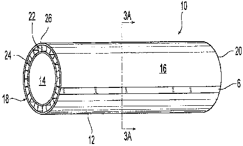
[0017] Figures 3 is a perspective view of an illustrative embodiment of a
stent graft
having a longitudinal radiopaque marker.
[0018] Figures 3A is a cross-sectional view of the stent graft of Figure 3
along line
3A--3A,
[0019] Figure 3B is an exploded perspective view of the stent graft of Figure
3.
[0020] Figure 4 is a perspective view of another embodiment of a stent graft
having a
longitudinal radiopaque marker.
[0021] Figures 5 and 6 are additional embodiments of a graft device having a
radiopaque marker,
[00221 Figure 7 is another embodiment of a graft device having a radiopaque
marker.
-6-
SUBSTITUTE SHEET (RULE 26)
CA 02626598 2008-04-18
WO 2007/056761 PCT/US2006/060702
[0023] Figure 8 is a fluoroscopic view of different embodiments of a bare
stent and
graft devices having a radiopaque marker.
Mode(s) For Carrying Out the Invention
[0024] Figures 1-8 illustrate the preferred embodiments. Shown in Figure 1 is
a
cross-section of one of the preferred embodiments of a graft device 100 having
at least one
radiopaque marker 106 embedded in an outer surface 104B of the device 100.
Alternatively
or in addition to, one or more radiopaque markers 102, 108 can be provided on
the inner
surface 104A, outer surface 104B or be dispersed or integrated with the graft
material 104 of
the device 100 between the inner surface 104A and the outer surface 104B.
[0025] The device 100 can be made from a graft material 104 which can be a non-
metallic material. Specifically, the non-metallic material 104 can include a
synthetic fiber or
fabric material such as, for example, Dacron, polyester, PTFE, ePTFE,
polyurethane,
polyurethane-urea, siloxane, and combinations thereof with an appropriate
amount of
additives added therein such as, for example, bio-active agents. In the
preferred
embodiments, the graft material 104 is expanded polytetrafluoroethylene or
"ePTFE."
[0026] The ePTFE material for graft 104 can be made by a variety of suitable
techniques, one of which is described as follows. A compounding of a polymeric
compound
is generated by sifting PTFE resin with a suitable amount of lubricant such
as, for example,
Isopar H, at 15-35% by weight of the PTFE to enable the PTFE to flow through
extrusion
equipment, The combined PTFE resin and lubricant are then placed in a shaker
device and
shaken so that the lubricant coats and penetrates each of the PTFE resin
particles. The
thoroughly mixed combination of PTFE resin and lubricant is then incubated in
a warming
cabinet overnight which is maintained at a temperature of approximately eighty-
five degrees
Fahrenheit (85 F). The incubation period is believed to allow for a further
and more equal
dispersion of the lubricant throughout the PTFE resin.
-7-
SUBSTITUTE SIHEET (RULE 26)
CA 02626598 2008-04-18
WO 2007/056761 PCT/US2006/060702
[0027] If desired, the PTFE resin can be further mixed and heated as part of
an
optional compounding process. For example, the PTFE resin can be compounded
with a
suitable hydroxyapatite (HA) material to produce a graft configured for
increased
biocompatibility and bioactivity in order to, for example, promote endothelial
cell growth for
the maintenance of graft patency and the reduction of intimal hyperplasia.
[0028] The PTFE resin or its compound can be preformed into a compressed
cylinder
by series of process steps. First, the resin can be poured into an inner
barrel of a preformer
by directing it through a funnel which is fit to the outside of the inner
barrel. Figure 2
illustrates a preferred embodiment of a divided preform barrel 40 which can be
used in
preforming a resin into a compressed cylinder. The divided preform barrel 40
preferably
includes an outer hollow cylindrical member 42, an optional inner hollow
cylindrical member
44, and a central solid cylindrical member 46. The inner hollow cylindrical
member 44 can
be concentrically contained within the outer hollow cylindrical member 42.
Details of a
similar process are shown and described in U.S. Patent Nos. 5,827,327;
5,641,443; and
6,190,590, each of which is incorporated in its entirety by reference.
(0029] The PTFE resin can be poured within a first area 52 located between the
outer
hollow cylindrical member 42 and a solid cylindrical member 46. The first area
52 can be
divided by one or more inner members 44 to define a secondary area 48 for
receipt of any
optionally added compound such as, for example, an HA compound material.
(0030] In one of the preferred embodiments, the outer hollow cylindrical
member 42
has a radius greater than the radius of the inner hollow cylindrical member
44. The diameter
of the components which comprise the preform barrel 40 will vary depending on
the size and
type of graft that is being produced. A preferred embodiment of the preform
barrel 40 can
have a radius of approximately 1.5 inches. The secondary area 48 between the
inner hollow
cylindrical member 44 and the central solid cylindrical member 46 can have a
radius of
-8-
SUBSTITUTE SHEET (RULE 26)
CA 02626598 2008-04-18
WO 2007/056761 PCT/US2006/060702
approximately 0.38 inches, the inner hollow cylindrical member 44 can have a
wall thickness
of approximately 0.07 inches, and the first area 52 located between the outer
hollow
cylindrical member 42 and the inner hollow cylindrical member 44 can have a
radius of
approximately 0.6 inches.
[0031] In addition, a radiopaque paste or resin can be partially or fully
embedded in a
portion of the outer or inner surfaces of the PTFE resin. Preferably, the
radiopaque paste can
be formed from a tantalum powder. Other radio-opaque materials which could be
used
include, but are not limited to, tungsten, gold, silver powder, Barium Sulfate
and the like.
The preferred radio-opaque material is also heat stable so that it can
tolerate sintering
temperature encountered during graft manufacturing. In one exemplary
embodiment, the
radio-opaque paste can be formed by mixing 4 grams of ePTFE, 6 grams of
tantalum and 2
grams of Isopar-H to produce a mixture containing sixty percent (60%)
tantalum. Preferably,
substantially all lubricant is evaporated after extrusion and sintering as
described herein.
Further in the alternative, the radiopaque paste can be formed from a Barium
Sulfate mixture.
For example, the radiopaque paste can include an ePTFE paste mixed with twenty
to forty
percent (20-40%) Barium Sulfate. In a preferred embodiment, the radiopaque
paste is formed
into an elongated strip that can be disposed along the length of the outer
surface of the PTFE
resin. Alternatively or in addition to, the radiopaque paste can form a
plurality of radiopaque
elements that can be aligned along the outer surface of the PTFE resin along
its length. The
radiopaque paste can be formed into any shape or form. For example, the paste
can be
formed as sutures, threads and other small pieces such as disks disposed
anywhere within the
PTFE resin. The continuous or elongated strip of radiopaque material can
provide the visual
cues to the clinician viewing the stent under fluoroscopy such as, for
example, location,
orientation or kinking.
-9-
SUBSTITUTE SHEET (RULE 26)
CA 02626598 2008-04-18
WO 2007/056761 PCT/US2006/060702
[00321 The assembly of PTFE resin and radiopaque paste markers can then be
compressed. The materials are compressed by placing the assembly into the
preform barrel
40 on a suitable press such as, for example, shown in Figure 3 of U.S. Patent
No, 5,827,327.
The press used during the compression of the polymeric compound is driven by a
suitable
power drive, which forces a top member toward a bottom member to compress the
material
within the divided preform barrel 40. FIallow cylindrical tubes of varying
thickness are used
to compress the material within the divided preform barrel 40 by slidably
reciprocating
around the inner hollow cylindrical member 44, the outer hollow cylindrical
member 42, and
the center solid cylindrical member 46 of the divided preform barrel 40. After
compressing
the materials contained within the preform barrel 40, the inner cylindrical
member 44 (if
used), the outer cylindrical member 42, and the center solid cylindrical
member 46 of the
divided preform barrel 40 are removed to obtain a compressed cylinder of
material.
Altematively, the dividers within the preform barrel may be removed prior to
compression,
without disturbing the interface between the different compounds, and then
compressed to
form a billet for extrusion. The compressed cylinder of material, or billet,
can be eo-extruded
via a suitable device such as, for example, the extruder shown in Figure 4 of
U.S. Patent No.
5,827,327. Briefly, the compressed cylinder of material is placed within an
extrusion barrel.
Force is applied to a ram, which in tum expels pressure on the compressed
cylinder of
material. The pressure causes the compressed cylinder of material to be
extruded around a
mandrel, through an extrusion die, and issue as a tubular extrudate. The
tubular extrudate can
be expanded to increase the porosity or alter the elasticity of the extrudate.
After extrusion or
expansion, the extrudate can be sintered in accordance with the expansion and
sintering
procedures undertaken with PTFE grafts which are known to those skilled in the
art.
[0033] In one embodiment, a PTFE billet can include an optional HA lumenal
layer
102 formed with a first outer strip of tantalum paste 106 and a second outer
strip of Barium
-10-
SUBSTITUTE SHEET (RULE 26)
CA 02626598 2008-04-18
WO 2007/056761 PCT/US2006/060702
Sulfate paste 108. The billet can be extruded through a suitable extruder at a
pressure from
about 500 to about 2000 psi. The reduction ratio (i.e., wall thickness of
billet to extruded graft
thickness) for the billet can be from about 50 to about 350. Table I below
shows a preferred
composition of a PTFE billet by weight.
[0034] Table 1
Ref. Formulation PTFE Tantalum Barium Hydroxy- Lube
Number Resin Weight (g) Sulfate apatite Weight
Weight (g) Weight (g) (g)
)
102 HA luminal 200 - 50 60
layer
106 Tantalum line 4 6 - - 2
108 Barium 4 - 6 - 2
Sulfate Line*
- PTFE base 500 100
The Barium Sulfate is preferably mixed with 10-200 milligrams (mg.) cobalt
blue (CAS no.
1345-16-0) to induce blue color.
[00351 The billets can be extruded to form various tubes I to 30 millimeters
(mm.) in
diameter, preferably 5 mm, to 6 mm. in diameter for peripheral vascular graft
applications.
More preferably, the diameter measured is the inner diameter of the tube. Each
extruded tube
can be expanded to various lengths to introduce different degrees of porosity
in the PTFE
material, thereby providing the expanded PTFE or ePTFE. The expanded tubes can
be
sintered at a suitable sintering temperature to cause the tube to maintain
essentially the
desired porosity and improve the physical characteristics of the expanded
ePTFE. The
expansion can potentially reduce the radio-opacity of the extruded material.
In general, higher
expansion gives reduced radio-opacity and/or visibility to the naked eye. It
is preferred to
add sufficient radio-opaque material or pigment material to produce a colored
marking after
expansion so that the graft shows adequate radio-opacity when viewed using
medical x-ray
imaging equipment. The sintering temperature can be similar to that of
standard ePTFE graft
processing, which can be from about 200 degrees Fahrenheit to 400 degrees
Fahrenheit, and
preferably about 300 degrees Fahrenheit. Other techniques to provide for the
graft device
-11-
SUBSTITUTE SHEET (RULE 26)
CA 02626598 2008-04-18
WO 2007/056761 PCT/US2006/060702
100 are shown and described in U.S. Patent Nos. 5,628,786; 6,053,943; and
6,203,735 and
U.S. Patent Application Publication Nos. 2004/0164445; 2004/0232588; and
2004/0236400,
each of which is incorporated in its entirety by reference.
[0036] In one embodiment, Barium Sulfate as a radio-opaque material is mixed
with a
biocompatible coloring agent to produce a blue color marking. Many
biocompatible coloring
agents or their mixtures can be used to produce desired color or shade. Black,
blue or green
colors are most preferred. Tantalum or tungsten metal provide black color as
well as radio-
opaque properties. In such case, no coloring agent may be needed. Many
biocompatible
coloring agents may be used, but colors that withstand high sintering
temperature without
substantial degradation are preferred. The preferred colored materials
include, but are not
limited to, cobalt blue, (Phthalocyaninato(2-)) copper, Chromium-cobalt-
aluminum oxide,
titanium oxide or mixtures thereof and the like.
[0037] Again referring to Figure 1, a tube preferably extruded by the process
described above can form the lumenal graft device 100. The graft device 100
further
preferably includes one or more elongated radiopaque markers or strips 106,
108 embedded
in a first inner surface 104A, a second surface 104B or in the material 104
between the first
and second surfaces 104A, 104B. More specifically, extrusion of the PTFE resin
and the
radiopaque marker provides for the device 100 with an ePTFE layer 104 with
first surface
104A and second surface 104B. In a preferred embodiment, the device 100
includes at least
one elongated portion 106 of radiopaque material on the outer surface 104B in
which the
radiopaque material is made of either tantalum powder or Barium Sulfate. The
elongated
portion 106 further preferably forms a continuous strip that runs along the
length of the
device 10. Alternatively, the graft device 100 can have one or more radiopaque
elements, in
any orientation line provided by the device 100 to improve visibility in a
suitable imaging
technique (e.g., x-ray imaging). More specifically, the radiopaque material
can form a series
-12-
SUBSTITUTE SHEET (R.ULE 26)
CA 02626598 2008-04-18
WO 2007/056761 PCT/US2006/060702
of radiopaque elements (not shown) aligned along the length of the outer
surface 104B of the
device 100.
[0038] Referring now to Figures 3-3A, shown is a preferred embodiment of an
encapsulated stent or "stent-graft" 10. The stent-graft 10 can generally
include a tubular
member 12 having an interior surface 14 and an exterior surface 16 which are
contained
between first and second ends 18, 20. An elongated radiopaque marker 6 is
preferably
provided on the exterior surface 16. As illustrated in FIGS. 3, 3A and 3B, the
tubular
member 12 preferably includes a balloon or pressure expandable tubular shaped
support
member 22 which is loaded over a first biocompatible flexible tubular member
24 that is held
on a mandrel (not shown). A second biocompatible flexible tubular member 26 is
then
preferably loaded over the first biocompatible tubular member/support member
combination
22, 24. The tubular shaped support member 22 preferably includes a stent
similar to that
shown or described in any one of U.S. Pat. Nos. 4,733,665; 6,053,941;
6,053,943; 5,707,386;
5,716,393; 5,860,999; and 6,572,647 each of which is incorporated in its
entirety by
reference. The stent utilized for the member 22 can be balloon expandable
stent, self-
expanding stent or memory-shaped plastic stent. The tubular members 24, 26 are
preferably
fused together to encapsulate the support member 22,
[00391 The tubular members 24, 26 of stent-graph 10 are preferably formed in a
manner substantially similar to the extruded graph device 100 described above.
In particular,
the first and second biocompatible flexible tubular members 24, 26 are
preferably made by
extruding a billet of expanded polytetrafluoroethylene (ePTFE). Alternatively,
the first and
second biocompatible flexible tubular members 24, 26 may also be made of
unexpanded
polytetrafluoroethylene (PTFE). The tubular member 26 is preferably extruded
along with a
radiopaque material to form at least one elongated radiopaque marker 6
embedded in the
outer surface of the tubular member 26. Alternatively or in addition to, the
tubular member
-13-
SUBSTITUTE SHEET (RULE 26)
CA 02626598 2008-04-18
WO 2007/056761 PCT/US2006/060702
24 can also be extruded along with a radiopaque material to form at least one
elongated
radiopaque marker embedded in the outer surface of the tubular member 24.
Further, the
pressure expandable tubular shaped support member 22 may be made of any
material having
the strength and elasticity to permit radial expansion and resist radial
collapse such as silver,
titanium, stainless steel, gold, and any suitable plastic material capable of
maintaining its
shape and material properties at various sintering temperatures for PTFE or
ePTFE.
[0040] Shown in Figure 3A is a cross-sectional view of the stent-graft 10 of
Figure 3
prior to fusing the graft or tubular members 24, 26 to the expansion member
22. The first
biocompatible flexible tubular member 24, preferably made of unsintered ePTFE,
forms the
innermost layer or luminal surface of the stent-graft 10, and covers the lumen
28 of the stent-
graft 10, thereby providing a smooth, inert biocompatible blood flow surface.
The tubular
support member 22, preferably a stent or similarly constructed structure,
forms the middle
layer located at the center of the stent-graft 10. Finally, the second
biocompatible flexible
tubular member 26, which is also preferably made of unsintered eP7'FE, forms
the outermost
layer or abluminal surface of the stent-graft 10.
100411 To form the stent-graft 10, the tubular shaped members 24, 22, and 26
can be
loaded onto one another. Pressure is applied to the graft/stentlgraft assembly
in order to fuse
the first and second biocompatible flexible tubular members 24, 26 to one
another through
the openings contained within the tubular support member 22. Where the tubular
support
member 22 is a stent frame, the first and second ePTFE tubular members 24, 26
are fused to
one another through the openings between the struts of the stent. The
graft/stent/graft
assembly is then heated at sintering temperatures to form a physical bond
between the ePTFE
layers. The resulting prosthesis is an unexpanded stent encapsulated within
ePTFE layers, or
specifically, an unexpanded stent having ePTFE layers on its luminal and
abluminal surfaces
in which the stent and ePTFE layers are inseparable. Alternatively, the
prosthesis can include
-14-
SUBSTITUTE SHEET (RULE 26)
CA 02626598 2008-04-18
WO 2007/056761 PCT/US2006/060702
hydroxyapatite on both its luminal and abluminal surfaces. Further, the ePTFE
layers may
also be fused or joined together around the ends of the unexpanded stent
thereby entirely
encasing the stent within ePTFE in both the radial and longitudinal
directions. The resulting
stent-graft 10 can be loaded onto a suitable delivery device such as, for
example, U.S. Patent
No, 6,756,007, which is incorporated in its entirety by reference. The stent-
graft 10 may
advantageously be used in a variety of medical applications including
intravascular treatment
of stenoses, aneurysms or fistulas; maintaining openings in the urinary,
biliary,
tracheobronchial, esophageal, renal tracts, vena cava filters; repairing
abdominal aortic
aneurysms; or repairing or shunting damaged or diseased organs such as, for
example,
Transjugular Intrahepatic Portosystemic Shunt (TIPS).
[00421 Procedures like TIPS can use an alternative embodiment of the stent-
graft 10
as shown in Figure 4. Shown in Figure 4 is a stent-graft 10' having a bare
stent portion 12'
embedded in a stent-graft, encapsulated or covered portion 14', i.e.,
a"hybrid' stent-graft. A
surgical procedure using the hybrid or stent-graft 10' may require
determination of where the
covered portion 14' ends during the procedure in order to allow blood flow
through the
uncovered stent-graft portion 12'. The encapsulated portion 14' of the hybrid
stent graft 10' is
preferably formed in a manner substantially similar for forming the stent-
graft 10 as
described above so as to include extrusion of at least an outer ePTFE or PTFE
member 26'
with an elongated radiopaque material to form the radiopaque marker 6'.
Accordingly, the
radiopaque marker or strip 6' on the covered portion 14' of the stent 10'
provides a medical
practitioner with a visual cue as to the actual position of the covered
portion while the
implantable prosthesis is inside a mammalian body. In addition, the radiopaque
marker or
strip 6' provides by its proximity, the position of the uncovered portion 12'
of the hybrid
stent-graft 10' to determine placement of the entire stent-graft 10' during
and subsequent to a
-15-
SUBSTITUTE SHEET (RULE 26)
CA 02626598 2008-04-18
WO 2007/056761 PCT/US2006/060702
surgical procedure. Moreover, the radiopaque strip 6' can eliminate the need
for "spoon" or
bead-type markers used at the non-encapsulated ends of the stent 10'.
[0043] Referring to Figures 5 and 6, shown are alternative embodiments of a
graft,
namely vascular bypass grafts 200 and 300 having a radiopaque elongation,
marker, or strip
embedded in the outer surface. Vascular bypass graft 200 is configured for
desired blood
flow characteristics for applications above the knees, whereas bypass graft
300 is configured
for blood flow characteristics below the knee. Regardless of the structural
configurations and
applications of the bypass grafts 200 and 300, the grafts 200, 300 can be
preferably formed
by extruded ePTFE material along with a radiopaque paste as described above to
provide the
elongated radiopaque markers or strips 204, 304. That is, a radiopaque paste
can be
embedded or incorporated by extrusion with the synthetic non-metallic material
(e.g.,
Dacron, polyester, PTFE, ePTFE, polyurethane, polyurethane-urea, siloxane, and
combinations thereof) to form grafts 200 aiid 300 with a radiopaque strip 204,
304 along at
least one of the luminal and abluminal surfaces of the grafts (200 or 300).
The material or
combinations of materials used (e.g., Dacron, polyester, PTFE, ePTFE,
polyurethane,
polyurethane-urea, siloxane, and combinations thereof) can include surface
modifying
additives or other materials. Examples of various grafts are shown and
described in U.S.
Patent Nos. 6,203,735; 6,039,755; and 6,790,226, each of which is incorporated
in its entirety
by reference.
[0044] Shown in FIG. 7 is another embodiment of the graft device 200. In the
device
200, the graft material can be formed in a manner as previously described
above using PTFE.
However, the radiopaque marker 206 is not extruded with the graft material.
Instead, the
radio-opaque marker 206 is preferably printed on the extruded PTFE forming the
tube of the
device 200 by a suitable printing technique, such as, for example, engraving,
mono-type,
offset, cliche transfer, ink-jet or gliced printing. The radio-opaque marker
206 can be a radio-
-16-
SUBSTITUTE SHEET (RUI,E 26)
CA 02626598 2008-04-18
WO 2007/056761 PCT/US2006/060702
opaque ink such as, for example, the ink produced by CT Medical, Inc, of
Norton Mass.
Preferably, the radiopaque ink is tungsten based in which tungsten is mixed as
a radio-opaque
component into the ink. In one embodiment, an ink composition for an
orientation line for an
ePTFE surface includes a suitable polymeric binder that adheres well to an
ePTFE surface, a
biocompatible dye or pigment, a radiopaque material and a solvent that
dissolves the
polymeric binder. In addition, the ink composition may contain inorganic white
solid
materials such as titanium dioxide (to adjust ink shade) and a viscosity
modifier. Although
many pigments or dyes may be used to make the orientation line, pigments or
dyes that have
a long history of human implantation are most preferred. The preferred color
compounds in
the ink include, but are not limited to: (Phthalocyaninato(2-)) copper, D&C
Blue No. 9, D&C
Green No. 5, Chlorophyllin-copper complex, oil soluble, Chromium-cobalt-
aluminum oxide,
Ferric ammonium citrate, D&C Blue No. 5, FD&C Blue No. 2, D&C Green No. 6,
Titanium
dioxide, carbon, Iron oxide, and the like. (Phthalocyaninato(2-)) copper is
the most preferred
green compound. The color of the ink (e.g., black, blue, etc.) may be
determined by viewing
under a light having a temperature of about 6500 degrees Kelvin. Hence, in
this embodiment,
the lines are not only visible to the unaided human eyes, they are also
visible to the human
eyes with a suitable fluoroscope imager.
[0045] With reference to Figure 8, it has been demonstrated that various
physical
embodiments of the device can be viewed under fluoroscopic examination. In
Figure 8, two
graft devices IOOA and 100B were provided along with a stent-graft 100c
(commercially
available under the trade name Fluency ) as referential datum for visibility
under a
fluoroscope imaging device through an Aluminum plate of 15 millimeters
thickness. The
reference stent-graft 100c was utilized with radiopaque ink that had 50%
tantalum and 50%
polycarbonate polyurethane as polymeric binder. The ink solution/dispersion
was prepared by
dissolving the polymer in tetrahydrofuran solvent. The dispersion was hand
painted using a
-17-
SUBSTITUTE SHEET (RULE 26)
CA 02626598 2008-04-18
WO 2007/056761 PCT/US2006/060702
paint brush to create a circular band on the stent graft surface. The band is
slightly visible
under fluoroscope so that the reference stent-graft 100c acts as referential
datum as to the
minimum radiopacity required. The aluminum plate is utilized to simulate the
density of
biological tissues by interposition of the plate (not shown) between the
fluoroscope and the
subject graft device. That is, the image of the reference stent-graft 100c in
Figure 8 provides
for an indication of the radiopacity of the stent as compared to the
background environment
on which the stent graft is placed in. Further, by having the Fluency stent
graft in the image,
a referential datum as to the effectiveness of the radiopaque line of the
preferred
embodiments is provided without resorting to observers with specialized
training or machine
visions, Consequently, as long as an ordinary observer can determine that the
lines provided
by the graft of the preferred embodiment in a fluoroscopic display medium has
a darker or
higher contrast image than the reference stent-graft 100c, then the
radiopacity of the line
would be deemed to be greater than a minimum level needed for the line to
function as a
radio-opaque marker in a mammalian body. Alternatively, a machine vision with
the ability
to recognize discrete levels of contrast can be utilized to provide an
objective indicator of the
effectiveness of the radiopacity of the radiopaque lines.
[00461 The graft device 100a was provided with two lines 106a and 108a where
each
line can be a combination of two different radiopaque materials: (1) Barium
Sulfate and (2)
Tantalum. Even though both lines are formed of different materials, both
materials form
generally similar solid black lines of radiopacity greater than the
referential bare stent in a
suitable imaging device, which is a black-and-white photographic print, as
shown in Figure
8. Alternatively, graft 100b was provided with two lines 106b and 108b where
106b is made
of 60% by weight Barium Sulfate material and a suitable colorant (e.g., cobalt
blue). This
line appears blue to the naked eye and is radio-opaque. The line 108B is made
using 60% (by
weight) of Tantalum powder and is black in color. The tantalum provides the
black coloring
-18-
SUBSTITUTE SHEET (RULE 26)
CA 02626598 2008-04-18
WO 2007/056761 PCT/US2006/060702
and generally no coloring agent is needed. These lines demonstrate that a
clinician (or even
an ordinary observer without any specialized training) would be able to
observe and
determine whether the graft device has been moved about in the body subsequent
to
implantation into an undesirable configuration by observing the orientation of
such line on a
fluoroscopic display medium (paper or graphical display monitor). For example,
where the
graft has twisted about its own axis, the display medium would show that the
line forms a
spiral, which is partly shown for graft 100A. Where the graft has rotated
about an axis
transverse to a longitudinal axis of the graft, i.e., a kink, the display
medium would show an
intersecting point rather than a smooth inflection curve, shown here in Figure
8 for graft
100B.
[0047] Although the graft device 100 has been described in relation to
specific
examples noted above, it should be emphasized that variations in the
configuration or
composition of ePTFE, radiopaque marker, stent framework, and other design
parameters can
be utilized with the graft device 100. For example, the weight percentage of
either the
tantalum powder or the Barium Sulfate in the graft device can vary. The
percentage of radio-
opaque composition in the graft will depend on the amount of radio-opacity
needed for a
given medical application and the amount of graft expansion subjected during
manufacturing.
The percentage of radio-opaque element such as tantalum or Barium Sulfate will
very from
5% to 70% most preferably from 20% to 60% and even more preferably from 50-60
%.
Finally, other types of bioactive agents can also be combined with the
radiopaque materials
described herein for the graft and the stent graft. The bioactive agents
include (but are not
limited to) pharmaceutic agents such as, for example, anti-
proliferative/antimitotic agents
including natural products such as vinca alkaloids (i.e. vinblastine,
vincristine, and
vinorelbine), paclitaxel, epidipodophyllotoxins (i.e. etoposide, teniposide),
antibiotics
(dactinomycin (actinomycin D) daunorubicin, doxorubicin and idarubicin),
anthracyclines,
-19-
SUBSTITUTE SHEET (RULE 26)
CA 02626598 2008-04-18
WO 2007/056761 PCT/US2006/060702
mitoxantrone, bleomycins, plicamycin (mithramycin) and mitomycin, enzymes (L-
asparaginase which systemically metabolizes L-asparagine and deprives cells
which do not
have the capacity to synthesize their own asparagine); antiplatelet agents
such as G(GP)
llb/llIa inhibitors and vitronectin receptor antagonists; anti-
proliferative/antimitotic alkylating
agents such as nitrogen mustards (mechlorethamine, cyclophosphamide and
analogs,
melphalan, chlorambucil), ethylenimines and methylmelamines
(hexamethylmelamine and
thiotepa), alkyl sulfonates-busulfan, nirtosoureas (carmustine (BCNU) and
analogs,
streptozocin), trazenes - dacarbazinine (DTIC); anti-proliferative/antimitotic
antimetabolites
such as folic acid analogs (methotrexate), pyrimidine analogs (fluorouracil,
floxuridine, and
cytarabine), purine analogs and related inhibitors (mercaptopurine,
thioguanine, pentostatin
and 2-chiorodeoxyadenosine {cladribine}); platinum coordination complexes
(cisplatin,
carboplatin), procarbazine, hydroxyurea, mitotane, aminoglutethimide; hormones
(i.e.
estrogen); anti-coagulants (heparin, synthetic heparin salts and other
inhibitors of thrombin);
fibrinolytic agents (such as tissue plasminogen activator, streptokinase and
urokinase),
aspirin, dipyridamole, ticlopidine, clopidogrel, abciximab; antimigratory;
antisecretory
(breveldin); anti-inflammatory: such as adrenocortical steroids (cortisol,
cortisone,
fludrocortisone, prednisone, prednisolone, 6a-methylprednisolone,
triamcinolone,
betamethasone, and dexamethasone), non-steroidal agents (salicylic acid
derivatives i.e.
aspirin; para-aminophenol derivatives i.e. acetominophen; indole and indene
acetic acids
(indomethacin, sulindac, and etodalac), heteroaryl acetic acids (tolmetin,
diclofenac, and
ketorolac), arylpropionic acids (ibuprofen and derivatives), anthranilic acids
(mefenamic
acid, and meclofenamic acid), enolic acids (piroxicam, tenoxicam,
phenylbutazone, and
oxyphenthatrazone), nabumetone, gold compounds (auranofin, aurothioglucose,
gold sodium
thiomalate); immunosuppressives: (cyclosporine, tacrolimus (FK-506), sirolimus
(rapamycin), azathioprine, mycophenolate mofetil); angiogenic agents: vascular
endothelial
-20-
SUBSTITUTE SHEET (RULE 26)
CA 02626598 2008-04-18
WO 2007/056761 PCT/US2006/060702
growth factor (VBGF), fibroblast growth factor (FGF); angiotensin receptor
blockers; nitric
oxide donors; anti-sense oligionucleotides and combinations thereof; cell
cycle inhibitors,
mTOR inhibitors, and growth factor receptor signal transduction kinase
inhibitors; retenoids;
cyclin/CDK inhibitors; HMG co-enzyme reductase inhibitors (statins); and
protease
inhibitors,
[0048] Furthermore, the radiopaque marker, when configured as a strip or
substantially straight line provides additional visual cues to the
practitioner or clinician
beyond graft location. Specifically, the straight line radiopaque marker can
indicate whether
there is any twisting of the graft during and subsequent to the implantation
procedure. This
feature is believed to be advantageous in that it allows for a clinician to
determine with
certainty whether the prosthesis has been implanted optimally in the body
without kinking or
twisting. That is, prior to the development of the prosthesis as described
herein, movements
of the arms and legs could cause the implanted prosthesis to kink or twist so
as to restrict
blood flow through the prosthesis without the clinician being aware of such
adverse
configurations after the implantation has been completed. The prosthesis, as
described
herein, allows the clinician to achieve an advantageous technique by ensuring
that the
prosthesis implanted by the clinician is properly configured inside the
mammalian body.
Alternatively, other types of indicia (e.g., date of manufacture, manufacture
etc.,) can be
provided by printing the radiopaque material onto the graft. The radio-opaque
lines can also
be encoded such as are used, for example, in bar coding of commercial goods.
For example
bar code, a series of black and white lines with certain thickness and heights
can be
interpreted by the machines as digital code which can be used in a computer
database.
[0049] As used herein, the singular form of "a," "an," and "the" include the
plural
referents unless specifically defined as only one. While the present invention
has been
disclosed with reference to certain preferred embodiments, numerous
modifications,
-21-
SUBSTITUTE SHEET (RULE 26)
CA 02626598 2008-04-18
WO 2007/056761 PCT/US2006/060702
alterations, and changes to the described embodiments are possible without
departing from
the sphere and scope of the present invention, as defined in the appended
claims. Moreover,
where methods, processes and steps described above indicate that certain
events occurring in
certain order, those skilled in the art would recognize that the ordering of
steps may be
modified and that such modifications are within the variations of the
described embodiments.
Accordingly, it is intended that the present invention not be limited to the
described
embodiments, but that it have the full scope defined by the language of the
following claims,
and equivalents thereof.
-22-
SUBSTITUTE SHEET (RULE 26)