Note: Descriptions are shown in the official language in which they were submitted.
CA 02629405 2013-11-05
DESCRIPTION
TISSUE ENGINEERING METHODS AND COMPOSITIONS
10 GRANT STATEMENT
This work was supported by grants AR49294 and GM08555 from the
United States National Institutes of Health. Thus, the U.S. government has
certain rights in the presently disclosed subject matter.
TECHNICAL FIELD
The presently disclosed subject matter generally relates to novel
methods and systems for differentiating adipose derived stem (ADS) cells to
provide cells and tissues suitable for laboratory and therapeutic
applications.
These cells, and other cells, can be employed alone or in conjunction with
unique biologically-compatible scaffold structures to generate differentiated
tissues and structures, both in vitro and in vivo.
TABLE OF ABBREVIATIONS
ADS adipose-derived stem
AN OVA analysis of variance
BaCl2 barium chloride
BMI body mass index
BMP bone morphogenic protein
CaCl2 calcium chloride
CAT chloramphenicol acetyl transferase
cm centimeter
CMV cytomegalovirus
COMP cartilage oligomeric protein
- 1 -
CA 02629405 2008-05-12
WO 2007/030811
PCT/US2006/035243
Dhfr - dihydrofolate reductase
DMEM - Dulbecco's Modified Eagle's Medium
DNA - deoxyribonucleic acid
dsDNA - double stranded DNA
EG - embryonic germ
EGF - epidermal growth factor
ePTFR - expanded PTFE
ES - embryonic stem
FACS - fluorescence-activated cell sorting
FBS - fetal bovine serum
FGF - fibroblast growth factor
g - gram
GAG - glycosaminoglycan
CF - growth factor
GFP - green fluorescent protein
GLA - glycine leucine alanine
hADS - human adipose-derived stem
Hprt - hypoxanthine phosphoribosyl transferase
HSC - hematopoietic stem cells
HSV-tk - herpes simplex virus-thymidine kinase
lEp - immediate early viral promoter
IGF-I - insulin-like growth factor
ITR - inverted terminal repeat
L - liter
LTR - long terminal repeat
mg - milligram
mL - milliliter
MLV - murine leukemia virus
mM - millimolar
mRNA - messenger RNA
MSC - mesenchymal stem cell
NaCI - sodium chloride
ng - nanogram
- 2 -
CA 02629405 2008-05-12
WO 2007/030811
PCT/US2006/035243
p - probability
PBS - phosphate buffered saline
FOR - polymerase chain reaction
PEEK - polyetheretherketone
PEG - polyethylene glycol
PEO _ polyethylene oxide
PGA - polyglycolic acid
PDGF - platelet-derived growth factor
Pgk - phosphoglycerate kinase
PLA - processed lipoaspirate
PLSD - protected least significant difference
PTFE - polyetetrafluoroethylene
PTHrP - parathyroid hormone-related protein
RGD - arginine glycine aspartic acid
RNA - ribonucleic acid
RSV - rous sarcoma virus
S.E.M. - standard error of measurement
SV40 - simian virus 40
TAF - transcription associated factor
TGF-a - transforming growth factor a
TG F-13 - transforming growth factorfl
VEGF - vascular endothelial growth factor
2-D - 2-dimensional
3-D - 3-dimensional
3H - tritium
pCi - microcuries
Pg - microgram
pm - micrometer
cyo - percent
# - number ,
< - less than or equal to
> - greater than or equal to
> - greater than
- 3 -
CA 02629405 2008-05-12
WO 2007/030811
PCT/US2006/035243
less than
equal to
plus or minus
plus
BACKGROUND
Many disease conditions or injuries of the body require the repair or
replacement of damaged tissues and/or structures, but the body itself may not
be able to replace or repair the tissue and/or structures satisfactorily or
within
an appropriate time scale. Accordingly, many methods of disease or injury
treatment involve augmenting the body's natural repair mechanisms and often
rely on the use of implantable biological scaffolds or prostheses. Tissue
engineering attempts to create three-dimensional tissue structures on which
cells and other biomolecules can be incorporated. These structures or
scaffolds
guide the organization, growth and differentiation of cells in the process of
forming functional tissue by providing physico-chemical cues.
For example, degenerative joint diseases such as osteoarthritis remain a
source of significant pain and disability, resulting in an economic burden of
over
40 billion dollars per year to the United States. Present treatment options
for
osteoarthritis are limited, and surgical management generally involves
replacement of the joint with a metal and polyethylene prosthesis. The short
life
span and loading tolerance of joint replacement makes this treatment
unacceptable for young, potentially active individuals. The treatment of
synovial
joints using tissue engineered grafts shows tremendous promise but its
application has been limited to the treatment of small cartilage defects in
the
knee joint.
Further, articular cartilage is avascular, aneural, and has limited capacity
for self-repair. Particularly, articular cartilage is a thin layer of soft
connective
tissue (0.5-5 mm thick) that covers the articulating surfaces of long bones in
synovial joints. The principal function of articular cartilage is to
redistribute
applied loads and to provide a low friction-bearing surface to facilitate
movement within these joints. Damage to this connective tissue in joints
results
in significant pain and morbidity, and currently, there are few options
available
- 4 -
CA 02629405 2008-05-12
WO 2007/030811
PCT/US2006/035243
for treatment. Some
treatment options include lavage, debridement,
nnicrofracture, and autologous and/or allogeneic osteochondral/chondral grafts
(reviewed in Hunziker (2002) Osteoarthritis Cartilage 10:432-463.
The success rates from these treatment options vary greatly, and some
show promise. However, in many of the studies, the results suggest fibrous
tissue formation, apoptosis, and further cartilage degeneration nonetheless
occur (Furukawa etal. (1980) J Bone Joint Surg Am 62:79-89; Kim etal. (1991)
J Bone Joint Surg Am 73:1301-1315; Shapiro etal. (1993) J Bone Joint Surg
Am 75:532-553; Nehrer etal. (1999) Clin Orthop Relat Res 365:149-162; Tew
etal. (2000) Arthritis Rheum 43:215-225; Mitchell and Shephard (2004) Clin
Orthop Relat Res 423:3-6. Autologous chondrocyte transplants studies have
also shown an inability to produce hyaline cartilage repair tissue,
specifically
over long time periods Brittberg et al. (1996) Clin Orthop Relat Res 326:270-
283; Brittberg (1999) Clin Orthop Re/at Res 367(Suppl):S147-155; Nehrer etal.
(1999) Clin Orthop Re/at Res 365:149-162; Breinan etal. (2001) J Orthop Relat
Res 19:482-492, and even though some clinical studies have shown some
promising results Brittberg etal. (1994) N Engl J Med 331:889-895; Breinan et
al. (1997) J Bone Joint Surg Am 79:1439-1451; Minas and Nehrer (1997)
Orthopedics 20:525-538; Gilloglv et al. (1998) J Orthop Sports Phys Ther
28:241-251, as with the other treatment options, randomized, controlled trials
are needed to truly ascertain the efficacy of these procedures. Given the
success rate to date of current cartilage remodeling, repair, regrowth, and/or
regeneration treatment options, combined with the burgeoning economic
burden cartilage pathology and osteoarthritis has on society (Jackson et al.
(2001) Clin Orthop Re/at Res 391(Suppl):S14-25), novel tissue engineering
approaches are needed to establish improved options for the treatment of
cartilage defects and osteoarthritis, among other maladies.
In recent years, the identification of mesenchymal stem cells has led to
advances in tissue regrowth and differentiation. Such cells are pluripotent
cells
found in bone marrow and periosteum, capable of differentiating into various
mesenchymal or connective tissues. For example, such bone-marrow derived
stem cells can be induced to develop into myocytes upon exposure to agents
such as 5-azacytidine (Wakitani etal., (1995) Muscle Nerve, 18(12), 1417-26).
- 5 -
CA 02629405 2008-05-12
WO 2007/030811
PCT/US2006/035243
It has been suggested that such cells are useful for repair of tissues such as
cartilage, fat, and bone (see, e.g., U.S. Pat. Nos. 5,908,784, 5,906,934,
5,827,740, 5,827,735), and that they also have applications through genetic
modification (see, e.g., U.S. Pat. No. 5,591,625). While the identification of
such cells has led to advances in tissue regrowth and differentiation, the use
of
such cells is hampered by several technical hurdles. One drawback to the use
of such cells is that they are very rare (representing as few as 1/2,000,000
cells), making any process for obtaining and isolating them difficult and
costly.
Additionally, bone marrow harvest is universally painful to the donor.
Moreover,
such cells are difficult to culture without inducing differentiation, unless
specifically screened sera lots are used, adding further cost and labor to the
use of such stem cells. Thus, there is a need for a more readily available
source for pluripotent stem cells, particularly cells that can be cultured
without
the requirement for costly prescreening of culture materials.
Other advances in tissue engineering have shown that cells can be
grown in specially-defined cultures to produce three-dimensional structures.
Spatial definition typically is achieved by using various acellular fiber
scaffolds
or matrices to support and guide cell growth and differentiation. While this
technique is still in its infancy, experiments in animal models have
demonstrated that it is possible to employ various acellular fiber scaffold
materials to regenerate whole tissues (see, e.g., Probst etal. (2000) BJU
Int.,
85(3), 362-367). While artificial fiber scaffolds have been developed, these
can
prove toxic either to cells or to patients when used in vivo, or do not
provide
adequate mechanical support required for tissue repair. Accordingly, there
remains a need for a scaffold material suitable for use as a substrate in
culturing and growing populations of cells, wherein the matrix, cell
combination
is tailored specifically for replacement of a target tissue. Ultimately, this
replacement tissue will serve to substantially function as the native tissue
it
seeks to replace.
Accordingly, the presently disclosed subject matter addresses needs in
the art for improved methods for producing improved tissue engineered
implantable compositions. This and other needs are addressed in whole or in
part by the presently disclosed subject matter.
- 6 -
CA 02629405 2008-05-12
WO 2007/030811
PCT/US2006/035243
SUMMARY
This Summary lists several embodiments of the presently disclosed
subject matter, and in many cases lists variations and permutations of these
embodiments. This Summary is merely exemplary of the numerous and varied
embodiments. Mention of one or more representative features of a given
embodiment is likewise exemplary. Such an embodiment can typically exist
with or without the feature(s) mentioned; likewise, those features can be
applied
to other embodiments of the presently disclosed subject matter, whether listed
in this Summary or not. To avoid excessive repetition, this Summary does not
list or suggest all possible combinations of such features.
Methods and compositions for treating a tissue pathology in a subject
are disclosed. In some embodiments, the subject is a mammalian subject.
In some embodiments, the method comprises providing to an adipose-
derived stem (ADS) cell in culture; exposing the ADS cell to an effective
amount
of a BMP-6 polypeptide or a functional fragment thereof, wherein the effective
amount of the BMP-6 polypeptide or the functional fragment thereof is
sufficient
to induce the ADS cell to differentiate into a cell capable of treating the
tissue
pathology in the subject; administering the cell to the subject. In some
embodiments, the effective amount of BMP-6 ranges from about 1 picogram/mL
to about 10 milligram/mL.
Also disclosed is a method for treating a tissue pathology in a subject,
comprising providing to an adipose-derived stem (ADS) cell in culture;
exposing
the ADS cell to an effective amount of a biologically active agent, wherein
the
effective amount of the biologically active agent is sufficient to induce the
ADS
cell to differentiate into a cell capable of treating the tissue pathology in
the
subject; and administering the cell to the subject.
Also disclosed is a composition for treating a tissue pathology in a
subject. The composition can comprise an adipose-derived stem (ADS) cell that
has been differentiated in vitro by exposure to an effective amount of a BMP-6
polypeptide or a functional fragment thereof; and a pharmaceutically
acceptable
carrier or excipient.
Also disclosed is composition for treating a tissue pathology in a subject,
the composition comprising an adipose-derived stem (ADS) cell; an effective
- 7 -
CA 02629405 2008-05-12
WO 2007/030811
PCT/US2006/035243
amount of a BMP-6 polypeptide or a functional fragment thereof; and a
pharmaceutically acceptable carrier or excipient.
Also disclosed is a composition for treating a tissue pathology in a
subject, the composition comprising an adipose-derived stem (ADS) cell that
has been differentiated in vitro by exposure to an effective amount of a
biologically active agent; and a pharmaceutically acceptable carrier or
excipient.
Also disclosed is a composition for treating a tissue pathology in a
subject, the composition comprising an adipose-derived stem (ADS) cell; an
effective amount of a biologically active agent; and a pharmaceutically
acceptable carrier or excipient.
The cell can be administered to a target tissue selected from the group
including but not limited to articular cartilage, non-articular cartilage,
auricular
cartilage, tracheal cartilage, laryngeal cartilage, nasal cartilage, growth
plate
cartilage, meniscus, labrum, intervertebral disc, tendon, ligament,
periodontal
ligament, fascia, and muscle. The target tissue can comprise multiple tissue
types that are integrated with one another selected from the group consisting
of
bone and cartilage, muscle and tendon, and ligament and bone. The tissue
pathology can comprise a compromise in the normal homeostasis of the tissue,
optionally culminating in degeneration of the tissue. The tissue pathology can
comprise loss, damage, injury, or combinations thereof to the tissue. The
treating can comprise tissue remodeling, repair, regrowth, resurfacing,
regeneration, or combinations thereof.
The ADS cells from an adipose depot selected from the group can be
selected from the group including but not limited to subcutaneous abdomen,
thigh, buttocks, infrapatellar fat pad, and combinations thereof. The ADS cell
can be selected from the group including but not limited ADS cells autologous
to the subject, ADS cells allogeneic to the subject, ADS cells xenogenic to
the
subject, and combinations thereof.
The isolated ADS cells can be transfected with an expression construct
encoding a biologically active agent, such as but not limited to at least one
of
BMP-6 polypeptide, a BMP-6 receptor polypeptide, or a functional fragment
thereof. The expression construct can comprise a regulatable promoter
operatively linked to at least one coding sequence. The expression vector
- 8 -
CA 02629405 2008-05-12
WO 2007/030811
PCT/US2006/035243
encoding a biologically active agent (such as but not limited to a BMP-6
polypeptide or a functional fragment thereof) can be administered in addition
to
the differentiated cell. The expression vector can be selected from the group
including but not limited to a viral vector, an adenovirus vector, an adeno-
associated virus vector, a plasmid, and a deoxyribonucleic acid molecule.
The ADS cell can be present in or on a biocompatible scaffold. The
biologically active agent, such as but not limited to a BMP-6 polypeptide or
functional fragment thereof, can be incorporated into the scaffold for
controlled
release over time.
The ADS cell can be exposed in culture to at least one other biologically
active agent, such as but not limited to a growth factor or cytokine.
Representative growth factors or cytokines including but are not limited a TGF-
13
member, an IGF-1, an FGF, an EGF, a PDGF, a parathyroid
hormone related peptide (PTHrP), an interleukin, and combinations thereof.
Another cell type other than the ADS cell can be administered along with
the ADS cell. The other cell type can be selected from the group including but
not limited to a chondrocyte, a fibroblast, an osteoblast, a myoblast, a
neuron, a
progenitor cell, and combinations thereof.
A subpopulation of differentiated ADS cells can be selected. The
subpopulation of differentiated ADS cells can be selected based on: (i)
expression of at least one cell surface marker can be selected from the group
including but not limited to CD10, CD13, CD31, CD34, CD36, CD44, CD49,
CD54, CD55, CD59, CD65 CD105, and CD166; (ii) differential expression of
aldehyde dehydrogenase (ALDH); (iii) differential expression of collagen 1;
(iv)
efflux of a dye such or a nucleic acid label; (v) telomere length or the
expression
of telomerase; (vi) expression of TGF-13 superfamily members; (vii) expression
of TGF-13 superfamily member receptor polypeptides; or (viii) combinations of
any of the foregoing. The dye can comprise Hoechst 33342. The
subpopulation of differentiated ADS cells can be selected by repeated passage
in culture.
The differentiated ADS cell can be identified as a cell suitable for use in
therapeutic restorative and regenerative techniques when gene expression
- 9 -
CA 02629405 2008-05-12
WO 2007/030811
PCT/US2006/035243
measurements, protein measurements, or combinations thereof meet
predetermined parameters.
The ADS cell can be passaged at least twice in culture, wherein the
passaging enhances an ability of the cell to express at least one
macromolecule
associated with a predetermined connective tissue upon exposure to a
biologically active agent, such as but not limited to BMP-6 or a functional
fragment thereof.
Also disclosed is a joint resurfacing implant adapted for use with a
predetermined joint. In some embodiments, the implant comprises a
biocompatible scaffold, wherein the scaffold can resurface at least a portion
of
an articulating surface of the predetermined joint upon implantation. In some
embodiments, the implant comprises: a scaffold comprising a biocompatible
material; and one or more cells, wherein the scaffold and one or more cells
can
resurface at least a portion of an articulating surface of a predetermined
joint
upon implantation. In some embodiments, the implant comprises a cell-seeded
biocompatible scaffold, wherein at least a fraction of the cells or scaffold
is
devitalized before implantation, and wherein the scaffold can resurface at
least
a portion of an articulating surface of the predetermined joint upon
implantation.
Methods for making and using the implants in joint resurfacing are also
disclosed.
The biocompatible material can comprise a material selected from the
group including but not limited to an absorbable material, a non-absorbable
material, and combinations thereof. The non-absorbable material can be
selected from the group including but not limited to a polytetrafluoroethylene
(PTFE), an expanded PTFE (ePTFE), a polyamide, a nylon, a polysulfone, a
cellulosic, an acrylic, tantalum, polyvinyl alcohol, carbon, ceramic, a metal,
an '
acrylic, a polycarbonate, a polyester, a polyether, a poly(ether ketone), a
poly(ether ether ketone), a poly(aryl ether ketone), a poly(ether ether ketone
ether ketone), a poly(ethylene terephthalate), a poly(methyl (meth)acrylate),
a
polyolefin, a polysulfone, a polyurethane, a polyethylene, a polypropylene, a
poly(vinyl chloride), a carbon fiber reinforced composite, a glass fiber
reinforced
composite, and combinations thereof. The absorbable material can be selected
from the group including but not limited to a polyglycolic acid (PGA), a
polylactic
-10-
CA 02629405 2008-05-12
WO 2007/030811
PCT/US2006/035243
acid (PLA), a polyglycolide-lactide, a polycaprolactone, a polydioxanone, a
polyoxalate, a polyanhydride, a poly(phosphoester), catgut suture, collagen,
silk, alginate, agarose, chitin, chitosan, hydroxyapatite, bioabsorbable
calcium
phosphate, hyaluronic acid, elastin, a polyorthoester, a poly(amino acid), a
pluronic/F-12, a poly(ethylene oxide)/poly(ethylene glycol) (PEO/PEG),
collagen, gelatin, a blood derivative, plasma, synovial fluid, serum, fibrin,
hyaluronic acid, a proteoglycan, elastin, and combinations thereof.
The scaffold can comprise biocompatible fibers. The fibers can comprise
a monofilament fiber, a multifilament fiber, a hollow fiber, a fiber having a
variable cross-section along its length, or a combination thereof. A two-
dimensional fiber scaffold can be utilized, comprising any woven, non-woven,
knitted, or braided fiber system. A three-dimensional fiber scaffold can be
utilized, comprising three orthogonally woven fiber systems, a plurality of
braided fiber systems, a plurality of circular woven fiber systems, or
combinations thereof.
The scaffold can comprise a three-dimensional fiber scaffold, the scaffold
comprising at least three systems of fibers, wherein (i) two of the three
fiber
systems define an upper layer, a lower layer, and a medial layer between the
upper layer and the lower layer within the three-dimensional fiber scaffold;
(ii)
one of the at least three fiber systems interconnects the upper layer, the
lower
layer and the medial layer; and (iii) the at least three fiber systems each
comprise a biocompatible material. The at least three fiber systems in atleast
one of the upper, medial, and lower layers can define a plurality of
interstices
within the fiber scaffold. The interstices can comprise a pore size ranging
from
about 1 pm to about 1,000 m , optionally about 10 !Am to about 500 m,
optionally from about 25 m to about 250 m, or optionally, from about 50 ium
to about 125 m.
The implant can comprise a shape that corresponds to a majority of an
articulating surface of the predetermined joint. The shape can be
substantially
that of the native predetermined joint.
One or more surfaces of the scaffold can be coated with a biomaterial
layer. The biomaterial layer can comprise a gel.
In some embodiments of the scaffold, the one or more cells can be
-11 -
CA 02629405 2008-05-12
WO 2007/030811
PCT/US2006/035243
selected from the group including but not limited to primary cells,
undifferentiated progenitor cells, stem cells, and combinations thereof. The
undifferentiated progenitor cells or stem cells can be selected from the group
including but not limited to stem or progenitor cells derived from adipose
tissue,
bone marrow, synovium, muscle, bone, cord blood, embryos, amniotic fluid,
periosteum, and combinations thereof. The primary cells can include but are
not limited to chondrocytes, osteoblasts, fibroblasts, fibrochondrocytes, and
combinations thereof.
The implant can comprise a biologically active material. The biologically
active material can be selected from the group including but not limited to a
growth factor, a cytokine, a chemokine, a collagen, gelatin, laminin,
fibronectin,
thrombin, lipids, cartilage oligomeric protein (COMP), thrombospondin, fibrin,
fibrinogen, Matrix-GLA (glycine-leucine-alanine) protein, chondrocalcin,
tenascin, a mineral, an RGD (Arginine-Glycine-Aspartic Acid) peptide or
RGD-peptide containing molecule, elastin, hyaluronic acid, a
glycosaminoglycan, a proteoglycan, water, an electrolyte solution, and
combinations thereof.
The predetermined joint can include but is not limited to a hip joint, a
knee joint, a shoulder joint, an ankle joint, thumb joint, finger joint, wrist
joint,
neck joint, spine joint, toe joint, temporomandibular joint, patella, and an
elbow
joint.
The joint resurfacing implant can be maintained in a bioreactor prior to
implantation for a time sufficient to provide tissue that can resurface at
least a
portion of an articulating surface of the predetermined joint.
In administering the implant, part or all tissues present at site of the joint
can be removed. The tissue to be removed can include but is not limited to
cartilage, bone, ligaments, meniscus, synovium, and combinations thereof. An
entire articulating surface of the joint can be resurfaced. At least a portion
of
one or more, two or more, etc., articulating surfaces of the joint can be
resurfaced in part or in all. At least a portion of all articulating surfaces
of the
joint can be resurfaced. All articulating surfaces of the joint can be
resurfaced.
Accordingly, it is an object of the presently disclosed subject matter to
provide novel tissue engineering methods and compositions. This and other
- 12-
CA 02629405 2008-05-12
WO 2007/030811
PCT/US2006/035243
objects are achieved in whole or in part by the presently disclosed subject
matter.
An object of the presently disclosed subject matter having been stated
above, other objects and advantages of the presently disclosed subject matter
will become apparent to those of ordinary skill in the art after a study of
the
following Description, Drawings, and non-limiting Examples.
BRIEF DESCRIPTION OF THE DRAWINGS
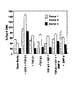
Figure 1 presents a bar graph of analyses of DNA content after 7 days in
culture (n .4/condition/donor). Data are expressed as mean percent of day 0
DNA S.E.M. Patterned bars represent donor 1, white bars represent donor 2,
and black bars represent donor 3.
Figures 2A and 2B present bar graphs of biosynthetic activity of hADS
from 3 donors under various growth factor treatments. [31-1]-proline DPM/tig
DNA (Figure 3A) and [35S]-sulfate DPM/j.ig DNA (Figure 3B) incorporation into
protein were determined. The x-axis values for each Figure are the same as in
Figure 2. All comparisons between growth factor conditions are significant at
p
<0.05. "+" indicates non-significance at p >0.05 (n .4/condition/donor). Data
are presented as mean S.E.M.) . Patterned bars represent donor 1, white
bars represent donor 2, and black bars represent donor 3.
Figures 3-6 depict gene expression analyses for AGC1 (Figure 4;
4/condition/donor. *, p < 0.0001 relative to Day 0 Control. #, p < 0.0001
relative
to Base Medium at Day 7); COL1A1 (Figure 5; .__4/condition/donor. *, p <0.05
relative to Day 0 Control. #, p < 0.0001 relative to Base Medium at Day 7);
COL2A1 (Figure 6; __4/condition/donor. *, p <0.05 relative to Day 0 Control.
#,
p < 0.05 relative to Base Medium at Day 7); and COL10A1 (Figure 7;
4/condition/donor. *, p < 0.005 relative to Day 0 Control. #, p < 0.05
relative to
Base Medium at Day 7). For each of Figures 4-7, data are presented as mean
S.E.M. For each of Figures 4-7, "o", "o", and "A" represent Donors 1, 2, and
3, respectively.
Figures 7A-7L depict photographs of the results of
immunohistochemistry of hADS cell-alginate beads after 7 days in culture. All
photographs were at 63X magnification. Figures 7A-7C demonstrate that three
- 13-
CA 02629405 2008-05-12
WO 2007/030811 PCT/US2006/035243
conditions (TGF-131 + Dex, TGF-i33 +IGF-1 + BMP-6, and BMP-6 only,
respectively) showed increased type I collagen staining over other conditions.
Figures 7D-7F demonstrate that type X collagen expression decreases with
BMP-6 with BMP-6 only having the least expression. Figures 7G-7I
demonstrate that all conditions showed increased type II collagen staining
over
control, but those with BMP-6 also have strongly staining matrix. Figures 7J-
7L
demonstrate that only those conditions with BMP-6 showed significant staining
of chondroitin sulfate with 3B3 antibody.
Figure 8 is a diagram of the steps of one embodiment of the presently
disclosed subject matter, involving the formation of a bioartificial hip
implant
from autologous stem cells.
DETAILED DESCRIPTION
The presently disclosed subject matter provides methods and
compositions for treating tissue pathologies in a subject, and methods for
making the compositions. In some embodiments an implantable composition
comprising one or more cells that can develop into one or more tissues at a
predetermined site for treatment in the subject is provided. Treatment can be
accomplished by implanting the composition at the predetermined site.
In some embodiments, the predetermined tissue types include but are
not limited to bone and cartilage, muscle and tendon, and ligament and bone.
In some embodiments the predetermined site comprises the resurfacing of the
articulating surface in a joint. In any of the presently disclosed
embodiments,
the sites for the intended replacement tissue can replace multiple tissue
types
with one implantation (e.g. one tissue replacement to replace bone, cartilage,
and the interface of bone and cartilage).
In some embodiments the tissue pathology can comprise a compromise
in the normal homeostasis of the tissue, optionally culminating in
degeneration
of the tissue. The tissue pathology can comprise loss, damage, degeneration,
injury, or combinations thereof to the tissue. The treatment can comprise
tissue
remodeling, repair, regrowth, replacement, regeneration, or combinations
thereof.
- 14 -
CA 02629405 2008-05-12
WO 2007/030811
PCT/US2006/035243
In some embodiments, the predetermined site comprises a target tissue
selected from the group including but not to limited articular cartilage, non-
articular cartilage, auricular cartilage, tracheal cartilage, laryngeal
cartilage,
nasal cartilage, growth plate cartilage, meniscus, labrum, and intervertebral
disc. Representative tissue types at the predetermined site also include but
are
not limited to musculoskeletal or dental connective tissues selected from the
group including but not limited to tendon, ligament, periodontal ligament,
fascia,
and muscle.
In some embodiments, the treatment is solely focused on the treatment
of the articular surface of a joint.
To produce the desired implants, the compositions of the presently
disclosed subject matter can be maintained under conditions suitable for them
to expand and divide to form the desired structures. In some applications,
this
is accomplished by transferring the compositions to a subject (i.e., in vivo)
typically at a site at which the new matter is desired. Thus, for example, the
presently disclosed subject matter can facilitate the regeneration of tissues
within an animal where the compositions are implanted into such tissues. In
other embodiments, the compositions can be prepared in vitro. For examples
cells present in the compositions can be induced to differentiate and expand
into tissues in vitro. In such applications, the cells can be cultured on
substrates or scaffolds that facilitate formation into three-dimensional
structures
conducive for tissue formation.
Definitions
All technical and scientific terms used herein, unless otherwise defined
below, are intended to have the same meaning as commonly understood by
one of ordinary skill in the art. References to techniques employed herein are
intended to refer to the techniques as commonly understood in the art,
including
variations on those techniques or substitutions of equivalent techniques that
would be apparent to one of skill in the art. While the following terms are
believed to be well understood by one of ordinary skill in the art, the
following
definitions are set forth to facilitate explanation of the presently disclosed
subject matter.
-15-
CA 02629405 2013-11-05
Following long-standing patent law tradition, the terms "a", "an", and
"the" are meant to refer to one or more as used herein, including the claims.
For example, the phrase "a cell" can refer to one or more cells.
The term "absorbable" is meant to refer to a material that tends to be
absorbed by a biological system into which it is implanted. Representative
absorbable fiber materials include, but are not limited to polyglycolic acid
(PGA), polylactic acid (PLA), polyglycolide-lactide, polycaprolactone,
polydioxanone, polyoxalate, a polyanhydride, a poly(phosphoester), catgut
suture, collagen, silk, chitin, chitosan, hydroxyapatite, bioabsorbable
calcium
phosphate, hyaluronic acid, and any other medically acceptable yet absorbable
fiber. Other absorbable materials include collagen, gelatin, a blood
derivative,
plasma, synovial fluid, serum, fibrin, hyaluronic acid, a proteoglycan,
elastin,
and combinations thereof.
The term "non-absorbable" is meant to refer to a material that tends not
to be absorbed by a biological system into which it is implanted.
Representative non-absorbable fiber materials include but are not limited to
polypropylene, polyester, polytetrafluoroethylene (PTFE) such as that sold
under the registered trademark TEFLON (E.1. DuPont de Nemours & Co.,
Wilmington, Delaware, United States of America), expanded PTFE (ePTFE),
polyethylene, polyurethane, polyamide, nylon, polyetheretherketone (PEEK),
polysulfone, a cellulosic, fiberglass, an acrylic, tantalum, polyvinyl
alcohol,
carbon, ceramic, a metal (e.g., titanium, stainless steel), and any other
medically acceptable yet non-absorbable fiber.
As used herein, the phrases "adipose-derived stem cell" and "ADS cell"
refer to a cell with, at a minimum, unipotent potential that can be isolated
from
adipose tissue and that can be differentiated along various mesodermal and
ectodermal lineages. Representative conditions are disclosed herein and have
been described in the art, such as in U.S. Patent No. 6,777,231 or Zuk et at.
(2002) Mol Biol Cell 13:4279-4295.
Adipose-derived stem cells can be isolted
using techniques described in these references. In some embodiments, an
ADS cell can be isolated from a subject by removing subcutaneous fat from the
subject, for example by liposuction. In some embodiments, an adipose-derived
- 16-
CA 02629405 2008-05-12
WO 2007/030811
PCT/US2006/035243
stem cell is isolated from a human, in which case it is referred to herein as
a
human adipose-derived stem (hADS) cell.
As used herein, the terms "anisotropic", "anisotropy", and grammatical
variations thereof, refer to properties of a scaffold and/or fiber system as
disclosed herein that can vary along a particular direction. Thus, the fiber
and/or scaffold can be stronger and/or stiffer in one direction versus
another. In
some embodiments, this can be accomplished by changing fibers (such as, but
not limited to providing fibers of different materials) in warp versus weft
directions, and/or in the Z direction, for example. Thus,
anisotropic
characteristics parallel native properties of a tissue, and it is desirable to
match
or approximate one or more native properties of the tissue in the implantable
composition.
Thus, strength can be provided in the direction needed and indeed it is
possible to restore properties of a tissue almost immediately without
necessarily
needing for cells to grow into functional tissues. However, in some
embodiments cells are provided and the growth into functional tissues is also
provided. Further, in some embodiments the scaffold can comprise materials at
least some, if not all of which, are absorbable materials, such that
degradation
of the scaffold occurs over time. Thus, in some embodiments, the scaffold is
replaced by tissue over time in the subject.
In some embodiments, the terms "anisotropic", "anisotropy" and
grammatical variations thereof, can also include, but is not limited to the
provision of more fiber in a predetermined direction. This can thus include a
change of diameter in a fiber over a length of the fiber, a change in diameter
at
each end of the fiber, and/or a change in diameter at any point or section of
the
fiber; a change in cross-sectional shape of the fiber; a change in density or
number of fibers in a volumetric section of the scaffold; and the use of
monofilament fibers and/or multifilament fibers in a volumetric section of the
scaffold; and can even include the variation in material from fiber system to
fiber system and along individual fibers in a volumetric section of the
scaffold.
As used herein, the term "bioartificial" can refer to an implantable
composition that comprises cells that were isolated, grown, and/or manipulated
in vitro, or the progeny of such cells. In some embodiments, a bioartificial
joint
- 17-
CA 02629405 2008-05-12
WO 2007/030811
PCT/US2006/035243
replacement implant as disclosed herein comprises a three-dimensional fiber
scaffold and one or more cells that can develop into tissues functioning
substantially as bone, cartilage, both bone and cartilage, or other tissues.
In
some embodiments, a bioartificial joint replacement implant as disclosed
herein
comprises a scaffold which is partly or wholly acellular. In some embodiments,
a bioartificial joint replacement implant as disclosed herein comprises a
scaffold
that has been partly or wholly decellularized or devitalized at some point in
time
after being seeded with cells.
The terms "biocompatible" and "medically acceptable" are used
synonymously herein and are meant to refer to a material that is compatible
with a biological system, such as that of a subject having a tissue (e.g., a
joint)
to be repaired, restored, and/or replaced in accordance with the presently
disclosed subject matter. Thus, the term "biocompatible" is meant to refer to
a
material that can be implanted internally in a subject as described herein.
The term "composite material", as used herein, is meant to refer to any
material comprising two or more components. One of the components of the
material can optionally comprise a matrix for carrying cells, such as a gel
matrix
or resin.
As used herein, the phrases "biologically active agent" and "biologically
active factor" are used interchangeably and can refer to a compound or mixture
of compounds that when added to a cell in culture induces the cell to enter
differentiation (e.g., differentiate at least one step further along a pathway
of
differentiation).
As used herein, the term "effective amount" refers to an amount of a
biologically active agent sufficient to produce a measurable response (e.g., a
biologically relevant response in a cell exposed to the differentiation-
inducing
agent) in the cell. In some embodiments, an effective amount of a
differentiation-inducing agent is an amount sufficient to cause a precursor
cell to
differentiate in in vitro culture into a cell of a tissue at predetermined
site of
treatment. It is understood that an "effective amount" can vary depending on
various conditions including, but not limited to the stage of differentiation
of the
precursor cell, the origin of the precursor cell, and the culture conditions.
- 18-
CA 02629405 2008-05-12
WO 2007/030811
PCT/US2006/035243
In some embodiments, an "effective amount" of a "biologically active
agent" can be determined by assaying the ability of different amounts of a
putative biologically active agent to induce the expression of a gene or genes
associated with development of a cell that can be used in providing treatment
of
a tissue pathology as disclosed herein. For example, expression of the gene
products aggrecan (for example, the human aggrecan gene product disclosed
as GENBANK Accession No. P16112, or a functional fragment or variant
thereof) and type II collagen (for example, the human aggrecan gene product
disclosed as GENBANK Accession No. NP 001835, or a functional fragment
or variant thereof) are associated with chondrogenic differentiation. In some
embodiments, a gene expression level of aggrecan and/or type II collagen is
measured before and after a given amount biologically active agent is provided
to a culture of ADS cells (for example, hADS cells), and the levels are
compared to determine if the amount of the biologically active agent provided
is
an "effective amount". In some embodiments, the expression of other genes
are similarly determined, including genes that are not associated with
particular
cartilaginous tissues including, but not limited to type I collagen and type X
collagen.
The term "expression vector" as used herein refers to a DNA sequence
capable of directing expression of a particular nucleotide sequence in an
appropriate host cell, comprising a promoter operatively linked to the
nucleotide
sequence of interest which is operatively linked to termination signals. It
also
typically comprises sequences required for proper translation of the
nucleotide
sequence. The construct comprising the nucleotide sequence of interest can
be chimeric. The construct can also be one that is naturally occurring but has
been obtained in a recombinant form useful for heterologous expression.
The term "gene expression" generally refers to the cellular processes by
which a biologically active polypeptide is produced from a DNA sequence and
exhibits a biological activity in a cell. As such, gene expression involves
the
processes of transcription and translation, but also involves post-
transcriptional
and post-translational processes that can influence a biological activity of a
gene or gene product. These processes include, but are not limited to RNA
synthesis, processing, and transport, as well as polypeptide synthesis,
- 19-
CA 02629405 2008-05-12
WO 2007/030811
PCT/US2006/035243
transport, and post-translational modification of polypeptides. Additionally,
processes that affect protein-protein interactions within the cell can also
affect
gene expression as defined herein.
The terms "heterologous gene", "heterologous DNA sequence",
"heterologous nucleotide sequence", "exogenous nucleic acid molecule", and
"exogenous DNA segment", as used herein, refer to a sequence that originates
from a source foreign to an intended host cell or, if from the same source, is
modified from its original form. Thus, a heterologous gene in a host cell
includes a gene that is endogenous to the particular host cell but has been
modified, for example by mutagenesis or by isolation from native
transcriptional
regulatory sequences. The terms also include non-naturally occurring multiple
copies of a naturally occurring nucleotide sequence.
Thus, the terms refer to a DNA segment that is foreign or heterologous
to the cell, or homologous to the cell but in a position within the host cell
nucleic
acid wherein the element is not ordinarily found. In some embodiments where
the heterologous DNA sequence comprises an open reading frame, the
heterologous DNA sequence is also referred to as a "transgene", although thel
term "transgene" is not limited to heterologous DNA sequences that comprise
an open reading frame.
The terms "inhomogeneous", "inhomogeneity", "heterogeneous",
"heterogeneity", and grammatical variations thereof, are meant to refer to a
scaffold and/or fiber as disclosed herein that does not have a homogeneous
composition along a given length or in a given volumetric section. In some
embodiments, an inhomogeneous tissue engineering construct as disclosed
herein comprises a composite material, such as a composite comprising a three
dimensional scaffold as disclosed herein, cells that can develop tissues that
substantially provide the function of bone, cartilage, other joint tissues, or
combinations thereof, and a matrix that supports the cells. In
some
embodiments, an inhomogeneous scaffold as disclosed herein can comprise
one or more component systems that vary in their properties according to a
predetermined profile, such as a profile associated with the tissue and/or
other
location in a subject where the scaffold will be implanted. Thus, it is an
aspect
of the terms "inhomogeneous", "inhomogeneity", "heterogeneous",
- 20 -
CA 02629405 2008-05-12
WO 2007/030811
PCT/US2006/035243
"heterogeneity", and grammatical variations thereof to encompass the control
of
individual materials and properties in a scaffold.
The terms "non-linear", "non-linearity", and grammatical variations
thereof, refer to a characteristic provided by a scaffold and/or fiber system
as
disclosed herein such that the scaffold and/or fiber system can vary in
response
to a strain. As would be appreciated by one of ordinary skill in the art after
review of the present disclosure, the scaffolds and/or fiber systems disclosed
herein provide stress/stain profiles that mimic that observed in a target such
as
predetermined tissue or joint. As such stress/strain responses are typically
described with reference to a plot, stress/strain responses can be referred to
as
"non-linear". Important non-linear properties of most biological tissues are
significant differences in the strength, stiffness, and/or other properties
associated with the magnitude of strain, as well as significant differences in
the
strength, stiffness, and/or other properties as measured in tension as
compared
to those measured in compression but along the same axis or direction.
When used in the context of a promoter, the term "linked" as used herein
refers to a physical proximity of promoter elements such that they function
together to direct transcription of an operably linked nucleotide sequence.
As used herein, the terms "nucleic acid" and "nucleic acid molecule"
mean any of deoxyribonucleic acid (DNA), ribonucleic acid (RNA),
oligonucleotides, fragments generated by the polymerase chain reaction (PCR),
and fragments generated by any of ligation, scission, endonuclease action, and
exonuclease action. Nucleic acids can be composed of monomers that are
naturally occurring nucleotides (such as deoxyri bon ucleotides and
ribonucleotides), or analogs of naturally occurring nucleotides (e.g., a-
enantiomeric forms of naturally-occurring nucleotides), or a combination of
both. Nucleic acids can be either single stranded or double stranded.
The terms "operatively linked" and "operably linked", as used herein,
refer to a promoter region that is connected to a nucleotide sequence (for
example, a coding sequence or open reading frame) in such a way that the
transcription of the nucleotide sequence is controlled and regulated by that
promoter region. Similarly, a nucleotide sequence is said to be under the
"transcriptional control" of a promoter to which it is operably linked.
Techniques
- 21 -
CA 02629405 2008-05-12
WO 2007/030811
PCT/US2006/035243
for operatively linking a promoter region to a nucleotide sequence are known
in
the art.
As used herein, the term "polypeptide" means any polymer comprising
any of the 20 protein amino acids, or amino acid analogs, regardless of its
size
or function. Although "protein" is often used in reference to relatively large
polypeptides, and "peptide" is often used in reference to small polypeptides,
usage of these terms in the art overlaps and varies. The term "polypeptide" as
used herein refers to peptides, polypeptides and proteins, unless otherwise
noted. As used herein, the terms "protein", "polypeptide" and "peptide" are
used interchangeably. The term "polypeptide" encompasses proteins of all
functions, including enzymes.
The term "promoter" or "promoter region" each refers to a nucleotide
sequence within a gene that is positioned 5' to a coding sequence of a same
gene and functions to direct transcription of the coding sequence. The
promoter region comprises a transcriptional start site, and can additionally
include one or more transcriptional regulatory elements. In
some
embodiments, a method of the presently disclosed subject matter employs a
promoter that is active in an endoderm-derived tissue. Exemplary such
promoters include promoters that are active in the liver, the pancreas, the
spleen, the lung, etc.
A "minimal promoter" is a nucleotide sequence that has the minimal
elements required to enable basal level transcription to occur. As such,
minimal
promoters are not complete promoters but rather are subsequences of
promoters that are capable of directing a basal level of transcription of a
reporter construct in an experimental system. Minimal promoters include but
are not limited to the CMV minimal promoter, the HSV-tk minimal promoter, the
simian virus 40 (SV40) minimal promoter, the human fl-actin minimal promoter,
the human EF2 minimal promoter, the adenovirus El B minimal promoter, and
the heat shock protein (hsp) 70 minimal promoter. Minimal promoters are often
augmented with one or more transcriptional regulatory elements to influence
the
transcription of an operably linked gene. For example, cell-type-specific or
tissue-specific transcriptional regulatory elements can be added to minimal
promoters to create recombinant promoters that direct transcription of an
- 22 -
CA 02629405 2008-05-12
WO 2007/030811
PCT/US2006/035243
operably linked nucleotide sequence in a cell-type-specific or tissue-specific
manner
Different promoters have different combinations of transcriptional
regulatory elements. Whether or not a gene is expressed in a cell is dependent
on a combination of the particular transcriptional regulatory elements that
make
up the gene's promoter and the different transcription factors that are
present
within the nucleus of the cell. As such, promoters are often classified as
"constitutive", "tissue-specific", "cell-type-specific", or "inducible",
depending on
their functional activities in vivo or in vitro. For example, a constitutive
promoter
is one that is capable of directing transcription of a gene in a variety of
cell
types. Exemplary constitutive promoters include the promoters for the
following
genes which encode certain constitutive or "housekeeping" functions:
hypoxanthine phosphoribosyl transferase (Hprt), dihydrofolate reductase (Dhfr;
Scharfmann etal. (1991) Proc Natl Acad Sci USA 88:4626-4630), adenosine
deaminase, phosphoglycerate kinase (Pgk), pyruvate kinase, phosphoglycerate
mutase, the 13-actin promoter (see e.g., Williams et al. (1993) J Clin Invest
92:503-508), and other constitutive promoters known to those of skill in the
art.
"Tissue-specific" or "cell-type-specific" promoters, on the other hand, direct
transcription in some tissues and cell types but are inactive in others.
The terms "replace", "replacement", and grammatical variations thereof,
refer to any qualitative or quantitative improvement in a target or
predetermined
tissue or site of treatment observed upon implantation of a composition as
disclosed herein. For example, these terms are not limited to full restoration
to
a normal healthy function, although these terms can refer to this. Rather,
these
terms are meant to any level of improvement observed in the tissue or at the
site.
The terms "reporter gene" and "marker gene" refer to an exogenous
gene encoding a product that is readily observed and/or quantitated. A
reporter
gene is exogenous in that it originates from a source foreign to an intended
host
cell or, if from the same source, is modified from its original form. Non-
limiting
examples of detectable reporter genes that can be operatively linked to a
transcriptional regulatory region can be found in Alam and Cook (1990) Anal
Biochem 188:245-254, and PCT International Publication No. WO 97/47763.
- 23
CA 02629405 2008-05-12
WO 2007/030811
PCT/US2006/035243
Exemplary reporter genes include the lacZ gene (see e.g., Rose and Botstein
(1983) Methods Enzymol 101:167-180), Green Fluorescent Protein (GFP;
Cubitt et al. (1995) Trends Biochem Sc! 20:448-455), luciferase, and
chloramphenicol acetyl transferase (CAT). Any suitable reporter and detection
method can be used, and it will be appreciated by one of skill in the art that
no
particular choice is essential to or a limitation of the presently disclosed
subject
matter.
The terms "resin", "matrix", or "gel" are used the art-recognized sense
and refer to any natural or synthetic solid, liquid, and/or colloidal material
that
has characteristics suitable for use in accordance with the presently
disclosed
subject matter. Representative "resin", "matrix", or "gel" materials thus
comprise biocompatible materials. In some embodiments, the "resin", "matrix",
or "gel" can occupy the pore space of a fiber scaffold as disclosed herein.
The terms "restore", "restoration", and grammatical variations thereof
refer to any qualitative or quantitative improvement in a target or
predetermined
tissue or and/or site of treatment observed upon implantation of a composition
as disclosed herein. Thus, these terms are not limited to full restoration of
the
tissue and/or site to a normal healthy function, although these terms can
refer
to this. Rather, these terms are meant to refer to any measurable and/or
observable level of improvement in the tissue and/or site.
The terms "resurface", "resurfacing", and grammatical variations thereof
refer to any qualitative or quantitative replacement of least the majority of
the
surface area of the surface of tissue upon implantation of a composition as
disclosed herein. These terms can also refer to any desired depth of
resurfacing; such as but not limited to a layer of micron thickness, to
multiple
layers of tissue including multiple tissue types, and/or to replacement of a
complete structure that provides a surface at the site of treatment. Thus,
these
terms are not limited to full replacement of the tissue and/or site, although
these
terms can refer to this. Rather, these terms are meant to refer to replacement
of any fraction of the native tissue beyond what is considered by one skilled
in
the art as a "focal defect". A representative surface is an articulating
surface of
a joint.
- 24 -
CA 02629405 2013-11-05
As used herein, the term "selectable marker" refers to a gene or gene
product that confers a growth advantage to a cell that expresses it. For
example, a selectable marker can allow a cell that expresses it to grow in the
presence of a chemical (e.g., a drug such as G418) that would inhibit the
growth of or kill cells that do not express the selectable marker. Selectable
marker genes include, but are not limited to antibiotic resistance genes, for
example the antibiotic resistance gene confers neomycin resistance (herein
referred to as the "neo gene").
The term "transcriptional regulatory sequence" or "transcriptional
regulatory element", as used herein, each refers to a nucleotide sequence
within the promoter region that enables responsiveness to a regulatory
transcription factor. Responsiveness can encompass a decrease or an
increase in transcriptional output and is mediated by binding of the
transcription
factor to the DNA molecule comprising the transcriptional regulatory element.
The term "transcription factor" generally refers to a protein that
modulates gene expression by interaction with the transcriptional regulatory
element and cellular components for transcription, including RNA Polymerase,
Transcription Associated Factors (TAFs), chromatin-remodeling proteins, and
any other relevant protein that impacts gene transcription.
The terms "viscoelastic", "viscoelasticity", and grammatical variations
thereof, are meant to refer to a characteristic provided by a scaffold and/or
fiber
system as disclosed herein that can vary with a time and/or rate of loading.
It is
thus envisioned that appropriately viscoelastic scaffolds and/or fiber systems
provide time and/or rate of loading characteristics that match or approximate
that observed in the predetermined tissue or site. This characteristic
pertains to
dissipation of energy, which can be provided by the scaffold itself and/or by
the
scaffold as a composite with cells growing therein, and can also be
accomplished by virtue of the choices of fibers that are included in the
scaffold.
As a particular example, it can be desirable to provide a scaffold that
approximates the viscoelastic properties of cartilage.
- 25 -
CA 02629405 2013-11-05
II. Cells and Reagents
I.A. Representative Cells for Joint Resurfacing
The presently disclosed implantable composition can comprise one or
more cells that can develop into a suitable replacement of a target tissue
(e.g.,
bone, cartilage, or both bone and cartilage). Particularly, the one or more
cells
comprise, or are derived from, a precursor cells, such as but not limited to a
stem cell. As used herein, the term "stem cell" refers to any unipotent,
multipotent, pluripotent and/or totipotent cell that can be differentiated
into a
desired lineage. As such, the presently disclosed subject matter can employ
stem cells that can be differentiated into a tissue appropriate for
replacement of
native pathological tissues. Representative stem cells include embryonic stem
(ES) cells, embryonic germ (EG) cells (e.g., pluripotent cells derived from
primordial germ cells), and somatic stem cells (alternatively referred to
herein
as "adult stem cells").
In some embodiments, the one or more cells described herein comprise
an adult stem cell. Adult stem cells can be derived from various adult tissues
including, but not limited to liver, bone marrow, umbilical cord blood, brain,
peripheral blood, blood vessels, skeletal muscle, adipose tissue, and skin.
Methods for the isolation, culturing, and manipulation of adult stem cells
from
various sources can be found in U.S. Patent Nos. 6,242,252 and 6,872,389
(hepatic stem cells); U.S. Patent No. 6,387,367 (hematopoietic/mesenchymal
stem cells); Kooler etal. (2004)J Exp Med 200:123-135 (placental cord blood);
Williams etal. (1999) The American Surgeon 65:22-26 (skeletal muscle); U.S.
Patent No. 6,777,231 (adipose tissue); and Blanpain et al. (2004) Ce//
118:635-648 (skin).
Representative techniques for deriving, growing, and manipulating ES
cells and EG cells are disclosed in the following publications: Evans and
Kaufman (1981) Nature 292:154-156; Martin (1981) Proc Natl Aced Sci USA
78:7634+7638; Robertson (1986) Trends Genet 2:9-13; PCT International
Patent Application Publications WO 96/22362; WO 97/32033; and WO
- 26 -
CA 02629405 2013-11-05
98/43679; and U.S. Patent Nos. 6,200,806; 6,090,622; 5,843,780; 5,690,926;
5,670,372; and 5,453,357; and references therein.
Thus, the presently disclosed subject matter provides in some
embodiments the use of the cells described herein for treatment of joint
disease. Currently, there are limited treatment options for osteoarthritis, as
one
example of joint disease. For advanced degeneration, the only current
treatment option is replacement of the joint with artificial materials, which
include polymers and metals, which effectively act as an artificial joint.
While
the joint replacement surgeries alleviate pain and restore some function in
many of the patients in the short-term, these joint replacements are not
intended for long-term use and often require difficult surgical revisions,
potentially leading to significant post-operative complications. Some post-
operative complications associated with the use of artificial materials
include
device related osteopenia, osteolysis, excessive wear of the bearing surfaces
of
the artificial device, and fracture of the bones supporting the implant.
Disclosed
herein for the first time are approaches for the complete resurfacing of the
diseased articular surface with a bioartificial implant, which avoids the
complications associated with the introduction of artificial materials due to
the
biologic nature of the composition of the implanted structure. Further
disclosed
herein is the use of progenitor, stem, or primary cells in conjunction with a
composition that comprises a medium capable of supporting the growth and
differentiation of the cells into functional tissue, but not necessarily
recapitulating the native structure of the tissue.
II.B. Adipose Derived Stem (ADS) Cells
The ADS and ADS-derived cells of an aspect of the presently disclosed
subject matter are useful in providing a source of differentiated and
functional
cells for research, transplantation, and development of tissue engineering
products for the treatment of mammalian disease and traumatic injury repair.
Thus, in some aspects, the presently disclosed subject matter provides
methods for differentiating ADS cells comprising culturing the cells in a
composition that comprises a medium capable of supporting the growth and
differentiation of the cells. The presently disclosed subject matter further
- 27 -
CA 02629405 2008-05-12
WO 2007/030811
PCT/US2006/035243
provides methods for the introduction of these cells into tissue defect areas
in
need of repair. In some embodiments the tissue defect areas can be treated by
exclusively using the ADS and ADS-derived cells of the presently disclosed
subject matter.
As an example of one tissue pathology, there are currently limited
treatment options for focal cartilage lesions. One treatment option involves
drilling into the subchondral bone and exposing the cartilage tissue to growth
factors and other molecular agents from the vascular supply found in the bone
in the hope that regeneration of the cartilage lesion occurs. A second
technique
involves the transfer of "healthy" cartilage from non-load bearing areas to
"unhealthy" areas to replace the degenerated cartilage. Thirdly, a cell-based
therapy exists that utilizes ex vivo cultured autologous chondrocytes
reimplanted at the defect site to regenerate the damaged tissue.
All three of these techniques are marked by varying degrees of success,
and accordingly, novel techniques and methodologies are needed for the
effective remodeling, repair, regrowth, and/or regeneration of cartilage
lesions.
The presently disclosed subject matter relates to replacement of damaged
cartilage as well as other tissues and has broad applications in the field of
tissue engineering and regenerative medicine.
ADS cells provide a readily accessible, abundant source of multipotent
progenitor cells for applications in tissue engineering and other cell-based
therapies. In particular, the potential use of ADS cells for the remodeling,
repair, regrowth, and/or regeneration of cartilage has been explored. However,
employing the chondrogenic differentiation techniques currently available in
the
art only results in a mild chondrogenic phenotype in in vitro culture. On the
contrary, disclosed herein for the first time are approaches for the
unambiguous
and robust differentiation of ADS cells along a lineage appropriate for
replacement/regeneration of pathological tissue such as degenerated, injured,
or damaged cartilage or other connective tissue, and use of these cells in
conjunction with a composition that comprises a medium capable of supporting
the growth and differentiation of ADS cells into functional tissue, but not
necessarily recapitulating the native structure of the tissue.
- 28 -
CA 02629405 2008-05-12
WO 2007/030811
PCT/US2006/035243
Thus, the presently disclosed subject matter provides in some
embodiments methods and systems for inducing specific phenotypes in ADS
cells for the treatment of various tissue pathologies. In contrast to current
technologies that are compromised by difficulties in obtaining appropriate
stem
cells and in differentiating the stem cells as desired in culture, the
presently
disclosed subject matter provides methods and systems for promoting ADS cell
differentiation at a significantly increased rate over previously known
methods.
In some embodiments of the presently disclosed subject matter,
methods and systems are provided for inducing differentiation comprising
providing to ADS cells in culture an effective amount of a biologically active
factor (e.g., BMP-6) or a functional fragment thereof.
In some embodiments of the presently disclosed subject matter,
methods and systems are provided for determining whether a cell has
differentiated into a desired phenotype. Particularly, because the cells of
the
presently disclosed subject matter have a specific phenotype, they can be
employed in tissue engineering. In this regard, the presently disclosed
subject
matter provides in some embodiments methods of maintaining the ADS cells
under conditions sufficient for them to expand and differentiate to form the
desired subject matter.
II.C. Isolation of ADS Cells
ADS cells (e.g., hADS cells) are isolated from a subject or obtained
directly from an established cell culture line. The subject can be alive or
dead,
so long as the ADS cells within the subject are viable. Typically, ADS cells
are
obtained from living donors, using well-recognized protocols such as surgical
or
suction lipectomy. Such cells can be isolated from the subject to be treated,
or
from a subject different from the subject to be treated. In some embodiments,
the subject from which the cells are isolated is of a different species than
the
subject into which the cells are to be transferred. Thus, in some embodiments,
the ADS cells can be derived from the adipose tissue of a primate, a higher
primate (e.g., baboon or ape), or from human adipose tissue, using the
methods described herein.
Thus, in some embodiments, the ADS cells are syngeneic (also referred
to herein as "autologous") to the subject into which the ADS cells and/or ADS-
- 29 -
CA 02629405 2008-05-12
WO 2007/030811
PCT/US2006/035243
derived cells are intended to be placed, and in some embodiments the one or
more cells are allogeneic (also referred to herein as "heterologous") to the
subject. In those embodiments where the one or more cells are allogeneic or
xenogeneic to the subject, the subject can be treated as necessary with
immunosuppressant drugs such as cyclosporin, azathioprines, or
corticosteroids using well-known techniques.
Representative
immunosuppressive drugs also include, but are not limited to, basiliximab
(SIMULECTO; available from Novartis Pharmaceuticals Corp., East Hanover,
New Jersey, United States of America), daclizumab (ZENAPAX , available
from Hoffmann-La Roche Inc., Nutley, New Jersey, United States of America),
muromonab CD3 (ORTHOCLONE OKT3O, available from Ortho Biotech
Products, L.P., Bridgewater, New Jersey, United States of America) and
tacrolimus (PROGRAF , available from Astellas Pharma US, Inc., Deerfield,
Illinois, United States of America).
As would be readily understood by one of skill in the art, ADS cells refer
to stem cells that originate from adipose tissue and are capable of self-
renewal.
By "adipose" is meant any fat tissue. ADS cells can be isolated from any
source of adipose tissue in the subject, although in some embodiments, the
ADS cells are isolated from an adipose depot in the body selected from the
group consisting of the subcutaneous abdomen, the thigh, the buttocks, and the
infrapatellar fat pad. Adipocytes can be harvested by liposuction on an
outpatient basis, a relatively non-invasive procedure with cosmetic effects
that
are acceptable to the vast majority of patients. It is well documented that
adipocytes are a replenishable cell population. Even after surgical removal by
liposuction or other procedures, it is common to see a recurrence of
adipocytes
in an individual over time.
ADS cells can comprise a primary cell culture or an immortalized cell
line. While stem cells represent less than 0.01% of the bone marrow's
nucleated cell population, there are up to 8.6x10⁴ stem cells per gram of
adipose tissue (Sen, et al. (2001) Journal of Cellular Biochemistry, 81:312-
319).
Ex vivo expansion over 2 to 4 weeks yields up to 500 million stem cells from
0.5 kilograms of adipose tissue. These cells can be used immediately or
cryopreserved for future autologous or allogeneic applications.
- 30 -
CA 02629405 2008-05-12
WO 2007/030811
PCT/US2006/035243
In addition, the isolated ADS cells can be further separated into
subpopulations of ADS cells based upon any observable, quantifiable, or other
trait of the cells for which a separation technique is available or can be
designed. In some embodiments, isolated ADS cells are separated into
subpopulations using fluorescence-activated cell sorting (FACS) based on the
appearance of one or more of cell surface markers. In some embodiments, the
following cell surface markers can be employed for separating ADS cells into
subpopulations: 0D10, 0D13, 0D31, CD34, CD36, CD44, CD49, CD54, CD55,
CD59, CD65 CD105, and CD166.
In some embodiments, the isolated ADS cells can be separated into
subpopulations of ADS cells based on differential expression of various genes.
In some embodiments, isolated ADS cells are separated into subpopulations
based on differential expression of aldehyde dehydrogenase (ALDH), various
members of the TGF-p superfamily, TGF-p superfamily receptor, and/or
telomerase activity. In some embodiments, the isolated ADS cells are
separated into subpopulations of ADS cells based on telomere length. In some
embodiments, the isolated ADS cells can be separated into subpopulations of
ADS cells based on efflux of macromolecules including, but not limited to dyes
(e.g., Hoechst 33342) or nucleic acid labels. In some embodiments, the
isolated ADS cells can be separated into subpopulations of ADS cells based on
responsiveness to a particular growth factor (e.g., BMP-6). It is understood
that
two or more of these separation strategies can be employed together to
produce subpopulations of ADS cells either before or after the induction of
differentiation.
Such isolated ADS cells and populations can be clonally expanded, if
desired, using a suitable method for cloning cell populations. For example, a
proliferated population of cells can be physically picked and seeded into a
separate plate (or the well of a multi-well plate). Alternatively, the cells
can be
subcloned onto a multi-well plate at a statistical ratio for facilitating
placing a
single cell into each well (e.g., from about 0.1 to about 1 cell/well). In
some
embodiments, the cells can be cloned by plating them at low density (e.g., in
a
petri-dish or other suitable substrate) and isolating them from other cells
using
devices such as a cloning rings. Alternatively, where an irradiation source is
- 31 -
CA 02629405 2008-05-12
WO 2007/030811
PCT/US2006/035243
available, clones can be obtained by permitting the cells to grow into a
monolayer and then shielding one and irradiating the rest of cells within the
monolayer. The surviving cells then can grow into a clonal population.
11Ø Genetic Manipulation of Cells
In some embodiments, the cells, for example ADS cells, can be
genetically modified, e.g., to express exogenous genes or to repress the
expression of endogenous genes. In some embodiments, the presently
disclosed subject matter provides methods of genetically modifying such cells
and populations. In accordance with these methods, the cells can be exposed
to an expression construct comprising a nucleic acid including a transgene,
such that the nucleic acid is introduced into the cell under conditions
appropriate for the transgene to be expressed within the cell. The transgene
generally is an expression cassette, including a coding polynucleotide
operably
linked to a suitable promoter. The coding polynucleotide can encode a protein,
or it can encode a biologically active (e.g., functional) fragment of a
protein.
Thus, for example, the coding polynucleotide can encode a gene
conferring resistance to a toxin, a hormone (such as peptide growth hormones,
hormone releasing factors, sex hormones, adrenocorticotrophic hormones,
cytokines (e.g., interferins, interleukins, lymphokines), etc.), a cell-
surface-
bound intracellular signaling moiety (e.g., cell adhesion molecules, hormone
receptors, etc.), a factor promoting a given lineage of differentiation, etc.
Of
course, where it is desired to employ gene transfer technology to deliver a
given
transgene, the sequence will be known. In some embodiments, the coding
polynucleotide encodes a growth factor. In some embodiments, the coding
polynucleotide encodes BMP-6 or a functional fragment thereof. In some
embodiments, the coding polynucleotide encodes BMP-6 receptor or a
functional fragment thereof.
The cells can be stably or transiently transfected or transduced with a
nucleic acid of interest using a plasmid, viral or alternative vector
strategy. With
respect to ADS cells, nucleic acids of interest include, but are not limited
to,
those encoding gene products which enhance the production of extracellular
matrix components found in cartilage; examples include , collagen type II, TGF-
/3, BMP, activin and insulin-like growth factor.
- 32 -
CA 02629405 2008-05-12
WO 2007/030811
PCT/US2006/035243
Thus, in some embodiments, the transduction of regulatory genes into
the cells, for example ADS cells, can be performed with viral vectors
(adenovirus, retroviruS, adeno-associated virus, or other vector) purified by
cesium chloride banding or any other well-known method at a multiplicity of
infection (viral units:cell) of between 10:1 to 2000:1. Cells can be exposed
to
the virus in serum-free or serum-containing medium in the absence or presence
of a cationic detergent such as polyethyleneimine or LipofectamineTM
(available
from Invitrogen, Carlsbad, California, United States of America) for a period
of 1
hour to 24 hours (Byk et al. (1998) Human Gene Therapy 9:2493-2502;
Sommer et al. (1999) Calcif. Tissue Int. 64:45-49) or in three dimensional
cultures by incorporation of the plasmid DNA vectors directly into a
biocompatible polymer (Bonadio etal. (1999) Nat. Med. 5:753-759).
In some embodiments, cells, for example ADS cells, are transfected with
the gene to be expressed to produce cells having stably incorporated therein
the DNA encoding the molecules to be expressed. Stable transfections can be
obtained by culturing and selecting for expression of the desired encoded
molecules. In some embodiments, the cells that exhibit stable expression can
be seeded onto or into the appropriate fiber matrix and implanted in a
subject.
For the tracking and detection of functional proteins encoded by these genes,
the viral or plasmid DNA vectors can contain a readily detectable marker gene,
such as the green fluorescent protein (GFP) orig-galactosidase enzyme, both of
which can be tracked by histochemical means.
Within the expression cassette, the coding polynucleotide can be
operably linked to a suitable promoter. Examples of suitable promoters include
prokaryotic promoters and viral promoters (e.g., retroviral inverted terminal
repeats (ITRs), long terminal repeats (LTRs), immediate early viral promoters
(lEp), such as herpes virus lEp (e.g., ICP4-lEp and ICP0-lEp), cytomegalovirus
(CMV) lEp, and other viral promoters, such as Rous Sarcoma Virus (RSV)
promoters, and Murine Leukemia Virus (MLV) promoters). Other suitable
promoters are eukaryotic promoters, such as enhancers (e.g., the rabbit /3-
globin regulatory elements), constitutively active promoters (e.g., the 13-
actin
promoter, etc.), signal specific promoters (e.g., inducible promoters such as
a
promoter responsive to RU486, etc.), and tissue-specific promoters. It is well
- 33 -
CA 02629405 2008-05-12
WO 2007/030811
PCT/US2006/035243
within the skill of the art to select a promoter suitable for driving gene
expression in a predefined cellular context. The expression cassette can
include more than one coding polynucleotide, and it can include other elements
(e.g., polyadenylation sequences, sequences encoding a membrane-insertion
signal or a secretion leader, ribosome entry sequences, transcriptional
regulatory elements (e.g., enhancers, silencers, etc.), and the like), as
desired.
The expression cassette containing the transgene can be incorporated
into a genetic vector suitable for delivering the transgene to the cells.
Depending on the desired end application, any such vector can be so employed
to genetically modify the cells (e.g., plasmids, naked DNA, viruses such as
adenovirus, adeno-associated virus, herpes viruses, lentiviruses,
papillomaviruses, retroviruses, etc.). Any method of constructing the desired
expression cassette within such vectors can be employed, many of which are
well known in the art (e.g., direct cloning, homologous recombination, etc.).
Of
course, the choice of vector will largely determine the method used to
introduce
the vector into the cells (e.g., by protoplast fusion, calcium-phosphate
precipitation, gene gun, electroporation, infection with viral vectors, etc.),
which
are generally known in the art.
In some embodiments, the genetically altered (e.g., ADS) cells can be
employed as bioreactors for producing the product of the transgene. In some
embodiments, the genetically modified cells are employed to deliver the
transgene and its product to a subject. For example, the cells, once
genetically
modified, can be introduced into the subject under conditions sufficient for
the
transgene to be expressed in vivo.
II.E. Induction of Differentiation for Tissue Replacement
Another object of the presently disclosed subject matter is to provide for
the identification and study of compounds that enhance the differentiation of
ADS cells (e.g., hADS cells) into cells capable of forming an extracellular
matrix
capable of functioning in place of the native tissue. Representative, non-
limiting
extracellular matrix proteins are disclosed in the Examples. Compounds that
enhance differentiation can be of value in the treatment of partial or full
cartilage
defects, osteoarthritis, traumatized cartilage, and cosmetic surgery of inborn
defects including cleft palate and deviated septum, among other treatments.
- 34 -
CA 02629405 2008-05-12
WO 2007/030811 PCT/US2006/035243
After isolation, ADS cells can be cultured in vitro for a time and under
conditions sufficient to induce one or more of the cells to undergo
differentiation. In some embodiments, one or more biologically active agents
are added to the culture medium. In some embodiments, a biologically active
agent comprises a BMP-6 polypeptide, or a functional fragment thereof. In
some embodiments, a biologically active agent comprises a BMP-6 polypeptide,
or a functional fragment thereof, in combination with one or more additional
growth factors and/or cytokines. It is understood that for any polypeptides
that
are employed as constituents of a biologically active agent, it is not
necessary
that full length polypeptides be employed, as functional fragments can also be
used. As used herein, the term "functional fragment" refers to a subsequence
of a polypeptide (or a subsequence of a nucleic acid encoding such a
polypeptide fragment) that is characterized by at least some activity in
differentiation when part of a biologically active agent.
In some embodiments, the method of inducing differentiation in stem
cells comprises (a) providing to a stem cell in culture an effective amount of
a
biologically active factor (e.g., BMP-6) or a functional fragment thereof; and
(b)
growing the stem cell (e.g. ADS cell) in culture for a time sufficient for
differentiation to occur, wherein differentiation is determined to have
occurred
when at least one stem cell (e.g. ADS cell) exhibits expression of a
macromolecule associated with the native, healthy tissue to be replaced (i.e.,
native extracellular composition of the tissue prior to injury, disease, or
degeneration). Representative, non-limiting extracellular matrix proteins are
disclosed in the Examples.
In some embodiments, the method further comprises passaging the
stem cell (e.g. ADS cell) repeatedly (e.g., at least twice) in culture,
wherein the
passaging enhances an ability of the cell to express at least one
macromolecule
associated with the predetermined target tissue upon exposure to a
biologically
active factor or a functional fragment thereof. Representative macromolecules
are disclosed in the Examples.
In some embodiments, the method further comprises providing to the
stem cells (e.g. ADS cells) a predetermined effective amount of a second
biologically active factor.
- 35 -
CA 02629405 2008-05-12
WO 2007/030811
PCT/US2006/035243
Represesentative biologically active factors can be selected from the
group including, but not limited to, growth factors, e.g., TGF-13 superfamily
members (including but not limited to BMP-6, TGF-/31, TGF-162, TGF-/33), IGF-
1, FGF, an EGF, a PDGF, a parathyroid hormone related peptide (PTHrP), an
interleukin, cytokines, chemokines, gelatins, laminins, fibronectins,
thrombins,
lipids, cartilage oligomeric proteins (COMP), thrombospondins, fibrins,
fibrinogens, Matrix-GLA protein, chondrocalcin, tenascin, a mineral, an RGD
peptide, an RGD-peptide containing molecule, elastin, hyaluronic acid, a
glycosaminoglycan, a proteoglycan and other molecules that alone or in
combination are capable of inducing the differentiation of an ADS cell into a
cell
or a cell that is further differentiated along the appropriate target lineage
than is
the ADS cell. In some embodiments a composition to be administered to cells
can consist essentially of a given biologically active agent (such as but not
limited BMP-6), and in some embodiments such a composition can consist of a
given biologically active agent.
In some embodiments, the biologically active agent is selected from the
group including but not limited to parathyroid hormone, a transforming growth
factor (e.g., a TGF-a and/or a TGF-fl), an insulin-like growth factor (e.g.,
IGF-I),
a platelet-derived growth factor (PDGF), a fibroblast growth factor (FGF), an
epidermal growth factor (EGF), a vascular endothelial growth factor (VEGF),
and combinations thereof. In some embodiments, the biologically active agent
is a bone morphogenetic protein (BMP), and in some embodiments the BMP is
BMP-6 (e.g., human BMP-6). In some embodiments, the human BMP-6
comprises amino acids 374-513 of GENBANKO Accession No. NP 001709, or
a functional fragment or variant thereof, and/or is encoded by a nucleic acid
comprising GENBANKO Accession No. NM_001718, or a function fragment or
variant thereof.
Further, it is to be understood that in some embodiments, the biologically
active agent (e.g., BMP-6) can be incorporated into an implantable composition
of the presently disclosed subject matter for controlled release over time.
II.F. Bone Morphogenic Proteins
Bone morphogenetic proteins (BMPs) are characterized by their ability to
promote, stimulate or otherwise induce the formation of cartilage and/or bone.
- 36 -
CA 02629405 2008-05-12
WO 2007/030811 PCT/US2006/035243
Accordingly, members of the BMP family of proteins can be used in
compositions to induce bone and/or cartilage formation, wound healing and
tissue repair, treatment of bone and/or cartilage defects, periodontal disease
and other tooth repair processes, treatment of osteoporosis and increase of
neuronal survival.
It has been presently discovered, as discussed herein in detail, that
BMP-6 in combination with several growth factors, supplements, and other
soluble mediators increases the gene expression and biosynthesis of collagen
and proteoglycan by several orders of magnitude as compared to other
differentiation protocols that have been previously published. Further, it has
also been shown herein that BMP-6 in the absence of other growth factors
strongly promotes a robust differentiation towards a generated tissue type
which may be used to replace many different tissue types (e.g., ligament,
tendon, IVD, meniscus, and cartilage). BMP-6 is a member of a different BMP
family subgroup than are BMP-2 and BMP-4, and is thus different from BMP-1,
-2, -3, -4, and -5. In some embodiments, the effective amount of BMP-6 ranges
from about 1 picogram/mL to about 10 milligram/rnL.
II.G. Detection of Differentiation
After culturing the cells in a suitable medium for a suitable time (e.g.,
several days to a week or more), the cells can be assayed to determine
whether, in fact, they have differentiated to acquire physical qualities of a
desired cell type. Thus, in some embodiments the presently disclosed subject
matter provides methods for testing ADS cell-derived cells.
One measurement of differentiation per se is telomere length,
undifferentiated stem cells having longer telomeres than differentiated cells;
thus, the cells can be assayed for the level of telomerase activity.
Alternatively,
RNA or proteins can be extracted from the cells and assayed (via Northern
hybridization, rtPCR, Western blot analysis, etc.) for the presence of markers
indicative of the desired phenotype.
Thus, methods for determining whether an ADS cell has differentiated
into a desired phenotype are also provided. In some embodiments, the methods
comprise (a) obtaining mRNA from an ADS cell that has been exposed to a
biologically active agent; and (b) determining from the mRNA a level of
- 31-
CA 02629405 2008-05-12
WO 2007/030811 PCT/US2006/035243
expression of at least one gene associated with a desired differentiation,
wherein
the level of expression determined for the at least one gene is indicative of
differentiation of the ADS cell. In some embodiments, the methods comprise
determining from the ADS cell a level of expression of at least one protein
associated with a desired differentiation. In some embodiments, the cells can
be
assayed immunohistochemically or stained, using tissue-specific stains.
In some embodiments, the methods comprise measuring expression of
at least one gene associated with a desired differentiation to determine when
at
least a subpopulation of the ADS cells have differentiated. In
some
embodiments, the appearance of an ADS cell-derived phenotype is tested
and/or confirmed by testing the ADS cell grown in culture to determine when
differentiation has occurred, wherein the testing comprises utilizing a
technique
for measuring gene expression to determine when the ADS cell has
differentiated, and wherein the technique for measuring gene expression
comprises measuring a level of expression of at least one gene associated with
a desired phenotype. In some embodiments, the at least one gene associated
with a desired differentiation is selected from the group consisting of
aggrecan,
type I collagen, type ll collagen, type X collagen, and combinations thereof.
Accordingly, the presently disclosed subject matter provides methods for
identifying cells, for example ADS-derived cells, suitable for use in
therapeutic
applications. In some embodiments, the differentiated ADS cell is identified
as
suitable for use in therapeutic restorative and regenerative techniques when
gene expression measurements, protein measurements, or combinations
thereof meet predetermined parameters, such as but not limited to those
disclosed in the Examples presented herein.
Other methods of assessing developmental phenotype are known in the
art, and any of them are appropriate. For example, the cells can be sorted by
size and granularity. Also, the cells can be used to generate monoclonal
antibodies, which can then be employed to assess whether they preferentially
bind to a given cell type. Correlation of antigenicity can confirm that the
stem
cell has differentiated along a given developmental pathway.
- 38 -
CA 02629405 2008-05-12
WO 2007/030811
PCT/US2006/035243
II.H. Administration of Cells to Subjects
As is known in the art, when the cells are to be administered to a subject,
it is preferable that the cell culture medium be biologically compatible with
the
subject. Stated another way, in some embodiments the cell culture medium
that is used to culture and induce cells does not contain any components that
would be expected to negatively affect the health of the subject after
administration of the induced cells. Thus, an appropriate cell culture medium
can also comprise one or more serum-free medium supplements. As used
herein, the term "serum-free medium supplement" refers to a supplement that
can be added to a medium to replace some or all of the serum that would
normally be added to the medium to support the propagation and/or
maintenance of cells in culture.
Serum-free medium supplements typically comprise about 10-25 mM
HEPES, about 1-4 grams per liter (g/L) sodium bicarbonate, up to about 5
micrograms per liter (.1g/L) hypoxanthine, up to about 10 jug/ thymidine, up
to
about 1.5 g/L sodium pyruvate, up to about 2.0 g/L L-glutamine, and up to
about 301,ig/L phenol red. Various trace elements and other growth factors can
also be added to serum-free medium supplements. An exemplary serum-free
medium supplement is OPTI-MEM I reduced serum medium supplement, sold
by Invitrogen Corp. (Carlsbad, California, United States of America) in powder
and liquid forms (Catalog Nos. 22600-050, 22600-134, 11058-021, 31985-062,
31985-070, 31985-088, and 51985-034). Other
serum-free medium
supplements include BIOGRO-1 and BIOGRO-2 (Biological Industries Ltd.,
Kibbutz Beit Haemek, Israel) and the Nutridoma family of serum free medium
supplements sold by Roche Applied Science (Indianapolis, Indiana, United
States of America).
For analysis and/or administration into a subject, the induced cells can
be treated with trypsin/EDTA in order to form a single cell suspension and
resuspended in an appropriate pharmaceutically acceptable carrier such as
phosphate-buffered saline. Representative carriers include, but are not
limited
to, calcium alginate, agarose, types I, II, IV or other collagen isoform,
fibrin,
poly-lactic/poly-glycolic acid, hyaluronate derivatives or other materials
(Perka
et al. (2000) J. Biomed. Mater. Res. 49:305 311; Sechriest et al. (2000) J.
- 39 -
CA 02629405 2008-05-12
WO 2007/030811
PCT/US2006/035243
Biomed. Mater. Res. 49:534 541; Chu et al. (1995) J. Biomed. Mater. Res.
29:1147 1154; Hendrickson et al. (1994) Orthop. Res. 12:485 497). Thus, in
some embodiments, the induced cells can be transplanted into a desired site in
a subject (i.e., a joint) to promote in situ repair or regeneration of
cartilage,
bone, or cartilage and bone. In some embodiments, two different types of cells
are administered, for example, another cell type is administered along with a
progenitor, stem, or primary cell. The other cell type can be selected from
the
group consisting of, but not limited to, a chondrocyte, a fibroblast, an
osteoblast, a myoblast, a neuron, a progenitor cell, and combinations thereof.
Thus, in some embodiments the cells can be administered to a target
tissue selected from the group consisting of, but not limited to, articular
cartilage, non-articular cartilage, auricular cartilage, tracheal cartilage,
laryngeal
cartilage, nasal cartilage, growth plate cartilage, meniscus, labrum, and
intervertebral disc. In some embodiments, the target tissue can comprise a
musculoskeletal or dental connective tissue selected from the group consisting
of, but not limited to, a tendon, ligament, periodontal ligament, fascia, and
muscle. It is to be understood that the target tissue can comprise multiple
tissue types that are integrated with one another, selected from the group
consisting of, but not limited to, bone and cartilage (e.g., an osteochondral
junction), muscle and tendon (e.g., a myotendinous junction), and ligament and
bone (e.g., an insertion site).
In some embodiments, the target tissue is the surface of an articulating
joint It is to be understood that the target tissue can comprise multiple
tissue
types that are integrated with one another, selected from the group consisting
of, but not limited to, bone and cartilage (e.g., an osteochondral junction),
muscle and tendon (e.g., a myotendinous junction), and ligament and bone
(e.g., an insertion site).
In some embodiments, the one or more cells to be administered to a
subject are present in a matrix (e.g., a gel) within the pores of a fiber
scaffold.
The fiber scaffold can be used as a substrate to facilitate the growth and/or
differentiation of cells. Thus, in some embodiments, the cells can be used to
grow pieces of functional cartilage and/or bone in vitro for implantation into
a
desired site in the patient (e.g., site of pathology or joint surface).
- 40 -
CA 02629405 2008-05-12
WO 2007/030811
PCT/US2006/035243
III. Scaffolds for Implantable Compositions
The presently disclosed subject matter provides in some embodiments
implantable compositions comprising scaffolds for use at a predetermined site
in a subject. In some embodiments the presently disclosed subject matter
provides a joint replacement or resurfacing implant (for example, an implant
that is intended to cover the majority of the articulating surface or surfaces
of a
joint) adapted for use with a predetermined joint that combines novel
composite
biomaterials with or without cells to produce a composite implant comprising
cells that differentiate into, and/or have differentiated to, tissues capable
of
substantial bone and/or cartilage function, among other functions. In contrast
to
current technologies that seek to repair small pieces of cartilage or focal
defects
in a predetermined joint, a joint replacement implant of the presently
disclosed
subject matter is used in some embodiments to resurface the majority or
entirety of surfaces of damaged or diseased joints.
A representative joint addressed in some embodiments of the presently
disclosed subject matter is the hip, due to the high incidence of hip
osteoarthritis. In some embodiments of the presently disclosed subject matter,
a joint replacement implant is engineered into a hemispherical shape for use
in
a hip replacement. In some embodiments, the joint replacement implant is
grown using human adult stem, other progenitor, and or primary cells seeded
onto three-dimensional woven composite biomaterial matrices that provide
desired biomechanical properties. A combination of growth modulating
materials as defined herein and physical stimuli can be used within a
bioreactor
to promote differentiation of integrated bone and cartilage within the joint
replacement implant. This approach can be used to fabricate implants, which
can provide substantial cartilage or cartilage/bone function for replacement
of
the surfaces of the joint (e.g., femoral head and/or acetabular cup),
shoulder,
knee, finger (e.g., thumb, phalanges, carpo-metacarpal, trapeziometacarpal),
temporomandibular joint, patella, elbow, ankle, or any other diarthrodial
joints.
A representative, non-limiting process that can be employed to form an
exemplary joint replacement implant, a bioartificial hip, is schematically
depicted
in Figure 8. As shown in Figure 8, liposuction can be employed to isolate
autologous (or heterologous) stem cells (including but not limited to adipose-
- 41 -
CA 02629405 2008-05-12
WO 2007/030811
PCT/US2006/035243
derived stem cells), which can then be expanded in culture. Once a sufficient
number of such stem cells are produced, the cells can be concentrated and
suspended in a matrix such as a gel biomaterial and applied to a three-
dimensional scaffold, which is then placed in culture or in a bioreactor until
sufficient growth and/or differentiation has occurred so that the resulting
joint
replacement implant can be implanted into the subject.
Thus, the presently disclosed subject matter represents a significant
departure from previous approaches in that a living tissue substitute for the
entire joint surface is provided, rather than a repair of an isolated defect.
Since
the joint replacement implant comprises living tissue, it can integrate with
the
subject's tissues and can require significantly less invasive surgery and
minimal
removal of native tissues. In some embodiments, a joint replacement implant is
provided that can be implanted using minimally invasive surgery as a temporary
replacement for an osteoarthritic hip or other joint surfaces. In
such
embodiments, standard prosthetic joint replacement surgery can be delayed
significantly, such as by 5-10 years or longer. This can be of interest in to
certain subjects, including younger and/or active subjects.
III .A. Fibers
Disclosed herein, in some embodiments, are joint replacement implants
comprising a fiber scaffold. Two-dimensional or three-dimensional fiber
scaffolds can be employed. These scaffolds can comprise systems of fibers,
wherein, for example for a three-dimensional fiber system, two of the three
fiber
systems define an upper layer, a lower layer, and a medial layer between the
upper layer and the lower layer within the three-dimensional fiber scaffold,
and
wherein one of the at least three fiber systems interconnects the upper layer,
the lower layer, and the medial layer. The at least three fiber systems can
each
comprise a biocompatible material, and the biocompatible material can
comprise an absorbable material, a non-absorbable material, or combinations
thereof.
Fibers can be monofilament, multifilament, or a combination thereof, and
can be of any shape or cross-section including, but not limited to bracket-
shaped (i.e., [), polygonal, square, I-beam, inverted T shaped, or other
suitable
shape or cross-section. The cross-section can vary along the length of fiber.
- 42 -
CA 02629405 2008-05-12
WO 2007/030811
PCT/US2006/035243
Fibers can also be hollow to serve as a carrier for macromolecules (e.g.,
antibiotics, growth factors, etc.), cells, and/or other materials as described
herein. In some embodiments, the fibers can serve as a degradable or non-
degradable carriers to deliver a specific sequence of growth factors,
antibiotics,
or cytokines, etc., embedded within the fiber material, attached to the fiber
surface, or carried within a hollow fiber.
Fiber diameters can be of any suitable length in accordance with
characteristics of the target or predetermined tissue in or at which the
implant is
to be placed. Representative size ranges include a diameter of about 1 micron,
about 5 microns, about 10 microns about 20 microns, about 40 microns, about
60 microns, about 80 microns, about 100 microns, about 120 microns, about
140 microns, about 160 microns, about 180 microns, about 200 microns, about
220 microns, about 240 microns, about 260 microns, about 280 microns, about
300 microns, about 320 microns, about 340 microns, about 360 microns, about
380 microns, about 400 microns, about 450 microns or about 500 microns
(including intermediate lengths). In various embodiments, the diameter of the
fibers can be less than about 1 micron or greater than about 500 microns.
Additionally, nanofibers fibers with diameters in the nanometer range (1-1000
nanometers) are envisioned for certain embodiments. Additionally, large fibers
with diameters up to 3.5 cm are envisioned for certain embodiments.
In some embodiments, representative fiber size ranges include 25 ni to
100 Jim in diameter. As would be apparent to one in ordinary skill in the art
upon review of the present disclosure, 25 jirn comprises approximately the
size
of a microsurgery suture. In some embodiments the diameter of the fibers
provides just enough integrity for the fiber to be held under tension and
therefore implemented in a process of making as disclosed herein.
In some embodiments, the distance between the fibers can range from
about 1 micron to about 1,000 microns. For example, the distance between the
fibers can be about 5 microns, about 10 microns, about 50 microns, about 70
microns, about 90 microns, about 100 microns, about 120 microns, about 140
microns, about 160 microns, about 180 microns, about 200 microns, about 220
microns, about 240 microns, about 260 microns, about 280 microns, about 300
microns, about 320 microns, about 340 microns, about 360 microns, about 380
- 43 -
CA 02629405 2008-05-12
WO 2007/030811
PCT/US2006/035243
microns, about 400 microns, about 450 microns or about 500 microns. In
various embodiments the distance between the fibers can be less than 1 micron
or greater than 500 microns.
In other embodiments of the presently disclosed subject matter, the
fibers or subset of fibers, can contain one or more therapeutic agents such
that
the concentration of the therapeutic agent or agents varies along the
longitudinal axis of the fibers or subset of fibers. The concentration of the
active agent or agents can vary linearly, exponentially or in any desired
fashion,
as a function of distance along the longitudinal axis of a fiber. The
variation can
be monodirectional; that is, the content of one or more therapeutic agents can
decrease from the first end of the fibers or subset of the fibers to the
second
end of the fibers or subset of the fibers. The content can also vary in a
bidirectional fashion; that is, the content of the therapeutic agent or agents
can
increase from the first ends of the fibers or subset of the fibers to a
maximum
and then decrease towards the second ends of the fibers or subset of the
fibers.
Thus, in some embodiments, the fibers serve as a degradable or
nondegradable carrier to deliver one or more specific sequences of growth
factors, antibiotics, cytokines, etc. that are embedded within the fiber
matter,
attached to the fiber surface, or carried within a hollow fiber.
For fibers that contain one or more therapeutic agents, the agent or
agents can include: a growth factor, an immunodulator, a compound that
promotes angiogenesis, a compound that inhibits angiogenesis, an anti-
inflammatory compound, an antibiotic, a cytokine, an anti-coagulation agent, a
procoagulation agent, a chemotactic agent, agents that promotes apoptosis, an
agent that inhibits apoptosis, a mitogenic agent, a radioactive agent, a
contrast
agent for imaging studies, a viral vector, a polynucleotide, therapeutic
genes,
DNA, RNA, a polypeptide, a glycosaminoglycan, a carbohydrate, a
glycoprotein, and combinations thereof.
In some embodiments, the three-dimensional fiber scaffold comprises a
3-D textile scaffold. In this case, the fiber systems are referred to as yarn
systems.
Fiber scaffolds suitable for inclusion with the presently disclosed subject
-44 -
CA 02629405 2008-05-12
WO 2007/030811 PCT/US2006/035243
matter can be derived from any suitable source (e.g., matrigel), or any of a
variety of commercial sources for suitable fiber scaffolds (e.g., polyglycolic
acid
can be obtained from sources such as Ethicon, Somerville, New Jersey, United
States of America).
In some embodiments, the fiber scaffold can be prepared in a hydrated
form or it can be dried or lyophilized into a substantially anhydrous form or
a
powder. Thereafter, the powder can be rehydrated for use as a cell culture
substrate, for example by suspending it in a suitable cell culture medium. In
this regard, the fiber scaffold can be mixed with other suitable scaffold
materials, such as described above.
In some embodiments, the fiber scaffold is biodegradable over time,
such that it will be absorbed into the subject as it develops. Suitable fiber
scaffolds, thus, can be formed from monomers such as glycolic acid, lactic
acid,
propyl fumarate, caprolactone, hyaluronan, hyaluronic acid, and the like.
Other
fiber scaffolds can include proteins, polysaccharides, polyhydroxy acids,
polyorthoesthers, polyanhydrides, polyphosazenes, or synthetic polymers
(particularly biodegradable polymers). In
some embodiments, suitable
polymers for forming the fiber scaffold can include more than one monomer
(e.g., combinations of the indicated monomers). Further, the fiber scaffold
can
include hormones, such as growth factors, cytokines, and morphogens (e.g.,
retinoic acid, arachidonic acid, etc.), desired extracellular matrix molecules
(e.g., fibronectin, laminin, collagen, etc.), or other materials (e.g., DNA,
viruses,
other cell types, etc.) as desired.
Polymers for use in the presently disclosed subject matter include single
polymer, co-polymer or a blend of polymers of poly(L-lactic acid), poly(DL-
lactic
acid), polycaprolactone, poly(glycolic acid) or polyanhydride. Naturally
occurring
polymers can also be used such as reconstituted or natural collagens or silks.
Those of skill in the art will understand that these polymers are just
examples of
a class of biodegradable polymer matrices that can be used in the presently
disclosed subject matter. Further
biodegradable matrices include
polyanhydrides, polyorthoesters, and poly(amino acids). Any such matrix can
be utilized to fabricate a biodegradable polymer matrix with controlled
properties for use in the presently disclosed subject matter.
- 45 -
CA 02629405 2008-05-12
WO 2007/030811
PCT/US2006/035243
Exemplary natural polymers include naturally occurring polysaccharides,
such as, for example, arabinans, fructans, fucans, galactans, galacturonans,
glucans, mannans, xylans (such as, for example, inulin), levan, fucoidan,
carrageenan, galatocarolose, pectic acid, pectins, including amylose,
pullulan,
glycogen, amylopectin, cellulose, dextran, dextrin, dextrose, glucose,
polyglucose, polydextrose, pustulan, chitin, agarose, keratin, chondroitin,
dermatan, hyaluronic acid, alginic acid, xanthan gum, starch and various other
natural homopolymer or heteropolymers, such as those containing one or more
of the following aldoses, ketoses, acids or amines: erythrose, threose,
ribose,
arabinose, xylose, lyxose, allose, altrose, glucose, dextrose, mannose,
gulose,
idose, galactose, talose, erythrulose, ribulose, xylulose, psicose, fructose,
sorbose, tagatose, mannitol, sorbitol, lactose, sucrose, trehalose, maltose,
cellobiose, glycine, serine, threonine, cysteine, tyrosine, asparagine,
glutamine,
aspartic acid, glutamic acid, lysine, arginine, histidine, glucuronic acid,
gluconic
acid, glucaric acid, galacturonic acid, mannuronic acid, glucosamine,
galactosamine, and neuraminic acid, and naturally occurring derivatives
thereof.
Accordingly, suitable polymers can include, for example, proteins, such as
albumin.
Exemplary semi-synthetic polymers include carboxymethylcellulose,
hydroxymethylcellulose, hydroxypropylmethylcellulose, methylcellulose, and
methoxycellulose. Exemplary synthetic polymers include polyphosphazenes,
polyethylenes (such as, for example, polyethylene glycol (including the class
of
compounds referred to as PLURONICS , commercially available from BASF,
Parsippany, N.J., U.S.A.), polyoxyethylene, and polyethylene terephthlate),
polypropylenes (such as, for example, polypropylene glycol), polyurethanes,
polyvinyl alcohol (PVA), polyvinyl chloride and polyvinylpyrrolidone,
polyamides
including nylon, polystyrene, polylactic acids, fluorinated hydrocarbon
polymers,
fluorinated carbon polymers (such as, for example, polytetrafluoroethylene),
acrylate, methacrylate, and polymethylmethacrylate, and derivatives thereof.
The polymeric materials can be selected from those materials which can
be polymerized or their viscosity altered by application of exogenous means.
For example, by application of light, ultrasound, radiation, or chelation,
alone or
in the presence of added catalyst, or by endogenous means, for example, a
-46-
CA 02629405 2008-05-12
WO 2007/030811
PCT/US2006/035243
change to physiological pH, diffusion of calcium ions (alginate) or borate
ions
(polyvinyl alcohol) into the polymer, or change in temperature.
It is also to be understood that while in some embodiments, the one or
more cells of the presently disclosed subject matter can be employed in
conjunction with the fibers of the 3-D fiber scaffold, it is also envisioned
that the
one or more cells can be present in a matrix comprising a gel or polymer phase
without a fiber scaffold. Further, it is to be understood that in some
embodiments, the differentiation-promoting factor (e.g., BMP-6) can be
incorporated into the 3-D fiber scaffold for controlled release over time.
III.B. Three-dimensional Weaving
In some embodiments, the presently disclosed subject matter provides a
novel 3-D weaving technology that can be employed to form composite
anatomically-shaped biomaterial scaffolds that can be impregnated with a
gelatinous material (fibrin, gelatin, alginate, agarose, etc.) to promote cell
growth. The fiber-reinforced scaffold can be woven to reproduce the
biomechanical properties of both cartilage and bone and can be coated with
biologically active factors in a site-specific manner to promote cell
differentiation, growth, and activity as required. One representative form of
the
scaffold is a largely hemispherical two-phase (bone/cartilage) construct.
Other
embodiments can include a "saddle" shaped two-phase (bone/cartilage)
construct. Other shapes can be made to mimic more complex joint architecture
including, but not limited to, the knee joint, with multiple tissues
(bone/cartilage/meniscus).
An advantage of presently disclosed weaving technology is that the
large, ordered, and interconnected pores or interstices of the 3-D weave allow
for consistent and even distribution of cells (including but not limited to
ADS or
ADS-derived cells) throughout the composite scaffold. The interstices comprise
a pore size ranging in some embodiments about 1 m to about 1,000 m, in
some embodiments about 5 pm to about 750 pm, in some embodiments from
about 10 im to about 500 m, in some embodiments from about 25 m to
about 250 pm, and in some embodiments from about 50 p,m to about 125 pm.
Such a structure provides sufficient area onto which the cells can grow and
proliferate.
-47 -
CA 02629405 2008-05-12
WO 2007/030811
PCT/US2006/035243
Three-dimensional fiber scaffolds are produced in some embodiments
using a 3-D weaving loom, specifically constructed to produce precise
structures from fine diameter fibers. A computer controlled weaving machine
can produce true 3-D shapes by placing fibers axially (x-warp direction),
transversely (y-weft, or filling direction), and vertically (z-thickness
direction).
Multiple layers of warp yarns are separated from each other at distances that
allow the insertion of the weft layers between them. Two layers of Z-yarns,
which are normally arranged in the warp direction, are moved (after the weft
insertion) up and down, in directions opposite to the other. This action is
followed by the "beat-up", or packing of the weft into the scaffold, and locks
the
two planar fibers (the warp and weft) together into a uniform configuration.
Change of yarn densities can be achieved for warp by altering the reed density
and warp arrangement and for weft by varying the computer program
controlling the take-up speed of a stepper motor.
In some embodiments, the three-dimensional fiber scaffold comprises
three orthogonally woven fiber systems, a plurality of braided fiber systems,
a
plurality of circular woven fiber systems, or combinations thereof.
In some embodiments the presently disclosed subject matter comprises
a 3-0 weave of fibers in three orthogonal directions. In comparison to
standard
weaving methods, this process eliminates fiber crimp and forms a true 3-D
structure. In general, most current 3-D textile composites are constructed by
laminating multiple 2-D structures together, and the lamination interface
between multiple layers is the weak point in the composite where debonding or
delamination can always occur. Because there is no "crimping" of the in-plane
fibers as in a standard woven matrix, the straightness of the presently
disclosed
scaffolds decreases buckling of individual fibers and significantly improves
their
strength and stiffness properties under both compressive and tensile stresses.
An advantage of the presently disclosed weaving technique is that each
fiber can be selected individually and woven into a construct. Using this
method of assembly, customized structures can be easily created by selectively
placing different constituent fibers (e.g., fibers of various material
composition,
size, and/or coating/treatment) throughout the scaffold. In this manner,
physical
and mechanical properties of the scaffold can be controlled (L e . , pore
sizes can
-48 -
CA 02629405 2013-11-05
be selected, directional properties can be varied, and discreet layers can be
formed). Using this technique, the inhomogeneity and anisotropy of various
tissues can be reproduced by constructing a scaffold that mimics the normal
stratified tissue network using a single, integral scaffold.
Setting of the yarn systems can be done via any of a number of art-
recognized techniques, including but not limited to ultrasonication, a resin,
infrared irradiation, heat, or any combination thereof. Setting of the yarn
systems within the scaffold in this manner provides cuttability and
suturability.
Sterilization can be performed by routine methods including, but not limited
to
autoclaving, radiation treatment, hydrogen peroxide treatment, ethylene oxide
treatment, and the like.
Representative methods for making three-dimensional textile structures
are also disclosed in U.S. Patent Nos. 5,465,760 and 5,085,252.
The
following patent publications are also incorporated herein by reference in
their
entireties: PCT International Patent Application Publication WO 01/38662
(published May 31, 2001); PCT International Patent Application Publication WO
02/07961 (published January 31, 2002); U.S. Patent Application Publication
2003/0003135 (published January 2, 2003), and PCT International Patent
Application Serial No. PCT/US06/14437, filed April 18, 2006.
111.C. Consolidation of Fiber Scaffolds with Cell-Seeded Hydrooel
As discussed herein above, the presently disclosed subject matter
provides in some embodiments a 3-D woven fiber scaffolds for use in joint
replacement. The scaffold can be used in its native form, as a composite
material in combination with other materials, as an acellular (non-viable)
matrix,
or combined with cells (such as but not limited to ADS and/or ADS-derived
cells) and/or growth modulating materials (e.g., growth factors) for use in
repair,
regeneration, and/or replacement of diseased or traumatized tissue (e.g., a
joint) and/or tissue engineering applications. An advantage of the presently
disclosed subject matter is the ability to produce biomaterial scaffolds and
composite matrices that have precisely defined mechanical properties that can
be inhomogeneous (vary with site), anisotropic (vary with direction),
nonlinear
(vary with strain), and/or viscoelastic (vary with time or rate of loading).
By
- 49 -
CA 02629405 2008-05-12
WO 2007/030811
PCT/US2006/035243
combining a fiber-based scaffold with a biocompatible resin or matrix, another
advantage of the composite matrix is that the microenvironment of embedded
cells can be controlled to promote appropriate cell growth and/or activity
while
providing for the prescribed mechanical properties. These characteristics can
arise from the combination of the two components.
In some embodiments, the fiber scaffold is mixed with cells (such as but
not limited to ADS or ADS-derived cells) and crosslinked to form a hydrogel
matrix containing the cells before or after implantation into the body. The
scaffold functions to provide a template for the integrated growth and
differentiation of the desired tissue. In some embodiments, a polymer forms
the
hydrogel within the body upon contact with a crosslinking agent. A hydrogel is
defined as a substance formed when an organic polymer (natural or synthetic)
is crosslinked via covalent, ionic or hydrogen bonds to create a three-
dimensional open-fiber scaffold structure which entraps water molecules to
form
a gel. Naturally occurring and synthetic hydrogel forming polymers, polymer
mixtures and copolymers can be utilized as hydrogel precursors. See, for
example, U.S. Patent No. 5,709,854 and WO 94/25080.
Hydrogels can be classified into two broad categories: reversible or
physical and irreversible or chemical. The networks in physical gels are held
together by molecular entanglements and/or secondary forces including ionic,
hydrogen bonding or hydrophobic forces. Physical hydrogels are characterized
by significant changes in the rheological properties as a function of
temperature, ionic concentration, and dilution. Chemical gels, also called
permanent gels, are characterized by chemically crosslinked networks. When
crosslinked, these gels reach an equilibrium swelling level in aqueous
solutions
which depends mainly on the crosslink density.
The preparation of hydrogels can be achieved by a variety of methods
well known to those of ordinary skill in the art. Physical gels can be formed
by:
heating or cooling certain polymer solutions (cool agarose, for example),
using
freeze-thaw cycles to form polymer microcrystals, reducing the solution pH to
form a hydrogen-bonded gel between two different polymers in the same
aqueous solution, mixing solutions of a polyanion and a polycation to form a
complex coacervate gel, gelling a polyelectrolyte solution with a multivalent
ion
- 50 -
CA 02629405 2008-05-12
WO 2007/030811
PCT/US2006/035243
of opposite charge, reticulation of linear polymers, grafting of synthetic
polymers onto naturally occurring macromolecules, and chelation of polycations
(Hoffman (2000) Advanced Drug Delivery Reviews, 43:3-12). Chemical gels
can be created by crosslinking polymers in the solid state or in solution with
radiation, chemical crosslinkers like glutaraldehyde, or multifunctional
reactive
compounds. They can also be made by copolymerizing a monomer and a
crosslinker in solution, copolymerizing a monomer and a multifunctional
macromer, polymerizing a monomer within a different solid polymer to form an
IPN gel, or chemically converting a hydrophobic polymer to a hydrogel (Hennick
and van Nostrum (2002) Advanced Drug Delivery Reviews, 54:13-26).
The presently disclosed subject matter, in some embodiments, provides
the use of hydrogel precursor materials and non-gelling proteins and
polysaccharides as scaffold materials themselves or within the core of the
fibers. Hydrogel precursor materials are the same materials as those that form
hydrogels, but they are not exposed to the agents or conditions that normally
gel the materials, or can be other proteins and polysaccharides that form gels
but not hydrogels. For example, alginate salts, such as sodium alginate, are
gelled in the presence of divalent cations, such as calcium, while other
materials create hydrogels via a change in pH or temperature. Certain
embodiments of the presently disclosed subject matter comprise the use of
precursor materials that are never gelled. Other embodiments of the presently
disclosed subject matter comprise the use of precursor materials in the
fabrication process that later can form gels or hydrogels. The formation of
gels
or hydrogels in the fiber layer can take place as a part of the fiber
fabrication
process, after the fiber has been fabricated, or after the application of an
appropriate type of external stimuli, including placing the fiber in vitro or
in vivo.
The terms "gel" or "hydrogel" as used herein is intended to include the formed
gel or hydrogel as well as the appropriate precursor molecules involved in the
formation of gels and hydrogels.
An exemplary method for combining the fiber-based scaffolds with a gel
matrix is via the utilization of a vacuum-assisted molding process.
Particularly,
the technique utilizes vacuum pressure to draw the gel while still in its
liquid
form into the 3-D fiber scaffold, effectively filling the pore spaces and
-51-
CA 02629405 2008-05-12
WO 2007/030811
PCT/US2006/035243
encapsulating the fibers. Once the gel has completely infused the scaffold, it
is
solidified by an appropriate cross-linking method to form the composite
construct. In some embodiments, cells and/or growth promoting materials are
seeded into the scaffolds by mixing them into a liquid gel prior to infusion
into a
scaffold.
Thus, the 3-D fiber performs, which in some embodiments are 3-D
orthogonally woven fiber performs, can be infiltrated with a cell-seeded or
acellular gel material to form a composite construct or bioartificial implant.
In
some embodiments, the cells can be primary cells (e.g., chondrocytes,
osteoblasts, fibroblasts, etc.) and/or undifferentiated progenitor cells
(e.g., stem
cells, including but not limited to ADS cells). The gel biomaterial can be one
of
many different types of crosslinkable, photocrosslinkable, temperature
sensitive, and/or other gel that can sustain cell growth and provide
mechanical
function to the scaffold. Possible gels include fibrin, alginate, agarose,
elastin,
chitosan, collagen, etc.
In some embodiments, to form the fiber scaffold, the cells are introduced
onto the scaffold such that they permeate into the interstitial spaces
therein.
For example, the matrix can be soaked in a solution or suspension containing
the cells, or they can be infused or injected into the matrix. In some
embodiments, a hydrogel is formed by crosslinking a suspension comprising
the fiber and the inventive cells dispersed therein. This particular method of
formation permits the cells to be dispersed throughout the fiber scaffold,
facilitating more even permeation of the fiber scaffold with the cells. As
would
be readily apparent to one of ordinary skill in the art, the composition can
include mature cells of a desired phenotype or precursors thereof,
particularly
to potentate the induction of the inventive stem cells to differential
appropriately
within the fiber scaffold (e.g., as an effect of co-culturing such cells
within the
fiber scaffold).
In some embodiments, cells can be employed to seed the scaffold, which
provides a template for the integrated growth and differentiation into tissue
capable of substantially functioning as cartilage and bone. By forming an
integrated "bone-cartilage" construct in the shape of a joint (e.g., a hip)
outside
the body, the implant can adhere to the bone surface of the joint and
integrate
- 52 -
CA 02629405 2008-05-12
WO 2007/030811
PCT/US2006/035243
appropriately. As would be readily apparent to one of skill in the art, cell
types,
such as ADS or ADS-derived cells, mesenchymal stem cells, primary
chondrocytes, or osteoblasts are useful for these applications.
In some embodiments, the scaffold can be coated on one or more
surfaces, before or after consolidation with a gel and/or cells, with a
material to
improve the mechanical, tribological, or biological properties of the
composite.
Such a coating material can be resorbable or non-resorbable and can be
applied by dip-coating, spray-coating, electrospinning, plasma spray coating,
and/or other coating techniques. The material can be a single or multiple
layers
or films. The material can also comprise randomly aligned or ordered arrays of
fibers. In some embodiments, the coating can comprise electrospun nanofibers.
The coating material can be selected from the group including, but not limited
to
polypropylene, polyester, polytetrafluoroethylene (PTFE), expanded PTFE
(ePTFE), polyethylene, polyurethane, polyamide, nylon, polyetheretherketone
(PEEK), polysulfone, a cellulosic, fiberglass, an acrylic, tantalum, polyvinyl
alcohol, carbon, ceramic, a metal, polyglycolic acid (PGA), polylactic acid
(PLA), polyglycolide-lactide, polycaprolactone, poly(ethylene glycol) (PEG),
polydioxanone, polyoxalate, a polyanhydride, a poly(phosphoester), catgut
suture, collagen, silk, chitin, chitosan, hydroxyapatite, bioabsorbable
calcium
phosphate, hyaluronic acid, elastin, lubricin, and combinations thereof.
In some embodiments a smooth surface coat on the scaffold is thus
provided if needed. In some embodiments, the surface coat can increase
durability and/or reduce friction of and/or at the surface.
In some embodiments, a fiber scaffold can be employed in any suitable
manner to facilitate the growth and generation of the desired tissue types or
structures. For example, the scaffold can be constructed using three-
dimensional or stereotactic modeling techniques. Thus, for example, a layer or
domain within the scaffold can be populated by cells primer for one type of
cellular differentiation, and another layer or domain within the scaffold can
be
populated with cells primed for a different type of cellular differentiation.
As
disclosed herein and as would be readily apparent to one of skill in the art,
to
direct the growth and differentiation of the desired structure, in some
embodiments, the scaffold can be cultured ex vivo in a bioreactor or
incubator,
- 53 -
CA 02629405 2008-05-12
WO 2007/030811
PCT/US2006/035243
as appropriate. In some embodiments, the structure is implanted within the
subject directly at the site in which it is desired to grow the tissue or
structure.
In further embodiments, the scaffold can be grafted on a host (e.g., an animal
such as a pig, baboon, etc.), where it can be grown and matured until ready
for
use, wherein the mature structure is excised from the host and implanted into
the subject.
Thus, provided in some embodiments is a novel scaffold for the growth
of tissues/organs both in vitro and in vivo. In particular embodiments,
provided
is a biodegradable scaffold of multiple layers made preferably with collagen
or
collagen composite material to be placed in either a bioreactor or a directly
into
a living bio-organism for the purpose of replacing a damaged and/or missing
organ such as bone, wherein the scaffold comprises mechanical structures for
stimulating cells.
In some embodiments, the presently disclosed subject matter provides
methods for producing an implant for use in joint resurfacing. In some
embodiments, the method comprises forming a three-dimensional fiber scaffold,
the scaffold comprising at least three systems of fibers; wherein (i) two of
the
three fiber systems define an upper layer, a lower layer and a medial layer
between the upper layer and the lower layer within the three-dimensional fiber
scaffold; (ii) one of the at least three fiber systems interconnects the upper
layer, the lower layer and the medial layer; and (iii) the at least three
fiber
systems each comprise a biocompatible material. One or more cells can be
disposed in the fiber scaffold such that the cells/matrix construct can
develop
into tissue capable of substantially functioning as bone, cartilage, or bone
and
cartilage. It is to be understood that the fiber scaffold or one or more of
the
fiber systems can provide one or more characteristics of joint to be replaced
upon implantation.
In some embodiments, the scaffold, before or after seeding with cells, is
molded into the appropriate shape using any standard manufacturing methods
including, but not limited to block molding, shape molding, vacuum molding,
press molding, compression molding, and combinations thereof.
In some embodiments, a portion or all of the cells seeded in the scaffold
are killed (i.e. devitalized) and/or removed prior to implantation. The
scaffold
- 54 -
CA 02629405 2008-05-12
WO 2007/030811 PCT/US2006/035243
can also be treated with DNase, RNase, and/or other enzymes to degrade
and/or remove any nucleic acids or genetic material before implantation. Thus,
in some embodiments an artificial "tissue" derived from the cell-seed scaffold
is
provided.
The presently disclosed subject matter also provides methods for
replacing a predetermined joint in a subject. In some embodiments, the method
comprises (a) providing a joint replacement implant comprising: (i) a three-
dimensional fiber scaffold formed of at least three systems of fibers, wherein
(1)
two of the three fiber systems define an upper layer, a lower layer and a
medial
layer between the upper layer and the lower layer within the three-dimensional
fiber scaffold; (2) one of the at least three fiber systems interconnects the
upper
layer, the lower layer and the medial layer; and (3) the at least three fiber
systems each comprise a bio-compatible material; and (ii) one or more cells
that can develop into tissue capable of substantially functioning as bone,
cartilage, or bone and cartilage, wherein the fiber scaffold, or one or more
of the
fiber systems, provide one or more characteristics of the predetermined joint
upon implantation; and (b) implanting at a site of the predetermined joint in
the
subject the implant provided in step (a) to thereby replace a joint in the
subject.
In some embodiments, the predetermined joint is selected from the group
consisting of a hip joint, a knee joint, a shoulder joint, an ankle joint, and
an
elbow joint, although the methods and compositions disclosed herein are not
restricted to just these joints.
IV. Bioreactor For Tissue Growth And Differentiation.
In some embodiments, a bioreactor is used to enhance growth and
differentiation of the cells. The bioreactor can enhance tissue
differentiation by
controlling the temperature, carbon dioxide, oxygen, and nitrogen
concentrations, physicochemical environment (e.g., pH, oxygen tension,
osmolarity), perfusion, and mechanical loading environment. The bioreactor
simultaneously can provide dynamic compressive loading to the joint
replacement implants as they grow in vitro.
Thus, as is known to those skilled in the art, bioreactors help in
establishing spatially uniform cell distribution on three-dimensional
scaffolds,
maintaining desired concentrations of gases and nutrients in the culture
- 55 -
CA 02629405 2008-05-12
WO 2007/030811
PCT/US2006/035243
medium, providing sufficient mass transfer to growing tissues, and exposing
developing tissues to physical stimuli.
For example, one or more ADS and/or ADS-derived cells can be grown
and/or differentiated in the bioreactor in any suitable cell culture medium.
Typically, cell culture media comprise a base medium such as Dulbecco's
Modified Eagle's Medium (DMEM) and/or Ham's Nutrient Mixture F12 (F12)
medium supplemented with one or more additives selected from the group
consisting of an animal serum (e.g., bovine serum) or a reduced serum
supplement (e.g., OPTI-MEM I reduced serum medium supplement,
lnvitrogen Corp., Carlsbad, California, United States of America), an
antibiotic
(e.g., penicillin and/or streptomycin), and one or more amino acids such as
glutamine. Other additives that can be employed are known to those skilled in
the art, and can include insulin/transferrin/selenium supplement (ITS,
available
from Invitrogen Corp., Carlsbad, California, United States of America),
essential
and non-essential amino acids, salts, buffers, and peptides and polypeptides
such as growth factors, cytokines, etc. Upon a review of the present
disclosure,
the skilled artisan will understand how to optimize the concentrations of the
various components in order to facilitate the growth and/or differentiation of
the
cells.
In some embodiments, the cell culture medium further comprises a
growth modulating material. As used herein, the phrase "growth modulating
material" refers to a molecule or group of molecules that individually or in
combination promotes the growth, survival, and/or differentiation of the one
or
more cells that can develop into a tissue of a predetermined site, such as but
not limited to bone, cartilage, or both bone and cartilage. Typically,
although
not exclusively, the growth promoting material can be present in the medium
and/or on or in the scaffold on which the one or more cells is growing.
In some embodiments, the implantable composition can be maintained in
the bioreactor prior to implantation for a time sufficient to provide tissue
comprising tissue capable of replacing tissue at a predetermined site, for
example but not limited to bone, cartilage, or both bone and cartilage.
In some embodiments, the bioreactor provides an in vitro environment
that embodies chemical and mechanical signals that regulate tissue
- 56 -
CA 02629405 2013-11-05
development and maintenance in vivo. The bioreactor culture vessels can
include, but are not limited to, spinner flasks, rotating vessels, a perfused
chamber, or a perfused column. The bioreactor thus can have the ability to
apply a variety of (mechanical) signals to the cells.
Bioreactors, especially bioreactors used for tissue regeneration
processes, are well known. Reference is hereby made, e.g., to U.S. Patent
Nos. 67306,169, 6,197,575, 6,080,581, 5,677,355, 5,433,909, 5898,040.
V. Formulation
The compositions of the presently disclosed subject matter comprise in
some embodiments a composition that includes a carrier, particularly a
pharmaceutically acceptable carrier. As disclosed herein above, any suitable
pharmaceutical formulation can be used to prepare the compositions for
administration to a subject.
For example, suitable formulations can include aqueous and non-
aqueous sterile injection solutions that can contain anti-oxidants, buffers,
bacteriostatics, bactericidal antibiotics and solutes which render the
formulation
isotonic with the bodily fluids of the intended recipient; and aqueous and non-
aqueous sterile suspensions which can include suspending agents and
thickening agents. The formulations can be presented in unit-dose or multi-
dose containers, for example sealed ampoules and vials, and can be stored in a
frozen or freeze-dried (lyophilized) condition requiring only the addition of
sterile
liquid carrier, for example water for injections, immediately prior to use.
Some
exemplary ingredients are SDS, in one example in the range of 0.1 to 10 mg/ml,
in another example about 2.0 mg/ml; and/or mannitol or another sugar, for
example in the range of about 10 to 100 mg/ml, in another example about 30
mg/ml; and/or phosphate-buffered saline (PBS). =
It should be understood that in addition to the ingredients particularly
mentioned herein, the formulations of the presently disclosed subject matter
can include other agents conventional in the art with regard to the type of
formulation in question. For example, sterile pyrogen-free aqueous and non-
aqueous solutions can be used.
- 57 -
CA 02629405 2008-05-12
WO 2007/030811
PCT/US2006/035243
The therapeutic regimens and compositions of the presently disclosed
subject matter can also be used with additional adjuvants or biological
response
modifiers including, but not limited to, cytokines and other immunomodulating
compounds.
VI. Administration
Administration of the compositions of the presently disclosed subject
matter can be by any method known to one of ordinary skill in the art. In some
embodiments, suitable methods for administration of the cells of the presently
disclosed subject matter include, but are not limited to injection into the
target
tissue or target site. The term "target tissue" as used herein refers to an
intended site for engraftment following administration to a subject.
In some embodiments, the compositions comprise cells present in a
matrix (e.g., a gel) within the pores of a fiber scaffold. The fiber scaffold
can be
implanted at a pre-determined site (i.e., a joint) to replace, repair, and/or
restore
a target tissue and/or structure at the particular site of insertion. In some
embodiments, the fiber scaffold can be implanted in a subject to alleviate
tissue
loss, damage, injury, or combinations thereof.
The fiber scaffolds can be implanted into the subject at the site in need
of treatment using standard surgical techniques. In some embodiments, the
fiber scaffold is constructed, seeded with cells and cultured in vitro prior
to
implantation. The cells can be cultured in the device, tested for viability,
and
then implanted. In some embodiments, the fiber scaffold is constructed,
seeded with cells and cultured in vivo after or during implantation. In some
embodiments, the scaffold is implanted without cells.
In some embodiments, the fiber scaffolds can be used for delivery of
multiple different cell types. The scaffold can be implanted in one or more
different areas of the body to suit a desired application.
In addition, there are situations where it could be desirable to use more
than one matrix, each implanted at the most optimum time for growth of the
attached cells to form a functioning three-dimensional structure from the
different matrices.
- 58 -
CA 02629405 2008-05-12
WO 2007/030811
PCT/US2006/035243
VII. Dose
An effective dose of a composition of the presently disclosed subject
matter is administered to a subject in need thereof. A "treatment effective
amount" or a "therapeutic amount" is an amount of a therapeutic composition
(e.g., induced ADS cells in a pharmaceutically acceptable carrier or
excipient)
sufficient to produce a biologically or clinically relevant response in a
subject
being treated. The actual number of induced ADS cells, as an example, in the
compositions of the presently disclosed subject matter can be varied so as to
administer a number of the induced ADS cells that is effective to achieve the
desired therapeutic response for a particular subject. The selected dosage
level will depend upon several factors including, but not limited to the
ability of
the induced ADS cells or their progeny to engraft the target tissue, the route
of
administration, combination with other drugs or treatments, the severity of
the
condition being treated, and the condition and prior medical history of the
subject being treated.
The potency of a composition can vary, and therefore a "treatment
effective amount" can vary. However, using standard assay methods, one
skilled in the art can readily assess the potency and efficacy of the induced
ADS cells of the presently disclosed subject matter, and adjust the
therapeutic
regimen accordingly. After review of the disclosure of the presently
disclosed subject matter presented herein, one of ordinary skill in the art
can
tailor the dosages to an individual subject, taking into account the
particular
formulation, method of administration to be used with the composition, and
particular disease treated. Further calculations of dose can consider subject
height and weight, severity and stage of symptoms, and the presence of
additional deleterious physical conditions. Such adjustments or variations, as
well as evaluation of when and how to make such adjustments or variations, are
well known to those of ordinary skill in the art of medicine.
VIII. Subjects
The subjects treated in the presently disclosed subject matter are in
some embodiments human subjects, although it is to be understood that the
presently disclosed subject matter is effective with respect to all vertebrate
animals, including mammals, which are intended to be included in the term
- 59 -
CA 02629405 2008-05-12
WO 2007/030811
PCT/US2006/035243
"subject". Moreover, a mammal is understood to include any mammalian
species in which treatment or prevention of a disease is desirable,
particularly
agricultural and domestic mammalian species.
More particularly provided is the treatment of mammals such as humans,
as well as those mammals of importance due to being endangered (such as
Siberian tigers), of economic importance (animals raised on farms for
consumption by humans) and/or social importance (animals kept as pets or in
zoos) to humans, for instance, carnivores other than humans (such as cats and
dogs), swine (pigs, hogs, and wild boars), ruminants (such as cattle, oxen,
sheep, giraffes, deer, goats, bison, and camels), and horses. Also provided is
the treatment of birds, including the treatment of those kinds of birds that
are
endangered, kept in zoos, as well as fowl, and more particularly domesticated
fowl, for example, poultry, such as turkeys, chickens, ducks, geese, guinea
fowl, and the like, as they are also of economic importance to humans. Thus,
contemplated is the treatment of livestock, including, but not limited to,
domesticated swine (pigs and hogs), ruminants, horses, poultry, and the like.
IX. Kits
All the essential materials and reagents required for the various aspects
of the presently disclosed subject matter can be assembled together in a kit.
The kits typically include vials comprising the desired components in close
confinement for commercial sale such as in, e.g., injection or blow-molded
plastic containers. Irrespective of the number or type of containers, the kits
of
the presently disclosed subject matter can be typically packaged with
instructions for use of the kit components.
As discussed above, the cells, populations, scaffolds, and compositions
of the presently disclosed subject matter can be used in tissue engineering
and
regeneration. The disclosed scaffolds can conveniently be employed as part of
a cell culture kit. Accordingly, the presently disclosed subject matter can
provide a kit including the presently disclosed scaffolds and one or more
other
components, such as hydrating agents (e.g., water, physiologically-compatible
saline solutions, prepared cell culture media, serum or derivatives thereof
etc.),
cell culture substrates (e.g., culture dishes, plates, vials, etc.), cell
culture media
(whether in liquid or powdered form), antibiotic compounds, hormones, and the
- 60 -
CA 02629405 2008-05-12
WO 2007/030811 PCT/US2006/035243
like. While the kit can include any such ingredients, it can include all
ingredients necessary to support the culture and growth of desired cell types
upon proper combination. Of course, if desired, the kit also can include cells
(typically frozen), which can be seeded into the fiber scaffold as described
herein.
By way of example, any of the steps for isolating one of the cell sources
disclosed in the presently disclosed subject matter can also provide a kit for
isolating such reagents from adipose tissues. The kit can include a device for
isolating adipose tissue from a patient (e.g., a cannula, a needle, an
aspirator,
etc.), as well as a device for separating stem cells (e.g., through methods
described herein or through methods commonly known by one of ordinary skill
in the art). The kit can be employed, for example, as a bedside source of stem
cells that can then be re-introduced from the same individual as appropriate.
Thus, the kit can facilitate the isolation of ADS cells for implantation in a
patient
needing regrowth of a desired tissue type, even in the same procedure. In this
respect, the kit can also include a medium for differentiating the cells, such
as
those set forth herein. As appropriate, the cells can be exposed to the medium
to prime them for differentiation within the patient as needed. In addition,
the kit
can be used as a convenient source of stem cells for in vitro manipulation
(e.g.,
cloning or differentiating as described herein). In some embodiments, the kit
can be employed for isolating a fiber scaffold as described herein.
EXAMPLES
The following Examples have been included to illustrate modes of the
presently disclosed subject matter. In light of the present disclosure and the
general level of skill in the art, those of skill will appreciate that the
following
Examples are intended to be exemplary only and that numerous changes,
modifications, and alterations can be employed without departing from the
scope of the presently disclosed subject matter.
- 61 -
CA 02629405 2008-05-12
WO 2007/030811
PCT/US2006/035243
Materials and Methods using in the Examples
Cell Culture. hADS cells from three female donors were purchased from
Zen-Bio Inc. (Durham, North Carolina, United States of America). All hADS
cells were originally obtained from subcutaneous abdominal adipose tissue, and
all donors were non-smokers and non-diabetics. Lot L012202 was derived from
a 34 year old donor with BM I of 22.24; L062801 from a 37 year old with BM I
of
23.29; and L031502 from a 47 year old with BMI of 29.08. The cells were
plated on 225 cm2 culture flasks (Corning, Corning, New York, United States of
America) at an initial density of 8,000 cells/cm2 in expansion medium.
Expansion medium comprised DMEM/F12 (Cambrex Bio Science,
Walkersville, Maryland, United States of America), 10% FBS (Hyclone, Logan
Utah, = United States of America), 1% penicillin-streptomycin-fungizone
(Invitrogen GIBCO Corp., Carlsbad, California, United States of America),
0.25 ng/ml TGF-/31 (R&D Systems, Minneapolis, Minnesota, United States of
America), 5 ng/ml EGF (Roche Diagnostics, Indianapolis, Indiana, United
States of America), and 1 ng/ml bFGF (Roche Diagnostics, Indianapolis,
Indiana, United States of America).
Culture media was replaced every other day, and the cultures were
allowed to reach 90% confluence before trypsinizing and replating at 8000
cell/cm2. The hADS cells were passaged to cell stage P4 at which point they
were trypsinized off the culture plates and resuspended in 1.2% alginate
solution at 5x10⁶ cells/mL. Using a 1-mL pipetter, the alginate-cell
suspension
was dropped into a 102 mM CaCl2 solution making spherical alginate beads.
Each bead was approximately 0.4 cm in diameter, containing approximately
150,000 cells.
The hADS cells were then cultured in seven different culture conditions
for seven days. One of the culture conditions, which served as a control,
consisted of DMEM-high glucose (Invitrogen GIBCO Corp., Carlsbad,
, California, United States of America), 10% FBS, 1% penicillin-
streptomycin, and
ascorbic-2-phosphate (37.5 g/ml). For differentiation induction, 1% ITS+
premix (0.62 pg/ml insulin, 0.62 pg/ml transferrin, and 0.62 ng/ml selenium;
Collaborative Biomedical, Becton Dickinson, Bedford, Massachusetts, United
States of America) was added to the control medium in addition to the growth
- 62 -
CA 02629405 2008-05-12
WO 2007/030811
PCT/US2006/035243
s= Myr .tervv ,,,,, ktrkkik krkkkro ...is kr
factors listed in Table 1. The alginate beads were cultured in 24-well tissue
culture plates (Corning Life Sciences, Corning, New York, United States of
America) with three beads per well and with 1 mL of medium in each well.
Culture medium was replaced every other day.
Table 1
Growth Factor Combinations for ADS Cell Differentiation
Growth Factors Group 1 Group 2 Group 3 Group 4 Group 5 Group 6
100 nM
X
DEX
ng/ml
X X
TGF-131
10 ng/ml
X X X
TGF-,63
100 ng/ml
X X
I G F-1
500 ng/ml
X X
rhBMP-6
DNA, [31-11-proline, and 1-35S1-sulfate Assays. For the last 24 hours of
10 culture, 10 ii,Ci/mL of [3F1]-proline and 5 laCi/mL of [35S}-sulfate
were added to
each of the different culture conditions in order to quantify total protein
and
GAG synthesis, respectively. After the beads were digested in papain, DNA
content (per three beads) was quantified using the PICOGREENO fluorescent
dsDNA assay (Molecular Probes, Eugene, Oregon, United States of America).
Radiolabel incorporation was quantified using a scintillation analyzer, and
the
resulting data were normalized to DNA content. AN OVA was used with Fisher's
PLSD post-hoc test to determine statistical significance between the different
conditions (a = 0.05).
RNA Isolation and Real Time PCR. Following seven days in culture, the
hADS cells were released from alginate using a solution of 150 nnM NaCI and
55 mM Na Citrate. RNA from these cells as well as from hADS cells frozen
down at Day 0 of the experiment was obtained using the RNeasy Mini kit from
- 63 -
CA 02629405 2008-05-12
WO 2007/030811
PCT/US2006/035243
Qiagen (Valencia, California, United States of America) and was treated with
an
RNAse-free DNAse (Qiagen). cDNA was synthesized from RNA using
ISCRIPTTm reverse transcriptase PCR (Bio-Rad, Hercules, California, United
States of America).
Using commercially bought primer-probes from Applied Biosystems
(Foster City, California, United States of America), real time PCR was used to
compare the resulting cDNA for five different genes: 18S rRNA (endogenous
control), aggrecan, type I collagen, type II collagen, and type X collagen.
The
amount of cDNA added per real time PCR reaction was standardized to 40 ng
cDNA. The 2- ct method was used for relative quantification of gene
expression Livak and Schmittqen (2001) Methods 25:402-408) to compare the
effects of the seven different culture conditions on hADS cell gene
expression.
ANOVA was used with Fisher's PLSD post-hoc test to determine statistical
significance between the different conditions (CL = 0.05).
lmmunohistochemistry. After seven days in culture, alginate beads from
each of the seven conditions and from each of the three donors were fixed for
four hours in a solution of 4% paraformaldehyde, 100 mM sodium cacodylate,
and 50 mM BaCl2 (the latter to irreversibly crosslink the alginate matrix) and
then washed overnight in a 100 mM sodium cacodylate, 50 mM BaCl2 buffer.
The beads were dehydrated with a series of increasing ethanol concentrations.
The beads were then cleared with xylene and then embedded in paraffin wax.
Immunohistochemistry was performed on 5 pm sections using
monoclonal antibodies to type I collagen (Sigma Chemical Co., St. Louis,
Missouri, United States of America), type II collagen (II-116B3 AB,
Developmental Studies Hybridoma Bank, The University of Iowa, Iowa City,
Iowa, United States of America), type X collagen (Sigma), and chondroitin
sulfate (3B3 antibody, gift from Dr. Virginia Kraus, Duke University Medical
Center, Durham, North Carolina, United States of America). DIGEST-ALLTm
(Zymed Laboratories, South San Francisco, California, United States of
America) was used for pepsin digestion on all sections except those stained
for
chondroitin sulfate. Sections to be stained for chondroitin sulfate were
treated
with trypsin (Sigma), then with soybean trypsin inhibitor (Sigma), and then
with
- 64 -
CA 02629405 2008-05-12
WO 2007/030811
PCT/US2006/035243
chondroitinase (Sigma) to allow the antibody to interact with a chondroitin-4-
sulfate epitope.
HISTOSTAIN -Plus ES Kit (Zymed) was used on all sections for
blocking, secondary antibody staining, and subsequent linking to horseradish
peroxidase. Aminoethyl carbazole (Zymed) was used as the enzyme
substrate/chromogen. The appropriate positive controls for each antibody were
prepared and examined to ensure antibody specificity: porcine cartilage for
type
H collagen and chondroitin sulfate, deep layer and calcified zone of cartilage
for
type X collagen, and meniscus for type I collagen. Negative controls showed
minimal background staining.
EXAMPLE 1
DNA Analysis
The growth factor and cytokine combinations disclosed herein resulted in
significant differences in hADS cells encapsulated within 1.2% alginate. In
order to normalize the [3F1]-proline and [35S]-sulfate incorporation results
and
also to evaluate the viability of the cells, dsDNA was measured at the time of
encapsulation in alginate and also at day 7, the terminal time point of the
study
(Figure 1).
A two-factor ANOVA showed significant DNA differences between
donors and growth factor conditions as well as an interactive effect between
the
donor and growth factor combination (p < 0.0001). A Fisher's PLSD post hoc
comparison also demonstrated significant differences between all conditions
except in the following conditions (p > 0.05): control medium and TGF-fl3 +
IGF-I; TGF-131 and TGF-/33; and TGF-/33 + IGF-I + BMP-6 and BMP-6.
EXAMPLE 2
Biosynthetic Activity
Significant differences in biosynthetic activity were also observed (see
Figures 2A and 2B). As with the DNA data, a two-factor ANOVA showed
significant differences between donors and growth factor conditions as well as
an interactive effect between the donor and growth factor combination (p <
0.0001).
Notably, [3F1]-proline incorporation was greatest within the
- 65 -
CA 02629405 2008-05-12
WO 2007/030811 PCT/US2006/035243
dexamethasone + TGF-fl1 group, and this combination resulted in a
statistically
significant increase in [3F1]-proline incorporation compared to the TGF-131
condition alone (p < 0.0001).
The addition of BMP-6 to TGF-,33 and IGF-I also resulted in a significant
increase in protein biosynthetic activity compared to the other growth factor
combinations using TGF-131 or TGF-I33 (without dexamethasone) (p <0.0001).
All growth factor combinations resulted in significant increases of [35S]-
sulfate
incorporation compared to the control medium alone (p <0.0001). Significant
differences in [35q-sulfate incorporation were also noted between the growth
factor conditions containing BMP-6 compared to all other conditions (p <0.05).
EXAMPLE 3
Gene Expression
As measured by quantitative RT-PCR, the growth factor combinations
disclosed herein displayed varying capabilities to induce differentiation with
hADS cells encapsulated in 1.2% alginate. As has been noted, mRNAs for two
positive markers of chondrogenesis were analyzed (aggrecan and collagen II),
and mRNAs of two negative markers of chondrogenesis were analyzed
(collagen I and collagen X). The results are expressed as relative
quantification
of mRNA levels compared to cells at the time of encapsulation (Day 0). As an
internal endogenous control for each gene transcript, expression of 18s rRNA
was also measured. Again, the data are represented using the ZAAct method
(Livak & Schmittgen, (2001) Methods 25:402-408), where the levels of 18S are
used to normalize the amount of mRNA transcript for each gene in the controls
(Day 0 cells) and the experimental groups at Day 7. The resulting data
represent the fold increase or decrease in gene expression for each gene
transcript relative to Day 0 cells (Figures 3-6). For all of the genes studied
(Figure 3-6), two factor ANOVA analyses revealed significant effects of both
donor and growth factor conditions as well as an interactive effect between
donor and growth factor conditions (p <0.0001).
For aggrecan gene expression, only the BMP-6 condition resulted in
statistically significant differences in gene expression versus all other
conditions
(p < 0.0001). The addition of BMP-6 to the control medium resulted in an
- 66 -
CA 02629405 2008-05-12
WO 2007/030811
PCT/US2006/035243
average increase in aggrecan gene expression across the 3 donors of
approximately 200 fold. Interestingly, this same statistically significance
increase in aggrecan gene transcript compared to all other conditions was not
noted in the condition in which BMP-6 was added to the culture along with TGF-
$3 and IGF-1.
The addition of BMP-6 and TGF-fl3 alone also resulted in a significant
increase in collal gene expression over Day 0 controls (p <0.05). Compared
to the control medium control group at seven days, the cocktail including TGF-
fl3, IGF-I, and BMP-6 resulted in significant upregulation of coll al gene
expression (p < 0.0001), whereas the addition of BMP-6 alone did not. All
conditions containing TGF-$1 or TGF-fl3 and not BMP-6 demonstrated
decreased COL1A1 gene expression when compared to the cocktail containing
TGF-133, 1GF-1, and BMP-6 (p <0.0001). Interestingly, however, no significant
differences were observed in comparing these same TGF-fl conditions to the
BMP-6 alone condition. Along these same lines, the cocktails containing TGF-
fl3 and IGF-I as well as the group containing TGF-fl3, IGF-I, and BMP-6 were
statistically different than the condition containing BMP-6 alone (p <0.05).
Compared to Day 0 controls, the addition of BMP-6 in the two conditions
or the condition containing TGF-fl3 alone resulted in a significant
upregulation
of col2a1 gene expression (p < 0.05). However, of these three conditions, only
the condition containing TGF-163, IGF-I, and BMP-6 resulted in a significant
increase in gene expression relative to the Day 7 control medium condition (p
=
0.0017). The two BMP-6 conditions also resulted in a significant upregulation
of
COL2A1 compared to either of the conditions containing TGF-/31 (p < 0.001).
Comparing TGF-$1 and TGF-fl3, TGF-fl3 shows a significant increase in
COL2A1 gene expression relative to TGF-181 + dexamethasone and relative to
TGF-fl3 + IGF-I suggesting that either dexamethasone or IGF-I inhibits COL2A1
when used in combination with TGF-133 (p < 0.05). Interestingly, the addition
of
BMP-6 restored the inhibitory nature of IGF-I (p < 0.05).
For COL10A1 gene expression, all of the conditions containing a TGF-fl
isoform resulted in a significant increase in COL10A1 mRNA transcript levels
compared to either the Day 0 control cells or the control medium at Day 7.
Conversely, the addition of exogenous BMP-6 significantly reduced the levels
of
- 67 -
CA 02629405 2008-05-12
WO 2007/030811
PCT/US2006/035243
COL10A1 transcript levels after seven days in culture relative to the Day 0
control cells and the TGF-fl isoform conditions.
EXAMPLE 4
lmmunohistochemistry
The same trends existing in the gene expression data were also evident
in the immunohistochemistry results. The negative controls exhibited minimal
background staining. In addition, the immunohistochemistry results from the
control showed insignificant staining for all antibodies studied. The most
robust
and interesting trends were seen in the TGF-fl1 + dexamethasone and the two
BMP-6 groups as shown in Figure 7.
Specifically, more intense staining for chondroitin 6-sulfate (3B3 epitope)
is seen with the BMP-6 group compared to the TGF-131 + dexamethasone
condition. This same trend was also observed with the collagen II antibody (II-
II6B3). The expression of collagen I showed little qualitative differences
across
the groups, though collagen I is noted in the pericellular and extracellular
matrix
with all the growth factors employed; whereas the addition of BMP-6 alone
resulted in a significant decrease in staining intensity for collagen X
compared
to the TGF-,61 + dexamethasone condition.
Discussion of Examples 1-4
The effects of BMP-6 on hADS cells have not previously been described.
The presently disclosed subject matter shows that BMP-6 is a strong inducer of
a phenotype that has some cartilage characteristics in hADS cells compared to
other growth factors. For aggrecan gene expression, the addition of BMP-6
alone, averaged across the three cell donors, resulted in a 205-fold increase
in
aggrecan gene expression (Figure 3). Somewhat surprisingly, the control
group, which included the base medium with only 10% FBS, showed consistent
increases in aggrecan gene expression roughly equal (on average) to the other
growth factors used in this study. Statistically significant differences were
noted
with COL1A1 gene expression between the conditions containing BMP-6 and
the other conditions.
- 68 -
CA 02629405 2008-05-12
WO 2007/030811 PCT/US2006/035243
Interestingly, the condition containing TGF-f33, IGF-I, and BMP-6
resulted in a significant increase in COL1A1 gene expression relative to other
conditions, while BMP-6 alone, compared to BMP-6 in conjunction with the
other two growth factors, showed a decrease in COL1A1 gene expression
potentially alluding to synergism between TGF-183, IGF-I, and BMP-6 in
promoting col1a1 gene expression (Figure 4). BMP-6 also consistently
increased COL2A1 gene expression across all 3 donors; this was seen in both
the multiple growth factor condition containing BMP-6 as well as BMP-6 alone.
The other growth factor combinations were able to induce COL2A1 gene
expression in two of the three donors. Again, and somewhat surprisingly, the
base medium was also able to induce a consistent increase in COL2A1 gene
expression, though this should be viewed in light of a significant decrease in
DNA content for this base medium condition over the seven-day time course
(Figure 1).
In stark contrast to previous studies in the art (Sekiya et al. (2001)
Biochem Biophys Res Commun 284:411-418; Sekiya etal. (2002) Proc Nat!
Acad Sci USA 99:4397-4402; lndrawattana etal. (2004) Biochem Biophys Res
Commun 320:914-919), the results presented herein not only demonstrate
BMP-6 as a strong inducer of two chondrogenic markers, aggrecan and
COL2A1, but also a strong inhibiter of the hypertrophic/endochondral
ossification pathway as measured by significant decreases in COL10A1 gene
expression and COL10A1 antibody staining compared to other conditions
(Figure 6 and Figure 7). One might argue that longer time periods in this
study
would reveal an increase in COL10A1 gene expression; however, it should be
noted that even at seven days in the studies by Sekiya et al., (Sekiya et al.
(2001) Biochem Biophys Res Commun 284:411-418; Sekiya etal. (2002) Proc
Natl Acad Sci USA 99:4397-4402Sekiya etal., 2001; Sekiya etal., 2002), PCR
analysis revealed an increase in COL10A1 gene expression relative to the Day
0 controls, which is directly opposite the results disclosed herein. Again, it
should be noted that this decrease in the COL10A1 gene expression was also
observed in the immunohistochemistry data (e.g., compare Figures 7F to 7D
and 7E).
- 69 -
CA 02629405 2008-05-12
WO 2007/030811
...õ, .
PCT/US2006/035243
õ
Exchanging TGF-101 for TGF-,83 also did not seem to have a profound
effect in promoting a differentiation effect as both isotypes exhibit similar
responses across all assays; this result is somewhat inconsistent with other
work, which showed that TGF-)33 was superior to TGF-131 in inducing
chondrogenesis in MSCs ( Barry etal. (2001) Exp Cell Res 268:189-200). 1GF-
1 also did not seem to have a strong effect in promoting synergism with TGF-
133
and with TGF-,83 and BMP-6; in fact the addition of1GF-1 and TGF-P to BMP-6
appears to partially inhibit the response of the cells to BMP-6. One potential
explanation would be that both TGF-183 and IGF-1 can initiate multiple
signaling
pathways different from that of BMP-6 and that downstream events associated
with these pathways can somehow compete and inhibit the ability of BMP-6 to
promote differentiation.
The most widely used growth cocktail for inducing chondrogenesis
includes TGF-131 and dexamethasone (Johnstone etal. (1998) Exp Cell Res
238:265-272) as discussed herein. While this combination of growth factors is
able to induce a chondrogenic response in other mesenchymal stem cells, the
presently disclosed subject matter demonstrates that this combination of
growth
factors is less than ideal for promoting chondrogenesis in hADS cells. This
condition showed the highest DNA content at Day 7 relative to Day 0 with the
- 20
levels of DNA staying consistent on average with Day 0 DNA levels (Figure 1).
This TGF-,61 + dexamethasone condition also showed the highest [31-1]-proline
biosynthesis rates over the last 24 hours of culture, indicating metabolically
active cells.
Although the highest rates of protein synthesis and highest levels of DNA
content are observed in the TGF-131 + dexamethasone condition, this condition
proved to be the weakest inducer of COL2A1 mRNA expression, which was
also observed in the collagen 2 immunohistological analysis. Increases in both
COL1A1 and COL10A1 gene expression along with a concomitant decrease in
both aggrecan gene expression and 3B3 immunohistological staining suggest
that this combination can induce an osteogenic phenotype.
Comparatively, the BMP-6 conditions also maintained higher DNA
amounts and biosynthetic activities after seven days across the three donors
as
compared to the other conditions without dexamethasone (Figures 1 and 2). A
- 70 -
CA 02629405 2008-05-12
WO 2007/030811
PCT/US2006/035243
decrease in COL2A1 gene expression when dexamethasone is added to TGF-
)31, compared to the TGF-/31 condition alone, suggests that dexamethasone is
somewhat inhibiting COL2A1.
Studies by Boden etal. (Boden etal. (1996) Endocrinology 137:3401-
3407; Boden etal. (1997) Endocrinology 138:2820-2828; Boden etal. (1998)
Endocrinology 139:5125-5134; Liu etal. (2004) Bone 35:673-681) indicate that
BMP-6 is also upregulated by glucocorticoids, suggesting that BMP-6 is
required for endochondral ossification and plays a role in bone formation in
MSCs. Conversely, the data disclosed herein suggest that BMP-6 does not
signal through the same pathway as in MSCs since a different response is
observed than that reported for MSCs.
As disclosed herein, hADS cells, respond in a vastly different fashion
than MSCs. Most notably and in direct contrast to the effects BMP-6 has on
other cell types in inducing a strong osteogenic phenotype, not only does BMP-
6 induce a novel phenotype as indicated by strong COL2A1 and aggrecan gene
expression and immunohistochemical data, it also appears to inhibit the
endochondral ossification pathway as evidenced by the significant decrease in
COL10A1 gene expression, also confirmed by immunohistochemistry.
As new paradigms for clinical intervention for musculoskeletal tissue
pathology are sought, the present disclosure suggests that hADS cells used in
conjunction with BMP-6 can be viable candidates for various remodeling,
repair,
regrowth, and/or regeneration strategies of various orthopaedic tissues, which
serve mechanical functions. Some embodiments include the use of hADS cells
that have been induced ex vivo to produce cartilaginous-like tissue and then
reimplanted. Some embodiments include a genetic engineering approach in
which the gene for either BMP-6 or the BMP-6 receptor could be inserted into
the genome of hADS cells and then delivered to the pathological site for in
vivo
repair/regeneration.
-71 -
CA 02629405 2013-11-05
REFERENCES
Alam & Cook (1990) Anal Biochem 188:245-254.
Awad et al. (2003) Tissue Eng 9:1301-1312.
Awad etal. (2004) Biomaterials 25:3211-3222.
Barry et al. (2001) Exp Cell Res 268:189-200
Blanpain etal. (2004) Cell 118:635-648.
Boden etal. (1996) Endocrinology 137:3401-3407.
Boden et al. (1997) Endocrinology 138:2820282-8.
Boden etal. (1998) Endocrinology 139:5125-5134.
Bonadio et a/. (1999) Nat. Med. 5:753-759.
Boskey et al. (2002) J Cell Biochem 84:509-519.
Breinan et al. (1997) J Bone Joint Surg Am 79:1439-1451.
Breinan et al. (2001) J Orthop Res 19:482-492.
Brittberq (1999) Suppl Clin Orthop Relat Res 367:S147-S155.
Brittberq et a/. (1994) N Engl J Med 331:889-895.
Brittberg et al. (1996) Clin Orthop Relat Res 326:270-283.
Byk et al. (1998) Human Gene Therapy 9:2493-2502.
Chenq etal. (2003) J Bone Joint Surg Am 85-A:1544-1552.
Chu eta!, (1995) J. Biomed. Mater. Res. 29:1147-1154.
Cubitt et al. (1995) Trends Biochem Sci 20:448-455.
De Ugarte et a/. (2003) Immunol Lett 89:267-270.
De Uqarte etal. (2003) Cells Tissues Organs 174:101-109.
Erickson et al. (2001) "Adipose tissue-derived stromal cells display a
chondrogenic phenotype in culture". 47th Annual Meeting of the
Orthopaedic Research Society, San Francisco, California, United States
of America.
Erickson et al. (2002) Biochem Biophys Res Commun 290:763-769.
- 72 -
CA 02629405 2008-05-12
WO 2007/030811
PCT/US2006/035243
Evans and Kaufman (1981) Nature 292:154-156.
Ferguson et al. (2004) J Orthop Res 22:687-696.
Fukumoto t al. (2003) Osteoarthritis Cartilage 11:55-64.
Furukawa et al. (1980) J Bone Joint Surg Am 62:79-89.
GENBANK(R) Accession Nos. NM_001718; NP_001709; NP_001835; and
P16112.
Gillogly etal. (1998) J Orthop Sports Phys Ther 28:241-251.
Gitelman etal. (1994) J Cell Biol 126:1595-1609.
Gitelman et al. (1995) Cell Growth Differ 6:827-836.
Grimsrud et al. (1999) J Bone Miner Res 14:475-482.
Grimsrud etal. (2001) J Orthop Res 19:18-25.
Gruber etal. (2000) Cytokine 12:1630-1638.
Gruber etal. (2003) Cytokine 23:133-137.
Hendrickson et al. (1994) Orthop. Res. 12:485-497.
Hennick and van Nostrum (2002)Advanced Drug Delivery Reviews, 54:13-26.
Hoffman (2000) Advanced Drug Delivery Reviews, 43:3-12.
Huang et al. (2002) Plast Reconstr Surg 1033-41; discussion 109:1042-1043.
Huang etal. (2004) Plast Reconstr Surg 113:585-594.
Hunziker (2002) Osteoarthritis Cartilage 10:432-463.
lndrawattana etal. (2004) Biochem Biophys Res Commun 320:914-919.
Ito etal. (1999) Biochim Biophys Acta 1451:263-270.
Iwasaki etal. (1997) Mech Dev 69:197-202.
Jackson etal. (2001) Suppl Clin Orthop Relat Res 391:S14-S25.
Johnstone et al. (1998) Exp Cell Res 238:265-272.
Kang etal. (2004). Gene Ther 11:1312-1320.
Kim etal. (1991) J Bone Joint Surg Am 73:1301-1315.
Kogler etal. (2004) J Exp Med 200:123-135.
Liu et al. (2004) Bone 35:673-681.
Livak and Schmittgen (2001) Methods 25:402-408.
Lyons etal. (1989) Genes Dev 3:1657-1668.
Mackay etal. (1998) Tissue Eng 4:415-428.
Martin (1981) Proc Nat! Acad Sci USA 78:7634-7638.
- 73 -
CA 02629405 2008-05-12
WO 2007/030811
PCT/US2006/035243
Minas and Nehrer (1997) Orthopedics 20:525-538.
Mitchell and Shepard (2004) Clin Orthop Re/at Res 423:3-6.
Nehrer etal. (1999) Clin Orthop Re/at Res 365:149-162.
Perka et al. (2000) J. Biomed. Mater. Res. 49:305-311.
Pittenger etal. (1999) Science 284:143-147.
Probst et al. (2000) BJU Int., 85(3), 362-367.
Robertson (1986) Trends Genet 2:9-13.
Rose and Botstein (1983) Methods Enzymol 101:167-180.
Safford et al. (2002) Biochem Biophys Res Commun 294:371-379.
Safford etal. (2004) Exp Neurol 187:319-328.
Sammons etal. (2004) Stem Cells Dev 13:273-280.
Scharfmann et al. (1991) Proc Nat! Acad Sc! USA 88:4626-4630.
Sechriest et al. (2000) J. Biomed. Mater. Res. 49:534-541
Sekiya etal. (2001) Biochem Biophys Res Commun 284:411-418.
Sekiya et al. (2002) Proc Nat! Acad Sci USA 99:4397-402.
Sen, et al. (2001) Journal of Cellular Biochemistry, 81:312-319.
Shapiro et al. (1993) J Bone Joint Surg Am 75:532-553.
Solloway et al. (1998) Dev Genet 22:321-339.
Sommer et al. (1999) Calcif. Tissue Int. 64:45-49.
Tew et al. (2000) Arthritis Rheum 43:215-225.
Vortkamp etal. (1996) Science 273:613-622.
Wakitani etal., (1995) Muscle Nerve, 18(12), 1417-1426
Wickham etal. (2003) Clin Orthop Re/at Res 421:196-212.
Williams et al. (1993) J Clin Invest 92:503-508.
Williams etal. (1999) The American Surgeon 65:22-26.
Yoo etal. (1998) J Bone Joint Surg Am 80:1745-1757.
Zuk etal. (2001) Tissue Eng 7:211-228.
Zuk et al. (2002) Mol Biol Cell 13:4279-4295.
PCT International Patent Application Publication WO 94/25080.
PCT International Patent Application Publication WO 96/22362.
PCT International Patent Application Publication WO 97/32033.
PCT International Patent Application Publication WO 97/47763.
- 74 -
CA 02629405 2008-05-12
WO 2007/030811
PCT/US2006/035243
PCT International Patent Application Publication WO 98/43679.
PCT International Patent Application Publication WO 01/38662.
PCT International Patent Application Publication WO 02/07961.
U.S. Patent Application Publication 2003/0003135.
U.S. Patent No. 5,085,252.
U.S. Patent No. 5,433,909.
U.S. Patent No. 5,453,357.
U.S. Patent No. 5,465,760.
U.S. Patent No. 5,591,625.
U.S. Patent No. 5,670,372.
U.S. Patent No. 5,677,355.
U.S. Patent No. 5,690,926.
U.S. Patent No. 5,709,854.
U.S. Patent No. 5,827,735.
U.S. Patent No. 5,827,740.
U.S. Patent No. 5,843,780.
U.S. Patent No. 5,898,040.
U.S. Patent No. 5,906,934.
U.S. Patent No. 5,908,784.
U.S. Patent No. 6,080,581.
U.S. Patent No. 6,090,622.
U.S. Patent No. 6,197,575.
U.S. Patent No. 6,200,806.
U.S. Patent No. 6,242,252.
U.S. Patent No. 6,306,169.
U.S. Patent No. 6,387,367.
U.S. Patent No. 6,777,231.
U.S. Patent No. 6,872,389.
It will be understood that various details of the presently disclosed
subject matter can be changed without departing from the scope of the
presently disclosed subject matter. Furthermore, the foregoing description is
for the purpose of illustration only, and not for the purpose of limitation.
- 75 -