Note: Descriptions are shown in the official language in which they were submitted.
CA 02629453 2008-05-12
WO 2007/059082
PCT/US2006/044090
METHOD OF TREATING OVARIAN AND RENAL CANCER USING
ANTIBODIES AGAINST T CELL IMIVIUNOGLOBULIN DOMAIN AND MUCIN
DOMAIN 1 (TIM-I) ANTIGEN
Background of the Invention
Field of the Invention
[0001] The
invention disclosed herein is related to antibodies directed to the
antigen T cell, immunoglobulin domain and mucin domain 1 (TIM-1) proteins and
uses of
such antibodies. In particular, there are provided fully human monoclonal
antibodies
directed to the antigen TIM-1. Nucleotide sequences encoding, and amino acid
sequences
comprising, heavy and light chain immunoglobulin molecules, particularly
sequences
corresponding to contiguous heavy and light chain sequences spanning the
framework .
regions and/or complementarity determining regions (CDRs), specifically from
FR1
through FR4 or CDR1 through CDR3, are provided. Hybridomas or other cell lines
expressing such immunoglobulin molecules and monoclonal antibodies are also
provided.
Description of the Related Art
[0002] A new
family of genes encoding T cell, immunoglobulin domain and
m.ucin domain (TIM) proteins (three in humans and eight in mice) have been
described
recently with emerging roles in immunity. Kuchroo et al., Nat Rev Immunol
3:454-462
(2003); McIntire et al., Nat Immunol 2:1109-1116 (2001). The TIM gene family
members
reside in chromosomal regions, 5q33.2 in human and 11B1.1 in mouse, and have
been
linked to allergy and autoimmune diseases. Shevach, Nat Rev Immunol 2:389-400
(2002);
Wills-Karp et al., Nat Immunol 4:1050-1052 (2003).
[0003] One TIM
family member, TIM-1, is also known as Hepatitis A virus
cellular receptor (HAVcr-1) and was originally discovered as a receptor for
Hepatitis A
virus (HAV) (Kaplan et al, EMBO J15(16):4282-96 (1996)). This gene was later
cloned as
kidney injury molecule 1 (KIM-1) (Ichimura et al., J Biol Chem 273:4135-4142
(1998);
Han et al., Kidney Jut 62:237-244 (2002)).
[0004] Kaplan
et al. isolated the cellular receptor for hepatitis A virus from a
cDNA library from a primary African Green Monkey Kidney (AGMK) cell line
expressing
the receptor. See U.S. Patent No. 5,622,861. The disclosed utility of the
polypeptides and
nucleic acids was to diagnose infection by hepatitis A virus, to separate
hepatitis A virus
CA 02629453 2008-05-12
WO 2007/059082
PCT/US2006/044090
from impurities in a sample, to treat infection as well as to prevent
infection by hepatitis A
virus. Furthermore, the polypeptides could be expressed in transformed cells
and used to
test efficacy of compounds in an anti-hepatitis A virus binding assay.
[0005] The
human homolog, hHAVcr-1 (aka TIM-1), was described by
Feigelstock et aL, J Virology 72(8): 6621-6628 (1998). The same molecules were
described
in PCT Publication Nos: WO 97/44460 and WO 98/53071 and U.S. Patent No.
6,664,385 as
Kidney Injury-related Molecules (KIM) that were found to be upregulated in
renal tissue
after injury to the kidney. The molecules were described as being useful in a
variety of
therapeutic interventions, specifically, renal disease, disorder or injury.
For example, PCT
Publication No. WO 02/098920 describes antibodies to KIM and describes
antibodies that
inhibit the shedding of KIM-1 polypeptide from KIM-1 expressing cells e.g.,
renal cells, or
renal cancer cells.
[0006] TIM-1 is
a type 1 membrane protein that contains a novel six-cysteine
immunoglobulin-like domain and a mucin threonine/serine.proline-rich (T/S/P)
domain.
TIM-1 was originally identified in rat. TIM-1 has been found in mouse, African
green
monkey, and humans (Feigelstock et al., J Virol 72(8):6621-8 (1998). The
African green
monkey ortholog is most closely related to human TIM-1 showing 77.6% amino
acid
identity over 358 aligned amino acids. Rat and mouse orthologs exhibit 50%
(155/310) and
45.6% (126/276) amino acid identity respectively, although over shorter
segments of
aligned sequence than for African green monkey. Monoclonal antibodies to the
Ig-like
domain of TIM-1 have been shown to be protective against Hepatitis A Virus
infection in
vitro. Silberstein et al., J Virol 75(2):717-25 (2001). In addition, Kim-1 was
shown to be
expressed at low levels in normal kidney but its expression is increased
dramatically in
postischemic kidney. Ichimura et al., J Biol Chem 273(7):4135-42 (1998). HAVCR-
1 is
also expressed at elevated levels in clear cell carcinomas and cancer cell
lines derived from
the same.
[0007] TIM-1
shows homology to the P-type "trefoil" domain suggesting that it
may have similar biological activity to other P-type trefoil family members.
Some trefoil
domain containing proteins have been shown to induce cellular scattering and
invasion
when used to treat kidney, colon and breast tumor cell lines. Prest et al.,
FASEB J
16(6):592-4 (2002). In addition, some trefoil containing proteins confer
cellular resistance
to anoikis, an anchorage-related apoptosis phenomenon in epithelium. Chen et
al., Biochem
Biophys Res Commun 274(3):576-82 (2000).
-2-
CA 02629453 2008-05-12
WO 2007/059082
PCT/US2006/044090
[0008] TIM-1
maps to a region of human chromosome 5 known as Tapr in the
murine sytenic region that has been implicated in asthma. Tapr, a major T cell
regulatory
locus, controls the development of airway hyperreactivity. Wills-Karp, Nature
Immunology
2:1095-1096 (2001); McIntire et aL, Nature Immunology 2:1109-1116 (2001).
Summary of the Invention
[0009]
Embodiments of the invention described herein are based upon the
development of human monoclonal antibodies, or binding fragments thereof, that
bind TIM-
1 and affect TIM-I function. TIM-1 is expressed at elevated levels in
pathologies, such as
neoplasms and inflammatory diseases. Inhibition of the biological activity of
TIM-I can
thus prevent inflammation and other desired effects, including TIM-1 induced
cell
proliferation. Embodiments of the invention are based upon the generation and
identification of isolated antibodies, or binding fragments thereof, that bind
specifically to
TIM-1.
[0010]
Accordingly, one embodiment of the invention includes isolated
antibodies, or fragments of those antibodies, that specifically bind to TIM-1.
As known in
the art, the antibodies can advantageously be, for example, monoclonal,
chimeric and/or
fully human antibodies. Embodiments of the invention described herein also
provide cells
for producing these antibodies.
[0011] Some
embodiments of the invention described herein relate to
monoclonal antibodies that bind TIM-1 and affect TIM-1 function. Other
embodiments
relate to fully human anti-TIM-1 antibodies and anti-TIM-1 antibody
preparations with
desirable properties from a therapeutic perspective, including strong binding
affinity for
TIM-1, the ability to neutralize TIM-1 in vitro and in vivo, and the ability
to inhibit TIM-1
induced cell proliferation.
[0012] In a
preferred embodiment, antibodies described herein bind to TIM-I
with very high affinities (Kd). For example a human, rabbit, mouse, chimeric
or humanized
antibody that is capable of binding TIM-1 with a Kd less than, but not limited
to, 10-7, le,
10-9, 10-10, 10-11, 10-12, 10-13 or 10-14 M, or any range or value therein.
Affinity and/or
avidity measurements can be measured by KinExA'' and/or BIACORE , as described
herein.
[0013] In one
embodiment, the invention provides an isolated antibody that
specifically binds to T cell, immunoglobulin domain and mucin domain 1 (TIM-
1). In some
-3-
CA 02629453 2008-05-12
WO 2007/059082
PCT/US2006/044090
embodiments, the isolated antibody has a heavy chain polypeptide comprising an
amino
acid sequence selected from the group consisting of SEQ ID NOs: 2, 6, 10, 14,
18, 22, 26,
30, 34, 38, 42, 46, and 50.
[0014] In
another embodiment, the invention provides an isolated antibody that
specifically binds to T cell, immunoglobulin domain and mucin domain 1 (TIM-1)
and has a
light chain polypeptide comprising an amino acid sequence selected from the
group
consisting of SEQ ID NOs: 4, 8, 12, 16, 20, 24, 28, 32, 36, 40, 44, 48, and
52.
[0015] In yet
another embodiment, the invention provides an isolated antibody
that specifically binds to TIM-1 and has a heavy chain polypeptide comprising
an amino
.acid sequence selected from the group consisting of SEQ ID NOs: 2, 6, 10, 14,
18, 22, 26,
30, 34, 38, 42, 46, and 50 and has a light chain polypeptide comprising an
amino acid
sequence selected from the group consisting of SEQ ID NOs: 4, 8, 12, 16, 20,
24, 28, 32,
36, 40, 44, 48, and 52.
[0016] Another
embodiment of the invention is a fully human antibody that
specifically binds to TIM-1 and has a heavy chain polypeptide comprising an
amino acid
sequence comprising the complementarity determining region (CDR) with one of
the
sequences shown in Table 4. It is noted that CDR determinations can be readily
accomplished by those of ordinary skill in the art. See for example, Kabat et
al., Sequences
of Proteins of Immunological Interest, Fifth Edition, NIH Publication 91-3242,
Bethesda
MD [1991], vols. 1-3.
[0017] Yet
another embodiment is an antibody that specifically binds to TIM-1
and has a light chain polypeptide comprising an amino acid sequence comprising
a CDR
comprising one of the sequences shown in Table 5. In certain embodiments the
antibody is
a fully human monoclonal antibody.
[0018] A
further embodiment is an antibody that binds to TIM-1 and comprises
a heavy chain polypeptide comprising an amino acid sequence comprising one of
the CDR
sequences shown in Table 4 and a light chain polypeptide comprising an amino
acid
sequence comprising one of the CDR sequences shown in Table 5. In certain
embodiments
the antibody is a fully human monoclonal antibody.
[0019] Another
embodiment of the invention is a fully human antibody that
binds to orthologs of TIM-1. A further embodiment herein is an antibody that
cross-
competes for binding to TIM-1 with the fully human antibodies described
herein.
-4-
CA 02629453 2008-05-12
WO 2007/059082
PCT/US2006/044090
[0020] Other
embodiments includes methods of producing high affinity
antibodies to TIM-1 by immunizing a mammal with human TIM-1, or a fragment
thereof,
and one or more orthologous sequences or fragments thereof.
[0021] It will
be appreciated that embodiments of the invention are not limited
to any particular form of an antibody. For example, the anti-TIM-1 antibody
can be a full
length antibody (e.g., having an intact human Fc region) or an antibody
fragment (e.g., a
Fab, Fab', F(ab')2, Fv, or single chain antibodies). In addition, the antibody
can be
manufactured from a hybridoma that secretes the antibody, or from a
recombinantly
produced cell that has been transformed or transfected with a gene or genes
encoding the
antibody.
[0022] Some
embodiments of the invention include isolated nucleic acid
molecules encoding any of the anti-TIM-1 antibodies described herein, vectors
having an
isolated nucleic acid molecule encoding the anti-TIM-1 antibody, and a host
cell
transformed with such a nucleic acid molecule. In addition, one embodiment of
the
invention is a method of producing an anti-TIM-1 antibody by culturing host
cells under
conditions wherein a nucleic acid molecule is expressed to produce the
antibody followed
by recovering the antibody from the host cell.
[0023] In other
embodiments the invention provides compositions, including an
antibody, or functional fragment thereof, and a pharmaceutically acceptable
carrier.
[0024] In some embodiments, the invention includes pharmaceutical
compositions having an effective amount of an anti-TIM-1 antibody in admixture
with a
pharmaceutically acceptable carrier or diluent. In yet other embodiments, the
anti-TIM-1
antibody, or a fragment thereof, is conjugated to a therapeutic agent. The
therapeutic agent
can be, for example, a toxin, a radioisotope, or a chemotherapeutic agent.
Preferably, such
antibodies can be used for the treatment of pathologies, including for
example, tumors and
cancers, such as ovarian, stomach, endometrial, salivary gland, lung, kidney,
colon,
colorectal, thyroid, pancreatic, prostate and bladder cancer, as well as other
inflammatory
conditions. More preferably, the antibodies can be used to treat renal and
ovarian
carcinomas.
[0025] In still
further embodiments, the antibodies described herein can be used
for the preparation of a medicament for the effective treatment of TIM-1
induced cell
proliferation in an animal, wherein said monoclonal antibody specifically
binds to TIM-1.
-5-
CA 02629453 2008-05-12
WO 2007/059082
PCT/US2006/044090
[0026] Yet
another embodiment is the use of an anti-TIM-1 antibody in the
preparation of a medicament for the treatment of diseases such as neoplasms
and
inflammatory conditions. In one embodiment, the neoplasm includes, without
limitation,
tumors and cancers, such as ovarian, stomach, endometrial, salivary gland,
lung, kidney,
colon, colorectal, thyroid, pancreatic, prostate and bladder cancer.
[0027] In yet
another aspect, the invention includes a method for effectively
treating pathologies associated with the expression of TIM-1. These methods
include
selecting an animal in need of treatment for a condition associated with the
expression of
TIM-I, and administering to said animal a therapeutically effective dose of a
fully human
monoclonal antibody, wherein said antibody specifically binds to TIM-1.
[0028]
Preferably a mammal and, more preferably, a human, receives the anti-
TIM-1 antibody. In a preferred embodiment, neoplasms are treated, including,
without
limitation, renal and pancreatic tumors, head and neck cancer, ovarian cancer,
gastric
(stomach) cancer, melanoma, lymphoma, prostate cancer, liver cancer, lung
cancer, renal
cancer, bladder cancer, colon cancer, esophageal cancer, and brain cancer.
[0029] Further
embodiments of the invention include the use of an antibody of
in the preparation of medicament for the effective treatment of neoplastic
disease in an
animal, wherein said monoclonal antibody specifically binds to TIM-1.
Treatable
neoplastic diseases include, for example, ovarian cancer, bladder cancer, lung
cancer,
glioblastoma, stomach cancer, endometrial cancer, kidney cancer, colon cancer,
pancreatic
cancer, and prostrate cancer.
[0030] In some
embodiments, the invention includes a method for inhibiting cell
proliferation associated with the expression of TIM-1. These methods include
selecting an
animal in need of treatment for TIM-1 induced cell proliferation and
administering to said
animal a therapeutically effective dose of a fully human monoclonal antibody,
wherein the
antibody specifically binds TIM-1. In other embodiments, cells expressing TIM-
1 are
treated with an effective amount of an anti-TIM-1 antibody or a fragment
thereof. The
method can be performed in vivo.
[0031] The
methods can be performed in vivo and the patient is preferably a
human patient. In a preferred embodiment, the methods concern the treatment of
neoplastic
diseases, for example, tumors and cancers, such as renal (kidney) cancer,
pancreatic cancer,
head and neck cancer, ovarian cancer, gastric (stomach) cancer, melanoma,
lymphoma,
-6-
CA 02629453 2008-05-12
WO 2007/059082
PCT/US2006/044090
prostate cancer, liver cancer, breast cancer, lung cancer, bladder cancer,
colon cancer,
esophageal cancer, and brain cancer.
[0032] In some
embodiments, the anti-TIM-1 antibody is administered to a
patient, followed by administration of a clearing agent to remove excess
circulating
antibody from the blood.
[0033] In some
embodiments, anti-TIM-1 antibodies can be modified to enhance
their capability of fixing complement and participating in complement-
dependent
cytotoxicity (CDC). In one embodiment, anti-TIM-1 antibodies can be modified,
such as by
an amino acid substitution, to alter their clearance from the body.
Alternatively, some other
amino acid substitutions can slow clearance of the antibody from the body.
[0034] In
another embodiment, the invention provides an article of manufacture
including a container. The container includes a composition containing an anti-
TIM-1
antibody, and a package insert or label indicating that the composition can be
used to treat
neoplastic or inflammatory diseases characterized by the overexpression of TIM-
1.
[0035] Yet
another embodiment provides methods for assaying the level of
TIM-1 in a patient sample, comprising contacting an anti-TIM-1 antibody with a
biological
sample from a patient, and detecting the level of binding between said
antibody and TIM-1
in said sample. In more specific embodiments, the biological sample is blood.
[0036] In one
embodiment, the invention includes an assay kit for detecting
TIM-1 and TIM-1 orthologs in mammalian tissues or cells to screen for
neoplastic diseases
or inflammatory conditions. The kit includes an antibody that binds to TIM-1
and a means
for indicating the reaction of the antibody with TIM-1, if present. Preferably
the antibody is
a monoclonal antibody. In one embodiment, the antibody that binds TIM-1 is
labeled. In
another embodiment the antibody is an unlabeled first antibody and the kit
further includes
a means for detecting the first antibody. In one embodiment, the means
includes a labeled
second antibody that is an anti-immunoglobulin. Preferably the antibody is
labeled with a
marker selected from the group consisting of a fluorochrome, an enzyme, a
radionuclide and
a radiopaque material.
[0037] Another
embodiment of the invention includes a method of diagnosing
diseases or conditions in which an antibody prepared as described herein is
utilized to detect
the level of TIM-1 in a patient sample. In one embodiment, the patient sample
is blood Or
blood serum. In further embodiments, methods for the identification of risk
factors,
-7-
CA 02629453 2015-02-17
=
diagnosis of disease, and staging of disease is presented which involves the
identification of
the overexpression of TIM-1 using anti-TIM-1 antibodies.
[0038] Embodiments of the invention described herein also pertain to
variants of
a TIM-1 protein that function as either TIM-1 agonists (mimetics) or as TIM-1
antagonists.
[0039] Another embodiment of the invention is the use of monoclonal
antibodies
directed against the TIM-1 antigen coupled to cytotoxic chemotherapic agents
or
radiotherapic agents such as anti-tumor therapeutics.
[0040] One embodiment provides an isolated antibody that blocks
simultaneous
binding to TIM-1 antigen by an antibody having a heavy chain sequence
comprising an the
amino acid sequence selected from the group consisting of SEQ ID NOS: 2, 6,
10, 14, 18,
22, 26, 30, 34, 38, 42, 46, and 54. Another embodiment provides an isolated
antibody that
binds to TIM-1 antigen and that cross reacts with an antibody having a heavy
chain
sequence comprising the amino acid sequence from the group consisting of SEQ
ID NOS:
2, 6, 10, 14, 18, 22, 26, 30, 34, 38, 42, 46, and 54.
[0041] Another embodiment of the invention provides an isolated antibody
that
binds to an epitope of SEQ ID NO: 87 on the TIM-1 antigen of SEQ ID NO: 50,
and that
cross reacts with an antibody having a heavy chain sequence comprising the
amino acid
sequence selected from the group consisting of SEQ ID NOS: 2, 6, 10, 14, 18,
22, 26, 30,
34, 38, 42, 46, and 54. In still another embodiment, the invention provides an
isolated
antibody that binds to an epitope of SEQ ID NO: 87 on the TIM-I antigen of SEQ
ID NO:
50, wherein said antibody blocks simultaneous binding to TIM-1 antigen by an
antibody
having a heavy chain sequence comprising the amino acid sequence selected from
the group
comprising SEQ ID NOS: 2, 6, 10, 14, 18, 22, 26, 30, 34, 38, 42, 46, and 54.
Brief Description of the Drawings
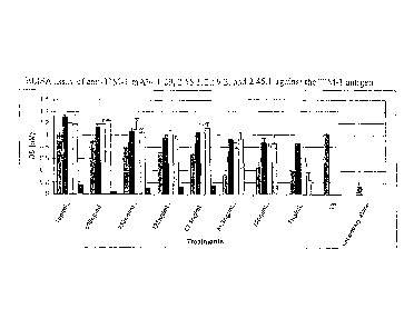
[0042] Figure 1 is a bar graph of the results of an ELISA assay of anti-
UM-1
monoclonal antibodies 1.29, 2.56.2, 2.59.2, and 2.45.1 against the TIM-1
antigen.
[0043] Figure 2 is a bar graph of the results of an ELISA assay of anti-
TIM-1
monoclonal antibodies 1.29, 2.56.2, 2.59.2, and 2.45.1 against irrelevant
protein.
[0044] Figure 3 shows staining of Renal Cell Cancer (3A) and Pancreatic
Cancer
(3B) with the anti-TIM-1 mAb 2.59.2.
-8-
CA 02629453 2008-05-12
WO 2007/059082
PCT/US2006/044090
[0045] Figure 4
is a bar graph of clonogenic assay results of anti-TIM-1
monoclonal antibody mediated toxin killing in the ACHN kidney cancer cell
line.
[0046] Figure 5
is a bar graph of clonogenic assay results of anti-TIM-1
monoclonal antibody mediated toxin killing in the BT549 breast cancer cell
line.
[0047] Figure 6
is a bar graph of the results of a clonogenic assay of CAKI-1
cells treated with Auristatin E (AE) conjugated antibodies.
[0048] Figure 7
is a bar graph of the results of a clonogenic assay of liT549 cells
treated with Auristatin E (AE) conjugated antibodies.
[0049] Figure 8
is a bar graph showing that anti-TIM-1 monoclonal antibodies
2.59.2, 2.56.2 and 2.45.1 significantly inhibit IL-4 release from Thl cells
compared to the
control PK16.3 mAb.
[0050] Figure 9
is a bar graph showing that anti-TIM-1 monoclonal antibodies
2.59.2 and 2.45.1 significantly inhibit IL-4 release from Th2 cells compared
to control
PK16.3 mAb.
[0051] Figure
10 is a bar graph showing that anti-TIM-1 monoclonal antibody
2.59.2 significantly inhibited IL-5 release from Thl cells compared to control
PK16.3 mAb.
[0052] Figure
11 is a bar graph showing that anti-TIM-1 monoclonal antibodies
2.59.2 and 1.29 significantly inhibited IL-5 release from Th2 cells compared
to control
PK16.3 mAb.
[0053] Figure
12 is a bar graph showing that anti-TIM-1 monoclonal antibodies
2.59.2, 1.29 and 2.56.2 significantly inhibited IL-10 release from Thl cells
compared to
control PK16.3 mAb.
[0054] Figure
13 is a bar graph showing that anti-TIM-1 monoclonal antibodies
2.59.2, 1.29 and 2.45.1 significantly inhibited IL-10 release from Th2 cells
compared to
control PK16.3 mAb.
[0055] Figure
14 is a bar graph showing that anti-TIM-1 monoclonal antibodies
2.592, 1.29 and 2.56.2 significantly inhibited IL-13 release from Thl cells
compared to
control PK16.3 mAb.
[0056] Figure
15 is a bar graph showing that anti-TIM-1 monoclonal antibodies
2.59.2 and 1.29 significantly inhibited IL-13 release from Th2 cells compared
to control
PK16.3 inAb
[0057] Figure
16 is a bar graph showing that anti-TIM-1 monoclonal antibodies
did not inhibit IFNy release from Thl cells compared to control PK16.3 inAb.
-9-
CA 02629453 2008-05-12
WO 2007/059082
PCT/US2006/044090
[0058] Figure
17 is a bar graph showing that anti-TIM-1 monoclonal antibodies
2.59.2 and 2.45.1 significantly inhibited IFNy release from Th2 cells compared
to control
PK16.3 mAb.
[0059] Figures
18A-18T are bar graphs showing BrdU incorporation assay
results from experiments in which the neutralization of various human anti-TIM-
1
monoclonal antibodies was assessed.
[0060] Figures
19A through 19D are line graphs showing the results of antibody
conjugate studies performed using the plant toxin Saporin conjugated to TIM-1-
specific
antibodies and irrelevant antibodies (Figures 19A-19C). Additional negative
controls
included irrelevant antibodies alone without toxin (Figure 19D).
[0061] Figure 20 is a graph showing tumor growth inhibition and complete
regression of IGROV1 ovarian carcinoma xenografts in athymic mice after
treatment with
6.25 to 50 mg/kg i.v. every 4 days for 4 treatments. The responses of tumor-
bearing animals
to reference drugs such as vinblastine (1.7 mg/kg i.v. q4d X4) and paclitaxel
(15.0 mg/kg
i.v. q2d X4) are also shown. Control groups were treated with either phosphate-
buffered
saline (PBS) or physiological saline. CR014-vcMMAE was toxic to the test
animals at 50
mg/kg/treatment (n= 1/6) and at 100 mg/kg/treatment (n= 6/6).
Detailed Description of the Preferred Embodiment
[0062]
Embodiments of the invention described herein are based upon the
generation and identification of isolated antibodies that bind specifically to
T cell,
immunoglobulin domain and mucin domain 1 (TIM-1). As discussed below, TIM-1 is
expressed at elevated levels in clear cell carcinomas and cancer cell lines
derived from the
same. Accordingly, antibodies that bind to TIM-1 are useful for -die treatment
and
inhibition of carcinomas. In addition, antibodies that bind TIM-1 are also
useful for
reducing cell migration and enhancing apoptosis of kidney cancer cells.
[0063]
Accordingly, embodiments of the invention described herein provide
isolated antibodies, or fragments of those antibodies, that bind to TIM-1. As
known in the
art, the antibodies can advantageously be, e.g., monoclonal, chimeric and/or
human
antibodies. Embodiments of the invention described herein also provide cells
for producing
these antibodies.
[0064] Another
embodiment of the invention provides for using these antibodies
for diagnostic or therapeutic purposes. For example, embodiments of the
invention provide
-10-
CA 02629453 2008-05-12
WO 2007/059082
PCT/US2006/044090
methods and antibodies for inhibiting the expression of TIM-1 associated with
cell
proliferation. Preferably, the antibodies are used to treat neoplasms such as
renal and
pancreatic tumors, head and neck cancer, ovarian cancer, gastric (stomach)
cancer,
melanoma, lymphoma, prostate cancer, liver cancer, breast cancer, lung cancer,
renal
cancer, bladder cancer, colon cancer, esophageal cancer, and brain cancer. In
association
with such treatment, articles of manufacture comprising these antibodies are
provided.
Additionally, an assay kit comprising these antibodies is provided to screen
for cancers or
tumors.
[0065]
Additionally, the nucleic acids described herein, and fragments and
variants thereof, may be used, by way of nonlimiting example, (a) to direct
the biosynthesis
of the corresponding encoded proteins, polypeptides, fragments and variants as
recombinant
or heterologous gene products, (b) as probes for detection and quantification
of the nucleic
acids disclosed herein, (c) as sequence templates for preparing antisense
molecules, and the
like. Such uses are described more fully in the following disclosure.
[0066]
Furthermore, the TIM-1 proteins and polypeptides described herein, and
fragments and variants thereof, may be used, in ways that include (a) serving
as an
innnunogen to stimulate the production of an anti-TIM-1 antibody, (b) a
capture antigen in
an immunogenic assay for such an antibody, (c) as a target for screening for
substances that
bind to a TIM-1 polypeptide described herein, and (d) a target for a TIM-1
specific antibody
such that treatment with the antibody affects the molecular and/or cellular
function
mediated by the target. TIM-1 polypeptide expression or activity can promote
cell survival
and/or metastatic potential. Conversely, a decrease in TIM-1 polypeptide
expression or
inhibition of its function reduces tumor cell survival and invasiveness in a
therapeutically
beneficial manner.
[0067] Single
chain antibodies (scFv's) and bispecific antibodies specific for
TIM-1 are useful particularly because it may more readily penetrate a tumor
mass due to its
smaller size relative to a whole IgG molecule. Studies comparing the tumor
penetration
between whole IgG molecules and scFv's have been have been described in the
literature.
The scFv can be derivatized with a toxin or radionuclide in order to destroy
tumor cells
expressing the TIM-1 antigen, in a manner similar to the IgG2 or IgG4 anti-TIM-
1 toxin
labeled or radionuclide derivatized whole antibodies already discussed, but
with the
advantage of being able to penetrate the tumor more fully, which may translate
into
-11-
CA 02629453 2008-05-12
WO 2007/059082
PCT/US2006/044090
increased efficacy in eradicating the tumor. A specific example of a
biologically active
anti-TIM-1 scFv is provided herein.
Sequence Listing
[0068] The
heavy chain and light chain variable region nucleotide and amino
acid sequences of representative human anti-TIM-1 antibodies are provided in
the sequence
listing, the contents of which are summarized in Table 1 below.
Table 1
mAb SEQ ID
Sequence
ID No.: NO:
Nucleotide sequence encoding the variable region and a portion of the 1
constant region of the heavy chain
Amino acid sequence of the variable region of the heavy chain 2
1.29
Nucleotide sequence encoding the variable region and a portion of the 3
constant region of the light chain
Amino acid sequence of the variable region of the light chain 4
Nucleotide sequence encoding the variable region and a portion of the 5
constant region of the heavy chain
-
Amino acid sequence of the variable region of the heavy chain 6
1.37
Nucleotide sequence encoding the variable region and a portion of the 7
constant region of the light chain
Amino acid sequence of the variable region of the light chain 8
Nucleotide sequence encoding the variable region and a portion of the 9
constant region of the heavy chain
Amino acid sequence of the variable region of the heavy chain 10
2.16
Nucleotide sequence encoding the variable region and a portion of the 11
constant region of the light chain
Amino acid sequence of the variable region of the light chain 12
-12-
CA 02629453 2008-05-12
WO 2007/059082
PCT/US2006/044090
Nucleotide sequence encoding the variable region and a portion of the 13
constant region of the heavy chain
Amino acid sequence of the variable region of the heavy chain 14
2.17
Nucleotide sequence encoding the variable region and a portion of the 15
constant region of the light chain
Amino acid sequence of the variable region of the light chain 16
Nucleotide sequence encoding the variable region and a portion of the 17
constant region of the heavy chain
Amino acid sequence of the variable region of the heavy chain 18
2.24
Nucleotide sequence encoding the variable region and a portion of the 19
constant region of the light chain
Amino acid sequence of the variable region of the light chain 20
Nucleotide sequence encoding the variable region and a portion of the 21
constant region of the heavy chain
Amino acid sequence of the variable region of the heavy chain 22
2.45
Nucleotide sequence encoding the variable region and a portion of the 23
constant region of the light chain
Amino acid sequence of the variable region of the light chain 24
Nucleotide sequence encoding the variable region and a portion of the 25
constant region of the heavy chain
Amino acid sequence of the variable region of the heavy chain 26
2.54
Nucleotide sequence encoding the variable region and a portion of the 27
constant region of the light chain
Amino acid sequence of the variable region of the light chain 28
Nucleotide sequence encoding the variable region and a portion of the 29
constant region of the heavy chain
Amino acid sequence of the variable region of the heavy chain 30
2.56
Nucleotide sequence encoding the variable region and a portion of the 31
constant region of the light chain
Amino acid sequence of the variable region of the light chain 32
-13-
CA 02629453 2008-05-12
WO 2007/059082
PCT/US2006/044090
Nucleotide sequence encoding the variable region and a portion of the 33
constant region of the heavy chain
Amino acid sequence of the variable region of the heavy chain 34
2.59
Nucleotide sequence encoding the variable region and a portion of the 35
constant region of the light chain
Amino acid sequence of the variable region of the light chain 36
Nucleotide sequence encoding the variable region and a portion of the 37
constant region of the heavy chain
Amino acid sequence of the variable region of the heavy chain 38
2.61
Nucleotide sequence encoding the variable region and a portion of the 39
constant region of the light chain
Amino acid sequence of the variable region of the light chain 40
Nucleotide sequence encoding the variable region and a portion of the 41
constant region of the heavy chain
Amino acid sequence of the variable region of the heavy chain 42
2.70
Nucleotide sequence encoding the variable region and a portion of the 43
constant region of the light chain
Amino acid sequence of the variable region of the light chain 44
Nucleotide sequence encoding the variable region and a portion of the 45
constant region of the heavy chain
Amino acid sequence of the variable region of the heavy chain 46
2.76
Nucleotide sequence encoding the variable region and a portion of the 47
constant region of the light chain
Amino acid sequence of the variable region of the light chain 48
Nucleotide sequence encoding the variable region and a portion of the 49
constant region of the heavy chain
Amino acid sequence of the variable region and a portion of the 50
2.70.2 constant region of the heavy chain
Nucleotide sequence encoding the variable region and a portion of the 51
constant region of the light chain
Amino acid sequence of the variable region and a portion of the 52
constant region of the light chain
Definitions
[0069] Unless
otherwise defined, scientific and technical terms used in
connection with the invention described herein shall have the meanings that
are commonly
-14-
CA 02629453 2015-02-17
understood by those of ordinary skill in the art. Further, unless otherwise
required by
context, singular terms shall include pluralities and plural terms shall
include the singular.
Generally, nomenclatures utilized in connection with, and techniques of, cell
and tissue
culture, molecular biology, and protein and oligo- or polynucleotide chemistry
and
hybridization described herein are those well known and commonly used in the
art.
Standard techniques are used for recombinant DNA, oligonucleotide synthesis,
and tissue
culture and transformation (e.g., electroporation, lipofection). Enzymatic
reactions and
purification techniques are performed according to manufacturer's
specifications or as
commonly accomplished in the art or as described herein. The foregoing
techniques and
procedures are generally performed according to conventional methods well
known in the
art and as described in various general and more specific references that are
cited and
discussed throughout the present specification. See e.g., Sambrook et al.
Molecular
Cloning: A Laboratory Manual (2d ed., Cold Spring Harbor Laboratory Press,
Cold Spring
Harbor, N.Y. (1989)), which is incorporated herein by reference. The
nomenclatures
utilized in connection with, and the laboratory procedures and techniques of,
analytical
chemistry, synthetic organic chemistry, and medicinal and pharmaceutical
chemistry
described herein are those well known and commonly used in the art. Standard
techniques
are used for chemical syntheses, chemical analyses, pharmaceutical
preparation,
formulation, and delivery, and treatment of patients.
[0070] As utilized in accordance with the present disclosure, the
following
terms, unless otherwise indicated, shall be understood to have the following
meanings:
[0071] The term "TIM-1" refers to T cell, immunoglobulin domain and mucin
domain 1. In one embodiment, TIM-1 refers to a polypeptide comprising the
amino acid
sequence of SEQ ID NO: 50.
[0072] The term "polypeptide" is used herein as a generic term to refer
to native
protein, fragments, or analogs of a polypeptide sequence. Hence, native
protein, fragments,
and analogs are species of the polypeptide genus. Preferred polypeptides in
accordance
with the invention comprise human heavy chain immunoglobulin molecules and
human
kappa light chain immunoglobulin molecules, as well as antibody molecules
formed by
combinations comprising the heavy chain immunoglobulin molecules with light
chain
immunoglobulin molecules, such as the kappa light chain immunoglobulin
molecules, and
vice versa, as well as fragments and analogs thereof.
-15-
CA 02629453 2008-05-12
WO 2007/059082
PCT/US2006/044090
[0073] The term
"polynucleotide" as referred to herein means a polymeric form
of nucleotides of at least 10 bases in length, either ribonucleotides or
deoxynucleotides or a
modified form of either type of nucleotide. The term includes single and
double stranded
forms of DNA.
[0074] The term
"isolated polynucleotide" as used herein shall mean a
polynucleotide of genomic, cDNA, or synthetic origin or some combination
thereof, which
by virtue of its origin the isolated polynucleotide (1) is not associated with
all or a portion of
a polynucleotide in which the isolated polynucleotide is found in nature, (2)
is operably
linked to a polynucleotide which it is not linked to in nature, or (3) does
not occur in nature
as part of a larger sequence.
[0075] The term
"isolated protein" referred to herein means a protein of cDNA,
recombinant RNA, or synthetic origin or some combination thereof, which by
virtue of its
origin, or source of derivation, the "isolated protein" (1) is not associated
with proteins
found in nature, (2) is free of other proteins from the same source, e.g.,
free of murine
proteins, (3) is expressed by a cell from a different species, or (4) does not
occur in nature.
[0076] The term
"oligonucleotide" referred to herein includes naturally
occurring, and modified nucleotides linked together by naturally occurring,
and non-
naturally occurring oligonucleotide linkages. Oligonucleotides are a
polynucleotide subset
generally comprising a length of 200 bases or fewer. Preferably
oligonucleotides are 10 to
60 bases in length and most preferably 12, 13, 14, 15, 16, 17, 18, 19, or 20
to 40 bases in
length.
Oligonucleotides are usually single stranded, e.g. for probes; although
oligonucleotides may be double stranded, e.g. for use in the construction of a
gene mutant.
Oligonucleotides described herein can be either sense or antisense
oligonucleotides.
[0077]
Similarly, unless specified otherwise, the lefthand end of single-stranded
polynucleotide sequences is the 5' end; the lefthand direction of double-
stranded
polynucleotide sequences is referred to as the 5' direction. The direction of
5' to 3' addition
of nascent RNA transcripts is referred to as the transcription direction;
sequence regions on
the DNA strand having the same sequence as the RNA and which are 5' to the 5'
end of the
RNA transcript are referred to as upstream sequences; sequence regions on the
DNA strand
having the same sequence as the RNA and which are 3' to the 3' end of the RNA
transcript
are referred to as downstream sequences.
[0078] The term
"naturally-occurring" as used herein as applied to an object
refers to the fact that an object can be found in nature. For example, a
polypeptide or
-16-
CA 02629453 2014-05-09
polynucleotide sequence that is present in an organism (including viruses)
that can be
isolated from a source in nature and which has not been intentionally modified
by man in
the laboratory or otherwise is naturally-occurring.
[0079] The term "naturally occurring nucleotides" referred to herein
includes
deoxyribonucleotides and ribonucleotides. The term "modified nucleotides"
referred to
herein includes nucleotides with modified or substituted sugar groups and the
like. The
term "oligonucleotide linkages" referred to herein includes oligonucleotides
linkages such
as phosphorothioate, phosphorodithioate, phosphoroselenoate,
phosphorodiselenoate,
phosphoroanilothioate, phoshoraniladate, phosphoroamidate, and the like. See,
e.g.,
LAPlanche et aL, NucL Acids Res. 14:9081 (1986); Stec et al., J. Am. Chem.
Soc. 106:6077
(1984); Stein et aL, NucL Acids Res. 16:3209 (1988); Zon et al., Anti-Cancer
Drug Design
6:539 (1991); Zon et al., Oligonucleotides and Analogues: A Practical
Approach, pp. 87-
108 (F. Eckstein, ed., Oxford University Press, Oxford England (1991)); Stec
et al., U.S.
Patent No. 5,151,510; Uhlmann and Peyman, Chemical Reviews 90:543 (1990) .
An oligonucleotide can include
a label for detection, if desired.
100801 The term "operably linked" as used herein refers to positions of
components so described are in a relationship permitting them to function in
their intended
manner. A control sequence operably linked to a coding sequence is ligated in
such a way
that expression of the coding sequence is achieved under conditions compatible
with the
control sequences.
[0081] The term "control sequence" is used herein refers to
polynucleotide
sequences which are necessary to effect the expression and processing of
coding sequences
to which they are ligated. The nature of such control sequences differs
depending upon the
host organism; in prokaryotes, such control sequences generally include
promoter,
ribosomal binding site, and transcription termination sequence; in eukaryotes,
generally,
such control sequences include promoters and transcription termination
sequence. The term
control sequences is intended to include, at a minimum, all components whose
presence is
essential for expression and processing, and can also include additional
components whose
presence is advantageous, for example, leader sequences and fusion partner
sequences.
[0082] The term "selectively hybridize" referred to herein means to
detectably
and specifically bind. Polynucleotides, oligonucleotides and fragments thereof
described
herein selectively hybridize to nucleic acid strands under hybridization and
wash conditions
-17-
CA 02629453 2008-05-12
WO 2007/059082
PCT/US2006/044090
that minimize appreciable amounts of detectable binding to nonspecific nucleic
acids. High
stringency conditions can be used to achieve selective hybridization
conditions as known in
the art and discussed herein. Generally, the nucleic acid sequence homology
between the
polynucleotides, oligonucleotides, and fragments described herein and a
nucleic acid
sequence of interest will be at least 80%, and more typically with preferably
increasing
homologies of at least 85%, 90%, 95%, 99%, and 100%.
[0083] Two
amino acid sequences are homologous if there is a partial or
complete identity between their sequences. For example, 85% homology means
that 85%
of the amino acids are identical when the two sequences are aligned for
maximum
matching. Gaps (in either of the two sequences being matched) are allowed in
maximizing
matching; gap lengths of 5 or less are preferred with 2 or less being more
preferred.
Alternatively and preferably, two protein sequences (or polypeptide sequences
derived from
them of at least 30 amino acids in length) are homologous, as this term is
used herein, if
they have an alignment score of at more than 5 (in standard deviation units)
using the
program ALIGN with the mutation data matrix and a gap penalty of 6 or greater.
See
Dayhoff, M.O., in Atlas of Pr otein Sequence and Structure, pp. 101-110
(Volume 5,
National Biomedical Research Foundation (1972)) and Supplement 2 to this
volume, pp. 1-
10. The two sequences or parts thereof are more preferably homologous if their
amino
acids are greater than or equal to 50% identical when optimally aligned using
the ALIGN
program.
[0084] The term
"corresponds to" is used herein to mean that a polynucleotide
sequence is homologous (i.e., is identical, not strictly evolutionarily
related) to all or a
portion of a reference polynucleotide sequence, or that a polypeptide sequence
is identical
to a reference polypeptide sequence.
[0085] In
contradistinction, the term "complementary to" is used herein to mean
that the complementary sequence is homologous to all or a portion of a
reference
polynucleotide sequence. For illustration, the nucleotide sequence "TATAC"
corresponds
to a reference sequence "TATAC" and is complementary to a reference sequence
"GTATA."
[0086] The
following terms are used to describe the sequence relationships
between two or more polynucleotide or amino acid sequences: "reference
sequence,"
"comparison window," "sequence identity," "percentage of sequence identity,"
and
"substantial identity." A "reference sequence" is a defined sequence used as a
basis for a
-18-
CA 02629453 2008-05-12
WO 2007/059082
PCT/US2006/044090
sequence comparison; a reference sequence may be a subset of a larger
sequence, for
example, as a segment of a full-length cDNA or gene sequence given in a
sequence listing
or may comprise a complete cDNA or gene sequence. Generally, a reference
sequence is at
least 18 nucleotides or 6 amino acids in length, frequently at least 24
nucleotides or 8 amino
acids in length, and often at least 48 nucleotides or 16 amino acids in
length. Since two
polynucleotides or amino acid sequences may each (1) comprise a sequence
(i.e., a portion
of the complete polynucleotide or amino acid sequence) that is similar between
the two
molecules, and (2) may further comprise a sequence that is divergent between
the two
polynucleotides or amino acid sequences, sequence comparisons between two (or
more)
molecules are typically performed ,by comparing sequences of the two molecules
over a
comparison window to identify and compare local regions of sequence
similarity. A
"comparison window," as used herein, refers to a conceptual segment of at
least 18
contiguous nucleotide positions or 6 amino acids wherein a polynucleotide
sequence or
amino acid sequence may be compared to a reference sequence of at least 18
contiguous
nucleotides or 6 amino acid sequences and wherein the portion of the
polynucleotide
sequence in the comparison window may comprise additions, deletions,
substitutions, and
the like (i.e., gaps) of 20 percent or less as compared to the reference
sequence (which does
not comprise additions or deletions) for optimal alignment of the two
sequences. Optimal
alignment of sequences for aligning a comparison window may be conducted by
the local
homology algorithm of Smith and Waterman, Adv. App!. Math., 2:482 (1981), by
the
homology alignment algorithm of Needleman and Wunsch, J. MoL Biol., 48:443
(1970), by
the search for similarity method of Pearson and Lipman, Proc. Natl. Acad. Sci.
(U.S.A.),
85:2444 (1988), by computerized implementations of these algorithms (GAP,
BESTFIT,
FASTA, and TFASTA in the Wisconsin Genetics Software Package Release 7.0,
(Genetics
Computer Group, 575 Science Dr., Madison, Wis.), Geneworks, or MacVector
software
packages), or by inspection, and the best alignment (i.e., resulting in the
highest percentage
of homology over the comparison window) generated by the various methods is
selected.
[0087] The term
"sequence identity" means that two polynucleotide or amino
acid sequences are identical (i.e., on a nucleotide-by-nucleotide or residue-
by-residue basis)
over the comparison window. The term percentage of sequence identity is
calculated by
comparing two optimally aligned sequences over the window of comparison,
determining
the number of positions at which the identical nucleic acid base (e.g., A, T,
C, G, U, or I) or
residue occurs in both sequences to yield the number of matched positions,
dividing the
-19-
CA 02629453 2015-02-17
number of matched positions by the total number of positions in the comparison
window
(i.e., the window size), and multiplying the result by 100 to yield the
percentage of sequence
identity. The terms "substantial identity" as used herein denotes a
characteristic of a
polynucleotide or amino acid sequence, wherein the polynucleotide or amino
acid
comprises a sequence that has at least 85 percent sequence identity,
preferably at least 90 to
95 percent sequence identity, more usually at least 99 percent sequence
identity as
compared to a reference sequence over a comparison window of at least 18
nucleotide (6
amino acid) positions, frequently over a window of at least 24-48 nucleotide
(8-16 amino
acid) positions, wherein the percentage of sequence identity is calculated by
comparing the
reference sequence to the sequence which may include deletions or additions
which total 20
percent or less of the reference sequence over the comparison window. The
reference
sequence may be a subset of a larger sequence.
[0088] As used herein, the twenty conventional amino acids and their
abbreviations follow conventional usage. See Immunology - A Synthesis (2nd
Edition, E.S.
Golub and D.R. Gren, Eds., Sinauer Associates, Sunderland, Mass. (1991))
Stereoisomers (e.g., D-amino acids) of the twenty
conventional amino acids, unnatural amino acids such as a-, a-disubstituted
amino acids,
N-alkyl amino acids, lactic acid, and other unconventional amino acids may
also be suitable
components for polypeptides described herein. Examples of unconventional amino
acids
include: 4-hydroxyproline, y -carboxyglutamate, a-N,N,N-trimethyllysine, s-N-
acetyllysine,
0-phosphoserine, N-acetylserine, N-formylmethionine, 3-methylhistidine, 5-
hydroxylysine,
cy-N-methylarginine, and other similar amino acids and imino acids (e.g., 4-
hydroxyproline). In the polypeptide notation used herein, the lefthand
direction is the
amino terminal direction and the righthand direction is the carboxy-terminal
direction, in
accordance with standard usage and convention.
[0089] As applied to polypeptides, the term "substantial identity" means
that
two peptide sequences, when optimally aligned, such as by the programs GAP or
BESTFIT
using default gap weights, share at least 80 percent sequence identity,
preferably at least 90
percent sequence identity, more preferably at least 95 percent sequence
identity, and most
preferably at least 99 percent sequence identity. Preferably, residue
positions which are not
identical differ by conservative amino acid substitutions. Conservative amino
acid
substitutions refer to the interchangeability of residues having similar side
chains. For
example, a group of amino acids having aliphatic side chains is glycine,
alanine, valine,
-20-
CA 02629453 2008-05-12
WO 2007/059082
PCT/US2006/044090
leucine, and isoleucine; a group of amino acids having aliphatic-hydroxyl side
chains is
serine and threonine; a group of amino acids having amide-containing side
chains is
asparagine and glutamine; a group of amino acids having aromatic side chains
is
phenylalanine, tyrosine, and tryptophan; a group of amino acids having basic
side chains is
lysine, arginine, and histidine; and a group of amino acids having sulfur-
containing side
chains is cysteine and methionine. Preferred conservative amino acids
substitution groups
are: valine-leucine-isoleucine, phenylalanine-tyrosine, lysine-arginine,
alanine-valine,
glutamic-aspartic, and asparagine-glutamine.
[0090] As
discussed herein, minor variations in the amino acid sequences of
antibodies or immunoglobulin molecules are contemplated as being encompassed
by the
invention described herein, providing that the variations in the amino acid
sequence
maintain at least 75%, more preferably at least 80%, 90%, 95%, and most
preferably 99%
sequence identity to the antibodies or immunoglobulin molecules described
herein. In
particular, conservative amino acid replacements are contemplated.
Conservative
replacements are those that take place within a family of amino acids that are
related in their
side chains. Genetically encoded amino acids are generally divided into
families: (1)
acidic=aspartate, glutamate; (2) basic=lysine, arginine, histidine; (3) non-
polar=alanine,
valine, leucine, isoleucine, proline, phenylalanine, methionine, tryptophan;
and (4)
uncharged polar=glycine, asparagine, glutamine, cysteine, serine, threonine,
tyrosine. More
preferred families are: serine and threonine are aliphatic-hydroxy family;
asparagine and
glutamine are an amide-containing family; alanine, valine, leucine and
isoleucine are an
aliphatic family; and phenylalanine, tryptophan, and tyrosine are an aromatic
family. For
example, it is reasonable to expect that an isolated replacement of a leucine
with an
isoleucine or valine, an aspartate with a glutamate, a threonine with a
serine, or a similar
replacement of an amino acid with a structurally related amino acid will not
have a major
effect on the binding or properties of the resulting molecule, especially if
the replacement
does not involve an amino acid within a framework site. Whether an amino acid
change
results in a functional peptide can readily be determined by assaying the
specific activity of
the polypeptide derivative. Assays are described in detail herein. Fragments
or analogs of
antibodies or immunoglobulin molecules can be readily prepared by those of
ordinary skill
in the art. Preferred amino- and carboxy-termini of fragments or analogs occur
near
boundaries of functional domains. Structural and functional domains can be
identified by
comparison of the nucleotide .and/or amino acid sequence data to public or
proprietary
-21-
CA 02629453 2014-05-09
sequence databases. Preferably, computerized comparison methods are used to
identify
sequence motifs or predicted protein conformation domains that occur in other
proteins of
known structure and/or function. Methods to identify protein sequences that
fold into a
known three-dimensional structure are known. Bowie et al., Science, 253:164
(1991).
Thus, the foregoing examples demonstrate that those of skill in the art can
recognize
sequence motifs and structural conformations that may be used to deft=
structural and
functional domains described herein.
[0091] Preferred amino acid substitutions are those which: (1)
reduce
susceptibility to proteolysis, (2) reduce susceptibility to oxidation, (3)
alter binding affinity
for forming protein complexes, (4) alter binding affinities, and (4) confer or
modify other
physicochemical or functional properties of such analogs. Analogs can include
various
muteins of a sequence other than the naturally-occurring peptide sequence. For
example,
single or multiple amino acid substitutions (preferably conservative amino
acid
substitutions) may be made in the naturally-occurring sequence (preferably in
the portion of
the polypeptide outside the domain(s) forming intermolecular contacts). A
conservative
amino acid substitution should not substantially change the structural
characteristics of the
parent sequence (e.g., a replacement amino acid should not tend to break a
helix that occurs
in the parent sequence, or disrupt other types of secondary structure that
characterizes the
parent sequence). Examples of art-recognized polypeptide secondary and
tertiary structures
=
are described in Proteins, Structures and Molecular Principles (Creighton,
Ed., W. H.
Freeman and Company, New York (1984)); Introduction to Protein Structure (C.
Branden
and J. Tooze, eds., Garland Publishing, New York, N.Y. (1991)); and Thornton
et al.,
Nature, 354:105 (1991) .
[0092] The term "polypeptide fragment" as used herein refers to a
polypeptide
that has an amino-terminal and/or carboxy-terminal deletion, but where the
remaining
amino acid sequence is identical to the corresponding positions in the
naturally-occurring
sequence deduced, for example, from a full-length cDNA sequence. Fragments
typically
are at least 5, 6, 8 or 10 amino acids long, preferably at least 14 amino
acids long, more
preferably at least 20 amino acids long, usually at least 50 amino acids long,
and even more
preferably at least 70 amino acids long. The term "analog" as used herein
refers to
polypeptides which are comprised of a segment of at least 25 amino acids that
has
substantial identity to a portion of a deduced amino acid sequence and which
has at least
one of the following properties: (1) specific binding to a TIM-1, under
suitable binding
-22-
CA 02629453 2015-02-17
conditions, (2) ability to block appropriate TIM-1 binding, or (3) ability to
inhibit the
growth and/or survival of TIM-1 expressing cells in vitro or in vivo.
Typically, polypeptide
analogs comprise a conservative amino acid substitution (or addition or
deletion) with
respect to the naturally occurring sequence. Analogs typically are at least 20
amino acids
long, preferably at least 50 amino acids long or longer, and can often be as
long as a full-
length naturally-occurring polypeptide.
[0093] Peptide
analogs are commonly used in the pharmaceutical industry as
non-peptide drugs with properties analogous to those of the template peptide.
These types
of non-peptide compounds are termed peptide mimetics or peptidomimetics.
Fauchere, J.
Adv. Drug Res., 15:29 (1986); Veber and Freidinger, TINS, p.392 (1985); and
Evans et al.,
J. Med. Chem., 30:1229 (1987) Such
compounds are often developed with the aid of computerized molecular modeling.
Peptide
mimefics that are structurally similar to therapeutically useful peptides may
be used to
produce an equivalent therapeutic or prophylactic effect. Generally,
peptidomimetics are
structurally similar to a paradigm polypeptide (i.e., a polypeptide that has a
biochemical
property or pharmacological activity), such as human antibody, but have one or
more
peptide linkages optionally replaced by a linkage selected from the group
consisting of: --
CH2NH--, --CH2S--, --CH2-CH2--, --CH=CH--(cis and trans), --COCH2--, --
CH(OH)CH2--,
and ¨CH2S0--, by methods well known in the art. Systematic substitution of one
or more
amino acids of a consensus sequence with a D-amino acid of the same type
(e.g., D-lysine
in place of L-lysine) may be used to generate more stable peptides. In
addition, constrained
peptides comprising a consensus sequence or a substantially identical
consensus sequence
variation may be generated by methods known in the art (Rizo and Gierasch,
Ann. Rev.
Biochem., 61:387 (1992) ; for example,
by adding internal
cysteine residues capable of forming intramolecular disulfide bridges which
cyclize the
peptide.
[0094] "Antibody" or
"antibody peptide(s)" refer to an intact antibody, or a
binding fragment thereof that competes with the intact antibody for specific
binding.
Binding fragments are produced by recombinant DNA techniques, or by enzymatic
or
chemical cleavage of intact antibodies. Binding fragments include Fab, Fab',
F(a1:02, Fv,
and single-chain antibodies. An antibody other than a bispecific or
bifunctional antibody is
understood to have each of its binding sites identical. An antibody
substantially inhibits
adhesion of a receptor to a counterreceptor when an excess of antibody reduces
the quantity
-23-
CA 02629453 2015-02-17
of receptor bound to counterreceptor by at least about 20%, 40%, 60% or 80%,
and more
usually greater than about 85% (as measured in an in vitro competitive binding
assay).
[0095] Digestion of antibodies with the enzyme, papain, results in two
identical
antigen-binding fragments, known also as "Fab" fragments, and a "Fe" fragment,
having no
antigen-binding activity but having the ability to crystallize. Digestion of
antibodies with
the enzyme, pepsin, results in the a "F(ab')2" fragment in which the two arms
of the
antibody molecule remain linked and comprise two-antigen binding sites. The
F(ab')2
fragment has the ability to crosslink antigen.
[0096] "Fv" when used herein refers to the minimum fragment of an
antibody
that retains both antigen-recognition and antigen-binding sites.
[0097] "Fab" when used herein refers to a fragment of an antibody which
comprises the constant domain of the light chain and the CH1 domain of the
heavy chain.
[0098] The term "epitope" includes any protein determinant capable of
specific
binding to an immunoglobulin or T-cell receptor. Epitopic determinants usually
consist of
chemically active surface groupings of molecules such as amino acids or sugar
side chains
and usually have specific three dimensional structural characteristics, as
well as specific
charge characteristics. An antibody is said to specifically bind an antigen
when the
dissociation constant is l .tM, preferably 100 nM and most preferably 10 TIM.
[0099] The term "agent" is used herein to denote a chemical compound, a
mixture of chemical compounds, a biological macromolecule, or an extract made
from
biological materials.
[0100] The term "pharmaceutical agent" or "drug" as used herein refers to
a
chemical compound or composition capable of inducing a desired therapeutic
effect when
properly administered to a patient. Other chemistry terms herein are used
according to
conventional usage in the art, as exemplified by The McGraw-Hill Dictionary of
Chemical
Terms (Parker, S., Ed., McGraw-Hill, San Francisco (1985))
[0101] The term "antineoplastic agent" is used herein to refer to agents
that have
the functional property of inhibiting a development or progression of a
neoplasm in a
human, particularly a malignant (cancerous) lesion, such as a carcinoma,
sarcoma,
lymphoma, or leukemia. Inhibition of metastasis is frequently a property of
antineoplastic
agents.
-24-
CA 02629453 2008-05-12
WO 2007/059082
PCT/US2006/044090
[0102] As used herein, "substantially pure" means an object species is
the
predominant species present (i.e., on a molar basis it is more abundant than
any other
individual species in the composition), and preferably a substantially
purified fraction is a
composition wherein the object species comprises at least about 50 percent (on
a molar
basis) of all macromolecular species present. Generally, a substantially pure
composition
will comprise more than about 80 percent of all macromolecular species present
in the
composition, more preferably more than about 85%, 90%, 95%, and 99%. Most
preferably,
the object species is purified to essential homogeneity (contaminant species
cannot be
detected in the composition by conventional detection methods) wherein the
composition
consists essentially of a single macromolecular species.
[0103] "Active" or "activity" in regard to a TIM-1 polypeptide refers
to a
portion of a TIM-1 polypeptide which has a biological or an immunological
activity of a
native TIM-1 polypeptide. "Biological" when used herein refers to a biological
function
that results from the activity of the native TIM-1 polypeptide. A preferred
biological
activity includes, for example, regulation of cellular growth.
[0104] "Label" or "labeled" as used herein refers to the addition of a
detectable
moiety to a polypeptide, for example, a radiolabel, fluorescent label,
enzymatic label
chemiluminescent labeled or a biotinyl group. Radioisotopes or radionuclides
may include
3H, 14c, 15N, 35s, 90y, 99n, 1111n, 125,-, 1 3 1
¨ -I, fluorescent labels may include rhodamine,
lanthanide phosphors or FITC and enzymatic labels may include horseradish
peroxidase, 13-
galactosidase, luciferase, alkaline phosphatase.
[0105] "Mammal" when used herein refers to any animal that is
considered a
mammal. Preferably, the mammal is human.
[0106] "Liposome" when used herein refers to a small vesicle that may
be useful
for delivery of drugs that may include the TIM-1 polypeptide described herein
or antibodies
to such a TIM-1 polypeptide to a mammal.
[0107] The term "patient" includes human and veterinary subjects.
Antibody Structure
[0108] The basic whole antibody structural unit is known to comprise a
tetramer.
Each tetramer is composed of two identical pairs of polypeptide chains, each
pair having
one "light" (about 25 kDa) and one "heavy" chain (about 50-70 kDa). The amino-
terminal
portion of each chain includes a variable domain of about 100 to 110 or more
amino acids
-25-
,
CA 02629453 2014-05-09
primarily responsible for antigen recognition. The carboxy-terminal portion of
each chain
defines a constant region primarily responsible for effector function. Human
light chains
are classified as kappa and lambda light Chains. Human heavy chains are
classified as mu,
delta, gamma, alpha, or epsilon, and defme the antibody's isotype as 1gM, IgG,
IgA, and
IgE, respectively. Within light and heavy chains, the variable and constant
regions are
: joined by a "J" region of about 12 or more amino acids, with the heavy chain
also including
a "D" region of about 10 more amino acids. See generally, Fundamental
Immunology Ch. 7
(Paul, W., ed., 2d ed. Raven Press, N.Y. (1989)).
The variable regions of each light/heavy chain pair form the antibody
= binding site.
[0109] The variable domains all exhibit the same general structure of
relatively
conserved framework regions (FR) joined by three hyper variable regions, also
called
complementarity determining regions or CDRs. The CDRs from the heavy and light
chains
of each pair are aligned by the framework regions, enabling binding to a
specific epitope.
From N-terminal to C-terminal, both light and heavy chains comprise the
domains FR1,
CDR1, FR2, CDR2, FR3, CDR3 and FR4. The assignment of amino acids to each
region is
in accordance with the definitions of Kabat, Sequences of Proteins of
Immunological
Interest (National Institutes of Health, Bethesda, Md. (1987 and 1991)), or
Chothia & Lesk,
J. MoL Biol. 196:901-917 (1987); Chothia et aL, Nature 342:878-883 (1989).
[0110] A bispecific or bifunctional antibody is an artificial hybrid
antibody
having two different heavy/light chain pairs and two different binding sites.
Bispecific
antibodies can be produced by a variety of methods including fusion of
hybridomas or
linking of Fab' fragments. See, e.g., Songsivilai & Lachmann, Clin. Exp.
ImmunoL 79:
315-321(1990), Kostelny et aL, ImmunoL 148:1547-1553 (1992). Bispecific
antibodies
do not exist in the form of fragments having a single binding site (e.g., Fab,
Fab', and Fv).
[0111] It will be appreciated that such bifunctional or bispecific
antibodies are
contemplated and encompassed by the invention. A bispecific single chain
antibody with
specificity to TIM-1 and to the CD3 antigen on cytotmdc T lymphocytes can be
used to
direct these T cells to tumor cells expressing TIM-1 and cause apoptosis and
eradication of
the tumor. Two bispecific scFv constructs for this purpose are described
herein. The scFv
components specific for TIM-1 can be derived from anti-TIM-1 antibodies
described herein.
In some embodiments, the anti-TIM-1 antibody components disclosed in Tables 4
and 5 can
be used to generate a biologically active scFv directed against T1M-1. In a
preferred
-26-
CA 02629453 2008-05-12
WO 2007/059082
PCT/US2006/044090
embodiment, the scFv components are derived from mAb 2.70. The anti-CD3 scFv
component of the therapeutic bispecific scFv was derived from a sequence
deposited in
Genbank (accession number CAE85148). Alternative antibodies known to target
CD3 or
other T cell antigens may similarly be effective in treating malignancies when
coupled with
anti-TIM-1, whether on a single-chain backbone or a full IgG.
Human Antibodies and Humanization of Antibodies
[0112]
Embodiments of the invention described herein contemplate and
encompass human antibodies. Human antibodies avoid certain of the problems
associated
with antibodies that possess murine or rat variable and/or constant regions.
The presence of
such murine or rat derived proteins can lead to the rapid clearance of the
antibodies or can
lead to the generation of an immune response against the antibody by a mammal
other than
a rodent.
Human Antibodies
[0113] The
ability to clone and reconstruct megabase-sized human loci in YACs
and to introduce them into the mouse germline provides a powerful approach to
elucidating
the functional components of very large or crudely mapped loci as well as
generating useful
models of human disease. An important practical application of such a strategy
is the
"humanization" of the mouse humoral immune system. Introduction of human
immunoglobulin (Ig) loci into mice in which the endogenous Ig genes have been
inactivated
offers the opportunity to develop human antibodies in the mouse. Fully human
antibodies
are expected to minimize the immunogenic and allergic responses intrinsic to
mouse or
mouse-derivatized Mabs and thus to increase the efficacy and safety of the
antibodies
administered to humans. The use of fully human antibodies can be expected to
provide a
substantial advantage in the treatment of chronic and recurring human
diseases, such as
inflammation, autoimmunity, and cancer, which require repeated antibody
administrations.
[0114] One
approach toward this goal was to engineer mouse strains deficient in
mouse antibody production with large fragments of the human Ig loci in
anticipation that
such mice would produce a large repertoire of human antibodies in the absence
of mouse
antibodies. This general strategy was demonstrated in connection with our
generation of the
first XenoMouse strains as published in 1994. See Green et aL, Nature
Genetics 7:13-21
(1994). The XenoMousee strains were engineered with yeast artificial
chromosomes
-27-
CA 02629453 2014-05-09
(YACs) containing 245 kb and 190 kb-sized germline configuration fragments of
the human
heavy chain locus and kappa light chain locus, respectively, which contained
core variable
and constant region sequences. Id. The XENOMOUSE strains are available from
Abgenix, Inc. (Fremont, CA). Greater than approximately 80% of the human
antibody
repertoire has been introduced through introduction of megabase sized,
germline
configuration YAC fragments of the human heavy chain loci and kappa light
chain loci,
respectively, to produce XenoMouseC mice.
[0115] The production of the XENOMOUSE is further discussed and
delineated in U.S. Patent Application Serial Nos. 07/466,008, filed January
12, 1990,
07/610,515, filed November 8, 1990, 07/919,297, filed July 24, 1992,
07/922,649, filed
July 30, 1992, filed 08/031,801, filed March 15,1993, 08/112,848, filed August
27, 1993,
08/234,145, filed April 28, 1994, 08/376,279, filed January 20, 1995, 08/430,
938, April 27,
1995, 08/464,584, filed June 5, 1995, 08/464,582, filed June 5, 1995,
08/463,191, filed June
5, 1995, 08/462,837, filed June 5, 1995, 08/486,853, filed June 5, 1995,
08/486,857, filed
June 5, 1995, 08/486,859, filed June 5, 1995, 08/462,513, filed June 5, 1995,
08/724,752,
filed October 2, 1996, and 08/759,620, filed December 3, 1996 and U.S. Patent
Nos.
6,162,963, 6,150,584, 6,114,598, 6,075,181, and 5,939,598 and Japanese Patent
Nos. 3 068
180 B2, 3 068 506 B2, and 3 068 507 B2. See also Mendez et al., Nature
Genetics 15:146-
156 (1997) and Green and Jakobovits, J. Exp. Med. 188:483-495 (1998). See also
European
Patent No. EP 0 463 151 El, grant published June 12, 1996, International
Patent
Application No., WO 94/02602, published February 3, 1994, International Patent
Application No., WO 96/34096, published October 31, 1996, WO 98/24893,
published June
11, 1998, WO 00/76310, published December 21, 2000.
[0116] Alternative approaches have utilized a "minilocus" approach, in
which an
exogenous Ig locus is mimicked through the inclusion of pieces (individual
genes) from the
1g locus. Thus, one or more VH genes, one or more DH genes, one or more JH
genes, a mu
constant region, and a second constant region (preferably a gamma constant
region) are
formed into a construct for insertion into an animal. This approach is
described in U.S.
Patent No. 5,545,807 to Surani et al. and U.S. Patent Nos. 5,545,806,
5,625,825, 5,625,126,
5,633,425, 5,661,016, 5,770,429, 5,789,650, 5,814,318, 5,877,397, 5,874,299,
and
6,255,458 each to Lonberg and Kay, U.S. Patent No. 5,591,669 and 6,023,010 to
-28-
CA 02629453 2015-02-17
=
Kiimpenfort and Berns, U.S. Patent Nos. 5,612,205, 5,721,367, and 5,789,215 to
Berns et
at., and U.S. Patent No. 5,643,763 to Choi and Dunn, and GenPharm
International U.S.
Patent Application Serial Nos. 07/574,748, filed August 29, 1990, 07/575,962,
filed August
31, 1990, 07/810,279, filed December 17, 1991, 07/853,408, filed March 18,
1992,
07/904,068, filed June 23, 1992, 07/990,860, filed December 16, 1992,
08/053,131, filed
April 26, 1993, 08/096,762, filed July 22, 1993, 08/155,301, filed November
18, 1993,
08/161,739, filed December 3, 1993, 08/165,699, filed December 10, 1993,
08/209,741,
filed March 9, 1994. See
also European Patent No. 0 546 073 Bl, International Patent Application Nos.
WO
92/03918, WO 92/22645, WO 92/22647, WO 92/22670, WO 93/12227, WO 94/00569, WO
94/25585, WO 96/14436, WO 97/13852, and WO 98/24884 and U.S. Patent No.
5,981,175.
See further
Taylor et al., 1992, Chen et at., 1993, Tuaillon et al., 1993, Choi et al.,
1993, Lonberg et al.,
(1994), Taylor et at., (1994), and Tuaillon et al., (1995), Fishwild et al.,
(1996) .
[0117] While chimeric
antibodies have a human constant region and a murine
variable region, it is expected that certain human anti-chimeric antibody
(HACA) responses
will be observed, particularly in chronic or multi-dose utilizations of the
antibody. Thus, it
would be desirable to provide fully human antibodies against TIM-1 in order to
vitiate
concerns and/or effects of human anti-mouse antibody (HAMA) or HACA response.
Humanization and Display Technologies
[0118] Antibodies
with reduced immunogenicity can be generated using
humanization and library display techniques. It will be appreciated that
antibodies can be
humanized or primatized using techniques well known in the art. See e.g.,
Winter and
Harris, Immunol Today 14:43-46 (1993) and Wright et al., Grit, Reviews in
Immunol.
12:125-168 (1992). The antibody of interest can be engineered by recombinant
DNA
techniques to substitute the CH1, CH2, CH3, hinge domains, and/or the
framework domain
with the corresponding human sequence (see WO 92/02190 and U.S. Patent Nos.
5,530,101,
5,585,089, 5,693,761, 5,693,792, 5,714,350, and 5,777,085). Also, the use of
Ig cDNA for
construction of chimeric immunoglobulin genes is known in the art (Liu et al.,
P.N.A.S.
84:3439 (1987) and J. Immuno1.139:3521 (1987)). mRNA is isolated from a
hybridoma or
other cell producing the antibody and used to produce cDNA. The cDNA of
interest can be
-29-
CA 02629453 2008-05-12
WO 2007/059082
PCT/US2006/044090
amplified by the polymerase chain reaction using specific primers (U.S. Pat.
Nos. 4,683,195
= and 4,683,202). Alternatively, an expression library is made and screened
to isolate the
sequence of interest encoding the variable region of the antibody is then
fused to human
constant region sequences. The sequences of human constant regions genes can
be found in
Kabat et al., "Sequences of Proteins of Immunological Interest," N.I.H.
publication no. 91-
3242 (1991). Human C region genes are readily available from known clones. The
choice
of isotype will be guided by the desired effector functions, such as
complement fixation, or
activity in antibody-dependent cellular cytotoxicity. Preferred isotypes are
IgG I, IgG2 and
IgG4. Either of the human light chain constant regions, kappa or lambda, can
be used. The
chimeric, humanized antibody is then expressed by conventional methods.
Expression
vectors include plasmids, retroviruses, YACs, EBV derived episomes, and the
like.
[0119]
Antibody fragments, such as Fv, F(ab')2 and Fab can be prepared by
cleavage of the intact protein, e.g., by protease or chemical cleavage.
Alternatively, a
truncated gene is designed. For example, a chimeric gene encoding a portion of
the F(ab')2
fragment would include DNA sequences encoding the CH1 domain and hinge region
of the
H chain, followed by a translational stop codon to yield the truncated
molecule.
[0120]
Consensus sequences of H and L J regions can be used to design
oligonucleotides for use as primers to introduce useful restriction sites into
the J region for
subsequent linkage of V region segments to human C region segments. C region
cDNA can
be modified by site directed mutagenesis to place a restriction site at the
analogous position
in the human sequence.
[0121]
Expression vectors include plasmids, retroviruses, YACs, EBV derived
episomes, and the like. A convenient vector is one that encodes a functionally
complete
human CH or CL immunoglobulin sequence, with appropriate restriction sites
engineered so
that any VH or VL sequence can be easily inserted and expressed. In such
vectors, splicing
usually occurs between the splice donor site in the inserted J region and the
splice acceptor
site preceding the human C region, and also at the splice regions that occur
within the
human CH exons. Polyadenylation and transcription termination occur at native
chromosomal sites downstream of the coding regions. The resulting chimeric
antibody can
be joined to any strong promoter, including retroviral LTRs, e.g., SV-40 early
promoter,
(Okayama et al., Mol. Cell. Bio. 3:280 (1983)), Rous sarcoma virus LTR (Gorman
et al.,
P.1V.A.S. 79:6777 (1982)), and moloney murine leukemia virus LTR (Grosschedl
et al., Cell
41:885 (1985)). Also, as will be appreciated, native Ig promoters and the like
can be used.
-30-
CA 02629453 2008-05-12
WO 2007/059082
PCT/US2006/044090
[0122] Further,
human antibodies or antibodies from other species can be
generated through display-type technologies, including, without limitation,
phage display,
retroviral display, ribosomal display, and other techniques, using techniques
well known in
the art and the resulting molecules can be subjected to additional maturation,
such as
affinity maturation, as such techniques are well known in the art. Wright and
Harris, supra.,
Hanes and Plucthau, PNAS USA 94:4937-4942 (1997) (ribosomal display), Parmley
and
Smith, Gene 73:305-318 (1988) (phage display), Scott, TIBS 17:241-245 (1992),
Cwirla et
al., PNAS USA 87:6378-6382 (1990), Russel et al., NucL Acids Res. 21:1081-1085
(1993),
Hoganboom et al., Immunol. Reviews 130:43-68 (1992), Chiswell and McCafferty,
TIB TECH 10:80-84 (1992), and U.S. Patent No. 5,733,743. If display
technologies are
utilized to produce antibodies that are not human, such antibodies can be
humanized as
described above.
[0123] Using
these techniques, antibodies can be generated to TIM-1 expressing
cells, TIM-1 itself, forms of TIM-1, epitopes or peptides thereof, and
expression libraries
thereto (see e.g. U.S. Patent No. 5,703,057) which can thereafter be screened
as described
above for the activities described above.
Antibody Therapeutics
[0124] In
certain respects, it can be desirable in connection with the generation
of antibodies as therapeutic candidates against TIM-1 that the antibodies be
capable of
fixing complement and participating in complement-dependent cytotoxicity
(CDC). Such
antibodies include, without limitation, the following: murine IgM, murine
IgG2a, murine
IgG2b, murine IgG3, human IgM, human IgG1 , and human IgG3. It will be
appreciated
that antibodies that are generated need not initially possess such an isotype
but, rather, the
antibody as generated can possess any isotype and the antibody can be isotype
switched
thereafter using conventional techniques that are well known in the art. Such
techniques
include the use of direct recombinant techniques (see, e.g., U.S. Patent No.
4,816,397), cell-
cell fusion techniques (see, e.g., U.S. Patent Nos. 5,916,771 and 6,207,418),
among others.
[0125] In the
cell-cell fusion technique, a myeloma or other cell line is prepared
that possesses a heavy chain with any desired isotype and another myeloma or
other cell
line is prepared that possesses the light chain. Such cells can, thereafter,
be fused and a cell
line expressing an intact antibody can be isolated.
-31-
CA 02629453 2008-05-12
WO 2007/059082
PCT/US2006/044090
[0126] By way
of example, the TIM-1 antibody discussed herein is a human
anti-TIM-1 IgG2 antibody. If such antibody possessed desired binding to the
TIM-1
molecule, it could be readily isotype switched to generate a human IgM, human
IgG1 , or
human IgG3 isotype, while still possessing the same variable region (which
defines the
antibody's specificity and some of its affinity). Such molecule would then be
capable of
fixing complement and participating in CDC.
Design and Generation of Other Therapeutics
[0127] Due to
their association with renal and pancreatic tumors, head and neck
cancer, ovarian cancer, gastric (stomach) cancer, melanoma, lymphoma, prostate
cancer,
liver cancer, breast cancer, lung cancer, renal cancer, bladder cancer, colon
cancer,
esophageal cancer, and brain cancer, antineoplastic agents comprising anti-TIM-
1
antibodies are contemplated and encompassed by the invention.
[0128]
Moreover, based on the activity of the antibodies that are produced and
characterized herein with respect to TIM-1, the design of other therapeutic
modalities
beyond antibody moieties is facilitated. Such modalities include, without
limitation,
advanced antibody therapeutics, such as bispecific antibodies, immunotoxins,
and
radiolabeled therapeutics, generation of peptide therapeutics, gene therapies,
particularly
intrabodies, antisense therapeutics, and small molecules.
[0129] In
connection with the generation of advanced antibody therapeutics,
where complement fixation is a desirable attribute, it can be possible to
sidestep the
dependence on complement for cell killing through the use of bispecifics,
immunotoxins, or
radiolabels, for example.
[0130] For
example, in connection with bispecific antibodies, bispecific
antibodies can be generated that comprise (i) two antibodies one with a
specificity to TIM-1
and another to a second molecule that are conjugated together, (ii) a single
antibody that has
one chain specific to TIM-1 and a second chain specific to a second molecule,
or (iii) a
single chain antibody that has-specificity to TIM-1 and the other molecule.
Such bispecific
antibodies can be generated using techniques that are well known for example,
in
connection with (i) and (ii) see, e.g., Fanger et al., Immunol Methods 4:72-81
(1994) and
Wright and Harris, supra and in connection with (iii) see, e.g., Traunecker et
al., Int. J.
Cancer (Suppl.) 7:51-52 (1992). In each case, the second specificity can be
made to the
-32-
CA 02629453 2008-05-12
WO 2007/059082
PCT/US2006/044090
heavy chain activation receptors, including, without limitation, CD16 or CD64
(see, e.g.,
Deo etal., 18:127 (1997)) or CD89 (see, e.g., Valerius et al., Blood 90:4485-
4492 (1997)).
Bispecific antibodies prepared in accordance with the foregoing would be
likely to kill cells
expressing TIM-1, and particularly those cells in which the TIM-1 antibodies
describ'ed
herein are effective.
[0131] With
respect to immunotoxins, antibodies can be modified to act as
immunotoxins utilizing techniques that are well known in the art. See, e.g.,
Vitetta, Iminunol
Today 14:252 (1993). See also U.S. Patent No. 5,194,594. In connection with
the
preparation of radiolabeled antibodies, such modified antibodies can also be
readily
prepared utilizing techniques that are well known in the art. See, e.g.,
Junghans et al., in
Cancer Chemotherapy and Biotherapy 655-686 (2d ed., Chafner and Longo, eds.,
Lippincott Raven (1996)). See also U.S. Patent Nos. 4,681,581, 4,735,210,
5,101,827,
5,102,990 (RE 35,500), 5,648,471, and 5,697,902. Each of immunotoxins and
radiolabeled
molecules would be likely to kill cells expressing TIM-1, and particularly
those cells in
which the antibodies described herein are effective.
[0132] In
connection with the generation of therapeutic peptides, through the
utilization of structural information related to TIM-1 and antibodies thereto,
such as the
antibodies described herein (as discussed below in connection with small
molecules) or
screening of peptide libraries, therapeutic peptides can be generated that are
directed against
TIM-1. Design and screening of peptide therapeutics is discussed in connection
with
Houghten et al., Biotechniques 13:412-421 (1992), Houghten, PNAS USA 82:5131-
5135
(1985), Pinalla et al., Biotechniques 13:901-905 (1992), Blake and Litzi-
Davis,
BioConjugate Chem. 3:510-513 (1992). Immunotoxins and radiolabeled molecules
can also
be prepared, and in a similar manner, in connection with peptidic moieties as
discussed
above in connection with antibodies.
[0133] Assuming
that the TIM-1 molecule (or a form, such as a splice variant or
alternate form) is functionally active in a disease process, it will also be
possible to design
gene and antisense therapeutics thereto through conventional techniques. Such
modalities
can be utilized for modulating the function of TIM-1. In connection therewith
the
antibodies, as described herein, facilitate design and use of functional
assays related thereto.
A design and strategy for antisense therapeutics is discussed in detail in
International Patent
Application No. WO 94/29444. Design and strategies for gene therapy are well
known.
However, in particular, the use of gene therapeutic techniques involving
intrabodies could
-33-
CA 02629453 2008-05-12
WO 2007/059082
PCT/US2006/044090
prove to be particularly advantageous. See, e.g., Chen et al., Human Gene
Therapy 5:595-
601 (1994) and Marasco, Gene Therapy 4:11-15 (1997). General design of and
considerations related to gene therapeutics, is also discussed in
International Patent
Application No. WO 97/38137.
[0134] Small
molecule therapeutics can also be envisioned. Drugs can be
designed to modulate the activity of TIM-1, as described herein. Knowledge
gleaned from
the structure of the TIM-1 molecule and its interactions with other molecules,
as described
herein, such as the antibodies described herein, and others can be utilized to
rationally
design additional therapeutic modalities. In this regard, rational drug design
techniques
such as X-ray crystallography, computer-aided (or assisted) molecular modeling
(CAMM),
quantitative or qualitative structure-activity relationship (QSAR), and
similar technologies
can be utilized to focus drug discovery efforts. Rational design allows
prediction of protein
or synthetic structures which can interact with the molecule or specific forms
thereof which
can be used to modify or modulate the activity of TIM-1. Such structures can
be
synthesized chemically or expressed in biological systems. This approach has
been
reviewed in Capsey et al., Genetically Engineered Human Therapeutic Drugs
(Stockton
Press, NY (1988)). Further, combinatorial libraries can be designed and
synthesized and
used in screening programs, such as high throughput screening efforts.
TIM-1 Agonists And Antagonists
[0135]
Embodiments of the invention described herein also pertain to variants of
a TIM-1 protein that function as either TIM-1 agonists (mimetics) or as TIM-1
antagonists.
Variants of a TIM-1 protein can be generated by mutagenesis, e.g., discrete
point mutation
or truncation of the TIM-1 protein. An agonist of the TIM-1 protein can retain
substantially
the same, or a subset of, the biological activities of the naturally occurring
form of the TIM-
1 protein. An antagonist of the TIM-1 protein can inhibit one or more of the
activities of the
naturally occurring form of the TIM-1 protein by, for example, competitively
binding to a
downstream or upstream member of a cellular signaling cascade which includes
the TIM-1
protein. Thus, specific biological effects can be elicited by treatment with a
variant of
limited function. In one embodiment, treatment of a subject with a variant
having a subset
of the biological activities of the naturally occurring form of the protein
has fewer side
effects in a subject relative to treatment with the naturally occurring form
of the TIM-1
protein.
-34-
CA 02629453 2008-05-12
WO 2007/059082
PCT/US2006/044090
[0136] Variants
of the TIM-1 protein that function as either TIM-1 agonists
(mimetics) or as TIM-1 antagonists can be identified by screening
combinatorial libraries of
mutants, e.g., truncation mutants, of the TIM-1 protein for protein agonist or
antagonist
activity. In one embodiment, a variegated library of TIM-1 variants is
generated by
combinatorial mutagenesis at the nucleic acid level and is encoded by a
variegated gene
library. A variegated library of TIM-1 variants can be produced by, for
example,
enzymatically ligating a mixture of synthetic oligonucleotides into gene
sequences such that
a degenerate set of potential TIM-1 sequences is expressible as individual
polypeptides, or
alternatively, as a set of larger fusion proteins (e.g., for phage display)
containing the set of
TIM-1 sequences therein. There are a variety of methods which can be used to
produce
libraries of potential TIM-1 variants from a degenerate oligonucleotide
sequence. Chemical
synthesis of a degenerate gene sequence can be performed in an automatic DNA
synthesizer, and the synthetic gene then ligated into an appropriate
expression vector. Use
of a degenerate set of genes allows for the provision, in one mixture, of all
of the sequences
encoding the desired set of potential TIM-1 variant sequences. Methods for
synthesizing
degenerate oligonucleotides are known in the art (see, e.g., Narang,
Tetrahedron 39:3
(1983); Itakura et al., Annu. Rev. Biochem. 53:323 (1984); Itakura et al.,
Science 198:1056
(1984); Ike et aL,NucL Acid Res. 11:477 (1983).
Radioinimuno & Immunochemotherapeutic Antibodies
[0137]
Cytotoxic chemotherapy or radiotherapy of cancer is limited by serious,
sometimes life-threatening, side effects that arise from toxicities to
sensitive normal cells
because the therapies are not selective for malignant cells. Therefore, there
is a need to
improve the selectivity. One strategy is to couple therapeutics to antibodies
that recognize
tumor-associated antigens. This increases the exposure of the malignant cells
to the ligand-
targeted therapeutics but reduces the exposure of normal cells to the same
agent. See Allen,
Nat. Rev. Cancer 2(10):750-63 (2002).
[0138] The TIM-
1 antigen is one of these tumor-associated antigens, as shown
by its specific expression on cellular membranes of tumor cells by FACS and
IHC.
Therefore one embodiment of the invention is to use monoclonal antibodies
directed against
the TIM-1 antigen coupled to cytotoxic chemotherapic agents or radiotherapic
agents as
anti-tumor therapeutics.
-35-
CA 02629453 2008-05-12
WO 2007/059082
PCT/US2006/044090
[0139]
Radiolabels are known in the 'art and have been used for diagnostic or
therapeutic radioimmuno conjugates. Examples of radiolabels includes, but are
not limited
to, the following: radioisotopes or radionuclides (e.g., 3H, 14C, 15N, 35S,
90Y, 99Tc,
111In, 1251, 1311, 177Lu, Rhenium-186, Rhenium-188, Samarium-153, Copper-64,
Scandium-47). For example, radionuclides which have been used in
radioimmunoconjugate
guided clinical diagnosis include, but are not limited to: 131 I, 125 I, 123
I, 99 Tc, 67 Ga, as
well as 111 In. Antibodies have also been labeled with a variety of
radionuclides for
potential use in targeted immunotherapy (see Peirersz et al., 1987).
Monoclonal antibody
conjugates have also been used for the diagnosis and treatment of cancer
(e.g., Immunol.
Cell Biol. 65:111-125). These radionuclides include, for example, 188 Re and
186 Re as
well as 90 Y, and to a lesser extent 199 Au and 67 Cu. I-(131) have also been
used for
therapeutic purposes. U.S. Patent No. 5,460,785 provides a listing of such
radioisotopes.
Radiotherapeutic chelators and chelator conjugates are known in the art. See
U.S. Patent
Nos. 4,831,175, 5,099,069, 5,246,692, 5,286,850, and 5,124,471.
[0140]
Immunoradiopharmaceuticals utilizing anti-TIM-1 antibodies can be
prepared utilizing techniques that are well known in the art. See, e.g.,
Junghans et al., in
Cancer Chemotherapy and Biotherapy 655-686 (2d ed., Chafner and Longo, eds.,
Lippincott
Raven (1996)), U.S. Patent Nos. 4,681,581, 4,735,210, 5,101,827, RE 35,500,
5,648,471,
and 5,697,902.
[0141]
Cytotoxic immunoconjugates are known in the art and have been used as
therapeutic agents. Such immunoconjugates may for example, use maytansinoids
(U.S.
Patent No. 6,441,163), tubulin polymerization inhibitor, auristatin (Mohammad
et al., Int. J.
Oncol. 15(2):367-72 (1999); Doronina et al., Nature Biotechnology 21(7):778-
784 (2003)),
dolastatin derivatives (Ogawa et al., Toxicol Lett. 121(2):97-106 (2001);
21(3)778-784),
Mylotarg (Wyeth Laboratories, Philadelphia, PA); maytansinoids (DM1), taxane
or
mertansine (ImmunoGen Inc.). Immunotoxins utilizing anti-TIM-1 antibodies may
be
prepared by techniques that are well known in the art. See, e.g., Vitetta,
Immunol Today
14:252 (1993); U.S. Patent No. 5,194,594.
[0142]
Bispecific antibodies may be generated using techniques that are well
known in the art for example, see, e.g., Fanger et al., Immunol Methods 4:72-
81(1994);
Wright and Harris, supra; Traunecker et al., Int. J. Cancer (Suppl.) 7:51-52
(1992). In each
case, the first specificity is to TIM-1, the second specificity may be made to
the heavy chain
activation receptors, including, without limitation, CD16 or CD64 (see, e.g.,
Deo et al.,
-36-
CA 02629453 2008-05-12
WO 2007/059082
PCT/US2006/044090
18:127 (1997)) or CD89 (see, e.g., Valerius et al., Blood 90:4485-4492
(1997)). Bispecific
antibodies prepared in accordance with the foregoing would kill cells
expressing TIM-1.
[0143]
Depending on the intended use of the antibody, i.e., as a diagnostic or
therapeutic reagent, radiolabels are known in the art and have been used for
similar
purposes. For example, radionuclides which have been used in clinical
diagnosis include,
125 L 123 L
but are not limited to: 131 I, 99
Tc, 67 Ga, as well as 111 In. Antibodies have also
been labeled with a variety of radionuclides for potential use in targeted
immunotherapy.
See Peirersz et al., (1987). Monoclonal antibody conjugates have also been
used for the
diagnosis and treatment of cancer. See, e.g., Inimunol. Cell Biol. 65:111-125.
These
radionuclides include, for example, <sup>188</sup> Re and <sup>186</sup> Re as well as
<sup>90</sup> Y, and
to a lesser extent <sup>199</sup> Au and <sup>67</sup> Cu. I-(131) have also been used for
therapeutic
purposes. U.S. Pat. No. 5,460,785 provides a listing of such radioisotopes.
[0144] Patents
relating to radiotherapeutic chelators and chelator conjugates are
known in the art. For example, U.S. Pat. No. 4,831,175 of Gansow is directed
to
polysubstituted diethylenetriaminepentaacetic acid chelates and protein
conjugates
containing the same, and methods for their preparation. U.S. Pat. Nos.
5,099,069,
5,246,692, 5,286,850, and 5,124,471 of Gansow also relate to polysubstituted
DTPA
chelates.
[0145]
Cytotoxic chemotherapies are known in the art and have been used for
similar purposes. For example, U.S. Pat. No. 6,441,163 describes the process
for the
production of cytotoxic conjugates of maytansinoids and antibodies. The anti-
tumor
activity of a tubulin polymerization inhibitor, auristatin PE, is also known
in the art.
Mohammad et al., Int. J. Oncol. 15(2):367-72 (Aug 1999).
Preparation of Antibodies
[0146] Briefly,
XenoMousee lines of mice were immunized with TIM-1
protein, lymphatic cells (such as B-cells) were recovered from the mice that
express
antibodies and were fused with a myeloid-type cell line to prepare immortal
hybridoma cell
lines, and such hybridoma cell lines were screened and selected to identify
hybridoma cell
lines that produce antibodies specific to TIM-1. Alternatively, instead of
being fused to
myeloma cells to generate hybridomas, the recovered B cells, isolated from
immunized
XenoMouse lines of mice, with reactivity against TIM-1 (determined by e.g.
ELISA with
TIM-1-His protein), were then isolated using a TIM-1-specific hemolytic plaque
assay.
-37-
CA 02629453 2014-05-09
=
Babcook et al., Proc. NatL Acad. ScL USA, 93:7843-7848 (1996). In this assay,
target cells
such as sheep red blood cells (SRBCs) were coated with the TIM-1 antigen. In
the presence .
of a B cell culture secreting the anti-TIM-1 antibody and complement, the
formation of a
plaque indicates specific TIM-1-mediated lysis of the target cells. Single
antigen-specific
plasma cells in the center of the plaques were isolated and the genetic
information that
encodes the specificity of the antibody isolated from single plasma cells.
[0147] Using reverse-transcriptase PCR, the DNA encoding the variable
region
of the antibody secreted was cloned and inserted into a suitable expression
vector,
preferably a vector cassette such as a pcDNA, more preferably the pcDNA vector
containing the constant domains of immunoglobulin heavy and light chain. The
generated
vector was then be transfected into host cells, preferably CH6 cells, and
cultured in
conventional nutrient media modified as appropriate for inducing promoters,
selecting
transformants, or amplifying the genes encoding the desired sequences.
[0148] In general, antibodies produced by the above-mentioned cell lines
possessed fully human IgG2 heavy chains with human kappa light chains. The
antibodies
possessed high affinities, typically possessing Kd's of from about 10-6
through about 10-
11 M, when measured by either solid phase and solution phase. These raAbs can
be
stratified into groups or "bins" based on antigen binding competition studies,
as discussed
below.
101491 As will be appreciated, antibodies, as described herein, can be
expressed
in cell lines other than hybridoma cell lines. Sequences encoding particular
.antibodies can
be used for transformation of a suitable mammalian host cell. Transformation
can be by
any known method for introducing polynucleotides into a host cell, including,
for example
packaging the polynucleotide in a virus (or into a viral vector) and
transducing a host cell
with the virus (or vector) or by transfection procedures known in the art, as
exemplified by
U.S. Patent Nos. 4,399,216, 4,912,040, 4,740,461, and 4,959,455.
The transformation procedure used depends upon the
host to be transformed. Methods for introduction of heterologous
polynucleotides into
mammalian cells are well known in the art and include dextran-mediated
transfection,
calcium phosphate precipitation, polybrene mediated transfection, protoplast
fusion,
electroporation, encapsulation of the ' polynucleotide(s) in liposom.es, and
direct
microhljection of the DNA into nuclei.
-38-
CA 02629453 2008-05-12
WO 2007/059082
PCT/US2006/044090
[0150]
Mammalian cell lines available as hosts for expression are well known in
the art and include many immortalized cell lines available from the American
Type Culture
Collection (ATCC), including but not limited to Chinese hamster ovary (CHO)
cells, HeLa
cells, baby hamster kidney (BHK) cells, monkey kidney cells (COS), human
hepatocellular
carcinoma cells (e.g., Hep G2), and a number of other cell lines. Cell lines
of particular
preference are selected through determining which cell lines have high
expression levels
and produce antibodies with constitutive TIM-1 binding properties.
Therapeutic Administration and Formulations
[0151] The
compounds of the invention are formulated according to standard
practice, such as prepared in a carrier vehicle. The term "pharmacologically
acceptable
carrier" means one or more organic or inorganic ingredients, natural or
synthetic, with
which the mutant proto-oncogene or mutant oncoprotein is combined to,
facilitate its
application. A suitable carrier includes sterile saline although other aqueous
and non-
aqueous isotonic sterile solutions and sterile suspensions known to be
pharmaceutically
acceptable are known to those of ordinary skill in the art. In this regard,
the term "carrier"
encompasses liposomes and the antibody (See Chen et al., Anal. Biochem. 227:
168-175
(1995) as well as any plasmid and viral expression vectors.
[0152] Any of
the novel polypeptides of this invention may be used in the form
of a pharmaceutically acceptable salt. Suitable acids and bases which are
capable of
forming salts with the polypeptides of the present invention are well known to
those of skill
in the art, and include inorganic and organic acids and bases.
[0153] A
compound of the invention is administered to a subject in a
therapeutically-effective amount, which means an amount of the compound which
produces
a medically desirable result or exerts an influence on the particular
condition being treated.
An effective amount of a compound of the invention is capable of ameliorating
or delaying
progression of the diseased, degenerative or damaged condition. The effective
amount can
be determined on an individual basis and will be based, in part, on
consideration of the
physical attributes of the subject, symptoms to be treated and results sought.
An effective
amount can be determined by one of ordinary skill in the art employing such
factors and
using no more than routine experimentation.
[0154] The
compounds of the invention may be administered in any manner
which is medically acceptable. This may include injections, by parenteral
routes such as
-39-
CA 02629453 2008-05-12
WO 2007/059082
PCT/US2006/044090
intravenous, intravascular, intraarterial, subcutaneous, intramuscular,
intratumor,
intraperitoneal, intraventricular, intraepidural, or others as well as oral,
nasal, ophthalmic,
rectal, or topical. Sustained release administration is also specifically
included in the
invention, by such means as depot injections or erodible implants. Localized
delivery is
particularly contemplated, by such means as delivery via a catheter to one or
more arteries,
such as the renal artery or a vessel supplying a localized tumor.
[0155]
Biologically active anti-TIM-1 antibodies as described herein can be used
in a sterile pharmaceutical preparation or formulation to reduce the level of
serum TIM-1
thereby effectively treating pathological conditions where, for example, serum
TIM-1 is
abnormally elevated. Anti-TIM-1 antibodies preferably possess adequate
affinity to
potently suppress TIM-1 to within the target therapeutic range, and preferably
have an
adequate duration of action to allow for infrequent dosing. A prolonged
duration of action
will allow for less frequent and more convenient dosing schedules by alternate
parenteral
routes such as subcutaneous or intramuscular injection.
[0156] When
used for in vivo administration, the antibody formulation must be
sterile. This is readily accomplished, for example, by filtration through
sterile filtration
membranes, prior to or following lyophilization and reconstitution. The
antibody ordinarily
will be stored in lyophilized form or in solution. Therapeutic antibody
compositions
generally are placed into a container having a sterile access port, for
example, an
intravenous solution bag or vial having an adapter that allows retrieval of
the formulation,
such as a stopper pierceable by a hypodermic injection needle.
[0157] The
route of antibody administration is in accord with known methods,
e.g., injection or infusion by intravenous, intraperitoneal, intracerebral,
intramuscular,
intraocular, intraarterial, intrathecal, inhalation or intralesional routes,
or by sustained
release systems as noted below. The antibody is preferably administered
continuously by
infusion or by bolus injection.
[0158] An
effective amount of antibody to be employed therapeutically will
depend, for example, upon the therapeutic objectives, the route of
administration, and the
condition of the patient. Accordingly, it is preferred that the therapist
titer the dosage and
modify the route of administration as required to obtain the optimal
therapeutic effect.
Typically, the clinician will administer antibody until a dosage is reached
that achieves the
desired effect. The progress of this therapy is easily monitored by
conventional assays or
by the assays described herein.
-40-
CA 02629453 2008-05-12
WO 2007/059082
PCT/US2006/044090
[0159]
Antibodies, as described herein, can be prepared in a mixture with a
pharmaceutically acceptable carrier. This therapeutic composition can be
administered
intravenously or through the nose or lung, preferably as a liquid or powder
aerosol
(lyophilized). The compositiOn can also be administered parenterally or
subcutaneously as
desired. When administered systemically, the therapeutic composition should be
sterile,
pyrogen-free and in a parenterally acceptable solution having due regard for
pH, isotonicity,
and stability. These conditions are known to those skilled in the art.
Briefly, dosage
formulations of the compounds described herein are prepared for storage or
administration
by mixing the compound having the desired degree of purity with
physiologically
acceptable carriers, excipients, or stabilizers. Such materials are non-toxic
to the recipients
at the dosages and concentrations employed, and include buffers such as TRIS
HC1,
phosphate, citrate, acetate and other organic acid salts; antioxidants such as
ascorbic acid;
low molecular weight (less than about ten residues) peptides such as
polyarginine, proteins,
such as serum albumin, gelatin, or immunoglobulins; hydrophilic polymers such
as
polyvinylpyrrolidinone; amino acids such as glycine, glutamic acid, aspartic
acid, or
arginine; monosaccharides, disaccharides, and other carbohydrates including
cellulose or its
derivatives, glucose, mannose, or dextrins; chelating agents such as EDTA;
sugar alcohols
such as marmitol or sorbitol; counterions such as sodium and/or nonionic
surfactants such as
TWEEN, PLURONICS or polyethyleneglycol.
[0160]
Sterile compositions for injection can be formulated according to
conventional pharmaceutical practice as described in Remington: The Science
and Practice
of Pharmacy (20th ed, Lippincott Williams & Wilkens Publishers (2003)). For
example,
dissolution or suspension of the active compound in a vehicle such as water or
naturally
occurring vegetable oil like sesame, peanut, or cottonseed oil or a synthetic
fatty vehicle
like ethyl oleate or the like can be desired. Buffers, preservatives,
antioxidants and the like
can be incorporated according to accepted pharmaceutical practice.
[0161] Suitable examples of sustained-release preparations include
semipermeable matrices of solid hydrophobic polymers containing the
polypeptide, which
matrices are in the form of shaped articles, films or microcapsules. Examples
of sustained-
release matrices include polyesters, hydrogels (e.g., poly(2-hydroxyethyl-
methacrylate) as
described by Langer et al., J. Biomed Mater. Res., (1981) 15:167-277 and
Langer, Chem.
Tech., (1982) 12:98-105, or poly(vinylalcohol)), polylactides (U. S . Pat. No.
3,773,919, EP
58,481), copolymers of L-glutamic acid and gamma ethyl-L-glutamate (Sidman et
al.,
-41-
.
CA 02629453 2008-05-12
WO 2007/059082
PCT/US2006/044090
Biopolymers, (1983) 22:547-556), non-degradable ethylene-vinyl acetate (Langer
et al.,
supra), degradable lactic acid-glycolic acid copolymers such as the LUPRON
DepotTM
(injectable microspheres composed of lactic acid-glycolic acid copolymer and
leuprolide
acetate), and poly-D-(-)-3-hydroxybutyric acid (EP 133,988).
[0162] While
polymers such as ethylene-vinyl acetate and lactic acid-glycolic
acid enable release of molecules for over 100 days, certain hydrogels release
proteins for
shorter time periods. When encapsulated proteins remain in the body for a long
time, they
can denature or aggregate as a result of exposure to moisture at 37 C,
resulting in a loss of
biological activity and possible changes in immunogenicity. Rational
strategies can be
devised for protein stabilization depending on the mechanism involved. For
example, if the
aggregation mechanism is discovered to be intermolecular S-S bond formation
through
disulfide interchange, stabilization can be achieved by modifying sulfhydryl
residues,
lyophilizing from acidic solutions, controlling moisture content, using
appropriate additives,
and developing specific polymer matrix compositions.
[0163]
Sustained-released compositions also include preparations of crystals of
the antibody suspended in suitable formulations capable of maintaining
crystals in -
suspension. These preparations when injected subcutaneously or
intraperitonealy can
produce a sustained release effect. Other compositions also include
liposomally entrapped
antibodies. Liposomes containing such antibodies are prepared by methods known
per se:
U.S. Pat. No. DE 3,218,121; Epstein et al., Proc. NatL Acad. Sci USA, (1985)
82:3688-
3692; Hwang et al., Proc. Natl. Acad. ScL USA, (1980) 77:4030-4034; EP 52,322;
EP
36,676; EP 88,046; EP 143,949; 142,641; Japanese patent application 83-118008;
U.S. Pat.
Nos. 4,485,045 and 4,544,545; and EP 102,324.
[0164] The
dosage of the antibody formulation for a given patient will be
determined by the attending physician taking into consideration various
factors known to
modify the action of drugs including severity and type of disease, body
weight, sex, diet,
time and route of administration, other medications and other relevant
clinical factors.
Therapeutically effective dosages can be determined by either in vitro or in
vivo methods.
[0165] An
effective amount of the antibodies, described herein, to be employed
therapeutically will depend, for example, upon the therapeutic objectives, the
route of
administration, and the condition of the patient. Accordingly, it is preferred
for the therapist
to titer the dosage and modify the route of administration as required to
obtain the optimal
therapeutic effect. A typical daily dosage might range from about 0.001mg/kg
to up to
-42-
CA 02629453 2008-05-12
WO 2007/059082
PCT/US2006/044090
100mg/kg or more, depending on the factors mentioned above. Typically, the
clinician will
administer the therapeutic antibody until a dosage is reached that achieves
the desired
effect. The progress of this therapy is easily monitored by conventional
assays or as
described herein.
[0166] It will
be appreciated that administration of therapeutic entities in
accordance with the compositions and methods herein will be administered with
suitable
carriers, excipients, and other agents that are incorporated into formulations
to provide
improved transfer, delivery, tolerance, and the like. These formulations
include, for
example, powders, pastes, ointments, jellies, waxes, oils, lipids, lipid
(cationic or anionic)
containing vesicles (such as Lipofectinn4), DNA conjugates, anhydrous
absorption pastes,
oil-in-water and water-in-oil emulsions, emulsions carbowax (polyethylene
glycols of
various molecular weights), semi-solid gels, and semi-solid mixtures
containing carbowax.
Any of the foregoing mixtures can be appropriate in treatments and therapies
in accordance
with the present invention, provided that the active ingredient in the
formulation is not
inactivated by the formulation and the formulation is physiologically
compatible and
tolerable with the route of administration. See also Baldrick P.
"Pharmaceutical excipient
development: the need for preclinical guidance." ReguL ToxicoL PharmacoL
32(2):210-8
(2000), Wang W. "Lyophilization and development of solid protein
pharmaceuticals." Int.
J. Pharm. 203(1-2):1-60 (2000), Charman WN "Lipids, lipophilic drugs, and oral
drug
delivery-some emerging concepts." J Pharm Sci .89(8):967 -78 (2000), Powell et
al.
"Compendium of excipients for parenteral formulations" PDA J Pharm Sci TechnoL
52:238-311 (1998) and the citations therein for additional information related
to
formulations, excipients and carriers well known to pharmaceutical chemists.
[0167] It is
expected that the antibodies described herein will have therapeutic
effect in treatment of symptoms and conditions resulting from TIM-1
expression. In
specific embodiments, the antibodies and methods herein relate to the
treatment of
symptoms resulting from TIM-1 expression including symptoms of cancer. Further
embodiments, involve using the antibodies and methods described herein to
treat cancers,
such as cancer of the lung, colon, stomach, kidney, prostrate, or ovary.
Diagnostic Use
[0168] TIM-1
has been found to be expressed at low levels in normal kidney but
its expression is increased dramatically in postischemic kidney. Ichimura et
al., J. Biol.
-43-
CA 02629453 2015-02-17
Chem. 273(7):4135-42 (1998). As immunohistochemical staining with anti-TIM-1
antibody
shows positive staining of renal, kidney, prostate and ovarian carcinomas (see
below), TIM-
1 overexpression relative to normal tissues can serve as a diagnostic marker
of such
diseases.
[0169] Antibodies, including antibody fragments, can be used to
qualitatively or
quantitatively detect the expression of TIM-1 proteins. As noted above, the
antibody
preferably is equipped with a detectable, e.g., fluorescent label, and binding
can be
monitored by light microscopy, flow cytometry, fluorimetry, or other
techniques known in
the art. These techniques are particularly suitable if the amplified gene
encodes a cell
surface protein, e.g., a growth factor. Such binding assays are performed as
known in the
art.
[0170] In situ detection of antibody binding to the TIM-1 protein can be
performed, for example, by immunofluorescence or immunoelectron microscopy.
For this
purpose, a tissue specimen is removed from the patient, and a labeled antibody
is applied to
it, preferably by overlaying the antibody on a biological sample. This
procedure also allows
for determining the distribution of the marker gene product in the tissue
examined. It will
be apparent for those skilled in the art that a wide variety of histological
methods are readily
available for in situ detection.
Epitone Mapping
[0171] The specific part of the protein immunogen recognized by an
antibody
may be determined by assaying the antibody reactivity to parts of the protein,
for example
an N terminal and C terminal half. The resulting reactive fragment can then be
further
dissected, assaying consecutively smaller parts of the immunogen with the
antibody until
the minimal reactive peptide is defined. Anti-TIM-1 mAb 2.70.2 was assayed for
reactivity
against overlapping peptides designed from the antigen sequence and was found
to
specifically recognize the amino acid sequence PLPRQNHE (SEQ ID NO:96)
corresponding to amino acids 189-202 of the TIM-1 immunogen (SEQ ID NO: 50).
Furthermore using an alanine scanning technique, it has been determined that
the second
proline and the asparagine residues appear to be important for inAb 2.70.2
binding.
[0172] Alternatively, the epitope that is bound by the anti-TIM-1
antibodies of
the invention may be determined by subjecting the TIM-1 immunogen to SDS-PAGE
either
in the absence or presence of a reduction agent and analyzed by
immunoblotting. Epitope
-44-
CA 02629453 2008-05-12
WO 2007/059082
PCT/US2006/044090
mapping may also be performed using SELDI. SELDI ProteinChip (LumiCyte)
arrays
used to define sites of protein-protein interaction. TIM-1 protein antigen or
fragments
thereof may be specifically captured by antibodies covalently immobilized onto
the
PROTEINCHIP array surface. The bound antigens may be detected by a laser-
induced
desorption process and analyzed directly to determine their mass.
[0173] The
epitope recognized by anti-TIM-1 antibodies described herein may
be determined by exposing the PROTEINCHIP Array to a combinatorial library of
random
peptide 12-mer displayed on Filamentous phage (New England Biolabs). Antibody-
bound
phage are eluted and then amplified and taken through additional binding and
amplification
cycles to enrich the pool in favor of binding sequences. After three or four
rounds,
individual binding clones are further tested for binding by phage ELISA assays
performed
on antibody-coated wells and characterized by specific DNA sequencing of
positive clones.
Examples
[0174] The
following examples, including the experiments conducted and results
achieved are provided for illustrative purposes only and are not to be
construed as limiting
upon the invention described herein.
Example 1
Preparation of monoclonal antibodies that bind TIM-1
[0175] The
soluble extracellular domain of TIM-1 was used as the immunogen
to stimulate an immune response in XenoMouse animals. A DNA (CG57008-02),
which
encodes the amino acid sequence for the TIM-1 extracellular domain (minus the
predicted
N-terminal signal peptide) was subcloned to the baculovirus expression vector,
pMelV5His
(CuraGen Corp., New Haven, CT), expressed using the pBlueBac baculovirus
expression
system (Invitrogen Corp., Carlsbad, CA), and confirmed by Western blot
analyses. The
nucleotide sequence below encodes the polypeptide used to generate antibodies.
TCTGTAAAGGTTG GTGGAGAGGCAGGTC CATCTGTCACACTACCCTGCCACTAC
AGTGGAGCTGTCACATCAATGTGCTGGAATAGAGGCTCATGTTCTCTATTCACA
TGCCAAAATGGCATTGTCTGGACCAATGGAACCCACGTCACCTATCGGAAGGA
CACACGCTATAAGCTATTGGGGGACCTTTCAAGAAGGGATGTCTCTTTGACCAT
AGAAAATACAGCTGTGTCTGACAGTGGCGTATATTGTTGC CGTGTTGAGCACCG
TGGGTGGTTCAATGACATGAAAATCACCGTATCATTGGAGATTGTGCCACCCAA
GGTCACGACTACTCCAATTGTCACAACTGTTCCAACCGTCACGACTGTTCGAAC
GAGCACCACTGTTCCAACGACAACGACTGTTCCAACGACAACTGTTCCAACAAC
AATGAGCATTCCAACGACAACGACTGTTCCGACGACAATGACTGTTTCAACGAC
-45-
CA 02629453 2015-02-17
AACGAGCGTTCCAACGACAACGAGCATTCCAACAACAACAAGTGTTCCAGTGA
CAACAACGGTCTCTACCTTTGTTCCTCCAATGCCTTTGCCCAGGCAGAACCATG
AACCAGTAGCCAC n CACCATCTTCACCTCAGCCAGCAGAAACCCACCCTACGA
CACTGCAGGGAGCAATAAGGAGAGAACCCACCAGCTCACCATTGTACTCTTAC
ACAACAGATGGGAATGACACCGTGACAGAGTCTTCAGATGGCCTTTGGAATAA
CAATCAAACTCAACTGTTCCTAGAACATAGTCTACTG (SEQ ID NO:53)
[01761 The amino acid sequence encoded thereby is as follows:
SVKVGGEAGPSVTLPCHYSGAVTSMCWNRGSCSLFTCQNGIVWTNGTHVTYRKDT.
RYKLLGDLSRRDVSLIIENTAVSDSGVYCCRVEHRGWFNDMICITVSLEIVPPKVTT
TPIVTTVPTVTTVRTSTTVPTTTTVPTTIYPTTMSIPTTTTVPTTMTVSTTTSVPTTTSI
PTTTSVPVTTTVSTFVPPMPLPRQNHEPVATSPSSPQPAETHPTTLOGAIRREPTSSPL,
YSYTTDGNDTV l'ESSDGLWNNNQTQLFLEHSLL (SEQ ID NO: 5Q)
[01771 To facilitate purification of recombinant TIM-1, the expression
construct
can incorporate coding sequences for the V5 binding domain V5 and a HIS tag.
Fully
human IgG2 and IgG4 monoclonal antibodies (mAb), directed against TIM-1 were
generated from human antibody-producing XenoMouse strains engineered to be
deficient
in mouse antibody production and to contain the majority of the human antibody
gene
repertoire on megabase-sized fragments from the human heavy and kappa light
chain loci as
previously described in Yang et al., Cancer Res. (1999). Two XenoMouse
strains, an
hIgG2 (xmg-2) strain and an IgG4 (3C-1) strain, were immunized with the TIM-1
antigen
(SEQ ID NO: 50). Both strains responded well to immunization (Tables 2 and 3).
Table 2
Serum titer of XENOMOUSE hIgG2 strain immunized with TIM-1 antigen.
Group 1: 5 mice (hIgG2 strain); mode of immunization = footpad
Reactivity to TIM-1
Titers via hIgG
Mouse ID Bleed After 4 inj. Bleed After 6 inj.
M716-1 600,000 600,000
M716-2 600,000 500,000
M716-3 200,000 400,000
M716-4 300,000 200,000
M716-5 400,000 400,000
Negative Control 75 110
Positive Control 600,000
-46-
CA 02629453 2008-05-12
WO 2007/059082
PCT/US2006/044090
Table 3
Serum titer of XENOMOUSE IgG4 strain immunized with TIM-1 antigen
Group 2: 5 mice (IgG4 strain); mode of immunization = footpad
Reactivity to TIM-1
Titers via hIgG
Mouse ID Bleed After 4 inj. Bleed After 6 inj.
M326-2 15,000 73,000
M326-3 7,500 60,000
M329-1 27,000 30,000
M329-3 6,500 50,000
M337-1 2,500 16,000
Negative Control <100 90
Positive Control 600,000
[0178]
Hybridoma cell lines were generated from the immunized mice. Selected
hybridomas designated 1.29, 1.37, 2.16, 2.17, 2.24, 2.45, 2.54 2.56, 2.59,
2.61, 2.70, and
2.76 (and subclones thereof) were further characterized. The antibodies
produced by cell
lines 1.29 and 1.37 possess fully human IgG2 heavy chains with human kappa
light chains
while those antibodies produced by cell lines 2.16, 2.17, 2.24, 2.45, 2.54
2.56, 2.59, 2.61,
2.70, and 2.76 possess fully human IgG4 heavy chains with human kappa light
chains.
[0179] The
amino acid sequences of the heavy chain variable domain regions of
twelve anti-TIM-1 antibodies with their respective germline sequences are
shown in Table 4
below. The corresponding light chain variable domain regions amino acid
sequence is
shown in Table 5 below. "X" indicates any amino acid, preferably the germline
sequence in
the corresponding amino acid position. The CDRs (CDR1, CDR2, and CDR3) and FRs
(FR1, FR2, and FR3) in the immunoglobulins are shown under the respective
column
headings.
-47-
Table 4. Heavy Chain Analysis
1;
mAb SEQ ID D FR1 CDR1 FR2 CDR2 FR3
CDR3 J C)
NO:
N
0
55 Germline QVQLVESGGGVVQP GFTFSSYGMH WVRQAPGKG
VIWYDGSNKYYADSVKG RFTISRDNSKNTLYLQMN XXDY WGQGTLVTVSSA 0
--.1
GRSLRLSCAAS LEWVA
SLRAEDTAVYYCAR 0
2.54 26 VH3-33/--/JH4b QVQLEQSGGGVVQP GFTFTNYGLH
WVRQAPGKG VIWYDGSHKFYADSVKG RFTISRDNSKNTLFLQMN DLDY
WGQGTLVTVSSA (A
0
GRSLRLSCAAS LDWVA
SLRAEDTAVYYCTR 0
00
56 Germline QVQLVESGGGVVQP GFTFSSYGMH WVRQAPGKG
VIWYDGSNKYYADSVKG RFTISRDNSKNTLYLQMN XXYDSSXXXYGMDV
WGQGTTVTVSSA l,.)
GRSLRLSCAAS LEWVA
SLRAEDTAVYYCAX
2.76 46 VH3-33/D3-22/JH6b XXXXEQSGGGVVQP
GFTFSSYGMY WVRQAPGKG VIWYDGSNKYYADSVKG RFTISRDNSKNTLYLQMN DFYDSSRYHYGMDV
WGQGTTVTVSSA
GRSLRLSCAAS LEWVA
SLRAEDTAVYYCAR
57 Germline QVQLQESGPGLVKP GGSISSGGYYWS WIRQHPGKG YIYYSGSTYYNPSLKS
RVTISVDTSKNQFSLKLS XXXXSSSWYXXFDY WGQGTLVTVSSA
SQTLSLTCTVS LEWIG
SVTAADTAVYYCAR
2.59 34 VH4-31/D6-
13/JH4b XXXXXQSGPRLVKP GGSISSDGYYWS WIRQHPGKG YIYYSGSTFYNPSLKS
RVAISVDTSKNQFSLKLS ESPHSSNWYSGFDC WGQGTLVTVSSA
SQTLSLTCTVS LEWIG
SVTAADTAVYYCAR
58 Germline QVQLVESGGGVVQP GFTFSSYGMH WVRQAPGKG
VIWYDGSNKYYADSVKG RFTISRDNSKNTLYLQMN DYYDSSXXXXXFDY WGQGTLVTVSSA
GRSLRLSCAAS LEWVA
SLRAEDTAVYYCAR 0
2.70 42 QVQLVESGGGVVQP GFIFSRYGMH
WVRQAPGKG VIWYDGSNKLYADSVKG RFTISRDNSKNTLYLQMN DYYDNSRHHWGFDY WGQGTLVTVSSA
GRSLRLSCAAS LKWVA
SLRAEDTAVYYCAR 0
N
2.24 18 QVQLEQSGGGVVQP GFTFSRYGMH
WVRQAPGKG VIWYDGSNKLYADSVKG
RFTISRDNSKNTLYLQMN DYYDNSRHHWGFDY WGQGTLVTVSSA m
VH3-33/D3-22/JH4b
GRSLRLSCAAS LKWVA
SLRAEDTAVYYCAR N
to
2.61 38 QVQLVEAGGGVVQP GFTFRSYGMH
WVRQAPGKG VIWYDGSNKYYTDSVKG
RFTISRDNSKNTLYLQMN DYYDNSRHHWGFDY WGQGTLVTVSSA .1.
in
GRSLRLSCAAS LKWVA
SLRAEDTAVYYCVR W
2.56 30 QVQLVESGGGVVQP GFTFSSYGMH
WVRQAPGKG VIWYDGSHKYYADSVKG
RFTISRDNSKNTLYLQMN DYYDTSRHHWGFDC WGQGTLVTVSSA N.)
GRSLRLSCAAS LEWVA
SLRAEDTAVYYSAR 0
0
59 Germline EVQLVESGGGLVKP GFTFSNAWMS
WVRQAPGKG RIKSKTDGGTTDYAAPVKG RFTISRDDSKNTLYLQMN XDXXXDY
WGQGTLVTVSSA op
i
GGSLRLSCAAS LEWVG
SLKTEDTAVYYCTX 0
2.16 10 VH3-15/D3-16/JH4b XXXXEQSGGGVVKP
GFTFSNAWMT WVRQAPGKG RIKRRTDGGTTDYAAPVKG RFTISRDDSKNTLYLQMN VDNDVDY
WGQGTLVTVSSA in
i
GGSLRLSCAAS LEWVG
NLKNEDTAVYYCTS H
N.)
60 Gemnline QVQLQESGPGLVKP GGSVSSGGYYWS WIRQPPGKG YIYYSGSTNYNPSLKS
RVTISVDTSKNQFSLKLS XXXWXXXFDY WGQGTLVTVSSA
SETLSLTCTVS LEWIG
SVTAADTAVYYCAR
1.29 2 VH4-61/D1-
7/JH4b QVQLQESGPGLVKP GGSVSSGGYYWS WIRQPPGKG FIYYTGSTNYNPSLKS
RVSISVDTSKNQFSLKLS DYDWSFHFDY WGQGTLVTVSSA
SETLSLTCTVS LEWIG
SVTAADAAVYYCAR
61 Germline EVQLVESGGGLVKP GFTFSNAWMS
WVRQAPGKG RIKSKTDGGTTDYAAPVKG RFTISRDDSKNTLYLQMN XXXSGDY
WGQGTLVTVSSA
GGSLRLSCAAS LEWVGSLKTEDTAVYYCTT
2.45 22 VH3-15/D6-19/JH4b XXXXXQSGGGLVKP
GFTFSNAWMT WVRQAPGKG -RIKRKTDGGTTDYAAPVKG RFTISRDDSENTLYLQMN VDNSGDY
WGQGTLVTVSSA
GGSLRLSCAAS LEWVG
SLETEDTAVYYCTT
.
IV
62 Germline EVQLVESGGGLVQP GFTFSSYWMS WVRQAPGKG
NIKQDGSEKYYVDSVKG RFTISRDNAKNSLYLQMN XDY WGQGTLVTVSSA r)
GGSLRLSCAAS LEWVA
SLRAEDTAVYYCAR
1.37 6 VH3-7/--/JH4b EVQLVESGGGLVQP GFTFTNYWMS
WVRQAPGKG NIQQDGSEKYYVDSVRG RFTISRDNAKNSLYLQMN WDY
WGQGTLVTVSSA
GGSLRLSCAAS LEWVA-
SLRAEDSAVYYCAR l,.)
63 Germline EVQLVESGGGLVQP GFTFSSYSMN WVRQAPGKG
YISSSSSTIYYADSVKG RFTISRDNAKNSLYLQMN XFDY WGQGTLVTVSSA =
0
GGSLRLSCAAS LEWVS
SLRDEDTAVYYCAX 0
2.17 14 VH3-48/--/JH4b QVQLEQSGGGLVQP GFTFSTYSMN
WVRQAPGKG -YIRSSTSTIYYAESLKG RFTISSDNAKNSLYLQMN DFDY
WGQGTLVTVSSA =
4=.
GGSLRLSCAAS LEWVS
SLRDEDTAVYYCAR 4=.
0
o
-48-
o
Table 5. Light Chain Analysis
1:1
nAb SEQ /D J FR1 CDR1 FR2 CDR2 FR3
CDR3 J
0
NO:
N
64 Germline EIVLTQSPGTLSLS RASQSVSSSYLA WYQQKPGQAPR GASSRAT
GIPDRFSGSGSGTDFTLTISRL QQYGSSXXLT FGGGTKVEIKR 0
0
PGERATLSC ,LLIY EPEDFAVYYC
--.1
2.54 28 A27/JK4 ETQLTQSPGTLSLS RASQSVSNNYLA WYQQKPGQAPR GASSRAT
GIPDRFSGSGSGTDFTLTISRL QQYGSSLPLT FGGGTKVEIKR
CA
PGERVTLSC LLIY EPEDCAECYC
,
0
65 Germline DIVMTQSPLSLPVT RSSQSLLHSNGYN WYLQKPGQSPQ LGSNRAS
GVPDRFSGSGSGTDFTLKISRV MQALQTXXT FGGGTKVEIKR 00
lµ.)
PGEPASISC YLD LLIY EAEDVGVYYC
2.16 12 XXXLTQSPLSLPVT RSSQSLLHSNGYNWyLQKPGQSPQ LGSNRAS
GVPDRFSGSGSGTDFTLKISRV MQALQTPLT FGGGTKVDIKR
A3/JK4 PGEPASISC YLD LLIY EAEDIGLYYC
2.45 24 XXXXTQSPLSLPVT RSSQSLLHSNGYNWYLQKPGQSPQ LGSNRAS
GVPDRFSGSGSGTDFTLKISRV MQALQTPLT FGGGTKVEIKR
PGEPASISC YLD LLIY EAEDVGVYYC
66 Germline DIQMTQSPSSLSAS RASQGIRNDLG WYQQKPGKAPK AASSLQS
GVPSRFSGSGSGTEFTLTISSL LQHNSYPLT FGGGTKVEIKR
VGDRVTITC RLIYQPEDFATYYC
_
1.29 4 A30/JK4 DIQMTQSPSSLSAS RASQGIRNDLG WYQQKPGKAPK AASSLQS
GVPSRFSGSGSGTEFTLTISSL LQHNSYPLT FGGGTKVEIKR
IGDRVTITC RLIY QPEDFATYYC
67 Germline DIVMTQTPLSSPVT RSSQSLVHSDGNTWIQQRPGQPPR KISNRFS
GVPDRFSGSGAGTDFTLKISRV MQATQFPXIT FGQGTRLEIKR 0
LGQPASISC YLS LLIY EAEDVGVYYC
o
2.17 16 A23/JK5 EIQLTQSPLSSPVT RSSQSLVHSDGDTWLQQRPGQPPR KISTRFS
GVPDRFSGSGAGTDFTLKISRV MQTTQIPQIT FGQGTRLEIKR N
m
LGQPASISC YLN LLIY ETDDVGIYYC
N
ko
68 Germline DIQMTQSPSSLSAS RASQSISSYLN WYQQKPGKAPK AASSLQS
GVPSRFSGSGSGTDFTLTISSL QQSYSTPPT FGQGTKVEIKR II.
VGDRVTITC LLIY QPEDFATYYC
in
W
2.24 20 012/JK1 DIQLTQSPSSLSAS RASQSIYSYLN WYQQKPGKAPK AASSLQS
GVPSRFSGSGSGTDFTLTISSL QQSYSTPPT FGQGTKVEIKR
N
VGDRVTITC LLIY QPEDFATYYC
o
69 Germline DIVMTQTPLSSPVT RSSQSLVHSDGNTWWQRPGQPPR KISNRFS
GVPDRFSGSGAGTDFTLKISRV MQATQFPQT FGQGTKVEIKR o
op
i
LGQPASISC YLS LLIY EAEDVGVYYC
o
1.37 8 A23/JK1 DIVMTQTPLSSTVI RSSQSLVHSDGNTWMQQRPGQPPR MISNRFS
GVPDRFSGSGAGTDFTLKISRV MQATESPQT FGQGTKVEIKR in
i
LGQPASISC YLN LLIY EAEDVGVYYC
H
70 Germline DIVMTQTPLSLPVT RSSQSLLDSDDGNWYLQKPGQSPQ TLSYRAS
GVPDRFSGSGSGTDFTLKISRV MQRIEFPIT FGQGTRLEIKR N
PGEPASISC TYLD LLIY EAEDVGVYYC
2.70 44 DIVMTQTPLSLPVT RSSRSLLDSDDGNWYLQKPGQSPQ TLSYRAS
GVPDRFSGSGSGTDFTLKISRV MQRVEFPIT FGQGTRLEIKR
PGEPASISC TYLD LLIY EAEDVGVYYC
2.56 32 01/JK5 EIVMTQTPLSLPVT RSSQSLLDSEDGNWYLQKPGQSPQ TLSHRAS
GVPDRFSGSGSGTDFTLKISRV MQRVEFPIT FGQGTRLEIKR
PGEPASISC TYLD LLIY EAEDVGVYCC
2.76 48 XXXXTQCPLSLPVT RSSQSLLDSDDGNWYLQKPGQSPQ TVSYRAS
GVPDRFSGSGSGTDFTLKISRV MQRIEFPIT FGQGTRLEIKR
PGEPASISC TYLD LLIY EAEDVGVYYC
71 Germline EIVLTQSPDFQSVT RASQSIGSSLH WYQQKPDQSPK YASQSFS
GVPSRFSGSGSGTDFTLTINSL HQSSSLPFT FGPGTKVDIKR IV
PKEKVTITCLLIK EAEDAATYYC
r)
2.59 36 A26/JK3 XXXXTQSPDFQSVT RASQSIGSRLH WYQQKPDQSPK YASQSFS
GVPSRFSGSGSGTDFTLTINSL HQSSNLPFT FGPGTKVDIKR
PKEKVTITC LLIK EAEDAATYYC
C71,
U0
72 Germline DIQMTQSPSSLSAS RASQGIRNDLG WYQQKPGKAPK AASSLQS
GVPSRFSGSGSGTEFTLTISSL LQHNSYPXX FGQGTKLEIKR lµ.)
0
VGDRVTITC RLIY QPEDFATYYC
=
CA
2.61 40 A30/JK2 DIQMTQSPSSRCAS RASQGIRNDLA WYQQKPGKAPKAASSLQS
GVPSRFSGSRSGTEFTLTISSL LQHNSYPPS FGQGTKLEIKR
VGDRVTITC RLIY QPEDFAAYYC
4=.
4=.
0
o
-49-
o
CA 02629453 2008-05-12
WO 2007/059082
PCT/US2006/044090
[0180] Human
antibody heavy chain VH3-33 was frequently selected in
productive rearrangement for producing antibody successfully binding to TIM-1.
Any
variants of a human antibody VH3-33 germline in a productive rearrangement
making
antibody to TIM-1 is within the scope of the invention. Other heavy chain V
regions
selected in TIM-1 binding antibodies included: VH4-31, VH3-15, VH4-61, V113-7
and
VH3-48. The light chain V regions selected included: A27, A3, A30, A23, 012,
01, and
A26. It is understood that the kfc XenoMouse may be used to generate anti-TIM-
1
antibodies utilizing lambda V regions.
[0181] The
heavy chain variable domain germ line usage of the twelve anti-
TIM-1 antibodies is shown in Table 6. The light chain variable domain germ
line usage is
shown in Table 7 (below).
-50-
Table 6. Germ Line Usage of the Heavy Chain Variable Domain Regions
0
mAb v Heavy V #N' N D1 DI Sequence #N's N D2
D2 #N's N JH J Constant CDRI CDR2 CDR3
t.)
o
Sequence s Sequen
Sequence Region o
---1
_ ce
-
o
til
o
_
2.16 VH3-15 TGTACC 5 TCA D3-16
CGATAA - N.A - - N.A - - N.A - -N.A - 7
TGACGTG MO (304-343) GACTAC G4 (344-529) 64-93 136-192 289-309 o
oe
(1-285) GT (291-296)
t.)
2.70 VH3-33 GAGAGA 0 D3-22 TTACTATGAT -N.A - - N.A - - N.A - - N.A - 15
AGACATCA JH4b (322-364) TTTGAC 04 (365-502) 70-99 142-192 289-330
(1-290) (291-306) AATAGT (SEQ CTGGGGG
ID NO: 73) (SEQ ID NO:
74)
_
.
2.59 VH _ 4-31 GAGAGA 8 ATC D6-13
ATAGCAGCAA - N.A - - N.A - -N.A - - N.A - 5 TCGGG
JH4b (315-358) CTTTGA G4 (359-545) 61-96 139-186 283-324
(2-284) CCC (293-309) CTGGTAC
TC (SEQ ID NO: 75) _
2.24 VH3-33 GAGAGA 0 D3-22 TTACTATGAT - N.A - - N.A - - N.A - - N.A - 15
AGACATCA JH4b (328-370) TTTGAC G4 (371-568) 76-105 148-198 295-336
(1-296) (297-312) AATAGT (SEQ CTGGGGG
0
ID NO: 76) (SEQ ID NO:
77)
o
n.)
1.29 VH4-61 GAGAGA 5 TTA D1-7 ACTGGA
- NA - -N.A- -N.A- - N.A - 6 GC1TCC JH4b (311-355)
ACTTTG G2 (356-491) 70-105 148-195 292-321 a)
n.)
(1-293) TG (299-304)
ko
2.61 VH3-33 GAGAGA 0 D3-22
TTACTATGAT -N.A - -N.A - -N.A - -
N.A - 15 AGACATCA JH4b (328-370) TTTGAC G4 (371-534) 76-105 148-198 295-336
11.
in
' (1-296) (297-312) AATAGT (SEQ CTGGGGG
u..)
ID NO: 78) (SEQ ID NO:
n.)
79)
o
o
2.76 VH3-33 TGCGAG 6 GGA D3-22
CTATGATAGT - N.A - - N.A - - N.A -
- N.A - 7 CGTTACC 1116b (308-358) ACTACG 04 (359-544) 64-93 136-186 283-324
op
(1-281) 'ITT (288-300) AGT (SEQ ID
ol
NO: 80)
in
_
1
2.54 VH3-33 GCGAGA - - - N.A - - N.A -
- N.A - - N.A - -N.A- - N.A - 2 TC JH4b (299-340)
TTGACT 04 (341-537) 76-105 148-198 295-306 H
IV
(1-296) N.A N.A -
-
1.37 VH3-7 (7- GCGAGA - - -NA- - N.A -
- -N.A- - N.A - - N.A - - MA - 3 TGG JH4b (304-343)
GACTAC G2 (344-469) 82-111 154-204 301-309
300) N.A N.A -
-
2.17 VH3-48 TGTGCG - - - N.A - - N.A -
. -N.A - -N.A - -N.A - - N.A - 5 CGGGA JH4b (297-
340) CTTTGA G4 (341-538) 76-105 148-198 295-306
(2-291) N.A N.A -
-
2.45 VH3-15 CCACAG 7 TCG D6-19 CAGTGG
- N.A - - N.A - -N.A- - N.A - 0 JH4b (300-340)
TGACTA G4 (341-526) 61-90 133-189 286-306 IV
n
(2-286) ATA (294-299)
1-3
, A
2.56 VH3-33 GAGAGA 0 D3-22
TTACTATGAT -N.A - -N.A - -N.A - -N.A -
20 CGAGTCGG JH4b (322-364) TTTGAC G4 (365-527) 70-99 142-192 289-330 c4
(1-290) (291-301) A (SEQ ID NO: CATCACTG
t.)
o
81) GGGG (SEQ
o
o
ID NO: 82)
-a-,
.6.
.6.
=
,.z
-51-
o
Table 7. Germ Line Usage of the Light Chain Variable Domain Regions
0
t..)
mAb VI V Sequence #N's N JL J Sequence Constant Region
CDR1 CDR2 CDR3 0
0
2.70 01 (46-348) TTTCCT 0 JK5 (349-385) ATCACC
IGKC (386-522) 115-165 211-231 328-354 --.1
0
2.59 A26 (1-272) TTTACC 0 JK3 (273-310) ATTCAC
IGKC (311-450) 58-90 136-156 253-279 CA
2.24 012 (1-287) CCCTCC 0 JK1 (288-322) GACGTT
IGKC (323-472) 70-102 148-168 265-291 0
pe
1.29 A30 (46-331) ACCCTC 0 JK4 (332-367) TCACTT IGKC (368-504) 115-
147 193-213 310-336
2.56 01 (46-348) TTTCCT 0 JK5 (349-385) ATCACC
IGKC (386-521) 115-165 211-231 328-354
2.61 A30 (1-287) CCCTCC 3 CAG
JK2 (291-322) TTTTGG IGKC (323-470) 70-102 148-168 265-291
2.76 01 (1-290) GTTTCC 0 31(5 (291-328) GATCAC
IGKC (329-419) 58-108 154-174 271-297
1.37 A23 (43-344) TCCTCA 0 JK1 (345-379) GACGTT IGKC (380-454) 112-
159 205-225 322-348
2.17 A23 (1-302) TCCTCA 1 A
JK5 (304-340) ATCACC IGKC (341-490) 70-117 163-183 280-309
2.54 A27 (1-286) GCTCAC 4 TCCC
JK4 (291-328) GCTCAC IGKC (329-480) 70-105 151-171
268-297 0
2.16 A3 (2-290) AACTCC 2 GC
JK4 (293-328) TCACTT IGKC (329-447) 61-108 154-174 271-297
o
2.45 A3 (1-287) AACTCC 2 GC
JK4 (290-325) TCACTT IGKC (326-465) 58-105 151-171
268-294 iv
m
I\)
ko
.1.
in
w
I\)
o
o
co
1
o
in
1
H
"
Iv
n
1-i
cp
t..,
o
o
o
-,i-::--,
.6.
.6.
o
o
-52-
,:::,
CA 02629453 2008-05-12
WO 2007/059082
PCT/US2006/044090
[0182] The
sequences encoding monoclonal antibodies 1.29, 1.37, 2.16, 2.17,
2.24, 2.45, 2.54 2.56, 2.59, 2.61, 2.70, and 2.76, respectively, including the
heavy chain
nucleotide sequence (A), heavy chain amino acid sequence (B) and the light
chain
nucleotide sequence (C) with the encoded amino acid sequence (D) are provided
in the
sequence listing as summarized in Table 1 above. A particular monoclonal
antibody, 2.70,
was further subcloned and is designated 2.70.2, see Table 1.
Example 2
Antibody reactivity with membrane bound TIM-1 protein by FACS.
[0183]
Fluorescent Activated Cell Sorter (FACS) analysis was performed to
demonstrate the specificity of the anti-TIM-1 antibodies for cell membrane-
bound TIM-1
antigen and to identify preferred antibodies for use as a therapeutic or
diagnostic agent. The
analysis was performed on two renal cancer cell lines, ACHN (ATCC#:CRL-1611)
and
CAKI-2 (ATCC#:HTB-47). A breast cancer cell line that does not express the TIM-
1
antigen, BT549, was used as a control. Table 8 shows that both antibodies
2.59.2 and
2.70.2 specifically bound to TIM-1 antigen expressed on ACHN and CAKI-2 cells,
but not
antigen negative BT549 cells. Based on the Geo Mean Ratios normalized to the
irrelevant
antibody isotype control (pK16), ACFIN cells had a higher cell surface
expression of TIM-1
protein than CAKI-2 cells.
Table 8
Geo Mean Ratio (relative to negative control)
Antibody BIN ACHN CAKI-2 BT549
2.59.2 1 15.2 7.7 1.4
2.70.2 6 19.4 8.8 1.8
1.29 1 17.9 1.2
2.16.1 2 7.9 1.5
2.56.2 5 12.2 1.5
2.45.1 8 4.3 1.1
-53-
CA 02629453 2008-05-12
WO 2007/059082
PCT/US2006/044090
Example 3
Specificity of the anti-TIM-1 monoclonal antibodies
[0184] The anti-
TIM-1 antibodies bound specifically to TIM-1 protein but not
an irrelevant protein in an ELISA assay. TIM-1 antigen (with a V5-HIS tag)
specific
binding results for four of the anti-TIM-1 monoclonal antibodies (1.29,
2.56.2, 2.59.2, and
2.45.1) as well as an isotype matched control mAb PK16.3 are shown in Figure
1. The X
axis depicts the antibodies used in the order listed above and the Y axis is
the optical
density. The respective binding of these antibodies to the irrelevant protein
(also with a V5-
HIS tag) is shown in Figure 2.
ELISA Protocol.
[0185] A 96-
well high protein binding ELISA plate (Corning Costar cat. no.
3590) was coated with 50 !IL of the TIM-1 antigen at a concentration of
51.1.g/mL diluted in
coating buffer (0.1M Carbonate, p119.5), and incubated overnight at 4 oC. The
wells were
then washed five times with 200-300 1.1L of 0.5% Tween-20 in PBS. Next, plates
were
blocked with 2004 of assay diluent (Pharmingen, San Diego, CA, cat. no.
26411E) for at
least 1 hour at room temperature. Anti-TIM-1 monoclonal antibodies were then
diluted in
assay diluent with the final concentrations of 7, 15, 31.3, 62.5, 125, 250,
500 and 1000
ng/mL. An anti-V5-HRP antibody was used at 1:1000 to detect the V5 containing
peptide
as the positive control for the ELISA. Plates were then washed again as
described above.
Next 50 1AL of each antibody dilution was added to the proper wells, then
incubated for at
least 2 hours at room temp. Plates were washed again as described above, then
50 I.LL of
secondary antibody (goat anti-human-HRP) was added at 1:1000 and allowed to
incubate
for 1 hour at room temp. Plates were washed again as described above then
developed with
100 pl.. of TMB substrate solution/well (1:1 ratio of solution A+B)
(Pharmingen, San
Diego, CA, cat. no. 2642KK). Finally, the reaction was stopped with 50 1.tI,
sulfuric acid
and the plates read at 450nm with a correction of 550nm.
Example 4
Antibody Sequences
[0186] In order
to analyze structures of antibodies, as described herein, genes
encoding the heavy and light chain fragments out of the particular hybridoma
were cloned.
Gene cloning and sequencing was accomplished as follows. Poly(A)+ mRNA was
isolated
-54-
CA 02629453 2008-05-12
WO 2007/059082
PCT/US2006/044090
from approximately 2 X 10⁵ hybridoma cells derived from immunized XenoMouse
mice
using a Fast-Track kit (Invitrogen). The generation of random primed cDNA was
followed
by PCR. Human VII or human Vic family specific variable domain primers (Marks
et. al.,
1991) or a universal human VH primer, MG-30 (CAGGTGCAGCTGGAGCAGTCIGG)
(SEQ ID NO:83) were used in conjunction with primers specific for the human:
Cy2 constant region (MG-40d; 5'-GCT GAG GGA GTA GAG TCC TGA GGA-3'
(SEQ ID NO:84));
Cyl constant region (HG1; 5' CAC ACC GCG GTC ACA TGG C (SEQ ID
NO:85)); or
Cy3 constant region (HG3; 5' CTA CTC TAG GGC ACC TGT CC (SEQ ID
NO:86))
or the human Cic constant domain (1102; as previously described in Green et
al., 1994).
Sequences of human MAbs-derived heavy and kappa chain transcripts from
hybridomas
were obtained by direct sequencing of PCR products generated from poly(A) RNA
using
the primers described above. PCR products were also cloned into pCRII using a
TA
cloning kit (Invitrogen) and both strands were sequenced using Prism dye-
terminator
sequencing kits and an AI31 377 sequencing machine. All sequences were
analyzed by
alignments to the "V BASE sequence directory" (Tomlinson et al., MRC Centre
for Protein
Engineering, Cambridge, UK) using Mac Vector and Geneworks software programs.
[0187] In each
of Tables 4-7 above, CDR domains were determined in
accordance with the Kabat numbering system. See Kabat, Sequences of Proteins
of
Immunological Interest (National Institutes of Health, Bethesda, Md. (1987 and
1991)).
Example 5
Epitope binning and BiaCoree affinity determination
Epitope binning
[0188] Certain
antibodies, described herein were "binned" in accordance with
the protocol described in U.S. Patent Application Publication No. 20030157730,
published
on August 21, 2003, entitled "Antibody Categorization Based on Binding
Characteristics."
[0189] MxhIgG
conjugated beads were prepared for coupling to primary
antibody. The volume of supernatant needed was calculated using the following
formula:
(n+10) x 50111, (where n = total number of samples on plate). Where the
concentration was
-55-
CA 02629453 2008-05-12
WO 2007/059082
PCT/US2006/044090
known, 0.5pg/mL was used. Bead stock was gently vortexed, then diluted in
supernatant to
a concentration of 2500 of each bead per well or 0.5X10⁵ /mL and incubated on
a shaker in
the dark at room temperature overnight, or 2 hours if at a known concentration
of 0.5 g/mL.
Following aspiration, 50 L of each bead was added to each well of a filter
plate, then
washed once by adding 100 L/well wash buffer and aspirating. Antigen and
controls were
added to the filter plate 50pL/well then covered and allowed to incubate in
the dark for 1
hour on shaker. Following a wash step, a secondary unknown antibody was added
at
50 L/well using the same dilution (or concentration if known) as used for the
primary
antibody. The plates were then incubated in the dark for 2 hours at room
temperature on
shaker followed by a wash step. Next, 504/well biotinylated mxhIgG diluted
1:500 was
added and allowed to incubate in the dark for 1 hour on shaker at room
temperature.
Following a wash step, 50 L/well Streptavidin-PE was added at 1:1000 and
allowed to
incubate in the dark for 15 minutes on shaker at room temperature. Following a
wash step,
each well was resuspended in 804 blocking buffer and read using a Luminex
system.
[0190] Table 9
shows that the monoclonal antibodies generated belong to eight
distinct bins. Antibodies bound to at least three distinct epitopes on the TIM-
1 antigen.
Determination of anti-TIM-1 mAb affinity using BiaCore analysis
[0191] BiaCore
analysis was used to determine binding affinity of anti-TIM-1
antibody to TIM-1 antigen. The analysis was performed at 25 C using a BiaCore
2000
biosensor equipped with a research-grade CM5 sensor chip. A high-density goat
a human
antibody surface over a CM5 BiaCore chip was prepared using routine amine
coupling.
Antibody supernatants were diluted to ¨ 5 pg/mL in HBS-P running buffer
containing 100
pg/mL BSA and 10 mg/mL carboxymethyldextran. The antibodies were then captured
individually on a separate surface using a 2 minute contact time, and a 5
minute wash for
stabilization of antibody baseline.
[0192] TIM-1
antigen was injected at 292 nM over each surface for 75 seconds,
followed by a 3-minute dissociation. Double-referenced binding data were
obtained by
subtracting the signal from a control flow cell and subtracting the baseline
drift of a buffer
inject just prior to the TIM-1 injection. TIM-1 binding data for each m_Ab
were normalized
for the amount of mAb captured on each surface. The normalized, drift-
corrected responses
were also measured. The kinetic analysis results of anti-TIM-1 mAB binding at
25 C are
listed in Table 9 below.
-56-
CA 02629453 2015-02-17
Table 9
Competition Bins and KDs for TIM-1-specific mAbs
Bin Antibody Affinity nIVI
by BIAcorn
2.59 0.38
1
1.29 3.64
2 2.16 0.79
3 2.17 2.42
1.37 2.78
4 2.76 0.57
2.61 1.0
2.24 2.42
2.56 1.1
6 2.70 2.71
7 2.54 3.35
8 2.45 1.15
Example 6
Epitope Mapping
[0193] Anti-TIM-1 mAb 2.70.2 was assayed for reactivity against
overlapping
peptides designed from the TIM-1 antigen sequence. Assay plates were coated
with the
TIM-1 fragment peptides, using irrelevant peptide or no peptide as controls.
Anti-TIM-1
mAb 2.70.2 was added to the plates, incubated, washed and then bound antibody
was
detected using anti-human Ig HRP conjugate. Human antibody not specific to TIM-
1, an
isotype control antibody or no antibody served as controls. Results showed
that mAb 2.70.2
specifically reacted with a peptide having the amino acid sequence
PMPLPRQNHEPVAT
(SEQ ID NO:87), corresponding to amino acids 189-202 of the TIM-1 immunogen
(SEQ ID
NO: 50).
101941 Specificity of mAb 2.70.2 was further defined by assaying against
the
following peptides:
A) PMPLPRQNHEPVAT (SEQ ID NO:87)
B) PMPLPRQNHEPV (SEQ ID NO:88)
C) PMPLPRQNHE (SEQ ID NO:89)
D) PMPLPRQN (SEQ ID NO:90)
E) PMPLPR (SEQ ID NO:91)
-57-
CA 02629453 2008-05-12
WO 2007/059082
PCT/US2006/044090
F) PLPRQNHEP VAT (SEQ ID NO:92)
G) PRQNHEPVAT (SEQ ID NO:93)
H) QNHEP VAT (SEQ ID NO:94)
I) HEPVAT (SEQ ID NO:95)
[0195] Results
showed mAb 2.70.2 specifically bound to peptides A, B, C, and
F, narrowing the antibody epitope to PLPRNHE (SEQ ID NO:96)
[0196] As shown
in Table 10, synthetic peptides were made in which each
amino acid residue of the epitope was replace with an alanine and were assayed
for
reactivity with mAb 2.70.2. In this experiment, the third proline and the
asparagines
residues were determined to be critical for mAb 2.70.2 binding. Furthermore,
assays of
peptides with additional N or C terminal residues removed showed mAb 2.70.2
binding was
retained by the minimal epitope LPRQNH (SEQ ID NO:97)
Table 10
SEQ ID mAb 2.70.2
NO: Reactivity
P MP L P RQNHE 89
P MP AP R QNHE 98
P MP L AR QNHE 99
P MP L P AQNHE 100
P MPLPR ANHE 101
P MP L P R QAHE 102
P MP L P RQNAE 103
PLP RQNHE 104
L P R QNHE 105
P L P R-QNHE 106
L P R QNHE 107
Example 7
Immunohistochemical (IHC) analysis of TIM-1 expression in normal and tumor
tissues
[0197]
Immunohistochemical (IHC) analysis of TIM-1 expression in normal and
tumor tissue specimens was performed with techniques known in the art.
Biotinylated fully
-58-
CA 02629453 2008-05-12
WO 2007/059082
PCT/US2006/044090
human anti-TIM-1 antibodies 2.59.2, 2.16.1 and 2.45.1 were analyzed.
Streptavidin-HRP
was used for detection.
[0198]
Briefly, tissues were deparaffinized using conventional techniques, and
then processed using a heat-induced epitope retrieval process to reveal
antigenic epitopes
within the tissue sample. Sections were incubated with 10% normal goat serum
for
minutes. Normal goat serum solution was drained and wiped to remove excess
solution.
Sections were incubated with the biotinylated anti-TIM-1 mAb at 511g/mL for 30
minutes at
25 C, and washed thoroughly with PBS. After incubation with streptavidin-HRP
conjugate
for 10 minutes, a solution of diaminobenzidine (DAB) was applied onto the
sections to
visualize the immunoreactivity. For the isotype control, sections were
incubated with a
biotinylated isotype matched negative control mAb at 5 vtg/mL for 30 minutes
at 25 C
instead of biotinylated anti-TIM-1 mAb. The results of the IHC studies are
summarized in
Tables 11 and 12.
[0199] The
specimens were graded on a scale of 0-3, with a score of 1+
indicating that the staining is above that observed in control tissues stained
with an isotype
control irrelevant antibody. The corresponding histological specimens from one
renal
tumor and the pancreatic tumor are shown in Figure 3 (A and B). In addition to
these the
renal and pancreatic tumors, specimens from head and neck cancer, ovarian
cancer, gastric
cancer, melanoma, lymphoma, prostate cancer, liver cancer, breast cancer, lung
cancer,
bladder cancer, colon cancer, esophageal cancer, and brain cancer, as well the
corresponding normal tissues were stained with anti-TIM-1 mAb 2.59.2. Overall,
renal
cancer tissue samples and pancreatic cancer tissue samples highly positive
when stained
with anti-TIM-1 mAb 2.59.2. No staining in normal tissues was seen. These
results
indicate that TIM-1 is a marker of cancer in these tissues and that anti-TIM-1
mAb can be
used to differentiate cancers from normal tissues and to target TIM-1
expressing cells in
vivo.
Table 11
Immunohistology Renal tumors expression of TIM-1 protein
detected by anti-TIM-1 mAb 2.59.2
_ Specimen Cell Type Histology Score
1 Malignant cells Not known 0
1 Other Not cell associated 2
2 Malignant cells Clear Cell 2
3 Malignant cells Clear Cell 0
-59-
CA 02629453 2008-05-12
WO 2007/059082
PCT/US2006/044090
4 Malignant cells Clear Cell 3
Malignant cells Clear Cell 2 (occasional)
6 Malignant cells Not known 2
7 Malignant cells , Clear Cell 2
8 Malignant cells , Clear Cell 0
9 Malignant cells Clear Cell 2 (occasional)
Malignant cells Clear Cell 1-2
11 Malignant cells Not known 3 (many)
12 Malignant cells Clear Cell 1-2
12 Other Not cell associated 2
13 Malignant cells Clear Cell 2 (occasional)
14 Malignant cells Clear Cell 1-2
Malignant cells Clear Cell 3-4
16 Malignant cells Not known 1-2
17 Malignant cells Not known 4 (occasional)
18 Malignant cells Not known 1-2
19 Malignant cells Clear Cell 0
Malignant cells Clear Cell 3-4
21 Malignant cells Clear Cell 2 (occasional)
22 Malignant cells Clear Cell 3
23 Malignant cells Clear Cell 2
24 Malignant cells Not known 3-4 occasional
Malignant cells Not known 2-3
26 Malignant cells Not known 3
27 Malignant cells Clear Cell 2
27 Other Not cell associated 2
28 Malignant cells Not known 2
29 Malignant cells Clear Cell 2-3
Malignant cells Clear Cell 2
31 Malignant cells Clear Cell 2-3
32 Malignant cells , Clear Cell 0
33 Malignant cells Clear Cell 0
_
34 Malignant cells Clear Cell 2
34 Other Not cell associated 2
Malignant cells Clear Cell 2-3
36 Malignant cells Clear Cell 3
37 Malignant cells Not known 3
38 Malignant cells Clear Cell 3
39 Malignant cells Not known 2
Malignant cells Clear Cell 2-3
Table 12
Normal Human Tissue Immunohistology with anti-TIM-1 mAb 2.59.2
Tissue Score
Specimen 1 Specimen 2
Adrenal Cortex 0 0
-60-
CA 02629453 2008-05-12
WO 2007/059082 PCT/US2006/044090
Adrenal Medulla 0 1
Bladder:Smooth muscle 0 0 _
Bladder: Transitional Epithelium 3 0
Brain cortex: Blia 0 0
Brain cortex: Neurons 0 0
Breast: Epithelium 0 0
Breast: Stroma 0 0
Colon: Epithelium 0 0
Colon: Ganglia 0 NA
Colon: Inflammatory compartment 3-4 (occasional) 3
(occasional)
Colon: Smooth muscle 1 (occasional) 0
Heart: Cardiac myocytes 0 0
Kidney cortex: Glomeruli 2-3 2 _
Kidney cortex: Tubular epithelium 2 2-3 _
Kidney medulla:Tubular 2 0
epithelium
Kidney medulla: other NA 2-3
Liver: Bile duct epithelium 0 0
Liver: Hepatocytes 1-2 1
Liver: Kupffer cells 0 0
Lung :Airway epithelium 0 0
Lung: Alveolar macrophages 2 (occasional)-3 2-3 (occasional)
Lung: other 3 NA
Lung: Pneumocytes 2-3 (occasional) 2-3 (occasional)
Ovary: Follicle 2 (occasional) 1-2 _
Ovary: Stroma 1 1 (occasional)
Pancreas: Acinar epithelium 0 1 (occasional)
Pancreas:Ductal epithelium 0 0 .
Pancreas:Islets of Langerhans 0 0 _
Placenta: Stroma 0 0
Placenta:Trophoblasts 0 0 _
Prostate: Fibromuscular stroma 0 0
Prostate: Glandular epithelium 0 0
Skeletal muscle: Myocytes 0 0
Skin: Dermis 0 0
Skin: Epidermis 0 0
Small intestine: Epithelium 0 0
Small intestine: Ganglion 0 0
Small intestine: Inflammatory 0 0
compaftment
-
Small intestine: Smooth muscle 0 0
cells
Spleen: Red pulp 0 2 (rare)
Spleen: white pulp 0 0 -
Stomach: Epithelium 0 0
_ _
Stomach: Smooth Muscle Cells 0 0 _
Tstis: Leydig cells 2 1-2
_ _
Testis: Seminiferous epithelium 1 2
-61-
CA 02629453 2008-05-12
WO 2007/059082
PCT/US2006/044090
Thymus: Epithelium 0 0
Thymus: Lymphocytes 2 (rare) 2 (occasional)
Thyroid: Follicular epithelium 0 0
Tonsil: Epithelium 0 0
Tonsil: Lymphocytes 3 (occasional) 2 (occasional)
Uterus: Endometrium 0 0
Uterus: Myometrium 0 0
Example 8
Antibody mediated toxin killing
[0200] A
clonogenic assay as described in the art was used to determine whether
primary antibodies can induce cancer cell death when used in combination with
a saporin
toxin conjugated secondary antibody reagent. Kohls and Lappi, Biotecliniques,
28(1):162-5
(2000).
Assay Protocol
[0201] ACHN and
BT549 cells were plated onto flat bottom tissue culture plates
at a density of 3000 cells per well. On day 2 or when cells reached ¨25%
confluency,
100 ng/well secondary mAb-toxin (goat anti-human IgG-saporin; Advanced
Targeting
Systems; HUM-ZAP; cat. no. IT-22) was added. A positive control anti-EGFR
antibody,
mAb 2.7.2, mAb 2.59.2, or an isotype control mAb was then added to each well
at the
desired concentration (typically 1 to 500 ng/mL). On day 5, the cells were
trypsinized,
transferred to a 150 mm tissue culture dish, and incubated at 37 C. Plates
were examined
daily. On days 10-12, all plates were Giemsa stained and colonies on the
plates were
counted. Plating efficiency was determined by comparing the number of cells
prior to
transfer to 150 mm plates to the number of colonies that eventually formed.
[0202] The
percent viability in antigen positive ACHN and antigen negative
BT549 cell lines are presented in Figure 4 and Figure 5 respectively. In this
study, the
cytotoxic chemotherapy reagent 5 Fluorouracil (5-FU) was used as the positive
control and
induced almost complete killing, whereas the saporin conjugated-goat anti-
human
secondary antibody alone had no effect. A monoclonal antibody (NeoMarkers MS-
269-
PABX) generated against the EGF receptor expressed by both cell lines was used
to
demonstrate primary antibody and secondary antibody- saporin conjugate
specific killing.
The results indicate that both cell lines were susceptible to EGFR mAb
mediated toxin
-62-
CA 02629453 2008-05-12
WO 2007/059082
PCT/US2006/044090
killing at 100 ng/mL. At the same dose, both the anti-TIM-1 mAb 2.59.2 and the
anti-TIM-
1 mAb 2.70.2 induced over 90% ACHN cell death as compared to 0% BT549 cell
death.
Antibody toxin conjugate mediated killing: Clonogenic Assay
[0203] CAKI-1
and BT549 cells were plated onto flat bottom tissue culture
plates at a density of 3000 cells per well. On day 2 or when cells reach ¨25%
confluency,
various concentrations (typically 1 to 1000 ng/ml) of unconjugated and
Auristatin E (AE)-
conjugated mAb, which included anti-EGFR, anti-TIM-1 mAb 2.7.2, anti-TIM-1 mAb
2.59.2 or isotype control mAb, were added to cells. Each of these antibodies
was conjugated
to AE. The monoclonal antibody (NeoMarkers MS-269-PABX) generated against the
EGF
receptor, which is expressed by both cell lines, was used as a positive
control to demonstrate
specific killing mediated by AE-conjugated antibody. On day 5, the cells were
trypsinized,
transferred to a 150 mm tissue culture dish, and incubated at 37 C. Plates
were examined
daily. On days 10-12, all plates were Giemsa stained and colonies on the
plates were
counted. Plating efficiency was determined by counting the cells prior to
transfer to 150 mm
plates and compared to the number of colonies that eventually formed.
[0204] The
percent viability in antigen positive CAKE-1 and antigen negative
BT549 cell lines are presented in Figures 6 and 7, respectively.
[0205] The
results indicate that unconjugated and AE-conjugated isotype control
mAb had no effect on growth of both CAKI-1 and BT549 cells. However, both cell
lines
were susceptible to AE-EGFR mAb mediated toxin killing in a dose-dependent
fashion. At
the maximum dose, both anti-TIM-1 mAbs (2.59.2 and 2.70.2) induced over 90 %
CAKE-1
cell death when compared to their unconjugated counterparts. The response was
dose
dependent. At the same dose range, both anti-TIM-1 mAbs 2.59.2 and 2.70.2 did
not affect
the survival of BT549 cells.
Example 9
Human Tumor Xenograft Growth Delay Assay
[0206] A tumor
growth inhibition model was used according to standard testing
methods. Geran et al., Cancer Chemother. Rep. 3:1-104 (1972). Athymic nude
mice
(nu/nu) were implanted with either tumor cells or tumor fragments from an
existing host, in
particular, renal (CaKi-1) or ovarian (OVCAR) carcinoma tumor fragments were
used.
These animals were then treated with an anti-TIM-1 antibody immunotoxin
conjugate, for
-63-
CA 02629453 2008-05-12
WO 2007/059082
PCT/US2006/044090
example, mAb 2.70.2 AE conjugate at doses ranging from 1 to 20 mg/kg body
weight, twice
weekly for a period of 2 weeks. Tumor volume for treated animals was assessed
and
compared to untreated control tumors, thus determining the tumor growth delay.
[0207] After
reaching a volume of 100 mm3 animals are randomized and
individually identified in groups of 5 individuals per cage. Protein or
antibody of interest
was administered via conventional routes (intraperitoneal, subcutaneous,
intravenous, or
intramuscular) for a period of 2 weeks. Twice weekly, the animals are
evaluated for tumor
size using calipers. Daily individual animal weights are recorded throughout
the dosing
period and twice weekly thereafter. Tumor volume is determined using the
formula:
Tumor volume (in mm3) = (length x width x height) x 0.536. The volume
determinations
for the treated groups are compared to the untreated tumor bearing control
group. The
difference in time for the treated tumors to reach specific volumes is
calculated for 500
1000, 1500 and 2000 mm3. Body weights are evaluated for changes when compared
to
untreated tumor bearing control animals. Data are reported as tumor growth in
volume
plotted against time. Body weights for each experimental group are also
plotted in graph
form.
[0208] Results
show that the treatment is well tolerated by the mice.
Specifically, complete regressions were noted in both the IGROV1 ovarian (6.25
mg/kg i.v.
q4dX4) and the Caki-1 (3.3 mg/kg i.v. q4dx4) renal cell carcinoma models. No
overt
toxicity was observed in mice at doses up to 25 mg/kg (cumulative dose of 100
mg/kg).
These data indicate that treatment with anti-TIM-1 mAb AE conjugate inhibits
tumor
growth of established CaKi-1 and OVCAR tumors, thus making these antibodies
useful in
the treatment of ovarian and renal carcinomas.
Example 10
Treatment of Renal Carcinoma with anti-TIM-1 antibodies
[0209] A
patient in need of treatment for a renal carcinoma is given an
intravenous injection of anti-TIM-1 antibodies coupled to a cytotoxic
chemotherapic agent
or radiotherapic agent. The progress of the patient is monitored and
additional
administrations of anti-TIM-1 antibodies are given as needed to inhibit growth
of the renal
carcinoma. Following such treatment, the level of carcinoma in the patient is
decreased.
-64-
CA 02629453 2008-05-12
WO 2007/059082
PCT/US2006/044090
Example 11
FACS analysis of expression of TIM-1 protein on CD4+ T cells
[0210]
Mononuclear cells were isolated from human blood diluted 1:1 in PBS,
by spinning over Ficoll for 20 minutes. The mononuclear cells were washed
twice at 1000
rpm with PBS ¨Mg and Ca and re-suspended in Miltenyi buffer (Miltenyi Biotec
Inc.,
Auburn, CA); PBS, 0.5% BSA, 5 mM EDTA at approximately 108 cells/mL. 20 pit of
CD4
Miltenyi beads were added per 107 cells and incubated for 15 minutes on ice.
Cells were
washed with a 10-fold excess volume of Miltenyi buffer. A positive selection
column (type
VS+) (Miltenyi Biotec Inc., Auburn, CA) was washed with 3 mL of Miltenyi
buffer. The
pelleted cells were re-suspended at 108 cells per mL of Miltenyi buffer and
applied to the
washed VS column. The column was then washed three times with 3 mL of Miltenyi
buffer. Following this, the VS column was removed from the magnetic field and
CD4+
cells were eluted from the column with 5 mL of Miltenyi buffer. Isolated CD4+
lymphocytes were pelleted and re-suspended in DMEM 5% FCS plus additives (non-
essential amino acids, sodium pyruvate, mercaptoethanol, glutamine,
penicillin, and
streptomycin) at 106 cells/mL. 1x10⁶ freshly isolated resting CD4+ T cells
were
transferred into flow cytometry tubes and washed with 2 mL/tube FACS staining
buffer
(FSB) containing PBS, 1% BSA and 0.05% NaN3. Cells were spun down and
supernatant
removed. Cells were blocked with 20% goat serum in FSB for 30 minutes on ice.
Cells
were washed as above and incubated with 10 g/mL of primary human anti-TIM-1
mAb or
control PK16.3 mAb in FSB (200 juL) for 45 minutes on ice followed by washing.
Secondary goat anti-human PE conjugated antibody was added at 1:50 dilution
for 45
minutes on ice in the dark, washed, resuspended in 500 mt of PBS containing 1%
formaldehyde and kept at 4 C until flow cytometry analysis was performed.
[0211] FACS
analysis was performed to determine the expression of TIM-1
protein as detected with five anti-TIM-1 monoclonal antibodies (2.59.2, 1.29,
2.70.2, 2.56.2,
2.45.1) on human and mouse resting CD4+ T cells, as well as human activated
and human
polarized CD4+ T cells. These analyses demonstrate that freshly isolated
resting human
CD4+ T cells do not express TIM-1, while a major fraction of polarized human
Th2 and
Thl cells do express TIM-1.
[0212] FACS
Analysis of the Expression of the TIM-1 protein on human CD4+
Th2 cells using five anti-TIM-1 monoclonal antibodies is shown in Table 13.
The
-65-
CA 02629453 2008-05-12
WO 2007/059082
PCT/US2006/044090
experiment is described in the left-hand column and the labeled antibody is
specified along
the top row. Data is reported as the geometric mean of the fluorescence
intensity.
Table 13
FAGS Analysis of the Expression of the TIM-1 protein on human CD4+ Th2 cells
Geometric mean of fluorescence intensity
Experiment Control Anti-TIM-1 mAb
PK16.3 1.29 2.45.1 2.56.2 2.59.2 2.70.2
Resting Human 4.6 4.7 5.1 6 4.9 N/A
CD4+ T cells
Polarized 8.4 22.3 42.4 564.1 22 27.8
Human CD4+
Th2 Cells
[0213] Table 14
demonstrates that over the course of 5 days, continual
stimulation of T cells results in an increase in TIM-1 expression, as measured
by anti-TIM-1
mAb 2.70.2, as compared to the control PK16.3 antibody. Furthermore, addition
of matrix
metalloproteinase inhibitor (MMPI) did not measurably increase TIM-1
expression,
demonstrating that the receptor is not shed from T cells under these
experimental
conditions. Thus, expression of the TIM-1 protein and specific antibody
binding is specific
to activated ml and Th2 cells, which in turn, are characteristic of
inflammatory response,
specifically asthma.
Table 14
Percent of activated T cells that express TIM-1
Day 0 Day 1 Day 2 Day 4 Day 5
- MMPI 1 3 3 1 1
Control
PKI6.3
+ MMPI 1 2 6 2 2
- MM:PI 1 8 10 5 13
TIM-1
2.70.2 +MARI 1 10 14 10 19
-66-
CA 02629453 2008-05-12
WO 2007/059082
PCT/US2006/044090
Example 12
Cytokine assays
[0214] IL-4, IL-
5, IL-10, IL-13, and IFNy production levels by activated Thl
and Th2 cell were measured in culture supernatants treated with anti-TIM-1
antibodies
using standard ELISA protocols. Cytokine production by Thl or Th2 cells
treated with
anti-TIM-1 antibodies was compared to Thl or Th2 cells treated with the
control PK16.3
antibody. In addition, the following samples were run in parallel as internal
controls: i)
anti-CD3 treated Thl or Th2 cells, where no cytokine production is expected
because of the
absence of co-stimulation, ii) anti-CD3/anti-CD28 stimulated Thl or Th2 cells,
expected to
show detectable cytokine production, and iii) untreated Thl or Th2 cells. CD4+
T cells
were isolated as described in the Example above. Isolated CD4+ lymphocytes
were then
spun down and re-suspended in DMEM 5% FCS plus additives (non-essential amino
acids,
sodium pyruvate, mercaptoethanol, glutamine, penicillin, and streptomycin) at
106 cells/mL.
Falcon 6-well non-tissue culture treated plates were pre-coated overnight with
anti-CD3 (2
g/mL) and anti-CD28 (10 i_tg/mL) (6004 total in Dulbecco's PBS) overnight at 4
C. The
plates were washed with PBS and CD4+ lymphocytes were suspended at 500,000
cells/mL
in Th2 medium: DMEM+ 10% FCS plus supplements and IL-2 5ng/mL, IL-4 5 ng/mL,
anti-IFN gamma 514/mL and cells were stimulated 4-6 days at 37 C and 5% CO2
in the
presence of 5 ptg/mL of mAb recognizing the TIM-1 protein or isotype matched
negative
control mAb PK16.3.
[0215] In
another set of experiments, CD4+ lymphocytes were suspended at
500,000 cells/mL in Thl medium: DMEM+ 10% FCS plus supplements and IL-2 5
ng/mL,
IL12 5 ng/mL, anti-IL-4 5 g/mL and stimulated 4-6 days 37 C temp and 5% CO2 in
the
presence of 5 [tg/mL TIM-1 or isotype matched control mAb PK16.3. Cells were
washed
two times in DMEM and resuspended in DMEM, 10% FCS plus supplements and 2
ng/mL
IL-2 (500,000 cells/mL) in the presence of 5 ,g/mL TIM-1 mAb or control
PK16.3 mAb
and cultured (rested) for 4-6 days at 37 C and 5% CO2. The process of
activation and
resting was repeated at least once more as described above With the addition
of anti-CD95L
(anti-FAS ligand) to prevent FAS-mediated apoptosis of cells. Falcon 96-well
non-tissue
culture treated plates pre-coated overnight with anti-CD3 mAb at 500 ng/mL and
costimulatory molecule B7H2 (B7 homolog 2) 51.1g/mL were washed and 100 IA, of
TIM-1
mAb treated Thl or Th2 (200,000 cells) added per well. After 3 days of
culture, the
-67-
CA 02629453 2008-05-12
WO 2007/059082
PCT/US2006/044090
supernatants were removed and IL-4, IL-5, IL-10, IL-13, and IFN1 levels were
determined
by ELISA (Pharmingen, San Diego, CA or R&D Systems, Minneapolis, MN).
[0216] As
demonstrated below, anti-TIM-1 mAb significantly inhibited release
of the tested cytokines by Thl and Th2 cells (see Figures 8-17). Results where
inhibition of
cytokine production is significant (p=.02-.008), are marked on the bar graphs
with an
asterisk. Tables 15 and 16 summarize the bar graphs in Figures 8-17.
Table 15
Cytokine Inhibition in CD4+ Thl cells using anti-TIM-1 antibodies in two
independent
human donors
Experiments that demonstrate significant inhibition of cytokine production are
marked with an asterisk: P= 0.01 to 0.05 *; P=0.005 to 0.009 **; P=0.001 to
0.004 ***
Donor 12+17 Percentage of Control Antibody
Cytokines
Anti-TIM-1 IL-5 I1-4 IL-10 IL-13 INF y
mAbs
TH1 2.56.2 100.17 28.49 * 63.76 * 86.45 93.69
2.45.1 90.23 39.78 * 83.98 96.25 100.6
1.29 94.63 81.05 60.77** 7395*** 93.51
2.59.2 66.62 * 31.40 * 68.99 * 54.5 *** 128.12
-68-
CA 02629453 2008-05-12
WO 2007/059082
PCT/US2006/044090
Table 16
Cytokine Inhibition in CD4+ Th2 cells using anti-TIM-1 antibodies in two
independent
human donors
Experiments that demonstrate significant inhibition of cytokine production are
marked with an asterisk: P= 0.01 to 0.05 *;P=0.005 to 0.009 **; P=0.001 to
0.004 ***
Donor 12+17 Percentage of Control Antibody
Cytokines
Anti-TIM-1 IL-5 IL-4 IL-10 IL-13 IN!? 7
mAbs
TH2 2.56.2 112.07 103.46 93.97 86.45 88.30
2.45.1 148.7 25.66 *** 55.97 * 86.81 25.66 *
1.29 80.26 112.54 4445 * 48.91 ** 112.54
2.59.2 23.62 * 19.17 ** 43.86 * 43.71 *** 19.18 *
[0217] A
summary of Th2 cytokine inhibition data obtained from multiple
experiments with different donors is provided in Table 17. Each experiment
used purified
CD4+ cells isolated from whole blood samples from two independent donors.
Cytokine
production is reported as the percent of cytokine production detected using
the control
PK16.3 mAb. The anti-TIM-1 mAb used in each experiment is specified along the
bottom
row. Results that report significant cytokine inhibition are underlined in
Table 17 below.
The use of "ND" indicates that the experiment was not performed. These results
do reflect
donor dependent variability but show that rnAbs 2.59.2 and 1.29 reproducibly
block one or
more of the 'Th2 cytokines.
-69-
CA 02629453 2008-05-12
WO 2007/059082
PCT/US2006/044090
Table 17
Summary of Cytokine Inhibition using anti-TIM-1 mAbs 2.59.2 and 1.29 in 5
independent
human donor groups
Results of experiments that report inhibition greater than 50% of that seen
using the control
PK16.3 antibody are underlined.
Donor ID 12+17 12+14 13+14 14 12
Cytokine
IL-4 19 626 130 ND ND
IL-5 24 5 122 67 2
IL-10 44 83 19 45 109
IL-13 44 ND 17 100 91
Anti-TIM-1 Anti-TIM-1 mAb 1.29
mAb 2.59.2
Example 13
Construction, expression and purification of anti-TIM-1 scFv.
[0218] The VL and VII domains of mAb 2.70 were used to make a scFv
construct. The sequence of the anti-TIM-1 scFv was synthesized by methods
known in the
art.
[0219] The nucleotide sequence of anti-TIM-1 scFv is as follows:
ATGAAATACCTGCTGCCGACCGCTGCTGCTGGTCTGCTGCTCCTCGCTGCCCAG
CCGGCCATGGCCGATATTGTGATGACCCAGACTCCACTCTCCCTGCCCGTCACC
CCTGGAGAGCCGGCCTCCATCTCCTGCAGGTCTAGTCGGAGCCTCTTGGATAGT
GATGATGGAAACAC C TATTTGGAC TGGTACCTGCAGAAGCCAGGGCAGTCTCC
ACAGCTCCTGATCTACACGCTTTCCTATCGGGCCTCTGGAGTCCCAGACAGGTT
CAGTGGCAGTGGGTCAGGCACTGATTTCACACTGAAAATCAGCAGGGTGGAGG
CTGAGGATGTTGGAGTTTATTACTGCATGCAACGTGTAGAGTTTCCTATCACCTT
CGGCCAAGGGACACGACTGGAGATTAAACTTTCCGCGGACGATGCGAAAAAGG
ATGCTGCGAAGAAAGATGACGCTAAGAAAGACGATGCTAAAAAGGACCTCCAG
GTGCAGCTGGTGGAGTCTGGGGGAGGCGTGGTCCAGCCTGGGAGGTCCCTGAG
ACTCTCCTGTGCAGCGTCTGGATTCATCTTCAGTCGCTATGGCATGCACTGGGTC
CGCCAGGCTCCAGGCAAGGGGCTGAAATGGGTGGCAGTTATATGGTATGATGG
AAGTAATAAACTCTATGCAGACTCCGTGAAGGGCCGATTCACCATCTCCAGAGA
CAATTCCAAGAACACGCTGTATCTGCAAATGAACAGCCTGAGAGCCGAGGACA
CGGCTGTGTATTACTGTGCGAGAGATTACTATGATAATAGTAGACATCACTGGG
-70-
CA 02629453 2008-05-12
WO 2007/059082
PCT/US2006/044090
GGTTTGACTACTGGGGCCAGGGAACC CTGGTCACCGTCTCCTCAGCTAGCGATT
ATAAGGACGATGATGACAAATAG (SEQ ID NO:108)
[0220] The amino acid sequence of mature anti-TIM-1 scFv is as follows:
DIVMTQTPLSLPVTPGEPASIS CRS SRSLLDSDDGNTYLDWYLQKPGQSPQLLIYTLS
YRASGVPDRF S GSGS GTDFTLKISRVEAEDVGVYYCMQRVEFPITFGQGTRLEIKLS
ADDAKKDAAKKDDAKKDDAKKDLQVQLVES GGGVVQPGRSLRLS CAAS GFIF SR
YGMHWVRQAPGKGLKWVAVIWYDGSNKLYADSVKGRFTISRDNSKNTLYLQMN
SLRAEDTAVYYCARDYYDNSRHHWGFDYWGQGTLVTVSSASDYKDDDDK (SEQ
ID NO:109)
[0221] The synthesized DNA can be inserted into the pET-20b(+)
expression
vector, for periplasmic expression in E. coli. Cells are grown and the
periplasmic proteins
prepared using standard protocols. Purification of the anti-TIM-1 scFv is
achieved using an
anti-FLAG M2 affinity column as per the manufacturer's directions. The
predicted
molecular weight of the mature protein is 30222.4 daltons. This purified scFv
is used in the
assays described below to test for biological activity. The scFv construct is
comprised of a
signal peptide (SP), VL (VL1) derived from mAb 2.70, a linker (L4) based on
the 25 amino
acid linker 205C, the VII (VH1) derived from inAb 2.70, and a Tag (in this
case the FLAG
tag). It will be obvious to those skilled in the art that other SP, linker and
tag sequences
could be utilized to get the same activity as the anti-TIM-1 scFv antibody
described herein.
Example 14
Construction, expression and purification of anti-TIM-1 and anti-CD3
bispecific scFv1
[0222] The basic formula for the construction of this therapeutic
protein is as
follows:
SP1 ¨VL1 ¨ Ll ¨ VH1 ¨ L2 ¨ VH2 ¨ L3 ¨ VL2 ¨ Tag
[0223] The signal peptide SP1 is the same as IgG kappa signal peptide
VKIII
A27 from Medical Research Council (MRC) Centre for Protein Engineering,
University of
Cambridge, UK.
[0224] Other signal peptides can also be used and will be obvious to
those
skilled in the art. This protein is designed to be expressed from mammalian
cells. The
predicted molecular weight of the mature cleaved protein is 54833.3 dalton. Li
corresponds
to the (Gly4Ser)3 linker, while linker 2 (L2) corresponds to the short linker
sequence:
GGGGS. L3 is an 18 amino acid linker. VH2 corresponds to the anti-CD3 variable
heavy
chain domain from Genbank (accession number CAE85148) while VL1 corresponds to
the
-71-
CA 02629453 2008-05-12
WO 2007/059082
PCT/US2006/044090
anti-CD3 variable light chain domain from Genbank (accession number CAE85148).
The
tag being used for this construct is a His tag to facilitate purification and
detection of this
novel protein. Standard protocols are used to express and purify this His
tagged protein,
which is tested for activity and tumor cell killing in the protocols described
below.
[0225] The
amino acid and nucleic acid numbering for the components
comprising the anti-TIM-1 and anti-CD3 bispecific scFv1 is as follows:
SP: -20 to ¨1 aa; -60 to ¨1 nt
VL1: 1-113 aa; 1-339nt
Li: 114-128 aa; 340-384nt
VH1: 129-251 aa; 385-753nt
L2: 252-256 aa; 754-768nt
VH2: 257-375 aa; 769-1125nt
L3: 376-393 aa; 1126-1179nt
VL2: 394-499 aa; 1180-1497nt
Tag: 500-505 aa; 1498-1515nt
[0226] The
nucleotide sequence of anti-TIM-1 and anti-CD3 bispecific scFv1 is
as follows:
ATGGAAACCCCAGCGCAGCTTCTCTTCCTCCTGCTACTCTGGCTCCCAGATACC
ACCGGAGATATTGTGATGACCCAGACTCCACTCTCCCTGCCCGTCACCCCTGGA
GAGCCGGCCTCCATCTCCTGCAGGTCTAGTCGGAGCCTCTTGGATAGTGATGAT
GGAAACACCTATTTGGACTGGTACCTGCAGAAGCCAGGGCAGTCTCCACAGCTC
CTGATCTACACGCTTTCCTATCGGGCCTCTGGAGTCCCAGACAGGTTCAGTGGC
AGTGGGTCAGGCACTGATTTCACACTGAAAATCAGCAGGGTGGAGGCTGAGGA
TGTTGGAGTTTATTACTGCATGCAACGTGTAGAGTTTCCTATCAC CTTCGGCCAA
GGGACACGACTGGAGATTAAAGGTGGTGGTGGTTCTGGCGGCGGCGGCTCCGG
TGGTGGTGGTTCCCAGGTGCAGCTGGTGGAGTCTGGGGGAGGCGTGGTCCAGC
CTGGGAGGTCCCTGAGACTCTCCTGTGCAGCGTCTGGATTCATCTTCAGTCGCT
ATGGCATGCACTGGGTCCGCCAGGCTCCAGGCAAGGGGCTGAAATGGGTGGCA
GTTATATGGTATGATGGAAGTAATAAACTCTATGCAGACTCCGTGAAGGGCCGA
TTCACCATCTCCAGAGACAATTCCAAGAACACGCTGTATCTGCAAATGAACAGC
CTGAGAGCCGAGGACACGGCTGTGTATTACTGTGCGAGAGATTACTATGATAAT
AGTAGACATCACTGGGGGTTTGACTACTGGGGCCAGGGAACCCTGGTCACCGTC
TCCTCAGGAGGTGGTGGATCCGATATCAAACTGCAGCAGTCAGGGGCTGAACT
GGCAAGACCTGGGGCCTCAGTGAAGATGTCCTGCAAGACTTCTGGCTACACCTT
TACTAGGTACACGATGCACTGGGTAAAACAGAGGCCTGGACAGGGTCTGGAAT
GGATTGGATACATTAATCCTAGCCGTGGTTATACTAATTACAATCAGAAGTTCA
AGGACAAGGCCACATTGACTACAGACAAATC CTCCAGCACAGCCTACATGCAA
CTGAGCAGCCTGACATCTGAGGACTCTGCAGTCTATTACTGTGCAAGATATTAT
GATGATCATTACTGC CTTGACTACTGGGGCCAAGGCACCACTCTCACAGTCTCC
TCAGTCGAAGGTGGAAGTGGAGGTTCTGGTGGAAGTGGAGGTTCAGGTGGAGT
-72-
CA 02629453 2014-05-09
CGACGACATTCAGCTGACCCAGTCTCCAGCAATCATGTCTGCATCTCCAGGGGA
GAAGGTCACCATGACCTGCAGAGCCAGTTCAAGTGTAAGTTACATGAACTGGT
ACCAGCAGAAGTCAGGCACCTCCCCCAAAAGATGGATTTATGACACATCCAAA
GTGGCTTCTGGAGTCCCTTATCGCTTCAGTGGCAGTGGGTCTGGGACCTCATAC
TCTCTCACAATCAGCAGCATGGAGGCTGAAGATGCTGCCACTTATTACTGCCAA
CAGTGGAGTAGTAACCCGCTCACGTTCGGTGCTGGGACCAAGCTGGAGCTGAA
ATAG (SEQ ID NO:110)
[0227] The protein sequence of mature anti-TIM-1 and anti-CD3 bispecific
scFv1 is as follows:
DIVMTQ'TPLSLPVTPGEPASISCRSSRSLLDSDDGNTYLDWYLQKPGQSPQLLIYTLS
YRASGVPDRFSGSGSGTDFTLKISRVEAEDVGVYYCMQRVEFPITFGQGTRLETKGG
GGSGGGGSGGGGSQVQLVESGGGVVQPGRSLRLSCAASGFIBSRYGMHWVRQAPG
KGLKWVAVIWYDGSNKLYADSVKGRFTISRDNSKNTLYLQMNSLRAEDTAVYYC
ARDYYDNSRHHWGFDYWGQGTLVTVSSGGGGSDEKLQQSGAELARPGASVKMSC
KTSGYTFTRYTMHWVKQRPGQGLEWIGYINPSRGYTNYNQKFKDKATI=KS SS
TAYMQLS S LTS ED SAVYYCARYYDDHYCLDYWGQGTTLTVS SVEGGSGGS GGSG
GSGGVDDIQLTQSPAIMSASPGEKVTMTCRAS SSVSYMNWYQQKS GTSPKRWIYD
TSKVASGVPYRFSGSGSGTSYSLTISSMEAEDAATYYCQQWSSNPLTFGAGTKLEL
K (SEQ IDNO:111)
Example 15
Construction, expression and purification of anti-TIM-1 and anti-CD3
bispecific scFv2:
[0228] The basic formula for the construction of this novel therapeutic
protein is
as follows:
SP1 ¨ VL1 ¨ L4 ¨ VH1 ¨ L2 ¨ VH2 ¨ L4 ¨ VL2 ¨ Tag
[0229] The signal peptide SP1 is IgG kappa signal peptide VKIII A27
from=
Medical Research Council (MRC) Centre for Protein Engineering, University of
Cambridge, UK.
Other signal peptides and linkers could
also be used to get additional biologically active bispecific single chain
antibodies. The
protein being described in this example is also designed to be expressed from
mammalian
cells and is similar to the anti-TIM-1 and anti-CD3 bispecific scFv1, except
that it utilizes a
different linker as indicated in the basic formula above (L4, as described
earlier), and that a
Flag tag is used instead of the His tag as in the first example.
[0230] The predicted molecular weight of the mature cleaved protein is
58070.0
dalton. The tag being used for this construct is a FLAG tag to facilitate
purification and
detection of this novel protein. Standard protocols are used to express this
secreted protein
-73-
CA 02629453 2008-05-12
WO 2007/059082
PCT/US2006/044090
and purify it, which is tested for activity and tumor cell killing in the
protocols described
below.
[0231] The
amino acid and nucleic acid numbering for the components
comprising the anti-TIM-1 and anti-CD3 bispecific scFv2 is as follows:
SP: -20 to ¨1 aa; -60 to ¨int
VL1: 1-113 aa; 1-339nt
Li: 114-138 aa; 340-414nt
VH1: 139-261 aa; 415-783nt
L2: 262-266 aa; 784-798nt
VH2: 267-385 aa; 799-1155nt
L3: 386-410 aa; 1156-1230nt
VL2: 411-516 aa; 1231-1548nt
Tag: 517-524 aa; 1549-1572nt
[0232] The
nucleotide sequence of anti-TIM-1 and anti-CD3 bispecific scFv2 is
as follows:
ATGGAAACCCCAGC GCAGCTTCTCTTCCTCCTGCTACTCTGGCTCCCAGATACC
ACCGGAGATATTGTGATGACCCAGACTCCACTCTCCCTGCCCGTCAC CCCTGGA
GAGCCGGCCTCCATCTCCTGCAGGTCTAGTCGGAGCCTCTTGGATAGTGATGAT
GGAAACACCTATTTGGACTGGTACCTGCAGAAGCCAGGGCAGTCTCCACAGCTC
CTGATCTACACGCTTTCCTATCGGGCCTCTGGAGTCCCAGACAGGTTCAGTGGC
AGTGGGTCAGGCACTGATTTCACACTGAAAATCAGCAGGGTGGAGGCTGAGGA
TGTTGGAGTTTATTACTGCATGCAACGTGTAGAGTTTCCTATCACCTTCGGCCAA
GGGACACGACTGGAGATTAAACTTTCCGCGGACGATGCGAAAAAGGATGCTGC
GAAGAAAGATGACGCTAAGAAAGACGATGCTAAAAAGGACCTGCAGGTGCAG
CTGGTGGAGTCTGGGGGAGGCGTGGTCCAGC CTGGGAGGTCCCTGAGACTCTCC
TGTGCAGCGTCTGGATTCATCTTCAGTCGCTATGGCATGCACTGGGTCCGCCAG
GCTCCAGGCAAGGGGCTGAAATGGGTGGCAGTTATATGGTATGATGGAAGTAA
TAAACTCTATGCAGACTCCGTGAAGGGCCGATTCACCATCTCCAGAGACAATTC
CAAGAACACGCTGTATCTGCAAATGAACAGCCTGAGAGCCGAGGACACGGCTG
TGTATTACTGTGCGAGAGATTACTATGATAATAGTAGACATCACTGGGGGTTTG
ACTACTGGGGCCAGGGAACCCTGGTCACCGTCTCCTCAGGAGGTGGTGGATCCG
ATATCAAACTGCAGCAGTCAGGGGCTGAACTGGCAAGACCTGGGGCCTCAGTG
AAGATGTCCTGCAAGACTTCTGGCTACACCTTTACTAGGTACACGATGCACTGG
GTAAAACAGAGGCCTGGACAGGGTCTGGAATGGATTGGATACATTAATCCTAG
CCGTGGTTATACTAATTACAATCAGAAGTTCAAGGACAAGGCCACATTGACTAC
AGACAAATCCTCCAGCACAGCCTACATGCAACTGAGCAGCCTGACATCTGAGG
ACTCTGCAGTCTATTACTGTGCAAGATATTATGATGATCATTACTGCCTTGACTA
CTGGGGCCAAGGCACCACTCTCACAGTCTCCTCACTTTCCGCGGACGATGCGAA
AAAGGATGCTGCGAAGAAAGATGACGCTAAGAAAGACGATGCTAAAAAGGAC
CTGGACATTCAGCTGACCCAGTCTCCAGCAATCATGTCTGCATCTCCAGGGGAG
AAGGTCACCATGACCTGCAGAGCCAGTTCAAGTGTAAGTTACATGAACTGGTAC
-74-
CA 02629453 2008-05-12
WO 2007/059082
PCT/US2006/044090
CAGCAGAAGTCAGGCACCTCCCCCAAAAGATGGATTTATGACACATCCAAAGT
GGCTTCTGGAGTCCCTTATCGCTTCAGTGGCAGTGGGTCTGGGACCTCATACTCT
CTCACAATCAGCAGCATGGAGGCTGAAGATGCTGCCACTTATTACTGCCAACAG
TGGAGTAGTAACCCGCTCACGTTCGGTGCTGGGACCAAGCTGGAGCTGAAAGA
TTATAAGGACGATGATGACAAATAG (SEQ ID NO:112)
[0233] The
protein sequence of mature anti-TIM-1 and anti-CD3 bispecific
scFv2 is as follows:
[0234]
DIVMTQTPLSLPVTPGEPASISCRSSRSLLDSDDGNTYLDWYLQKP
GQSPQLLIYTLSYRASGVPDRFSGSGSGTDFTLKISRVEAEDVGVYYCMQRVEFPIT
FGQGTRLEIKLSADDAKKDAAKKDDAKKDDAKKDLQVQLVESGGGVVQPGRSLR
LSCAASGFIFSRYGMHWVRQAPGKGLKWVAVIWYDGSNKLYADSVKGRFTISRDN
SKNTLYLQMNSLRAEDTAVYYCARDYYDNSRHHWGFDYWGQ GTLVTVS S GGGG
SDIKLQQ SGAELARP GAS VKMSCKTS GYTFTRYTMHWVKQRPGQGLEWIGYINPS
RGYTNYNQKFKDKATLTTDKSS STAYMQLSSLTSEDSAVYYCARYYDDHYCLDY
WGQGTTLTVSSLSADDAKKDAAKKDDAKKDDAKKDLDIQLTQSPAIMSASPGEKV
TMTCRASSSVSYMNWYQQKSGTSPKRWIYDTSKVASGVPYRFSGSGSGTSYSLTIS
SMEAEDAATYYCQQWSSNPLTFGAGTKLELKDYKDDDDK (SEQ ID NO:113)
Example 16
Anti-TIM-1 scFy species biological activity
ELISA Analysis:
[0235] To
determine if the anti-TIM-1 and anti-CD3 bispecific scFv1 and scFv2
antibodies bind to specific antigen, ELISA analysis is performed. lug/ml of
specific
antigen (TIM-1 antigen (CG57008-02) is bound to ELISA plates overnight in
carbonate/bicarbonate buffer (pH approximately 9.2-9.4). Plates are blocked
with assay
diluent buffer purchased from Pharmingen San Diego, CA), and various
concentrations of
the anti-TIM-1 scFy bispecific antibodies are added for 1 hour at room temp.
Plates are
washed in 0.01% Tween 20 in PBS, followed by addition of HRP-conjugated mAb to
either
the 6-His tag (Invitrogen, Carlsbad, CA) or the FLAG peptide tag or (Sigma,
St. Louis,
MO) in assay diluent for 60 minutes at room temperature. Color is developed
with TMB
substrate (Pharmingen), and the reaction stopped with H2SO4. Plates are read
at A450 nm,
and the O.D. value taken as a measure of protein binding.
FACS analysis
[0236] Binding
of the anti-TIM-1 and anti-CD3 bispecific scFv1 and scFv2
antibodies, as well as the anti-TIM-1 scFy antibody to cells expressing the
antigens
-75-
CA 02629453 2008-05-12
WO 2007/059082
PCT/US2006/044090
recognized by the anti-TIM-1 human mAbs is examined by FACS analysis. Cells
(such as
ACHN) are washed in PBS and resuspended in FACS buffer consisting of ice cold
PBS
with addition of 1% BSA or 1% FBS. The resuspended cells are then incubated on
ice with
various concentrations of the bispecific antibody for 30 minutes. Cells are
washed to
remove non-bound antibody. Bound antibody is detected by binding of a
secondary labeled
mAb (phycoerythrin or FITC labeled) that specifically recognizes the 6-his tag
or the
FLAG-tag that is engineered on the bispecific antibody sequence. Cells are
washed and
analyzed for binding of the anti-tag mAb by FACS analysis. Binding of
bispecific mAb
plus anti-tag mAb is compared to binding of the anti-tag mAb alone.
Cytotoxicity analysis
[0237] To
determine if the bispecific antibody has functional activity as defined
by the ability of the bispecific to target T cells to TIM-1 expressing normal
or tumor cells,
the bispecific antibody is tested in a Cytotoxicity assay. T cells are
obtained from the low
density cells derived from centrifugation of blood over density separation
medium (specific
density 1.077). T cells can be used in a heterogeneous mix from the peripheral
blood
mononuclear cell fraction (which also contains B cells, NK cells and
monocytes) or further
purified from the low-density cells using MACS separation and negative or
positive
selection. Killing in assays with T cells derived from the blood directly will
have less
cytolytic activity than cells that have been stimulated in vitro with PHA,
cytokines,
activating monoclonal antibodies or other stimulators of polyclonal T cell
activation.
Therefore, these activators will be used to further boost the activity of T
cells in the
functional assays. Many variations of cytotoxicity assays are available.
Cytotoxicity assays
measure the release of natural products of the cells metabolism upon lysis,
such as LDH.
Other assays are based around labeling cells with various agents such as
radioactive
chromium (51Cr), DELFIA BATDA, CSFE or similar labeling agents and detecting
release
or change in live cells bound by the agent.
[0238] DELFIA
cytotoxicity assays (PerkinElmer Life and Analytical Sciences,
Inc. Boston, MA) offer a non-radioactive method to be used in cell mediated
cytotoxicity
studies. The method is based on loading cells with an acetoxymethyl ester of a
fluorescence
enhancing ligand. After the ligand has penetrated the cell membrane the ester
bonds are
hydrolyzed within the cell to form a hydrophilic ligand, which no longer
passes through the
membrane. After cytolysis the released ligand is introduced to a europium
solution to form a
-76-
CA 02629453 2008-05-12
WO 2007/059082
PCT/US2006/044090
fluorescent chelate. The measured signal correlates directly with the amount
of lysed cells.
Target cells are resuspended to a concentration of 2x10⁶/ml. 10 I of DELFIA
BATDA was
mixed in a tube with 2 ml of target cells according to the manufacturers
instructions.
Various concentrations of T cells are added to a fixed concentration of
labeled target cells
(5000 cells per well) in 96 well U-bottom plates, and incubated for at least 2
hours at 37 C.
The plates are spun at approximately 200g, followed by the aspiration of 20 I
of
supernatant, which was then added to a europium solution (200 1) in a
separate plate. The
plate is incubated for 15 minutes at room temperature, followed by analysis on
a SAFIRE
(Tecan, Maennedorf, Switzerland) according to the manufacturer's instructions.
Signal in
the test wells are compared to signal in 100% lysis well (10% lysis buffer in
place of T
cells) and cell with medium alone (spontaneous release), and % specific lysis
is calculated
from the formula
%specific lysis = (test¨ spontaneous release)/100% lysis x100.
BIAcore kinetic analysis of scFv constructs
[0239] Kinetic
measurements to determine the affinity for the scFv constructs
(monomer as well as bispecific, containing at least 1 scFv moiety binding to
TIM-1) are
measured using the methods described earlier for the whole antibodies of this
invention.
scFv-containing antibody protein affinities to TIM-1 are expected to be within
a factor of
10, i.e. between 0.271 ¨27.1 nM, of the affinity given for mAb 2.70.
Example 17
Ability of anti-TIM-1 mAb to inhibit the proliferation of human ovary
carcinoma cells
[0240] Several
fully human monoclonal antibody clones were isolated from the
immunizations described above and their ability to inhibit the proliferative
potential of
OVCAR-5 (human ovary carcinoma) cells was analyzed using the 5-bromo-2-
deoxyuridine
(BrdU) incorporation assay (described in International Patent Application No.
WO
01/25433).
[0241] In the
BrdU assay, OVCAR-5 cancer cells (Manassas, VA) were
cultured in Dulbeccos Modification of Eagles Medium (DMEM) supplemented with
10%
fetal bovine serum or 10% calf serum respectively. The ovarian cancer cell
line was grown
-77-
CA 02629453 2008-05-12
WO 2007/059082
PCT/US2006/044090
to confluence at 37 C in 10% CO2/air. Cells were then starved in DMEM for 24
hours.
Enriched conditioned medium was added (10 4/100 1.tL of culture) for 18 hours.
BrdU (10
p,M) was then added and incubated with the cells for 5 hours. BrdU
incorporation was
assayed by colorimetric immunoassay according to the manufacturer's
specifications
(Boehringer Mannheim, Indianapolis, IN).
[0242] The
capability of various human anti-TIM-1 monoclonal antibodies to
neutralize was assessed. The results provided in Figures 18A-18T are presented
in a bar
graph format to assist in comparing the levels of BrdU incorporation in OVCAR5
cells
upon exposure to various human anti-TIM-lmonoclonal antibodies described
herein. As
positive and negative controls, OVCAR5 cells were cultured in the presence of
either
complete media (complete) or restricted serum-containing media (starved). In
addition, the
monoclonal antibody PK16.3 was included as a negative treatment control
representing a
human IgG antibody of irrelevant specificity. Human anti-TIM-1 monoclonal
antibodies
described herein were used at varying doses (10-1000 ng/mL) as compared to a
control run
utilizing varying concentrations.
Example 18
Antibody conjugate studies
[0243]
Additional antibody conjugate studies were performed using the plant
toxin saporin conjugated to anti-TIM-1-specific mABs (1.29 and 2.56.2) and
various
irrelevant antibodies, including, PK16.3 (Figures 19A-19C). Additional
negative controls
included anti-TIM-1-specific mAB 2.56.2 and irrelevant antibody PK16.3 without
toxin
(Figure 19D). Four cancer cell lines, three kidney cancer cell lines (ACHN,
CAKI, and
7860) and one breast cancer cell line (BT549), were treated for 72 hours with
saporin-
antibody conjugates or antibodies alone, after which time BrdU was added to
monitor
proliferation over a 24 hour period. The results are described in Figures 19A-
20C for the
kidney cancer cell lines and Figure 19D for the breast cancer cell line. All
three kidney
cancer cell lines were sensitive to treatment with saporin-TIM-1-specific
antibody
conjugates as evidenced by a measurable decrease in BrdU incorporation.
Treatment of the
same cell lines with conjugated irrelevant antibodies had little or no effect
demonstrating
antigen dependent antiproliferative effects. The same studies performed with
the BT549
-78-
CA 02629453 2008-05-12
WO 2007/059082
PCT/US2006/044090
cell line showed that the TIM-1-specific antibody 2.56.2 showed no
antiproliferative effect
either alone or when conjugated to sapoiin. The negative controls for these
studies
appeared to work well with no cytotoxic effects
Example 19
Sequences
[0244] Below
are sequences related to monoclonal antibodies against TIM-1.
With regard to the amino acid sequences, bold indicates framework regions,
underlining
indicates CDR regions, and italics indicates constant regions.
Anti-TIM-1 mAb 1.29
[0245]
Nucleotide sequence of heavy chain variable region and a portion of
constant region:
5'TGGGTCCTGTCCCAGGTGCAGCTGCAGGAGTCGGGCCCAGGACTGGTGAAGC
CTTCGGAGACCCTGTCCCTCACCTGCACTGTCTCTGGTGGCTCCGTCAGCAGTG
GTGGTTACTACTGGAGCTGGATCCGGCAGCCCCCAGGGAAGGGACTGGAGTGG
ATTGGGTTTATCTATTACACTGGGAGCACCAACTACAACCCCTCCCTCAAGAGT
CGAGTCTCCATATCAGTAGACACGTCCAAGAACCAGTTCTCCCTGAAGCTGAGC
TCTGTGACCGCTGCGGACGCGGCCGTGTATTACTGTGCGAGAGATTATGACTGG
AGCTTCCACTTTGACTACTGGGGCCAGGGAACCCTGGTCACCGTCTCCTCAGCC
TCCACCAAGGGCCCATCGGTCTTCCCCCTGGCGCCCTGCTCCAGGAGCACCTCC
GAGAGCACAGCGGCCCTGGGCTGCCTGGTCAAGGACTACTTCCCCGAACCGGT
GACGGTGTCGTGGAACTCAGGCGCTCT3' (SEQ ID NO:1)
[0246] Amino
acid sequence of heavy chain variable region and a portion of
constant region encoded by SEQ ID NO:1:
WVLSQVQLQESGPGLVICPSETLSLTCTVSGGSVSSGGYYWSWIRQPPGKGLEWI
GFIYYTGSTNYNPSLKSRVSISVDTSKNQFSLKLSSVTAADAAVYYCARDYDWSF
HFDYWGQGTLVTVSSASTKGPSVFPLAPCSRSTSESTAALGCL VKDYFPEPVTVSWNSG
A (SEQ ID NO:114)
[0247]
Nucleotide sequence of light chain variable region and a portion of
constant region:
5' CAGCTCCTGGGGCTCCTGCTGCTCTGGTTCCCAGGTGCCAGGTGTGACATCCA
GATGACCCAGTCTCCATCCTCCCTGTCTGCATCTATAGGAGACAGAGTCACCAT
CACTTGCCGGGCAAGTCAGGGCATTAGAAATGATTTAGGCTGGTATCAGCAGA
AACCAGGGAAAGCCCCTAAGCGCCTGATCTATGCTGCATCCAGTTTGCAAAGTG
GGGTCCCATCAAGGTTCAGCGGCAGTGGATCTGGGACAGAATTCACTCTCACAA
TCAGCAGCCTGCAGCCTGAAGATTTTGCAACTTATTACTGTCTACAGCATAATA
-79-
CA 02629453 2008-05-12
WO 2007/059082
PCT/US2006/044090
GTTACCCTCTCACTTTCGGCGGAGGGACCAAGGTGGAGATCAAACGAACTGTG
GCTGCACCATCTGTCTTCATCTTCCCGCCATCTGATGAGCAGTTGAAATCTGGA
ACTGCCTCTGTTGTGTGCCTGCTGAATAACTTCTATCCCAGAGAGGCCAAAGTA
CAGTGGAAGGTGGATAACGCC3' (SEQ ID NO:3)
[0248] Amino
acid sequence of light chain variable region and a portion of
constant region encoded by SEQ ID NO:3:
QLLGLLLLWFPGARCDIQMTQSPSSLSASIGDRVT1TCRASQGIRNDLGWYQQICPG
KAPKRLIYAASSLQS GVPSRFSGSGSGTEFTLTISSLQPEDFATYYCLOHNSYPLT
FGGGTICVEIKRTVAAPSVFIFPPSDEQLKSGTASVVCLLNNFYPREAKVQWKVDNA
(SEQ ID NO:115)
Anti-TIM-1 mAb 1.37
[0249]
Nucleotide sequence of heavy chain variable region and a portion of
constant region:
5'CAGTGTGAGGTGCAGCTGGTGGAGTCTGGGGGAGGCTTGGTCCAGCCTGGGG
GGTCCCTGAGACTCTCCTGTGCAGCCTCTGGATTCACCTTTACTAACTATTGGAT
GAGCTGGGTCCGCCAGGCTCCAGGGAAGGGGCTGGAGTGGGTGGCCAACATAC
AGCAAGATGGAAGTGAGAAATACTATGTGGACTCTGTGAGGGGCCGATTCACC
ATCTCCAGAGACAACGCCAAGAACTCACTGTATCTGCAAATGAACAGCCTGAG
AGCCGAGGACTCGGCTGTGTATTACTGTGCGAGATGGGACTACTGGGGCCAGG
GAACCCTGGTCACCGTCTCCTCAGCCTCCACCAAGGGCCCATCGGTCTTCCCCC
TGGCGCCCTGCTCCAGGAGCACCTCCGAGAGCACAGCGGCCCTGGGCTGCCTG
GTCAAGGACTACTTCCCCGAACCGGTGAGCGGTGTCGTGGAAC3' (SEQ ID
NO:5)
[0250] Amino
acid sequence of heavy chain variable region and a portion of
constant region encoded by SEQ ID NO:5:
QCEVQINESGGGLVQPGGSLRLSCAASGFTFTNYWMSWVRQAPGKGLEWV AN
IQODGSEKYYVDSVRGRFTISRDNAKNSLYLQMNSLRAEDSAVYYCARWDYWG
QGTLVTVSSASTKGPSVFPLAPCSRSTSESTAALGCL VKDYFPEPVSGVVE (SEQ ID
NO:116)
[0251]
Nucleotide sequence of light chain variable region and a portion of
constant region:
5'CTTCTGGGGCTGCTAATGCTCTGGGTCCCTGGATCCAGTGGGGATATTGTGAT
GACCCAGACTCCACTCTCCTCAACTGTCATCCTTGGACAGCCGGCCTCCATCTCC
TGCAGGTCTAGTCAAAGCCTCGTACACAGTGATGGAAACACCTACTTGAATTGG
CTTCAGCAGAGGCCAGGCCAGCCTCCAAGACTCCTAATTTATATGATTTCTAAC
CGGTTCTCTGGGGTCCCAGACAGATTCAGTGGCAGTGGGGCAGGGACAGATTTC
ACACTGAAAATCAGCAGGGTGGAAGCTGAGGATGTCGGGGTTTATTACTGCAT
GCAAGCTACAGAATCTCCTCAGACGTTCGGCCAAGGGACCAAGGTGGAAATCA
-80-
CA 02629453 2008-05-12
WO 2007/059082
PCT/US2006/044090
AACGAACTGTGGCTGCACCATCTGTCTTCATCTTCCCGCCATCTGATGAGCAGTT
GAAATCTGGAAGGGCCTCTGTTG3' (SEQ ID NO:7)
[0252] Amino
acid sequence of light chain variable region and a portion of
constant region encoded by SEQ ID NO:7:
LLGLLMLWVPGSSGDIVMTQTPLSSTVILGQPASISCRSSOSLVHSDGNTYLNWLQ
QRPGQPPRLLIYMISNRFSGVPDRFSGSGAGTDFTLKISRVEAEDVGVYYCMQA
TESPCITFGQGTKVE1KRTVAAPSVFIFPPSDEQLKSGRASV (SEQ ID NO:117)
Anti-TIM-1 mAb 2.16
[0253]
Nucleotide sequence of heavy chain variable region and a portion of
constant:
5' GAGCAGTCGGGGGGAGGCGTGGT AAAGCCTGGGGGGTCTCTT AGACTCTCCT
GTGCAGCCTCTGGATTCACTTTCAGTAACGCCTGGATGACCTGGGTCCGCCAGG
CTCCAGGGAAGGGGCTGGAGTGGGTTGGCCGTATTAAAAGGAGAACTGATGGT
GGGACAACAGACTACGCTGCACCCGTGAAAGGCAGATTCACCATCTCAAGAGA
TGATTCAAAAAACACGCTGTATCTGCAAATGAACAACCTGAAAAACGAGGACA
CAGCCGTGTATTACTGTACCTCAGTCGATAATGACGTGGACTACTGGGGCCAGG
GAACCCTGGTCACCGTCTCCTCAGCTTCCACCAAGGGCCCATCCGTCTTCCCCCT
GGCGCCCTGCTCCAGGAGCACCTCCGAGAGCACAGCCGCCCTGGGCTGCCTGGT
CAAGGACTACTTCCCCGAACCGGTGACGGTGTCGTGGAACTCAGGCGCCCTGAC
CAGCGGCGTGCACACCTTCCCGGCTGTCCTACAGTCCTCAGGACTCT3' (SEQ ID
NO:9)
[0254] Amino
acid sequence of heavy chain variable region and a portion of
constant region encoded by SEQ ID NO:9:
3000CEQSGGGVVIGIGGSLRLSCAASGFTFSNAWMTWVRQAPGKGLEWVGRIK
RRTDGGTTDYAAPVKGRFTISRDDSKNTINLQMNNLKNEDTAVYYCTSVDNDV
DYWGQGTLVTVSSASTKGPSVFPLAPCSRSTSESTAALGCL VKDYFPEPVTVSWNSGA
LTSGVHTFPAVLQSSGL (SEQ ID NO:118)
[0255]
Nucleotide sequence of light chain variable region and a portion of
constant region:
5'CTGACTCAGTCTCCACTCTCCCTGCCCGTCACCCCTGGAGAGCCGGCCTCCAT
CTCCTGCAGGTCTAGTCAGAGCCTCCTGCATAGTAATGGATACAACTATTTGGA
TTGGTACCTGCAGAAGCCAGGGCAGTCTCCACAGCTCCTGATCTATTTGGGTTC
TAATCGGGCCTCCGGGGTCCCTGACAGGTTCAGTGGCAGTGGATCAGGCACAG
ATTTTACACTGAAAATCAGCAGAGTGGAGGCTGAGGATATTGGTCTTTATTACT
GCATGCAAGCTCTACAAACTCCGCTCACTTTCGGCGGAGGGACCAAGGTGGAC
ATCAAACGAACTGTGGCTGCACCATCTGTCTTCATCTTCCCGCCATCTGATGAG
CAGTTGAAATCTGGAACTGCCTCTGTTGTGTGCCTGCTGAATAACTTCTATCCCA
GAGAGGCCAAAGTACAG3' (SEQ ID NO:11)
-81-
CA 02629453 2008-05-12
WO 2007/059082
PCT/US2006/044090
[0256] Amino
acid sequence of light chain variable region and a portion of
constant region encoded by SEQ ID NO:11:
XXXLTQSPLSLPVTPGEPASISCRSSQ SLLHSNGYNYLDWYLQKP GQ SP QLLIYL
GSNRASGVPDRFSGSGSGTDFTLKISRVEAEDIGLYYCMQALQTPLTFGGGTKV
DIKRTVAAPSVFIFPPSDEQLKSGTASVVCLLNNFYPREAKVQ (SEQ ID NO:119)
Anti-TIM-1 inAb 2.17
[0257]
Nucleotide sequence of heavy chain variable region and a portion of
constant region:
5'CAGGTGCAGCTGGAGCAGTCGGGGGGAGGCTTGGTACAGCCTGGGGGGTCCC
TGAGACTCTCCTGTGCAGCCTCTGGATTCACCTTCAGTACCTATAGCATGAACT
GGGTCCGCCAGGCTCCAGGGAAGGGGCTGGAGTGGGTTTCATACATTAGAAGT
AGTACTAGTACCATATACTATGCAGAGTCC CTGAAGGGCCGATTCACCATCTCC
AGCGACAATGCCAAGAATTCACTATATCTGCAAATGAACAGCCTGAGAGACGA
GGACACGGCTGTGTATTACTGTGCGCGGGACTTTGACTACTGGGGCCAGGGAAC
CCTGGTCACCGTCTCCTCAGCTTCCACCAAGGGCCCATCCGTCTTCCCCCTGGCG
CCCTGCTCCAGGAGCACCTCCGAGAGCACAGCCGCCCTGGGCTGCCTGGTCAAG
GAC TACTTCCCCGAACCGGTGACGGTGTCGTGGAAC TCAGGCGC CCTGACCAGC
GGCGTGCACACCTTCCCGGCTGTCCTACAGTCCTCAGGACTCTACTCCCTCAGC
A3' (SEQ ID NO:13)
[0258] Amino
acid sequence of heavy chain variable region and a portion of
constant region encoded by SEQ ID NO:13:
QVQLEQSGGGLVQPGGSLRLSCAASGFTF STYSMNWVRQAPGKGLEWVSYIRS
STSTIYYAESLKGRFTISSDNAKNSLYLQMNSLRDEDTAVYYCARDFDYWGQGT
LVTVSSASTKGPSVFPLAPCSRSTSESTAALGCLVKDYFPEPVI'VSWNSGALTSGVHTFP
AVLQSSGLYSLS (SEQ ID NO:120)
[0259]
Nucleotide sequence of light chain variable region and a portion of
constant region:
5'GAAATCCAGCTGACTCAGTCTCCACTCTCCTCACCTGTCACCCTTGGACAGCC
GGCCTCCATCTCCTGCAGGTCTAGTCAAAGCCTCGTACACAGTGATGGAGACAC
CTACTTGAATTGGCTTCAGCAGAGGCCAGGCCAGCCTCCAAGACTCCTAATTTA
TAAGATTTCTACCCGGTTCTCTGGGGTCCCTGACAGATTCAGTGGCAGTGGGGC
AGGGACAGATTTCACACTGAAAATCAGCAGGGTGGAGACTGACGATGTC GGGA
TTTATTACTGCATGCAAACTACACAAATTCCTCAAATCACCTTCGGCCAAGGGA
CACGACTGGAGATTAAACGAACTGTGGCTGCACCATCTGTCTTCATCTTCCCGC
CATCTGATGAGCAGTTGAAATCTGGAACTGCCTCTGTTGTGTGCCTGCTGAATA
ACTTCTATCCCAGAGAGGCCAAAGTACAGTGGAAGGTGGATAACGCCCTCCAA
TCGGGTA3' (SEQ ID NO:15)
-82-
=
CA 02629453 2008-05-12
WO 2007/059082
PCT/US2006/044090
[0260] Amino
acid sequence of light chain variable region and a portion of
constant region encoded by SEQ ID NO:15:
EIQLTQSPL SSPVTLGQPASISCRS SO SLVHSDGDTYLNVVL QQRP GQPPRLLIYKI
STRFSGVPDRFSGSGAGTDFTLKISRVETDDVGWYCMOTTQIPQITFGQGTRLEI
KRTVAAPSVFIFPPSDEQLKSGTASVVCLLIOIFYPREAKVQWKVDNALQSG (SEQ ID
NO:121)
Anti-TIM-1 mAb 2.24
[0261]
Nucleotide sequence of heavy chain variable region and a portion of
constant region:
5'CAGGTGCAGCTGGAGCAGTCGGGGGGAGGCGTGGTCCAGCCTGGGAGGTCCC
TGAGACTCTCCTGTGCAGCGTCTGGATTCACCTTCAGTCGCTATGGCATGCACT
GGGTCCGCCAGGCTCCAGGCAAGGGGCTGAAATGGGTGGCAGTTATATGGTAT
GATGGAAGTAATAAACTCTATGCAGACTCCGTGAAGGGCCGATTCACCATCTCC
AGAGACAATTCCAAGAACACGCTGTATCTGCAAATGAACAGCCTGAGAGCCGA
GGACACGGCTGTGTATTACTGTGCGAGAGATTACTATGATAATAGTAGACATCA
CTGGGGGTTTGACTACTGGGGCCAGGGAACCCTGGTCACCGTCTCCTCAGCTTC
CACCAAGGGCCCATCCGTCTTCCCCCTGGCGCCCTGCTCCAGGAGCACCTCCGA
GAGCACAGCCGCCCTGGGCTGCCTGGTCAAGGACTACTTCCCCGAACCGGTGAC
GGTGTCGTGGAACTCAGGCGCCCTGACCAGCGGCGTGCACACCTTCCCGGCTGT
CCTACAGTCCTCAGGACTCTACTCCCTCAGCA (SEQ ID NO:17)
[0262] Amino
acid sequence of heavy chain variable region and a portion of
constant region encoded by SEQ ID NO:17:
QVQLEQ SGGGVVQPGRSLRLSCAASGFTF SRYGMI-INVVRQAPGKGLICWVAVINV
YDGSNKLYADSVKGRFTISRDNSICNTLYLQMNSLRAEDTAVYYCARDYYDNSR
HHWGFDYWGQGTLVTVSSASTKGPSVFPLAPCSRSTSESTAALGCLVKDYFPEPVTVS
WNSGALTSGVHTFPAVLQSSGLYSLS (SEQ ID NO:122)
[0263]
Nucleotide sequence of light chain variable region and a portion of
constant region:
5'GACATCCAGCTGACCCAGTCTCCATCCTCCCTGTCTGCATCTGTAGGAGACAG
AGTCACCATCACTTGCCGGGCAAGTCAGAGTATTTATAGTTATTTAAATTGGTA
TCAGCAGAAACCAGGGAAAGCCCCTAAGCTCCTGATCTATGCTGCATCCAGTTT
GCAAAGTGGGGTCCCATCCAGGTTCAGTGGCAGTGGATCTGGGACAGATTTCAC
TCTCACCATCAGCAGTCTGCAACCTGAAGATTTTGCAACTTACTACTGTCAACA
GAGTTACAGTACCCCTCCGACGTTCGGCCAAGGGACCAAGGTGGAAATCAAAC
GAACTGTGGCTGCACCATCTGTCTTCATCTTCCCGCCATCTGATGAGCAGTTGA
AATCTGGAACTGCCTCTGTTGTGTGCCTGCTGAATAACTTCTATCCCAGAGAGG
CCAAAGTACAGTGGAAGGTGGATAACGCCCTCCAATCGGGTA3' (SEQ ID
NO:19)
-83-
CA 02629453 2008-05-12
WO 2007/059082
PCT/US2006/044090
[0264] Amino
acid sequence of light chain variable region and a portion of
constant region encoded by SEQ ID NO:19:
DIQL/MT/LQSPSSLSASVGDRVTITCRASOSIYSYLNWYQQKPGKAPKILIYAAS
SLOSGVPSRFSGSGSGTDFTLTISSLQPEDFATYYCQ0SYSTPPTFGQGTKVEIKR
TVAAPSVFIFPPSDEQLKSGTASVVCLLIVNFYPREAKVQWKVDNALQSG (SEQ ID
NO:123)
Anti-TIM-1 mAb 2.45
[0265]
Nucleotide sequence of heavy chain variable region and a portion of
constant region:
5'CAGTCGGGGGGAGGCTTGGTAAAGCCTGGGGGGTCCCTTAGACTCTCCTGTG
CAGCCTCTGGATTCACTTTCAGTAACGCCTGGATGACCTGGGTCCGCCAGGCTC
CAGGGAAGGGGCTGGAGTGGGTTGGCCGTATTAAAAGGAAAACTGATGGTGGG
ACAACAGACTACGCTGCACCCGTGAAAGGCAGATTCACCATCTCAAGAGATGA
TTCAGAAAACACGCTGTATCTGCAAATGAACAGCCTGGAAACCGAGGACACAG
CCGTGTATTACTGTACCACAGTCGATAACAGTGGTGACTACTGGGGCCAGGGAA
CCCTGGTCACCGTCTCCTCAGCTTCCACCAAGGGCCCATCCGTCTTCCCCCTGGC
GCCCTGCTCCAGGAGCACCTCCGAGAGCACAGCCGCCCTGGGCTGCCTGGTCAA
GGACTACTTCCCCGAACCGGTGACGGTGTCGTGGAACTCAGGCGCCCTGACCAG
CGGCGTGCACACCTTCCCGGCTGTCCTACAGTCCTCAGGACTCTCT3' (SEQ ID
NO:21)
[0266] Amino
acid sequence of heavy chain variable region and a portion of
constant region encoded by SEQ ID NO:21:
XX,XXXQSGGGLVKPGGSLRLSCAASGFTFSNAWMTWVRQAPGKGLEVVVGRIK
RKTDGGTTDYAAPVKGRFTISRDDSENTLYLOINSLETEDTAVYYCTTVDNSG
DYWGQGTLVTVSSASTKGPSVFPLAPCSRSTSESTAALGCLVKDYFPEPVTVSWNSGA
LTSGVHTFPAVLQSSGLS (SEQ ID NO:124)
[0267]
Nucleotide sequence of light chain variable region and a portion of
constant region:
'ACTCAGTCTCCACTCTCCCTGCCCGTCACCCCTGGAGAGCCGGCCTCCATCTC
CTGCAGGTCTAGTCAGAGCCTCCTGCATAGTAATGGATACAACTATTTGGATTG
GTACCTGCAGAAGCCAGGGCAGTCTCCACAGCTCCTGATCTATTTGGGTTCTAA
TCGGGCCTCCGGGGTCCCTGACAGGTTCAGTGGCAGTGGATCAGGCACAGATTT
TACACTGAAAATCAGCAGAGTGGAGGCTGAGGATGTTGGGGTTTATTACTGCAT
GCAAGCTCTACAAACTCCGCTCACTTTCGGCGGAGGGACCAAGGTGGAGATCA
AACGAACTGTGGCTGCACCATCTGTCTTCATCTTCCCGCCATCTGATGAGCAGTT
GAAATCTGGAACTGCCTCTGTTGTGTGCCTGCTGAATAACTTCTATCCCAGAGA
GGCCAAAGTACAGTGGAAGGTGGATAACGCCCTCA3' (SEQ ID NO:23)
-84-
CA 02629453 2008-05-12
WO 2007/059082
PCT/US2006/044090
[0268] Amino
acid sequence of light chain variable region and a portion of
constant region encoded by SEQ ID NO:23:
XXXXTQSPLSLPVTPGEPASISCRS SO SLLHSNGYNYLDWYLQKP GQSP QLLIYL
GSNRASGVPDRFSGSGSGTDFTLKISRVEAEDVGVYYCMOALOTPLTFGGGTKV
EIKRTVAAPSVFIFPPSDEQLKSGTASVVCLLNNFYPREAKVQWKVDNAL (SEQ ID
NO:125)
Anti-TIM-1 mAb 2.54
[0269]
Nucleotide sequence of heavy chain variable region and a portion of
constant region:
' CAGGTGCAGCTGGAGCAGTCGGGGGGAGGCGTGGTCCAGCCTGGGAGGTCCC
TGAGACTCTCCTGTGCAGCGTCTGGATTCACCTTCACTAACTATGGCTTGCACTG
GGTCCGCCAGGCTC CAGGCAAGGGGCTGGATTGGGTGGCAGTTATATGGTATG
ATGGAAGTCATAAATTCTATGCAGACTCCGTGAAGGGCCGATTCACCATCTCCA
GAGACAATTCCAAGAACACGCTCTTTCTGCAAATGAACAGCCTGAGAGCCGAG
GACACGGCTGTGTATTACTGTACGCGAGATCTTGACTACTGGGGCCAGGGAACC
CTGGTCACCGTCTCCTCAGCTTCCACCAAGGGCCCATCCGTCTTCCCCCTGGCGC
CCTGCTCCAGGAGCACCTCCGAGAGCACAGCCGCCCTGGGCTGCCTGGTCAAG
GACTACTTCCCCGAACCGGTGACGGTGTCGTGGAACTCAGGCGCCCTGACCAGC
GGCGTGCACACCTTCCCGGCTGTCCTACAGTCCTCAGGACTCTACTCCCTCAGC3
' (SEQ ID NO:25)
[0270] Amino
acid sequence of heavy chain variable region and a portion of
constant region encoded by SEQ ID NO:25:
QVQLEQSGGGVVQPGRSLRLSCAASGFTFTNYGLI-INVVRQAPGKGLDWVAVIW
YDGSHKEYADSVICGRFTISRDNSICNTLFLQMNSLRAEDTAVYYCTRDLDYWGQ
GTLVTVSSASTKGPSVFPLAPCSRSTSESTAALGCL VKDYFPEPVTVSWNSGALTSGVHT
FPAVLQSSGLYSLS (SEQ ID NO:126)
[0271]
Nucleotide sequence of light chain variable region and a portion of
constant region:
5 ' GAAACGCAGCTGACGCAGTCTCCAGGCACCCTGTCTTTGTCTC CAGGGGAAA
GAGTCACCCTCTCCTGCAGGGCCAGTCAGAGTGTTAGCAACAACTACTTAGCCT
GGTACCAGCAGAAACCTGGCCAGGCTCCCAGGCTCCTCATCTATGGTGCATCCA
GCAGGGCCACTGGCATCCCAGACAGGTTCAGTGGCAGTGGGTCTGGGACAGAC
TTCACTCTCACCATCAGCAGACTGGAGCCTGAAGATTGTGCAGAGTGTTACTGT
CAGCAATATGGTAGCTCACTCCCGCTCACTTTCGGCGGAGGGACCAAGGTGGA
GATCAAACGAACTGTGGCTGCACCATCTGTCTTCATCTTCCCGCCATCTGATGA
GCAGTTGAAATCTGGAACTGCCTCTGTTGTGTGCCTGCTGAATAACTTCTATCCC
AGAGAGGCCAAAGTACAGTGGGAAGGTGGGATAACGCCCTCCAATCGGGTA3'
(SEQ ID NO:27)
-85-
CA 02629453 2008-05-12
WO 2007/059082
PCT/US2006/044090
[0272] Amino
acid sequence of light chain variable region and a portion of
constant region encoded by SEQ ID NO:27:
ETQLTQSPGTLSLSPGERVTLSCRASO SVSNNYLAWYQQKPGQAPRLLIYGAS S
RATGIPDRFSGSGSGTDFTLTISRLEPEDCAECYCQQYGSSLPLTFGGGTKVEIK
RTVAAPSVFIFPPSDEQLKSGTASVVCLLNNFYPREAKVQWEGGITPSNRV (SEQ ID
NO:127)
Anti-TIM-1 mAb 2.56
[0273]
Nucleotide sequence of heavy chain variable region and a portion of
constant region:
5'GTCCAGTGTCAGGTGCAGCTGGTGGAGTCTGGGGGAGGCGTGGTCCAGCCTG
GGAGGTCCCTGAGACTCTCCTGTGCAGCGTCTGGATTCACCTTCAGTAGCTATG
GCATGCACTGGGTCCGCCAGGCTCCAGGCAAGGGGCTGGAGTGGGTGGCAGTT
ATATGGTATGATGGAAGTCATAAATACTATGCAGACTCCGTGAAGGGCCGATTC
ACCATCTCCAGAGACAATTCCAAGAACACGCTGTATCTGCAAATGAACAGCCTG
AGAGC CGAGGACACGGCTGTGTATTACTCTGCGAGAGATTACTATGATACGAGT
CGGCATCACTGGGGGTTTGACTGCTGGGGCCAGGGAACCCTGGTCACCGTCTCC
TCTGCTTCCACCAAGGGCCCATCCGTCTTCCCCCTGGCGCCCTGCTCCAGGAGC
ACCTCCGAGAGCACAGCCGCCCTGGGCTGCCTGGTCAAGGACTACTTCCCCGAA
CCGGTGACGGTGTC GTGGAACTCAGGCGCCCTGACCAGCGGCGTGCACACCTTC
CCGGC3' (SEQ ID NO:29)
[0274] Amino
acid sequence of heavy chain variable region and a portion of
constant region encoded by SEQ ID NO: 29:
VQCQVQLVESGGGVVQPGRSLRLSCAASGFTFSSYGMHVVVRQAPGKGLEVVVA
VIWYDGSHKY/LYA/TDSVKGRFTISRDNSKNTLYLQMNSLRAEDTAVYYSARDY
YDTSRI-THWGFDCWGQGTLVTVSSASTKGPSVFPLAPCSRSTSESTAALGCLVKDYFP
EPVTVSWNSGALTSGVHTFP (SEQ ID NO:128)
[0275]
Nucleotide sequence of light chain variable region and a portion of
constant region:
5' CAGCTCCTGGGGCTGCTAATGCTCTGGGTCCCTGGATCCAGTGAGGAAATTGT
GATGACCCAGACTCCACTCTCCCTGCCCGTCACCCCTGGAGAGCCGGCCTCCAT
CTCCTGCAGGTCTAGTCAGAGCCTCTTGGATAGTGAAGATGGAAACACCTATTT
GGACTGGTACCTGCAGAAGCCAGGGCAGTCTCCACAGCTCCTGATCTATACGCT
TTCCCATCGGGCCTCTGGAGTCCCAGACAGGTTCAGTGGCAGTGGGTCAGGCAC
TGATTTCACACTGAAAATCAGCAGGGTGGAGGCTGAGGATGTTGGAGTTTATTG
CTGCATGCAACGTGTAGAGTTTC CTATCACCTTCGGCCAAGGGACACGACTGGA
GATTAAACGAACTGTGGCTGCACCATCTGTCTTCATCTTCCCGCCATCTGATGA
GCAGTTGAAATCTGGAACTGCCTCTGTTGTGTGCCTGCTGAATAACTTCTATCCC
AGAGAGGCCAAAGTACAGTGGAAGGTGGATAACGC3' (SEQ ID NO:31)
-86-
CA 02629453 2008-05-12
WO 2007/059082
PCT/US2006/044090
[0276] Amino
acid sequence of light chain variable region and a portion of
constant region encoded by SEQ ID NO:31:
QLLGLLML WVPGSSEEIVMTQ TPL SLPVTPGEPASISCRS S QSLLD SEDGNTYLDW
YLQICPGQSP QLLIYTLSHRAS GVPDRFSGSGSGTDFTLICISRVEAEDVGVYC CM
QRVEFPITFGQGTRLEIKRTVAAPSVFIFPPSDEQLKSGTASVVCLLNNFYPREAKVQW
KVDN (SEQ ID NO:129)
Anti-TIM-1 mAb 2.59
[0277]
Nucleotide sequence of heavy chain variable region and a portion of
constant region:
5'CAGTCGGGCCCAAGACTGGTGAAGCCTTCACAGACCCTGTCCCTCACCTGCAC
TGTCTCTGGTGGCTCCATCAGTAGTGATGGTTACTACTGGAGCTGGATCCGCCA
GCACCCAGGGAAGGGCCTGGAGTGGATTGGGTACATCTATTACAGTGGGAGCA
CCTTCTACAACCCGTCCCTCAAGAGTCGAGTTGCCATATCAGTGGACACGTCTA
AGAACCAGTTCTCCCTGAAGCTGAGCTCTGTGACTGCCGCGGACACGGCCGTGT
ATTACTGTGCGAGAGAATCCCCTCATAGCAGCAACTGGTACTC GGGCTTTGACT
GCTGGGGCCAGGGAACCCTGGTCACCGTCTCCTCAGCTTCCACCAAGGGCCCAT
CCGTCTTCCCCCTGGCGCCCTGCTCCAGGAGCACCTCCGAGAGCACAGCCGCCC
TGGGCTGCCTGGTCAAGGACTACTTTCCCCGAACCGGTGACGGTGTCGTGGAAC
TCAGGCGCCCTGAC CAGCGGCGTGCACACCTTCCCGGCTGTCCTACAGTCCTCA
GGACTCTCT3' (SEQ ID NO:33)
[0278] Amino
acid sequence of heavy chain variable region and a portion of
constant region encoded by SEQ ID NO:33:
VCXXXQSGPRLVICPSQTLSLTCTVSGGSISSDGYYW SWIRQHPGKGLEWIGYTY
YSGSTFYNP SLKSRVAISVDTSKNQFSLICL SSVTAADTAVYYCARESPHSSNWYS
GFDCWGQGTLVTVSSASTKGPSVFPLAPCSRSTSESTAAL GCL VKDYFPRTGDGVVEL
RRPDQRRAHLPGCPTVLRTL (SEQ ID NO:130)
[0279]
Nucleotide sequence of light chain variable region and a portion of
constant region:
'ACTCAGTCTCCAGACTTTCAGTCTGTGACTCCAAAGGAGAAAGTCACCATCAC
CTGCCGGGCCAGTCAGAGCATTGGTAGTAGGTTACACTGGTACCAGCAGAAAC
CAGATCAGTCTCCAAAGCTCCTCATCAAGTATGCTTCCCAGTCCTTCTCAGGGG
TCCCCTCGAGGTTCAGTGGCAGTGGATCTGGGACAGATTTCACCCTCACCATCA
ATAGCCTGGAAGCTGAAGATGCTGCAACGTATTACTGTCATCAGAGTAGTAATT
TACCATTCACTTTCGGCCCTGGGACCAAAGTGGATATCAAACGAACTGTGGCTG
CACCATCTGTCTTCATCTTCCCGCCATCTGATGAGCAGTTGAAATCTGGAACTGC
CTCTGTTGTGTGCCTGCTGAATAACTTCTATCCCAGAGAGGCCAAAGTACAGTG
GAAGGTGGATAACGCCCTC3' (SEQ ID NO:35)
-87-
CA 02629453 2008-05-12
WO 2007/059082
PCT/US2006/044090
[0280] Amino
acid sequence of light chain variable region and a portion of
constant region encoded by SEQ ID NO:35:
VOCXTQSPDFQSVTPICEKVTITCRASQSIGSRLHWYQQKPDQSPICLLIKYASQ SF
SGVP SRFSGSGSGTDFTLTINSLEAEDAATYYCHQ S SNLPFTFGP GTKVDIKRTVA
APSVFIFPPSDEQLKSGTASVVCLLNNFYPREAKVQWKVDNAL (SEQ ID NO:131)
Anti-TIM-1 mAb 2.61
[0281]
Nucleotide sequence of heavy chain variable region and a portion of
constant region:
5'CAGGTGCAGCTGGTGGAGGCTGGGGGAGGCGTGGTCCAGCCTGGGAGGTCCC
TGAGACTCTCCTGTGCAGCGTCTGGATTCACCTTCAGAAGCTATGGCATGCACT
GGGTCCGCCAGGCTCCAGGCAAGGGGCTGAAATGGGTGGCAGTTATATGGTAT
GATGGAAGTAATAAATACTATACAGACTCCGTGAAGGGCCGATTCACCATCTCC
AGAGACAATTCCAAGAACACGCTGTATCTGCAAATGAACAGCCTGAGAGCCGA
GGACACGGCTGTGTATTACTGTGTGAGAGATTACTATGATAATAGTAGACATCA
CTGGGGGTTTGACTACTGGGGCCAGGGAACCCTGGTCACCGTCTCCTCAGCTTC
CACCAAGGGCCCATCCGTCTTCCCCCTGGCGCCCTGCTCCAGGAGCACCTCCGA
GAGCACAGCCGCCCTGGGCTGCCTGGTCAAGGACTACTTCCCCGAACCGGTGAC
GGTGTCGTGGAACTCAGGCGCCCTGACCAGGCGGCGTGCACACCTTCCCGGC3'
(SEQ ID NO:37)
[0282] Amino
acid sequence of heavy chain variable region and a portion of
constant region encoded by SEQ ID NO:37:
QVQLVE/QAGGGVVQPGRSLRLSCAASGF TFRSYGMHWVRQAP GKGLICWVAV
IWYDGSNKY/LYTDSVKGRFTISRDNSKNTLYLQMNSLRAEDTAVYYCVRDYYD
NSRHHWGFDYWGQGTLVTVSSASTKGPSVFPLAPCSRSTSESTAALGCLVKDYFPEP
VTVSWNSGALTRRIZAHLPG (SEQ ID NO:132)
[0283]
Nucleotide sequence of light chain variable region and a portion of
constant region:
5'GACATCCAGATGACCCAGTCTCCATCCTCCCGGTGTGCATCCGTAGGAGACAG
AGTCACCATCACTTGCCGGGCAAGTCAGGGCATCAGAAATGATTTAGCTTGGTA
TCAGCAGAAACCAGGGAAAGCCCCTAAGCGCCTGATCTATGCTGCATCCAGTTT
GCAAAGTGGGGTCCCATCAAGGTTCAGCGGCAGTAGATCTGGGACAGAATTCA
CTCTCACAATCAGCAGCCTGCAGCCTGAAGATTTTGCAGCTTATTACTGTCTCCA
GCATAATAGTTACCCTCCCAGTTTTGGCCAGGGGACCAAGCTGGAGATCAAACG
AACTGTGGCTGCACCATCTGTCTTCATCTTCCCGCCATCTGATGAGCAGTTGAA
ATCTGGAACTGCTAGCGTTGTGTGCCTGCTGAATAACTTCTATCCCAGAGAGGC
CAAAGTACAGTGGAAGGTGGATAACGCCCTCCAATCGGG3' (SEQ ID NO:39)
-88-
CA 02629453 2008-05-12
WO 2007/059082
PCT/US2006/044090
[0284] Amino
acid sequence of light chain variable region and a portion of
constant region encoded by SEQ ID NO:39:
DIQMTQSPSSRCASVGDRVTITCRASQGIRNDLAWYQQKPGICAPICRLIYAASSL
QaGVPSRFSGSRSGTEFTLTISSLQPEDFAAYYCLQHNSYPPSFGQGTICLEIKRTV
AAPSVFIFPPSDEQLKSGTASVVCLL1VNFYPREAKVQWKVDNALQS (SEQ ID NO:133)
Anti-TIM-1 mAb 2.70
[0285]
Nucleotide sequence of heavy 'chain variable region and a portion of
constant region:
' CATGTGCAGGTGCAGCTGGTGGAGTCTGGGGGAGGCGTGGTC CAGCCTGGGA
GGTCCCTGAGACTCTCCTGTGCAGCGTCTGGATTCATCTTCAGTCGCTATGGCAT
GCAC TGGGTCC GCCAGGC TCCAGGCAAGGGGC TGAAATGGGTGGCAGTTATAT
GGTATGATGGAAGTAATAAACTCTATGCAGACTCCGTGAAGGGCCGATTCACC
ATCTCCAGAGACAATTCCAAGAACACGCTGTATCTGCAAATGAACAGCCTGAG
AGCCGAGGACACGGCTGTGTATTACTGTGCGAGAGATTACTATGATAATAGTAG
ACATCACTGGGGGTTTGACTACTGGGGCCAGGGAACCCTGGTCACCGTCTCCTC
AGCTTCCACCAAGGGCCCATCCGTCTTCCCCC TGGC GCCCTGC TCCAGGAGCAC
CTCCGAGAGCACAGCCGCCCTGGGCTGCCTGGTCAAGGACTACTTCCCCGAACC
GGTGACGGTGTCGTGGAACTCAGGCGCCCTGA3' (SEQ ID NO:41)
[0286] Amino
acid sequence of heavy chain variable region and a portion of
constant region encoded by SEQ ID NO:41:
HVQVQLVESGGGVVQPGRSLRLSCAASGFIFSRYGMHIVVRQAPGKGLICWVAV
IWYDGSNKLYADSVICGRFTISRDNSKNTLYLQMNSLIUEDTAVYYCARDYYDN
SRHHWGFDYWGQGTLVTVSSASTKGPSVFPLAPCSRSTSESTAALGCLVKDYFPEPVT
VSWNSGAL (SEQ ID NO:134)
[0287]
Nucleotide sequence of light chain variable region and a portion of
constant region:
5 ' TCAGCTCCTGGGGCTGCTAATGCTCTGGGTC CCTGGATCAGTGAGGATATTGT
GATGACCCAGACTCCACTCTCCCTGCCCGTCACCCCTGGAGAGCCGGCCTCCAT
CTCCTGCAGGTCTAGTCGGAGCCTCTTGGATAGTGATGATGGAAACACCTATTT
GGACTGGTACCTGCAGAAGCCAGGGCAGTCTCCACAGCTCCTGATCTACACGCT
TTCCTATCGGGCCTCTGGAGTCCCAGACAGGTTCAGTGGCAGTGGGTCAGGCAC
TGATTTCACACTGAAAATCAGCAGGGTGGAGGC'TGAGGATGTTGGAGTTTATTA
CTGCATGCAACGTGTAGAGTTTCCTATCACCTTCGGCCAAGGGACACGACTGGA
GATTAAACGAACTGTGGCTGCACCATCTGTCTTCATCTTCCCGCCATCTGATGA
GCAGTTGAAATCTGGAACTGCCTCTGTTGTGTGCCTGCTGAATAACTTCTATCCC
AGAGAGGCCAAAGTACAGTGGAAGGTGGATAACGCCT3' (SEQ ID NO:43)
-89-
CA 02629453 2008-05-12
WO 2007/059082
PCT/US2006/044090
[0288] Amino
acid sequence of light chain variable region and a portion of
constant region encoded by SEQ ID NO:43:
SAPGAANALGPWISEDIVNITQTPLSLPVTPGEP ASISCRSSRSLLDSDDGNTYLDWY
LQKPGQSPQLLIYTLSYRASGVPDRFSGSGSGTDFTLKISRVEAEDVGVYYCMQ
RVEFPITFGQGTRLEIKRTVAAPSVFIFPPSDEQLKSGTASVVCLLNNFYPREAKVQWK
VDNA (SEQ ID NO:135)
Anti-TIM-1 mAb 2.70.2
[0289]
Nucleotide sequence of heavy chain variable region and a portion of
constant region:
' CGGCCGCCTATTTACCCAGAGACAGGGAGAGGCTCTTCTGTGTGTAGTGGTTG
TGCAGAGCCTCATGCATCACGGAGCATGAGAAGACATTCCCCTCCTGCCACCTG
CTCTTGTCCACGGTTAGCCTGCTGTAGAGGAAGAAGGAGCCGTCGGAGTCCAGC
ACGGGAGGCGTGGTCTTGTAGTTGTTCTCCGGCTGCCCATTGCTCTCCCACTCCA
CGGCGATGTCGCTGGGGTAGAAGCCTTTGACCAGGCAGGTCAGGCTGACCTGG
TTCTTGGTCATCTCCTCCTGGGATGGGGGCAGGGTGTACACCTGTGGCTCTCGG
GGCTGCCCTTTGGCTTTGGAGATGGTTTTCTCGATGGAGGACGGGAGGCCTTTG
TTGGAGACCTTGCACTTGTACTCCTTGCCGTTCAGCCAGTCCTGGTGCAGGACG
GTGAGGACGCTGACCACACGGTACGTGCTGTTGAACTGCTCCTCCCGCGGCTTT
GTCTTGGCATTATGCAC CTCCACGCCATCCACGTACCAGTTGAACTGGACCTCG
GGGTCTTCCTGGCTCACGTCCACCACCACGCACGTGACCTCAGGGGTCCGGGAG
ATCATGAGAGTGTCCTTGGGTTTTGGGGGGAACAGGAAGACTGATGGTCCCCCC
AGGAACTCAGGTGCTGGGCATGATGGGCATGGGGGACCATATTTGGACTCAAC
TCTCTTGTCCAC CTTGGTGTTGCTGGGCTTGTGATCTACGTTGCAGGTGTAGGTC
TTCGTGCCCAAGCTGCTGGAGGGCACGGTCAC CACGCTGCTGAGGGAGTAGAG
TCCTGAGGACTGTAGGACAGCCGGGAAGGTGTGCACGCCGCTGGTCAGGGCGC
CTGAGTTCCACGACACCGTCACCGGTTCGGGGAAGTAGTCCTTGACCAGGCAGC
CCAGGGCGGCTGTGCTCTCGGAGGTGCTCCTGGAGCAGGGCGCCAGGGGGAAG
ACGGATGGGCCCTTGGTGGAAGCTGAGGAGACGGTGACCAGGGTTCCCTGGCC
CCAGTAGTCAAACCCCCAGTGATGTCTACTATTATCATAGTAATCTCTCGCACA
GTAATACACAGCCGTGTCCTCGGCTCTCAGGCTGTTCATTTGCAGATACAGCGT
GTTCTTGGAATTGTCTCTGGAGATGGTGAATCGGCCCTTCACGGAGTCTGCATA
GAGTTTATTACTTCCATCATACCATATAACTGCCACCCATTTCAGCCCCTTGCCT
GGAGC CTGGCGGACCCAGTGCATGCCATAGCGACTGAAGATGAATCCAGACGC
TGCACAGGAGAGTCTCAGGGACCTCCCAGGCTGGACCACGCCTCCCCCAGACTC
CACCAGCTGCACCTGACACTGGACACCTTTTAAAATAGCCACAAGAAAAAGCC
AGCTCAGCCCAAACTCCATGGTGGTCGACT3' (SEQ ID NO:136)
[0290] Amino
acid sequence of heavy chain variable region and a portion of
constant region encoded by SEQ ID NO:136:
MEFGLSTVLFLVAILKGVQCQVQLVESGGGVVQPGRSLRLSCAASGFIFSRYGMHW
VRQAPGKGLKWVAVIWYDGSNKLYADSVKGRFTISRDNSKNTLYLQMNSLRA
EDTAVYYCARDYYDNSRHHWGFDYWGQGTLVTVSSASTKGPSVFPLAPCSRSTSE
STAALGCLVKDYFPEPVTVSWNSGALTSGVHTFPAVLQSSGLYSLSSVVTVPSSSLGTKTYT
-90-
CA 02629453 2008-05-12
WO 2007/059082
PCT/US2006/044090
CNVDHKPSNTKVDKRVESKYGPPCPSCPAPEFLGGPSVFLFPPKPKDTLMISRTPEVTC
VVVDVSQEDPEVQFNWYVDGVEVHNAKTKPREEQFNSTYRVVSVLTVLHQDWLNGKEY
KCKVSNKGLPSSIEKTISKAKGQPREPQVYTLPPSQEEMTKNQVSLTCLVKGFYPSDIAVE
WESNGQPEIVNYKTTPPVLDSDGSFFLYSRLTVDKSRWQEGNVFSCSVMHEALHNHYTQ
KSLSLSLGK (SEQ ID NO:137)
[0291]
Nucleotide sequence of light chain variable region and a portion of
constant region:
'AGTCGACCACCATGGAAACCCCAGCGCAGCTTCTCTTCCTCCTGCTACTCTGG
CTCCCAGATACCACCGGAGATATTGTGATGACCCAGACTCCACTCTCCCTGCCC
GTCACCCCTGGAGAGCCGGCCTCCATCTCCTGCAGGTCTAGTCGGAGCCTCTTG
GATAGTGATGATGGAAACACCTATTTGGACTGGTACCTGCAGAAGCCAGGGCA
GTCTCCACAGCTCCTGATCTACACGCTTTCCTATCGGGCCTCTGGAGTCCCAGAC
AGGTTCAGTGGCAGTGGGTCAGGCACTGATTTCACACTGAAAATCAGCAGGGT
GGAGGCTGAGGATGTTGGAGTTTATTACTGCATGCAACGTGTAGAGTTTCCTAT
CACCTTCGGCCAAGGGACACGACTGGAGATTAAACGAACTGTGGCTGCACCAT
CTGTCTTCATCTTCCCGCCATCTGATGAGCAGTTGAAATCTGGAACTGCCTCTGT
TGTGTGCCTGCTGAATAACTTCTATCCCAGAGAGGCCAAAGTACAGTGGAAGGT
GGATAACGCCCTCCAATCGGGTAACTCCCAGGAGAGTGTCACAGAGCAGGACA
GCAAGGACAGCACCTACAGCCTCAGCAGCACCCTGACGCTGAGCAAAGCAGAC
TACGAGAAACACAAAGTCTACGCCTGCGAAGTCACCCATCAGGGCCTGAGCTC
GCCCGTCACAAAGAGCTTCAACAGGGGAGAGTGTTAGGCGGCCG3' (SEQ ID
NO:138)
[0292] Amino
acid sequence of light chain variable region and portion constant
region by SEQ ID NO:138:
METPAQLLFLLLLWLPDTTGDIVIVITQTPLSLPVTPGEPASISCRSSRSLLDSDDGNT
YLDWYLQIUGQSPQLL1YTLSYRASGVPDRFSGSGSGTDFTLKISRVEAEDVGV
YYCMORVEFPITFGQGTRLEIKRTVAAPSVFIFPPSDEQLKSGTASVVCLLIVNFYPREA
KVQWKVDNALQSGNSQESVTEQDSKDSTYSLSSTLTLSKADYEKHKVYACEVTHQGLSSP
VTKSFNRGEC (SEQ ID NO:139)
Anti-TIM-1 mAb 2.76
[0293]
Nucleotide sequence of heavy chain variable region and a portion of
constant region:
5'GAGCAGTCGGGGGGCGGCGTGGTCCAGCCTGGGAGGTCCCTGAGACTCTCCT
GTGCAGCGTCTGGATTCACCTTCAGTAGCTATGGCATGTACTGGGTCCGCCAGG
CTCCAGGCAAGGGGCTGGAGTGGGTGGCAGTTATATGGTATGATGGAAGCAAT
AAATACTATGCAGACTCCGTGAAGGGCCGATTCACCATCTCCAGAGACAATTCC
AAGAACACGCTGTATCTGCAAATGAACAGCCTGAGAGCCGAGGACACGGCTGT
GTATTACTGTGCGAGGGATTTCTATGATAGTAGTCGTTACCACTACGGTATGGA
CGTCTGGGGCCAAGGGACCACGGTCACCGTCTCCTCAGCTTCCACCAAGGGCCC
ATCCGTC TTCCCCCTGGCGCC C TGCTC CAGGAGCACC TCCGAGAGCACAGCCGC
-91-
,
CA 02629453 2008-05-12
WO 2007/059082
PCT/US2006/044090
CCTGGGCTGCCTGGTCAAGGACTACTTCCCCGAACCGGTGACGGTGTCGTGGAA
CTCAGGCGCCCTGACCAGCGGCGTGCACACCTTCCCGGCTGTCCTACAGTCCTC
AGGACTCTCT3' (SEQ ID NO:45)
[0294] Amino
acid sequence of heavy chain variable region and a portion of
constant region encoded by SEQ ID NO:45:
XXXXEQSGGGVVQPGRSLRLSCAASGFTFS SYGMYWVRQAPGKGLEWVAVIW
YDGSNKYYAD SVKGRFTISRDNSKNTLYLQMNSLRAEDTAVYYCARDFYD S SR
YHYGMDVWGQGTTVTVSSASTKGPSVFPLAPCSRSTSESTAALGCL VKDYFPEPVTVS
WNSGALTSGVHTFPAVLQSSGLS (SEQ ID NO:140)
[0295]
Nucleotide sequence of light chain variable region and a portion of
constant region:
5'ACTCAGTGTCCACTCTCCCTGCCCGTCACCCCTGGAGAGCCGGCCTCCATCTC
CTGCAGGTCTAGTCAGAGCCTCTTGGATAGTGATGATGGAAACACCTATTTGGA
CTGGTACCTGCAGAAGCCAGGGCAGTCTCCACAGCTCCTGATCTATACGGTTTC
CTATCGGGCCTCTGGAGTCCCAGACAGGTTCAGTGGCAGTGGGTCAGGCACTGA
TTTCACACTGAAAATCAGCAGGGTGGAGGCTGAGGATGTTGGAGTTTATTACTG
CATGCAACGTATAGAGTTTCCGATCACCTTCGGCCAAGGGACCCGACTGGAGAT
TAAACGAACTGTGGCTGCACCATCTGTCTTCATCTTCCCGCCATCTGATGAGCA
GTTGAAATCTGGAACTGCCTCTGTTGTGTGCCTGCTGAATAA3' (SEQ ID NO :47)
[0296] Amino
acid sequence of light chain variable region and a portion of
constant region encoded by SEQ ID NO:47:
XX,XXTQCPLSLPVTPGEPASISCRSSQ SLLDSDDGNTYLDWYLQKPGQSPQLLIY
TVSYRAS GVPDRFSGSGSGTDFTLKISRVEAEDVGVYYCMQRIEFPITFGQ GTRL
EIKRTVAAPSVFIFPPSDEQLKSGTASVVCLLN (SEQ ID NO:141)
Example 20
In Vivo Studies Demonstrating Usefulness of Anti-Tim-1 Antibodies For the
Treatment of Ovarian Cancer
[0297] An in vivo study was performed to assess the potency and therapeutic
efficacy of the antibody-drug conjugate, CR014-vcMMAE, against an established
human
IGROV-1 ovarian xenograft in athymic mice.
Materials and Methods:
[0298] Test Animals: Five- to 6-week old athymic mice (CD-1 nu/nu females),
used
for human tumor xenografts, were obtained from Charles Rivers Laboratories
(Wilmington,
DE). Animals were housed in specific pathogen-free conditions, according to
the guidelines
-92-
=
CA 02629453 2008-05-12
WO 2007/059082
PCT/US2006/044090
of the Association for Assessment and Accreditation of Laboratory Animal Care
International (AAALAC International). Test animals were provided pelleted food
and water
ad libitum and kept in a room with conditioned ventilation (HVAC), temperature
(22
2 C), relative humidity (55% 15%), and photoperiod (12 hr). All studies were
carried out
with approved institutional animal care and use protocols. Contract
Research
Organizations. Experiments in vivo were conducted at Southern Research
Institute
(Birmingham, AL).
[0299] Human Ovarian Carcinoma Xenograft Model. The tumor inhibitory activity
of the CR014-MMAE immunoconjugate was measured in an anti-tumor xenograft
model
using athymic mice, according to published methods (Geran RI, Greenberg NH,
Macdonald
MM, Schumacher AM and Abbott BJ (1972) Protocols for screening chemical agents
and
natural products against animal tumors and other biological systems. Cancer
Chemother
Rep 3:1-104).
[0300] Briefly, test animals were implanted subcutaneously by trocar with
small
fragments of the IGROV1 carcinoma (30-60 mg) excised from athymic mouse tumor
donors. When tumors became established (day 20, 95 mg), the animals were pair-
matched
into groups (n= 6 mice/group), and treatment was administered by intravenous
injection
(tail vein).
[0301] The IGROV1 ovarian carcinoma was derived from a 47 yr. old woman in
1985, and was obtained from the American Type Culture Collection. The effects
of
treatment were monitored by repetitive tumor measurements across 2 diameters
with
Vernier calipers; tumor size (in mg) was calculated using a standard formula,
(W2 x L)/2,
assuming a specific gravity of 1Ø Tumor size and body weights were assessed
twice
weekly. Mice were examined daily, however, and moribund animals were humanely
euthanized if clinical indications of excessive pain or distress were noted
(i.e., prostration,
hunched posture, paralysis/paresis, distended abdomen, ulcerations, abscesses,
seizures,
and/or hemorrhages). Animals with tumors exceeding 2,000 mg were removed from
the
study and euthanized humanely.
[0302] Xenograft studies in the athymic mouse have been shown to effectively
demonstrate anti-tumor effects for a variety of agents which have been found
subsequently
to have activity against clinical cancer Johnson JI, Decker S, Zaharevitz D,
Rubinstein LV,
Venditti .TM, Schepartz S, Kalyandrug S, Christian M, Arbuck S, Hollingshead M
and
-93-
CA 02629453 2008-05-12
WO 2007/059082
PCT/US2006/044090
Sausville EA (2001) Relationships between drug activity in NCI preclinical in
vitro and in
vivo models and early clinical trials. Br J Cancer 84:1424-1431.
Results:
[0303] Anti-Tumor Effects In Vivo vs. IGROV1. Based on the potency and
cytotoxicity of CR014-veMMAE against TIM-1-expressing cells in vitro, the anti-
tumor
effects were examined in vivo.
[0304] The effects of vehicle control groups, reference agents and the CR014-
vcMMAE immunoconjugate on the growth of subcutaneous human IGROV1 ovarian
carcinoma are shown in Figure 20.
[0305] Tumors in animals treated with saline or PBS grew progressively until
the
tumor mass reached 2,000 mg at which time the animals were removed from the
study and
euthanized humanely. IGROV1 tumors have a high "take" rate in
immunocompromised
hosts (93 %) and a very low rate of spontaneous regression (0 %) (Dykes DJ,
Abbott BJ,
Mayo JG, Harrison Jr. SD, Laster Jr WR, Simpson-Herren L and Griswold Jr. DP
(1992)
Development of human tumor xenograft models for in vivo evaluation of new
antitumor
drugs, in Immunodeficient mice in Oncology, vol. 42 (Fiebig HH and Berger DPe
eds) pp 1-
22, Contrib. Oncol. Basel, Karger).
[0306] Two known anti-tumor reference agents, vinblastine sulfate (i.v., 1.7
mg/kg,
q4d X4) and paclitaxel (i.v., 24 mg/kg, q2d X4) were used in this study; these
agents were
administered at the maximum tolerated dose (MTD) determined in prior studies.
Vinblastine
produced a very slight, but not significant, anti-tumor effect (P < 0.20);
Paclitaxel, however,
showed significant tumor growth inhibition and produced complete regression of
the
ovarian tumors (n= 6/6); re-growth of tumors was not observed during the
observation
period (i.e., 101 days after the commencement of treatment). Paclitaxel, but
not vinblastine,
has known efficacy in clinical ovarian carcinoma (Madman, M., Taxol: an
important new
drug in the management of epithelial ovarian cancer. Yale J Biol Med, 1991.
64(6): p. 583-
90).
[0307] The anti-tumor effects of CR014-veMMAE administered i.v. to IGROV1-
bearing mice were remarkable. The CR014 immunoconjugate, when dosed at very
high
levels, however, produced lethal toxicity at 50 mg/kg/treatment (1/6= 17 %)
and 100
mg/kg/treatment (6/6= 100 %). Nevertheless, 5/6 animals dosed at 50
mg/kg/treatment
showed complete regression of the human ovarian carcinoma. Lower doses, such
as 25, 12.5
-94-
CA 02629453 2014-05-09
, .
and 6.25 mg/kg/treatment were therapeutically effective producing tumor growth
inhibition
which led to complete regressions for the majority of test animals. Tumors
that regressed
did not re-grow during the observation period.
[0308] The animals in this study (CR014-ONC-1, CGC-17) showed no abnormal
treatment effects on gross examination at doses below 100 mg/kg; at 50 mg/kg
inhibition of
body weight and fatal toxicity occurred in only one of six mice. Below 50
mg/kg/treatment
twice weekly body weight determinations showed no observable or statistically
significant
effects of treatment with CR014-va/v1MAE on body weight or weight gain.
[0309] Conclusions: CR014-vcMIVLAE produces substantial, dose-dependent anti-
tumor effects that began as tumor growth inhibition but soon led to complete
regression of
established human ovarian xenografts; the regressions were long-lived and re-
growth of
tumors after successfid therapy was not been noted during the observation
period (101 days
after first clay of treatment).
Equivalents
[0310] While the preferred embodiment of the invention has been
illustrated and
described, it is to be understood that this invention is capable of variation
and modification
by those skilled in the art to which it pertains, and is therefore not limited
to the precise
terms set forth, but also such changes and alterations which may be made for
adapting the
invention to various usages and conditions. Accordingly, such changes and
alterations are
properly intended to be within the full range of equivalents, and therefore
within the
purview of the following claims.
[0311] The invention and the manner and a process of making and using it has
been described in such full, clear, concise and exact terms so as to enable
any person skilled
in the art to which it pertains, or with which it is most nearly connected, to
make and use the
same.
-95-