Note: Descriptions are shown in the official language in which they were submitted.
DEMANDE OU BREVET VOLUMINEUX
LA PRESENTE PARTIE DE CETTE DEMANDE OU CE BREVET COMPREND
PLUS D'UN TOME.
CECI EST LE TOME 1 DE 2
CONTENANT LES PAGES 1 A 37
NOTE : Pour les tomes additionels, veuillez contacter le Bureau canadien des
brevets
JUMBO APPLICATIONS/PATENTS
THIS SECTION OF THE APPLICATION/PATENT CONTAINS MORE THAN ONE
VOLUME
THIS IS VOLUME 1 OF 2
CONTAINING PAGES 1 TO 37
NOTE: For additional volumes, please contact the Canadian Patent Office
NOM DU FICHIER / FILE NAME:
NOTE POUR LE TOME / VOLUME NOTE:
CA 02629749 2008-05-13
WO 2007/059298 PCT/US2006/044635
CHIMERIC IMMUNORECEPTOR USEFUL IN
TREATING HUMAN CANCERS
TECHNICAL FIELD
[0001] This invention relates to cancer therapy, and the use of genetically-
modified T-lymphocytes expressing chimeric iminunoreceptors in the treatment
of
human brain tumors and other cancers.
BACKGROUND OF THE INVENTION
[0002] Primary brain tumors are the third leading contributor to cancer-
related
mortality in young adults, are the second leading contributor in children, and
appear to be increasing in incidence both in the pediatric and geriatric
population1-4. Gliomas are the most common type of primary brain tumors;
20,000
cases are diagnosed and 14,000 glioma-related deaths occur annually in the
United
Statess-$. Gliomas are heterogeneous with respect to their malignant behavior
and,
in their most common and aggressive forms, anaplastic astrocytoma (AA-grade
III) and glioblastoma multiforme (GBM-grade IV), are rapidly progressive and
nearly uniformly lethal' 10. Currently available therapeutic modalities have
minimal curative potential for these high-grade tumors and often exacerbate
the
already severe morbidities imposed by their location in the central nervous
system.
Thus patients with malignant glioma are often struck in the most productive
period
of their lives; frequent deterioration of mental faculties and a high
case:fatality
ratio contribute to the unique personal and social impact of these tumors.
[0003] The cornerstones of oncologic management of malignant glioma are
resection and radiation therapy"-'G With modern surgical and radiotherapeutic
techniques the mean duration of survival has increased to 82 weeks for
glioblastoma multiforme and 275 weeks for anaplastic astrocytoma, although
5-year survival rates have only increased from 3 to 6% for glioblastoma
inultiforme and 12.1% for anaplastic astrocytomaG-8. The major prognostic
indicators for prolonged survival are younger age (<40yrs) and perfonnance
status
(KPS score >70)". Resections of >90% of bulky tumors are usually attempted
provided that vital functional anatomy is spared. When used in conjunction
with
post-operative radiation therapy, the impact of extent of resection on
duration of
CA 02629749 2008-05-13
WO 2007/059298 PCT/US2006/044635
survival is less clear18 '9. The addition of chemotherapy to resection and
radiation
provides only marginal survival advantage to patients with anaplastic
astrocytoma
or glioblastoma multiforme2o_23. Nitrosureas alone or in combination with
procarbazine and vincristine are the conventional drugs used in the community
and
appear to improve the 1-year and 2-year survival rates by 15% without
impacting
on the overall median survivalz4 25. More aggressive regimens incorporating
platinum-based drugs and topoisomerase inhibitors are under investigation26.
The
role of high-dose chemotherapy with stem cell rescue has not been
substantiated to
date2'-29.
[0004] Approximately 80% of recurrent tumors arise from radiographically
enhancing remnants of the original incompletely resected tumorlo 30 3'
Provided
recurrences are unifocal and amenable in their location to aggressive re-
resection,
this approach can extend survival duration, particularly for patients with
anaplastic
astrocytoma and those glioblastoma multifomze patients with a KPS >70.1o The
median survival of recurrent glioblastoma multiforme patients treated with re-
resection is 36 weeks'o 30 3' Radiation therapy in the form of either
brachytherapy
or stereotactic radiosurgery may extend the duration of survival in re-
resected
recurrent glioblastoma inultiforme patients by only 10-12 weeks32. The use of
chemotherapy in the setting of recurrent disease should be in the context of
available clinical trials, as its efficacy in this patient population is
unsubstantiated.
[0005] The continued dismal prognosis of malignant glioma has prompted the
clinical investigation of novel therapeutic entities, including, but not
limited to:
gene therapy (TK-suicide, antisense inhibition of tumor growth factor
receptors,
conditionally lethal viral vectors), immunotherapy (antibody, tumor cell
vaccines,
inununotoxins, adoptive transfer of activated lymphocytes), and anti-
angiogenesis
approaches33_40 The multiplicity of challenges faced in the development of
effective adjuvant therapies for malignant glioma include the extensive
infiltrative
growth of tumor cells into normal brain parenchyma, the capacity of soluble
factors elaborated from these tumors to attenuate the development of immune
responses, and the difficulty of establishing clinically meaningful
therapeutic
ratios when administering therapeutics into the central nervous system (CNS).
Early clinical evaluation of novel therapeutics is clearly indicated in this
patient
population.
2
CA 02629749 2008-05-13
WO 2007/059298 PCT/US2006/044635
[0006) Recently, receptors for transferrin and growth factors have been the
subject
of experimental glioma therapeutics utilizing ligands for these receptors
conjugated to toxins or radionucleotides as a delivery system~'. The
specificity of
this approach relies on the unique expression or over-expression of targeted
receptors on glioma cells compared to normal brain. Interestingly, some
receptor
complexes for interleukins utilized by the immune system are expressed by
gliomas, in particular high-affinity IL-13 receptors42-4g. Unlike the IL- 13
receptor
trimolecular complex utilized by the iinmune system, which consists of the IL-
13Rec1, the IL-4R(3, and ye, glioma cells overexpress a unique IL-13Ra2 chain
capable of binding IL-13 independently of the requirement for IL-4R(3 or yc4a
49; so
Like its homologue IL-4, IL-13 has pleotrophic immunoregulatory activity
outside
the CNSsI"13 Both cytokines stimulate IgE production by B lymphocytes and
suppress pro-inflammatory cytokine production by macrophages. The
immunobiology of IL-13 within the CNS is largely unknown.
[0007] Detailed studies by Debinski et al. using autoradiography with
radiolabeled
IL-13 have demonstrated abundant IL-13 binding on nearly all malignant glioma
tissues studied4z; 45; 16; 41 Moreover, the binding is highly homogeneous
within
tumor sections and from single cell analysis46 48 Scatchard analyses of IL-13
binding to human glioma cell lines reveals on average 17,000-28,000 binding
sites/cel145. Molecular analysis using probes specific for IL-13Ra2 mRNA fail
to
demonstrate expression of the glioma-specific receptor by normal brain
elements
in all CNS anatomic locations42; 43 Furthermore, autoradiography with
radiolabeled IL-13 failed to demonstrate detectable specific 11L-13 binding in
the
CNS, suggesting that the shared IL13Ra1/IL-4(3/yc receptor is also not
expressed
at detectable levels in the CNS46 . These findings were independently verified
using immunohistochemical techniques on non-pathologic brain sections with
antibodies specific for IL-13Ra1 and TL-4(354. Thus IL-13Ra2 stands as the
most
specific and ubiquitously expressed cell-surface target for glioma described
to
date.
[0008] As a strategy to exploit the glioma-specific expression of IL-13Ra2 in
the
CNS, molecular constructs of the IL-13 cytokine have been described that fuse
various cytotoxins (Pseudonaonas exotoxin and Diptheria toxin) to its carboxyl
terminalss-ss Internalization of these toxins upon binding to IL-13 receptors
is the
3
CA 02629749 2008-05-13
WO 2007/059298 PCT/US2006/044635
basis of the selective toxicity of these fusion proteins. These toxins display
potent
cytotoxicity towards glioma cells in vitro at picomolar concentrations55.
Human
intracranial glioma xenografts in immunodeficient mice can be eliminated by
intratumor injection of the IL-13-toxin fusion protein without observed
toxicitiesss
These studies support the initiation of clinical investigation utilizing IL-13-
directed immunotoxins loco-regionally for malignant glioma.
[0009] However, the binding of IL-13-based cytotoxins to the broadly expressed
IL-13Ra1/IL-4(3/yc receptor complex has the potential of mediating untoward
toxicities to normal tissues outside the CNS, and thus limits the systemic
administration of these agents. IL-13 has been extensively dissected at the
molecular level: structural domains of this cytokine that are important for
associating with individual receptor subunits have been mappedss; ss
Consequently, selected amino acid substitutions in IL-13 have predictable
effects
on the association of this cytokine with its receptor subunits. Amino acid
substitutions in IL-13's alpha helix A, in particular at amino acid 13,
disrupt its
ability to associate with IL-4(3, thereby selectively reducing the affinity of
IL-13 to
the IL-13Ra1/IL-4(3/yc receptor by a factor of fivess; '; ss Surprisingly,
binding of
.mutant IL-13(E13Y) to 1L-13Ra2 was not only preserved but increased relative
to
wild-type IL-13 by 50-fold. Thus, minimally altered IL-13 analogs can
simultaneously increase IL-13's specificity and affinity for glioma cells via
selective binding to IL-13Ra2 relative to normal tissues bearing IL-13Ra1/IL-
4(3/yc receptors.
[0010] Malignant gliomas represent a clinical entity that is highly attractive
for
immunotherapeutic intervention since 1) most patients with resection and
radiation
therapy achieve a state of minimal disease burden and 2) the anatomic location
of
these tumors within the confines of the CNS make direct loco-regional
administration of effector cells possible. At least two pathologic studies
have
demonstrated that the extent of perivascular lymphocytic infiltration in
malignant
gliomas correlates with an improved prognosis59-61. Animal model systems have
established that glioma-specific T cells, but not lymphokine-activated killer
(LAK)
cells, can mediate the regression of intracerebrally implanted gliomasG2-". T
cells,
unlike LAK cells, have the capacity to infiltrate into brain parenchyma and
thus
can target infiltrating tumor cells that may be distant from the primary
tumor.
4
CA 02629749 2008-05-13
WO 2007/059298 PCT/US2006/044635
Despite these findings, there is a substantial body of evidence that gliomas
actively
subvert immune destruction, primarily by the elaboration of immunosuppressive
cytokines (TGF-p2) and prostaglandins, which, inhibit the
induction/amplification
of glioma-reactive T cell responses1z-'4. These findings have prompted the
evaluation of ex vivo expanded anti-glioma effector cells for adoptive therapy
as a
strategy to overcome tumor-mediated limitations of generating responses in
vivo.
100111 At least ten pilot studies involving the administration of ex vivo
activated
lymphocytes to malignant glioma resection cavities have been reported to
date's-as
Despite the variety of effector cell types (LAK, TILs, alloreactive CTLs),
their
heterogeneous composition/variability of composition from patient to patient,
and
the often modest in vitro reactivity of these effector cells towards glioma
targets,
these studies, in aggregate, report an approximate 50% response rate in
patients
with recurrent/refractory disease with anecdotal long-term survivors. These
studies support the premise that a superior clinical effect of cellular
immunotherapy for glioma might be expected with homogenous highly potent
effector cells.
[0012] These pilot studies also report on the safety and tolerability of
direct
administration of ex vivo activated lymphocytes and interleukin-2 (IL-2), a T
cell
growth factor, into the resection cavity of patients with malignant glioma75
76 78 82
86-12. Even at large individual cell doses (>10' cells/dose), as well as high
cumulative cell doses (>27x10' cells), toxicities are modest, and typically
consist
of grade II or less transient headache, nausea, vomiting and fever. As noted
above,
these studies also employed the co-administration of rhIIL-2 to support the in
vivo
survival of transferred lymphocytes. Multiple doses given either concurrently
with
lymphocytes or sequentially after lymphocyte administration were tolerated at
doses as high as 1.2x10⁶ IU/dose for 12-dose courses of IL-2 delivered every
48-
hours.
[0013] Based on the findings outlined above, strategies to improve the anti-
tumor
potency of lyniphocyte effector cells used in glioma immunotherapy are under
development. One approach utilizes bi-specific antibodies capable of co-
localizing
and activating T lymphocytes via an anti-CD3 domain with glioma targets
utilizing
an epidermal growth factor receptor (EGFR) binding domain93-16. Preliminary
clinical experience with this bi-specific antibody in combination with
autologous
CA 02629749 2008-05-13
WO 2007/059298 PCT/US2006/044635
lymphocytes suggests that T cells are activated in situ in the resection
cavity.
Targeting infiltrating tumor cells within the brain parenchyma, however, is a
potentially significant limitation of this approach. T cells might have
significantly
increased anti-glioma activity if they are specific for target antigens
expressed by
gliomas. A growing number of human genes encoding tumor antigens to which T
lymphocytes are reactive have been cloned, including the SART-1 gene, which
appears to be expressed by nearly 75% of high-grade gliomas". Both dendritic
cell-based in vitro cell culture techniques, as well as tetramer-based T cell
selection technologies are making feasible the isolation of antigen-specific T
cells
for adoptive therapy. Since antigens like SART-1 are recognized by T cells in
the
context of restricting HLA alleles, antigen-specific approaches will require
substantial expansion in the number of antigens and restricting HLA alleles
capable of presenting theseantigens to be broadly applicable to the general
population of glioma patients.
[0014] Chimeric antigen receptors engineered to consist of an extracellular
single
chain antibody (scFvFc) fused to the intracellular signaling domain of the T
cell
antigen receptor complex zeta chain (scFvFc:() have the ability, when
expressed in
T cells, to redirect antigen recognition based on the monoclonal antibody's
specificity98. The design of scFvFc:C receptors with target specificities for
tuinor
cell-surface epitopes is a conceptually attractive strategy to generate
antitumor
immune effector cells for adoptive therapy as it does not rely on pre-existing
anti-
tumor immunity. These receptors are "universal" in that they bind antigen in a
MHC independent fasliion, thus, one receptor construct can be used to treat a
population of patients with antigen-positive tumors. Several constructs for
targeting human tumors have been described in the literature including
receptors
with specificities for Her2/Neu, CEA, ERRB-2, CD44v6, and epitopes selectively
expressed on renal cell carcinoma98-104These epitopes all share the common
characteristic of being cell-surface moieties accessible to scFv binding by
the
chimeric T cell receptor. In vitro studies have demonstrated that both CD4+
and
CD8+ T cell effector functions can be triggered via these receptors. Moreover,
animal models have demonstrated the capacity of adoptively transferred scFvFc:
expressing T cells to eradicate established tumors'os The function of primary
human T cells expressing tumor-specific scFvFc:C receptors have been evaluated
6
CA 02629749 2008-05-13
WO 2007/059298 PCT/US2006/044635
in vitro; these cells specifically lyse tumor targets and secrete an array of
pro-
inflaminatory cytokines including IL-2, TNF, IFN-y, and GM-CSF104 Phase I
pilot adoptive therapy studies are underway utilizing autologous scFvFc:(-
expressing T cells specific for HIV gp120 in HIV infected individuals and
autologous scFvFc:(-expressing T cells with specificity for TAG-72 expressed
on
a variety of adenocarcinomas, including breast and colorectal adenocarcinoma.
[0015] Investigators at City of Hope have engineered a CD20-specific scFvFc:C
receptor construct for the purpose of targeting CD20+ B-cell malignancy and an
Ll-CAM-specific chimeric immunoreceptor for targeting neuroblastoma' 6
Preclinical laboratory studies have demonstrated the feasibility of isolating
and
expanding from healthy individuals and lymphoma patients CD8+ CTL clones that
contain a single copy of unrearranged chromosomally integrated vector DNA and
express the CD20-specific scFvFc:( receptor107. To accomplish this, purified
linear
plasmid DNA containing the chimeric receptor sequence under the
transcriptional
control of the CMV immediate/early promoter and the NeoR gene under the
transcriptional control of the SV40 early promoter was introduced into
activated
human peripheral blood mononuclear cells by exposure of cells and DNA to a
brief
electrical current, a procedure called electroporation. Utilizing selection,
cloning,
and expansion methods currently employed in FDA-approved clinical trials at
the
Fred Hutchinson Cancer Research Center, Seattle, Washington, gene modified
CD8+ CTL clones with CD20-specific cytolytic activity have been generated from
each of six healthy volunteers in 15 separate electroporation procedures.
These
clones when co-cultured with a panel of human CD20+ lymphoma cell lines
proliferate, specifically lyse target cells, and are stiniulated to produce
cytokines.
SUMMARY OF THE INVENTION
[0016] The present invention relates to chimeric transmembrane
immunoreceptors,
named "zetakines," comprised of an extracellular domain comprising a soluble
receptor ligand linked to a support region capable of tethering the
extracellular
domain to a cell surface, a transmembrane region and an intracellular
signaling
domain. Zetakines, when expressed on the surface of T lymphocytes, direct T
cell
activity to those cells expressing a receptor for which the soluble receptor
ligand is
7
CA 02629749 2008-05-13
WO 2007/059298 PCT/US2006/044635
specific. Zetakine chimeric immunoreceptors represent a novel extension of
antibody-based immunoreceptors for redirecting the antigen specificity of T
cells,
with application to treatment of a variety of cancers, particularly via the
autocrine/paracrine cytokine systems utilized by human malignancy.
[0017] In one preferred embodiment exploiting the tumor-restricted expression
of
IL-13Ra2 by malignant glioma and renal cell carcinoma as a target for cellular
immunotherapy, a mutant of the IL- 13 cytokine, IL-13(E13Y), having selective
high-affinity binding to IL-13Ra2 has been converted into a type I
transmembrane
chimeric immunoreceptor capable of redirecting T cell antigen specificity to
IL-13Ra2-expressing tumor cells. This embodiment of the zetakine consists of
extracellular IL-13(E13Y) fused to human IgG4 Fc, transmembrane CD4, and
intracellular T cell antigen receptor CD3 complex zeta chain. Analogous
immunoreceptors can be created that are specific to any of a variety of cancer
cell
types that selectively express receptors on their cell surfaces, for which
selective
ligands are known or can be engineered.
[0018] Bulk lines and clones of human T cells stably transformed to express
such
an immunoreceptor display redirected cytolysis of the cancer cell type to
which
they are specific, while showing negligible toxicity towards non-target cells.
Such
engineered T cells are a potent and selective therapy for malignancies,
including
difficult to treat cancers such as glioma.
BRIEF DESCRIPTION OF THE FIGURES
[0019] Figure 1: Results of a Western Blot showing that the IL13zetakine
Chimeric Iinmunoreceptor is expressed as an intact glycosylated protein in
Jurkat
T cells.
[0020] Figure 2: Results of flow cytometric analysis showing that expressed
IL13zetakine cliimeric immunoreceptor trafficks to the cell-surface as a type
I
transmembrane protein.
[0021] Figure 3: Results of flow cytometric analysis showing the cell surface
phenotype of a representative primary human IL13zetakine+ CTL clone.
[0022] Figures 4A and 4B: Results of a chromium release assays showing (4A)
that the IL13zetakine+ CTL clone acquired glioma-specific re-directed
cytolytic
8
CA 02629749 2008-05-13
WO 2007/059298 PCT/US2006/044635
activity, and (4B) the profile of anti-glioma cytolytic activity by primary
human
IL13zetakine+ CD8+ CTL clones was observed in glioma cells generally.
[0023] Figure 5: Results of in vitro stimulation of cytokine production,
showing
that IL13zetakine+ CTL clones are activated for cytokine production by glioma
stimulator cells.
[0024] Figures 6A, 6B and 6C: Results of in vitro stimulation of cytokine
production, showing the specific inhibition of IL13zetakine+ CTL activation
for
cytokine production by anti-IL13R Mab and rhILl3.
[0025] Figures 7A and 7B: Results of growth studies showing (a) that
ILl3zetakine+ CD8+ CTL cells proliferate upon co-culture with glioma
stimulators,
and (b) the inhibition of glioma-stimulated proliferation of IL13zetakine}
CDB+
CTL cells by rhIL-13.
[0026] Figures 8A, 8B and 8C: Flow chart of the construction of
IL 13 zetakine/HyTK-pMG.
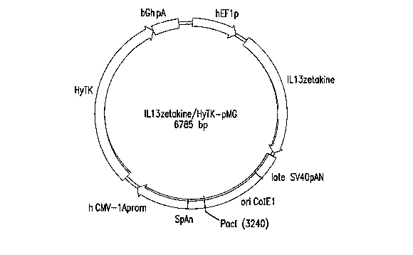
[0027] Figure 9: Plasmid map of ILl3zetakine/HyTK-pMG.
[0028] Figure 10: Nucleic acid sequence of the plasmid DNA vector (upper
strand:
SEQ ID NO:14; lower strand:SEQ ID NO:16) and the corresponding amino acid
sequence of ILl3zetakine (SEQ ID NO:17) and HyTK (SEQ ID NO:18).
[0029] Figure 11: Schematic diagram showing structure of IL13 zetakine insert.
DETAILED DESCRIPTION
[0030] An ideal cell-surface epitope for tumor targeting with genetically-
engineered re-directed T cells would be expressed solely on tumor cells in a
homogeneous fashion and on all tumors within a population of patients with the
same diagnosis. Modulation and/or shedding of the target molecule from the
tumor cell membrane may also impact on the utility of a particular target
epitope
for re-directed T cell recognition. To date few "ideal" tumor-specific
epitopes
have been defined and secondary epitopes have been targeted based on either
lack
of expression on critical normal tissues or relative over-expression on
tumors. In
the case of malignant glioma, the intracavitary administration of T cells for
the
treatment of this cancer permits the expansion of target epitopes to those
expressed
on tumor cells but not normal CNS with less stringency on expression by other
tissues outside the CNS. The concern regarding toxicity from cross-reactivity
of
9
CA 02629749 2008-05-13
WO 2007/059298 PCT/US2006/044635
based on the intracavitary route of administration and b) the low cell numbers
administered in comparison to cell doses typically administered systemically.
[0031] The IL-13Ra2 receptor stands out as the most ubiquitous and specific
cell-
surface target for malignant glioma47. Sensitive autoradiographic and
immunohistochemical studies fail to detect IL-13 receptors in the CNS16; as
Moreover, mutation of the IL-13 cytokine to selectively bind the glioma-
restricted
IL-13Ra2 receptor is a further safeguard against untoward reactivity of IL-13-
directed therapeutics against IL-13Ra1/IL-4(3+ normal tissues outside the
CNSs5; s' The potential utility of targeting glioma IL-13Ra2 the design and
testing
of a novel engineered chimeric immunoreceptor for re-directing the specificity
of
T cells that consists of an extracellular IL-13 mutant cytokine (E13Y)
tetliered to
the plasma membrane by human IgG4 Fc which, in turn, is fused to CD4TM and
the cytoplasmic tail of CD3 zeta. This chimeric immunoreceptor has been given
the designation of "IL-13 zetakine." The IL-13Ra2 receptor/I]L-l3(E13Y)
receptor-ligand pair is an excellent guide for understanding and assessing the
suitability of receptor-ligand pairs generally for use in zetakines. An ideal
zetakine comprises an extracellular soluble receptor ligand having the
properties of
IL-13(E13Y) (specificity for a unique cancer cell surface receptor, in vivo
stability
due to it being derived from a naturally-occurring soluble cell signal
molecule, low
immunogenicity for the same reason). The use of soluble receptor ligands as
distinct advantages over the prior art use of antibody fragments (such as the
scFvFc immunoreceptors) or cell adhesion molecules, in that soluble receptor
ligands are more likely to be stable in the extracellular enviromnent, non-
antigenic,
and more selective.
[0032] Chimeric immunoreceptors according to the present invention comprise an
extracellular domain comprised of a soluble receptor ligand linked to an
extracellular support region that tethers the ligand to the cell surface via a
transmembrane domain, in turn linked to an intracellular receptor signaling
domain. Examples of suitable soluble receptor ligands include autocrine and
paracrine growth factors, chemokines, cytokines, hormones, and engineered
artificial small molecule ligands that exhibit the required specificity.
Natural
ligand sequences can also be engineered to increase their specificity for a
particular target cell. Selection of a soluble receptor ligand for use in a
particular
CA 02629749 2008-05-13
WO 2007/059298 PCT/US2006/044635
zetakine is governed by the nature of the target cell, and the qualities
discussed
above with regard to the IL-13(E13Y) molecule, a preferred ligand for use
against
glioma. Examples of suitable support regions include the constant (Fc) regions
of
immunoglobins, human CD8 , and artificial linkers that serve to move the
targeting moiety away from the cell surface for improved access to receptor
binding on target cells. A preferred support region is the Fc region of an IgG
(such
as IgG4). Examples of suitable transmembrane domains include the
transmembrane domains of the leukocyte CD marlcers, preferably that of CD8.
Examples of intracellular receptor signaling domains are those of the T cell
antigen
receptor complex, preferably the zeta chain of CD3 also Fcy RIII costimulatory
signaling domains, CD28, DAP 10, CD2, alone or in a series with CD3zeta.
[0033] In the IL-13 zetakine embodiment, the human IL-13 eDNA having the
E13Y amino acid substitution was synthesized by PCR splice overlap extension.
A full length IL-13 zetakine construct was assembled by PCR splice overlap
extension and consists of the human GM-CSF receptor alpha chain leader
peptide,
IL-13(E13Y)-Gly-Gly-Gly, human IgG4 Fc, human CD4TM, and human
cytoplasmic zeta chain. This cDNA construct was ligated into the multiple
cloning
site of a modified pMG plasmid under the transcriptional control of the human
Elongation Factor-lalpha promoter (Invivogen, San Diego). This expression
vector co-expresses the HyTK cDNA encoding the fusion protein HyTK that
combines in a single molecule hygromycin phosphotransferase activity for in
vitro
selection of transfectants and HSV thymidine kinase activity for in vivo
ablation of
cells with ganciclovir from the CMV immediate/early promoter. Western blot of
whole cell Jurkat lysates pre-incubated with tunicamycin, an inhibitor of
glycosylation, with an anti-zeta antibody probe demonstrated that the expected
intact 56-kDa chimeric receptor protein is expressed. This receptor is heavily
glycosylated consistent with post-translational modification of the native IL-
13
cytokine108. Flow cytometric analysis of IL-13 zetakine+ Jurkat cells with
anti-
human IL-13 and anti-huinan Fc specific antibodies confirmed the cell-surface
expression of the IL-13 zetakine as a type I transmembrane protein.
[0034] Using established human T cell genetic modification methods developed
at
City of Hope107, primary human T cell clones expressing the IL-13 zetakine
chimeric immunoreceptor have been generated for pre-clinical functional
11
CA 02629749 2008-05-13
WO 2007/059298 PCT/US2006/044635
characterization. IL-13 zetakine+ CD8+ CTL clones display robust proliferative
activity in ex vivo expansion cultures. Expanded clones display re-directed
cytolytic activity in 4-hr chromium release assays against human IL-13Ra2+
glioblastoma cell lines. The level of cytolytic activity correlates with
levels of
zetakine expression on T cells and IL-13Ra2 receptor density on glioma target
cells. In addition to killing, IL- 13 zetakine+ clones are activated for
cytokine
secretion (IFN-y, TNF-ca, GM-CSF). Activation was specifically mediated by the
interaction of the IL- 13 zetakine with the IL-13Ra2 receptor on glioma cells
since
CTL clones expressing an irrelevant chimeric immunoreceptor do not respond to
glioma cells, and, since activation can be inhibited in a dose-dependent
manner by
the addition to culture of soluble IL- 13 or blocking antibodies against IL-13
on
T cell transfectants and IL-13Ra2 on glioma target cells. Lastly, IL- 13
zetakine-
expressing CD8+ CTL clones proliferate when stimulated by glioma cells in
culture. IL-13 zetakine+ CTL clones having potent anti-glioina effector
activity
will have significant clinical activity against malignant gliomas with limited
collateral damage to normal CNS.
[0035] An iinmunoreceptor according to the present invention can be produced
by
any means known in the art, though preferably it is produced using recombinant
DNA techniques. A nucleic acid sequence encoding the several regions of the
chimeric receptor can prepared and assembled into a complete coding sequence
by
standard techniques of molecular cloning (genomic library screening, PCR,
priiner-assisted ligation, site-directed mutagenesis, etc.). The resulting
coding
region is preferably inserted into an expression vector and used to transform
a
suitable expression host cell line, preferably a T lymphocyte cell line, and
most
preferably an autologous T lymphocyte cell line. A third party derived T cell
line/clone, a transformed humor or xerogenic immunologic effector cell line,
for
expression of the immunoreceptor. NK cells, macrophages, neutrophils, LAK
cells, LIK cells, and stem cells that differentiate into these cells, can also
be used.
In a preferred embodiment, lymphocytes are obtained from a patient by
leukopharesis, and the autologous T cells are transduced to express the
zetalcine
and administered back to the patient by any clinically acceptable means, to
achieve
anti-cancer therapy.
12
CA 02629749 2008-05-13
WO 2007/059298 PCT/US2006/044635
[0036] Suitable doses for a therapeutic effect would be between about 106 and
about 109 cells per dose, preferably in a series of dosing cycles. A preferred
dosing regimen consists of four one-weelc dosing cycles of escalating doses,
starting at about 10' cells on Day 0, increasing incrementally up to a target
dose of
about 108 cells by Day 5. Suitable modes of administration include
intravenous,
subcutaneous, intracavitary (for example by reservoir-access device),
intraperitoneal, and direct injection into a tumor mass.
[0037] The following examples are solely for the purpose of illustrating one
embodiment of the invention.
EXAMPLE 1: Construction of an immunoreceptor coding sequence
[0038] The coding sequence for an immunoreceptor according to the present
invention was constructed by de novo synthesis of the IL13(E13Y) coding
sequence using the following primers (see Fig. 8 for a flow chart showing the
construction of the immunoreceptor coding sequence and expression vector):
IL13PI:
EcoRI
TATGAATTCATGGCGCTTTTGTTGACCACGGTCATTGCTCTCACTTGCC
TTGGCGGCTTTGCCTCCCCAGGCCCTGTGCCTCCCTCTACAGCCCTCAG
GTAC [SEQ ID NO. 1]
IL13P2:
GTTGATGCTCCATACCATGCTGCCATTGCAGAGCGGAGCCTTCTGGTTC
TGGGTGATGTTGACCAGCTCCTCAATGAGGTACCTGAGGGCTGTAGAG
GGAG [SEQ ID NO. 2]
IL13P3:
CTCTGGGTCTTCTCGATGGCACTGCAGCCTGACACGTTGATCAGGGATT
CCAGGGCTGCACAGTACATGCCAGCTGTCAGGTTGATGCTCCATACCAT
GC [SEQ ID NO. 3]
IL13P4:
CCTCGATTTTGGTGTCTCGGACATGCAAGCTGGAAAACTGCCCAGCTGA
GACCTTGTGCGGGCAGAATCCGCTCAGCATCCTCTGGGTCTTCTCGATG
GC [SEQ ID NO. 4]
13
CA 02629749 2008-05-13
WO 2007/059298 PCT/US2006/044635
IL13P5:
BamHI
TCGGATCCTCAGTTGAACCGTCCCTCGCGAAAAAGTTTCTTTAAATGTA
AGAGCAGGTCCTTTACAAACTGGGCCACCTCGATTTTGGTGTCTCGG
[SEQ ID NO. 5]
[0039] The final sequence (417bp) was end-digested with EcoRI-BamHI, and
ligated into the plasmid pSK (stratagene, LaJolla, CA) as ligation 312#3.
Ligation
312#3 was mutagenized (stratagene kit, per manufacturer's instructions) to fix
a
deleted nucleotide using the primers 5': IL13 312#3 mut5-3
(CAACCTGACAGCTGGCATGTACTGTGCAGCCCTGGAATC [SEQ ID NO.
6]) and 3':IL13 312#3 mut3-5
(GATTCCAGGGCTGCACAGTACATGCCAGCTGTCAGGTTG [SEQ ID NO.
7]), and ligation 312#3 as a template, to form ligation 348#1
(IL13zetakine/pSK).
[0040] The coding Human GM-CSFR alpha chain Signal Peptide (hsp) coding
sequence was fused to the 5' end of IL13(E13Y) by standard PCR splice overlap
extension. The hsp sequence (101 bp) was obtained from the template ligation
301#10 (hsp/pSK) (human GCSF receptor a-chain leader sequence from liuman T
cell cDNA), using the primers 5': 19hsp5'
(ATCTCTAGAGCCGCCACCATGCTTCTCCTGGTGACAAGCCTTC [SEQ ID
NO. 8]) (Xbal site highlighted in bold), and 3': hsp-IL13FR
(GAGGGAGGCACAGGGCCTGGGATCAGGAGGAATG [SEQ ID NO. 9]).
The IL- 13 sequence (371 bp) was obtained using the primers 5': hsp-IL13FF
(CATTCCTCCTGATCCCAGGCCCTGTGCCTCCCTC [SEQ ID NO. 10]) and
3': ILl 3-IgG4FR (GGGACCATATTTGGACTCGTTGAACCGTCCCTCGC
[SEQ ID NO. 11]), and ligation 312#3 as template. Fusion was achieved using
the
101 bp hsp sequence and 371 bp IL13 sequence thus obtained, and the primers
5':
19hsp5' and 3': IL13-IgG4FR, to yeild a 438 bp fusion hsp-IL13 sequence.
[0041] A sequence encoding the IgG4 Fc region IgG4m:zeta was fused to the 3'
end of the hsp-IL13 fusion sequence using the same methods. The IgG4m:zeta
sequence (1119 bp) was obtained using the primers 5': ILl3-IgG4FF
(GCGAGGGACGGTTCAACGAGTCCAAATATGGTCCC [SEQ ID NO. 12])
and 3': ZetaN3' (ATGCGGCCGCTCAGCGAGGGGGCAGG [SEQ ID NO. 13])
(Notl site highlighted in bold), using the sequence R9.10 (IgG4mZeta/pSK) as
template. The 1119 bp IgG4m:zeta sequence was fused to the hsp-IL13 fusion
14
CA 02629749 2008-05-13
WO 2007/059298 PCT/US2006/044635
sequence using the respective sequences as templates, and the primers 5':
19hsp5'
and 3': ZetaN3', to yeild a 1522 bp hsp-ILl3-IgG4m:zeta fusion sequence. The
ends were digested witli Xbal-Notl, and ligated into pSK as ligation 351#7, to
create the plasmid IL13zetakine/pSK (4464 bp).
EXAMPLE 2: Construction of expression vector
[0042] An expression vector containing the IL13 zetalcine coding sequence was
created by digesting the ILl3zetakine/pSK of Example 1 with XbaI-NotI, and
creating blunt ends with Klenow, and ligating the resulting fragment into the
plasmid pMG~Pac (Invirogen) (first prepared by opening with SgrAI, blunting
with Klenow, and dephosphorylation with SAP), to yield the plasmid
IL13zetakine/pMG. See Fig. 8. The hygromycin resistance region of
IL13zetakine/pMG was removed by digestion with Notl-NheI, and replaced by the
selection/suicide fusion HyTK, obtained from plasmid CE7R/HyTK-pMG (Jensen,
City of Hope) by digestion with NotI-NheI, to create the expression vector
1L13zetakine/HyTK-pMG (6785 bp). This plasmid comprises the Human
Elongation Factor-la promoter (hEFlp) at bases 6-549, the IL13zetakine coding
sequence at bases 692-2185, the Simian Virus 40 Late polyadenylation signal
(Late SV40pAN) at bases 2232-2500, a ininimal E. coli origin of replication
(Ori
ColEl) at bases 2501-3247, a synthetic poly A and Pause site (SpAN) at bases
3248-3434, the Immeate-early CMV enhancer/promoter (h CMV-lAprom) at
bases 3455-4077, the Hygromycin resistance-Thymidine kinase coding region
fusion (HyTK) at bases 4259-6334, and the bovine growtli hormone
polyadenylation signal and a transcription pause (BGh pAn) at bases 6335-6633.
The plasmid has a Pacl linearization site at bases 3235-3242. The hEFlp and
IL13zetakine elements derived from ILl3zetakine/pMG, and the remaining
elements derived from CE7R/HyTk-pMG (and with the exception of the HyTK
element, ultimately from the parent plasmid pMG~Pac). In sum,
IL13zetakine/HyTK-pMG is a modified pMG backbone, expressing the
IL13zetakine gene from the hEFl promoter, and the HyTK fusion from the h
CMV-lA promoter. A map of the plasmid IL13zetakine/HyTK-pMG appears in
CA 02629749 2008-05-13
WO 2007/059298 PCT/US2006/044635
Fig. 9. The full nucleic acid sequence of the plasmid is shown in Fig. 10. The
sequence of the IL13zetakine insert is given as SEQ ID NO:15, below:
atgcttctcctggtgacaagccttctgctctgtgagttaccacacccagcattcctcctgatcccaggccctgtgcctc
cc
tctacagccctcaggtacctcattgaggagctggtcaacatcacccagaaccagaaggctccgctctgcaatggcagc
atggtatggagcatcaacctgacagctggcatgtactgtgcagccctggaatccctgatcaacgtgtcaggctgcagt
gccatcgagaagacccagaggatgctgagcggattctgcccgcacaaggtctcagctgggcagttttccagcttgcat
gtccgagacaccaaaatcgaggtggcccagtttgtaaaggacctgctcttacatttaaagaaactttttcgcgagggac
ggttcaacgagtccaaatatggtcccccatgcccaccatgcccagcacctgagttcctggggggaccatcagtcttcct
gttccccccaaaacccaaggacactctcatgatctcccggacccctgaggtcacgtgcgtggtggtggacgtgagcc
aggaagaccccgaggtccagttcaactggtacgtggatggcgtggaggtgcataatgccaagacaaagccgcggg
aggagcagttcaacagcacgtaccgtgtggtcagcgtcctcaccgtcctgcaccaggactggctgaacggcaagga
gtacaagtgcaaggtctccaacaaaggcctcccgtcctccatcgagaaaaccatctccaaagccaaagggcagccc
cgagagccacaggtgtacaccctgcccccatcccaggaggagatgaccaagaaccaggtcagcctgacctgcctg
gtcaaaggcttctaccccagcgacatcgccgtggagtgggagagcaatgggcagccggagaacaactacaagacc
acgcctcccgtgctggactccgacggctccttcttcctctacagcaggctaaccgtggacaagagcaggtggcagga
ggggaatgtcttctcatgctccgtgatgcatgaggctctgcacaaccactacacacagaagagcctctccctgtctctg
ggtaaaatggccctgattgtgctggggggcgtcgccggcctcctgcttttcattgggctaggcatcttcttcagagtga
a
gttcagcaggagcgcagacgcccccgcgtaccagcagggccagaaccagctctataacgagctcaatctaggacg
aagagaggagtacgatgttttggacaagagacgtggccgggaccctgagatggggggaaagccgagaaggaaga
accctcaggaaggcctgtacaatgaactgcagaaagataagatggcggaggcctacagtgagattgggatgaaagg
cgagcgccggaggggcaaggggcacgatggcctttaccagggtctcagtacagccaccaaggacacctacgacg
cccttcacatgcaggccctgccccctcgc (SEQ ID NO: 15).
EXAMPLE 3: Expression of the immunoreceptor
[0043] Assessment of the integrity of the expressed construct was first
delineated
by Wester blot probed with an anti-zeta antibody of whole cell lysates derived
froin Jurkat T cell stable transfectants107 cocultured in the presence or
absence of
tunicamycin, an inhibitor of glycosylation. Fig. 1. Jurkat T cell stable
transfectants (Jurkat-IL13-pMG bulk line) were obtained by electroporating
Jurkat
T cells with the IL13zetakine/HyTK-pMG expression vector, followed by
selection and expansion of positive transfectants. 2x10⁶ cells from the Jurkat-
16
CA 02629749 2008-05-13
WO 2007/059298 PCT/US2006/044635
IL13-pMG bulk line were plated per well in a 24-well plate with or without 5
g/ml, 10 g/ml, or 20 g/ml Tunicarnycin. The plate was incubated at 37 C for
22 hrs. Cells were harvested from each well, and each sample was washed with
PBS and resuspended in 50 l RIPA buffer (PBS, 1% NP40, 0.5% sodium
deoxycholate, 0.1% SDS) containing 1 tablet/lOml Complete Protease Inhibitor
Cocktail (Boehringer Mannheim, Indianapolis, IN). Samples were incubated on
ice for 30 minutes then disrupted by aspiration with syringe with 21 gauge
needle
then incubated on ice for an additional 30 minutes before being centrifuged at
4 C
for 20 minutes at 14,000 rpm. Samples of centrifuged lysate supematant were
harvested and boiled in an equal volume of sample buffer under reducing
conditions, then subjected to SDS-PAGE electrophoresis on a 12% acrylamide
gel.
Following transfer to nitrocellulose, membrane was allowed to dry O/N at 4 C.
Next morning, membrane was blocked in a Blotto solution containing 0.04 gm/inl
non-fat dried milk in T-TBS (0.02% Tween 20 in Tris buffered saline pH 8.0)
for 1
hour. Membrane was then incubated with primary mouse anti-human CD3(
monoclonal antibody (Pharmingen, San Diego, CA) at a concentration of 1 g/ml
for 2 hours, washed, and then incubated with a 1:3000 dilution (in Blotto
solution)
of goat anti-mouse IgG alkaline phosphatase conjugated secondary antibody (Bio-
Rad ImmunoStar Kit, Hercules, CA) for 1 hour. Prior to developing, membrane
was washed 4 additional times in T-TBS, and then incubated with 3 ml of
phosphatase substrate solution (Biorad ImmunoStar Kit, Hercules, CA) for 5
minutes at room teinperature. Membrane was then covered with plastic, and
exposed to x-ray film. Consistant with the known glycosylation pattern of wild-
type human IL-13, the electrophoretic mobility of expressed IL-13(E13Y)
zetakine
is demonstrative of a heavily glycosylated protein which, when expressed in
the
presence of tunicamycin, is reduced to an amino acid backbone of approximately
54 kDa.
[0044] The IL-13(E13Y) zetakine traffics to the cell surface as a homodimeric
type I transmembrane protein, as evidenced by flow cytometric analysis of
transfectants witli a phycoerythrin (PE)-conjugated anti human-IL13 monoclonal
antibody and a fluorescein isothiocyanate (FITC)-conjugated mouse anti-human
Fc
(gamma) fragment-specific F(ab')2 antibody. Fig. 2. Jurkat IL13zetakine-pMG
transfectants were stained with anti-human Fc(FITC) antibody (Jackson
17
CA 02629749 2008-05-13
WO 2007/059298 PCT/US2006/044635
ImmunoResearch, West Grove, PA), recombinant human IL13Ra2/human IgGl
chimera (R&D Systems, Minneapolis, MN) followed by FITC-conjugated anti
human-IgGl monoclonal antibody (Sigma, St. Louis, MO), and an anti-IL13(PE)
antibody (Becton Dickinson, San Jose, CA) for analysis of cell surface
chimeric
receptor expression. Healthy donor primary cells were also stained with FITC-
conjugated anti-CD4, anti-CD8, anti-TCR, and isotype control monoclonal
antibodies (Becton Dickinson, San Jose, CA) to assess cell surface phenotype.
For
each stain, 106 cells were washed and resuspended in 100 1 of PBS containing
2%
FCS, 0.2 mg/ml NaN3, and 5 l of stock antibody. Following a 30 minute
incubation at 4 C, cells were washed twice and either stained with a secondary
antibody, or resuspended in PBS containing 1% paraformaldehyde and analyzed
on a FACSCaliber cytometer.
EXAMPLE 4: Binding of IL13(E13Y) zetakine to IL13Ra2 receptor
[0045] IL-13(E13Y), tethered to the cell membrane by huinan IgG4 Fc (i.e.,
IL13(E13Y) zetakine), is capable of binding to its target IL13Ra2 receptor as
assessed by flow cytometric analysis using soluble IL13Rec2-Fc fusion protein.
Fig. 3. Cloned human PBMC IL13zetakine-pMG transfectants were obtained by
electroporating PBMC with the IL13zetakine/HyTK-pMG expression vector,
followed by selection and expansion of positive transfectants107.
IL13zetakine+
CTL clonal cells were stained with a fluorescein isothiocyanate (FITC)-
conjugated
mouse anti-human Fc (gamma) fragment-specific F(ab')2 (Jackson
ImmunoResearch, West Grove, PA), recoinbinant human IL13Ra2/human IgGl
chimera (R&D Systems, Minneapolis, MN) followed by FITC-conjugated anti
human-IgG1 monoclonal antibody (Sigma, St. Louis, MO), and a phycoerythrin
(PE)-conjugated anti human-IL13 monoclonal antibody (Becton Dickinson, San
Jose, CA) for analysis of cell surface chimeric receptor expression. Healthy
donor
primary cells were also stained with FITC-conjugated anti-CD4, anti-CD8, anti-
TCR, and isotype control monoclonal antibodies (Becton Dickinson, San Jose,
CA) to assess cell surface phenotype. For each stain, 10' cells were washed
and
resuspended in 100 1 of PBS containing 2% FCS, 0.2 mg/ml NaN3, and 5 l of
antibody. Following a 30 minute incubation at 4 C, cells were washed twice and
18
CA 02629749 2008-05-13
WO 2007/059298 PCT/US2006/044635
either stained with a secondary antibody, or resuspended in PBS containing 1%
paraformaldehyde and analyzed on a FACSCaliber cytometer.
[0046] Next, the immunobiology of the IL-13(E13Y) zetakine as a surrogate
antigen receptor for primary human T cells was evaluated. Primary human T
cells
were electroporated with the plasmid expression vector. Positive transformants
were selected with hygromycin, cloned in limiting dilution, then expanded by
recursive stimulation cyles with OKT3, IL-2 and irradiated feeder cells.
Clones
demonstrating IL 13zetakine expression by Western blot and FACS were then
subjected to functional evaluation in 4-hr chromium release assays against a
variety of IL-13a2/CD20- glioma cell lines (U251, SN-B19, U138), and the IL-
13(x-/CD20+ B cell lymphocyte line Daudi). These tests showed that
IL13zetakine
conferred cytolytic activity that was specific for glioma cells (Fig. 4a), and
that
this specific cytolytic activity is present for glioma cells as a class (Fig.
4b). The
cytolytic activity of MJ-IL13-pMG clones was assayed by employing 51Cr-labeled
SN-B19, U251, and U138 glioma cell lines (IL13(x2+/CD20-) and Daudi
(CD20+/IL13a2-) as targets. MJ-IL13 effectors were assayed 8-12 days following
stimulation. Effectors were harvested, washed, and resuspeded in assay media:
2.5x10⁵, 1.25x10⁵, 2.5x10⁴, and 5x10³ effectors were cultured in triplicate at
37 C
for 4 hours with 5x10³ target cells in 96-well V-bottom microtiter plates.
After
incubation, 1001i1 aliquots of cell-free supernatant were harvested and 51Cr
in the
supernatants was assayed with a y-counter. Percent specific cytolysis was
calculated as follows:
(~erimenta151Cr release) - (contro151Cr release) x 100
(Maximum 51Cr release) - (control 51Cr release)
Control wells contained target cells incubated in the presence of target cells
alone.
Maximum 51Cr release was determined by measuring the 51Cr released by labeled
target cells in the presence of 2% SDS. Bulk lines of stabley transfected
human
T cells consisting of approximately 40% IL-13(E13Y) zetakine TCRa/p+
lymphocytes displayed re-directed cytolysis specific for 13Ra2+ glioma targets
in
4-hr chromium release assays (>50% specific lysis at E:T ratios of 25:1), with
negligable acitivity against IL-13Ra2- targets (<8% specific lysis at E:T
ratios of
19
CA 02629749 2008-05-13
WO 2007/059298 PCT/US2006/044635
25:1). IL-13(E13Y) zetakine+CD8+TCRa/P+ CTL clones selected on the basis of
high-level binding to anti-IL-13 antibody also display redirected IL13Ra2-
specific
glioma cell killing. Fig. 4b.
[0047] IL-13 zetakine-expressing CD8+ CTL clones are activated and proliferate
wlien stimulated by glioma cells in culture. Figs. 5-7. MJ-IL13-pMG Cl. F2
responder cells expressing the IL13 zetakine were evaluated for receptor-
mediated
triggering of IFNy, GM-CSF, and TNFa production in vitro. 2x10⁶ responder
cells were co-cultured in 24-well tissue culture plates with 2x 10⁵ irradiated
stimulator cells (Daudi, Fibroblasts, Neuroblastoma 10HTB, and glioblastoma
U251) in 2 ml total. Blocking rat anti-human-IL13 monoclonal antibody
(Pharmingen, San Diego, CA), recombinant human IL13 (R&D Systems,
Minneapolis, MN), and IL13Ra2-specific goat IgG (R&D Systems, Minneapolis,
MN) were added to aliquots of U251 stimulator cells (2x 10⁵/ml) at
concentrations
of 1 ng/ml, 10 ng/ml, 100 ng/ml, and 1 g/ml, 30 minutes prior to the addition
of
responder cells. Plates were incubated for 72 hours at 37 C, after which time
culture supernatants were harvested, aliquoted, and stored at -70 C. ELISA
assays
for IFNy, GM-CSF, and TNFa were carried out using the R&D Systems
(Minneapolis, MN) kit per manufacturer's instructions. Samples were tested in
duplicate wells undiluted or diluted at 1:5 or 1:10. The developed ELISA plate
was evaluated on a microplate reader and cytokine concentrations determined by
extrapolation from a standard curve. Results are reported as picograms/ml, and
show strong activation for cytokine production by glioma stimulator cells.
Fig. 5,
Fig. 6.
[0048] Lastly, IL-2 independent proliferation of IL13zetakine+ CD8+ CTL was
observed upon co-cultivation with glioma stimulators (Fig. 7a), but not with
IL13
Ra2 stimulators. Proliferation was inhibited by the addition of rhIL-13
antibody
(Fig. 7b), showing that the observed proliferation was dependant on binding of
zetakine to the IL-13Ra2 glioma cell-sepcific receptor.
EXAMPLE 5: Preparation of IL-13 zetakine+ T cells suitable for therapeutic use
[0049] The mononuclear cells are separated from heparinized whole blood by
centrifugation over clinical grade Ficoll (Pharmacia, Uppsula, Sweden). PBMC
CA 02629749 2008-05-13
WO 2007/059298 PCT/US2006/044635
are washed twice in sterile phosphate buffered saline (Irvine Scientific) and
suspended in culture media consisting of RPMI 1640 HEPES, 10% heat
inactivated FCS, and 4 mM L-glutamine. T cells present in patient PBMC are
polyclonally activated by addition to culture of Orthoclone OKT3 (30ng/m1).
Cell
cultures are then incubated in vented T75 tissue culture flasks in the study
subject's
designated incubator. Twenty-four hours after initiation of culture rhIL-2 is
added
at 25 U/ml.
[0050] Three days after the initiation of culture PBMC are harvested,
centrifuged,
and resuspended in hypotonic electroporation buffer (Eppendorf) at 20x10G
cells/ml. 25 g of the plasmid IL13zetakine/HyTK-pMG of Example 3, together
witli 400 l of cell suspension, are added to a sterile 0.2 cm electroporation
cuvette. Each cuvette is subjected to a single electrical pulse of 250V/40 s
and
again incubated for ten minutes at RT. Surviving cells are harvested from
cuvettes, pooled, and resuspended in culture media containing 25 U/ml rhIL-2.
Flasks are placed in the patient's designated tissue culture incubator. Three
days
following electroporation hygromycin is added to cells at a final
concentration of
0.2 mg/ml. Electroporated PBMC are cultured for a total of 14 days with media
and IL-2 supplementation every 48-hours.
[0051] The cloning of hygromycin-resistant CD8+ CTL from electroporated
OKT3-activated patient PBMC is initiated on day 14 of culture. Briefly, viable
patient PBMC are added to a mixture of 100x10' cyropreserved irradiated feeder
PBMC and 20x106 irradiated TM-LCL in a volume of 200ml of culture media
containing 30 ng/ml OKT3 and 50 U/ml rhIL-2. This mastermix is plated into ten
96-well cloning plates with each well receiving 0.2 ml. Plates are wrapped in
aluminum foil to decrease evaporative loss and placed in the patient's
designated
tissue culture incubator. On day 19 of culture each well receives hygromycin
for a
final concentration of 0.2 mg/ml. Wells are inspected for cellular outgrowth
by
visualization on an inverted microscope at Day 30 and positive wells are
marked
for restimulation.
[0052] The contents of each cloning well with cell growth are individually
transferred to T25 flasks containing 50x106 irradiated PBMC, 1Ox106 irradiated
LCL, and 30ng/mlOKT3 in 25m1s of tissue culture media. On days 1,3,5,7,9,11,
and 13 after restimulation flasks receive 50U/ml rhIL-2 and 15mis of fresh
media.
21
CA 02629749 2008-05-13
WO 2007/059298 PCT/US2006/044635
On day 5 of the stiinulation cycle flasks are also supplemented with
hygromycin
0.2 mg/ml. Fourteen days after seeding cells are harvested, counted, and
restimulated in T75 flasks containing 150 x 106 irradiated PBMC, 30 x 106
irradiated TM-LCL and 30 ng/ml OKT3 in 50 mis of tissue culture media. Flasks
receive additions to culture of rhlL-2 and hygromycin as outlined above.
[0053] CTL selected for expansion for possible use in tlierapy are analyzed by
iinmunofluorescence on a FACSCalibur housed in CRB-3006 using FITC-
conjugated monoclonal antibodies WT/31 (a13TCR), Leu 2a (CD8), and OKT4
(CD4) to confirin the requisite phenotype of clones (apTCR+, CD4-, CD8+, and
IL13+). Criteria for selection of clones for clinical use include uniform TCR
a(3+,
CD4-, CD8+ and IL13+ as compared to isotype control FITC/PE-conjugated
antibody. A single site of plasmid vector chromosomal integration is confirmed
by
Southern blot analysis. DNA from genetically modified T cell clones will be
screened with a DNA probe specific for the plasmid vector. Probe DNA specific
for the HyTK in the plasmid vector is synthesized by random priming with
florescein-conjugated dUTP per the manufacture's instructions (Amersham,
Arlington Hts, IL). T cell genomic DNA is isolated per standard technique. Ten
micrograms of genomic DNA from T cell clones is digested overnight at 37 C
then electrophoretically separated on a 0.85% agarose gel. DNA is then
transferred to nylon filters (BioRad, Hercules, CA) using an alkaline
capillary
transfer method. Filters are hybridized overnight with probe in 0.5 M Na2PO~,
pH
7.2, 7% SDS, containing 10 g/mi salmon sperm DNA (Sigma) at 65 C. Filters
are then washed four times in 40 mM Na2PO4, pH 7.2, 1% SDS at 65 C and then
visualized using a chemiluminescence AP-conjugated anti-florescein antibody
(Ainersham, Arlington Hts, IL). Criteria for clone selection is a single band
unique
vector band.
[0054] Expression of the IL-13 zetakine is determined by Western blot
procedure
in which chimeric receptor protein is detected with an anti-zeta antibody.
Whole
cell lysates of transfected T cell clones are generated by lysis of 2 x 1C'
washed
cells in 1 ml of RIPA buffer (PBS, 1% NP40, 0.5% sodium deoxycholate, 0.1 %
SDS) containing 1 tablet/lOml Complete Protease Inhibitor Cocktail (Boehringer
Mannheim). After an eighty minute incubation on ice, aliquots of centrifuged
whole cell lysate supernatant are harvested and boiled in an equal volume of
22
CA 02629749 2008-05-13
WO 2007/059298 PCT/US2006/044635
loading buffer under reducing conditions then subjected to SDS-PAGE
electrophoresis on a precast 12% acrylamide gel (BioRad). Following transfer
to
nitrocellulose, membranes are bloclced in blotto solution containing .07 gm/ml
non-fat dried milk for 2 hours. Membranes are washed in T-TBS (.05% Tween 20
in Tris buffered saline pH 8.0) then incubated with primary mouse anti-human
CD3C monoclonal antibody 8D3 (Pharmingen, San Diego, CA) at a concentration
of 1 g/ml for 2 hours. Following an additional four washes in T-TBS,
membranes are incubated with a 1:500 dilution of goat anti-mouse IgG alkaline
phosphatase-conjugated secondary antibody for 1 liour. Prior to developing,
membranes are rinsed in T-TBS then developed with 30 ml of "AKP" solution
(Promega, Madison, WI) per the manufacturer's instructions. Criteria for clone
selection is the presence of a chimeric zeta band.
[0055] CD8+ cytotoxic T cell clones expressing the IL-13 zetakine chimeric
immunoreceptor recognize and lyse human glioblastoma target cells following
interaction of the chimeric receptor with the cell surface target epitope in a
HLA-
unrestricted fasliion. The requirements for target IL-13Ra2 epitope expression
and
class I MHC independent recognition will be confirmed by assaying each
a13TCR+,
CD8+, CD4-, IL-13 zetakine+ CTL clones against IL-13Ra2+ Daudi cell
transfectants and IL-13Ra2- Daudi cells. T cell effectors are assayed 12-14
days
following stimulation with OKT3. Effectors are harvested, washed, and
resuspended in assay media; and Daudi cell transfectants expressing IL-13Ra2.
2.5x10⁵, 1.25x10⁵, 0.25x10⁵, and 0.05x10⁵ effectors are plated in triplicate
at 37 C
for 4 hours with 5x10³ target cells in V-bottom microtiter plates (Costar,
Cambridge, MA). After centrifugation and incubation, 100 L aliquots of cell-
free
supernatant is harvested and counted. Percent specific cytolysis is calculated
as:
(Experimenta151Cr release) - (control51Cr release) x 100
(Maximum 51Cr release) - (contro151Cr release)
Control wells contain target cells incubated in assay media. Maximum 51Cr
release
is determined by measuring the 51Cr content of target cells lysed with 2% SDS.
Criteria for clone selection is >25% specific lysis of IL-13Ra2+ Daudi
23
CA 02629749 2008-05-13
WO 2007/059298 PCT/US2006/044635
transfectants at an E:T ratio of 5:1 and a <10% lysis of parental Daudi at the
same
E:T ratio.
EXAMPLE 6: Treatment of human glioma using IL-13 zetakine-expressing
T cells.
[0056] T cell clones genetically modified according to Example 5 to express
the
IL-13R zetakine chimeric immunoreceptor and HyTK are selected for:
a. TCRa/(3+, CD4-, CDB+, IL-13} cell surface phenotype as determined by
flow cytometry.
b. Presence of a single copy of chromosomally integrated plasmid vector
DNA as evidenced by Southern blot.
c. Expression of the IL-13 zetakine protein as detected by Western blot.
d. Specific lysis of human IL-13Ra2+ targets in 4-hr chromium release assays.
e. Dependence on exogenous IL-2 for in vitro growth.
f. Mycoplasma, fungal, bacterial sterility and endotoxin levels <5 EU/ml.
g. In vitro sensitivity of clones to ganciclovir.
Peripheral blood mononuclear cells are obtained from the patient by
leukapheresis, preferably following recovery from initial resection surgery
and at a
tiine at least three weeks from tapering off steroids and/or their most recent
systemic chemotherapy. The target leukapheresis mononuclear cell yield is 5x
109
and the target number of hygromycin-resistant cytolytic T cell clones is 25
with
the expectation that at least five clones will be identified that meet all
quality
control parameters for ex-vivo expansion. Clones are cryopreserved and
patients
monitored by serial radiographic and clinical examinations. When recurrence of
progression of disease is documented, patients undergo a re-resection and/or
placeinent of a reservoir-access device (Omaya reservoir) for delivering T
cells to
the tumor resection cavity. Following recovery from surgery and tapering of
steroids, if applicable, the patient commences with T cell therapy.
[0057] The patient receives a target of at least four one-week cycles of
therapy.
During the first cycle, cell dose escalation proceeds from an initial dose on
Day 0
of 10' cells, followed by 5x10' cells on Day 3 to the target dose of 108 cells
on Day
5. Cycle 2 commences as early as one week from commencement of cycle 1.
24
CA 02629749 2008-05-13
WO 2007/059298 PCT/US2006/044635
Those patients demonstrating tumor regression with residual disease on MRI may
have additional courses of therapy beginning no earlier than Week 7 consisting
of
repetition of Cycles 3 and 4 followed by one week of rest/restaging provided
these
treatments are well tolerated (max. toxicities <grade 3) until such time that
disease
progression or a CR is achieved based on radiographic evaluation.
[0058] Cell doses are at least a log less than doses given in studies
employing
intracavitary LAK cells (individual cell doses of up to 109 and cumulative
cell
numbers as high as 2.75x10¹⁰ have been safety administered), ex vivo expanded
TILs (up to 109 cells/dose reported with minimal toxicity) and allo-reactive
lymphocyte (starting cell dose 108 with cumulative cell doses up to 51.5x10)
delivered to a similar patient population75-85. The rationale for the lower
cell doses
as proposed in this protocol is based on the increased in vitro
reactivity/anti-tumor
potency of IL- 13 zetakine+ CTL clones compared to the modest reactivity
profile
of previously utilized effector cell populations. Low-dose repetitive dosing
is
favored to avoid potentially dangerous inflammatory responses that might occur
with single large cell number instillations. Each infusion will consist of a
single
T cell clone. The same clone will be administered throughout a patient's
treatment
course. On the days of T cell administration, expanded clones are aseptically
processed by washing twice in 50cc of PBS then resuspended in pharmaceutical
preservative-free normal saline in a volume that results in the cell dose for
patient
delivery in 2mls. T cells are instilled over 5-10 minutes. A 2ml PFNS flush
will
be administered over 5 minutes following T cells. Response to therapy is
assessed
by brain MRI +/- gandolinium, with spectroscopy.
[0059] Expected side-effects of administration of T cells into glioma
resection
cavities typically consist of self-limited nausea and vomiting, fever, and
transient
worsening of existing neurological deficits. These toxicities can be
attributed to
both the local inflammation/edema in the tumor bed mediated by T cells in
combination with the action of secreted cytokines. These side-effects
typically are
transient and less than grade II in severity. Should patients experience more
severe
toxicities it is expected that decadron alone or in combination with
ganciclovir will
attenuate the inflammatory process and ablate the infused cells. The
inadvertent
infusion of a cell product that is contaminated with bacteria or fungus has
the
potential of mediating serious or life-threatening toxicities. Extensive pre-
infusion
CA 02629749 2008-05-13
WO 2007/059298 PCT/US2006/044635
culturing of the cell product is conducted to identify contaminated tissue
culture
flasks and minimize this possibility. On the day of re-infusion, gram stains
of
culture fluids, as well as, endotoxin levels are performed.
[0060] Extensive molecular analysis for expression of IL-13Ra2 has
demonstrated
that this molecule is tumor-specific in the context of the CNS44; 46; 41; 14.
Furthermore, the only lluman tissue with demonstrable IL-13Ra2 expression
appears to be the testis42. This tumor-testis restrictive pattern of
expression is
reininiscent of the growing number of tumor antigens (i.e. MAGE, BAGE, GAGE)
expressed by a variety of human cancers, most notably melanoma and renal cell
carcinoma109-"'. Clinical experience with vaccine and adoptive T cell therapy
has
demonstrated that this class of antigens can be exploited for systemic tumor
immunotherapy without concurrent autoimmune attack of the testis"z-iia
Presumably this selectively reflects the effect of an intact blood-testis
barrier and
an immunologically privileged environment within the testis. Despite the
exquisite specificity of the mutant IL-13 targeting moiety, toxicities are
theoretically possible if cells egress into the systemic circulation in
sufficient
numbers and recognize tissues expressing the IL-13Ra1/IL-4(3 receptor. In
light of
this remote risk, as well as the possibility that instilled T cells in some
patients
may mediate an overly exuberant inflammatory response in the tumor bed, clones
are equipped with the HyTK gene which renders T cells susceptible to in vivo
ablation with ganciclovir"s-"s Ganciclovir-suicide, in combination with an
intra-
patient T cell dose escalation strategy, helps minimize the potential risk to
research
participants.
[0061] Side effects associated with therapy (headache, fever, chills, nausea,
etc.)
are managed using established treatments appropriate for the condition. The
patient receives ganciclovir if any new grade 3 or any grade 4 treatment-
related
toxicity is observed that, in the opinion of the treating physician, puts that
patient
at significant medical danger. Parentally administered ganciclovir is dosed at
10
mg/kg/day divided every 12 hours. A 14-day course will be prescribed but may
be
extended should symptomatic resolution not be achieved in that time interval.
Treatment with ganciclovir leads to the ablation of IL-13 zetakine HyTK+ CD8+
CTL clones. Patients should be hospitalized for the first 72 hours of
ganciclovir
therapy for monitoring purposes. If symptoms do not respond to ganciclovir
26
CA 02629749 2008-05-13
WO 2007/059298 PCT/US2006/044635
within 48 hours additional immunosuppressive agents including but not limited
to
corticosteroids and cyclosporin may be added at the discretion of the treating
physician. If toxicities are severe, decadron and/or other immunosuppressive
drugs along with ganciclovir are used earlier at the discretion of the
treating
physician.
27
CA 02629749 2008-05-13
WO 2007/059298 PCT/US2006/044635
REFERENCES
1. Davis FG, McCarthy BJ. Epidemiology of brain tumors. Curr Opin Neurol.
2000;13:635-640.
2. Davis FG, Malinski N, Haenszel W, et al. Primary brain tumor incidence
rates
in four United States regions, 1985- 1989: a pilot study. Neuroepidemiology.
1996;15:103-112.
3. Smith MA, Freidlin B, Ries LA, Simon R. Increased incidence rates but no
space-time clustering of childhood astrocytoma in Sweden, 1973-1992: a
population-based study of pediatric brain tumors. Cancer. 2000;88:1492-1493.
4. Ahsan H, Neugut AI, Bruce JN. Trends in incidence of primary malignant
brain
tumors in USA, 1981-1990. Int J Epidemiol. 1995;24:1078-1085.
5. Ashby LS, Obbens EA, Shapiro WR. Brain tumors. Cancer Chemother Biol
Response Modif. 1999;18:498-549.
6. Davis FG, Freels S, Grutsch J, Barlas S, Brem S. Survival rates in patients
with
primary malignant brain tumors stratified by patient age and tumor
histological
type: an analysis based on Surveillance, Epidemiology, and End Results (SEER)
data, 1973- 1991. JNeurosurg. 1998;88:1-10.
7. Duffner PK, Cohen ME, Myers MH, Heise HW. Survival of children with brain
tumors: SEER Program, 1973-1980. Neurology. 1986;36:597-601.
8. Davis FG, Freels S, Grutsch J, Barlas S, Brem S. Survival rates in patients
with
primary malignant brain tumors stratified by patient age and tumor
histological
type: an analysis based on Surveillance, Epidemiology, and End Results (SEER)
data, 1973- 1991. J Neurosurg. 1998;88:1-10.
9. Kolles H, Niedermayer I, Feiden W. [Grading of astrocytomas and
oligodendrogliomas]. Pathologe. 1998;19:259-268.
10. Huncharek M, Muscat J. Treatinent of recurrent high grade astrocytoma;
results of a systematic review of 1,415 patients. Anticancer Res. 1998;18:1303-
1311.
11. Loiseau H, Kantor G. [The role of surgery in the treatment of glial
tumors].
Cancer Radiother. 2000;4 Supp11:48s-52s.
12. Palma L. Trends in surgical management of astrocytomas and other brain
gliomas. Forum (Genova). 1998;8:272-281.
28
CA 02629749 2008-05-13
WO 2007/059298 PCT/US2006/044635
13. Azizi SA, Miyamoto C. Principles of treatment of malignant gliomas in
adults:
an overview. J Neurovirol. 1998;4:204-216.
14. Shapiro WR, Shapiro JR. Biology and treatment of malignant glioma.
Oncology (Huntingt). 1998;12:233-240.
15. Chamberlain MC, Kormanik PA. Practical guidelines for the treatment of
malignant gliomas. West J Med. 1998;168:114-120.
16. Ushio Y. Treatinent of gliomas in adults. Curr Opin Oncol. 1991;3:467-475.
17. Scott JN, Rewcastle NB, Brasher PM, et al. Long-term glioblastoma
multiforme survivors: a population-based study. Can J Neurol Sci. 1998;25:197-
201.
18. Finlay JL, Wisoff JH. The impact of extent of resection in the management
of
malignant gliomas of childhood. Childs Nerv Syst. 1999;15:786-788.
19. Hess KR. Extent of resection as a prognostic variable in the treatment of
gliomas. J Neurooncol. 1999;42:227-231.
20. van den Bent MJ. Chemotherapy in adult malignant glioma. Front Radiat Ther
Oncol. 1999; 3 3:174-191.
21. DeAngelis LM, Burger PC, Green SB, Cairncross JG. Malignant glioma: who
benefits from adjuvant chemotherapy? Ann Neurol. 1998;44:691-695.
22. Annstrong TS, Gilbert MR. Chemotherapy of astrocytomas: an overview.
Semin Oncol Nurs. 1998;14:18-25.
23. Prados MD, Russo C. Chemotherapy of brain tumors. Semin Surg Oncol.
1998;14:88-95.
24. Prados MD, Scott C, Curran WJ, Nelson DF, Leibel S, Kramer S.
Procarbazine, lomustine, and vincristine (PCV) chemotherapy for anaplastic
astrocytoma: A retrospective review of radiation therapy oncology group
protocols
coinparing survival with carmustine or PCV adjuvant chemotherapy. J Clin
Oncol.
1999;17:3389-3395.
25. Fine HA, Dear KB, Loeffler JS, Black PM, Canellos GP. Meta-analysis of
radiation therapy with and without adjuvant chemotherapy for malignant gliomas
in adults. Cancer. 1993;71:2585-2597.
26. Mahaley MS, Gillespie GY. New therapeutic approaches to treatment of
malignant gliomas: chemotherapy and immunotherapy. Clin Neurosurg.
1983;31:456-469.
29
CA 02629749 2008-05-13
WO 2007/059298 PCT/US2006/044635
27. Millot F, Delval 0, Giraud C, et al. High-dose chemotherapy with
hematopoietic stem cell transplantation in adults with bone marrow relapse of
medulloblastoma: report of two cases. Bone Marrow Transplant. 1999;24:1347-
1349.
28. Kalifa C, Valteau D, Pizer B, Vassal G, Grill J, Hartmann O. High-dose
chemotherapy in childhood brain tumours. Childs Nerv Syst. 1999;15:498-505.
29. Finlay JL. The role of high-dose chemotherapy and stem cell rescue in the
treatment of malignant brain tumors. Bone Marrow Transplant. 1996; 18 Suppl
3:S1-S5.
30. Brandes AA, Vastola F, Monfardini S. Reoperation in recurrent high-grade
gliomas: literature review of prognostic factors and outcome. Am J Clin Oncol.
1999;22:387-390.
31. Miyagi K, Ingram M, Techy GB, Jacques DB, Freshwater DB, Sheldon H.
Immunohistochemical detection and correlation between MHC antigen and cell-
mediated immune system in recurrent glioma by APAAP method. Neurol Med
Chir (Tokyo). 1990;30:649-655.
32. Bauman GS, Sneed PK, Wara WM, et al. Reirradiation of primary CNS
tumors. Int J Radiat Oncol Biol Phys. 1996;36:433-441.
33. Fine HA. Novel biologic therapies for malignant gliomas. Antiangiogenesis,
immunotherapy, and gene therapy. Neurol Clin. 1995;13:827-846.
34. Brandes AA, Pasetto LM. New therapeutic agents in the treatment of
recurrent
high-grade gliomas. Forum (Genova ). 2000;10:121-131.
35. Pollack IF, Okada H, Chambers WH. Exploitation of immune mechanisms in
the treatment of central nervous system cancer. Semin Pediatr Neurol.
2000;7:131-
143.
36. Black KL, Pikul BK. Gliomas--past, present, and future. Clin Neurosurg.
1999;45:160-163.
37. Riva P, Franceschi G, Arista A, et al. Local application of radiolabeled
monoclonal antibodies in the treatment of high grade malignant gliomas: a six-
year
clinical experience. Cancer. 1997; 80:2733-2742.
38. Liang BC, Weil M. Locoregional approaches to therapy with gliomas as the
paradigm. Curr Opin Oncol. 1998;10:201-206.
CA 02629749 2008-05-13
WO 2007/059298 PCT/US2006/044635
39. Yu JS, Wei MX, Cliiocca EA, Martuza RL, Tepper RI. Treatment of glioma
by engineered interleukin 4-secreting cells. Cancer Res. 1993;53:3125-3128.
40. Alavi JB, Eck SL. Gene therapy for malignant gliomas. Hematol Oncol Clin
North Am. 1998;12:617-629.
41. Debinski W. Recombinant cytotoxins specific for cancer cells. Ann N Y Acad
Sci. 1999; 8 8 6:297-299.
42. Debinski W, Gibo DM. Molecular expression analysis of restrictive receptor
for interleukin 13, a brain tumor-associated cancer/testis antigen. Mol Med.
2000;6:440-449.
43. Mintz A, Debinski W. Cancer genetics/epigenetics and the X chromosome:
possible new links for malignant glioma pathogenesis and immune-based
therapies. Crit Rev Oncog. 2000;11:77-95.
44. Joshi BH, Plautz GE, Puri RK. hiterleukin-13 receptor alpha chain: a novel
tumor-associated transmembrane protein in primary explants of human malignant
gliomas. Cancer Res. 2000;60:1168-1172.
45. Debinski W, Obiri NI, Powers, SK, Pastan I, Puri RK. Human glioma cells
overexpress receptors for interleukin 13 and are extremely sensitive to a
novel
chimeric protein composed of interleukin 13 and pseudomonas exotoxin. Clin
Cancer Res. 1995;1:1253-1258.
46. Debinski W, Gibo DM, Hulet SW, Connor JR, Gillespie GY. Receptor for
interleukin 13 is a marker and therapeutic target for human high-grade
gliomas.
Clin Cancer Res. 1999;5:985-990.
47. Debinski W. An immune regulatory cytokine receptor and glioblastoma
multiforme: an unexpected link. Crit Rev Oncog. 1998;9:255-268.
48. Debinski W, Slagle B, Gibo DM, Powers SK, Gillespie GY. Expression of a
restrictive receptor for interleukin 13 is associated with glial
transformation. J
Neurooncol. 2000;48:103-111.
49. Debinski W, Miner R, Leland P, Obiri NI, Puri RK. Receptor for interleukin
(IL) 13 does not interact with IL4 but receptor for IL4 interacts with IL13 on
human glioma cells. J Biol Chem. 1996;271:22428-22433.
50. Murata T, Obiri NI, Debinski W, Puri RK. Structure of IL-13 receptor:
analysis of subunit composition in cancer and immune cells. Biochem Biophys
Res
Commun. 1997;238:90-94.
31
CA 02629749 2008-05-13
WO 2007/059298 PCT/US2006/044635
51. Opal SM, DePalo VA. Anti-inflanunatory cytokines. Chest. 2000; 117:1162-
1172.
52. Romagnani S. T-cell subsets (Thl versus Th2). Ann Allergy Astluna
Immunol. 2000; 85 : 9-18.
53. Spellberg B, Edwards JE, Jr. Type 1/Type 2 immunity in infectious
diseases.
Clin Infect Dis. 2001;32:76-102.
54. Liu H, Jacobs BS, Liu J, et al. Interleukin-13 sensitivity and receptor
phenotypes of human glial cell lines: non-neoplastic glia and low-grade
astrocytoma differ from malignant glioma. Cancer Immunol Immunother.
2000;49:319-324.
55. Debinski W, Gibo DM, Obiri NI, Kealiher A, Puri RK. Novel anti-brain tumor
cytotoxins specific for cancer cells. Nat Biotechnol. 1998;16:449-453.
56. Debinski W, Gibo DM, Puri RK. Novel way to increase targeting specificity
to a human glioblastoma- associated receptor for interleukin 13. Int J Cancer.
1998;76:547-551.
57. Debinski W, Thompson JP. Retargeting interleukin 13 for
radioimmunodetection and radioimmunotherapy of human high-grade gliomas.
Clin Cancer Res. 1999;5:3143s-3147s.
58. Thompson JP, Debinski W. Mutants of interleukin 13 with altered reactivity
toward interleukin 13 receptors. J Biol Chem. 1999;274:29944-29950.
59. Brooks WH, Netsky MG, Levine JE. Immunity and tumors of the nervous
system. Surg Neurol. 1975;3:184-186.
60. Bullard DE, Gillespie GY, Mahaley MS, Bigner DD. Immunobiology of
human gliomas. Semin Oncol. 1986;13:94-109.
61. Coakham HB. Immunology of human brain tumors. Eur J Cancer Clin Oncol.
1984;20:145-149.
62. Holladay FP, Heitz T, Wood GW. Antitumor activity against established
intracerebral gliomas exhibited by cytotoxic T lymphocytes, but not by
lymphokine-activated killer cells. J Neurosurg. 1992;77:757-762.
63. Holladay FP, Heitz T, Chen YL, Chiga M, Wood GW. Successful treatment of
a malignant rat glioma with cytotoxic T lymphocytes. Neurosurgery. 1992;31:528-
533.
32
CA 02629749 2008-05-13
WO 2007/059298 PCT/US2006/044635
64. Kruse CA, Lillehei KO, Mitchell DH, Kleinschmidt-DeMasters B, Bellgrau D.
Analysis of interleukin 2 and various effector cell populations in adoptive
iminunotherapy of 9L rat gliosarcoma: allogeneic cytotoxic T lymphocytes
prevent
tuinor take. Proc Natl Acad Sci U S A. 1990;87:9577-9581.
65. Miyatake S, Nishihara K, Kikuchi H, et al. Efficient tumor suppression by
glioma-specific murine cytotoxic T lymphocytes transfected with interferon-
gamma gene. J Natl Cancer Inst. 1990;82:217-220.
66. Plautz GE, Touhalisky JE, Shu S. Treatment of murine gliomas by adoptive
transfer of ex vivo activated tumor-draining lymph node cells. Cell Immunol.
1997;178:101-107.
67. Saris SC, Spiess P, Lieberman DM, Lin S, Walbridge S, Oldfield EH.
Treatment of murine primary brain tumors with systemic interleukin-2 and tumor-
infiltrating lymphocytes. J Neurosurg. 1992;76:513-519.
68. Tzeng JJ, Barth RF, Clendenon NR, Gordon WA. Adoptive immunotherapy of
a rat glioma using lymphokine-activated killer cells and interleukin 2. Cancer
Res.
1990;50:4338-4343.
69. Yamasaki T, Kikuchi H. An experimental approach to specific adoptive
immunotherapy for malignant brain tumors. Nippon Geka Hokan. 1989;58:485-
492. ~
70. Yamasaki T, Handa H, Yamashita J, Watanabe Y, Namba Y, Hanaoka M.
Specific adoptive immunotherapy with tumor-specific cytotoxic T- lymphocyte
clone for murine malignant gliomas. Cancer Res. 1984;44:1776-1783.
71. Yamasaki T, Handa H, Yamashita J, Watanabe Y, Namba Y, Hanaoka M.
Specific adoptive immunotherapy of malignant glioma with long-term cytotoxic T
lymphocyte line expanded in T-cell growth factor. Experimental study and
future
prospects. Neurosurg Rev. 1984;7:37-54.
72. Kikuchi K, Neuwelt EA. Presence of immunosuppressive factors in brain-
tumor cyst fluid. J Neurosurg. 1983;59:790-799.
73. Yamanaka R, Tanaka R, Yoshida S, Saitoh T, Fujita K, Naganuma H.
Suppression of TGF-betal in human gliomas by retroviral gene transfection
enhances susceptibility to LAK cells. J Neurooncol. 1999;43:27-34.
74. Kuppner MC, Hamou MF, Bodmer S, Fontana A, de Tribolet N. The
glioblastoma-derived T-cell suppressor factor/transforming growth factor beta
2
33
CA 02629749 2008-05-13
WO 2007/059298 PCT/US2006/044635
inhibits the generation of lymphokine-activated killer (LAK) cells. hit J
Cancer.
1988;42:562-567.
75. Hayes RL. The cellular immunotherapy of primary brain tumors. Rev Neurol
(Paris). 1992;148:454-466.
76. Ingrain M, Buckwalter JG, Jacques DB, et al. Immunotherapy for recurrent
malignant glioma: an interim report on survival. Neurol Res. 1990;12:265-273.
77. Jaeckle KA. .hnmunotherapy of malignant gliomas. Semin Oncol.
1994;21:249-259.
78. Kruse CA, Cepeda L, Owens B, Johnson SD, Stears J, Lillehei KO. Treatment
of recurrent glioma with intracavitary alloreactive cytotoxic T lymphocytes
and
interleukin-2. Cancer J=unol Inununother. 1997;45 : 77-87.
79. Merchant RE, Baldwin NG, Rice CD, Bear HD. Adoptive immunotherapy of
malignant glioma using tumor-sensitized T lympliocytes. Neurol Res.
1997;19:145-152.
80. Nakagawa K, Kamezaki T, Shibata Y, Tsunoda T, Meguro K, Nose T. Effect
of lymphokine-activated killer cells with or without radiation therapy against
malignant brain tumors. Neurol Med Chir (Tokyo). 1995;35:22-27.
81. Plautz GE, Bamett GH, Miller DW, et al. Systemic T cell adoptive
immunotherapy of malignant gliomas. J Neurosurg. 1998;89:42-51.
82. Sankhla SK, Nadkarni JS, Bhagwati SN. Adoptive immunotherapy using
lymphokine-activated killer (LAK) cells and interleukin-2 for recurrent
malignant
primary brain tumors. J Neurooncol. 1996;27:133-140.
83. Sawamura Y, de Tribolet N. Immunotherapy of brain tumors. J Neurosurg Sci.
1990;34:265-278.
84. Thomas C, Schober R, Lenard HG, Lumenta CB, Jacques DB, Wechsler W.
Immunotherapy with stimulated autologous lyinphocytes in a case of a juvenile
anaplastic glioma. Neuropediatrics. 1992;23 :123-125.
85. Tsurushima H, Liu SQ, Tuboi K, et al. Reduction of end-stage malignant
glioma by injection witli autologous cytotoxic T lymphocytes. Jpn J Cancer
Res.
1999;90:536-545.
86. Barba D, Saris SC, Holder C, Rosenberg SA, Oldfield EH. Intratumoral LAK
cell and interleukin-2 therapy of human gliomas. J Neurosurg. 1989;70:175-182.
34
CA 02629749 2008-05-13
WO 2007/059298 PCT/US2006/044635
87. Hayes RL, Koslow M, Hiesiger EM, et al. Improved long term survival after
intracavitary interleukin-2 and lymphokine-activated killer cells for adults
with
recurrent malignant glioma. Cancer. 1995;76:840-852.
88. Ingram M, Jacques S, Freshwater DB, Techy GB, Shelden CH, Helsper JT.
Salvage immunotherapy of malignant glioma. Arch Surg. 1987;122:1483-1486.
89. Jacobs SK, Wilson DJ, Komblith PL, Grimm EA. Interleukin-2 or autologous
lymphokine-activated killer cell treatment of malignant glioma: phase I trial.
Cancer Res. 1986;46:2101-2104.
90. Jeffes EW, III, Beamer YB, Jacques S, et al. Therapy of recurrent high-
grade
gliomas with surgery, autologous mitogen-activated IL-2-stimulated (MAK)
killer
lymphocytes, and rIL-2: II. Correlation of survival with MAK cell tumor
necrosis
factor production in vitro. Lymphokine Cytokine Res. 1991;10:89-94.
91. Merchant RE, McVicar DW, Merchant LH, Young HF. Treatment of recurrent
malignant glioma by repeated intracerebral injections of human recombinant
interleukin-2 alone or in combination with systeinic interferon-alpha. Results
of a
phase I clinical trial. J Neurooncol. 1992;12:75-83.
92. Yoshida S, Takai N, Saito T, Tanaka R. [Adoptive immunotherapy in patients
with malignant glioma]. Gan To Kagaku Ryoho. 1987;14:1930-1932.
93. Davico BL, De Monte LB, Spagnoli GC, et al. Bispecific monoclonal
antibody anti-CD3 x anti-tenascin: an immunotherapeutic agent for human
glioma.
Int J Cancer. 1995;61:509-515.
94. Jung G, Brandl M, Eisner W, et al. Local immunotherapy of glioma patients
witll a combination of 2 bispecific antibody fragments and resting autologous
lymphocytes: evidence for in situ t-cell activation and therapeutic efficacy.
Int J
Cancer. 2001;91:225-230.
95. Pfosser A, Brandl M, Salih H, Grosse-Hovest L, Jung G. Role of target
antigen in bispecific-antibody-mediated killing of human glioblastoma cells: a
pre-
clinical study. Int J Cancer. 1999;80:612-616.
96. Yoshida J, Takaoka T, Mizuno M, Momota H, Okada H. Cytolysis of
malignant glioma cells by lymphokine-activated killer cells combined with anti-
CD3/antiglioma bifunctional antibody and tumor necrosis factor-alpha. J Surg
Oncol. 1996;62:177-182.
CA 02629749 2008-05-13
WO 2007/059298 PCT/US2006/044635
97. Imaizumi T, Kuramoto T, Matsunaga K, et al. Expression of the tumor-
rejection antigen SART1 in brain tumors. Int J Cancer. 1999;83:760-764.
98. Eshhar Z, Waks T, Gross G, Schindler DG. Specific activation and targeting
of cytotoxic lymphocytes tlirough chimeric single chains consisting of
antibody-
binding domains and the gamma or zeta subunits of the immunoglobulin and T-
cell receptors. Proc Natl Acad Sci U S A. 1993;90:720-724.
99. Haynes NM, Snook MB, Trapani JA, et al. Redirecting mouse CTL against
colon carcinoma: superior signaling efficacy of single-chain variable domain
chimeras containing TCR-zeta vs Fe epsilon RI-gamma. J Inununol.
2001;166:182-187.
100. Hombach A, Heuser C, Sircar R, et al. An anti-CD30 chimeric receptor that
mediates CD3-zeta-independent T- cell activation against Hodgkin's lymphoma
cells in the presence of soluble CD30. Cancer Res. 1998;58:1116-1119.
101. Hombach A, Scllneider C, Sent D, et al. An entirely humanized CD3 zeta-
chain signaling receptor that directs peripheral blood t cells to specific
lysis of
carcinoembryonic antigen- positive tumor cells. hit J Cancer. 2000;88:115-120.
102. Hombach A, Sircar R, Heuser C, et al. Chimeric anti-TAG72 receptors with
immunoglobulin constant Fc domains and gamma or zeta signalling chains. Int J
Mol Med. 1998;2:99-103.
103. Moritz D, Wels W, Mattern J, Groner B. Cytotoxic T lymphocytes with a
grafted recognition specificity for ERBB2-expressing tumor cells. Proc Natl
Acad
Sci U S A. 1994;91:4318-4322.
104. Weijtens ME, Willeinsen RA, Valerio D, Stam K, Bolhuis RL. Single chain
Ig/gamma gene-redirected human T lymphocytes produce cytokines, specifically
lyse tumor cells, and recycle lytic capacity. J Immunol. 1996;157:836-843.
105. Altenschmidt U, Klundt E, Groner B. Adoptive transfer of in vitro-
targeted,
activated T lymphocytes results in total tumor regression. J Immunol.
1997;159:5509-5515.
106. Jensen M, Tan G, Forman S, Wu AM, Raubitschek A. CD20 is a molecular
target for scFvFc:zeta receptor redirected T cells: implications for cellular
immunotherapy of CD20+ malignancy. Biol Blood Marrow Transplant. 1998;4:75-
83.
36
CA 02629749 2008-05-13
WO 2007/059298 PCT/US2006/044635
107. Jensen MC, Clarke P, Tan G, et al. Human T lymphocyte genetic
modification with naked DNA. Mol Ther. 2000;1:49-55.
108. Minty A, Chalon P, Derocq JM, et al. Interleulcin-13 is a new human
lymphokine regulating inflammatory and immune responses. Nature.
1993;362:248-250.
109. Boon T, Cerottini JC, Van den EB, van der BP, Van Pel A. Tumor antigens
recognized by T lymphocytes. Annu Rev Immunol. 1994;12:337-365.
110. Castelli C, Rivoltini L, Andreola G, Carrabba M, Renkvist N, Parmiani G.
T-
cell recognition of melanoma-associated antigens. J Cell Physiol. 2000;182:323-
331.
111. Chi DD, Merchant RE, Rand R, et al. Molecular detection of tumor-
associated antigens shared by human cutaneous melanomas and gliomas. Am J
Pathol. 1997;150:2143-2152.
112. Boon T, Coulie P, Marchand M, Weynants P, Wolfel T, Brichard V. Genes
coding for tumor rejection antigens: perspectives for specific immunotherapy.
Itnportant Adv Oncol. 1994;53-69.
113. Cebon J, MacGregor D, Scott A, DeBoer R. hnmunotherapy of melanoma:
targeting defined antigens. Australas J Dermatol. 1997;38 Suppl 1:S66-S72.
114. Greenberg PD, Riddell SR. Tumor-specific T-cell immunity: ready for prime
time? J Natl Cancer Inst. 1992;84:1059-1061.
115. Cohen JL, Saron MF, Boyer 0, et al. Preservation of graft-versus-
infection
effects after suicide gene therapy for prevention of graft-versus-host
disease. Hum
Gene Ther. 2000;11:2473-2481.
116. Drobyski WR, Morse HC, III, Burns WH, Casper JT, Sandford G. Protection
from lethal murine graft-versus-host disease without compromise of
alloengrafl,inent using transgenic donor T cells expressing a thymidine kinase
suicide gene. Blood. 2001;97:2506-2513.
117. Link CJ, Jr., Traynor A, Seregina T, Burt RK. Adoptive immunotherapy for
leukemia: donor lymphocytes transduced with the herpes simplex thymidine
kinase gene. Cancer Treat Res. 1999;101:369-375.
118. Spencer DM. Developments in suicide genes for preclinical and clinical
applications. Curr Opin Mol Ther. 2000;2:433-440.
37
DEMANDE OU BREVET VOLUMINEUX
LA PRESENTE PARTIE DE CETTE DEMANDE OU CE BREVET COMPREND
PLUS D'UN TOME.
CECI EST LE TOME 1 DE 2
CONTENANT LES PAGES 1 A 37
NOTE : Pour les tomes additionels, veuillez contacter le Bureau canadien des
brevets
JUMBO APPLICATIONS/PATENTS
THIS SECTION OF THE APPLICATION/PATENT CONTAINS MORE THAN ONE
VOLUME
THIS IS VOLUME 1 OF 2
CONTAINING PAGES 1 TO 37
NOTE: For additional volumes, please contact the Canadian Patent Office
NOM DU FICHIER / FILE NAME:
NOTE POUR LE TOME / VOLUME NOTE: