Note: Descriptions are shown in the official language in which they were submitted.
CA 02629931 2008-05-15
WO 2007/057644 PCT/GB2006/004208
Method
Field of the Invention
This invention relates particularly, though not exclusively, to the =field of
analysis of
biochemical pathways and to the interaction between biomolecules such as
proteins and
polypeptides.
Background of the Invention
There has been an explosion of biological information in the last few years
resulting from the
use of high throughput experimental techniques. These techniques range from
genome
sequencing, microarray analysis, yeast 2-hybrid protein-protein interaction
assays, to RNAi
screens and the use of automated image analysis to study cell biological
processes.
Following the successful implementation of each `-omic' technique,
opportunities and
bottlenecks are created at the interface between one set of techniques and the
next. One key
bottleneck is that of biochemistry.
Molecular machines, formed from complexes of proteins, are the building blocks
underlying
most cellular processes. Techniques such as the yeast 2-hybrid technique have
been used to
identify binary interactions between pairs of proteins on a genome-wide scale,
while
complementary analyses using complex purification and mass spectrometry are
starting to
identify the combinations of components that comprise each molecular machine.
However,
the detailed study of biochemical interactions requires the identification of
affinity binding
and rate constants, together with techniques for studying how these properties
are
physiologically regulated.
Standard 'test tube' biochemical techniques, including calorimetry and
fluorescence
anisotropy, require the time-consuming production and purification of
microgram to
CA 02629931 2008-05-15
WO 2007/057644
PCT/GB2006/004208
2
milligram quantities of soluble proteins and are therefore unlikely to be
scaled-up for high
throughput applications. A potentially more promising technique, Surface
Plasmon
Resonance (Biacore) requires somewhat less protein and can tolerate the
presence of
impurities (in some formats), but requires the careful timed flow of assay
protein, followed
by wash solutions over an immobilized binding partner and is therefore
unlikely to be easily
adapted for high throughput analyses. Typical protein requirements for Surface
Plasmon
Resonance and calorimetry are described below:
Isothermal Titration Calorimetry:
Measures: Kd, Stoichiometry (n), AG, AN
Requires: 1 ml of 10 M solution; 20nMoles; lmg of 50KDa protein.
Surface Plasmon Resonance (Biacore)
Measures: 'Con, Koff, Kd
Requires: 2mls of 100nM solution; 200pMoles; 10 g of 50KDa protein*.
* Calculation based on typical series of experiments required to establish a
binding affinity in
the ¨10nM range.
The high levels of proteins that are required in these assays result from the
requirement to
'saturate' binding ligand concentrations 4-10x higher than the dissociation
constant. This
imposes the typical requirements shown in Table 1.
Ka 4x Kd 1.1g/m1 for 50 KDa protein
1nM 4nM 0.2
10 nM 40nM 2
100nM 400nM 20
11IM 41AM 200
Table 1
CA 02629931 2008-05-15
WO 2007/057644
PCT/GB2006/004208
3
These levels of protein require time consuming and relatively expensive
conventional protein
synthesis and purification systems.
In vitro translation 'pull-down' experiments can be used to identify binding
interactions
between small amounts of radiolabelled protein produced in a translation
extract (100-15Ong
produced). However, like the yeast 2-hybrid technique, they suffer from the
disadvantage that
they are non-quantitative, they screen for picomolar to low nanomolar ranges
of affinities and
fail to screen for lower affinity interactions.
There is a requirement for new high-throughput biochemical techniques that are
quantitative,
sensitive, may use small vohunes of analyte, may require a low number of moles
of analyte,
may achieve high (up to mid-micromolar typically about 1-5 pM to about 10-20
1.1,M)
concentrations and may be adaptable for massively parallel analyses.
Summary of the Invention
According to one aspect of the invention there is provided a method of
measuring the affinity
of first and second biomolecules in which a first biomolecule is tethered by a
first tether
portion having a first tether portion length and a second biomolecule is
tethered by a second
tether portion having a second tether portion length, determining binding of
adjacent first and
second biomolecules to each other, varying at least one of the first and
second tether portion
lengths and determining binding of the first and second biomolecules.
The method of the invention is advantageous in that it allows very accurate
control of the
concentration of amounts of biomolecules and also in that it needs only very
small amounts
of the biomolecules - it allows equilibrium-binding studies to be carried out
in pico- to
attolitre voltunes. The low volumes allow a range of concentration-dependent
biochemical
assays to be performed with very low input amounts of each biomolecule. By way
of
example, in a theoretical determination comparing a method in accordance with
the invention
with conventional techniques, conventional calorimetry would require 20
nanomoles of
protein or 1 mg of protein of a 50KDa protein; Surface Plasmon Resonance would
require
CA 02629931 2008-05-15
WO 2007/057644 PCT/GB2006/004208
4
200 picomoles of protein corresponding to 10 micrograms for a 50KDa protein.
In contrast, a
comparable method in accordance with the invention would require only 10
attomoles and 50
picograms for a 50KDa protein. Typically, nano- to zeptolitre, preferably pico-
to attolitre,
volumes of first and/or second biomolecules are used.
In this context, the term "biomolecule" includes both naturally-occurring
molecules and
synthetic molecules.
The first and second biomolecules may be tethered to a solid support such as a
glass slide or
microbead. In one such configuration, the first and second biomolecules are
separately
tethered to the same solid support. Alternatively, the first and second
biomolecules may be
tethered together in a "Y-shaped" arrangement. In another embodiment, the
first and second
biomolecules may be joined by a tether but not tethered to a solid support, in
a "linear
molecule" arrangement. For example, the first and second biomolecules may be
tethered
together and present in a solution.
Where the biomolecules are tethered to a solid support, the biomolecules may
be randomly
arranged over the solid support. Alternatively, the biomolecules may be
tethered to discrete
portions of, or areas defined on, the solid support such that the first and
second biomolecules
can only interact if they "stretch" to span the gap between the discrete
portions. By
controlling the distance between the discrete portions and/or the tether
length, the proportion
of bound and free biomolecules may be altered allowing the determination of
affinity
described above. Discrete portions of the surface may be coupled to the first
and second
biomolecules using a range of techniques including photo, electron or ion beam
lithography
to sequentially deprotect portions for coupling. Alternatively, direct etching
/ modification
using atomic force microscope tips may be used to introduce selective regional
surface
modification. An advantage of this type of approach is that inter-molecular
distances may be
directly controlled rather than relying on mean distances. In principle, this
should allow for
more accurate control of biomolecule concentration.
CA 02629931 2008-05-15
WO 2007/057644
PCT/GB2006/004208
In a preferred embodiment where first and second biomolecules are tethered to
a solid
support, the method may be arranged to be operated in a nano-scale reaction
zone. The
formation of the reaction zone may be achieved by anchoring the tethers near
each other so
that at least some of the first and second biomolecules are closely adjacent
to each other, such
5 that the substantially hemispherical swept volumes defined by the free
ends of each
biomolecule overlap, allowing the first and second biomolecules to bind to
each other. The
volume of each hemispherical volume may be of the order of 2 x 10³ to 1 x
1012nm3. By
varying the length of the tether portions, the effective concentrations of the
biomolecules can
be controlled allowing quantitative analyses.
The stiffness of the tethers is considered in terms of the persistence length
(P) which is an
experimentally measured parameter that characterises the stiffness as a single
bending
parameter of a flexible rod. (Bustamante, C., J. F. et al 1994. Science.
265:1599 ¨1600; and
Marko, J. F., and E. D. Siggia. 1995. Stretching DNA. Macromolecules. 28:8759-
8770). In
the case of relatively long tethers (P > approx five times less than the
contour length of the
tether) made of polymers such as DNA in solution, the tether can be
represented by a worm-
like chain model that is characterised by, the persistence length (P).
Molecules much longer
than the persistence length (P equals approx 50-90nm for dsDNA) behave like
random coils
of a freely jointed chain with a segment length 2P and a Gaussian distribution
of segment
density.
In addition to varying the length of the tethers, the inter-anchor distance
can be altered to
vary the overlap of the swept volumes. This method can be used to alter the
stoichiometry of
tethered molecules and to fully exploit the special case when long flexible
tethers are used.
Computer simulations (Monte Carlo) have been used to calculate the
probabilities of free
DNA end distribution (for example, see Jian and Vologodskii (1997). A combined
wormlike-
chain and bead model for dynamic simulations of long linear DNA, J. Comp.
Physics 136
pp168-179.).
CA 02629931 2008-05-15
WO 2007/057644 PCT/GB2006/004208
6
The probability distribution of the free end of a tether in the nanotether
context is determined
by a number of factors including the stiffness of the tether, the temperature
and the ionic
conditions. By using long tethers in proportion to the persistence length
(e.g. greater than 5
times P), the inter-anchor distance can be varied to probe the effective
concentration gradient
within the substantially hemispherical swept volume.
In this concentration gradient, the lowest concentration would be closest to
the surface swept
by the tether at its full contour length (stretched straight). The
concentration would then
increase to a maximum close to the average centre of mass due to entropic
considerations.
The probability that two tethered biomolecules will interact at a particular
inter-anchor
distance will thus be dependent on the calculated probability distribution and
will increase as
the inter-anchor distance decreases. Approaches to alter the probability
distribution of
flexible tethers within their swept volumes (e.g. inducing bulk liquid flow or
the use of
vibrating supports) may be used to enhance the utility of tethers far beyond
their persistence
lengths (e.g. 20-40 times P).
By measuring the interaction distributions of a range of 'test case'
biomolecule interactions
(e.g. GSK-3 and Axin peptide; streptavidin ¨ biotin; antibody ¨ antigen), it
will be possible to
correlate the probability distribution with the inter-anchor distance and to
equate this directly
to an affinity binding constant. Alternatively mathematical models of this
process can also be
produced based on the studies of Jian (supra).
Both varying tether length and inter-anchor distance are proposed as methods
for varying
tethered biomolecule concentrations. At high length: P values, the probability
distribution
may be the most effective method of calculating affinity. hi a linear molecule
or Y-shaped
molecule embodiment, the first and second tether portion length may be varied
to vary the
biomolecule concentrations.
Techniques that measure the proportion of the first and second biomolecules
that are
molecularly close to each other can be used to quantify the proportion of
interacting first and
CA 02629931 2008-05-15
WO 2007/057644 PCT/GB2006/004208
7
second biomolecules. For example, the proportion of first and second
biomolecules that are
molecularly close to each other can be determined by Forster resonance energy
transfer
(FRET).
In a preferred method, the assay readout is the intensity of FRET between
fluorophores
coupled to the head oligonucleotides attached to the first and second
biomolecules. A laser,
appropriate to the excitation maximum for a fluorophore attached to the first
biomolecule, is
used to excite that fluorophore. Emission at the wavelength maximum from a
fluorophore
attached to the second biomolecule is recorded to assess the level of FRET. In
practice, for
FRET to occur, an excited molecule of the first fluorophore has to be
molecularly close
(<10nm) to the second fluorophore for energy to be transferred, leading to
emission at the
characteristic wavelength of the second fluorophore. This will occur when the
first and
second biomolecules are also molecularly close due to the formation of the
first
biomolecule/second biomolecule complexes. In a preferred variant of the FRET
technique,
fluorescence lifetime measurement (FRET/FLINI) is used to measure the time
dependence of
FRET since this technique offers improved sensitivity ('Fluorescence Lifetime
Imaging: An
emerging technique in Fluorescence Microscopy', C.G. Morgan, Chromosome
Research,
4(4), 261-263, 1996.). Appropriate controls (e.g. spots of the first and
second biomolecules
alone) will be used to normalise signal levels.
In a preferred embodiment, FRET may be measured using a lens to focus the
lasers on glass
slides containing arrays of tethered biomolecules. In other embodiments,
purpose-built
machines are arranged to overcome the limitations that confocal microscopes
have due to
their design for other purposes. In particular, the use of photomultipliers
and cooled charge-
coupled devices (CCDs) may enhance the sensitivity of detection of the low
level FRET
signals, potentially leading to the detection of FRET between tethered pairs
of single first and
second biomolecules (for example, see Walter et al., Biopolymers (Nucleic Acid
Sciences),
Vol. 61, 224-241 (2002)). Alternatively, Total Internal Reflection
Fluorescence Microscopy
(TIRF) may be used (Surface fluorescence microscopy with evanescent
illumination.,
CA 02629931 2008-05-15
WO 2007/057644
PCT/GB2006/004208
8
Axelrod, D., Light Microscopy in Biology, Lacey, A. (ed), Oxford University
Press, New
York, 399-423 (1999)).
In an alternative solution, nanoscale spheres or "quantum dots", nanocrystals
which absorb
light but quickly re-emit the light in a different colour, may be tethered in
place of the single
fluorophores. These conjugates may offer higher FRET efficiencies due to the
increased
number of fluorescent molecules. Alternatively, the nanoscale spheres would
allow
fluorescence correlation spectroscopy to be performed using a high-resolution
light confocal
microscope. For tethers longer than 2Kb (0.6[M), the formation of first
biomolecule/second
biomolecule complexes may be directly recorded due to the proportion of
fluorescent dot
pairs in proportion to those that show some separation.
The first and/or second tethers, or a single tether in the case of a linear
molecule, may be
formed from nucleotides. Preferably, a tether is generated from double
stranded DNA
(dsDNA). Alternatively, a tether may be made from other polymers such as
carbon nanotubes
(D. H. Jung et al Covalent attachment and hybridization of DNA
oligonucleotides on
patterned single-walled carbon nanotube films. Langmuir. 2004 Sep
28;20(20):8886-91.),
amyloid fibrils, or DNA crossover complexes for example DX hybrids, which
include an
even number (typically 4, 6, or 8) strands of DNA and are somewhat stiffer
than dsDNA; J.
Am. Chem. Soc. (2000), 122, 1848-1860, Construction, Analysis, Ligation, and
Self-
Assembly of DNA Triple Crossover Complexes). Furthermore, the 'stiffness' or
persistence
length (P) and electrostatic charge of tethers such as dsDNA may be modulated
by chemical
modification, interchelation with molecules such as ethidium bromide or other
suitable
interchelating agents, which are typically organic compounds to allow
insertion between the
dsDNA bases or are positively charged and polymeric to complex with DNA based
on
affinity for the negatively charged phosphate backbone of DNA. The stiffness
of DNA may
also be altered by complexing with DNA binding proteins along the length of
the tether. The
stiffness of other tethers may be modulated by other means. For example, the
stiffness of
tethers comprising DX hybrids may be modulated by varying the number of
strands; for
CA 02629931 2008-05-15
WO 2007/057644 PCT/GB2006/004208
9
tethers comprising carbon nanotubes by increasing the number of concentric
tubes forming
each nanotube.
In a preferred embodiment, variable-length dsDNA tether portions are ligated
together to
form a tether. For example, a tether body portion may be linked to head and
tail tether
portions to produce the tether. The nucleotides of the body portion may be
ligated to head
and tail portions in solution.
For non-nucleotide tethers, chemical cross-linking methods can be used to
attach head and
tail oligonucleotide linkers to the respective ends of the body portions of
the tethers.
Where the tethers comprise nucleotides, the or each tether portion length may
typically be of
the order of 50 basepairs (bp) to 50Kb preferably, 200 base pairs to 20 kbase
pairs, or 30 to
12 000 nm, preferably 60 to 6000nm, for other tethers.
The tethers may be tethered to a surface by means of anchors. The anchors may
be single-
stranded amino-modified oligonucleotides. The tethers (head, body and tail)
can then be
hybridised to a solid support to which anchor oligonucleotides have been
immobilised. The
solid support may be a modified glass substrate. Standard techniques may be
used to
covalently couple an anchor oligonucleotide (for example, see: Chrisey, L.A.,
Lee, G.U., and
O'Ferrall, E. (1996) Covalent attaclunent of synthetic DNA to self-assembled
monolayer
films Nucleic Acids Res. 24:3031-3039). A preferred method involves coupling
amino-
modified anchor oligonucleotides to a glass support treated with an agent such
as amino
silane and p-phenylene1,4 diisothiocyanate (PDC). Other substrates that can be
modified to
bind a tether to a surface are also contemplated, including agarose and
sepharose.
In one embodiment, the format of the support is a glass slide onto which
oligonucleotide
anchors or tethers are printed in arrays of spots using commercially available
split pin
arraying machines such as those available from Genetix. More specialist solid
supports,
based on miniaturisation techniques derived from microelectronics, may be used
in more
CA 02629931 2008-05-15
WO 2007/057644 PCT/GB2006/004208
sophisticated implementations that are designed to further miniaturise the
analysis and to
integrate better with readout systems.
In an alternative format, the support may be provided by microbeads that are
coupled in
5 formats that generate a unique relationship between a single bead and
tether combination.
This format enables the adaptation of the technology to microfluidic systems,
and may
enhance probe density particularly where relatively long tethers, say about 50
m, are used to
test for high affinity interactions at high inter-anchor distances. Suitable
microbeads may
include polystyrene, coated ferrous/ferric particles, gold particles,
sepharose, agarose, glass
10 or carbon.
In an arrayed-spot implementation, amino-terminal oligonucleotide anchors for
the first and
second biomolecules may be covalently coupled to the modified glass substrate.
A range of
other approaches can be used to vary the inter-tether distance. hi one
implementation, the
distance between the oligonucleotide anchors is increased by the use of a non-
specific amino
terminal oligonucleotide (which is designed not to bind to other tether
components) that is
titrated into the specific oligonucleotide mix. The greater the proportion of
the non-specific
oligonucleotide; the greater the resulting distance between specific
oligonucleotide anchor.
Alternatively, the proportion of modified silane molecules may be reduced
prior to
oligonucleotide coupling. Inter-anchor mean distances may be varied from
distances greater
than the tether length to the maximal oligonucleotide tether capacity. The
maximal coupling
density possible using published protocols (e.g. Chrisey et al 1996 supra) is
20pmoles of
bound DNA/cm2 which equates to an mean inter-anchor spacing of 1.6nm; Chrisey,
L.A., et
al (1996) supra. This inter-anchor density massively exceeds that required for
the most
probable range of anchor densities which would normally range from about 5nm
to about
1 lam.
In the above implementation, the non-specific amino-specific oligonucleotide
functions to
cap the reactive groups and will also make the surface of the support
electrostatically
negative, thereby minimizing the association of the negatively-charged DNA
tether with the
CA 02629931 2008-05-15
WO 2007/057644
PCT/GB2006/004208
11
surface. Alternatively, hydrophobic lipid groups may be coupled to the glass
surface to
discourage DNA-surface association due to the incompatibility of hydrophobic ¨
hydrophilic
associations. For example, suitable lipid groups may include phosphatidyl
ethanolamine.
In an alternative implementation, the sequences present in adjacent anchor
oligonucleotides
are synthesized in series (i.e. as a single oligonucleotide). This effectively
generates a
common anchor for both the first and second tethers and ensures that the swept
volumes
entirely overlap. There may be advantages to this approach if the binding of
very low
numbers (as low as 1 pair) of biomolecules were to be studied. In a variant of
this
implementation, where the first and second biomolecules are not tethered to a
separate solid
surface, the first and second biomolecules may be tethered at the respective
ends of a single
tether and measurements could be made in solution.
In a preferred method of connecting protein biomolecules to nucleic acid
tethers, protein
nucleic acid conjugates are produced according to the method described in:
Jung, G. Y., and
Stephanopoulos, G. (2004) A functional protein chip for pathway optimization
and in vitro
metabolic engineering Science 304, 428-431. This description is in turn based
on the original
method described in: Roberts, R. W., and Szostak, J. W. (1997) RNA-peptide
fusions for the
in vitro selection of peptides and proteins Proc. Natl. Acad. Sci. U.S. A 94,
12297-12302. In
short, the method involves the use of an in vitro translation reaction to
covalently attach a
nascent peptide by its C-terminus close to the 3' end of an mRNA-DNA
conjugate. An
additional class of methods for connecting protein biomolecules to nucleic
acid tethers that
can be used with equal preference to the method described above. These methods
generically
involve the synthesis of a fusion protein comprising the biomolecule of
interest attached to a
modified enzyme (shown schematically in the accompanying Figure 23; species A
fused to
species X). This type of system has been described for three different
enzymes, the Halo-
Tag, the AGT tag and cutinase (Hodneland, et al., (2002) Selective
immobilization of
proteins to self-assembled monolayers presenting active site-directed capture
ligands. Proc
Natl Acad Sci USA 99, 5048-5052; Keppler et al., (2004a) Labeling of fusion
proteins of 06-
alkylguanine-DNA alkyltransferase with small molecules in vivo and in vitro.
Methods 32,
CA 02629931 2008-05-15
WO 2007/057644 PCT/GB2006/004208
12
437-444; Keppler et al., (2004b) Labeling of fusion proteins with synthetic
fluorophores in
live cells. Proc Natl Acad Sci USA 101, 9955-9959; Temple et al., (2006). From
genome to
proteome: developing expression clone resources for the human genome. Hum Mol
Genet 15
Spec No 1, R31-43). Following synthesis, the fusion enzyme (X) is irreversibly
and
covalently coupled to a chemically synthesised substrate. In the systems
described, a wide
range of modified substrates have been generated. In the implementation
proposed, a Head
Set oligonucleotide is chemically synthesised that incorporates the substrate
species (Figure
23; Y). This synthesis of the covalently coupled oligonucleotide could also
incorporate the
Donor or Acceptor Fluorophore allowing a 1-step coupling and labelling
protocol.
There are three main advantages to the use of the approach described in Jung
and
Stephanopoulos (2004) supra in the context of the present invention. First,
the protein-
nucleic acid complex can be purified away from in vitro translation extract
proteins,
following annealing to the immobilised tethers and washing. Second, multiple
messenger
RNAs can be simultaneously translated and conjugated to their unique coding
nucleic acids;
this enables high throughput approaches to be taken to protein production.
Third, the proteins
produced in the in vitro translation extracts (-15Ong / translation) are in a
large excess over
that required to saturate a typical 100um diameter microarray spot of tethers
(--Spg of a
50KDa protein in a spot containing 30nm inter-anchor distance with 1 x 10⁷
molecules). Jung
and Stephanoupoulos (2004) showed that the density of `oligonucleotide
anchors' in their
approach was the primary determinant of the levels of immobilized nucleic acid-
protein
complexes. In an entirely analogous fashion, the proportion of tethers used in
a method in
accordance with the invention will determine the proportions of tethered first
and second
nucleic acid- protein complexes.
Alternative methods of making protein-nucleic acid conjugates may be used,
including the
direct chemical crosslinking of purified protein biomolecules to modified
oligonucleotides.
As an alternative, protein-nucleic acid complexes may be generated in situ by
annealing the
mRNA-DNA conjugate to the immobilized tether first and translating the
messenger RNA,
CA 02629931 2008-05-15
WO 2007/057644 PCT/GB2006/004208
13
whilst bound to the tether, by adding in vitro translation extracts to the
tethered messenger
RNA.
Alternatively, messenger RNA may be designed to generate protein fusions
between the
protein biomolecules of interest and a second protein domain X. The second
domain X may
be designed to have a very high affinity for an engineered component of the
head
oligonucleotide or the head end of the tether. For example, if the X domain is
a high affinity
specific DNA binding protein (e.g. lambda repressor), its cognate DNA site may
be
introduced into the head oligonucleotide complex to enable the nascent protein
to associate
with the tether via the DNA binding moiety. Alternatively, X could be a
molecule such as
streptavidin and a corresponding binding partner ¨ in this case biotin - would
be chemically
coupled to the head oligonucleotide.
Typical protein biomolecules include enzymes, antibodies and receptors. In
further
alternative methods, the first and/or second biomolecule may be a bioactive
molecule other
than a protein. The only requirement is that the alternative biomolecule is
capable of
maintaining its functional activity whilst being coupled to a tether.
Alternative molecules
include peptides, peptide analogues, such as synthetic amino acids,
combinatorial polymer
libraries, small molecules (say <1000 Daltons, for example chemically-
synthesized drugs),
polysaccharides, and catalytically active RNA species.
In a preferred method, nucleic acid protein conjugates are annealed through
complimentary
sequences, provided by either of the first and second biomolecules, close to
the 3' end of the
nucleic acid component to complementary sequences in a head oligonucleotide
tether portion.
This concentrates the nucleic acid conjugates from molarities typical of in
vitro translations
(e.g. lOnM) to the experimental concentrations.
Simple well-characterised equilibrium binding equations (Michaelis Menten) can
be used to
derive molecular interaction parameters based on the concentrations of the
first and second
biomolecules and the proportion of first biomolecule/second biomolecule bound.
CA 02629931 2008-05-15
WO 2007/057644 PCT/GB2006/004208
14
For example, in a typical experiment to accurately determine the Kd of an
interaction between
first and second biomolecules which are tethered to a support, first and
second biomolecules
having a range of tether lengths and inter-anchor distances is set up as an
array of spots using
appropriate combinations of anchors and tethers for the first and second
biomolecules. This
generates a standard range of concentrations. These concentrations are first
plotted against
the proportion of bound first/second biomolecule complex and the concentration
of the first
biomolecule (or the second biomolecule) required for half maximal binding is
determined
(this concentration is the IQ).
According to a further aspect of the invention there is therefore provided a
method of
determining the Kd of an interaction between first and second biomolecules by
determining
the proportion of bound first and second biomolecules for a range of
concentrations of the
first and second biomolecules and the determining the concentration of the
first or second
biomolecule required for half maximal binding of the first and second
biomolecules - the Kd.
The affinity of a first biomolecule to a library of second biomolecules may be
determined.
The library of biomolecules may comprise at least a significant portion of a
transcriptome or
proteome.
Methods in accordance with the invention offer the potential of screening
interactions
between a single biomolecule A and a library of molecules Bl, B2, B3... B. In
one format,
each spot is occupied by only biomolecule A and B1 or A and B2... A and B. In
a preferred
implementation for protein molecules, head tether portions recognising unique
(for example
coding) regions from the 3' end of messages B1, B2, B3 ... Bi, are generated
and coupled to
the core tethers as described earlier. Br, may be libraries of proteins
potentially representing a
transcriptome/ proteome. Alternatively, Bõ may be libraries of peptides used
for defining
interaction sites. Alternatively, Bn may be tethered libraries of chemical
compounds ranging
from small molecule compounds to libraries of synthetic polymers.
CA 02629931 2008-05-15
WO 2007/057644 PCT/GB2006/004208
By using an anchor/tether for the second biomolecule that can be cleaved
together with initial
saturating concentrations of the first and second biomolecules, it is possible
to determine K.
In this scheme, the rate of decay of first biomolecule/second biomolecule
complex levels is
monitored in real time following cleavage of the tether for the second
biomolecule. This type
5 of analysis is analogous to that used in surface plasmon resonance to
determine the Koff.
The effect of a third, tethered or non-tethered biomolecule on the interaction
between the
tethered first and second biomolecules may also be studied.
10 According to a further aspect of the invention there is provided a
method in which the Koff
value for an interaction between the first and second biomolecules is
determined by providing
initial saturating concentrations of the first and second biomolecules,
cleaving a second
biomolecule tether portion or anchor and monitoring any change in levels of
bound first and
second biomolecules.
According to a further aspect of the invention there is provided a method of
determining the
change in free energy (AG ) value for an interaction between a first and
second biomolecules,
the method comprising determining the proportion of bound biomolecules at a
first
temperature and determining the proportion of bound biomolecules at a second
temperature
and comparing the proportion of bound biomolecules at the respective first and
second
temperatures. Typically, the temperature of the biomolecules may be varied by
altering the
temperature of the experimental apparatus used to perform the method.
According to another aspect of the invention there is provided apparatus for
determining the
affinity of first and second biomolecules, the apparatus comprising a first
biomolecule
tethered by a first tether having a first tether length, a second biomolecule
tethered by a
second tether having a second tether length, means for determining binding of
adjacent first
and second biomolecules to each other, and means for varying at least one of
the first, and
second tether lengths.
CA 02629931 2013-06-10
16
At least one of the first and second biomolecules may be tethered to a surface
of the
apparatus. Preferably, both the first and second biomolecules are tethered to
a surface. The
biomolecules may be tethered separately to the surface or together in, for
example, a Y-
shaped or linear arrangement. Alternatively, the biomolecules may be tethered
together and 5
associated with the apparatus in the form of a solution.
The surface may be provided by a solid support. Preferably, the solid support
is a glass slide.
Alternatively, the solid support may be a micro bead.
In accordance with an aspect of the present invention there is provided a
method of
measuring the affinity of first and second biomolecules in which a first
biomolecule is
tethered by a first tether portion having a first tether portion length and a
second biomolecule
is tethered by a second tether portion having a second tether portion length,
determining
binding of adjacent first and second biomolecules to each other, varying at
least one of the
first and second tether portion lengths and determining binding of the first
and second
biomolecules, wherein the biomolecules are tethered to a support and/or
tethered together.
In accordance with a further aspect of the present invention there is provided
an apparatus for
determining the affinity of first and second biomolecules said apparatus
comprising a solid
support; a first biomolecule tethered by a first tether portion having a first
tether portion
length, a second biomolecule tethered by a second tether portion having a
second tether
portion length, and means for determining binding of adjacent first and second
biomolecules
to each other; wherein the biomolecules are tethered to said solid support
and/or tethered
together.
Other aspects of the apparatus may be provided by preferred method features
described
above.
Brief Description of the Drawings
Methods and apparatus in accordance with the invention will now be described,
by way of
1.5 example, with reference to the further accompanying Figures 1 to 22 in
which:
Figure 1 is a diagram showing a tethered biomolecule for use in a method of
the invention;
CA 02629931 2013-06-10
16a
Figure 2 is a diagram showing two tethered biomolecules for use in a method of
the 20
invention;
Figure 3A is a diagram showing the biomolecules of Figure 2 binding; Fig. 3B
illustrates the
biomoleenles of Fig. 3 A binding and illustrates the flexible nature of the
tethers;
Figure 4A and B are diagrams showing an array of tethered biomolecules for use
in a method
of the invention at different inter-tether spacings;
Figure 5 shows head tether portions for use in tethers in accordance with the
invention;
CA 02629931 2008-05-15
WO 2007/057644 PCT/GB2006/004208
17
Figure 6 shows a modified oligonucleotide for use in tethers in accordance
with the
invention;
Figure 7 shows the formation of tethers in accordance with the invention;
Figure 8 shows a further step in the formation of tethers in accordance with
the invention;
Figure 9 shows the production of biomolecule and tether conjugates;
Figure 10 illustrates a method in accordance with the invention;
Figure 11 illustrates the use of a method in accordance with the invention to
measure Koff;
Figure 12 is a scheme showing operation of a linear molecule arrangement of
biomolecules in
accordance with the invention in which:
A. is an illustration of spheres swept by the free ends of short and long
flexible tethers;
B. is an illustration of a possible conformation of free and bound variants
of a linear molecule representing an intra-molecular interaction
between biomolecules A and B;
C. is an illustration of free and bound variants undergoing inter-molecular
interactions between A and B;
Figure 13 is a diagram showing 'head-set' oligonucleotides used for forming
tethers in
which:
A. shows separate molecules of the form shown in Fig.12C;
B. shows a linear molecule with acceptor and donor head sets attached.
CA 02629931 2008-05-15
WO 2007/057644
PCT/GB2006/004208
18
This molecule takes the form shown in Fig. 12B;
Figure 14 shows time dependent decay of donor fluorescence due to FRET;
Figure 15 is a graph illustrating an Acceptor Head-Set Titration;
Figure 16 illustrates experimental measures of linear molecule affinity for:
a. a range of lengths; and
b. a range of concentrations;
Figure 17 illustrates a Y-shaped molecule in accordance with the invention;
Figure 18 illustrates a determination of Factor 'C' using a method in
accordance with the
invention as described below;
Figure 19 illustrates the design of biomolecules formed by oligonucleotides;
Figure 20 is a photograph of a gel analysis of biomolecules having various
length tether
portions;
Figure 21 shows results of FRET experiments using the oligonucleotides of
Figure 19; and
Figure 22 is a graph illustrating the variation of the proportion of bond
molecules with the
length of DNA tethers.
1 Overview of a method in accordance with the invention
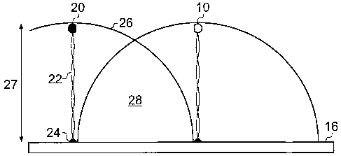
Figure 1 shows a single first biomolecule 10 tethered by a first tether 12
through anchor 14 to
surface 16. The first biomolecule 10 is free to move on the tether 12 about
anchor 14 in a
CA 02629931 2008-05-15
WO 2007/057644 PCT/GB2006/004208
19
substantially hemispherical volume 18. The volume of volume 18 is determined
by the first
tether length 19.
Figure 2 shows anchored first and second biomolecules 10 and 20. The second
biomolecule
20 is tethered by a second tether 22 via an anchor 24 and is also free to move
in a
substantially hemispherical volume 26. The volume of volume 26 is determined
by the
second tether length 27. The hemispherical volumes 18 and 26 overlap to define
a reaction
zone 28.
Figure 3A shows the first and second biomolecules 10 and 20 binding in the
reaction zone 28.
As shown in Fig. 3B the tethers are flexible and so the biomolecules occupy a
volume rather
than just a surface. Figure 4 shows varying the inter-tether spacing between
tethered
biomolecules. In Figure 4A, the biomolecules, for example 30 and 32, are
relatively spaced
apart. In Figure 4B, the biomolecules, for example 34 and 36, are relatively
close together.
An alternative to the random distribution of first and second biomolecules
tethers as
illustrated in Fig.4A and B is the targeting of first and second biomolecules
to discrete
portions on the surface of the substrate such that the first and second
biomolecules can only
interact if they stretch to span the gap between the surface patches. By
controlling the
distance between the discrete portions and/or the tether length, the
proportion of bound and
free biomolecules may be altered allowing the determination of affinity as
described herein.
2 Preparation of tethered array of biomolecules
The preparation of one form of tethered biomolecules for use in a method in
accordance with
the invention is shown in Figures 5 to 9. This involves joining a variable
length body tether
to three "adaptor" oligonucleotides. The head, body, tail and anchor
oligonucleotides are
combined as described below to generate an immobilised tether. Arrays of spots
containing
immobilised tethers are produced with different proportions of first and
second tether length
tethers. As described later, nucleic acid-protein covalent complexes are then
hybridised to the
immobilised tethers.
CA 02629931 2008-05-15
WO 2007/057644 PCT/GB2006/004208
a) Production of tether head portions
Tether body portions are generated from double stranded DNA (dsDNA) as shown
5 particularly in Figure 6. A tether body portion 50 has a single-stranded
upper portion
comprising a restriction enzyme half site X, which is complimentary to the
half-site X' of
tether head portion 38 or 40. The lower region of the body tether portion
includes a single
stranded section, generally designated as Y in Figure 6.
10 b) Production of tether body portions
Tether body portions are generated from double stranded DNA (dsDNA) as shown
particularly in Figure 6. A tether body portion 50 has a single-stranded upper
portion
comprising a restriction enzyme half site X, which is complimentary to the
half-site X' of
15 tether head portion 38 or 40. The lower region of the body tether
portion includes a single
stranded section, generally designated as Y in Figure 6.
c) Production of tether tail portions
20 Tether tail portions are designed to anneal and ligate to the dsDNA
tether body portion and
also to anneal to specific anchor oligonucleotides which are described below.
The tether tail
portions 52 and 54 shown in Figure 6 each comprise upper respective and lower
sections.
The upper section, generally designated as Y', is complimentary to the single
stranded
portion Y of tether body portion 50. The lower sections, generally designated
as 1 and 2, are
also single-stranded and are designed to anneal to the anchors described
below.
d) Assembly of the tethers
Separate tether production reactions are set up to generate pools of first or
second fluorophore
or to quantum dot labelled tethers with different tether lengths. The tether
head portions 38,
CA 02629931 2008-05-15
WO 2007/057644 PCT/GB2006/004208
21
40, tether body portions 50 and tether tail portions 52, 54 are assembled by
conventional
conditions under suitable conditions in solution as shown in Figure 6 to form
tethers 55 and
57. Typical conditions may be 50mM NaC1, HEPES buffer pH7.5 (10mM),and room
temperature.
e) Anchor-oligonucleotides
The assembled tethers 55, 57 can be anchored to a surface by means of anchors.
The anchors
are typically single-stranded amino-modified oligonucleotides. In a preferred
embodiment,
the solid support is a modified glass substrate prepared using standard
techniques to
covalently couple the anchor oligonucleotide. For example, see: Chrisey, L.A.,
Lee, G.U.,
and O'Ferrall, E. (1996) Covalent attachment of synthetic DNA to self-
assembled monolayer
films Nucleic Acids Res. 24:3031-3039. The amino-modified anchor
oligonucleotides are
coupled to glass treated with amino silane and p-phenylene1,4 diisothiocyanate
(PDC) (Fig.
7).
In the specific implementation described below (Figs 13-15), Forster Resonance
Energy
Transfer (FRET) coupled with Fluorescence Life-time Measurement (FLIN4) was
used to
determine the proportion of A and B that were molecularly close in an AB
complex. FLIM
exploits the time-dependence of FRET to allow more sensitive measurements of
the
proportions of A and B that are found in AB complexes. Both FRET and FLIM were
used in
the assays shown.
As shown in Figure 8, the tethers 55 and 57 are then hybridised to a solid
support 60 to which
anchor oligonucleotides 56, 58, each having single-stranded sections,
generally designated as
1 and 2 respectively, which are complimentary to corresponding sections 1 and
2 of the
tether tail portions 52, 54, have been previously immobilised.
CA 02629931 2008-05-15
WO 2007/057644
PCT/GB2006/004208
22
3 Production of tether/biomolecule conjugates
a) Use of in vitro translation
Protein biomolecule/nucleic acid conjugates which can hybidise to the tethers
are produced
according to the method described in: Jung, G. Y., and Stephanopoulos, G.
(2004). supra by
an in vitro translation reaction to covalently attach a nascent peptide by its
C-terminus close
to the 3' end of an mRNA-DNA conjugate. Tether protein complexes are then
hybridised to
the annealed arrays of tethers attached to their immobilised anchor
oligonucleotides.
This is schematically illustrated in Figure 9 where a first biomolecule,
indicated generally as
Protein A, is hybridized to the head portion of tether 55 and a second
biomolecule, indicated
generally as Protein B is hybridized to the head portion of tether 57.
Alternative methods of
making protein biomolecule-nucleic acid conjugates may be used, including the
direct
chemical crosslinking of purified first or second biomolecules to modified
oligonucleotides.
b) Generation of protein-nucleic acid complexes in situ
Alternatively, protein nucleic acid complexes may be generated in situ by
annealing the
mRNA-DNA conjugate to the immobilized tether first and translating the
messenger RNA
whilst bound to the tether by adding in vitro translation extracts to the
tethered messenger
RNA.
c) Use of protein-protein fusions
In another approach, the messenger RNA is engineered to generate protein
fusions between
the protein biomolecule of interest and a second protein domain X. The domain
X is
designed to have a very high affinity for an engineered component of a tether
head portion
oligonucleotide or the head end of the tether. For example, where the X domain
is a high
affinity specific DNA binding protein (e.g. lambda repressor), its cognate DNA
site is
CA 02629931 2008-05-15
WO 2007/057644
PCT/GB2006/004208
23
introduced into the head oligonucleotide complex to enable the nascent protein
to associate
with the tether via the DNA binding moiety. Alternatively, X is a molecule
such as
streptavidin and its binding partner ¨ in this case biotin - is chemically
coupled during
synthesis to a tether head portion oligonucleotide.
4 Annealing of nucleic acid ¨ protein conjugates to tethers
In the preferred method, nucleic acid biomolecule protein conjugates are
annealed through
complimentary sequences (A or B) close to the 3' end of the nucleic acid
component to
complementary sequences in the head tether portion as shown in Figure 9. This
concentrates
the nucleic acid conjugates from molarities typical of in vitro translations
(e.g. 10nM) to the
experimental concentrations (e.g. 3.7p,M based on a 200bp tether without any
tether overlap;
see Table 2) which shows the relationship between DNA length and other
parameters for a
individually-spaced tethered molecules.
Bases Length Volume Molarity
200 60nm 0.4aL 3.7AM
2Kb 600nm 0.4fL 3.7nM
20Kb 6um 0.4pL 3.7pM
Table 2
As noted above the tethers need not be made from dsDNA but may be rnade from
other
molecules such as DNA DX hybrids.
CA 02629931 2008-05-15
WO 2007/057644 PCT/GB2006/004208
24
Measurement of Affinity in Solution
The measurement of affinity between first and second biomolecules A and B can
also be
carried out in solution, allowing the basic principle underlying the tethering
principle to be
5 investigated using the simplified scheme shown in Figure 12A. In this
method, A and B are
attached at opposite ends of a single flexible tether allowing both molecules
to sweep out a
shared spherical volume that varies as a cubic function of the tether length.
As the length of
the single tether is reduced, the volume swept by A and B reduces and the
effective
concentration of A and B within the volume rises as a cubic function of the
tether length. This
scheme is formally analogous to the surface anchoring of tether biomolecules
described
above in that A and B can be regarded as being anchored to a surface that is
exactly half the
length of the joint tether such that the volumes swept by A and B exactly
overlap.
In the specific examples described below, Forster Resonance Energy Transfer
(FRET)
coupled with Fluorescence Life-time Measurement (FLIM) was used to determine
the
proportion of A and B that were molecularly close in an AB complex. FLIM
exploits the
time-dependence of FRET to allow more sensitive measurements of the
proportions of A and
B that are found in AB complexes. Both FRET and FLIM were used in the assays
shown.
(Backsai et al (2003) J Biomed Opt. 2003 Jul;8(3):368-75; Forster T (1965)
Delocalized
excitation and excitation transfer. In Modern Quantum Chemistry, part III. O.
Sinanoslu,
editor. Academic Press, New York. 93-137. Stryer L and Haugland RP, (1967)
Proceedings
of the National Academy of Science USA. 58: 719-730.).
Example 1
a) Oligonucleotide labelling and preparation of 'head sets'
The details of the test system are illustrated in Figure 13. The biomolecules
whose affinity
was measured were complementary strands of a DNA hybrid in which two 11 base
pair
overlaps recognise each other in a reversible reaction. The 11 base pair
interacting regions
CA 02629931 2008-05-15
WO 2007/057644 PCT/GB2006/004208
are single-stranded DNA extensions of longer double stranded DNA molecules
that contain
fluorophores A (Acceptor) and D (Donor) incorporated into the bases indicated
in bold
(Fig.13A). In the data shown, the fluorophore used as donor was Alexa Fluor
488 and at the
fluorophore used as acceptor was Alexa Fluor 555, both are manufactured by
Molecular
5 Probes. Both fluorophores were incorporated during oligonucleotide
synthesis and the
labelled oligonucleotides were subsequently annealed to form the structures
shown in Fig.
6A. The fluorophore-tagged double-stranded oligonucleotides are referred to as
a donor or
acceptor 'head set' to denote the presence of both the annealing 1 lbp
affinity region and the
presence of the fluorescent dyes.
b) Linear DNA tether preparation
To make the longer tethered molecules illustrated in Figure 13B and
schematically in Fig.
12A and 12B, the donor and acceptor head set oligonucleotides were ligated to
variable
length double stranded DNA regions by standard procedures. Briefly, the 'head
set'
oligonucleotides were cleaved with Bst X1 restriction enzyme and were ligated
to variable
length 'tether body' DNAs each of which contained a free BstX1 and Xbal site.
BstX1-
BstX1 and Xba-Xba ligations were used to generate the molecules as shown in
Fig.5B. These
were gel purified prior to analysis. The total lengths of the linear molecules
incorporating
both Donor and Acceptor head groups were: 515bp and 710bp.
c) Sample preparation, FRET and FLIM detection
Head sets or dual-labelled linear DNA molecules were diluted to the
concentrations described
in a final concentration of 70 mM NaC1, 10 mM Tris pH 8Ø 61.11 of each
solution was
introduced into one of the wells of a 50 well slide produced using a multi-
chambered
coverslip (Stratech Scientific, UK) together with a 22 x 50 mm coverslip
(Menzel-Glaser,
Germany).
CA 02629931 2008-05-15
WO 2007/057644 PCT/GB2006/004208
26
Samples were analysed using a frequency-doubled Ti:Sa laser providing short
optical pulses
(100fs duration) at 76Mhz repetition rate, with wavelength in the absorption
band of the
donor fluorophore (-470nm). The exciting light was weakly focused onto the
sample
allowing for a uniform illumination and collection over lmm well depth, to
maximize the
signal contribution over the fluorescence background of the coverslip. Low
excitation
intensities (0.05-10mW over 0.4mm spot diameter) were maintained to avoid
nonlinearities
and photodamage. Fluorescence light collected from a microscope objective was
spectrally
analysed using a spectrometer and detected by a cooled CCD camera for time-
integrated
FRET spectra. For time-resolved FLIM, fluorescence light was filtered by the
spectrometer
around the emission maximum of the donor fluorophore (520 5nm) and detected
by a single
channel fast photomultiplier (200ps time resolution) connected to a time-
correlated single
photon counting module. Background contributions were measured from the buffer
solution
without fluorophores in the same excitation and detection conditions and
properly subtracted
to the data.
d) Preparation of a Y-shaped molecule
The first and second tether portions for each biomolecule in a Y-shaped
molecule are
anchored to a single DNA strand such that the tethers are free to diffuse as
for the linear
molecule shown in Fig. 12B. The main advantage of this form of tethering
compared with
that of the single molecule is that the first and second tether portions are
free to interact
independent of the length of the intervening tether. By contrast, the linear
molecule is unable
to fold back on itself at lengths shorter than the persistence length (P)
which approximates to
between 90 and 120bp.
e) Data analysis
To determine the % maximal binding, we first determined the proportion of
bound and
unbound donor (R) at different donor and acceptor concentrations using the
following
procedure. The ratio between the bound and unbound decay spectra for different
acceptor
CA 02629931 2008-05-15
WO 2007/057644 PCT/GB2006/004208
27
concentrations was determined over time and plotted as shown in Fig.14 using
free labelled
head set oligonucleotides (The three curves shown represent 1. 50 nM
acceptor:50DM donor,
2. 200 nM acceptor: 50 nM donor, 3. 600 nM:50nM donor).
For each curve a numerical fit (dotted lines) to the decay curve (R(t)=U(l+R
exp(-t/T)) was
performed, where R=ratio between bound and unbound donor, t=time,
U=N(unbound)/N
(where N=concentration of donor in the absence of acceptor). '=decay constant.
R, U and
were directly determined from the numerical fit of the experimental data.
The proportion of bound donor = 12/(1+R) was plotted against acceptor
concentration as
shown in Fig.15 (percentage normalised to the maximum effect observed above
4000 nM
acceptor concentration). In Fig.15, the experimental curve of free donor and
acceptor head
sets was determined for a range of acceptor head set and a single (50nM) donor
head set
concentration. This allowed the detennination of the binding affinity of the 1
lbp overlap
head sets as 136nM. This matches closely to the theoretical determination of
176nM for the
same sequence. In Fig. 16, preliminary data from two llbp overlap linear
molecules (donor
at one end, acceptor at the other; open circles) is displayed on the same
scale.
f) Theoretical determination of DNA binding affinity
Assuming a chemical reaction between molecules A and D in order to form bound
molecule
AD:
A+12,4-).AD
(1)
as well as a reverse reaction and that the system is in equilibrium, we can
define dissociation
constant:
kd = [A] [D]l[AD]
(2)
CA 02629931 2008-05-15
WO 2007/057644 PCT/GB2006/004208
28
Note that according to the basic textbooks (see for example John SantaLucia,
Jr. and Donald
Hicks. (2004) Armu. Rev. Biophys. Biomol. Struct. 33, 415-40), people also use
equilibrium
constant keg =///cd,
kd=1/keq=exp(AG/RT)
(3)
where ka [mo1/1] is dissociation constant, AG [cal/mol] is change of the free
energy due to
reaction, R =1.987 [cal/(K mol)], T [K] absolute temperature. In order to
calculate kd we have
to calculate AG. In our case we have DNA headsets with different base pairs
overlap.
This can be done by methods and software developed by Prof. SantaLucia and co-
workers
John SantaLucia, Jr. and Donald Hicks. (2004) supra, Annu. Rev. Biophys.
Biomol. Struct.
33, 415-40.
In order to estimate properly AG for DNA molecules we have to take in account
folding and
hybridization prediction (M. Zuker. Nucleic Acids Res. 31 (13), 3406-15,
(2003)).
The final results are presented in the following tables. The theoretical
affinities were
calculated using methods described in the following references: John
SantaLucia, Jr. and
Donald Hicks. (2004) supra; M. Zuker, (2003) supra; and A V Fotin et al,
Nucleic Acids
Res. 26 (1998) p.1515.
30
CA 02629931 2008-05-15
WO 2007/057644 PCT/GB2006/004208
29
SALT CONCENTRATION 70 mM, TEMPERATURE=21 C (274.15K)
BASE dG[kcal/mol]/K dG[kcal/mol]/K. dG[kcal/mol]/K dG[kcal/mol]/K
dG[kcal/mol]/K
PAIRS [M] [M] SantaLucia [M] SantaLucia [M] Fotin [M]
Fotin
(thermodynamic Correction due correction due to correction due to
correction due to
prediction) to folding folding (net folding folding
(net
(thermodynamic hybridization (thermodynamic
hybridization
prediction) thermodynamics) prediction)
thermodynamics)
11 -13.5/ -9.14/ -6.01 / -10.3/ -7-17 /
5.0465E-
1.06304E-10 1.7685E-7 3.63052E-5 2.45826E-8 6
9 -9,79/ -5.99/ -5.84/ -6.59/ -6.44/
5.85332E-8 3.75616E-5 4.84797E-5 1.35357E-5 1.74701E-
5
7 -6.93/
7.59095E-6
-4.34/
6.219E-4
Table 3. Prediction of AG for DNA headsets for salt concentration 70m1\4.
5 SALT CONCENTRATION 35 m1\4, TEMPERATURE=21 C (274.15K)
BASE dG[kcal/mol]/K dG[kcal/mol]/K dG[kcal/mol]/K dG[kcal/mol]/K
dG[kcal/mol]/K
PAIRS [M] [M] SantaLucia [M] SantaLucia [M] Fotin [M]
Fotin
Correction due correction due to correction due to correction due to
to folding folding (net folding folding
hybridization
thermodynamics)
11 -12.75/ -8.5/ -5.72/ = --9.54/ -6.77/
3.80743E-10 5.25316E-7 5.94583E-5 8.95563E-8 9.966E-6
9 -9.19/ -5.48/ -5.36/ -5.99 / 3.75616E- 5.87/
1.6243E-7 8.94373E-5 1.09691E-4 5!!! 4.607E-5!!!
7 -6.48/
1.63209E-5
5 -4.04/
0.00104
Table 4. Prediction of AG for DNA headsets for salt concentration 35m1VI.
The results presented in Tables 3 and Table 4 for 11 base pairs with a
correction due to the
folding depend on the methods for the calculation which is used either
SantaLucia or Fotin.
The difference is one order of magnitude. For 9 base pairs the agreement
between two
methods is better.
CA 02629931 2008-05-15
WO 2007/057644 PCT/GB2006/004208
Results
a)
Determination of the binding affinity of the llbp overlap using free
oligonucleotides
5
An essential initial goal of these studies was to determine an accurate value
for the 1 lbp
affinity to enable later comparison with results using the nano-tether
methodology of the
invention. Standard titration reactions were carried out to identify the
dissociation constant
(Kd) for the oligonucleotides shown in Figure 13A. Essentially, this involved
creating
10 multiple samples with a fixed concentration of fluorescently-
labelled donor head-set
oligonucleotides (D; 50nM) with a variable concentration of fluorescently-
labelled acceptor
head set oligonucleotides (A; OnM-5000nM).
To determine the amount of D:A hybrids, the samples were analysed for the time-
dependence
15 of FRET-FLIM as described above. A representative plot from this
analysis is shown in
Figure 14. The rate of decay of the fluorescence signal is increased in the
presence of
increasing levels of fluorescently-labelled acceptor head sets showing
increased decay rate of
the donor fluorophore in the presence of the acceptor fluorophore that is a
time-dependent
characteristic of FRET. Importantly, labelled head sets that did not contain a
single-stranded
20 overhang showed no FRET/FLIM (data not shown), arguing that the
decay observed was due
to the inter-molecular hybridisation of the two head-sets.
The characteristic decay curves from FRET-FLIM analyses of the kind shown in
Figure 14
were transformed into relative FLIM values according to the method described
above and
25 were plotted in relation to the concentration of Acceptor Head-Set
oligonucleotide (Fig. 15).
Specifically in this figure the percentage maximum FLIM (y-axis) for the 1 lbp
overlap donor
head set was plotted against the acceptor head-set concentration. The curve
showed a
classical saturation response with a half maximal binding (Kd) concentration
of Acceptor
head set being calculated (FIT) to be 136nM. Linear regression analysis was
used to estimate
30
a value of 136n1V1 for the dissociation constant of the 1 lbp overlap in 70mM
NaCI. This
CA 02629931 2008-05-15
WO 2007/057644 PCT/GB2006/004208
31
value was very close to the theoretical value of 170nM that was calculated for
the sequence
from nearest-neighbour thermodynamic predictions (see above). This indicates
that the
FRET-FLIM method was able to accurately determine the proportion of bound
fluorophores.
b) Tether length-dependence of FLIM on linear molecules
The proportion of linear tether molecules found in the bound form increased as
the length of
the tether decreased according to predictions (Fig.12A,B). To test this,
linear molecules with
an 1 lbp overlap donor head set at one end and an acceptor head set at the
other were
generated as described above. The data obtained is represented in the table
below.
Preliminary data on FRET/FLIM for the linear molecules is indicated in open
circles in
Fig.16 and Table 5. (A more complete data set on a greater number of DNA
lengths is shown
in Figure 22). The preliminary data points are for a 515bp and a 710bp linear
DNA; each with
an llbp overlap. The ends of each molecule were labelled at one end with Alexa
Fluor 488
and at the other with Alexa Fluor 555. The actual concentration of each
molecule was 5nM
and the nominal tethered concentration of each molecule was 778nM and 2000nM
as
determined by assuming each molecule has a volume whose spherical radius is
the length of
the tether. As can be seen from the graph, the measured FRET values were much
higher than
expected based on the absolute molecular concentration (5nM) and were higher
for the
shorter molecule (515bp) than for the longer molecule (710bp). This data is
consistent with
the tether enhancing the concentration of the free ends in proportion to the
inverse of the
length of the tether. In addition, the data is consistent with the claims that
concentration can
be altered by varying the length of the tethers.
Length p [bp] f=[AD]/D U R T [ns] [A] TOT [D] TOT [A]
[nM] [nM] [nM]
710 0.1181 0.8905 0.134 0.5569 778.15 778.15 686.25
515 0.2922 0.7299 0.413 0.6724 2039.0 2039.0 1443.2
Table 5
CA 02629931 2008-05-15
WO 2007/057644
PCT/GB2006/004208
32
The percentage maximal FLIM was plotted against nominal tethered concentration
in Fig. 16
showing that FRET/FLIM and therefore binding increased at shorter tether
lengths. In Figure
16, the percentage maximum FLIM for the 1 lbp overlap donor head set was (as
in Figure 15)
is shown a gain for reference, plotted against the acceptor head-set
concentration. The data
shows that the measured percentage FLIM for each length of tether was
comparable to that
generated by using free concentrations of the same ligand as shown in Fig. 15,
suggesting
that the tethers maintain their ends within a volume similar to that generated
by a flexible
linear molecule.
Example 2
Experimental details:
The generation of the data shown involved the preparation of fluorophore-
labelled linear
DNA molecules and the measurement of time resolved FRET.
1. Preparation of reagents
a) Design of the Head Sets
The biomolecules whose affinity was measured are shown in Figure 19. The key
points are an
1 lbp overlap between two pairs of oligonucleotides that constitutes the
biological affinity to
be measured, together with covalently-coupled fluorophores that are required
for the
measurement of free and bound molecules using time-resolved FRET. These
molecules are
essentially the same as described in Example 1 above (Fig 13A) which contain
the same 1 lbp
overlap single-stranded DNA overlap. The main difference between those
sequences and the
sequences of this example is the presence of a BstX1 half site to allow
ligation onto the
Tether Body DNAs (Fig. 19C,D).
The overlapping oligonucleotide pairs are called 'Head Sets' and they are
distinguished by
the attached fluorophore. The donor fluorophore (Alexafluor 488) and Acceptor
(ATT0550)
CA 02629931 2008-05-15
WO 2007/057644
PCT/GB2006/004208
33
fluorophores were attached to the oligonucleotides during synthesis by
commercial suppliers
(Eurogentec) and are attached to bases indicated.
As a control, analogous fluorophore-labelled Head Set blunt-ended
oligonucleotides were
synthesised that have no single stranded overlap (Obp overlap; Fig.19B).
b) Annealing and ligation to the tether body
The 2 constituent oligonucleotides for the donor or acceptor Head Sets (25 M
final
concentration) were annealed by cooling from 90 C to room temperature over 1
hour in a
thermal cycler machine in annealing buffer (70mM NaC1 10mM Tris pH 7.4).
Following annealing, 1.5p.1 of a 2511M solution of each of the donor and
acceptor Heat Sets
(-5 fold molar excess) was ligated to various length 'Tether Body' DNAs to
generate linear
molecules with a terminal donor and acceptor Head Set according to standard
procedures
((Sambrook et al., 1989); Figure 19C,D). An gel analysis example of the
ligation reactants
and products is shown in Figure 20 (1% Agarose Gel stained with ethidium
bromide
according to standard procedures(Sambrook et al., 1989). This shows that the
linear Tether
Bodies increased in size following the ligation of the donor and acceptor Head
Sets.
The DNAs that were analysed by FRET (Fig.20) were 498bp, 692bp, 1052bp and
1752bp in
length following addition of the Head Sets. To ensure that each Tether Body
attached to 1
donor and 1 acceptor Head Set, the ligation overlap sequences were designed to
be different
in sequence and non-palindromic (Acceptor Headset 5'TCAC; Donor Headset
5'CACA).
This was achieved by BstX1 digestion of the Tether Body DNAs from plasmids
that
contained two BstX1 sites flanking the region Tether Body region of DNA.
Following ligation, the linear molecules were gel purified and quantified by
comparison with
known DNA standards. For FRET analysis, the samples were diluted to the
concentrations
CA 02629931 2008-05-15
WO 2007/057644 PCT/GB2006/004208
34
indicated and 5 1 was added to the wells of a multiwell chambered coverslip
(Grace Bio-labs;
CWCS 50R-1.0). The wells were sealed with a standard glass coverslip.
2. Time-Resolved FRET Analysis.
a) Data aquisition
Samples were analysed using a frequency-doubled Ti:Sa laser providing short
optical pulses
(100fs duration) at 76Mhz repetition rate, with wavelength in the absorption
band of the
donor fluorophore (-470nm). The exciting light was weakly focused onto the
sample
allowing for a uniform illumination and collection over lmm well depth, to
maximize the
signal contribution over the fluorescence background of the coverslip. Low
excitation
intensities (0.05-10mW over 0.4mm spot diameter) were maintained to avoid
nonlinearities
and photodamage. Fluorescent light collected from a microscope objective was
spectrally
analysed using a spectrometer and detected by a cooled CCD camera for time-
integrated
FRET spectra. For time-resolved FRET, fluorescence light was filtered by the
spectrometer
around the emission maximum of the donor fluorophore (520 5nm) and detected
by a single
channel fast photomultiplier (200ps time resolution) connected to a time-
correlated single
photon counting module. Background contributions were measured from the buffer
solution
without fluorophores in the same excitation and detection conditions and
properly subtracted
to the data.
b) Data Analysis
The time dependence of FRET can be seen in the Donor and Acceptor dynamics
shown in
Figure 21. The maximal fluorescence intensity of each trace was normalised to
1. As
expected, the proximity of the Donor Fluorophore to the Acceptor Fluorophore
(due to the
binding of the 1 lbp overlap sequences) resulted in a rapid decay of Donor
fluorescence by
comparison with unligated Donor Head Set oligonucleotides (Fig. 21A solid
curves). A
corresponding enhancement of Acceptor fluorophore dynamics was observed by
comparison
CA 02629931 2008-05-15
WO 2007/057644 PCT/GB2006/004208
with the unligated Acceptor Head Set. Importantly, no energy transfer was
observed in
analogous experiments involving the Obp overlap linear molecules (Fig. 21B),
indicating that
the 1 lbp overlap was required for the changes in fluorescence dynamics.
5 The proportion of bound (circular conformation) to total number of
molecules Pbouncl/Dtot]
is proportional to the probability that the molecules are in the circular
conformation and was
calculated as described above.
The variation of the proportion of bound molecules with the length of DNA is
shown in
10 Figure 22. The proportion of bound molecules closely matched the
theoretical values
predicted from models of DNA end concentration (Jm factor) as calculated from
according to
Rippe et al., (Rippe, 2001) (see x symbols) . It is important to note that the
theoretical curve
shows a maximum local end concentration (Jm factor) close to the persistence
length of the
DNA. A similar peak is observed in the experimental data. The practical basis
for the
15 maximum is that below a certain length, (the persistence length), the
DNA ends cannot fold
back to bind each other due to the stiffness of the DNA. By contrast, once the
DNA has
exceeded a length required to fold back on itself a maximum level of binding
is observed.
Further increases in length result in a lower probability that the free ends
will encounter each
other. These characteristic changes in binding are the property that the
methods of the
20 invention will use to determine the binding affinity of unknown binding
partners.
The second major point to note is that the proportion of bound molecules was
not markedly
affected by a 10 fold dilution (10nM to 1nM; Figure 22). This contrasts with
the strong
concentration dependence of the free molecules as shown in Figure 15. This
fact is consistent
25 with our contention that nanotether affinity measurements should be
highly sensitive.
Theoretically, the sensitivity of measurements using methods in accordance
with the
invention should only require multiple measurements of a single pair of
molecules. However,
in some implementations of the method, many molecule pairs will be probed
simultaneously
(>100,000) to maximise the signal output.
CA 02629931 2008-05-15
WO 2007/057644 PCT/GB2006/004208
36
Discussion
The results show that tethered biomolecules at each end of a linear DNA tether
occupy an
effective volume that is close to that predicted on the basis of the swept
volume of their
contour length, d (4/3.pi.(d/2)3). The data shows that varying the length of
the tether alters the
effective concentration of the ends.
These results show that tether length can be used as a direct way of
controlling the
concentration of biomolecules at the free ends of the tethers and that a high
'concentration' of
tethered biomolecules can be obtained via an intra-molecular interaction
between a pair of
biomolecules at the end of the tether.
The results show that FRET/FLIM analysis is a practical way of assessing the
proportion of
bound biomolecules attached at the end of the tethers. As the percentage of
interacting
molecules is dependent on the length of the tether and not on the
concentration of the tethers,
it is, in principle, possible to measure the affinity of a pair of tethered
molecules by taking
multiple FRET/FLIM readings on a single tethered molecule. However, with the
sensitivity
of existing fluorescence technologies, we estimate that FRET/FLIM analyses
will require at
least 10,000 tethered molecule pairs necessary to estimate an equilibrium
binding constant
since readings will need to be made with as few as 10% of bound pairs 1,000
molecules.
Nonetheless, this number of molecules is in the attomole range and argues that
the technique
should be as sensitive as discussed above.
Although the preferred method for tethering the biomolecules is to attach them
to a solid
surface, the connection of two biomolecules via a single flexible tether is
essentially a minor
practical modification of the linear tether system described since the primary
control over
tethered biomolecule concentration will be attained by altering the tether
length. However,
the surface tethered method should also allow fine control of the overlap
between swept
volumes by altering the inter-anchor distances (see Figures 1 to 4).
CA 02629931 2008-05-15
WO 2007/057644 PCT/GB2006/004208
37
Nonetheless, we note that the linear molecule and other implementations of the
nano-tether
biochemistry approach such as the Y-shaped molecule (Figure 17) may have
distinct
advantages in settings in which the detection molecules are introduced into
containers
containing factors that may alter the affinity of the two biomolecules. For
example, vesicle
preparations containing the linear or Y-shaped molecules could be used to
monitor the
concentration of a metabolite that alters the affinity of a first biomolecule
for a second
biomolecule and is free to diffuse into the vesicle. Potential containers
include test tubes,
microwell plates, membrane-bound containers that allow diffusion of
metabolites, but retain
the linear molecule, cells (e.g. microinjection of molecules), and organisms
(e.g. Zebra fish
embryos).
6 Format for Methods/Apparatus of the Invention
a) Glass slide format
In one embodiment, the format of the support is a glass slide onto which
oligonucleotides
have been printed in arrays of spots using a split pin arraying machine.
b) Microbead format
In an alternative format, the support is provided by microbeads that are
coupled in formats
that generate a unique relationship between a single bead and tether
combination. This
format enables the adaptation of the technology to microfluidic systems.
c) Controlling the inter-anchor spacing
Using the preferred arrayed-spot implementation, mentioned in section 6a)
above, amino-
terminal oligonucleotide anchors for the first and second biomolecules are
covalently coupled
to the modified glass substrate. A non-specific amino-terminal oligonucleotide
(designed not
to bind tether components) is titrated into the anchor oligonucleotide
coupling mix to vary the
CA 02629931 2008-05-15
WO 2007/057644 PCT/GB2006/004208
38
inter-anchor coupling distance where appropriate. Inter-anchor mean distances
are varied
from lengths greater than the tether length to the maximal oligonucleotide
tether capacity
(maximal capacity is 20pmoles of bound DNA/cm2 which equates to an mean inter-
anchor
spacing of 1.6nm; Chrisey, L.A., et al (1996)). This inter-anchor density
massively exceeds
that required for the typical range of anchor densities (e.g. to bring 200bp
(60nm) tethers
within a mean 30nm of each other requires a mean spacing of 30nm).
The non-specific oligonucleotide functions to cap the reactive groups and will
also make the
surface electrostatically negative, thereby minimizing the association of the
negatively-
charged DNA tether with the surface. Alternatively, hydrophobic lipid groups
may be
coupled to the glass surface to discourage DNA-surface association due to the
incompatibility
of hydrophobic ¨ hydrophilic associations.
In an alternative implementation, the sequences present in the anchor
oligonucleotide
(sequences 1 and 2; of anchors 56 and 58 in Figure 8) are synthesized in
series (i.e. 5'
sequence 1 ¨ sequence 2-3' as a single oligonucleotide). This will effectively
generate a
common anchor for both tethers A and B and will ensure that the swept volumes
entirely
overlap. There may be advantages to this approach if very low numbers (as low
as 1 pair of
biomolecules) were to be studied as part of further developments of the
technique.
7 Assay readout
a) Slide Mounted Systems
In one embodiment of the invention, the assay readout is the intensity of
Forster resonance
energy transfer (FRET) between the different fluorophores 42, 43 coupled to
the tether head
portions 38 and 40 or elsewhere in the nucleic acid portion of the tether as
shown in Figure
10. A laser appropriate to the excitation maximum (M) for fluorophore is used
to excite that
fluorophore. Emission at the wavelength maximum (N2) from the fluorophore 43
is recorded
to assess the level of FRET (Figure 10). Alternatively, fluorophore 43 may be
selected to
CA 02629931 2008-05-15
WO 2007/057644 PCT/GB2006/004208
39
quench fluorescence from fluorophore 42 through FRET. In practice, for FRET to
occur, an
excited molecule of one fluorophore has to be molecularly close (<10nm) to
another
fluorophore for energy to be transferred leading to emission at the
characteristic wavelength
of the other fluorophore. This will occur in methods of the invention when the
first and
second biomolecules are also molecularly close due to the formation of
complexes between
the first and second biomolecules. Thus, the proportion of first and second
biomolecule
present within a tethered biomolecule spot is quantified by the intensity of
FRET.
Appropriate controls (e.g. spots of the fluorophores 42 and 43 alone) are used
to normalise
signal levels.
FRET is measured using a confocal microscope on glass slides containing arrays
of tethered
biomolecules.
b) Use of nanoscale spheres or quantum dots
In an alternative solution, nanoscale solids, such as spheres or "quantum
dots" (Doty, R. C. et
al Cell Mol. Life Sci. 61 (15) 1843-9), are tethered in place of the single
fluorophores.
These conjugates may offer higher FRET efficiencies due to the increased
number of
fluorescent molecules. Alternatively, the nanoscale solids would allow
fluorescence
correlation spectroscopy to be performed using a high-resolution light
confocal microscope.
For tethers longer than 2Kb (0.6 m), the formation of first and second
biomolecule
complexes can be directly recorded due to the proportion of fluorescent dots
pairs in
proportion to those that show some separation.
8 Data Analysis
Simple well characterised equilibrium binding equations (Michaelis Menten) are
used to
derive molecular interaction parameters based on the concentrations of the
first and second
biomolecules and the proportion of those biomolecules which are bound.
CA 02629931 2008-05-15
WO 2007/057644 PCT/GB2006/004208
9 Applications
Additional applications of, methods of the present invention, the nano-tether
biochemistry
technique, in all formats (linear, Y-shaped and attached) include:
5
a) Determination of Kd
For example, in a typical experiment to accurately determine the Kd of an
interaction, a range
of tether lengths and inter-anchor distances are set up as an array of spots
using appropriate
10 combinations of anchors and tethers for the first and second
biomolecules. This generates a
standard range of concentrations. These concentrations are plotted against the
proportion of
bound first and second biomolecule complex and the concentration of the first
biomolecule
(or the second biomolecule) required for half maximal binding is determined
(This
concentration is the KJ).
b) Library screening
The method of invention allows the screening of interactions between a single
molecule A
and a library of molecules Bl, B2, B3... B. In this format, each spot is
occupied by only A
and B1 or A and B2... A and B. In a preferred implementation for protein
molecules, head
tethers recognising unique (for example coding) regions from the 3' end of
messages Bl, B2,
B3 ... Bn are generated and coupled to the core tethers as described earlier.
Bn can be a
library of proteins potentially representing the transcriptome/ proteome.
Alternatively, Bõ
can be libraries of peptides used to defining interaction sites or tethered
libraries of chemical
compounds ranging from small molecule compounds to libraries of synthetic
polymers.
c) Koff measurement
As illustrated schematically in Figure 11, by using an anchor/tether for a
second biomolecule
that can be cleaved together with initial saturating concentrations of the
first and second
CA 02629931 2008-05-15
WO 2007/057644 PCT/GB2006/004208
41
biomolecules, it is possible to determine Koff. In this arrangement, the rate
of decay of levels
of the complex of the first and second biomolecules is monitored in real time
following
cleavage of the tether for the second biomolecule. This type of analysis is
analogous to that
used in surface plasmon resonance to determine the K.
This may be achieved in two ways. For situations involving slow Koff rates,
restriction
enzyme digestion of the anchor/tether releases the second biomolecule and
allows it to
diffuse away from the first biomolecule. For faster Koff rates, a modified
oligonucleotide
containing a photo-cleavable moiety is incorporated into the single stranded
region of the
anchor. The photocleavage is initiated using a different wavelength of light
from that used in
FRET analysis. By knowing Koff and Ka, Km can be calculated based on the
equation
Ka=Koff/Kon
d) Screening for Modulators of biological systems
By establishing binding constants between the first and second biomolecules
that are close to
the Kd, it is possible to set up binding reactions at concentrations of the
first and second
biomolecules that are close to the Kd and thus are particularly sensitive to
screens for soluble
modulators of their interaction. These modulating molecules will collectively
be called "C".
Examples of C include: a purified interacting protein, a protein that modifies
A and B (e.g. a
kinase), a drug molecule or candidate, complex mixtures of proteins that
contain one or more
components that alter AB complex formation (e.g. cell extracts, blood serum,
other biological
fluids). C could be a solution of a single molecule or complex mixtures of
compounds (e.g.
biological extracts or bodily fluids). C may itself be tethered to a third
tether or alternatively
it may be non-tethered, for example, in solution according to the application.
The presence of
C can be tested in a number of formats as described below with reference to
Figure 18.
Format 1. An array of different first and second tethered biomolecule pairs is
treated with C
to determine the range of binding reactions that C affects. The tether lengths
of A and B can
be adjusted such that their effective concentration would be close to the Kd
of the AB
CA 02629931 2008-05-15
WO 2007/057644 PCT/GB2006/004208
42
complex. At this concentration, 50% of AB would be present in the complex and
the
interaction would be most sensitive to factors that alter the strength of AB
interaction. Factor
'C' may increase or decrease the affinity of A for B by interacting with or
modifying either or
both of A and B.
Format 2. The same tethered pair of first and second biomolecules is treated
with different C
compounds by tethering the first and second in separate reaction wells (e.g.
microwell plates).
Format 3. Continuous Flow. The same tethered pair of first and second
biomolecules
arranged in a column format or an array of microbeads or in a microfluidic
system will be
treated in a flow of C. In this case, C may be a sequential series of test
solutions or fractions
from a separation (e.g. Chromatography column eluates in a combinatorial
chemistry system,
or protein fractions from a cellular extract).
Format 4. Measurement of concentration of known 'C's. When the affinity of a
Factor 'C' for
A, B or an AB complex is known, the concentration of Factor C can be measured.
This could
be used for example to determine the proportion of biomolecules in clinical
samples such as
serum.