Note: Descriptions are shown in the official language in which they were submitted.
CA 02630790 2008-05-22
WO 2007/067743 PCT/US2006/046895
OPTICAL DETERMINATION OF GLUCOSE UTILIZING BORONIC ACID
ADDUCTS-II
CROSS-REFERENCE TO RELATED APPLICATIONS
This applicat:ion is a continuation-in-part of U.S. Ser. No. 11/296,898 filed
December 7,
2005, which is a part of U.S. Ser. No. 10/456,895, filed June 5, 2003, which
is a continuation-in-
part of prior U.S. application Ser. No. 09/731,323, filed December 5, 2000,
now U.S. Patent No.
6,627,177, issued September 30, 2003, which preceding applications are hereby
incorporated by
reference in their entirety.
STATEMENT OF GOVERNMENTAL SUPPORT
This invention was not made with Government Support.
REFERENCE TO SEQUENCE LISTING, COMPUTER PROGRAM, OR COMPACT
DISK
None.
BACKGROUND OF THE INVENTION
Field of the InventiOn
Embodiments of this invention relate to an improved optical method and/or
sensor for
measuring the concentration of polyhydroxy substituted organic molecules in
aqueous or organic
media. In one application, the method and sensor monitor the concentration of
sugars, i.e.,
glucose or fructose, in aqueous solution in vitro. In particular, the method
and sensor are adapted
to monitor the concentration of sugars, i.e., glucose or fructose, in blood
while implanted
intravascularly.
Description of Related Art
There has been an ongoing effort over many years to use fluorescence
techniques to
measure polyhydroxyl compound (e.g., glucose) concentrations in body fluids.
Although the
term "glucose" is used herein below, it is to be understood that the
concentration of most
polyhydroxyl-containing organic compounds (carbohydrates, 1,2-diols, 1,3-diols
and the like) in
a solution are determined. But in spite of the intense effort, no practical
system has been
developed and comr.nercialized for in vivo monitoring. Several attempts have
been made to
1
CA 02630790 2008-05-22
WO 2007/067743 PCT/US2006/046895
detect glucose by fluorescence using dyes to which a boronic acid group has
been attached.
Boronic acids are known to bind sugars reversibly. When the boronic acid
functionalized dye
binds to a sugar, the pioperties of the dye are affected. These changes have
been used in the past
to measure sugar concentration.
One use of this approach to a glucose sensor was reported by Russell, U.S.
Patent
5,137,833 (See also Russell & Zepp, U.S. Patent 5,512,246), which disclosed
the use of a
boronic acid functianalized dye that binds to glucose and generates a signal
dependent on
glucose concentration. James et al U.S. Patent 5,503,770 used the same
principle but combined a
fluorescent dye, an amine quenching functionality, and a boronic acid in a
single complex
moiety, the fluorescence emission from which varies with the extent of glucose
binding. Van
Antwerp et al U.S. 1'atent 6,002,954 and U.S. 6,011,984 combined features of
the previously
cited references and also taught fabrication of a device that is purported to
be implantable. A.E.
Colvin, Jr. in U.S. Patent 6,304,766 disclosed optical-based sensing devices,
especially for in situ
sensing in humans.
Patents of interest include but are not limited to:
Russell, US :Patent 5,137,833 (1992)
James et al., US Patent 5,503,770 (1996)
Russell & Zepp, US Patent 5,512,246 (1996)
Van Antwerp et al., US Patent 6,002,954 (1999)
Van Antwerp and Mastrototaro, US Patent 6,011,984 (2000)
Related U.S. patents of interest include:
Wolfbeis et al., US Patent 4,586,518 (1986)
Gallop & Paz, US Patent 4,659,817 (1989)
Yafuso & Hui, US Patent 4,798,738 (1989)
Yafuso & Hui, US Patent 4,886,338 (1989)
Saaski et al., 'US Patent 5,039,491 (1991)
Lanier et al., 'US Patent 5,114,676 (1992)
Woltbeis et a1., US Patent 5,232,858 (1993)
Colvin, US Patent 5,517,313 (1996)
2
CA 02630790 2008-05-22
WO 2007/067743 PCT/US2006/046895
Sundrehagen et al., US Patent 5,631,364 (1997)
James et al., TJS Patent 5,763,238 (1998)
Siegmund et al., US Patent 5,711,915 (1998)
Bamard & Rouilly, US Patent 5,852,126 (1998)
Colvin, US Patent 5,894,351 (1999)
Alder et al., iJS Patent 5,922,612 (1999)
Arnold et al., US Patent 6,063,637 (2000)
Song et al., L-S Patent 6,046,312 (2000)
Kimball et al., US Patent 6,139,799 (2000)
Clark et al., US Patent 6,040,194 (2000)
Schultz, US :Patent 6,256,522 (2001)
Walt, et al.,1JS Patent 6,285,807 (2001)
Colvin US Patent 6,304,266 (2001)
Van Antwerj?, et al., US Patent 6,319,540 (2001)
Related articles and publications of interest include:
Yoon & Czarnik, .I. Amer. Chem. Soc. (1992) 114, 5874-5875.
James, Linnsme, & Shinkai, Chem. Commun. (1996), 281-288.
Suenaga et a.l., Tetrahedron Letters (1995), 36, 4825-4828.
Eggert et al., J. Org. Chem. (1999), 64, 3 846-3852.
Wolfbeis et al., Analytica Chimica Acta (1995), 304, 165-170.
Wang et al., Organic Letters (1999), 1, 1209-1212.
Chen et al., Proc. Nat. Acad. Sci. (1999), 96, 12287-12292.
P.D. Hale et: al., Analytica Chirnica Acta (1999), 248, 155-161.
A.E. Colvinõ Jr. et al., Johns Hopkins Technical Digest, Vol. 12, # 17, p. 378
(1996).
Cappuccio, et al., J. Fluorescence, 2004, 14, 521-533.
3
CA 02630790 2008-05-22
WO 2007/067743 PCT/US2006/046895
Camara et al., Tetrahedron Letters, 2002, 43, 1139-141.
Suri, et al., Angewandte Chemie Int. Ed., 2003, 42, 5857-5859.
Suri, et al., Langmuir, 2003, 19, 5145-5152.
Cordes et al.., Langmuir, 2005, 21, 6540-6547.
Some General References Concerning Polyviologens Include
H. Sato et al., .7: Appl. Polym. Sci., 1979, 24, 2075-2085.
W. Geuder ~t al, Tetrahedron, 1986, 42, 1665-1677.
M. Lieder et al., J. Electroanal. Chem., 1996, 411, 87-94.
S. Heinen et al., Angew. Chem. Int. Ed., 2000, 39, 806-809.
A. Factor et al., Polymer Letters, 197.1, 9, 289-295.
M. Okawara et al., J. Polym. Sci. Polym. Chem., 1979, 17, 927-930.
H. Kamogarn.a et al, J Polym. Sci. Polym. Chem., 1979, 17, 3149-3157.
M.S. Simon e;t al., J. Polym. Sci. Polym. Chem., 1975, 13, 1-16.
J. Stepp et al., J Electrochem. Soc., 1997, 144, L155-L157.
P.D. Hale et al, Mo1. Cryst. Liq. Cryst., 1990, 190, 259-264.
P.D. Hale et al, Anal. Chim. Acta, 1991, 248, 155-161.
E. Avram et ztl, Eur. Polym. J., 2001, 37, 1901-1906.
T.Endo et al, J. Polyin. Sci. A: Polym. Chem., 1990, 28, 2509-2516.
References of a general nature include:
A.W. Czarnit: (ed), Fluorescent Chemosensors for Ion and Molecule Recognition,
ACS
Washington, D.C. 1992.
F.W. Scheller et al., (eds), Frontiers in Biosensorics I Fundamental Aspects,
Birkhauser
Verlag, Basel 1997.
J.R. Lakowic;:, Principles of Fluorescence Spectroscopy. 2nd ed. Kluwer
Academics/Plenum Publishers, New York, New York (1999).
4
CA 02630790 2008-05-22
WO 2007/067743 PCT/US2006/046895
R.P. Haugland, Handbook of Fluorescent Probes and Research Chemicals 6'h ed.
Molecular Probes Inc., Eugene, Oregon (1996).
Gunter WulfF, et al., "Molecular Imprinting for the Preparation of Enzyme
Analogous
Polymers", pp. 10-28 in R.A. Bartsch and M. Maeda (eds) Molecular and Ionic
Recognition with
Imprinted Polymers. ACS Symposium 703 American Chemical Society 1998.
Washington, D.C.
H. Murakam:i, et al., "Glucose Detection by Electrochemical Methods Using a
Viologen
Boronic Acid Derivative", Chem. Letters (Japan), (2000) (8) p. 940-1.
Some references concerning the technology of the quantum dots include:
D. Ishii, et al., Nature 2003, 423, 628-632.
D. Larson, et al., Science 2003, 300, 1434-1436.
W.C. Chan, et al., Current Opinion in Bictechnology 2002, 13, 40-46.
W.C. Chan, et al., Science 1998, 281, 2016-2018.
C. Niemeyer, Angewandte Chemie-Int. Ed. 2001, 40, 4128-4158. =
M. Bruchez, et al., Science 1998, 28 I, 2013-2016.
S. L. Dgunov, et al., Journal of Physical Chemistry A 1995, 102, 5652-5653.
Y. Nosabi, et al., JPhys Chem 1988, 92, 255-256.
D. Duonghong, J. Am Chem Soc, 198 L 103, 4685-4690.
C. Landes, et al., Journal ofPhysical Chemistry 11 2001, 105. 29X!-29&6.
K.M. Gattas -- Asfina et al., J. Phys. Chem. B, 2003, 104, 10464-69.
All patents, aYticles, references, standards and the like cited in this
application are
incorporated herein by reference in their entirety.
All of these prior art sensors are deficient in one or more aspects, such as
operability
under physiological conditions, stability of operation, simplicity of design,
reliability,
implantability, and sensitivity. The present invention overcomes these
deficiencies.
BRIEF SUMMARY OF THE INVENTION
In preferred ernbodiments, the present invention concerns an optical method
and an
optical device for dete:rmining the concentration of polyhydroxyl compounds in
aqueous media,
5
CA 02630790 2008-05-22
WO 2007/067743 PCT/US2006/046895
especially for determining in vivo, especially sugars such as glucose or
fructose, in physiological
media. These compounds, the analytes, are in a system with a fluorescence
sensing device
comprised of a light source, a detector, and the active components including a
fluorophore D
(fluorescent dye and the like), a quencher and an optional polymer matrix M.
When excited by
light of appropriate -wavelength, the fluorophore emits light (fluoresces).
The intensity of the
light is dependent on the extent of quenching. The fluorophore and quencher Q
may be
independent entities, optionally they are immobilized in or covalently
attached to a polymeric
matrix that is permeable to or in contact with the compounds of interest to be
detected and
quantified. In other embodiments, the fluorophore D and quencher Q may be
covalently bonded
to one another.
In one aspect, the present invention comprises a class of fluorescence
quenching
compounds that are responsive to the presence of polyhydroxyl compounds such
as glucose in
aqueous media at or near physiological pH. In other words, the quenching
efficiency is
controlled by the concentration of these compounds in the medium. The quencher
is comprised
of a viologen substituted with at least one boronic acid group wherein the
adduct is immobilized
in or covalently bonded to a polymer. The quencher, dye and polymer may also
be covalently
bonded to each other.
In another as:pect, the present invention is a class of polymeric fluorescent
dyes, which
are susceptible to quenching by the viologen/boronic acid adduct. Useful dyes
include pyranine
derivatives (e.g., hyclroxypyrene trisulfonamide ("HTPS") derivatives and the
like) and
aminopyrene trisulfonic acid derivatives ("APTS"). (See Figures lA, 1B, 1C and
17),
In one embociiment, the dye is comprised of a hydroxypyrene trisulfonamide
moiety
bonded to a polymer. Converting sulfonic acid groups to sulfonamide groups
shifts the pKa of
pyranine into a range: that may be more suitable for measurement at
physiological pH. This
conversion also shifts the absorbance of the dye to longer wavelengths thereby
allowing it to be
more efficiently excited by light from a blue LED, which is one preferred
light source for an
implanted sensor. These derivatives are typically prepared by reacting a
trisulfonyl chloride
intermediate with 1) a polyamine, 2) an amine functional ethylenically
unsaturated monomer,
which adduct is subsequently polymerized, 3) or an amine functional polymer.
In one
embodiment, the dye is a fully substituted derivative having no residual free
sulfonic acid groups
on the pyrene ring.
In another aspect, the present invention is a composite water-compatible
polymer matrix,
preferably a hydroge:l, which comprises the dye and quencher moieties. The
matrix is a water-
6
CA 02630790 2008-05-22
WO 2007/067743 PCT/US2006/046895
swellable copolymer, preferably crosslinked, to which the dye and quencher
moieties may be
covalently bonded by a linking group L. In one embodiment, the matrix is an
interpenetrating
polymer network (IPN) with the dye incorporated in one polymer network and the
quencher in
the other polymer network. In another embodiment, the matrix is a semi-IPN
wherein the dye
5' component is a high molecular weight water-soluble or dispersible polymer
trapped in a
crosslinked network comprised of quencher monomer and suitable hydrophilic
comonomers.
Optionally, the quencher may be in the water-compatible or dispersible
component and the dye
within the network. Further both dye and quencher may be separately
incorporated in water-
soluble or dispersiblil- polymers wherein dye and quencher are both trapped in
an inert polymer
matrix. Optionally, the components are separated from the analyte solution by
a membrane,
which is impermeab.~e to the components, but permeable to the analyte.
Optionally, the matrix is
molecularly imprinted to favor association between dye and quencher, and to
enhance selectivity
for specific sugars, e.g., glucose, over other polyhydroxy compounds. One
preferred method for
enhancing interactio:n between dye and quencher is to functionalize the dye
moiety with
negatively charged gxoups such as carboxylate, sulfonate, phosphonate, and
phosphate.
In one preferred aspect, the present invention concerns a device for measuring
the
concentration of glucose in vivo by means of an optical sensor. The device
preferably comprises
of a visible light source, preferably a blue LED light source, a
photodetector, a light conduit
(optical wave guide) such as an optical fiber assembly, and a water-insoluble
polymer matrix
comprising a fluorophore susceptible to quenching by a viologen, a
viologen/boronic acid
quencher, and a glucose permeable polymer, wherein the matrix is in contact
with said conduit
and with the mediuni containing the analyte.
In one embodiment, a device is disclosed for optically determining an analyte
concentration. The device comprises an analyte permeable component; a
fluorophore associated
with the analyte perrneable component and configured to absorb light at a
first wavelength and
emit light at a second wavelength; a quencher associated with the analyte
permeable component
and configured to modify the light emitted by the fluorophore by an amount
related to the
analyte concentration, wherein the quencher comprises a boronic acid
substituted viologen; a
light source; and a d--tector.
The analyte permeable component may comprise a polymer nzatrix. In a preferred
variation, the polymer matrix is a hydrogel. The hydrogel may be formed by
polymerization of
hydrophilic monomers or by cross-linking hydrophilic polymers. Preferably, the
hydrophilic
monomers are seleci.ed from the group consisting of 2-hydroxyethyl-
methacrylate, polyethylene
glycol methacrylate, methacrylic acid, hydroxyethyl acrylate, N-vinyl
pyrrolidone, acrylamide,
7
CA 02630790 2008-05-22
WO 2007/067743 PCT/US2006/046895
N,N'-dimethyl acrylamide, methacrylaminopropyl trimethylammonium chloride,
diallyl
dimethyl ammoniurr.i chloride, and sodium sulfopropyl methacrylate, optionally
having cross-
linkers selected fron=i ethylene dimethacrylate, PEGDMA, methylene-bis-
acrylamide and
trimethylolpropane triacrylate, and combinations thereof.
In one embociiment, the polymer matrix is insoluble in water. The water-
insoluble
polymer matrix may be prepared from monomers selected from the group
consisting of
HPTS(Lys-MA)3, B:PTS-MA, HPTS-C02-MA, APTS-BuMA, and APTS-DegMA.
In one embocliment, the water-insoluble polymer matrix comprises copolymers of
HEMA
and polyethylene glycol dimethacrylate or N,N -dimethylacrylamide and
methylene-bis-
acrylamide.
In another v<<riation, the analyte permeable component comprises a membrane,
which
confines the fluorop:hore and the quencher.
In preferred embodiments, the fluorophore comprises a substituted pyrene. The
substituted pyrene rnay comprise a pyrene sulfonate derivative. The pyrene
sulfonate derivative
may be selected fror.n the structure:
0
0
OH (I
K-SI rHz
iu
il
H K=-II II-R,
or u a
wherein Rl, :RZ, and R3 are each NHR~, R4 is -CH2-CHZ(-O-CH2-CH2)õ-X';
wherein Xl is -OH, OCH3 ,-CO2H, -CONH2, -SO3H, or -NH2; and
n is between about 70 and 10,000.
In one preferred embodiment of the device, the boronic acid substituted
viologen fiuther
comprises an aromatic boronic acid moiety.
The boronic acid quencher may be prepared from a precursor selected from the
group
consisting of:
~
CA 02630790 2008-05-22
WO 2007/067743 PCT/US2006/046895
rm,rh
qr+ll}a m ne
-~ '-'~ Q ,
CX
4X
QY77U)
AkWlh
rHwrq u;uu~
't.YQ
t<14~R:
:xQ
4X
Im
Rca11Fh
iK47Na
and
The viologen inay be prepared from a precursor selected from the group
consisting of:
9
CA 02630790 2008-05-22
WO 2007/067743 PCT/US2006/046895
+ _ - + 4Br
N/ \ !
p4 + \ ~ s+\
2Br- NH HO B-OH FIO-BOH -
NH
-JN+ + O
(Hp)2B O~ IMABP
8 (OH) 2
NH
O
P-3,3' roBBV NH
O ~ N+ / \ N+ -
~
B(C7H) 2 (HO)2B
P2-3,3 -oBBV
_ =MQ_J N a9r-
HO~Na
HO'
NH
IM ABP
In preferred e:mbodiments, the quencher is configured to bind an amount of the
analyte.
In one variation, the quencher is configured to reduce the light emitted by
the fluorophore. In a
further variation, the quencher may be configured to reduce the light emitted
by the fluorophore
by an amount inverseely related to the amount of bound analyte.
In preferred embodiments of the disclosed device, the analyte comprises a
polyhydroxyl-
substituted organic rr-olecule. More preferably, the analyte is glucose.
In another preferred embodiment of the disclosed device, the light source is a
blue light
emitting diode (LED).
In accordance: with another preferred embodiment, a device is disclosed for
optically
determining an analyte concentration in a physiological fluid. The device
comprises: an analyte
permeable component; a fluorophore associated with the analyte permeable
component and
configured to absorb light at a first wavelength and emit light at a second
wavelength, wherein
the fluorophore comprises a substituted pyrene; a quencher associated with the
analyte
permeable component and configured to modify the light emitted by the
fluorophore by an
amount related to the analyte concentration; a light source; and a detector.
In accordance with another preferred embodiment, an analyte sensor is
disclosed. The
analyte sensor comprises: a fluorophore configured to absorb light at a first
wavelength and emit
CA 02630790 2008-05-22
WO 2007/067743 PCT/US2006/046895
light at a second wavelength; and a quencher configured to modify the light
emitted by the
fluorophore by an arnount related to the analyte concentration, wherein the
quencher comprises a
boronic acid substituted viologen.
In accordance with another preferred embodiment, an analyte sensor is
disclosed
comprising: a fluorophore dye comprising a pyrene derivative configured to
absorb light at a firsi
excitation wavelength and emit light at a second emission wavelength: and a
quencher
configured to bind an analyte, wherein the quencher is operably coupled to the
fluorophore dye,
and wherein the quencher is configured to modulate the light emitted by the
fluorophore dye in
relation to the binding of the analyte.
A method is also disclosed for optically determining the concentration of an
analyte in a
sample. The metho+i comprises: contacting an analyte sensor described above
with the sample;
applying light to the sensor; detecting emitted light; and determining the
concentration of the
analyte. -
The concentration of analyte may be determined continuously over a period of
time.
The sample :is a fluid, and preferably a physiological fluid. More preferably,
the sample
is a physiological fluid in a living mammal.
In one varialion to the disclosed method, the light is applied at a first
excitation
wavelength. Preferably, the light is applied by a light emitting diode (LED).
In another
variation, the light is detected at a second emission wavelength. In
variations to the disclosed
method, the light may be applied substantially continuously or the light may
be applied
periodically.
In preferred embodiments, the method is adapted to optically determine the
concentration
of polyhydroxyl-substituted organic analytes. More preferably, the analyte is
glucose.
In one embodiment, the contacting step may fiu ther comprise implanting the
analyte
sensor subcutaneou:31y. Alternatively, the contacting step may further
comprise implanting the
analyte sensor within a blood vessel. The blood vessel may be an artery or a
vein. Preferably,
the analyte sensor is implanted in a human.
In one embodiment, the analyte sensor also comprises a biocompatible coating.
A method oi.'making an analyte sensor is disclosed in accordance with one
preferred
embodiment of the :present invention. The method comprises: reacting a
dipyridyl with an
alkylating agent cornprising an arylboronic acid to produce a N,N'-bis-
benzylboronic acid
11
CA 02630790 2008-05-22
WO 2007/067743 PCT/US2006/046895
viologen; and operably coupling the N,N'-bis-benzylboronic acid viologen to a
fluorophore
capable of being quenched by a viologen.
The alkylatirLg agent'is preferably a halomethylphenylboronic acid wherein
halo is chloro
or bromo.
In another preferred embodiment of the present invention, compositions of
matter are
disclosed. The compositions may be selected from:
14 ~ H 0
NaC1~s f 1 .r~ ~~}~2?,, = ~d~'~' Nsbg3 I ' r~(O-CH2CH2)r, -CH2-CH2O.1~
I J 'I 1 l ~
NApyS I f SO:IJa NaOyS. oim
a
0
NFi ~
o-(cNyCH 24)n-
}L-rrH
N r~ N
(e~t}kn l~N~ .
0' ~
t~ il
_ or ~v' ~1 ~ . 's1
O(CI'I=CHxa)n>
. / ' Ni. N~_~ 'N~ ~~fe=
~ :f ~ ~-ON -+6-0 l( ~,
6{OF{i2 (~101t3 v N4 C++ ~
~GHzk,
tJr
Ã?(CI42 CFIsf})n.
NM
~=
m 0 - 6; n 0-10; and the dyes include the free acid and conjugate salt
thereof.
Specific exanlples of the above-described compositions of matter include:
H Na03S H. aao;~a. = rJ ~ \ '"''r'0 TtfT "
Na03S 1 r S03Na
I~~G-;,S ' 84ytJ9
APTS-9uKdk A PTS-DegMA
12
CA 02630790 2008-05-22
WO 2007/067743 PCT/US2006/046895
ae~-
~ NH _N~.-~ =
~ a-a+ lafl-9. %
or+
N-~ N
NH
a~
~~ 1-lABP ,~
NN
r--/
0
NH
ra= N ~ 2Br
a(oK)t tN4h9
R 2-3,3-o g 8 V
In one variation to the disclosed device, the analyte permeable component may
include
quantum dot moieties.
In further variations, to the device, the analyte permeable component may be a
polymer
independently selectcd from the group consisting of HEMA, PEGMA, methacrylic
acid,
hydroxyethyl acrylate, N-vinyl pyrrolidone, acrylamide, N,N'-dimethyl
acrylamide,
methacryloylaminopropyl trimethylammonium chloride, diallyl dimethyl ammonium.
chloride,
vinyl benzyl trimethvl ammonium chloride, sodium sulfopropyl methacrylate with
crosslinkers
including ethylene dimethacrylate, PEGDMA, methylene bis acrylamide,
trimethylolpropane
triacrylate, and combinations thereof.
In another prE;ferred embodiment of the present invention, the viologen may
comprise
two or more boronic acid moieties.
In another prE:ferred embodiment, the fluorophore may be present having at
least one
negative charge.
In another err.ibodiment, the invention is a device which incorporates the
components
listed above which w=ork together to determine the analyte.
In the present invention, the term "polymer" to which the fluorophores are
attached
excludes those polyhydroxy polymers that react or combine with boronic acid
compounds. The
useful polymers may be anionic, cationic or non-ionic, and are hydrolytically
stable and
compatible with in vivo fluid.
In one aspect, this invention is a class of fluorescence quenching compounds
that are
responsive to the presence of poly hydroxy compounds such as glucose in
aqueous media; i.e.,
13
CA 02630790 2008-05-22
WO 2007/067743 PCT/US2006/046895
the quenching efficieiicy is controlled by the concentration of said compounds
in the medium.
When said quenchers are combined with a fluorophore, they are useful for
measuring the
concentration of glucose in physiological fluid, such as blood. The
fluorophore may be
fluorescent organic dye, a fluorescent organometallic compound or metal
chelate, a fluorescent
conjugated polymer, a fluorescent quantum dot or nanoparticle or combinations.
The quencher is
comprised of a viologen substituted with two or more boronic acid groups. In
one embodiment,
the quencher is comprised of a viologen derived from 3,3'-dipyridyl
substituted on the nitrogens
with ortho benzyl boronic acid groups, said adduct optionally containing one
or more additional
cationic groups, said adduct preferably being covalently bonded to a polymer.
The receptor that
provides glucose recognition is an aromatic boronic acid. The boronic acid of
this invention is
bonded to a viologen and reacts reversibly with glucose in blood or other body
fluids, in the pH
range of about 6.8 to 7.8 and at body temperature to form boronate esters, the
extent of reaction
being related to glucose concentration in the medium, over the concentration
range from about
50 to greater than 401) mg/dl. Preferably, two or more boronic acid groups are
attached to the
viologen molecule arid spaced to allow cooperative binding to glucose. The
fluorophore and
quencher are incorporated into a hydrogel or are confined by a membrane
sufficiently permeable
to glucose to allow equilibrium to be established in less than 10 minutes. The
viologen-boronic
acid moiety can be a unit in the polymer backbone or a pendant group on the
polymer chain.
Optionally, it can be attached to a surface; e.g., as a self-assembled
monolayer or multilayer. In
another aspect, this invention is a polymer matrix, preferably a hydrogel,
which comprises the
fluorophore and queiicher moieties. The matrix is a water soluble or swellable
copolymer,
preferably crosslinked, to which the fluorophore and quencher moieties are
covalently bonded;
more preferably the :natrix is an interpenetrating polymer network (IPN) with
the fluorophore
incorporated in one polymer network and the quencher in the other. Optionally,
the matrix is
molecularly imprinted to favor association between fluorophore and quencher,
and to enhance
selectivity for gluco:,e over other poly hydroxy compounds. Monomers useful
for making said
matrix include hydroxyethyl methacrylate, hydroxy ethyl acrylate, acrylamide,
and N,N-
dimethyl acrylamide, and the like. A typical synthesis of the viologen and the
sensing polymer
and a demonstration of glucose sensing is provided herein.
In another aspect, this invention is a device for measuring the concentration
of glucose in
blood in vivo, said device being comprised of an LED light source, a
photodetector, a light
conduit such as an optical fiber, and a polymer matrix comprised of a
fluorophore susceptible to
quenching by a viologen, an ortho benzyl boronic acid substituted viologen
quencher, and a
glucose permeable polymer, said matrix being in contact with said conduit and
with the medium
14
CA 02630790 2008-05-22
WO 2007/067743 PCT/US2006/046895
containing the analyte. Typically said sensor is incorporated into a catheter
for insertion into a
blood vessel.
In another aspect of the method, the Dye D is selected from a discrete
moleciule D' or
polymer D 2 of pyranine derivatives having the structure of
0
11
Ri -S OH
11
O
' \ I
-
(~ i
R2 -S
0
where R', R2 and R3 are each -NH-CH2-CH2(-O-CH2-CH2).-X';
0
11
Ri-S NH2
.
~
I I
0 II
R2 11 (1
-R'
O O
wherein X.' is selected from -CH2 -OCH3, -COaH, -CONH2, -SO3H, or -NH2; and n
is
between about 70 and 10,000, and preferably between 100 and 1,000.
In another aspect of the method, the Dye D' or D2 is prepared from pyranine
derivatives having the structure of:
p
11
X -s O-Z
11
0
x=CLBr
il
X -S S-X
1
0 lo
CA 02630790 2008-05-22
WO 2007/067743 PCT/US2006/046895
or from a dye monomer selected from the group consisting of:
R\
N,S O-2
R6 11
0
I I
Rv ~R4
N
R6 N-il II R6
O
where R4 := -H, and
R5 is selected from: -R6-NH-(C=O)-(C=CH2)-R7, -R6-O-(C=O)-(C=CHa)-R',
-CHa-C6H4-CH=CH2 or -CHZ-CH=CHa
where in F:6 is a lower alkylene of 2 to 6 carbons and R7 =-H, or -CH3
where Z is a blocking group that is removed by hydrolysis selected from:
-(C=O)-R$-Y
where Rg is a lower alkylene of 1 to 4 carbon atoms and
Y is selected from -H, -OH, -COZH, -SO3H, -(C=O)-NH-R9, or -CO2-R9
where R9 is a lower alkylene of 1 to 4 carbon atoms.
Preferably a (lye moiety D' as a discrete compound or a pendant group is
prepared from
pyranine derivatives selected from:
R'\
N-s O-z
R'g (
0
I l
R 3- N~~ts
R 8 N-il II Rls
0 0
16
CA 02630790 2008-05-22
WO 2007/067743 PCT/US2006/046895
where R'$ is -H or L-A where L is a linking group and A is selected from -COOH
and -
SO3H; and
R19 is -H or is selected from RS above with the proviso that when the dye is
D2 at least
one of R'$ or R19 is a polymerizable group and each sulfonamide group is
substituted with one -
H.
In another as:pect, Q' is a discrete compound with a molecular weight (MW) at
least
twice the MW of the analyte which is water soluble or dispersible having at
least one boronic
acid substituent wherein said compound is isolated from the body by a semi-
permeable
membrane. Preferably Q' as a discrete compound contains two boronic acid
substituents.
In another aspect the quencher Q' is selected from:
B(OH)2 ~
2X
C\1 N
~B~ Kl2
with the proviso that for above structure no ortho derivatives are included,
and
('HO).ZB ~ ~B(OH)a
(D
N ' / N
wherein the boronic acid groups are in the ortho-, meta- and para- positions.
For the dye Dõ note that D1 and D2 are defined with the proviso that the dye
D' and D 2 do
not include a diazo linlcage -N=N-.
For the quencher Q, Q' and Q2 are defined with the proviso that the quencher Q
1 and Q2
do not include a diaza linkage -N N-.
For the in vivo applications, described herein, viologens that are N- benzyl-2-
boronic acid
adducts of 4.4'-dipyridyl in the presence of a polymer are excluded.
In a preferred embodiment, the fluorophore may be present having at least one
negative
charge.
17
CA 02630790 2008-05-22
WO 2007/067743 PCT/US2006/046895
BRIEF DESCRIPTION OF THE DRAWINGS
Figure 1A is the structural formula of (8-hydroxypyrene - 1,3,6-N, N', N" -
tris-
(methoxypolyethoxvethyl (n-125) sulfonamide) (HPTS-PEG).
Figure 1B is the structural formula of 8-acetoxypyrene - 1,3,6-N, N', N" -tris-
(rnethacrylpropylamidosulfonamide) (acetoxy-HPTS-MA).
Figure 1 C is the structural formula of 8-hydroxypyrene-1,3,6-N,N',N"-tris
(carboxypropylsulfonamide) (HPTS-C02).
Figures 2A ti) 2G are schematic representations of structures of quenchers Ql
as the
dihalide salts.
Figure 2A is trans-l,2-bis(4,4'-N,N'-(benzyl-4-boronic acid)-
pyridinium)ethylene
dibromide;
Figure 2B is 1,7-N,N'-bis(benzyl-3-boronic acid)-phenanthrolinium dibromide;
Figure 2C is 'benzyl viologen (BV)-a comparative quencher;
Figure 2D is .4,4-N,N'-bis-(benzyl-2-boronic acid)-dipyridinium dibromide
(oBBV);
Figure 2E is 4,4'-N,N'-bis-(benzyl-3-boronic acid)-dipyridinium dibromide
(mBBV);
Figure 2F is 4,4'-NN'-bis-(benzyl-4-boronic acid)-dipyridinium dibromide
(pBBV);
Figure 2G is.V, N'-bis (benzyl-(2, 3, or 4)-boronic acid-4,7-phenantliolinium
halide (4,7-
phen-o, m, or p-BBV');
Figure 3A is an unsymmetrical glucose responsive viologen, and Figures 3B to
31 are
schematic representations of structures of quencher precursors:
Figure 3A is 4-N-(benzyl-2-boronic acid)-4'-N'-(benzyl)-dipyridinium bromide
chloride;
Figure 3B is 4-N-(benzyl-3-boronic acid)-4'-N'-(benzyl-4-ethenyl)-dipyridinium
bromide
chloride (m-SBBV);
Figure 3C is 4'=-N-(benzyl-2-boronic acid)-4'-N'-(benzyl-4-ethenyl)-
dipyridinium bromide
chloride (o-SBBV); and
Figure 3D is -!-N-(benzyl-4-boronic acid)-4'-N'-(benzyl-4-ethenyl)-
dipyridinium bromide
chloride (p-SBBV).
18
CA 02630790 2008-05-22
WO 2007/067743 PCT/US2006/046895
Figure 3E is trans-l,2-bis-4-N-(benzyl-4-boronic acid)-4'-N'-(benzyl-4-
ethenyl)dipyridinium-4-ethylene dibromide;
Figure 3F is 4-N-(benzyl-3-boronic acid)-4'-N'-(benzyl-3-ethenyl)-3
phenanthrolinium
dibromide;
Figure 3G is 4,4'-N,N-bis-[benzyl-(3-methylene-4-vinyl-pyridinium bromide)-5-
(boronic
acid)]-dipyridinium clibromide) (m-BBVBP);
Figure 3H is 4-N-(benzyl-3-(boronic acid)-7-n-[benzyl-3-(methylene-(1-oxy-3-
(oxybenzylvinyl)-prc-pane))-5-boronic acid] -4,7-phenanthrolinium dibromide;
Figure 31 is 4,4'-N,N-bis-[benzyl-(3-methylene-4-vinylpyridiniumbromide)-5-
(boronic
acid)]--4,7-phenanthrolinium dibromide;
Figures 4A and 4B are schematic representations of the structures of the
interpenetrating
polymer network (IPN) polymers and semi-IPN polymers respectively of the
invention.
Figure 5 is a graphic representation of the response of benzyl viologen
(0.OO1M) and
4,4'-N,N'-bis-(benzy1-3-boronic acid)-dipyridinium dibromide (m-BBV) showing
modulation of
m-BBV quenching ei"ficiency toward HPTS-PEG (1 x10"5 M) as a function of
glucose
concentration.
Figure 6 is a;;raphic representation of the response of ortho-, meta-, and
parabenzyl
boronic acid viologei:i (BBV) (0.001M) showing modulation of quenching
efficiencies to HPTS-
PEG (1 x 10"5-M) as a function of glucose concentration.
Figure 7 is aStern-Volmer plot of m-BBV quenching of HPTS-PEG in pH 7.4
phosphate
buffer.
Figure 8 is a:schematic representation of one embodiment of the in vitro probe
as it
would be used in a p:rocess stream and is also an embodiment illustrating the
use of the sensing
polymer assembly.
Figure 9 is aSchematic representation of a second embodiment of the in vitro
probe as it
would be used in a process stream to monitor for polyhydroxyl organic
compounds, e.g., glucose
or fructose.
Figure 10 is a schematic cross-sectional representation of the in vitro probe
of Figure 9. It
is also a representation of the in vivo sensing polymer assembly of Figure 9.
19
CA 02630790 2008-05-22
WO 2007/067743 PCT/US2006/046895
Figure 11 is a graphic representation of the two-component system of 4,7-phen
m-SBBV
and HPTS-MA, plott;ing fluorescence intensity versus time in seconds in a pH
7.4 buffer.
Figure 12A is a graphic representation bf the fluorescence emission spectra of
8-
hydroxypyrene-1,3,6==N,N',N"- (carboxypropyl sulfonamide) (HPTS-C02) with
increasing m-
BBV. It plots fluorescence intensity versus wavelength (nm) from 0 to 1 mM.
Figure 12B is a graphic representation of the fluorescence emission response
to glucose
of 8-hydroxypyrene-1,3,6-N,N',N"- (carboxypropyl sulfonamide) (HPTS-C02)/m-
BBV. It plots
fluorescence intensity versus wavelength (nm) for 0 to 1800 mg/dL.
Figure 13 is a graphic representation of the glucose response of 8-
hydroxypyrene-1,3,6-
N,N',N"- (carboxypropyl sulfonamide) (HPTS-C02) with m-BBV. It plots F/F
versus glucose
(mg/dL).
Figure 14 is a graphic representation of fluorescence intensity versus time
(sec) for a two-
component system of m-BBVBP and HPTS-MA.
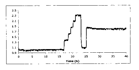
Figure 15 is a graphic representation of glucose response in fluorescence
intensity for
hydrogel glued (VetBond) to Imm PMMA fiber versus time in seconds.
Figure 16 is similar to Figure 15 and is the response at different glucose
concentrations
versus time in seconds.
Figure 17 is the structure of HPTS(Lys-MA) as prepared in Example 47.
Figure 18 is a;raphic representation of the glucose response of hydrogel from
Example
54.
Figure 19 is aI;raphic representation of the characteristic fluorescence
response in
addition of a quantum dot solution followed by addition of quencher to glucose
to the quencher
solution at pH 7.4.
Figure 20 is a graphic representation of a Stern Volmer Plot of 0-BVV2+ and
BVa+
quenching the fluorescence of amine and carboxyl substituted quantum dots
(2x10⁻⁷) M at pH 7.4.
Figure 21 is a graphic representation of glucose response cures obtained by
using o-
BBV2+ quenching the fluorescence amine and carboxyl substituted quantum dots
at pH 7.4.
Figure 21A is zi graphic representation of glucose response and of hydrogel
comprising 1-
MABP and APTS-BurAA with F/F plotted against time in hours.
CA 02630790 2008-05-22
WO 2007/067743 PCT/US2006/046895
Figure 21B is a graphic representation of glucose response of the hydrogel
comprising 1-
MABP and APTS-DegMA with F/F plotted against glucose level in mM.
Figure 22A is a graphic representation of glucose response of the hydrogel
containing
P2-3,3'-oBBV and APTS-DegMA with F/F plotted against time in hours.
Figure 22B is a graphic representation of glucose response of the hydrogel'
containing P2-
3, 3'-oBBV and APTS-DegMA with F/F plotted against glucose level in mM.
Figure 23 is a graphic representation of DMAA hydrogel comprising P-BOB-APTS-
DegMA showing glucose response as a function of time.
Figure 24 is a graphic representation of the relative intensity of light as a
function of
glucose concentratioii for P-BOB:APTS-DegMA hydrogel.
Figure 25 is a graphic representation of glucose sensing for the polyviologen
quencher
showing F/F versus glucose concentration in mM.
DETAILEED DESCRIPTION OF THE PREFERRED EMBODIMENT
Definitions
As used herein:
" Bis-viologer.i" refers to compounds in which two viologens are coupled
together.
"Boronic acid." refers to a structure -B(OH)2. It is recognized by those
skilled in the art
that a boronic acid may be present as a boronate ester at various stages in
the synthesis of the
quenchers of this invention. Boronic acid is meant to include such esters.
"Detector" rei7ers to a device for monitoring light intensity such as a photo
diode.
"Fluorophore"' refers to a substance that when illuminated by light at a
particular
wavelength emits light at a longer wavelength; i.e., it fluoresces.
Fluorophores include organic
dyes, organometallic compounds, metal chelates, fluorescent conjugated
polymers, quantum dots
or nanoparticles and combinations of the above. Fluorophores may be discrete
moieties or
substituents attached to a polymer. "Fluorescent dye" or "dye" is selected
from a discrete
compound or a reactive intermediate which is convertible to a second discrete
compound, or to a
polymerizable compound D1; or D2 which is pendant group or chain unit in a
polymer prepared
from said reactive ini.ermediate or polymerizable compound, which polymer is
water-soluble or
water-dispersible or is a water-insoluble polymer, said polymer which is
optionally crosslinked.
21
CA 02630790 2008-05-22
WO 2007/067743 PCT/US2006/046895
"Fluorescent -conjugated polymers" refers to a polymer in which the structure
as a whole
behaves as a fluoropliore. A typical example is polyphenylene vinylene, i.e.,
a conjugated
carbon-carbon double bond is present and conjugation is sufficient for the
polymer to have
fluorescent propertie:a.
"HEMA" refers to 2-hydroxyethylmethacrylate.
"Light source" or "excitation light source" refers to a device that emits
light preferably of
a selected wavelengtli. The "light source" may encompass any device that emits
electromagnetic
radiation such as a xenon lamp, medium pressure mercury lamp, a light emitting
diode (LED) all
of which are commercially available.
"Linking group" refers to L, Ll or L 2 which are divalent moieties, that
covalently connect
the sensing moiety to the polymer or matrix. Examples of L, Ll or L 2 include
those which are
each independently st-llected from a direct bond or, a lower alkylene having 1
to 8 carbon atoms,
optionally terminated with or interrupted by one or more divalent connecting
groups selected
from sulfonamide (-SO2NH-), amide -(C=O)N-, ester -(C=O)-O-, ether.-O-,
sulfide -S-, sulfone
(-SOZ-), phenylene -C6H4-, urethane -NH(C=0)-0-, urea -NH(C=0)NH-, thiourea -
NH(C=S)-
NH-, amide -(C=O)NH-, amine -NR- (where R is defined as alkyl having 1 to 6
carbon atoms)
and the like.
"Polyviologen" refers generally to compounds comprising two or more viologen
units
coupled together, including bis-viologens, wherein the viologen rings are
close enough that the
electron affinity of the coupled compound as measured by reduction potential
is enhanced over
that of a single viologen.
"Polyviologer.i boronic acid" refers to a polyviologen substituted with at
least two boronic
acid groups.
"Quencher" ("Q") refers to a compound that, when operably coupled to a
fluorophore,
reduces the emission of the fluorophore. In one embodiment, the quencher and
fluorophore may
be deemed operably coupled when the quencher and fluorophore are in close
enough proximity
to one another to intei-act-wherein the interaction results in the reduced
fluorescence. In
preferred embodiments, Q is further configured to bind analyte, preferably
glucose, wherein
analyte binding modulates the quenching activity of Q. Quencher Q may be
selected from a
discrete compound, a reactive intermediate which is convertible to a second
discrete compound
or to a polymerizable compound or Q is a pendant group or chain unit in a
polymer prepared
22
CA 02630790 2008-05-22
WO 2007/067743 PCT/US2006/046895
from said reactive intermediate or polymerizable compound, which polymer is
water-soluble or
dispersible or is an insoluble polymer, said polymer is optionally
crosslinked.
"Quantum dots" ("qd") refers to when electrons and holes in material are
confined to
ultra-small regions of'space (typically 1-25 nm), the material structure
enters the regime of size
quantization, wherein the electronic energy levels of the system become
discrete rather than
quasi-continuous, and the optical and electronic properties of the materials
become strongly size-
dependent. Such structures are termed quantum dots or nanocrystals, quantum
rods, or quantum
wells depending upoii their shape and dimensionality of the quantum
confinement. They include
semiconductor crystals with a diameter of a few nanometers typically surface
treated with
functional groups to ynake them water-dispersible.
"In vivo" refers to analysis in a living mammal, preferably a human being. In
vivo
measurements take p:lace under physiological conditions of temperature,
pressure, medium,
analyte concentration. and pH as found, e.g., in a human body.
"IPN" or "interpenetrating polymer network " refers to a combination of two or
more
network polymers synthesized in juxtaposition (see L.H. Sperling,
Interpenetrating Polymer
Networks, ACS Advances in Chemistry Series 239, 1994, from August 25-30,1991
New York
ACS Meeting).
"Pyridinium" refers to structures (linking groups or pendant groups comprised
of units,
i.e., pyridine rings substituted on the nitrogen and optionally on carbons in
other positions on the
ring. Substituents on carbon include vinyl groups and substituents on nitrogen
include the
methylene group of a benzyl boronic acid.
"Semi-IPN" c-r semi-interpenetrating polymer network" refers to a combination
of
polymers in which one component is soluble and the other polymer is a network
(see Sperling
above).
"Onium" refers to a heteroaromatic ionic compound having a formal positive
charge on
the heteroatom, which in the case of viologen is a nitrogen.
"PEG" or "polyethylene glycol" refers to polymer or chain segments, which
contain
oxyethylene (-OCH7=-CH2-) repeating units.
"PEGDMA" refers to polyethylene glycol terminated with two methacrylate
groups.
"PEGMA" refers to polyethylene glycol terminated with one methacrylate group.
23
CA 02630790 2008-05-22
WO 2007/067743 PCT/US2006/046895
"Physiological pH" refers to the pH range of 7.3-7.5 normally existing in the
blood of a
healthy living human being. In critically ill patients, a physiological pH
between about 6.8 to 7.8
is often observed.
"Visible ligh-t range" refers to light in the spectrum between about 400 and
800 nm.
"Viologen" refers generally to compounds having the basic structure of a
nitrogen
containing conjugate;d N-substituted heterocyclic aromatic bis-onium salt,
such as 2,2'-, 3,3'- or
4,4'-N,N'bis-(benzy:l) bipyridium dihalide (i.e., dichloride, bromide
chloride), etc. Viologen also
includes the substituted phenanthroline compounds. A number of important
advances are encompassed within the preferred embodiments of
the present inventiori concerns a number of important advances. These include
but are not
limited to a method t3nd an in vivo device for determining carbohydrate, 1,2-
diol or 1,3-diol
levels in liquids selected from aqueous or organic liquids or combinations
thereof or in a
physiological fluid, respectively. A series of fluorophore dyes, a series of
boronic acid
substituted quenchers, and combinations of interacting water-compatible and
water-soluble and
organic solvent-compatible and organic solvent-soluble organic polymers are
used. These
aspects are discussed in more detail below. The components are discussed
first, and their
combination to produce the method and the device follows.
uencher
The moiety that provides glucose recognition in the present invention is an
aromatic
boronic acid. More specifically, the boronic acid of this invention is
covalently bonded to a
conjugated nitrogen-containing heterocyclic aromatic bis-onium structure,
e.g., a viologen, (see
for example Figures 3A to 31) in which the boronic acid reacts reversibly with
glucose in
aqueous media at pF[ from about 6.8 to 7.8 to form boronate esters. The extent
of reaction at a
specific pH is relateci to glucose concentration and the acidity (as measured
by pKa) of the
boronic acid.
Bis-onium salts of this invention are prepared from conjugated heterocyclic
aromatic
dinitrogen compounds. The conjugated heterocyclic aromatic dinitrogen
compounds are selected
from dipyridyls, dipyridyl ethylenes, dipyridyl phenylenes, phenanthrolines,
and diazafluorenes,
wherein the nitrogen, atoms are in a different aromatic ring and are able to
form an onium salt. It
is understood that al:l isomers of said conjugated heterocyclic aromatic
dinitrogen compounds in
which both nitrogens can be substituted are useful in this invention. Bis-
onium salts derived
from 4,4-dipyridyl Eu1d 4,7-phenanthroline are included. The viologen boronic
acid adducts are
discrete compounds or are water-compatible pendant groups or units in a chain
of a water-
24
CA 02630790 2008-05-22
WO 2007/067743 PCT/US2006/046895
soluble or water-dispersible polymer with a molecular weight greater than
10,000 or are bonded
to an insoluble polyrner matrix. One or more boronic acid groups are attached
to the viologen
moieties.
For the polyrneric quencher precursors, inultiple options are available for
the boronic
acid moiety to be attcLched to two different nitrogens in the heteroaromatic
centrally located
group. These are:
a) a polymerizable group on a first aromatic moiety is attached to one
nitrogen and a
second aromatic group containing at least one -B(OH)2 group is attached to the
second nitrogen;
b) one or more boronic acid groups are attached to a first aromatic moiety
which is
attached to one nitrogen and one boronic acid and a polymerizable group are
attached to a second aromatic group which second aromatic group is attached to
the
second nitrogen;
c) one boronic acid group and a polymerizable group are attached to a first
aromatic
moiety vrhich first aromatic group is attached to one nitrogen, and a boronic
acid
group and a polymerizable group are attached to a second aromatic moiety which
is
attached to the a second nitrogen; and
d) one boronic acid is attached to each nitrogen and a polymerizable or
coupling groups
is attached to the heteroaromatic ring. Preferred embodiments have two boronic
acid
moieties and one polymerizable group or coupling group.
Representative viologens with one boronic acid group include the following:
1. boronic acid substituted viologens of the structure:
(D
2X
Y2 -(CH2)n IQ 0 el Nl
where n=1-3, preferably n is 1, and where L is a linking group, i.e., Ll or L2
as defined
herein and M is a polymer matrix as defined herein, and
where Y2 is phenyl boronic acid (m- and p-isomers) or naphthyl boronic acid,
preferably a phenyl boronic acid, and
2. as a substituent on the heterocyclic ring of a viologen.
CA 02630790 2008-05-22
WO 2007/067743 PCT/US2006/046895
The viologen is contemplated to include combinations of the above. The
precursor from
which the viologen/boronic acid is derived is an unsymmetrically substituted
viologen, such as
with a boronic acid functional group on one end and a polymerizable group,
such as a vinyl
group, on the other (;;ee Figures 3A-31). The viologen/boronic acid moiety
(i.e. the quencher) is a
pendant group or a cliain unit in a water soluble or dispersible polymer, or a
unit in a crosslinked,
hydrophilic polymer or hydrogel sufficiently permeable to glucose to allow
equilibrium to be
established. In a pref -.rred embodiment, greater intensities of signals are
observed when the
viologen comprises two or more boronic acid moieties.
In another en:ibodiment, the quencher Q1 or Q2 is prepared from a quencher
precursor
selected from the group consisting of o-, m-, and p- boronic acids:
HO' f OHz'r~ 2~ 2X
T
f or.
OH
F
z+t-[Vr=-GH2 CHZ-[N}z=--Z
4X
HO~ ON
where V is a(iinitrogen containing conjugated heterocyclic aromatic group
selected from
isomers of dipyridyls., dipyridyl ethylenes, dipyridyl phenylenes,
phenanthrolines, or
diazafluorenes; wherein the two nitrogen atoms are each in a different
aromatic ring and the
nitrogens are in all positions capable of forming an onium salt and where Z'
or Z2 is
independently a substituent on nitrogen and is either a polymerizable
ethylenically unsaturated
group or a coupling group, optionally including a boronic acid substituted
xylylene linking
group.
Said polymerizable groups are selected from but not limited to:
(i) -R10-CO,-C(Rl l)==CHa, -R1O-NH-(C=0)-C(R)=CH2, or -CH2-C6H4-CH=CH2,
where Ra0 is a lower alkylene or hydroxyalkylene of 2 to 6 carbon atoms and
where R' 1= -l.~ or -CH3.
(ii) Said coupling groups are selected from but not limited to:
-R12-Z3
26
CA 02630790 2008-05-22
WO 2007/067743 PCT/US2006/046895
where R12 is a linking group, preferably -CH2C6H4-CH2- or alkylene of 2 to 6
carbon
atoms anci
Z3 is a reactive functional group, capable of forming a covalent bond with a
coreactant. Such
groups include but are not limited to -OH, -SH, -COZH, or -NH2.
Q' is a discrel:e compound or a pendant group or a chain unit (linear or
branched) of a
water-soluble or dispersible polymer. Q2 is a pendant group or chain unit in a
water insoluble
polymer matrix M' - L2 - Q2. Preferably the matrix is a crosslinked hydrogel.
In another aspect, Q' or Q2 is prepared from a precursor selected from:
C;Hz .y e
~ Ct~ n'~ L q ~ . 6
z'CHs. õ'. CHx-(V.1..FCHz ,~ GHz- z
B(QH)z 2X' ar ~
8(aH4 2X-
B(OH)z B(t?C-I)x
Where V' is the same as V, and Z4 and Z5 are polymerizable groups or coupling
groups such as Z' anci Z2 covalently linked to a quaternary nitrogen group,
including N,N-
dimethylammonium,md pyridinium, which is further bonded to the methylene
groups on the
quencher precursor.
Thus, Z4 or Z'' include 2, 3 or 4-(CH2=CH)-pyridinium; 2, 3, or 4-(CH2=C (CH3)-
(C=O)NH-(CH2),-pvridinium; -N-(CH3)2 -(CHz) ,-O(C=O) C(CH3) =CH2); -O-(CH2)", -
0-
(C=0) -C(CH3)=CH2; -O-(CH2)w,-O-(C=O)CH=CH2; and -O-(CH2),,,-NH-(C=O)
C(CH3)=CH2;
and w is a integer from 2 to 6, or Z4 and Z5 are Z' and Z2, bonded to the
methylene group on the
viologen precursor through a heteroatom, preferably -O-.Subsequent reaction of
the
polymerizable groups, or coupling groups results in the binding of the
quencher precursor to a
water soluble or dispersible polymer or to a polymer matrix, M as a pendant
group, a chain unit,
or an end group in said polymers
Preferred que:achers Q2 are prepared from precursors comprising viologens
derived from
3.3'-dipyridyl substituted on the nitrogens with benzylboronic acid groups and
at other positions
on the dipyridyl rings, with a polymerizable group or a coupling group.
Representative viologens
include:
27
CA 02630790 2008-05-22
WO 2007/067743 PCT/US2006/046895
14 - z6
2X
+Nm M~+
R, R,t R + R*'
Where L4 is independently selected from L, Ll or L2 as defined herein, Z6 is
independently selected from Z1, Z2, Z3, Z4 or Z5 as defined herein and R' is -
B(OH)2 and R" is a
polymerizable or a coupling group as is defined herein.
Other exampk;s of novel quencher precursors include:
NH
0
26r- ~
NH zBr O NH
N +N\- F\ F
Q N
Ri RZ Ra Ry ~-s
B(OH)2 (HO)2B
3,3'-oBBV: Rc=H, R2=B(OH)2 3,3'-FoBBV
3,3'-m BBV: R 1 =B(OH)Z, R2= H
O O~ \
~NH
NH
26 0 r NH 2Br O NH
(HO)Zg N~ \ ,
/-\ N +N~_ / + _
+N~
(HO)2B B(OH)2 (HO)2B
3,;,'-mBBV
4 , 3-o B B V
O
2Br
N N
/ \
/ \ NN
(HO)2B /
B(OH)2
n+ n+ +N~
LD
3,3'-B PV
RI is a boronic acid in the ortho-, meta-, or para- positions on the benzyl
ring. R2 is a
hydrogen or optionally a polymerizable group or a coupling group as defined
herein or a
substituent specifically used to modify the acidity of the boronic acid,
28
CA 02630790 2008-05-22
WO 2007/067743 PCT/US2006/046895
Boronic acid substituted polyviologens are another class of preferred
quenchers.
The term "polyviologen" includes: a discrete compound comprised of two or more
viologens
covalently bonded together by a linking group, a polymer comprised of viologen
repeat units in
the chain, a polymer with viologen groups pendant to the chain, a dendrimer
comprised of
viologen units, preferably including viologen terminal groups, an oligomer
comprised of
viologen units, preferably including viologen endgroups, and combinations
thereof. Polymers in
which mono-viologei:i groups are a minor component are not included. The
preferred quenchers
are substituted with at least two boronic acid groups and are water-soluble or
dispersible
polymers or hydrogel.s comprised of polyviologen boronic acids. Alternatively,
the polyviologen
boronic acid is directly bonded to an inert substrate. Quencher precursors
comprised of
polyviologen boronic. acids include low molecular weight polyviologen boronic
acids further
substituted with polynerizable groups or coupling groups
In a specific embodiment, the polyviologen boronic acid precursors are bis-
viologen
derivatives prepared by covalently linking two viologen units wherein said
adducts are further
substituted with boronic acids, and polymerizable groups, or coupling groups.
Preferably the
precursor is substituted with only one such polymerizable group or coupling
group attached
directly to the linking: group. The linking group is bonded to one nitrogen in
the heterocyclic
aromatic ring of each viologen unit, or to a carbon in the ring of each
viologen unit, or one bond
is to a ring carbon in Dne viologen and to a nitrogen in the other. Two or
more boronic acid
groups are attached to the quencher precursor. Preferably the linking group is
selected to
enhance cooperative binding of the boronic acid groups to glucose.
The moiety that connects the two viologen units is a linking group, L, as
defined
previously with the p;:oviso that L is optionally further substituted with a
boronic acid group, a
polymerizable group, or a coupling group or combinations thereof. In some
cases, the linking
group may be a segment of a polymer chain in which the viologens are pendant
groups or units
in said chain.
Fluorophore Dye
Dyes useful ir.i the embodiments of this invention (See Fig. 1 A, 1 B and 1 C)
are excited
by light of wavelength about or greater than 400 nm (preferably 430 nm), with
a Stokes shift
large enough that the excitation and emission wavelengths are separable, being
at least 10 nm,
and preferably greater than or equal to about 30 nm. These dyes are
susceptible to quenching by
electron acceptor molecules, such as viologens, are resistant to photo-
bleaching, and are stable
against photo-oxidation, hydrolysis, and biodegradation. Dyes useful in the
present invention
have an apparent Stern-Volmer quenching constant when tested with methyl
viologen of about
29
CA 02630790 2008-05-22
WO 2007/067743 PCT/US2006/046895
50 or greater and pre:ferably greater than 100. A general description of the
Stern-Volmer test is
found below in Prep<<ration A. Preferred dyes include polymeric derivatives of
hydroxypyrene
trisulfonic acid and aminopyrene trisulfonic acid. In some cases, the dye is
bonded to a polymer
through the sulfonann.ide functional groups. The polymeric dyes are water-
soluble, water-
insoluble but swellable or dispersible in water or may be crosslinked. A
preferred dye as a
polymer is for exarnple, a water soluble PEG adduct of 8-hydroxypyrene-1,3,6-
N,N',N"-
tris(methoxypolyethcaxylethyl (n-125) sulfonamide) (formed by reaction of
acetoxypyrene
trisulfonyl chloride with aminoethyl PEG monomethyl ether. The resulting dye
polymer has a
molecular weight of at least about 10,000 such that, when it is trapped in a
hydrogel or network
polymer matrix, it is incapable of diffusing out of the matrix into the
surrounding aqueous
medium.
Representative dyes as discrete compounds are the tris adducts formed by
reacting 8-
acetoxypyrene-1,3,6-tTisulfonylchloride (HPTS-Cl) with an amino acid, such as
amino butyric
acid. Hydroxypyrene trisulfonamide dyes bonded to a polymer and bearing one or
more anionic
groups are most preferred, such as copolymers of 8-hydroxypyrene-l-N-
(methacrylamidoprofrylsulfonamido)-N',N"-3,6-bis(carboxypropylsulfonamide)
HPTS-C02-MA
with HEMA, PEGMA, etc.
Other exampl-.s include soluble copolymers of 8-acetoxypyrene-1,3,6-N, N', N"-
tris(methacrylamidopropylsulfonamide) with HEMA, PEGMA, or other hydrophilic
comonomers. The phenolic substituent in the dye is protected during
polymerization by a
blocking group that can be removed by hydrolysis after completion of
polymerization. Such
blocking groups, which are suitable for example acetoxy, trifluoroacetoxy, and
the like are well
known in the art. Other preferred dyes include polymeric derivatives of
aminopyrene trisulfonic
acid [APTS] in which the dye is bonded to the polymer as a pendant group or a
unit in the
polymer chain. The dye is bonded to the polymer through a sulfonamide linkage
or preferably
through an amine linking group. Some polymerizable APTS derivatives include:
CA 02630790 2008-05-22
WO 2007/067743 PCT/US2006/046895
H 0
M.I. 03S N pJ
I I ~
03S ~ .- S03M
M"'= positive counter.,ion
0
HOOC
H rS . ,;, NH2
~
( ~ ~
M 03S ~ ~ SO3M",
It is preferred that, for sensing to occur, the sensing moieties (analyte,
dye, quencher) are
in close enough physical proximity to allow interaction, e.g., mixed on a
molecular level and in
equilibrium with the species to be detected. While not bound by any theory or
mechanism, it
appears that ionic int(,-raction between dye and quencher leads to the
formation of a ground state
dye/viologen complex and the intensity of the fluorescence emitted by the dye
is attenuated.
Binding of glucose to the quencher produces a negatively charged boronate
ester which weakens
the complex resulting in an increase in intensity dependent on the extent of
glucose
binding. Changes in the molecular conformations of the complexed species are
also likely
because of steric inte:ractions resulting from analyte binding which
influences the
signal. Further, the boronate ester may interact with the viologen thereby
altering its quenching
efficacy. The specific: nature of this interaction is not yet established, but
boronate formation
may shift the reduction potential of the viologen. The reduction potential is
an indicator of the
ability of the viologea to accept an electron. The remarkably enhanced
quenching efficiency of
the polyviologen/boronic adducts and increased modulation that obtains from
glucose binding
indicates that a redox mechanism may be involved. A redox couple between dye
and quencher
followed by electron exchange between viologen moieties assist in keeping the
dye in a non-
excitable state. Boror.Late ester formation interferes with this process.
Quantum Dot (qd) E nbodiments
Fluorescent qi..zantum dot semiconductor nanoparticles have found increasing
use as
replacements for traditional organic fluorophores in such applications as
biomolccule tagging,
tissue imaging and ion sensing. Interest in fluorescent quantum dots (qd's)
derives from their
broad absorption, narrow emission, intense brightness, and good photostability
relative to
organic dyes. Surprisingly, though, despite the large and diverse set of
fluorescence-based
31
CA 02630790 2008-05-22
WO 2007/067743 PCT/US2006/046895
sensing systems for glucose, no methods for glucose detection utilizing
inherently fluorescent
qd's have yet been reported. The two-component approach to glucose sensing
described herein
allows for considerable flexibility in choosing the quencher/receptor and
fluorophore
components depending on the particular requirements of the sensing
application. For example,
fluorophore componE;nts are selected to provide any in a range of desired
excitation or emission
wavelengths while a particular quencher/receptor may be chosen for reasons of
its
monosaccharide bind!ing selectivity. Some of the advantages of qd's are
realized in the two-
component system to. sense changes in glucose concentration in aqueous
solution.
Fluorescent qd's are constructed of inorganic semiconductor core materials
such as CdTe
and CdSe, coated wi1:h an insulating shell material such as ZnS and further
treated to provide
desired surface chemistry. For the preparation of water-soluble core shell
qd's, surface
functionalization with phosphonate, carboxyl, or amine groups is often
employed. The particular
surface chemistry allows for the qd's to bind to molecules of interest such as
proteins and also
determines their solubility, aggregation behavior and sensitivity to quenching
processes. Several
groups have observeel quenching of qd fluorescence using methyl viologen
(1VIV2+). The process
is believed to occur tl.lrough excited state electron transfer from the qd to
the viologen resulting in
reduction of the viologen to MV*+. Previous studies had shown that viologens
were extremely
efficient in statically quenching the fluorescence of many organic dyes
through complex
formation with the fl uorophore. The fluorescence of core shell quantum dots
bearing polar
surface groups such as carboxyl and amine is similarly quenched through
complex formation
with the boronic acid-substituted viologen quenchers.
Two sets of commercially available core shell CdSe quantum dots were
identically
prepared except for tl.leir surface fictionalization: one set was prepared
with carboxyl groups on
the surface, the other with amine groups. Both sets had a fairly narrow
fluorescence emission
centered at 604 nm. 7:'hese qd's indeed functioned in this system in a manner
similar to that of
organic dyes: showing a decrease in fluorescence upon addition of viologen
quencher. Robust
fluorescence recover~ was observed upon addition of glucose to the quenched qd
solutions
(Figure 18).
The sensitivity of both quantum dot sets fluorescence quenching by the boronic
acid
substituted viologen ~:)-BBV2+ was determined in pH 7.4 aqueous solution
(Figure 20). The
fluorescence of both -the carboxyl and amine substituted qd's were sensitive
to quenching by o-
BBVZ+, with the carboxyl substituted quantum dots showing a stronger
sensitivity to quenching
than the amine substituted dots. Fluorescence of both sets of qd's was also
similarly quenched by
simple unsubstituted benzyl viologen (BV2}) though to a lesser degree than
with boronic acid
32
CA 02630790 2008-05-22
WO 2007/067743 PCT/US2006/046895
substituted viologen. Significantly, while the degree of ionization of the
surface group was not
determined, the carboxyl-substituted dots are expected to exist primarily in
their anionic form at
pH 7.4 whereas the amine dots would most likely be neutral. The enhanced
sensitivity of the
carboxyl-substituted qd's may be due to electrostatic attraction between the
cationic viologen
quencher and the anionic surface groups on the qd.
Previous studies had shown that choice of an appropriate ratio of quencher to
fluorophore
was critical for a strong and linear signal response across the physiological
glucose range. When
experimenting with several different quencher-to-quantum dot ratios generally
the same behavior
was observed as with, traditional organic dyes where higher ratios tended to
give larger, more
linear fluorescence signals in response to addition of glucose (Figure 3).
Both sets of qd's were screened for glucose response at quencher:qd ratios of
50, 200,
500, and 1000 to 1. For both the amine and carboxyl substituted qd's, optimal
results were
obtained using the 1000:1 quencher-to-quantum dot ratio. Significantly, the
use of quantum
dots allows for a large signal response and a considerable degree of recovery
of the initial,
unquenched quantum dot fluorescence after addition of 100 nM glucose (Figure
21).
Results using quantum dots in a hydrogel in two component sensing systems for
the
detection of sugars are in Example 60.
Polymer Matrix for Sensors
For in vivo applications, the sensor is preferably used in a moving stream of
physiological fluid, e.g., within a blood vessel, which contains one or more
polyhydroxyl organic
compounds or is implanted in tissue such as muscle which contains said
compounds. Therefore,
it is preferred that none of the sensing moieties escape from the sensor
assembly. Thus, for use in
vivo, the sensing components are part of an organic polymer sensing assembly.
Soluble dyes and
quenchers can be cor.Lfined by a semi-permeable membrane that allows passage
of the analyte but
blocks passage of the= sensing moieties. This can be realized by using as
sensing moieties soluble
molecules that are substantially larger than the analyte molecules (molecular
weight of at least
twice that of the analyte or greater than 1000 preferably greater than 5000);
and employing a
selective semipermeEtble membrane such as a dialysis or an ultrafiltration
membrane with a
specific molecular weight cutoff between the two so that the sensing moieties
are quantitatively
retained.
Preferably the sensing moieties are immobilized in an insoluble polymer
matrix, which is
freely permeable to glucose, see Figure 8. The polymer matrix may be comprised
of organic,
inorganic or combinzttions of polymers thereof. The matrix may be composed of
biocompatible
33
CA 02630790 2008-05-22
WO 2007/067743 PCT/US2006/046895
materials. Alternative:ly, the matrix is coated with a second biocompatible
polymer that is
permeable to the anal.ytes of interest.
One function of the polymer matrix is to hold together and immobilize the
fluorophore
and quencher moieties providing an operable coupling between these moities,
while at the same
time allowing contact with the analyte, and binding of the analyte to the
boronic acid. To achieve
this effect, the matrix. is preferably insoluble in the medium, and in close
association with it by
establishing a high stirface area interface between matrix and analyte
solution. For example, an
ultra-thin film or microporous support matrix may be used. Alternatively, the
matrix is swellable
in the analyte solution, e.g., a hydrogel matrix is used for aqueous systems.
In some instances,
the sensing polymers are bonded to a surface such as the surface of a light
conduit, or
impregnated in a microporous membrane. In all cases, the matrix preferably
does not interfere
with transport of the analyte to the binding sites so that equilibrium can be
established between
the two phases. Techniques for preparing ultra-thin films, microporous
polymers, microporous
sol-gels, and hydroge;ls are established in the art. All useful matrices are
defined as being analyte
permeable.
Hydrogel polymers are preferred for embodiments of this invention. The term,
hydrogel,
as used herein refers to a polymer that swells substantially, but does not
dissolve in water. Such
hydrogels may be lin.ear, branched, or network polymers, or polyelectrolyte
complexes, with the
proviso that they contain no soluble or leachable fractions. Typically,
hydrogel networks are
prepared by a crosslinking step, which is performed on water-soluble polymers
so that they swell
but do not dissolve in aqueous media. Alternatively, the hydrogel polymers are
prepared by
copolymerizing a mixture of hydrophilic and crosslinking monomers to obtain a
water swellable
network polymer. Su:ch polymers are formed either by addition or condensation
polymerization,
or by combination pi-ocess. In these cases, the sensing moieties are
incorporated into the polymer
by copolymerization using monomeric derivatives in combination with network-
forming
monomers. Alternatively, reactive moieties are coupled to an already prepared
matrix using a
post polymerization reaction. Said sensing moieties are units in the polymer
chain or pendant
groups attached to the chain.
The hydrogel.s useful in this invention may also be monolithic polymers, such
as a single
network to which both dye and quencher are covalently bonded, or multi-
component hydrogels.
Multi-component hydrogels include interpenetrating networks, polyelectrolyte
complexes, and
various other blends of two or more polymers to obtain a water swellable
composite, which
includes dispersions of a second polymer in a hydrogel matrix and alternating
microlayer
assemblies.
34
CA 02630790 2008-05-22
WO 2007/067743 PCT/US2006/046895
Monolithic hydrogels are typically formed by free radical copolymerization of
a mixture
of hydrophilic monoiners, including but not limited to HEMA, PEGMA,
methacrylic acid,
hydroxyethyl acrylati-1, N-vinyl pyrrolidone, acrylamide, N,N'-dimethyl
acrylamide, and the like;
ionic monomers include methacryloylaminopropyl trimethylammonium chloride,
diallyl
dimethyl ammonium chloride, vinyl benzyl trimethyl ammonium chloride, sodium
sulfopropyl
methacrylate, and the like; crosslinkers include ethylene dimethacrylate,
PEGDMA, N,N'-
methylene-bis-acrylamide trimethylolpropane triacrylate, and the like. The
ratios of monomers
are chosen to optimize network properties including permeability, swelling
index, and gel
strength using principles well established in the art. In one embodiment, the
dye moiety is
derived from an ethylenically unsaturated derivative of a dye molecule, such
as 8-
acetoxypyrene-1,3,6-N, N', N"-tris(methacrylamidopropylsulfonamide), the
quencher moiety is
derived from an ethylenically unsaturated viologen such as 4-N-(benzyl-3-
boronic acid)-4'-N'-
(benzyl-4ethenyl)-dipyridinium dihalide (m-SBBV) and the matrix is made from
HEMA and
PEGDMA. The concentration of dye is chosen to optimize emission intensity. The
ratio of
quencher to dye is adjusted to provide sufficient quenching to produce the
desired measurable
signal.
Alternatively, a monolithic hydrogel may be formed by a condensation
polymerization.
For example, acetoxy pyrene trisulfonyl chloride is reacted with an excess of
PEC'r diamine to
obtain a tris- (amino PEG) adduct dissolved in the unreacted diamine. A
solution of excess
trimesoyl chloride ar.id an acid acceptor is reacted with 4-N-(benzyl-3-
boronic acid)-4'-N'-
(2hydroxyethyl) bipyridinium dihalide to obtain an acid chloride functional
ester of the viologen.
The two reactive mixtures are brought into contact with each other and allowed
to react to form
the hydrogel, e.g., b), casting a thin film of one mixture and dipping it into
the other.
Polymers that are capable of reacting with boronic acids to form boronate
esters under
the conditions of this method are not preferred as matrix polymers. Such
polymers have 1,2- or
1,3- dihydroxy substituents, including but not limited to cellulosic polymers,
polysaccharides,
polyvinyl alcohol and its copolymers and the like.
Multi-component hydrogels wherein the dye is incorporated in one component and
the
quencher in another are preferred for making the sensor of this invention.
Further, these systems
are optionally molecularly imprinted to enhance interaction between components
and to provide
selectivity for glucose over other polyhydroxy analytes. Preferably, the
multicomponent system
is an interpenetrating polymer network (IPN) or a semi-interpenetrating
polymer network (semi-
IPN).
CA 02630790 2008-05-22
WO 2007/067743 PCT/US2006/046895
The IPN polymers are typically made by sequential polymerization. First, a
network
comprising the quencher is formed. The network is then swollen with a mixture
of monomers
including the dye monomer and a second polymerization is carried out to obtain
the IPN
hydrogel.
The semi-IPN hydrogel is formed by dissolving a soluble polymer containing dye
moieties in a mixture of monomers including a quencher monomer and
polymerizing.
Alternatively, a soluble quencher polymer is dissolved in a monomer mixture
containing the dye
monomer and the mi:cture polymerized. In either case, the molecular weight of
the soluble
component must be sufficiently high (about or greater than 10,000) that it
cannot diffuse out of
the network, i.e., it becomes physically bound in or trapped by the matrix.
In Figure 4A, one group of polymer chains 41, 42, 43 and 44 contain the
quencher, for
example quencher Q2. A second group of polymer chains 45, 46 and 47 containing
the dye, for
example, dye D2, is formed at about the same time or sequentially. The points
of crosslinking of
the polymers are designated as 48 and 49. In Figure 4B, one group of polymer
chains 51, 52, 53
and 54 contain the quencher, for example, quencher Q2. Dye DI is to a pendant
group on a
second polymer 56. C'=rosslinking points 57 are designated.
Molecular Imprinting
Optionally, th+: polymers of this invention are molecularly imprinted. In one
embodimeiit,
an organic salt is fornied from a monomeric quencher cation and a monomeric
dye anion. The
organic salt is then copolymerized, under conditions such that the ion pairs
remain at least
partially associated, to form a monolithic hydrogel matrix. Alternatively, the
quencher monomer
is polymerized to forn:i a first polymer, which is then ion exchanged to
obtain a polyelectrolyte
with anionic dye counterion. The latter is then copolymerized with suitable
monomers to form an
interpenetrating dye polymer, which is associated through ionic bonding with
the quencher
polymer. The combination is either an IPN polymer or a semi-IPN polymer. In
another
embodiment, the polymers of this invention are molecularly imprinted to
enhance selectivity for
glucose over other polyhydroxyl compounds, such as fructose, by first forming
a bis boronate
ester of glucose with & polymerizable viologen boronic acid. This ester is
then copolymerized
and hydrolyzed to obttiin a glucose-imprinted polymer. This polymer is
subsequently used to
form an IPN with a dye polymer.
In one aspect, in-SBBV is mixed with glucose in about a 2:1 molar ratio in
aqueous
organic solvent, e.g., -vvater/dioxane. The product bis-boronate ester is
recovered by distilling off
the solvents under vacuum. The product is next copolymerized with HEMA and
PEGDMA to
36
CA 02630790 2008-05-22
WO 2007/067743 PCT/US2006/046895
obtain a first hydrogel following the procedures described in Example 14.
Glucose is then
leached from the hydrogel by conditioning in dilute hydrochloric acid. After
conditioxung in
deionized water, the llydrogel is contacted with the dye monomer of Exaniple
28 to form a
complex between the anionic dye and the cationic quencher polymer. A second
stage
polymerization with more HEMA and PEGDMA is then carried out to form a
molecularly
imprinted IPN hydrogel.
The individuztl components in a multi-component hydrogel are made by the same
or a
different polymerization scheme. For example, in an IPN polymer, a first
network is formed by
free radical polymerization, the second by condensation polymerization.
Likewise, in a semi-IPN
polymer, the soluble component is formed by condensation polymerization and
the network by
free radical polymerization. For example, a quencher polymer, such as poly
4,4'-N,N'-bis(1,3-
xylylene-5-boronic acid) bipyridinium dihalide is formed by condensing 4,4'-
dipyridyl with 3,5-
bis-bromomethyl phe:nylboronic acid. The quencher polymer is dissolved in a
reaction mixture
containing 8-acetoxypyrene-1,3,6-N, N', N"-
tris(methacrylamidopropylsulfonamide) as
described above, and the solution is polymerized to obtain the semi-IPN
hydrogel.
The quencher polymer described above is an example of a polyviologen boronic
acid.
In a specific embodiment, a high molecular weight water-soluble dye is
prepared by
condensing acetoxyp;yrene trisulfonyl chloride with aminoethyl PEG monomethyl
ether to obtain
the 8-hydroxypyrene-=1,3,6-N,N',N"-tris-(methoxypolyethoxyethyl (n-125)
sulfonamide). The
PEG dye polymer is dissolved in a mixture comprised of HEMA, PEGDMA, 4-N-
(benzyl-3-
boronic acid)-4' N'-(benzyl-4-ethenyl)-dipyridinium dihalide (m-SBBV), aqueous
alcohol and
free radical initiator and polymerized to obtain a semi-IPN hydrogel. After
hydrolysis with dilute
base and leaching wilh deionized water, the hydrogel is affixed to a
bifurcated optical fiber light
conduit such that it c~m be exposed to and equilibrate with the body fluid.
The light conduit
together with appropi=iate filters is connected to a blue light emitting diode
(LED) light source
and a silicon photodetector together with an electronic controller and
associated measurement
instrumentation. The sensor is placed in the tip of a catheter, which is
inserted in the body in the
desired location. The sensor is excited by light of about 475 nm and the
fluorescence intensity
monitored at about 520 nm. The level of glucose in the body fluid is
determined from the
intensity of the emission.
A Single Component Viologen Sensor
In another embodiment the invention is a boronic acid substituted viologen
covalently
bonded to a fluorophore. An example of a single component viologen sensor as a
discrete
37
CA 02630790 2008-05-22
WO 2007/067743 PCT/US2006/046895
compound is shown as Example 39. Preferably, the adduct is a polymerizable
compound or is a
unit in a polymer. Orie such adduct is prepared by first forming an
unsymmetrical viologen from
4,4'-dipyridyl by attaching a benzyl-3-boronic acid group. to one nitrogen and
an aminoethyl
group to the other. T]1e viologen is condensed sequentially first with 8-
acetoxy-pyrene-1,3,6-
trisulfonyl chloride in a 1:1 mole ratio followed* by reaction with excess PEG
diamine to obtain a
prepolymer mixture. An acid acceptor is included in both steps to scavenge the
byproduct acid.
The prepolymer mixture is crosslinked by reaction with a polyisocyanate to
obtain a hydrogel.
The product is treated with base to remove the blocking group. Incomplete
reaction products and
unreacted starting materials may be leached out of the hydrogel by exhaustive
extraction with
deionized water before further use. The product is responsive to glucose when
used as the
sensing component as described herein.
Alternatively,, said adducts are ethylenically unsaturated monomers for
example dimethyl
bis-bromomethyl ber.izene boronate is reacted with excess 4,4-dipyridyl to
form a half viologen
adduct. After removing the excess dipyridyl, the adduct is further reacted
with an excess of
bromoethylamine hy-irochloride to form the bis-viologen adduct. This adduct is
coupled to a
pyranine dye by reaction with 8-acetoxypyrene trisulfonyl chloride in a 1: 1
mole ratio in the
presence of an acid acceptor followed by reaction with excess
aminopropylmethacrylamide.
Finally, any residual amino groups are reacted with methacrylol chloride.
After purification the
dye/viologen monomer is copolymerized with HEN1A and PEGDMA to obtain a
hydrogel.
One advantage of this group of viologens is that dye and quencher are held in
close
proximity by covalent bonds (i.e., fixed in an operably coupled
configuration), which could lead
to increased sensitivi-ty. A disadvantage is that making these adducts is a
formidable synthetic
challenge and change:s in composition are difficult to implement.
Characterization and
purification of the product is equally difficult. Therefore, the embodiments
in which dye and
quencher are separate; entities are preferred. The combination of components
described herein
produces a device for the determination of polyhydroxy substituted organic
molecules in
physiological fluids.
Batch Optical Methoi of Analysis for Glucose
Measurements are carried out in a conventional luminescence spectrometer. A
solution
containing a dye and quencher of this invention buffered to pH = 7.4 is
prepared and loaded into
a cuvet. The sample is excited by light of wavelength suitable for the dye
being used and the
fluorescence intensity measured. A fixed amount of the unknown glucose
containing solution is
added to the solution and the measurement is repeated. The change in intensity
is used to
calculate glucose concentration by reference to a calibration curve determined
separately by
38
CA 02630790 2008-05-22
WO 2007/067743 PCT/US2006/046895
measuring a standard series of glucose solutions and plotting the results as
intensity change as a
function of concentration. In this method, the sensing components need to be
stable only for the
time of the test, and the reaction with glucose need not be reversible.
Ontical Method of Process Stream Analysis
A flow-through cell is fabricated for the luminescence spectrometer. A sensing
polymer
is mounted in the cell such that it is exposed on one surface to the
excitation light and on the
other to the process stream. A baseline is established by passing the process
stream free of
glucose through the cell and measuring the steady state fluorescence. The
process stream is then
passed through the cell and the fluorescence intensity monitored as a function
of time. Glucose
concentration is determined by reference to a calibration curve as described
above. In this
method, the sensor rnust be stable over time of operation and the reaction
with glucose must be
reversible. Further, the sensing moieties must be immobilized and not leach
out into the process
stream.
Device Confi uration
Figure 8 is a>chematic representation of the device as used for one time or
continuous
monitoring for sugar,, i.e., glucose. The sensing polymer 81, which contains
the dye and
quenched may be attached to an optional support 82. For some embodiments an
optional semi-
permeable polymer r.nembrane 83A is present. For other applications it may be
useful to have an
optimal biocompatible coating 83B covering the assembly. The light source 84
is connected to
an optical filter 85 to an optical fiber 86 to the sensing polymer 81.
Detector 87 is connected to
an optical filter 88 to an optical fiber 89, which connects to sensing
polynier 81. Light source 84
and detector 87 are both connected to electronic controller 90. The optical
fibers are optional
depending inter alia on the light source. Thus the system can detect changes
in the sensing
polymer based on the glucose content of the physiological fluid. The device
useful in a process
stream and for in vivo implanting and monitoring is shown in Figures 9 and 10.
Figure 9 shows
the optical device. Figure 10 is the cross sectional representation of the
probe. For Figure 9, light
source 11 (visible) is connected by optical fiber 12 to active cell 13.
Semipermeable membrane
14 allows the analytE; to enter and exit freely from cell 13. Optical fiber 15
conveys the altered
light to filter 16, and optional photomultiplier to 17 to produce the analyte
spectrum for analysis.
As shown in Figures 9 and 10, cell 13 includes the selectively permeabl.e
membrane such
that the mixture of polymer 21, dye 22, and quencher 23 are retained in cell
13 under the
conditions of analysis. The light enters cell 14 via optical fiber 12. Within
the active portion of
14A of cell 14, the polymer 21, dye 22 and quencher 33, contact analyte 24,
which has
selectively entered the cell causing a quantitative and reproducible change in
the spectrum. This
39
CA 02630790 2008-05-22
WO 2007/067743 PCT/US2006/046895
modified light signal travels optical fiber 15 to photomultiplier 17 to be
analyzed. One skilled in
the art will recognize that the sensing moieties of this invention can be used
in other implantable
fluorescence sensing devices known in the art. The components for the
quencher, fluorophore
and analyte permeabl.e component (aka, matrix) are described herein and in the
claims. All are
incorporated by reference in this specification.
EXPERIMENTAL
Reagents and solvents are used as received from commercial supplier unless
otherwise
noted. (See Chem Sources USA, which is published annually.)
The following examples are provided to be descriptive and exemplary only. They
are not
to be construed to limiting in any manner or fashion.
Procedure A
FLUORESCENCE SPECTROSCOPY ANALYSIS OF THE APPARENT STERN-
VOLMER QUENCHING CONSTANT OF METHYL VIOLOGEN WITH A
FLUORESCENT DYE
The apparent Stern-Volmer quenching constant is derived from the slope of a
SternVolmer plot of relative fluorescence intensity (FdF) versus concentration
of quenched (M).
See J.R. Lakowicz, (1.999) Principles of Fluorescence Spectroscopy Second
Edition, Kluwer
Academic/Plenum PLiblishers, New York, pp. 237-289. One skilled in the art is
in general able to
perform this analysis for any fluorescent dye/quenched pair in a particular
solvent of interest.
This general Stem-Volmer analysis is used in determining the Stern-Volmer
quenching constants
in 0.1 ionic strength pH 7.4 phosphate buffer.
In order to avoid concentration quenching effects, the concentration of the
dye is
generally adjusted so that the optical density of the dye, at excitation
Xma,,<0.5 absorption units.
Once the desired dye concentration is determined, a stock dye solution is
prepared in which the
concentration is 5 times greater than that desired in the final measurements.
For example, a dye
for which the desired final concentration, which gives an optical density of
excitation Xn,,G0.5
absorption units, is 1 x 10"5 M, would require a stock solution in which the
concentration is 5 x
10"5 M.
Once determiiied, as is described above, 10 mL of dye stock solution of the
appropriate
concentration is madet by weighing out the appropriate mass of dye and placing
the solid into a
10 mL volumetric flask. The flask is then filled to the 10 mL mark with 0. 1
ionic strength pH
7.4 phosphate buffer.
CA 02630790 2008-05-22
WO 2007/067743 PCT/US2006/046895
A stock solution of methyl viologen (25 mL, 0.0025 M) was prepared in a 10-mL
volumetric flask with. pH 7.4 phosphate buffer (0.1 ionic strength). Seven
different solutions
containing methyl viologen were then prepared in pH 7.4 phosphate buffer as
described below in
Table 1:
Tab:le 1. Volume dye Volume quencher Volume Final
Standard Standard Buffer (dye) (Quenched)
(mL) (jnL) (mL) (M) (M)
1 0.00 4.00 1.00E-05 0.00E+00
1 0.20 3.80 1.00E-05 1.00E-04
1 0.30 3.70 1.00E-05 1.50E-04
1 0.50 3.50 1.00E-05 2.50E-04
1 1.00 3.00 1.00E-05 5.OOE-04
1 1.50 2.50 1.00E-05 7.50E-04
1 2.00 2.00 1.00E-05 1.OOE-03
Each sample :is then in-turn analyzed in a luminescence spectrometer set at
the
appropriate excitatioil wavelength and the appropriate emission wavelength
range for the
corresponding dye. The instrumental settings (slit widths, scan speed, optical
filters, excitation
wavelength, emissioii wavelength range) are held constant throughout the
analysis of the series
of samples). The emission fluorescence intensity is then determined as the
integration of the
fluorescence intensity over the emission wavelength range by the trapezoidal
rule approximation
method. The integrated values are plotted on the y-axis and the quenched
concentrations are
plotted on the x-axis and the slope of the resulting line is calculated by
linear regression as the
Stem-Volmer quenching constant. One skilled in the art will realize that based
on quenching
mechanism the Stern-Volmer plot may not result in a linear relationship.
However through the
use of the appropriate, mathematical relationships, which is known and
understood by one skilled
in the art, the apparent Stern-Volmer quenching constant is calculated and
used for comparison.
Preparation A
SYNTHESIS OF D:IMETHYL-(4-BROMOMETHYL)-BENZENEBORONATE
An oven-dried, 100-mL round bottom flask was cooled under argon, fitted with a
magnetic stirring bar, and charged with (4-bromomethyl)-benzeneboronic acid
(12.49 mmols,
2.684 g). The flask vfas sealed with a septum and charged with pentane (55
mL). The suspension
was stirred at room trmperature and upon addition of freshly distilled CH3OH
(3.16 g, 4 mL, 97
mmols) the solution instantly clarified. After stirring for 20 minutes, the
solution was dried over
MgSOa, then over ClIC1a (to remove excess CH3OH). The supematant was
cannulated, under
argon, through a gla:.s-fritted funnel (medium), and the pentane subsequently
removed in vacuo.
The remaining yellow oil was further dried under reduced pressure (0.1 torr, I
hr). Yield: 1.6 g,
41
CA 02630790 2008-05-22
WO 2007/067743 PCT/US2006/046895
6.59 mmols (56 %). 'H-NMR (CD30D, ppm): 4.5 (s, 2H), 7.4 (d, 2H), 7.55 (d,
2H). "B-NMR
(CH3OH, ppm): 29 (:;). Similar procedures were used to prepare the
corresponding 2- and 3-
isomers. The products were used to make the boronic acid-viologen compounds of
Examples.l-
3,5and6.
Preparation B
SYNTHESIS OF 8-.ACETOXY-PYRENE-1.3,6-TRISULFONYL CHLORIDE
Trisodium-8-,icetoxy-pyrene-1,3,6-trisulfonate (acetoxy-HPTS, 11.33 g, 20
mmol) was
suspended in 30 mL of thionyl chloride to which 5 drops of
dimethylformarr.rnide was added. The
suspension was reflu:ced for 3 hr., during which time it became a brown
solution. The solution
was then cooled to 2_'; O C under an argon atmosphere. Thionyl chloride was
then distilled off
under vacuum (2 Tor.r) leaving a yellow residue. The yellow residue was
transferred to three
separate centrifuge tubes along with 60 mL of dichloromethane. The suspensions
were then
centrifuged and the supernatant solutions transferred to a dry round bottom
flask. The residue
remaining in the centrifuge tubes was washed an additional four times each
with 10 mL portions
of dichloromethane. ':['he supernatant solutions were combined and left
overnight under an argon
atmosphere and some precipitation was observed. The dichloromethane solution
was added to
250 mL of pentane ca.using precipitation of a large amount of yellow solid.
The supernatant was
removed by a double-.ended needle and the yellow solid was dried on high
vacuum (0.2 Torr).
Yield: 8.62 g, 15.5 m:nol (78 %), 'H-NMR (500 MHz, CDC13, ppm): 2.682 (s, 3H),
8.833, (d,
J=10Hz, 1H), 8.915 (s, 1H), 9.458 (d, J=10Hz, 1H), 9.509 (d, J=10 Hz, 1H),
9.630 (s, 1H), 9.685
(d, J= 10Hz, 1H). This product was used in Examples 7, 9, 13, 14 and 15_
Preparation C
SYNTHESIS OF 4-(4-PYRIDYL)-N-(BENZYL-4-ETHENYL)-PYRIDINIUM
CHLORIDE
An oven-dried, 100-mL round bottom flask was cooled under argon, fitted with a
magnetic stirring bar, and charged with 4,4'-dipyridyl (12.50 g, 80 mmols).
The flask was sealed
with a septum and charged with CH3OH (20 rnL). The homogenous solution was
stirred at room
temperature while 4-(:;hloromethyl)styrene (2.82 mL, 20 mmols) was added
dropwise via
syringe. After stirring the solution at room temp for 48 hours, the solvent
was removed in vacuo.
Dry tetrahydrofuran (50 mL) was added to the reaction flask via cannula and
the mixture stiffed
for three days, at which point the stirring was stopped, the solids allowed to
settle, and the
solvent was removed as much as possible via cannula. This process was repeated
two more
times, in each case reclucing the mixing time to 24 hours. After the third
trituration the mixtu.re
was filtered under nitrogen and washed with dry diethyl ether (200 mL) via
cannula. The cake
42
CA 02630790 2008-05-22
WO 2007/067743 PCT/US2006/046895
was dried by passing dry nitrogen through it under pressure for I hour, and
finally by applying
vacuum (0.1 Torr, 1 h). Yield: 5.56 g, 18 mmols (90%), 'HNMR (D20, ppm); 9.12
(d, 2H), 8.86,
(d, 2H), 8.48 (d, 2H), 7.98 (d, 2H), 7.67 (d, 2H), 7.57 (d, 2H), 6.87 (dd,
1H), 5.92 (s, 2H),.5.45
(d, 1 H). This compound was used in Examples 5 and 6.
Preparation D
SYNTHESIS OF N-BENZYL-4-ETHENYL-4,7-PHENANTHROLINIUM CHLORIDE
(4,7-PHEN SV)
A flame dried, side armed 100-mL round bottom flask, equipped with a magnetic
stirring
bar, was cooled unde;r argon and charged with 4,7-phenanthroline (2.14 g,
11.86 mmols). The
flask was equipped with a reflux condenser attached to an argon (g) line and
charged with 4-
(chloromethyl)styreni-I (0.905 g, 0.836 mL, 5.93 mmols) and anhydrous CH3CN
(20 mL) through
the side arm. The sohztion was heated to reflux under argon (g) for 17 h, then
cooled to room
temperature and prec:ipitated with diethyl ether (30 mL). The suspension was
allowed to settle
and the supernatant removed via cannula. The remaining residue along with 15
mL of solvent
was cannulated into a. centrifuge tube, triturated with acetone (20 mL), and
centrifuged (process
repeated 4 times). The brownish/pink solid was triturated with diethyl ether
(3 x 20 mL) and
dried under reduced pressure. Yield: 0.376 g, 1.13 mrnols (19%). 'H NMR (250
MHz, CD30D,
ppm): 5.266 (d, 1H, 11 Hz), 5.80 (d, 1H, J=17.75 Hz), 6.482 (s, 2H), 6.708
(dd, 1H, Jj=11 Hz,
J2=17.75 Hz), 7.374 (d, 1H, J=8 Hz), 7.496 (d, 1H, J=8 Hz) 8.00, (dd, IH, JJ=4
Hz, J2=8.5 Hz),
8.453 (dd, 1H, J1=6 Hz, J2=8.5 Hz), 8.60 (d, 1H, J=10 Hz), 8.697 (d, 1H, J=10
Hz), 9.20 (d, 1H,
J=4 Hz), 9.50 (d, 1H, J=8.25 Hz), 9.65 (d, 1H, J=5.75 Hz), 10.188 (d, IH,
J=8.5 Hz). 13C NMR
(62.5 MHz, CD3OD):, 62.40, 121.344, 124.899, 126.023, 128.454, 129.031,
130.778, 132.161,
133.893, 134.242, 137.205, 139.848, 140.410, 140.699, 144.041, 147.976,
149.541, 154.661.
This compourid was used in Examples 25.
EXAMPLE 1
SYNTHESIS OF 4,4'-N,N' BIS-(BENZYL-3-BORONIC ACID)
DIPYRIDINIUM DIBROMIDE
An oven-driecl, 50-mL centrifuge tube was cooled under argon, fitted with a
magnetic
stirring bar, and charged with 4,4'-bipyridyl (0.469 g, 3 mmols). The tube was
sealed with a
septum and charged Nvith CH3OH (7 mL). The homogenous solution was stirred at
room
temperature while freshly prepared dimethyl-(3-bromomethyl)-benzeneboronate
(1.82 g, 7.5
mmols) was added via syringe: After stirring the solution for 15 hours, the
reaction vessel was
43
CA 02630790 2008-05-22
WO 2007/067743 PCT/US2006/046895
centrifuged (4 min at 3200 RPM) and the CH3OH cannulated to a separate flask.
The remaining
yellow solid was triturated with acetone:water (24: 1, VN, 25mL), stirred
vigorously on a vortex
mixer and centrifuged. The acetone.solution was removed by cannula and the
trituration process
repeated two more tirnes. The solid was then triturated with diethyl ether
using the same process.
The pale yellow solid, in the centrifuge tube, was then dried on the high
vacuum (0.6 torr, 2 hr).
Yield: 0.956g, 1.63 nunols (54%). MP: decomposition> 230 C. 'H-NMR (D20, ppm):
6.093 (s,
4H), 7.715, (dd, 2H, .f r=7.5 Hz, J2=7.5 Hz), 7.788 (d, 1 H, J-7.5 Hz), 7.984
(s, 1 H), 8.002 (d, 1 H,
J=7.5 Hz), 8.662 (d, 4H, J=7 Hz), 9.293 (d, 4H, J=7 Hz)."B-NMR (CH3OH, ppm):
29 (s).
This compound was used in Examples 16-18 and Figure 6 below.
EXAMPLE 2
SYNTHESIS OF 4,4'-N,N'-BIS-(BENZYL-4-BORONIC ACID)
DIPYRIDINIUM DIBROMIDE
An oven-dried, 50-mL centrifuge tube was cooled under argon, fitted with a
magnetic
stirring bar, and charged with 4,4' -dipyridyl (0.234 g, 1.5 mmols). The tube
was sealed with a
septum and charged with anhydrous CH3OH (7 mL). The homogenous solution was
stirred at
room temperature wl.iile freshly prepared dimethyl-(4-bromomethyl)-
benzeneboronate(1.09 g,
4.5 mmols) was added via syringe. After stirring the solution for 15 hours,
the reaction vessel
was centrifuged (4 min at 3200 RPM) and the CH3OH cannulated to a separate
flask. The
remaining yellow sotid was triturated with acetone:water (24: 1, V/V, 25mL),
stirred vigorously
on a vortex mixer, and centrifuged. The acetone solution was removed by
cannula and the
trituration process repeated two more times. The solid was then triturated
with diethyl ether
using the same procE;ss. The pale yellow solid, in the centrifuge tube, was
then dried under
reduced pressure (0.6 torr, 2 hr). Yield: 0.723 g, 1.63 mmols (82%). MP:
decomposition greater
than 230EIC. 1H-NIbIR (D20 ppm): 6.116 (s, 4H), 7.670 (d, 4H, J=8.25 Hz),
8.017 (d, 4H, J=8.25
Hz), 8.698 (d, 4H, J==6.5 Hz), 9.325 (d, 4H, J=6.5 Hz). "B-NMR (CH3OH, ppm):
29 (s). See
Examples 17 and 18 and Figure 6.
44
CA 02630790 2008-05-22
WO 2007/067743 PCT/US2006/046895
EXAMPLE 3
SNNTHESIS OF 4,4'-N,N'-BIS-(BENZYL-2-BORONIC ACID)
DIPYRIDINIUM DIBROMIDE
(a) An oven-dried, 50-mL centrifuge tube was cooled under argon and fitted
with a
magnetic stirring bar. 4,4'-Bipyridyl (1.5 mmol, 0.234 g) was weighed out into
the tube which
was then sealed with a septum and charged with CH3OH (7 mL). The homogenous
solution was
stirred at room temperature while mixing. Freshly prepared dimethyl-(2-
bromomethyl)benzeneboronate (4.5 mmols, 1.2 mL, 1.09 g) was added via syringe
to the
reaction tube and the resulting brown/orange solution was stirred at room
temperature (ambient)
for 15 hrs. The reaction vessel was then centrifuged (4 min at 3200 RPM) and
the CH3OH
cannulated to a separate flask. The remaining yellow solid was triturated with
diethyl ether (25
mL), stirred vigorously using a vortex mixer, and centrifuged. The ether
solution was removed
by cannula and the tri.turation process repeated three more times. The pale
yellow solid, in the
centrifuge tube, was ihen dried under reduced pressure (0.6 torr, 2 hr). The
yield was 70 %.
'HNMR (D20, ppm): 6.21 (s, 2H), 7.72, (m, 3H), 7.91 (d, 1H), 8.60 (d, 2H),
9.18 (d,2H).
~ 'BNMR (CH3OH, ppm) 30.2 (broad s).
This compourid was found to quench the fluorescence of the dye of Example 8
and to
respond to glucose. See Example 17.
EXAMPLE 4
SYNT'HESIS OF 1,7-N,N'-BIS(BENZYL-3-BORONIC ACID)-
PHENANTHROLINIUM DIBROMIDE
An oven-driect, 50-mL centrifuge tube was cooled under argon, fitted with a
magnetic
stirring bar, and charged with 1,7-phenanthroline (0.288 g, 1.6 mmols). The
tube was then sealed
with a septum, charged with CH3OH (4 mL), and freshly prepared dimethyl-
(3bromomethyl)-
benzeneboronate (0.972 g, 4 mmols) was added via syringe. The homogenous
solution was
stirred at room temperature for 15 hrs, and then refluxed for 2 hrs. The
reaction mixture was
cooled to room temperature under argon and the CH3OH was removed in vacuo. The
yellow/orange solid vias triturated overnight with acetone:water (40 mL, 24:
1, F//T/), then with
diethyl ether (2 x 40 rnL). The suspension was filtered through a glass-
fritted funnel (medium),
and the solid isolated under argon. Yield: 0.495g, 0.812 mmols(51%).
MP:>230EIC. 'H-
NMR(D20, ppm): 6.f;04(1H),7.638(1H),8.025(m,2H), 8.2505 (d, 111, 8.5 Hz),
8.483 (in, 6H)
CA 02630790 2008-05-22
WO 2007/067743 PCT/US2006/046895
8.73 8(d,1H, J=8.5 I-3 z), 9.315 (d, 1 H, J=5.75 Hz), 9.605 (d, 1H, J=5.75
Hz), 10.098 (d, 1 H,
J=8.5 Hz) 10.269 (d, 111, J=8.5 Hz). "B-NMR (CH3OH, ppm): 28 (s).
This compouiid was found to quench the fluorescence of the dye of Example 8
and
respond to glucose.
EXAMPLE 5
SYNTHESIS OF 4-N-(BENZYL-4-BORONIC ACID)-4'-N'-
(.BENZYL-4-I.ETHENYL)DIPYRIDINIUM BROMIDE CHLORIDE (P-SBBV)
An oven-drie3, 50-mL centrifuge tube was cooled under argon, fitted with a
magnetic
stirring bar, and char,ged with 4-(4-pyridyl)-N-(benzyl-4-ethenyl)-pyridinium
chloride (0.463 g,
1.5 mmols). The tube was sealed with a septum and charged with acetonitrile (6
mL). The
resulting pinklorange: suspension was stirred at room temperature while
freshly prepared
dimethyl-(4-bromomethyl)-benzeneboronate (0.486 g, 2 mmols) was added via
syringe. After
stirring the suspension for 23 hrs the reaction vessel was centrifuged (4 min
at 3200 RPM) and
the acetonitrile canni.tlated to a separate flask. The remaining yellow solid
was triturated with
acetone:water (25mL., 24: 1; U/kg, stirred vigorously on a vortex mixer, and
centrifuged. The
acetone solution was removed by cannula and the trituration process repeated
two more times.
The solid was then triturated with diethyl ether using the same process. The
bright yellow solid,
in the centrifuge tube, was then dried under reduced pressure (0.5 torr, 1
hr). Yield: 0.43 1 g,
0.824 mmols (55%). MP: > 200 C. iH-NMR (D20 ppm): 5.405 (d, 1H, J= 11.5 Hz),
5.929 (d,
2H, J = 17.5 Hz), 5.934 (s, 2H), 5.981 (s, 2H), 6.832 (dd, 2H, J, = 17.5 Hz,
J2 =1 I Hz), 7.523 (d,
2H, J= 9 Hz), 7.562 (d, 2H, J = 8 Hz), 7.626 (d, 2H, J= 8 Hz), 7.8815 (d, 2H,
J= 8.5 Hz), 8.566
(dd, 4H, J, = 3.6 Hz, J2 =1.5 Hz), 9.1855 (dd, 4H, J, = 6.5 Hz, J2= 6 Hz). "B-
NMR (CH3OH,
ppm): 28 (s).
This compourid was used to quench the fluorescence of the dye of Example 8 and
to
respond to glucose.
EXAMPLE 6
SYNTHESIS OF 4-N-(BENZYL-3-BORONIC ACID)-4'-N'-
(BENZYL-4-1:THENYL)-DIPYRIDINIUM BROMIDE CHLORIDE (M-SBBV)
An oven-dried, 50-mL centrifuge tube was cooled under argon, fitted with a
magnetic
stirring bar, and charged with 4-(4-pyridyl)-N-(benzyl-4-ethenyl)-pyridinium
chloride (0.463 g,
46
CA 02630790 2008-05-22
WO 2007/067743 PCT/US2006/046895
1.5 mmols). The tube was sealed with a septum and charged with acetonitrile (6
mL). The
resulting pink/orangF: suspension was stirred at room temperature while
freshly prepared
dimethyl-(3-bromorr.tethyl)-benzeneboronate (0.486 g, 2 mmols) was added via
syringe. After
stirring the suspensic-n for 23 hours the reaction vessel was centrifuged (4
min at 3200 RPM) and
the acetonitrile canrnxlated to a separate flask. The remaining yellow solid
was triturated with
acetone:water (25mi,, 24: 1, V/V), stirred vigorously on a vortex mixer, and
allowed to sit
overnight. The acetone solution was removed by cannula and the solid then
triturated with
diethyl ether (3 x 25 mL); each time the triturant was removed via cannula.
The remaining bright
yellow solid, in the centrifuge tube, was then dried under reduced pressure
(0.0 15 torr, 3 hr).
Yield: 0.584g, 1.12 r.amols (74%). MP: decomposition greater than 150 C. 'H-
NMR (D2Oppm):
5.5165 (d, 1H, J= 10.75 Hz), 6.0435 ppm (d,1H, J = 17.8 Hz), 6.095 (s, 2H),
6.049 (s, 2H),
6.9433 (dd,1H, Ji, = 11.5 Hz, J2 =17.9 Hz), 7.626 (m, 4H), 7.724 (m, 2H),
7.979 (s, 1H), 7.994
(d, IH, J=7.5 Hz), 8.648 (d, 4H), 9.280 (d, 4H). "B-NMR (CH3OH, ppm): 28 (s).
This compound was used to make the polymers of Examples 10, 11, 12, and 14.
EXAMPLE 7
SYNT]EIESIS OF 8-ACETOXYPYRENE - 1,3,6-N, N', N" -TRIS-
(1VIET]IOXYPOLYETHOXYETHYL (NM125) SULFONAMIDE)
A 250-mL round bottom flask was equipped with a magnetic stirring bar and
charged
with 170 mL of dry tetrahydrofuran (THF). Methoxy-polyethyleneglycol (PEG)-
amine (5.65 g,
5630 g/mol, 1 mmol) was added to the flask along with 0.5 grams of granular
CaH2. The mixture
was heated to 300C for 24 hr with stirring. Diisopropylethylamine (0.6 mL,
129.24 MW, 0.742
g/mL, 3.4 mmol) was added to the flask and the mixture allowed to stir for an
additional hour.
The flask was cooled to room temperature and filtered through an air sensitive
glass fritted
filtration apparatus tci remove excess CaH2 and Ca(OH)2. The THF solution was
placed back into
a 250 mL round bottom flask with magnetic stir bar and heated to 30 C with
stirring. 8-acetoxy-
pyrene- 1,3,6-trisulfonyl chloride (0. 185 g, 624.8 g/mol, 0.3 mmol) was added
to the warm THF
solution. The solutioii immediately turned a deep blue color and faded to a
red wine color over
15 min. The reaction was stirred at 30 C for 24 hr. The solvent was removed by
rotary
evaporation and the residue was dissolved in 100 mL of 1 M HCI. The aqueous
solution was
extracted with methylene chloride (3 x 100 mL). The methylene chloride
fractions were
combined and the solvent was removed by reduced pressure evaporation to yield
compound as a
red solid. Yield: about 5.5 g(-97%). FTIR (KBr pellet, crri'): 842, 963, 1060,
1114, 1150, 1242,
47
CA 02630790 2008-05-22
WO 2007/067743 PCT/US2006/046895
1280, 1343, 13 60, 14-68, 1732, 2525, 2665, 2891. 1. This product was then
used in Examples 8
and 11, 16 and 17.
EXAMPLE 8
8-HYDROXYPYRENE - 1,3,6-N, N', N" -TRIS-
(METI'[OXYPOLYETHOXYETHYL (N-125) SULFONAMIDE)
8-Acetoxypyr,-.ne - 1,3,6-N,N',N-tris-(methoxypolyethoxyethyl (n-125)
sulfonamide)
(5.5 g, 0.32 numols) vias dissolved in 100 mL of 1 M NaOH and stirred for 2
hr. The aqueous
solution was neutraliZed to pH 7 and extracted with methylene chloride (3 x
100 mL). The
methylene chloride fractions were combined and reduced to approximately 10 mL
by rotary
evaporation. The concentrated methylene chloride solution was then added
dropwise into 400
mL of vigorously stiri-ed diethyl ether in an Erlenmeyer flask. The diethyl
ether was filtered
using a Buchner furuie.l. The product was isolated as an orange powder. Yield:
5.425 g, 0.31
mmol (94%). FTIR (F:Br pellet, cm"'): 842, 963, 1060, 1110, 1150, 1242, 1281,
1343, 1360,
1468, 2888. This compound was identified as the trisubstituted sulfonamide
derivative by
Fourier Transform Ini:rared (FTIR). The sulfonic acid IR stretch occurs at
1195.7 cm i. There is
no 1195.7 cm"1 stretcli in the FTIR of this compound. Instead a stretch of
1110 cm'1, assigned to
the sulfonamide, is ol:iserved. When dissolved in pH 7.4 buffer, the
fluorescence of this
compound is quenched by methyl viologen with an apparent Stern-Volmer
quenching constant
of 319M' '.
This was quenched by the products of Examples 1, 2 and 3 and used in Examples
11, 16,
17,18and19.
EXAMPLE 9
8-ACETOXYPYRENE-1,3,6-N, N', N"-
TRI:S(METHACRYLAMIDOPROPYLSULFONAMIDE)
(ACETOXY-HPTS-MA)
A 100-mL round bottom flask was charged with aminopropyl-3 -methacrylamide-HCl
salt (2.68 g, 15 mmol) and 50-mL of acetonitrile to give a white suspension.
Water was added
dropwise while stirring until all of the white suspension had disappeared
producing two layers.
Potassium carbonate xvas added and the suspension was stirred for 15 minutes.
The supernatant
was transferred to a 500-mL round bottom flask and the potassium carbonate was
washed with
48
CA 02630790 2008-05-22
WO 2007/067743 PCT/US2006/046895
50-mL acetonitrile, vvhich was then combined in the 500-mL round bottom flask.
A yellow
solution of acetoxy-HPTS-Cl (1.03 g, 1.8 mmol), 200-mL acetonitrile, and 20-mL
dichloromethane was added under argon to the 500-mL round bottom flask
containing the free
amine in acetonitrile causing the solution to turn dark red with precipitate
formation. The
solution was stirred fbr 1 hr and the supernatant was transferred and
concentrated under vacuum
to give a dark residuE;. The residue was extracted with water (1000 mL) and a
50:50
acetonitrile/ethyl acetate solution (700 mL). The organic extract was washed
with an additional
1000 mL water. The organic extract was dried over magnesium sulfate and
concentrated on a
rotary evaporator to give a red residue, which was dissolved in methanol. The
methanol solution
was concentrated anci the resulting red residue was dried under high vacuum to
give a red solid,
which was the unprotected HPTS-MA. Yield: 420 mg, 0.5 mmol, 28 %. 'H-NMR (500
MHz,
D4 -methanol, ppm): 1.617 (p, J=6.5Hz, 8H), 1.781 (s, 3H), 1.767 (s, 6H),
2.934 (p, J=6.5Hz,
9H), 3.158 (mult. 8I1), 5.211 (t, J=1.5Hz), 5.229 (t, J=1.5Hz), 5.488 (s, 1H),
5.510 (s, 2H), 8.290
(s, 1H), 8.837 (d, J=9.SHz, 1H), 8.913 (d, J=9.5Hz, 1H), 8.988 (d, J=1.5Hz
1H), 9.201 (d,
J=9.5Hz, 111), 9.222. (s, I H). Unprotected HPTS-MA (100 mg, 0. 1 mmol) was
then suspended
in 10 ml, acetic anhydride and a catalytic amount of sodium acetate was added
and the
suspension refluxed :For 2 hr. Acetic anhydride and acetic acid were removed
under vacuum and
the resulting brown residue was extracted with 20 mL acetonitrile. The extract
was dripped into
150 mL, diethyl ether causing the precipitation of a brown solid. Yield: 75
mg, 0.09 mmol (86
%).
This monomer was used in Examples 13, 14 and 15.
EXAMPLE 10
COPOLYMERIZATION OF 4-N-(BENZYL-3-BORONIC ACID)-4'-N'-(BENZYL-4
ETH:ENYL)-DIPYRIDINIUM BROMIDE CHLORIDE INTO
A WATER-SOLUBLE POLYMER
A 50-mL cone-shaped round bottom flask was charged with 2-hydroxyethyl
methacrylate
(1.50g, 11.5 mmols), 4-N-(benzyl-3-boronic acid)-4-N'-(benzyl-4-
ethenyl)dipyridinium bromide
chloride (0. 1 g, 0. 191 mmols), and 3-((methacryloylamino)propyl)) trimethyl
ammonium
chloride (0.50 g, 2.2"7 mmols). After the flask was sealed with a septum, the
solution was
vigorously stirred on a vortex mixer. The vessel was then charged with
isopropyl alcohol:water
(8 mL, 1:1, V/V) and deoxygenated with argon for one hr. Concurrently, in a
separate 100-mL,
side-armed round bo=ttom flask, a solution of 2,2'azobisisobutyronitrile
(AIBN, 100 mg, 0.609
49
CA 02630790 2008-05-22
WO 2007/067743 PCT/US2006/046895
mmols) in isopropyl alcohol:water (5 mL) was prepared. The flask was equipped
with a
magnetic stir bar and a condenser, and deoxygenated with argon for one hour.
The entire
manometric solution was taken-up by syringe and 1 mL was added, through the
sidearm, to the
AIBN solution. The AIBN reaction vessel was then placed in a 70 C oil bath and
the remaining
manometric mixture added via syringe pump over 6 hrs (1.5 mL/hr). The
resulting orange
solution was cooled to room temperature under argon and the solvent carefully
removed in
vacuo. The amorphotis solid was dissolved in CH3OH (20 mL) and quantitatively
transferred to a
centrifuge tube via cannula. After addition of diethyl ether (20 mL) and
formation of a white
precipitate, the product was isolated via centrifugation (4 min at 3200 RPM).
It was washed with
diethyl ether (30 mQ, dried under reduced pressure (0.5 torr 3 hrs), and
isolated under an inert
atmosphere of argon. Yield: 1.345g, (67 Wt %). The amount of viologen moiety
incorporated
into the polymer was determined, by UV absorbance, to be greater than 99% of
the expected
value.
This product was used in Example 19.
EXAMPLE 11
SEMI-IPN: THE THIN FILM COPOLYMERIZATION OF 4-N-
(BENZYL-3-B:ORONIC ACID)-4'-N-(BENZYL-4-ETHENYL)-DIPYRIDINIUM
BROMIDE CHLORIDE USING HPTS-PEG
A 10-mL volumetric flask was charged with 2-hydroxyethyl niethacrylate (3.525
g, 27.08
mmols), 4-N-(benzyl-3-boronic acid)-4'-N'-(benzyl-4-ethenyl)-dipyridinium
bromide chloride
(0.039 g, 0.075 mmols), 3-((methacryloylamino)propyl) trimethyl ammonium
chloride (0.3 g,
1.36 mmols), polyethylene glycol dimethacrylate (1.11 g, 1. 11 mmols), 2,2'-
azobis (2-(2-
imidazolin-2-yl)propane)dihydrochloride (0.025 g, 0.077 mmols), and 8-
hydroxypyrene - 1,3,6-
N, N', N" -tris-(methoxypolyethoxyethyl (n-125) sulfonamide) (0.013 g, 7.5 x
104 mmols); it
was filled to the 10-:mL mark with isopropyl alcohol:water (1:1, V/V). After
the solution was
vigorously stirred oii the vortex mixer it was transferred, via pipette, to a
50-mL, cone-shaped
round bottom flask and deoxygenated with argon for one hour. The monomer
solution was
taken-up by syringe and the syringe attached to the polymerization chamber.
The solution was
then inserted into the cell, under argon, to fill the entire cavity of the
cell. The chamber was
sealed with Teflon plugs and wrapped in two ZIPLOC freezer bags. The entire
unit was
submerged in a 40 C waterbath and heated for 17 hrs. The polymerization
chamber was removed
from the bath and the bags, and subsequently disassembled to afford a thin
green polymeric film.
CA 02630790 2008-05-22
WO 2007/067743 PCT/US2006/046895
The polymeric film w'as leached and stored under pH 7.4 phosphate-buffer. This
product was
used in Example 12.
* The polyme:rization chamber was comprised of (1) An IR cell-holder: two
stainless
steel plates fashioned to contain the cell and the LUER LOC ports; (2) A
Cell: two glass plates
containing a TEFLOl'1 0.02" spacer in between, with holes drilled through the
top plate and
spacer; and (3) A Gasket: a precision-cut rubber spacer used to the seal the
cell to the cell-holder.
EXAMPLE 12
FLUORESCENCE SPECTROSCOPY ANALYSIS OF SEMI-IPN COPOLYMER OF 4-
N-(BENZYL-3-BORONIC ACID)-41-N'-(BENZYL-4-ETHENYL)-DIPYRIDINIUM
BROMIDE CHLORIDE (M-SBBV) USING HPTS-PEG
A 10-mm path length, 5-mL glass cuvet, which was open on both sides was
equipped
with two disposable polyethylene cuvet caps. Holes were drilled through the
caps such that the
threads of a 10/32 standard thread, 1/8" I.D. hose end adapter were screwed
into place. A thin
sheet of plastic was ttien cut into a 35 x 9 mm rectangle and a window 6 x 15
mm was cut out of
the center. Two f ttin~;s were constructed from small septa to put pressure on
the plastic mask to
hold the polymer in place within the cuvet. The height of the septa was 9 mm.
The flow-through-
cell was then assembled such that the polymer film was in the center of the
cuvet and the plastic
mask directly over it, effectively framing the film with its window. The
pressure fittings were
then put in place using tweezers, one at the bottom of the cell and one at the
top. The outside
walls of the cuvet caps, which sits inside the cuvet, were coated with vacuum
grease and inserted
into the cuvet to seal the cell. The cell was placed into a Perkin-Elmer LS50B
spectrophotometer
equipped with a front surface adapter. The cell was oriented so that its side,
touching the
polymer, was facing the excitation beam of the instrument (face-first in the
front surface
adapter). 1/8" TYC'rO:V PTFE tubing was connected to the hose adapters of the
flow-through-
cell. The orientation of the front surface adapter was optimized so that the
emission detector was
sensing only the surface of the polymer. A peristaltic pump was used to
circulate pH 7.4
phosphate buffer (ionic strength 0.1) through the cell at a rate of 30 mL per
minute. The time
drive function of the Perkin-Elmer LS50B software was used to acquire
fluorescence intensity
readings every ten sec for an integration time of two sec. The excitation
frequency was set at 475
nm and the emission slit width at 536 nm. The excitation and emission slit
widths were set at 2.5
nm. A base line value: of 358 (fluorescence intensity) was established with
buffer solution. The
51
CA 02630790 2008-05-22
WO 2007/067743 PCT/US2006/046895
peristaltic pump weis stopped and the pumping solution was changed to 1800
mg/dl glucose in
pH 7.4 phosphate buffer.
The fluorescence intensity increased 127 units to a value of 485,
corresponding to a 35%
signal increase (S/N ratio = 72). After switching back to buffer the signal
approached the
expected baseline value of 358.
EXAMPLE 13
8-HYDROXYPYRENE-1,3,6-N, N', N"-
TRIS(METHACRYLAMIDOPROPYLSULFONAMIDE) HYDROGEL POLYMER
A 16-mm NMR tube modified with a female 14/20 ground glass joint was charged
with a
mixture of isopropyl alcohol/water (1:1, 1.5 mL), HEMA (750 mg), polyethylene
glycoldimethacrylale (PEGDMA, n-25) (200mg), 3-(methacrylamido)
propyltrimethyI
ammonium chloride: (TMAC) (50 mg), 8-acetoxypyrene-1,3,6-N, N', N"-
tris(methacrylamidc-propylsulfonamide) (acetoxy-HPTS-MA) (1 mg, 1.2 x 10⁻⁶
mols), and (2,2'-
azobis-2(2-imidazolin-2-yl) propane) hydrochloride (VA-044 free radical
initiator) (5 mg). All
solids were dissolved with the aid of a vortex mixer. The NMR tube was then
fitted with a male
14/20 ground glass joint TEFLON stop cock to vacuum adapter. The mixture was
then de-
oxygenated via 4 fre:eze/pump/thaw cycles (-78 C, 1 torr, 5 min. and thawed
under nitrogen. The
NMR tube was then heated in a water bath at 40 C,(0.5 C for 12 hr. The glass
NMR tube was
carefully broken to free the polymer plug. The polymer was dialyzed in 200 mL
of de-ionized
water with triethylar.nine (5 drops) (de-ionized water and amine solution was
changed every 24
hr for 7 days) to rem.ove the acetoxy protecting group on the acetoxy-HPTS-MA.
The resulting
polymer plug was ci:it into about 5-mm slices and analyzed by fluorescence
spectroscopy.
Excitation and emission spectra of the gels are substantially identical to
spectra obtained
for the PEG adduct (Example 12). Samples of the polymer gel suspended in pH
7.4 buffer are
visibly fluorescent when examined in daylight. The fluorescence is noticeably
diminished when
m-SBBV, o-SBBV, or p-SBBV was added to the aqueous phase. The fluorescence was
recovered when glucose is added to the solution. Similar gels were prepared
with dye
concentrations of 0.0,5 to 5 mg/g polymer (dry weight). All were yellow-green
to orange in color
and were visibly flucrescent when examined in day (natural) light.
The fluorescence was quenched when the hydrogels were exposed to aqueous o-,.m-
, and
p-BBV (benzyl boronic acid viologens).
52
CA 02630790 2008-05-22
WO 2007/067743 PCT/US2006/046895
EXAMPLE 14
IPN: COPOLYMERIZATION OF 4-N-(BENZYL-3-BORONIC ACID)-4'-N-
(BENZYL-4-ETHENYL)-DIPYRIDINIUM BROMIDE CHLORIDE
(M-SBBV) USING HPTS-MA POLYMER
Manometric quenched solution: A 10-mL volumetric flask was charged with 2-
hydroxy
ethyl methacrylate (27.08 mmols, 3.525 g), 4-N-(benzyl-3-boronic acid)-4'-
N'(benzyl-4-
ethenyl)-dipyridiniLnn bromide chloride (0.197 mmols, 0.103 g),
3((methacryloylamino)propyl)
trimethyl ammonimn chloride (1.36 mmols, 0.30 g), polyethylene glycol
dimethacrylate (1. 11
mmols, 1. 11 g), and 2,2'-azobis (2-(2-imidazolin-2-yl)propane)dihydrochloride
(0.077 mmols,
0.025 g); it was filled to the 10-mL mark with isopropyl alcohol:water (1: 1,
V/V). The solution
was vigorously stirred on the vortex mixer until homogenous.
Polymeric Dlye Powder: A 1 0-mL volumetric flask was charged with 2-hydroxy
ethyl
methacrylate (27.08 mmols, 3.525 g), 3-((methacryloylamino)propyl) trimethyl
ammonium
chloride (1.36 mmols, 0.3 g), polyethylene glycol dimethacrylate (1. 11 mmols,
1. 11 g), 2,2'-
azobis (2-(2-imidaz+)lin-2-yl)propane)dihydrochloride (0.077 mmols, 0.025 g),
and 8-
Acetoxypyrene-1,3,6-N, N', N"-tris(methacrylamidopropylsulfonamide) (7.5 x 10-
4 mmols, 6.6 x
10-4 g); it was filled to the 10-mL mark with isopropyl alcohol:water (1:1,
V/V). After the
solution was vigorously stirred on the vortex mixer it was transferred, via
pipette, to a 50-mL
round-bottom flask and the flask was sealed with a rubber septum. It was
deoxygenated with
argon for 30 minute3. The manometric solution was taken-up by syringe and the
needle was
capped with a rubber stopper. It was then transferred to an argon-filled glove
box along with the
polymerization char.nber. The syringe was attached to the polymerization
chamber and the
solution was inserteil into the cell, under argon, to fill the entire cavity.
The chamber was sealed
with TEFLON plugs and wrapped in a ZIPLOC freezer bag. The entire unit was
transferred to
an oven and heated to 40 C for 14 hrs. The polymerization chamber was removed
from the oven
and the bags, and subsequently disassembled to afford a thin green polymeric
film. The film was
leached with 500 mL of distilled water (pH 5) for six hours; fresh water was
replaced every two
hours. The thin film was then dried under reduced pressure (40 C, 20 in Hg, 3
hours), brought to
-196 C and crushed into a fine powder using a mortar and pestle.
Interpenetrating network copolymer: A 50-mL round-bottom flask was charged
with
manometric quenched-solution (5.2 mL) and polymeric dye-powder (0.169 g). The
mixture was
vigorously stirred or.- the vortex mixer for 10 minutes to allow the liquid to
be imbibed by the
dye particles and then deoxygenated with argon for 15 minutes. The
heterogeneous solution was
53
0
CA 02630790 2008-05-22
WO 2007/067743 PCT/US2006/046895
taken-up by syringe and the needle was capped with a rubber stopper. It was
then transferred to
an argon-filled glove box along with the polymerization chamber* (*See Example
11). The
syringe was attached to the polymerization chamber and the solution was
inserted into the cell,
under argon, to fill the entire cavity. The chamber was sealed with TEFLON
plugs and wrapped
in aZIPLOC freezer bag. The entire unit was transferred to an oven and heated
to 40 C for 14
hrs. The polymerization chamber was removed from the oven and the bag, and
subsequently
disassembled to afford a thin, orange, gel-integrated polymeric film. The film
was placed in a pH
8-NaOH solution fo:r 12 hours, then leached and stored in pH 7.4 phosphate-
buffer.
This product was used in Example 20.
EXAMPLE 15
TWO COMPONENT SYSTEM: THE THIN FILM COPOLYMERIZATION OF 4-N-
(BENZYL-3-BCIRONIC ACID)-4'-N-(BENZYL-4-ETHENYL)-DIPYRIDINIUM
BROMIDE CHLORIDE (M-SBBV) USING HPTS-MA
A 10-mL voliametric flask was charged with 2-hydroxyethyl methacrylate (3.525
g, 27.08
mmols), 4-N-(benzyl-3-boronic acid)-4'-N'-(benzyl-4-ethenyl)-dipyridinium
bromide chloride
(0.039 g, 0.075 mmols), 3-((methacryloylamino)propyl) trimethyl ammonium
chloride (0.3 g,
1.36 mmols), polyetl;aylene glycol dimethacrylate (1.11 g, 1.11 mmols),
2,2'azobis (2-(2-
imidazolin-2-yl)prop,ane)dihydrochl.oride (0.025 g, 0.077 mmols) and 8-
acetoxypyrene-1,3,6-N,
N', N"-tris(methacry:lamidopropylsulfonamide) (6.6 x 10⁻⁴ g, 7.5 x 10⁻⁴ nmols)
it was filled to
the 10-mL mark with isopropyl alcohol:water (1:1, V/V). After the solution was
vigorously
stirred on a vortex mixer it was transferred, via pipette, to a 50-mL, cone-
shaped round bottom
flask and the flask wets sealed with a rubber septum; it was deoxygenated with
argon for 30
minutes. The manomi-Itric solution was taken-up by syringe and the needle was
capped with a
rubber stopper. It was then transferred to an argon-filled glove box along
with the
polymerization chamber* (*See Example 11). The syringe was attached to the
polymerization
chamber and the solution was inserted into the cell, under argon, to fill the
entire cavity. The
chamber was sealed vtith TEFLON plugs and wrapped in two ZIPLOC freezer
bags. The
entire unit was submerged in a 40 C water-bath and heated for 12 hrs. The
polymerization
chamber was removed from the bath and the bags, and subsequently disassembled
to afford a
thin green polymeric fitlm. The polymeric film was placed in a pH 8 NaOH
solution for 12 hours,
then leached and stored in pH 7.4 phosphate buffer. This product was used in
Example 21.
54
0
CA 02630790 2008-05-22
WO 2007/067743 PCT/US2006/046895
EXAMPLE 16
FLUORESCENC'E SPECTROSCOPY ANALYSIS OF 4,4'-N,1V'-BIS(BENZYL-2, 3, OR
4-BORONIC ACID)-BIPYRIDINIUM DIBROMIDE WITH 8-HYDROXYPYRENE -
1,3,6-N, N', N" -1['RIS-(METHOXYPOLYETHOXYETHYL (N-125) SULFONAMIDE)
HPTS-PEG
A stock solution of HPTS-PEG (10 mL, 5 x 10⁻⁵ M) was prepared in a 10-mL
volumetric
flask with pH 7.4 pltosphate buffer (0.1 ionic strength). Similarly, a m-BBV
solution (25 mL,
0.0025 M) was prepared. Seven different solutions containing HPTS-PEG and rn-
BBV were then
prepared in pH 7.4 phosphate buffer as described below in Table 2.
Table 2.
Volume Volume Volume Final Final
HPTS-PEG standard buffer (HPTS-PEG) BBV
standard (m-BBV)(M)
(M) (mL) (mL) (M) (mg/DL)
0.00 4.00 1.00E-05 0.00E+00
l 0.20 3.80 1.00E-05 1.005-04
0.30 3.70 1.00E-05 1.505-04
1 0.50 3.50 1.00E-05 2.505-04
1 1.00 3.00 1.00E-05 5.005-04
1.50 2.50 1.0 E-05 7.505-04
1 2.00 2.00 1.00E-05 1.005-03
Each sample was then analyzed on the Perkin-Elmer LS50-B luminescence
spectrometer.
The instrumental se~:tings were:
Excitation AAjavelength - 473 nm
Emission Wavelength Range - 480-630 nm
Excitation S:lit Width - 0 nm (Instrumental dependent minimum)
Emission Slit Width - 0 nm (Instrumental dependent minimum)
Optical filter - none
Scan Speed == 100 nm/sec
The instrumental settings (slit widths, scan speed, optical filters,
excitation wavelength,
emission wavelengtll range) were held constant throughout the series analysis.
The emission
fluorescence intensi-ty was then quantified by integration (the trapezoidal
rule approximation
method) of the fluorescence intensity curve between 480 and 630 nrn. The
apparent Stern-
Volmer quenching constant was determined to be 520 M"t (see Figure 7).
CA 02630790 2008-05-22
WO 2007/067743 PCT/US2006/046895
EXAMPLE 17
GLUCOSE SENSING ABILITY OF 4,4'-N,N'-BIS(BENZYL-2,3 OR 4-BORONIC ACID)-
BIPYRIDINIUM DIBROMIDE WITH 8-HYDROXYPYRENE - 1,3,6-N, N', N" -TRIS-
(METHOXYPOLYETI-IOXYETHYL (N-125) SULFONAMIDE) (HPTS-PEG)
ANALYZED BY FLUORESCENCE SPECTROSCOPY
(a) A stock solution of HPTS-PEG (10 mL, 5 x 10⁻⁵ M) was prepared in a 10-mL
volumetric flask witit pH 7.4 phosphate buffer (0.1 ionic strength).
Similarly, a m-BBV solution
(25 mL, 0.0025 M) and -D-Glucose (10 mL, 0.250 M) solution were prepared.
Seven different
solutions containing :HPTS-PEG, m-BBV, and -D-Glucose were then prepared in pH
7.4
phosphate buffer as described below in Table 3:
Table 3.
Volume Volume Volume Volume Final Final Final
HPTS-PEG m-BBV' Glucose Buffer (HPTS-PEG) (m-BBV) (Glucose)
Stock (mL) stock (ml.) Stock (mL) (mL) (M) (M) (mg/dL)
2 0.00 2.00 1.00E-05 1.00E-03 0.00
2 0.02 1.98 1.00E-05 1.00E-03 18.02
1 2 0.04 1.96 1.00E-05 1.00E-03 36.03
2 0.20 1.80 1.00E-05 I.OOE-03 180.16
1 2 0.40 1.60 1.00E-05 1.00E-03 360.32
2 1.00 1.00 1.00E-05 1.00E-03 900.80
2 2.00 0.00 1.00E-05 1.00E-03 1801.60
The pH of each sample was independently determined using a pH nieter to assure
that the
pH was constant throughout the series to within 0.02 pH units.
Each sample was then analyzed on the Perkin-Elmer LS50-B luminescence
spectrometer.
The instrumental settings were the same as Example 16.
The relative integrated values, were then used to construct a calibration
curve: plotting
F/Fo vs. glucose conceittration (mg/dL), where Fo is the integrated
fluorescence intensity of the
first sample in Table 3containing 0 mg/dL glucose.
(a) Evaluation of glucose sensitivity with HPTS-PEG. The glucose sensing
ability of
benzyl viologen was compared to that of 4,4'-N,N'-bis(benzyl-3-boronic acid)-
bipyridiniuin
dibromide in the presence of HPTS-PEG dye. The apparent Stem-Volmer quenching
constant
for benzyl viologen with HPTS-PEG was determined as described in Procedure A,
and found to
be 559M"1. The glucose sensitivity of benzyl viologen in the presence of HPTS-
PEG was
determined in the same :manner. The signal from the benzyl viologen/HPTS-PEG
solution did
56
CA 02630790 2008-05-22
WO 2007/067743 PCT/US2006/046895
not respond to changes in glucose concentration. The glucose sensitivity of
4,4'-N,N'-bis
(benzyl-3-boronic acid)-bipyridinium dibromide is shown in Figure 5 together
with the glucose
sensitivity of benzyl viologen.
(b) Similarly, (a) is repeated except that the 4,4'-N,N'-Bis (benzyl-3-
boronic
acid)bipyridiniurn dibromide is replaced with 4, 4'-N,N'-bis -(benzyl-4-
boronic acid) dipyridyl
dibromide. The ortho and para isomers were analyzed in the same way. The
results for glucose
sensitivity are comparable. The results are plotted in Figure 6.
EXAMPLE 18
COMPARISON OF GLUCOSE SENSITIVITY OF BENZYL VIOLOGEN VS. 4,4' N,N'-
BIS(BENZYL-3-BORONIC ACID)-BIPYRIDINIUM DIBROMIDE WITH HPTS-PEG
The glucose sensing ability of benzyl viologen was compared to that of 4,4'-
NN'-
bis(benzyl-3-boronic acid)-bipyridinium dibromide in the presence of HPTS-PEG
dye. The
apparent Stern-Volmer quenching constant for benzyl viologen with HPTS-PEG was
determined
as described in Procedure A, and found to be 559 M"1. The glucose sensitivity
of benzyl viologen
in the presence of HPTS-PEG was determined as in example 17. The signal from
the benzyl
viologen/HPTS-PEG solution did not respond to changes in glucose
concentration. The glucose
sensitivity of 4,4'-N,IV'-bis(benzyl-3-boronic acid)-bipyridinium dibromide,
as found in Example
17, is shown in Figure 5 together with the glucose sensitivity of benzyl
viologen.
EXAMPLE 19
FLUORESC'ENCE SPECTROSCOPY ANALYSIS OF WATER SOLUBLE
COPOLYMER OF 4-N-(BENZYL-3-BORONIC ACID)-4'-N'-(BENZYL-4-ETHENYL)-
10iIPYRIDINIUM BROMIDE CHLORIDE (M-SBB 19
m-SBBV (50 mL, 2.5 mM) copolymer from Example 10 was prepared in pH 7.4
phosphate buffer and pH balanced.( 0.02 pH units) with NaOH solution. Six
different solutions
of poly m-SBBV (the: analyte, 0, 0.10, 0.15, 0.25, 0.50, 0.75, 1.0 mM)
containing HPTS-PEG
(dye, 1 x 10⁻⁵ M) were then prepared and analyzed on the spectrofluorimeter.
The analyte/dye
solutions were contained in a standard 10-mm path length, quartz cuvet, and
the
spectrofluorimeter w,is set to an excitation and emission frequency of 473 and
533, respectively.
The excitation and ernission slit widths were set to 0 nm. After the
fluorescence spectra were
obtained for the solutions mentioned above, additional spectra of the
analyte/dye solutions were
57
CA 02630790 2008-05-22
WO 2007/067743 PCT/US2006/046895
obtained in the presertce and absence of glucose and fructose. The apparent
differences in spectrz
were quantified as areas under the curve. The difference in areas was then
determined to be
representative of the polymer response to glucose or fructose, e.g., in the
absence of glucose or
fructose the representative area under the curve was determined to be
26479.45. Upon addition
of different concentrations of glucose, the areas changed accordingly as
indicated in Table 4.
Table 4.
Ch<<nge in Fluorescence Intensity of 1.0 mM poly m-SBBV/HPTS-PEG
Solutions After Addition of Glucose; Represented as the Area Under the Curve
(Glucose) (mg/dl) Area Under Curve
0 26479.45
18 26934.93
36 27163.92
180 27988.86
360 28221.08
900 28810.57
1800 29434.23
Thus, the fluorescence intensity increase by 11 fo upon addition of 1800
mg/dl of glucose
and 14.6% upon addii;ion of 1800 mg/dl of fructose.
EXAMPLE 20
FLUORESCENCE SPECTROSCOPY ANALYSIS OF IPN: COPOLYMER OF 4-N
~
(BENZYL-3-BORONIC ACID)-41-.1N-BENZYL-4-ETHENYL)-DIPYRIDINIUM
BROMIDE CHLORIDE (M-SBBV) USING DISPERSED HPTS-MA HYDROGEL
See Example 12 for procedures.
A peristaltic pump was used to circulate 7.4 phosphate buffer (ionic strength
0.1) through
the cell at a rate of 30 mL per minute.
The time drivi-, function of the Perkin-Elmer LS50B software was used to
acquire
fluorescence intensity readings every ten seconds with an integration time of
two seconds. The
excitation frequency was set at 475 nm and the emission frequency was set at
536 nm. The
excitation and emission slit width were set at 15 nm and 20 nrn, respectively.
A base line value
of 249 (fluorescence :intensity) was established with buffer solution. The
peristaltic pump was
stopped and the pumping solution was changed to 1800 mg/dl glucose in pH 7.4
phosphate
buffer.
58
CA 02630790 2008-05-22
WO 2007/067743 PCT/US2006/046895
The fluorescence intensity increased 25 units to a value of 274, corresponding
to a 10%
signal increase (S/N ratio=43). After switching back to buffer the signal
approached the expected
baseline value of 249.
EXAMPLE 21
FLUORESCENCE SPECTROSCOPY ANALYSIS OF TWO COMPONENT SYSTEM:
THIN FILM COPOLYMER HYDROGEL OF 4-N-(BENZYL-3- BORONIC ACID)-4'-N'-
(BENZYL-4-ETHIENYL)-DIPYRIDINIUM BROMIDE CHLORIDE (M-SBBV) USING
ACETOXY-HPTS-MA
See Example 12 for analysis procedures.
A peristaltic pump was used to circulate pH 7.4 phosphate buffer (ionic
strength 0.1)
through the cell at a rate of 30 mL per minute. The time drive function of the
Perkin-Elmer
LS50B software was used to acquire fluorescence intensity readings every ten
sec with an
integration time of two sec. The excitation frequency was set at 475 nm and
the emission
frequency was set at :i36 nm. The excitation and emission slit widths were set
at 7 nm. A base
line value of 490 (flwxescence intensity) was established with buffer
solution. The peristaltic
pump was stopped and the pumping solution was changed to 400 mg/dl glucose in
pH 7.4
phosphate buffer.
The fluorescence intensity increased nine units to a value of 499,
corresponding to a
1.5% signal increase (S/N ratio = 6.5). The process of switching solutions was
repeated. The
buffer gave an expected base line of 490. After changing to 1800 mg/dl glucose
in pH 7.4-
phosphate buffer the ~:luorescence intensity rose 35 units to a value of 525,
corresponding to a
7.6% signal increase (S/N = 15.0). Finally, the base line dropped to the
expected value of 490
when buffer was pumped through the system.
59
CA 02630790 2008-05-22
WO 2007/067743 PCT/US2006/046895
EXAMPLE 22
FLUORESCENCE SPECTROPHOTOMETRIC DETERMINATION OF GLUCOSE
CONCENTRAT]:ON IN AN AQUEOUS SAMPLE WITH 4,4'-N,N'-BIS(BENZYL-3-
BORONIC ACID)-BIPYRIDINIUM DIBROMIDE (M-BBV) AND 8-HYDROXYPYRENE
- 1,3,6-N, N', N" -TRIS-(METHOXYPOLYETHOXYETHYL (N-125) SULFONAMIDE)
(HPTS-PEG)
A stock solution of HPTS-PEG (10 ml, 5 x 10⁻⁵ M) is prepared in a 10-mL
volumetric
flask with pH 7.4 phosphate buffer (0.1 ionic strength). Similarly, a m-BBV
solution (25 mL,
0.0025 M) and -D-Glucose (10 mL, 0.250 M) solution are prepared. Seven
different solutions
containing HPTS-PE,G, m-BBV, and -D-Glucose are then prepared in pH 7.4
phosphate buffer as
described below in Table 5.
Table 5.
Volume Volume Volume Volume Final Final Final
HPTS-PEG m-13BV Glucose Buffer (HPTS-PEG) (ni-BBV) (Glucose)
Stock (mL) stocl: (mL) Stock (mL) (mL) (M) (M) (mg/dL)
2 0.00 2.00 1.00E-05 1.00E-03 0.00
2 0.02 1.98 1.00E-05 1.00E-03 18.02
1 2 0.04 1.96 1.00E-05 1.00E-03 36.03
2 0.20 1.80 1.00E-05 1.OOE-03 180.16
1 2 0.40 1.60 1.00E-05 1.00E-03 360.32
1 2 1.00 1.00 1.00E-05 1.00E-03 900.80
1 2 2.00 0.00 1.00E-05 1.00E-03 1801.60
The pH of earh sample is independently determined using a pH meter to assure
that the
pH is constant throughout the series to within f0.02 pH units.
See Example 17 for the analysis procedures.
Two mL of a n aqueous glucose solution of unknown concentration are placed in
a 5-mL
volumetric flask to vthich is added 1 mL of HPTS-PEG stock solution and 2 mL
of m-BBV stock
solution. The sample is mixed, placed into an appropriate cuvet and the
fluorescence emission
intensity of the sample is analyzed as previously described. The fluorescence
emission intensity
is then quantified by integration (using the trapezoidal rule approximation
method) of the
fluorescence emission intensity curve between 480 and 630 nm. The glucose
concentration for
the unknown can be determined by comparison of the quantified value for the
fluorescence
emission intensity oi'the sample of unknown glucose concentration to the
calibration curve on
the y-axis and reading the corresponding glucose concentration on the x-axis.
The glucose
CA 02630790 2008-05-22
WO 2007/067743 PCT/US2006/046895
concentration read ofi"the calibration chart is then adjusted for the 5/2
dilution factor to
determine the glucose; concentration of the unknown sample.
EXAMPLE 23
FLUORESCENCE SPECTROPHOTOMETRIC DETERMINATION OF GLUCOSE
CONCENTF:ATION IN AN AQUEOUS SAMPLE WITH THE THIN FILM
COPOLYMER OF4-N-(BENZYL-3-BORONIC ACID)-4'-N'- (BENZYL-4 ETHENYL)-
DIPYRIDINIUM E-ROMIDE CHLORIDE USING HPTS-PEG (SEMI-IPN THIN FILM)
The thin film copolymer is prepared as described in Example 11 and mounted in
the
fluorescence spectrorneter as described in Exarnple 12. Seven 100 ml stock
solutions of -D-
Glucose (0, 18, 36, 1:30, 360, 900, and 1800 mL/dL) are then prepared in pH
7.4 phosphate
buffer. The 7 solutions are sequentially circulated through the flow through
cell and the
fluorescence emissio:n intensities analyzed as described in Example 13. In
each case the
fluorescence emission intensity is allowed to stabilize prior to changing
solutions. A calibration
curve is constructed plotting the stabilized fluorescence intensity values vs.
the corresponding
glucose concentrations. The pH value of an aqueous glucose sample of unknown
concentration is
determined with a pI-I meter and adjusted to pH 7.4 :1:0.02 with concentrated
acid or base. The
unknown sample is circulated through the flow through cell and the
fluorescence emission
intensity observed uritil it stabilizes. The glucose concentration for the
unknown sample is
circulated through the flow through cell and the fluorescence emission
intensity observed until it
stabilizes. The glucose concentration for the unknown can be determined by
comparison of its
quantified value for the stable fluorescence emission intensity to the
calibration curve on the y-
axis and reading the corresponding glucose concentration on the x-axis. The
final determined
glucose concentration for the unknown sample is adjusted for any dilution
factor caused by
adjusting the pH of the sample.
EXAMPLE 24
SYNTHESIS OF 4-N-(BENZYL-3-BORONIC ACID)-4,7-PHENANTHROLINIUM
BROMIDE (4,7-PHEN-M-BV)
~ \ 6 ~ + 2)2 Br
90 C, 24 h B(QMe)2
61
CA 02630790 2008-05-22
WO 2007/067743 PCT/US2006/046895
An oven-dried., 250-mL round bottom flask equipped with a magnetic stirring
bar was
cooled under argon, and charged'with 4,7-phenanthroline (6.16 g, 34.2 mmols).
The flask was
equipped with a reflux condenser attached to an argon (g) line and charged
with N,N-
dimethylformamide (30 mL). The suspension was dissolved by heating and kept at
90 C while
freshly prepared dime:thyl-(3-bromomethyl)-benzeneboronate (5.562 g, 22.8
mrnols) was added
via syringe. The reaction was monitored by TLC and after three hours showed
the disappearance
of the boronate ester. The reaction mixture was cooled to room temperature
under argon (g) and
the orange suspensior.i transferred, via cannula, to a moisture sensitive
fritted funnel. The salmon
colored solid was collected, washed with acetone (4 x 50 mL) and dried under
reduced pressure
overnight. Yield: 3.652 g, 17.7 mmols (78%). 1H NMR (500 MHz, CD3OD, ppm):
3.31 (s, 6H).
6.487 (s, 2H), 7.427 (mult., 2H), 8.002 (dd, 1H, J= 10 Hz), 8.451 (dd, lHm J,
= 6 Hz, J2 = 8.5
Hz). 13C NMR (125 MHz, CD3OD): 61.48, 119.825, 123.258, 124.429, 124.493,
128.279,
128.472, 129.194, 132.161, 132.707, 133.990, 138.161, 139.107, 142.428,
146.358, 147.947,
153.080, 163.379. "El NMR (80 MHz, MeOH, ppm): 27.4 (s, broad).
This compour.[d was used in Example 31.
EXAMPLE 25
SYNTHESIS OF 4.-N-(BENZYL-3-BORONIC ACID)-N-7-(BENZYL-4-ETHENYL) -4,7-
PHENANT'HROLINIUM BROMIDE CHLORIDE (4,7-PHEN-M-SBBV)
CH3CN Ci Br
+
\ /~ Cl + l / Acetnne:Water N \
B(OMe)2 E(0,1)2
N-Benzyl-4-ethenyl- 4,7-phenanthrolinium chloride (0.243 g, 0.730 mmols) was
suspended in CH3CN (2 mL) in a flame dried, sidearmed 25-mL round bottom
flask, equipped
with a magnetic stirring bar and reflux condenser. Dimethyl-(3-bromomethyl)-
benzeneboronate
(2.8 g, 11.5 mmols) vvas added via syringe through the side area and the
suspension heated to
reflux for 64 h under argon (g). The solution was cooled to room temperature
and precipitated
with diethyl ether (1 C, mL). The suspension was allowed to settle and the
supernatant removed
via cannula. The remaining residue along with 3 mL of solvent was cannulated
into a centrifuge
tube, triturated with acetone water (50/50, V/V, 20 mL), and centrifuged
(process repeated four
times). The beige/yellow solid was triturated with diethyl ether (3 x 20 mL)
and dried under
reduced pressure. Yield: 0.354 g, 0.615 mmols (84%). 'H NMR (250 MHz, D20,
ppm): 5.223 (d,
1H, 11.25 Hz), 5.715 (d, 1H, J= 17.75 Hz), 6.434 (d, 4H), 6.605 (dd, 1H, Jt =
11.25 Hz, J2=
62
CA 02630790 2008-05-22
WO 2007/067743 PCT/US2006/046895
17.75 Hz), 7.446 (mu:lt., 8H), 8.604 (mult., 1H), 8.92 (d, 2H, J = 3.5 Hz),
9.698 (d, 2H, J= 5.75
Hz), 10.214 (d, 2H, J= 9 Hz). CH3OH, ppm): 29.5 (s, broad). This compound was
used in
Example 26.
63
CA 02630790 2008-05-22
WO 2007/067743 PCT/US2006/046895
EXAMPLE 26
TWO COMPONENT SYSTEM: THE THIN FILM COPOLYMERIZATION OF 4-N-
(BENZY'L-3-BORONIC ACID)-7-N'-(BENZYL-4-ETHENYL)-4,7-
PHENANTHItOLINIUM BROMIDE CHLORIDE (4,7-PHEN-M-SBBV) AND
ACETOXY- HPTS-MA
Cl' gr ~ Q
o
B(C?H)2
H~..m 4 ~ ~ ",,,,,'=,~,
6 8 m
A 10-mL volumetric flask was charged with 2-hydroxy ethyl methacrylate (3.525
g,
27.08 mmols), 4,7-ph.enanthrolinium -(benzyl-3-boronic acid)- N'-(benzyl-4-
ethenyl) bromide
chloride (m-SBBV) (0.086g, 0.15mmols), 3-((methacryloylamino)propyl) trimethyl
ammonium
chloride (0.3 g, 1.36 inmols), polyethylene glycol dimethacrylate (1.11 g,
1.11 mmols), 2,2'-
azobis (2-(2-imidazol.in-2-yl)propane)dihydrochloride (0.025 g, 0.077 mmols)
and 8-
acetoxypyrene-1,3,6-N, N', N"-tris(methacrylarnidopropylsulfonamide) (6.6 x 10-
4 g, 7.5 x 10⁻⁴
mmols); it was filled to the 10-mL mark with isopropyl alcohol:water (1:1,
V/V). After the
solution was vigorously stirred on a vortex mixer it was transferred to an
argon-filled glove box
along with the polymerization chamber. * (*See Example 11.) The syringe was
attached to the
polymerization chamber and the solution was inserted into the cell, under
argon, to fill the entire
cavity. The chamber was sealed with LUER-LOC plugs and wrapped in two ZIPLOC
Freezer
bags. The entire unit was transferred to a 40 C oven and heated for 18 hrs.
The polymerization
chamber was removed from the oven and allowed to reach room temperature. It
was
disassembled and the: orange film was leached with a pH 8-NaOH solution for 7
hours
effectively turning it green. The green film was stored in pH 7.4 phosphate-
buffer for 14 hrs.
This polymer is characterized in Example 32.
64
CA 02630790 2008-05-22
WO 2007/067743 PCT/US2006/046895
EXAMPLE 27
PREPARATION OF 8-ACETOXY PYRENE-I-METHACRYLOYLAMINOOROPYL-
3,5- BIS-CARBOXYPROPYL SULFONAMIDE (HPTS-C02-MA) DISODIUM SALT
~
....JL ~H~-..," o-~
Ct-S O 1. ' ...0 Eq.
0 '~' ~N
0 ~ (]Na O p
O + lsomers
O O
S-Ct 2 1.0 1E4= CI=F131t'~~~~~ ' NaOH NeO,,~~~~, N.S ONa
11 O p HO OM n
A] 00-m1 round bottom flask equipped with a stir) bar and rubber septum was
charged
with (1-acetoxy-3, 6, 8-pyrene trisulfonyl chloride) (0.5 mmols 272.91 mg) and
40 ml of THF. A
sample of sodium 4-.nnino-butyrate (1 mmol, 125. 10 mg) was placed into a
small test tube with
2 ml of THF and 0.26 ml deionized water. The suspension was vortexed for a
short period and
taken up into a 3 ml plastic syringe. A sample of N-(3-aminopropyl)
methacrylamide HC1 was
placed into a small test tube with 5 ml of THF and 0.55 mi of I M aqueous
NaOH. The
suspension was vortexed for a short period and taken up into a 10 ml plastic
syringe. The
solution in the 100 niL round bottom flask was stirred rapidly and charged
with 5.2 ml deionized
water, followed by d.ropwise addition of the sodium 4-amino-butyrate
suspension to produce a
bright red solution vihich faded to yellow after 10 min. of stirring. The
flask was then charged
with the N-(3-aminopropyl) methacrylamide. HCl suspension by dropwise addition
again
producing a red solution, which faded to yellow. The solution was stirred for
4 hr. After this
period, the solvent was removed by rotoevaporation and then high vacuum. The
solid in the flask
was taken up into a rninimum amount of methanol and precipitated with diethyl
ether. The
precipitate was colle.cted by centrifugation and the precipitation repeated to
produce the final
product(s). IH-NMF: (500 MHz, CD3 D ppm): 1.601 (M, J=8 Hz), 1.829 (Q, J=5Hz),
2.392 (T,
J=2.5Hz), 2.584 (S)., 2.890 (T, J=7.5 Hz), 2.933 (T, MHz), 5.519 (1), J=176.5
Hz), 8.306 (S),
8.526 (S), 8.616 (1):, J=9.5 Hz), 9.062 (13, J=9.5 HZ), 9.130 (13, J=9.5 HZ),
9.225 (1), J=10 Hz),
9.305 (S), 9.317 (S), 9.338 (S), 9.358 (S), 9.440 (S). These are mixtures of
specific isomers.
This producl: was used in Example 37.
CA 02630790 2008-05-22
WO 2007/067743 PCT/US2006/046895
EXAMPLE 28
PREPARATTON OF 8-ACETOXY-1,3,6-PYRENETRICARBOXYPROPYL
SULFONAMIDE (ACETOXY-HPTS-COZ)
II
Q--=-õ , *y 1
sk~
1)M!ih ,p~ ~~ L
~~ ~ ~ a 11 I(1(
A round bottc-m flask was charged with 4-aminobutyric acid (5.156g, 50 mmols).
Methanol (50 mL) was added followed by sodium hydroxide (2g, 50 mmols). The
solution was
stirred until it became homogeneous, at which point the methanol was removed
on a rotary
evaporator. The tan solid was further dried by coevaporations with
acetonitrile to remove water.
Preparation of HPTS=-CO_
An oven dried round bottom flask was cooled under argon, fitted with a
magnetic stirring
bar, charged with 8-acetoxy-1,3,6-pyrene trisulfonylchloride (460 mg, 0.83
mmols), and sealed
with a septum. DMSO (20 mL) was added to give a homogenous yellow solution. A
second oven
dried round bottom flask was cooled under argon, fitted with a magnetic
stirring bar, charged
with the 4-aminosodiumbutyrate (415 mg, 3.32 mmols), and sealed with a septum.
DMSO (20
mL) was added via double ended needle, and after a few minutes of stirring,
the first solution
containing 8-acetoxy=1,3,6-pyrene trisulfonylchloride in DMSO was cannulated
in drop wise to
give a deep red homogeneous solution. After six hours approximately one third
of the solution
was removed, and DMSO was distilled off under vacuum. The resulting brown
material was
washed with a small amount of acetonitrile, which was filtered through cotton
and dripped into
Et20 to precipitate a>mall amount (48 mg) of brown/red hygroscopic solid. 'H-
NMR (250 MHz,
D20, ppm): 2 (p, 6H), 2.4 (t, 6H), 2.61 (s, 3H), 3 (t, 6H), 8.2 (d, 1
H),8.4(s, 1 H), 8.6 (d, 1 H), 9.2
(d, 114), 9.4 (s, 1 H).
The acetoxy protecting groups was removed by treatment with aqueous NaOH. The
pKa
value was then detern:iined to be around 6.8.
The hydroxyl==material was then used in a Stem-Volmer quenching study with m-
BBV
and gave a Stern-Voliner quenching constant of 25419.
66
CA 02630790 2008-05-22
WO 2007/067743 PCT/US2006/046895
Following the: Stern-Volmer study the HPTS-C02/m-BBV combination was used in a
glucose response stucly. This combination showed sensitivity to small changes
in glucose
concentration, with a fairly linear response to glucose in the physiological
range (0-400 mg/dL).
A glucose concentration study was performed using HPTS-C02 with 4, 7-phen-BBV
utilizing the Ocean Gptics Inc. Model# SF 2000. Fiber Optics, 380 Main Street,
Dunedin, FL
34698, spectrophotoineter for fluorescence with a computer controller ADC 1000
Rev B and
again it was observed that increasing glucose concentration gave increased
fluorescence
intensity.
EXAMPLE 29
PREPARATION OF 2-(3,5-BIS-BROMOMETHYL-PHENYL)-(1,3,2)-
DIOXABORINANE
er ~ \ st
{ \ ~
1 / +
+ ---
aK ar+ Q
0 q
Preparation of the Boronic Ester:
An oven dried round bottom flask with side arm was cooled under nitrogen,
fitted with a
magnetic stir bar, and charged with 3,5 -dimethylphenyl boronic acid, (5 g, 33
mmol) followed
by pentane to prod=e a 0.5M heterogeneous solution. The flask was then fitted
with an oven-
dried reflux condenser, sealed with septum, and purged with nitrogen. The
solution was stirred
while 1,3-propanediol (14.5 mL) was added via double ended needle, then the
solution was
heated to reflux until it became homogenous (approximately 20 min.). The
solution was cooled
to room temperature: under a nitrogen atmosphere. Magnesium sulfate and
calcium chloride were
quickly added, the apparatus was purged with nitrogen, and the solution was
gently heated for 1
hr. The solution wa;; then cooled to room temperature under nitrogen and
stirring was stopped.
The supemate was transferred to a separate oven dried round bottom flask,
which had been
cooled under nitrog-,n and sealed with a septum. The remaining solids were
washed with
pentane, and this was combined with the first pentane layer. The pentane was
removed in vacuo
on a rotary evaporator with an argon bleed to yield a yellow solid. MP:58-60
C.
67
CA 02630790 2008-05-22
WO 2007/067743 PCT/US2006/046895
Dibromination:
An oven drieci round bottom flask with side arm was cooled under nitrogen,
fitted with a
magnetic stir bar, charged with N-bromosuccinimide (13.4 g, 73.26 mmol) and
AIBN (1.094 g,
6.66 mmol), fitted wi.th a reflux condenser, sealed with a septum, and purged
with nitrogen for
several minutes. The boronic ester was dissolved in chloroform (250 mL,
distilled over CaH2)
and cannulated into the round bottom containing N-bromosuccinimide and AIBN.
The apparatus
was vented through zt nitrogen bubbler attached to an HBr trap consisting of
aqueous sodium
sulfite, and the solution was heated to a vigorous reflux while stirring.
After 3.5 hr., the pale
yellow solution was removed from heating and cooled to room temperature under
nitrogen. The
solution was concentrated in vacuo on a rotary evaporator with an argon bleed
to give an orange
solution from which succinimide byproduct was removed by filtration under
argon. The filtrate
was further concentrated on a rotary evaporator with an argon bleed to give a
viscous, deep
orange liquid. Pentar.Ee (-250 ml) was slowly added to this viscous liquid
while stirring to
precipitate the crude product. The pentane supernate was filtered and the
solids were collected
on a medium glass fritted filter under argon atmosphere. The solid was dried
in vacuum to 60
millitorr. Yield: 71 /a. MP: 124-125 0 C. 'H-NMR (500 MHz, CDC13 2.059-2.081
(quint, 2H,
J=5.5 Hz), 4.163-4.185 (t, 4H, J=5.5 Hz), 4.5 (s, 4H), 7.479 (t, 'H), 7.721-
7.725 (d, 2H, J=2 Hz).
13C-NMR (500 MHz, CDC13,ppm): 27.476, 33.262, 62.162, 131.845,134.459,137.694.
1'B
NMR(250 MHz,CD(,'13, ppm): 25.52.
This compound is used in Example 30 and 35.
EXAMPLE 30
SYNTHESIS OF 3-(3-BROMOMETHYL-5-(1,3,2)DIOXABORINAN-2-YL-
BENZYLOXY)-PROPAN-1-OL
Nai-f
4H UH
CH3CN C)H ONa
3r Br
aH ONa
CH3CN
0'810 0'~'0
68
CA 02630790 2008-05-22
WO 2007/067743 PCT/US2006/046895
An oven-dried, 250-mL round bottom flask equipped with a magnetic stirring bar
and
reflux condenser was cooled under argon and charged with NaH (0.800 g of 60%
in mineral oil,
20 mmols). The powcier was washed with pentane (3 x 100 mL) and dried in
vacuum.
Acetonitrile (50 mL) was added by syringe and the mixture stirred at room
temperature. 1,3-
Propane diol (10 mL) was added dropwise over ten min. to form a white
insoluble precipitate.
The suspension was vigorously stirred for one hour at which time 20 mL was
taken up by
syringe and added dropwide to a 250-mL round bottom flask charged with 2-(3,5-
Bis-
bromomethyl-phenyl:)-(1,3,2)dioxaborinane (2.865 g, 8.2 mmols) and
acetonitrile (50 mL). The
mixture was stirred for 12 hr at room temperature. A reflux condenser was
attached along with a
vacuum adapter and the reaction mixture was heated to reflux under argon for
two hours. The
acetonitrile was removed in vacuo and the residue purified by flash
chromatography
(EtOAc:hexane, 2:1). Removal of solvents gave a suspension of white solids in
a yellow oil,
which when analyzed. by thin layer chromatography showed no starting material.
The crude
mixture containing 1,3-propane diol was used without further purification.
This compour.id was used in Example 31.
EXAMPLE 31
SYNTHESIS OF 4-N-(BENZYL-3-(DIMETHYL)BORONATE)-7-N-(BENZYL-3-
(1,3,2,))DIOXABORINAN-2-YL)-5-METHYLENOXY-PROPANOL-4,7-
PHENANTHROLINIUM DIBROMIDE (4,7-PHEN-M-BBVOH)
e-
o~~~oH - ~~
~_.... M ~ r~ r, ~ / ~ ' ~.. , ~.-- ~r l o~ c~--
g"'Q,
~ 0 OMF gCpMe~= ~' ~ , ~} o,_f)
,
The material f'rom Example 30 was retained in a 100-mL round bottom flask with
a side
arm, and the flask wa> equipped with a magnetic stirring bar and a reflux
condenser. The flask
was charged with 4-N"-(benzyl-3-(dimethyl)boronate)-4,7-phenanthrolinium
bromide (4,7-Phen-
m-BV) (0.797, 1.88 nimols), DMF (4 mL), and CH3OH (3 mL). The suspension was
heated to
100 C for 48 hrs and kept under a blanket of argon throughout the reaction.
The reaction mixture
was cooled to room temperature under argon and kept stirring. The suspension
was cannulated
into ice-cold diethyl ether (100 mL) and allowed to precipitate over one hr.
The supernatant was
cannulated to a separeLte vessel and the beige/red residue was triturated with
THF (50 mL). The
mixture was sonicatecl at 40 C for 120 min and the resultant fine powder was
washed with
69
CA 02630790 2008-05-22
WO 2007/067743 PCT/US2006/046895
diethyl ether (3 x 50 rnL). The solids were collected on a fritted funnel
under argon and dried
under reduced pressure (0.929 g, 49.4% yield).
This compound was used in Example 34.
EXAMPLE 32
FLUORESCENCE SPECTROSCOPY ANALYSIS OF TWO COMPONENT SYSTEM:
THIN FILM COPOLYMER HYDROGEL OF 4-N-(BENZYL-3-BORONIC ACID)-7-N-
(BENZYL-4-ETl:iENYL)-4,7-PHENANTHROLINIUM CHLORIDEBROMIDE (4,7-
PHEN-M-SBBV) USING HPTS-MA
0
It
N' s Or"-
HN-----" Q
O
r
g
LN- + O ~ \ % 0
-\ B(OH)2 HN-g S-NH
11 õ \-~
HN 0
N
The fluorescence was measured according to the procedures of Example 17.
A base line value of 441 (fluorescence intensity) was established with buffer
solution.
The peristaltic pump was stopped and the pumping solution was changed to 400
mg/dl glucose
in pH 7.4 phosphate buffer. The fluorescence intensity increased twelve units
to a value of 453,
corresponding to a 2.7% signal increase. The process of switching solutions
was repeated. The
solution was changetl to 400 mg/dl fructose in pH 7.4 phosphate buffer. The
buffer gave a base
line of 443. The fluorescence intensity increased fourteen units to a value of
457, corresponding
to a 3.2% signal increase. Finally, pH 7.4 phosphate buffer was pumped through
the system to
achieve a baseline oi'446_
These results are found in Figure 11.
CA 02630790 2008-05-22
WO 2007/067743 PCT/US2006/046895
EXAMPLE 33
SYNT.H[ESIS OF 4,7-N,N-BIS(BENZYL-3-BORONIC ACID)-4-7-
PHE]VANTHROLINIUM DIBROMIDE (4,7-PHEN-M-BBV)
~ ~ - j \ 2Br
(E~~)=B ~ ~'~ " {Br \ ~ D F~~
_. / N~
13(oH)2 sa c tHOha r\ ~ B(OH)2
An oven-dried., 100-mL round bottom flask equipped with a magnetic stirring
bar and
reflex condenser was cooled under argon, and charged with 4,7-phen-m-BV (0.814
g, 1.92
mmols) and 3-bromornethylphenylboronic acid (1.77 g, 8.24 mmols). The system
was purged
with argon and charged with dry DMF (35 mL). The suspension was heated to 80 C
for 48 hours
under a blanket of argon. The mixture was cooled to room temperature under
argon and dripped
into ice-cold diethyl ether:acetone (1:1, 500 mL) containing 1 M HCl (10
drops). The precipitate
was filtered and washed multiple times with cold acetone and subsequently
dried under reduced
pressure. Yield: 0.913 g, 1.50 mmols (78%). 'H NMR (250 MHz, CD3OD, ppm):
6.526 (s, 4H),
7.668 (m., 4H), 7.426 (m, 4H), 8.660 (q, 2H, J= 4.5 Hz), 9.833 (d, 2H, J, = 6
Hz), 9.117 (s,
2H,), 10.387 (d, 2H, J:= 9 Hz).
"B NMR (80 lvIHz, CD3OD, ppm): 30 (s, broad). This compound quenched the dye
of
Example 28 and responded to glucose.
This compounci was evaluated according to the procedures of Example 17. The
Stern-
Volmer quenching coristant was 2598M-1.
The glucose response was measured using 180 mg/dL, the fluorescence intensity
changed
from 257 to 291.
EXAMPLE 34
SYNTHESIS OF 4-N-(BENZYL-3-(BORONIC ACID)-'7-N-[BENZYL-3-(METHYLENE-
(1-OXY-3-(OXYBENZYLVINYL)-PROPANE))-S-BORONIC ACIDj-4,7-
PHENANTHROLINIUM DIBROMIDE
2x~
ct
71
CA 02630790 2008-05-22
WO 2007/067743 PCT/US2006/046895
An oven-dried, 100-mL round bottom flask equipped with a magnetic stirring bar
was
charged with 4,7-phen m-BBVOH (0.491 g, 0.641 mmols) and 4-vinylbenzylchloride
(0.137 g,
0.9 mmols). Freshly activated NaH (0.048 g, 2 mmols) was suspended in DMF (10
mL) and
cannulated into the 100-mL flask. The mixture was stirred at room temperature
for 46 hr then
quenched with acetone (30 mL) and 1 M HCl (10 drops), and allowed to stir
overnight (-20 hr).
The suspension was ciripped into cold diethyl ether (200 mL) and the
precipitate allowed to
settle. The supernataiit was removed after centrifugation and the residue
dissolved in the
minimum amount of methanol. Acetone: diethyl ether (1:1, 20 mL) was added and
the
precipitate was kept sit 4 C overnight. The suspension was filtered and washed
with diethyl ether
multiple times and dried under reduced pressure. Yield: 0.201 g, 0.247 mmols,
3 8.5%). 1 H-NMR
(500 MHz, D20, ppni):1.73 (d, 2H), 3.5 81 (d, 2H), 3.707 (d, 2H) , 4.7 (s,
4H), 5.565 (d, l H),
6.090 (d, I H), 6.554 (m, 8H), 6.980(dd, 1 H), 7.66 (m, 7H), 8.150 (d, 1H),
8.737 (d, 1 H), 8.804
(d, 1H), 9.261 (d, 1H), 9.515 (d, 1H), 9.605 (d, 1H), 10.024 (d, 1H), " B NMR
(80 MHz,
CD3OD, ppm): 30 (s., broad). This compound quenched the dye of Example 28 and
showed a
response to glucose.
EXAMPLE 35
PREPARATION OF 4,4'-N,N-BIS-[BENZYL-(3-BROMOMETHYL)-5-(BORONIC
ACID)]-DIPYRIDINIUM DIBROMIDE (M-BBVBP)
C"'10
O-B
8r Br
KII-IDN +, Q+N -- -- ~ 6r
O..8N0 tN
DMF 8r \ / 28r
RT,72h
Or._.f
An oven-dried, 100-mL round bottom flask equipped with a magnetic stirring bar
was
cooled under argon, and charged with 4,4'-dipyridyl (0.394 g, 2.52 mmols) and
2-(3,5-bis-
bromomethyl-pheny:l)-[1,3,2]dioxaborinane (2.63 g, 7.56 mmols) and sealed with
a septum. The
flask was purged wi1h argon and charged with N,N-dimethylformamide (10 mL).
The solution
was stirred at room temperature for 72 hr and the resultant suspension
cannulated, via a plastic
cannula, to an acetone: diethyl ether solution (1:1, 300 mL). The precipitate
was filtered through
an air sensitive fritted funnel and washed multiple times with diethyl ether
under a blanket of
72
CA 02630790 2008-05-22
WO 2007/067743 PCT/US2006/046895
argon. The bright yel;low solids were dried under reduced pressure and
isolated under argon.
Yield: 1.632 g, 1.92 rnmols, 76%.
The compound was used in Example 36.
EXAMPLE 36
SYNTHESIS OF 4,4'-N,N-BIS-[BENZYL-(3-METHYLENE-4-VINYL-PYRIDINIUM
BROMIDE)-5-i(BORONIC ACID)]-DIPYRIDINIUM DIBROMIDE) (M-BBVBP)
O-B (HO)-29
b Q
Br \ i~ N\\ / \ oN
'nN O \ f
Br \ / ----3- - N
p--O MeOH B(OH)2 4Br
p-1 reflux
An oven-dried, side-armed 50-mL round bottom flask equipped with a magnetic
stirring
bar and reflux conderiser was cooled under argon, and charged with m-BBVBBr
(500 mg, 0.587
mmols). The solid was dissolved in the minimum amount of anhydrous CH3OH (6
mL) and 4-
vinylpyridine (63 mg, 0.60 mmols) was added through the side arm. The solution
was stirred at
room temperature for 15 h and then heated to reflux for six hr. Additional 4-
vinylpyridine (63
mg, 0.60 mmols) was added and the mixture refluxed for 4 days. The dark green
solution was
cooled to room tempf:rature under argon and the CH3OH removed in vacuum. The
crude oil was
vigorously stirred with acetone: water (40:1) along with 1M HCI (5 drops) 4 x
30 mL for ten min
and the supernatant decanted. The residue was recrystallized from boiling
methanol:ethanol (1:1,
50 mL) to yield dark green crystals. The solids were collected onto a fritted
funnel and washed
with ice-cold ethanol (95% in water) and diethyl ether. Subsequent drying
under reduced
pressure gave a pea-green powder. Yield: 0.446 g, 0.506 mmols, 86%. 'H NMR
(500 MHz, D20,
ppm): 5.87 (m, 2H), 6.055 (m, 8H), 6.400 (m, 2H), 7.44 (d, 2H), 7.899 (m, 6H),
8.612 (d, 8H),
9.225 (d, 8H). "B NMR (80 MHz, CD3OD, ppm): 30 ppm (s, broad).
The compound was used in Examples 37 and 40.
73
CA 02630790 2008-05-22
WO 2007/067743 PCT/US2006/046895
EXAMPLE 37
TWO COMPONENT SYSTEM: THIN FILM COPOLYMERIZATION OF ln-BBVBP
WITH HPTS-C02 MA
A 10-mL volumetric flask was charged with 2-hydroxyethyl methacrylate (3.525
g, 27.08
mrnols), m-BBVBP (9.617 mg, 7.5 x 10⁻⁴ mmols), polyethylene glycol
dirn.ethacrylate (1.11 g,
1.11 mmols), 2,2'-azobis [2-(2-imidazolin-2-yl)propane]dihydrochloride (0.025
g, 0.077 nimols)
and HPTS CO2 MA (1.26 mg, 1.5 x 10"3 mmols); it was filled to the 10-mL mark
with
methanol:water (1:1, V/V). After the solution was vigorously stirred on a
vortex mixer, it was
transferred to a 50-mL round bottom flask and the flask was sealed with a
rubber septum. It was
deoxygenated with argon for 20 minutes. The manometric solution was taken-up
by syringe and
the needle was capped with a rubber stopper. It was then transferred to an aa-
gon-filled glove box
along with the polyrr.ierization chamber described in Example 16.
The green filin was stored in pH 7.4 phosphate buffer until used in Example
38.
EXAMPLE 38
FLUORESCENC]E SPECTROSCOPY ANALYSIS OF TWO COMPONENT SYSTEM:
THIN FILM COPOLYMER HYDROGEL OF 4,4'-N,N-BIS-(BENZYL-(3-
(METHYLENE-4-VINYLPYRIDINIUMBROMIDE)-5-(BORONIC ACID))]-
Dl[PYRIDINIUM DIBROMIDE USING HPTS-C02 MA
(-iO)2 e f ~ u
.~." "u ~
Q
a 0
N,i
~ f BtC?N;I~ 48Q Ci
a[
The fluorescesnce was measured according to the procedures of Example 12.
The time drive function of the Perkin-Elmer LS50B software was used to acquire
fluorescence intensity readings every ten seconds with an integration time of
two seconds. The
excitation frequency was set at 463 nm and the emission frequency was set at
518 nm. The
excitation slit width was set at 15 nm and the emission at 4.3 nm. A base line
value of 451
(fluorescence intensity) was established with buffer solution. The
peristal.tic pump was stopped
and the pumping sol-ution was changed to 360 mg/dl glucose in pH 7.4 phosphate
buffer. The
74
CA 02630790 2008-05-22
WO 2007/067743 PCT/US2006/046895
fluorescence intensity increased 29 units to a value of 458, corresponding to
a 1.6% signal
increase. The process of switching solutions was repeated. The buffer gave an
expected base line
of 451.
EXAMPLE 39
A SINGLE COMPONENT VIOLOGEN SENSOR HPTS-m-BV
CI O CIHaN----\ O 8r e
Br
N ~ N + THF
CH,cIZ HN s 22 c ~
H~~/-NH2 tloC
(HO)zB
Ct CI (HO)zB
CIHzN~
O
CIH3N--; ~r HN
HN 0 N' N CHSOH / \
+ ! \ Q Br
(HO)28
C1
(HO)zB
K O'P
/ \ O
a"O CIHaN-N CH~OH, (CH3)ZCO / ~
~5 O~ O H20 Q p /\ O
O HN K2CO3 K ~-S '
/ + f\ e O 10
Hr O~N~
e oQg. ~ i CI ~/ \ iN H O
~ i5G f \ HN
(HO)2e
Br
(HO)zB
(a)-- An overt dried round bottom flask was cooled under argon, fitted with a
magnetic
stirring bar, charged with 4-chloromethylbenzoylchloride (1.89 g, 10 mmols),
and sealed with a
rubber septum. Dichloromethane (25 mL) was added and the solution was stirred
and cooled on
an ice water bath. 1,:3-Propanediamine (0.89 g, 12 mmol) was added drop wise
causing an
immediate white precipitate. The white solid was collected under argon on a
medium fritted
glass filter and washed with cold dichloromethane. The white solid was dried
under vacuum
(100 mtorr, 3 h) to give 2.61 grams (99 % yield) of 4-chioromethylbenzoyl-(1-
amidopropyl-3-
ammonium chloride). 'H NMR. (500 MHz, D20, ppm): 1.7-1.8 (m), 2.5, 2.8 (t),
3.3 (q), 4.8 (s),
7.5 (d), 7.8 (d), 8.6 (t).
CA 02630790 2008-05-22
WO 2007/067743 PCT/US2006/046895
(b) ( rn-BV) An oven dried round bottom flask was cooled under argon, fitted
with a
magnetic stirring bar, charged with 3-bromomethylphenylboronic acid (0.64 g, 3
mmols), and
sealed with a rubber :>eptum. TI-IF (50 mL) was added to give a slightly
cloudy yellow solution.
A second oven dried round bottom flask was cooled under argon, fit with a
magnetic stir bar,
charged with 4,4'-bipyridine (1.87 g, 12 mmols), and sealed with a rubber
septum. THF (5 mL)
was added via double ended needle, and after a few minutes of stirring, the
solution containing
4,4'-bipyridine in THF was added drop wise to the 3-bromomethylphenylboronic
acid solution.
After 30 minutes sorrte yellow precipitate begins to form, the solution was
stirred at room
temperature overnigh.t and a large amount of precipitate formed. The solution
was then
centrifuged and the supernatant transferred via double ended needle. The
yellow solid was
washed with THF (3ac 10 mL) and dried under vacuum (100 mtorr, 3 h) to give
0.88 grams (79%
yield) mBV. 1H NMR (500 MHz, CD3OD, ppm): 5.9 (s), 7.46 (m), 7.6 (m), 8.0 (m),
8.5, 8.7, 9.2;
"B NMR (250 MHz, CD3OD, ppm): 30.8
(c) m-ABBV- - An oven dried round bottom flask was cooled under argon, fitted
with a
magnetic stirring bar, charged with 4-chloromethylbenzoyl-(1-amidopropyl-3-
ammonium
chloride) (263 mg, 1 mmol) and sealed with a rubber septum. Methanol (30 mL)
was added and
the solution stirred. niBV (371 mg, 1 mmol) was dissolved in methanol (10 mL)
and added drop
wise to the solution containing 4-chloromethylbenzoyl-(1-amidopropyl-3-
ammonium chloride).
The solution was heated to reflux. After 48 hours the solution was cooled to
room temperature
under argon. 10 mL of the solution was removed with a syringe and precipitated
in acetone (100
mL). The supernatani: was decanted off and the white solid collected and dried
under vacuum to
give 44 mg of m-AB13V. 'H NMR (500 MHz, D20, ppm): 2.1, 2.2, 3.45, 4.9, 6.0,
7_6, 8.6, 9.2;
"B NMR (250 MHz, CD3OD, ppm): 31.7.
(d) AIO - - Aii oven dried round bottom flask was cooled under argon, fitted
with a
magnetic stirring bar, charged witJn m-ABBV (44 mg, 0.075 mmol) and sealed
with a rubber
septum. Methanol (10 mL) was added followed by water (2 mL). K2C03 was added
and the
solution stirred. 1-Acetoxy-3,6-8-trisulfonylchloride (acetoxy-HPTS-Cl) (38
mg, 0.068 mmol)
was dissolved in methanol (15 mL) to give a yellow suspension, acetone (5 mL)
was added to
give a homogeneous solution. The acetoxy-HPTS-Cl solution was added to the m-
ABBV
dropwise via syringe. The solution immediately became red and after a few
minutes of stirring a
precipitate began to fbrm. The solution was stirred at room temperature
overnight, then
transferred to a centrifuge tube. After centrifugation the supernatant was
transferred to a round
bottom flask and concentrated on a rotary evaporator. Residual water was
removed by co-
evaporation with acet:onitrile, and the resulting black solid was dried under
vacuum to give 55
76
CA 02630790 2008-05-22
WO 2007/067743 PCT/US2006/046895
mg (70% yield) of 8-acetoxy- 1 -m-ABBV-pyrene-3,6-bissulfonic acid (AIO). 'H
NMR (500
MHz, D20, ppm): 2.01-2.08, 2.14, 2.8, 3.1, 3.4, 5.7, 5.88, 7.45, 7.55, 7.7,
7.8, 7.99, 8.07, 8.17,
8.6, 8.7, 8.8, 8.9, 9.05.
(e) The final isolated material was then used in a glucose study as described
in Example
17. First a 5x10⁻⁴ M stock solution of AIO was prepared in a 25 mL volumetric
flask, but before
diluting completely with pH 7.4 (0.1 ionic strength) phosphate buffer the
solution was made
basic (pH 10) to ensure all the acetoxy protecting group was removed. The
solution was then
adjusted back to pH 7.4 and diluted to 25 mL. Next a 5x10"5 M stock solution
was then used to
prepare seven 5 ml samples with varying amounts of glucose. The analysis was
done on a
Perkin-Elmer LS50-:B luminescence spectrometer with the following instrument
settings:
Excitatio;a Wavelength 463 nm
Emission Wavelength Range 450-650 nm
Excitation Slit Width 15 nm
Emission Slit Width 15 nm
Emission Filter 1% T attenuator
Scan Speed 100 nm/sec
This compound was highly responsive to glucose. Addition of 18 mg/dL resulted
in a
signal increase from 827 to 908. Addition of more concentrated glucose
solutions did not cause
any additional increase in fluorescence intensity due to the material being
saturated with small
amounts of glucose.
77
CA 02630790 2008-05-22
WO 2007/067743 PCT/US2006/046895
EXAMPLE 40
TWO COMPOPIENT SYSTEM: THE THIN FILM COPOLYMERIZATION OF m-
BBVBP WITH HPTS MA
O
(Ho)2e HN- S ~
o ~ 1 O
HN
~
N~l6 N
e HN-S S-NH
~. t B(OH)Z 4sr U O
O
)40*
A 10-mL volumetric flask was charged with 2-hydroxy ethyl methacrylate (3.525
g,
27.08 mmols), m-BEVBP (12.3 mg, 0.015 mmols), polyethylene glycol
dimethacrylate (1.11 g,
1.11 mmols), 2,2"-azobis [2-(2-imidazolin-2-yl)propane]dihydrochloride (0.025
g, 0.077 mmols)
and HPTS MA (1.32 mg, 1.5 x 10'3 mmols). It was filled to the 10-mL mark with
methanol:
water (1:1, VIV). Afier the solution was vigorously stirred on a vortex mixer
it was transferred to
a 50-mL round bottom flask and the flask was sealed with a rubber septum; it
was deoxygenated
with argon for 20 minutes. The manometric solution was taken-up by syringe and
the needle was
capped with a rubber stopper. It was then transferred to an argon-filled glove
box along with the
polymerization chamber.* (*See Ex.1 1) The syringe was attached to the
polymerization chamber
and the solution was inserted into the cell, under argon, to fill the entire
cavity. The chamber was
sealed with LUER-LOCK plugs and wrapped in a ZIPLOC freezer bag. The entire
unit was
transferred to a 40 oven and heated for 10 hrs. The polymerization chamber
was removed from
the oven and allowed to reach room temperature. It was disassembled and the
film was leached
with a pH 8 NaOH solution for four hours. The film was stored in pH 7.4
phosphate buffer until
analyzed in Example 41.
78
CA 02630790 2008-05-22
WO 2007/067743 PCT/US2006/046895
EXAMPLE 41
FLUORESCENCE SPECTROSCOPY ANALYSIS OF TWO COMPONENT SYSTEM:
THIN FILM COPOLYMER HYDROGEL OF 4,4'-N,N-BIS-[BENZYL-(3-METHYLENE-
4-VINYLPYRIDINIUMBROMIDE)-5-(BORONIC ACID)]-DIPYRIDINIUM
DIBROMIDE (M-BBVBP) USING HPTS-MA
4
HN-S O~
(HO)2e /_\ \ HN---/ O I 0
~j= CJ p =~
N~, \ eN O O
O \ ~ HN-S S-NH
e(OHh O 4
40 HN~ ~NH
~
O
O
See Example 12 for analysis procedure.
A peristaltic pump was used to circulate pH 7.4 phosphate buffer (ionic
strength 0.1)
through the cell at a a-ate of 30 mL per minute. The time drive function of
the Perkin-Elmer
LS50B software was used to acquire fluorescence intensity readings. The sample
was irradiated
using the pulse funct'ion (every two seconds) and readings captured every ten
seconds with an
integration time of tvio see. The excitation frequency was set at 475 nm and
the emission
frequency was set at 525 nm. The excitation slit width was set at 15 nm and
the emission at 4
nm. A base line value of 464 (fluorescence intensity) was established with
buffer solution. The
peristaltic pump was stopped and the pumping solution was changed to 360 mg/dl
glucose in pH
7.4 phosphate buffer. The fluorescence intensity increased 29 units to a value
of 493,
corresponding to a 63 1o signal increase. The process of switching solutions
was repeated. The
buffer gave an expeci:ed base line of 464. After changing to 100 mg/dl glucose
in pH 7.4
phosphate buffer the -fluorescence intensity rose 20 units to a value of 484,
corresponding to a
4.3% signal increase. Finally, the base line dropped to the expected value of
464 when buffer
was pumped through the system. The results are shown in Figure 14.
EXAMPLE 42
PREPARATION OF PMMA
A 1 mm thick :piece of PMMA is cut to the size of a polymerization chamber
with a
dremel tool. The PMP/IA is then cleaned with hexanes on a kimwipes, followed
by isopropanol
79
CA 02630790 2008-05-22
WO 2007/067743 PCT/US2006/046895
on a kimwipes, and subsequently placed in and soaked in isopropanol for two
hours. The PMMA
article is then dried fbr one hour at 40 C in a vacuum oven under nitrogen.
EXAMPLE 43
PREPARATION OF AMIDE SOLUTION
NH2 "Bu[J(2.OM tn cyalohexane) *.~Ar~NHa + H2N,''~,,~NH2
~N
A dry 50 mL round bottom flask is fitted with a magnetic stir bar and rubber
septum,
cooled under argon, and charged with 1,2-ethanediamine. The flask is then
purged with argon for
20 min. Butyllithium. ("BuLi 2.0 M in cyclohexane) is added drop wise at room
temperature via
syringe over 30 minutes. Following the addition of "BuLi, the solution is
stirred for 3 hours.
EXAMPLE 44
AMINE FUNCTIONALIZATION OF PMMA SURFACE
NIN~ NH~
O~ .OGH3 CL. ,10CH3 0, NH O, NH
= Lei HNH2
PMMA cydohexane PMMA
H2NN H 2
Dry PMMA e-nd the lithium amide solution are transferred to a dry box, which
is then
flushed with argon. ']'he PMMA surface is then exposed to the lithium amide
solution by
dripping the amide solution onto PMMA with a Pasteur pipet. The amide solution
is left in
contact with PMMA for two min and the amide is then quenched with milli-Q
water. Amide
treated PMMA is then placed into the vacuum oven and dried at 40 C for one
hour.
CA 02630790 2008-05-22
WO 2007/067743 PCT/US2006/046895
EXAMPLE 45
METHA+ZRYLATE FUNCTIONALIZATION OF PMMA SURFACE
NHZ NH2
1~ O NH O NH
0, ,, NH O. .N1"H Ci
CNH O'O.N
PMMA dodecane
triethytarnin PMMA
Amine functionalized PMMA is then transferred to the dry box. Dodecane,
methacryloylchloride, and triethylamine are placed into dry box, and the dry
box is flushed with
argon. 5mL dodecane: (25 mmole), 5mL methacryloylchloride (51 mmole), and
0.5mL
triethylamine (3.5 mrnole) are mixed, and this heterogeneous solution is
dripped onto the amine
functionalized PMMA surface. The solution is left in contact with PMMA for 15
min, then
rinsed with isopropanol.
EXAMPLE 46
COVALENT AT'TACHMENT OF SENSING HYDROGEL POLYMER TO PMMA
SURFACE
0 O 4er0
N ~OH~
OH ~ D ~ N Qtl;j
~ SemsfnQ ComPene~x
O
~NFI NH ~ '~ (jtC
~~J 11 a VA-044 (Inltlatoii ~ ;~KH
q~ ~}1 ~~ ~{ Me0HM10 4H$NH
~ 40 C.12h PMMA O
' I RbtMA
~ 1-~-~-~~
The polymeriz;ation chamber is assembled in the usual manner, surface modified
PMMA
is placed into the char.nber as the back plate with the methacrylate
functionality on the inside of
the polymerization chamber. The top glass plate is treated with dimethylsilane
(1% in toluene)
and thoroughly rinsed with hexanes before assembling. A monomer mix is then
prepared in the
81
CA 02630790 2008-05-22
WO 2007/067743 PCT/US2006/046895
usual manner using a 1:20 dye/quencher ratio, the polymerization chamber is
filled in the dry
box, transferred to the vacuum oven, and maintained at 40 C for 18 hr. The
polymerization
chamber is then removed and disassembled. The sandwiched film is placed into a
water bath,
which is brought to pH 10, and left stirring for 12 hr. The PMMA with
covalently attached
sensing hydrogel is then placed into pH 7.4 buffer and placed into the
refrigerator.
The PMMA tiound sensing hydrogel is then cut to fit into the flowthrough
cuvette. This
film is examined in flow-through experiments using the Perkin Elmer LS50B
spectrophotometer,
and also using the Ocean Optics SF2000 spectrophotometer using a glass optical
fibers. In both
spectrometers, a measurable change in fluorescence is observed with variance
in glucose
concentration.
EXAMPLE 47
SYNTHESIS OF HPTS(LYS-MA)3:
A 500 mL round-bottomed flask with magnetic stir bar was charged with 3.0 g of
Boc-
protected lysine and 10 mL of milliQ water and stirring was commenced. 0.80 G
of NaOH were
added and allowed to dissolve followed by addition of 85 mL of acetonitrile.
0.600 g of
AcOHPTSCI dissolved in 10 mL of tetrahydrofuran were then added dropwise to
the stirring
solution to produce a:red-orange color. The flask was sealed with a septum and
allowed to
continue overnight. After 22 hr the reaction was stopped and the mixture
settled into two phases:
a deep red lower aqueous layer and a clear green upper organic layer. The
lower aqueous layer
was removed with a Pasteur pipet and was added dropwise to a 50 mL centrifuge
tube filled with
mL of 3M HCI to produce a yellow precipitate. The precipitate was concentrated
by
centrifugation and the acidic supernatant was decanted. The process was
repeated 5 times until
all of the red aqueous material had been precipitated. The yield and purity of
this crude material
were not determined. The combined solids were dissolved in 30 mL of a 50%
MeOH/50% pH
25 7.4 buffer solution.
This material Nvas next separated on a C18 reverse phase Biotage
chromatography
column having a UV-vis detector in three injections using water/methanol
eluent. The combined
fractions of interest were evaporated to dryness on the rotary evaporator.
This red material was
redissolved in 8 mL M:eOH and filtered with a 1.0 um syringe filter, dried
again on the rotary
30 evaporator and dried completely on the high vacuum overnight. The mass of
the dried orange
colored material -HPTS(Lys-Boc)3 was determined to be 0.815 g. 1H NMR analysis
revealed
that the Boc protecting; group remained largely in place.
82
CA 02630790 2008-05-22
WO 2007/067743 PCT/US2006/046895
The product was next redissolved in 20 mL of trifluoroacetic acid and allowed
to stir
overnight in order to remove the Boc protecting group. After deprotection, the
excess acid was
neutralized by addition of triethylamine and pH 7.4 buffer solution to give a
total volume of 30
mL. A portion of this material was injected on C 18 reverse phase Biotage
chromatography
column using water/inethanol eluent. The fractions of interest were combined
and dried on rotary
evaporator and high 'vacuum to give 66 mg of highly pure material (HPTS(Lys)3.
This purified dye was placed in a 100 mL round bottomed flask with a magnetic
stir bar
and dissolved in 1 m:L of milliQ water and 0.3 mL of 3M NaOH and the bright
green solution
was stirred. 10 ML of tetrahydrofuran were added and the flask was sealed with
a septum. 0.2
ML of methacryloyl chloride were added via a syringe causing a color change
from green to
deep red-brown. The reaction was allowed to continue overnight and was stopped
after 24 hr.
3M NaOH 1.2 mL were added to bring the pH to 10 in order to form the sodium
salt of the
product. The stir bar was then removed and the product was dried under vacuum
overnight. After
additional C 18 colun:in chromatography, the final product was isolated as the
pink-colored
sodium salt. Mass = 87 mg. The product was characterized using 'H NMR and the
spectrum
showed clean product with appropriate signals and integration. The structure
is shown in
Fig._1 7.
EXAMPLE 48
SYNTHESIS OF HPTS (LYS-MA)3 : BBVBP 0.02" HYDROGEL:
(a) Preparatian of monomer mixture (1:20 dye:quencher ratio):
A 20 mL scir.itillation vial was charged with 0.560 g of PEG-DMA, 1.767 g of
HEMA,
12 mg of VA-044 (a polymerization initiator), 2 mg of HPTS(Lys-MA)3 dissolved
in 1 mL
water, and 100 mg of BP (a quencher) dissolved in 1 mL of water. The mixture
was placed on a
vortex mixer until al t the materials had dissolved, then the total volume was
brought to 5 mL by
addition of milliQ water. The solution was then transferred to a 25 mL round
bottom flask that
was sealed with a septum before the flask was placed in an ice bath. A syringe
needle attached to
a nitrogen line was ii:iserted into the flask and the solution was degassed
for 15 minutes. 3 mL of
the degassed solutioii was withdrawn using a syringe, corked and placed in the
drybox for
addition to the polynlerization chamber.
(b) Polymerization of the monomer mixture:
83
CA 02630790 2008-05-22
WO 2007/067743 PCT/US2006/046895
At the same time, the polymerization charnber was prepared in the conventional
manner,
degassed, and placed in the drybox. The monomer mixture was added to the
polymerization
chamber under argon in the drybox. The chamber was sealed and placed in a
vacuum oven at
40 C overnight. After heating for 16 hr, the temperature was raised to 70 C
for one hr, then the
chamber was removed from the oven and allowed to cool at ambient. After
cooling, the chamber
was disassembled anci the glass plate to which the then film was attached was
placed in a pH 10
water bath. After one day in the water bath, the thin film was cut into
cuvette-sized pieces and
stored in pH 7.4 buffi:r and refrigerated.
(c) Performan.ce for the thin film:
A single piece of the thin film was mounted inside a flow-through cuvette with
lines
attached that allow for different solutions to be run through the cuvette
while the fluorescence
intensity is being measured. After running pH 7.4 buffer over the thin film
for several hours a
steady baseline was established. The solution was then switched to 90 mg/dL
glucose solution in
pH 7.4 buffer resultiYig in an increase of 10% in fluorescence intensity.
Changing the solution
from 90 mg/dL to a 180 mg/dL glucose solution caused a further 3% increase in
fluorescence
intensity. A change from 180 mg/dL to a 360 mg/dL caused an additional 2 %
increase in
fluorescence intensity. Finally, when the solution was returned to pH 7.4, the
fluorescence
intensity dropped by 10%.
EXAMPLE 49
OPTICAL FIBER WITH SENSING HYDROGEL
Assembly of PMMA Optical Fiber
A lmm diameter PMMA optical fiber (South Coast Fiber Optics) is assembled by
first
filling an SMA-905 ( Thor Labs part # 11040A) connector with Epotec two part
epoxy resin, then
pushing the optical fiber through the connector so that about 5mm of optical
fiber protrudes
through the back sidv of the SMA connector. The fiber/connector is then placed
into a vacuum
oven at 40 C for 14 hr. A small glass capillary is filled with Epotec two part
epoxy resin, and the
distal end of the optical fiber is inserted through so that about 5mm of the
optical fiber protrudes
through the glass capillary, the fiber is then placed into the vacuum oven at
40 C for 14 hr. The
fiber is removed froin the vacuum oven. The proximal end of the fiber is cut
with a razor blade
almost flush with the SMA connector, polished with 5 micron Aluminum Oxide
Fiber polishing
film until flush with SMA connector, then polished with 1 micron Aluminum
Oxide Fiber
84
CA 02630790 2008-05-22
WO 2007/067743 PCT/US2006/046895
polishing film to bufi: The distal end is cut with a razor blade almost flush
with the glass
capillary, polished with 5 micron Aluminum Oxide Fiber polishing film until
flush with glass
capillary, then polished with 1 micron Aluminum Oxide Fiber polishing film to
buff. Both the
distal end and the proximal end of the fiber are cleaned with isopropanol, and
finally blown
clean and dry with ceuined air.
Hydro eg l Preparatioii
A 0.001 inch sensing hydrogel comprised of BBVBP and HPTS(LysMA)3 was
prepared as described in Example 48.
Attachment of H, drugel to an Optical Fiber
A small amount of VetBondTM (3M) was applied to the edges of the distal end of
the
PMMA optical fiber. The distal end of the PMMA optical fiber was then
contacted with the
sensing hydrogel piece lying on a metal spatula. After about 60 sec the fiber
was lifted off the
spatula with the sensing hydrogel affixed. The sensing hydrogel was then
trimmed with a razor
blade to be approxirnately the same diameter as the PMMA optical fiber. See
Fig. 15 and 16.
The glass flo=w through cell was used. The inlet had a small diameter TYGON
tubing
pushed through a rubber septum wrapped with parafilm. The outlet had a large
diameter
TYGON tubing pla.ced directly over the glass arm. The fiber with sensing
hydrogel affixed was
pushed through a rubber gasket in a plastic cap, which fit onto the glass flow
through cell. The
volume of the glass oell and tubing was 120 mL. Aquarium sealant was used to
seal up the top
where the fiber weni: through the cap. The solution is circulated using a
Masterflex peristaltic
pump at a rate of 14 mL/min.
Glucose Response Fluorescent Time Studv
The excitatian source was a blue LED housed in the Ocean Optics SF2000 device.
The
detector was also house in the Ocean Optics SF2000 device. The Ocean Optics
SF2000 is
connected to a lap top computer via the USB 2000. A piece of calamet (unit of
X) filter was
placed inside the SMA connector leading into the detector. A glass bifurcated
cable is then
attached to the Ocean Optics device. The proximal end of the PMMA optical
fiber is then
attached to the distal end of the glass optical fiber. The Ocean Optics device
is set as follows for
emission acquisitioris: Integration time=1000msec.; Average=5; Boxcar=25;
Flash Delay=Imsec;
strobe lamp/enable is checked; correct for electrical dark is checked;
emission monitored at 546
nm. First pH 7.4 phosphate buffer is calculated. At 60487 seconds 20mM glucose
(in pH 7.4
phosphate buffer) solution is pumped through (140 mL) then recirculated
resulting in an 11%
increase in Fluorescence Signal. At 67585 seconds pH 7.4 phosphate buffer is
pumped through
CA 02630790 2008-05-22
WO 2007/067743 PCT/US2006/046895
(140 mL) then recirci.tlated resulting in an 11% decrease in fluorescent
signal as shown in Figure
15. Figure 16 is similar to Figure 15 and shows the glucose response for
different sample
concentrations of glucose versus time in seconds.
EXAMPLE 50
[3,3']BIPYDRIDINYL-S-CARBONTRILE
To a 50-mL oven-dried round bottomed flask with a sideann and condenser, was
added
5-bromo-3-cyanopyridine (2.2 g, 12 mmol), 3-pyridineboronic acid (1.23 g, 10
mmol), and
anhydrous lA-dioxane (10 mL) under argon. A degassed aqueous solution ofNa2CO3
(2 M, 10
nmL) was then added. via syringe to the vigorously stirred reaction mixture,
followed by the
addition of Pd(OAcZ ~(0.11 g, 0.5 mmol) and PPh3 (0.52 g, 2 mrnol). The
reaction flask was then
degassed using 5 argon/vacuum back-fill cycles, then stirred for 2 h at 95 C.
After cooling to
arribient temperature, water was added (40 mL), and the reaction was extracted
with ethyl acetate
(3 x 100 mL). The combined organics were washed with brine (2 x 75 mL), dried
with
magnesium sulfate, and evaporated under reduced pressure. The residue was
chromatographed
on silica gel (pretreati;d with 10% triethylamine) using 20% ethyl acetate in
dichloromethane to
give 0.6 g (34 % yiel(l) of white solid. 'H NMR (CDCh, 500 MHz)_ 7.47 (dd, J--
8.5,5.0 Hz,
1H), 7.89 (dt, J= 8.5, 2.0 Hz, IH), 8.15 (t, J= 2.5 Hz, IH), 8.72 (dd, .I=
5.0,1.5 Hz, IH), 8.85 (d,
J-- 2.0 Hz, 1H), 8.91 (d, J= 2.0 Hz, 1H), 9.03 (d, J= 2.5 Hz,1H);13C NMR
(CDC13, 125 MHz)
.110.42, 116.20, 124.7; 131.33, 133.95, 134.54, 137.37, 148.09, 150.46,
151.36, 151.53; MS
(ES!) rnJz calcd for C. t a HgN3 (M+H) ": 182.06, found 182.1.
EXAMPLE 51
C-[3,3' ] BIPYDRIDINYL-S-YL-METHYLAMINE
To a solution of CoC12 (0.86 g, 6.6 mml) in methanol (20 mL), was added NaBH4
(1.25
g, 33 mmol) portionwise, resulting in an exothermic reaction with H2
evolution. The reaction
was stirred for 10 min.., and the black precipitate that formed was filtered,
washed with methanol
and air-dried. The black solid was added to a suspension of [3,3']bipyridinyl-
5carbonitrile (0.6 g,
3.3 mmol) in methanc-l (40 mL). After cooling to 0 C, NaBH4 (1g) was added,
and the reaction
was stirred at ambient temperature for 12 h. Then, 3 M HCI (200 mL) was added,
the methanol
was removed under reduced pressure, and the acidic aqueous layer was washed
with ether (100
86
CA 02630790 2008-05-22
WO 2007/067743 PCT/US2006/046895
mL), then basified with conc. NaOH, extracted with ethyl acetate, dried with
Na2SO4a and
evaporated to a yellaw oil (0.15 g).
EXAMPLE 52
N-[3,3']E-IPYRIDINYL-5-YLMETHYL-2METHYL-ACRYLAMIDE
To a cooled solution of C-[3,3']Bipyridinyl-5-yl-methylamine (1.16 g, 6.2
mmol) in
dichioromethane (100 mL) was added methacryloyl chloride dropwise. After
stirring for 7 h at
ambient temperature,, the reaction was quenched with 1 M NaOH, and extracted
with
dichloromethane (2 x 100 mL). The combined organics were washed with sat.
NaHCO3, brine,
dried with Na2SO4, euid evaporated to a yellow oil (1.65 g) which was
chromatographed en silica
gel (pretreated with ).0 fo triethylamine) using a methanol gradient (0 - 3%)
in dichloromethane
to give 0.76 g of clear oil.
EXAMPLE 53
SYNTHESIS OF P3,3'-OBDV
To a solution of N-[3,3']-Bipyridinyl-5-ylmethyl-2methyl-acrylamide (0.15 g,
0.59) in
DMF (25 mL), was added o-bromomethylphenylboronic acid (0.29 g, 1.36 mml), and
the
reaction was stirred at 55 C for 48 h. After cooling to ambient temperature,
acetone (100 mL)
was added to the yellow solution to induce precipitation. The white
precipitate was collected by
centrifugation, washed with acetone, and dried under a stream of argon to
yield 0.1 g (25 %
yield) of product.
EXAMPLE 54
Hydrogel Containinl; P3,3'-oBBV and APTS-Lys-s-MA
1) CuSO4 5H2O,
O OH NaOH, Na2CO3 O ~~\v0 0Cu0 0
H2NNH2 2) O N N/ ~N N
--~H HZ H2 HIlIr
26
ion exchange
(DOWEX-NHq)
0 COOH
N" v 1" 'NH2
27
87
CA 02630790 2008-05-22
WO 2007/067743 PCT/US2006/046895
0
HN--\~COO-
NH2 CIO2S ~ NH2 0~5 NH NH2
S03CI 27 O 11
D M NaOH 3Na+
reflux, 16 h A MeOH/DCM
C102S ~ SOZCi -03g S03
31
APTS-1 Lys-MA
32
IVE-Methacryl,:)yl-(S)-lysine (27). Methacryloyl chloride (4.8 mL, 50 mmol)
was slowly
added, via syringe, to a cooled (0 C) solution of lysine monohydrochloride
(8.0 g, 43.6 mmol),
CuSO4'5H2O (5.46 g, 21.8 mmol), NaOH (3.6 g, 90 mmol), and Na2CO3 (4.6 g, 43.6
mmol) in
H2O (80 mL). The reaction was stirred at RT for 2 h. The resulting blue
precipitate was filtered
and washed with HaG, acetone, ether, then H20 again. After air-drying, the
violet-blue solid (26)
was purified by ion exchange: ca. 40 mL of DOWEX 50WX8-400 resin was treated
with 1 M
NH4OH (100 mL), and the suspension was poured into a column and washed with
conc.
NH4OH. The copper complex (26) was dissolved in conc. NH4OH (2 mL), loaded
onto the
column, and eluted with ca. 500 mL of cone. NH4OH. The solution was evaporated
in vacuo,
and dried under high -vac. to yield pure 27 as a white solid (5.5 g, 60%). 'H
NMR (250 MHz,
D20) S: 1.53 (m, 2H),, 1.67 (m, 2H), 1.98 (m, 2H), 2.02 (s, 3H), 3.37 (t, J=
6.5, 2H), 3.83, (t, J
6.0, 1H), 5.53, (s, 1H), 5.77 (s, IH); 13CNMR (69.3 MHz, D20) S: 17.75, 21.85,
28.12, 30.14,
39.14, 54.71, 120.82, 139.22, 171.93, 174.79. See Makromol. Chem. 1980, 181,
2183-2197.
APTS-Cl (31)õ A dry 50-mL round-bottom flask with a side-arm and condenser,
was
charged with 1-aminopyrene (0.50 g, 2.3 mmol) and CHZCIa (10 mL) under argon.
To this clear
brown solution was aclded chlorosulfonic acid (2 mL, 30 mmol) dropwise, via
syringe, and the
reaction was refluxed for 16 h. After cooling to RT, the reaction mixture was
poured into a
beaker of crushed ice. The red-colored water (containing some solid) was
extracted with CHZCIa
several times. All CH;,C12 portions (amber-colored) were combined, dried with
Na2SO~, filtered
and evaporated to give 31 as a dark red solid (0.47 g, 40 %). 'H NMR (250 MHz,
CDC13) S: 8.35
(s, 1H), 8.59 (d, J= 9.5, 1H), 9.14 (d, J= 9.75, 1H), 9.23 (d, J= 9.5, 1H),
9.48 (d, J= 9.75, 1H),
9.49, (s, 1 H).
APTS-Lys-E-NiA (32). To a solution of 31 (0.47 g, 0.92 mmol) in CH2Cla (100
mL) was
added a solution of 1VE=-Methacryloyl-(S)-lysine (0.63 g, 2.9 mmol) and NaOH
(0.23 g, 5.5 mmol)
in CH3OH (20 mL). T1ze clear, amber-colored solution became greenish, and
orange precipitate
formed when the basic lysine was added. The heterogeneous reaction mixture was
stirred for 16
88
CA 02630790 2008-05-22
WO 2007/067743 PCT/US2006/046895
h, then filtered and vrashed several times with CH2Cl2. After drying under
reduced pressure, 32
was obtained as an orange solid (0.63 g, 95%).
Hydrogel Containin~; P3,3-oBBV and APTS-Lys-s-MA
In a 1 mL vol.umetric flask was added HEMA (354 mg, 2.45 mmol), PEG-DMA (mw =
1000, 111mg, 0.111inmol), SPM (28 mg, 0.114), APTS-Lys-s-MA (0.2 mL of a 0.01M
solution,
0.002 mmol), P3,3-oBBV (0.014 g, 0.02 mmol), VA-044 (2.4 mg, 0.0074 mmol).
Polymerized
at 40C for 24 h. See Fig._1 8.
Structure of P3,3-oBBV:
2Br O\~
NJH
N_ N
B(OH)2 (HO)2B
Observations With P3,3'-oBBV
A solution ofP3,3-oBBV in water remained colorless when exposed to UV light
(254nm
or 365nm) for extended periods of time (several hours). A 4,4' -oBBV
derivative, however, was
observed to turn pink: colored under these same conditions.
A hydrogel composed of P3,3-oBBV and a polymerizable dye did not change color
when
exposed to the same aforementioned conditions. It also did not change colors
when exposed to
continuous illumination at 467nm (argon laser).
EXAMPLE 55
SYNTHESIS OF APTS-BUMA
~ O
APT;;, NaO,s
Cy ~
NaBH;,CN,
PCC fiCOH
NL'ts CCM H~ Y v GH Lkd H. ( I~
C+
Ik 241
~ NaO,S 80,Na
2 ~
APTS-BuMA
A. Synthesis of CornDound 1
Methacryloyl chloride (5.86mL, 60mmol) was added dropwise to a cooled solution
(0 C)
of 1,4-butanediol (533mL, 60mmol) and pyridine (30mL) in dichloromethane
(30mL). The
reaction was stirred at room temperature for 2 h., quenched with 1M HCI
(5OmL), and extracted
89
CA 02630790 2008-05-22
WO 2007/067743 PCT/US2006/046895
with dichloromethan.e (3x100mL). The combine DCM layers were washed with 3M
HCI (DCM -
3x100mL), dried with magnesium sulfate, and evaporated to a pink oil which was
chromatographed on silica gel using hexanes/ethylacetate (6:4) to give 3.7g
(40% yield) of clear
oil.
B. Synthesis of Compound 2
A solution of'Compound 1 (3.7g, 23mmol) in dichloromethane (lOmL) was added to
a
suspension of pyridinium chlorochromate (7.4g, 34.5mmol) and celite (5g) in
dichloromethane
(30mL). The reaction was stirred at room temperature for 4 h. Diethylether
(200mL) was added
and the reaction was filtered through celite. The dark brown filtrate was
evaporated to a black oil
which was then chromatographed on silica gel using 100% dichloromethane to
yield 2g (56%
yield) of clear oil.
C. Synthesis of APTS-BuMA
To a solution of 8-aminopyrenetrisulfonic acid trisodium salt (APTS) (0.6g,
1.15nunol)
in dry methanol (20r.nL) was added Compound 2(0.18g, 1.15mmol) and glacial
acetic acid
(1 mL, l7rnmol). A solution of sodium cyanoborohydride (0.3g, 4.7mmol) in dry
methanol
(IOmL) was then added, and the reaction was allowed to stir at arnbient
temperature overnight.
The starting material and product (st;50:50) were observed by TLC, so the
reaction was heated at
55 C for 4 h. The re<<etion mixture was evaporated, and the resulting residue
was redissolved in a
minimal amount of Nvater and purified by flash column chromatography on silica
gel
(isopropanol/ammonium hydroxide 9:1 to 3:1 gradient). Isolated 0.15g (20%
yield) of orange
powder. (Various AI'TS derivatives are described in PCT Int. Pub. No.
W02004/027388.)
EXAMPLE 56
Synthesis of APTS-DEGMA
APTS, NaO3 H
1I NaBH3CN, S I~ N_---O-'O
PCC O\~O~iO~ \ AcOH F ~ 0
II OCM, celite
0 R7 MeOH, ( , 1 h
H O 50 C, 2h Na03S 03Na
3
APTS-DegMA
A. Synthesis of Compound 3
A solution of'diethylene glycol monomethacrylate (4 g, 23mmol) in
dichloromethane
(l OmL) was added to a suspension of pyridinium chlorochromate (7.4g,
34.5mmol) and celite
(8g) in dichloromethane (40mL). The reaction was stirred at ambient
temperature for I h.
CA 02630790 2008-05-22
WO 2007/067743 PCT/US2006/046895
Diethylether (200mL) was added and the reaction was filtered through celite.
The dark brown
filtrate was evaporated to a black oil, which was then chromatographed on
silica gel using 0% to
5% ethyl acetate in dichloromethane to yield 1.4g (36 % yield) of light green
oil.
B. Synthesis of APTS-DegMA
To a solution of 8-aminopyrenetrisulfonic acid trisodium salt (APTS) (0.23g,
0.44mmol)
in dry methanol (10r,nL) was added Compound 3 (0.3g, 1.77mmol) and glacial
acetic acid
(0.4mL, 6.6mmol). A solution of sodium cyanoborohydride (0.12g, 1.77mmol) in
dry methanol
(10mL) was then adcied, and the reaction was let stir at 50 C for 2 h. The
reaction mixture was
evaporated, and the resulting residue was redissolved in a minimal amount of
methanol and
purified by flash cohunn chromatography on silica gel (isopropanol/ammonium
hydroxide 7:1 to
4:1 gradient). Isolated 0.145g (48% yield) of orange powder.
EXAMPLE 57
SYNTHESIS OF IMA-BP
O Br_
Br~Br NH Br~N, O
I~, a a I~ N
O.B.O B(OH)z H~ + + 48r-
C~
+N \ / / \ N
7 B 9
d B-OH HO-B
\ HO OH
Br- N/ \ /_\N 29r-
8r I ~ Br a &~~N ~ c - / \ + - 1MABP
~
0,8,~ B(OH)a (HO)2B \
10 11
a) CH2CI2 / CH3OH (3:1), 40 C, 22 h, 18% (9), 52% (11); b) 4,4-dipyridyl,
DMF, 00 C, 48 h, 50%: c) 1. 4,4'-dipyridyl, DMF, 80
C, 5 min; 2. acetone, 74%; d) DMF, 80'C, 48 h, 56%.
This illustrate=s the procedure for synthesizing 1-IVIABP, which has one
polymerizable
group attached to the dipyridyl unit. MABP has two polymerizable groups. It is
synthesized in
the same way except that compound 1 I is not used in step d. Instead 2 units
of compound 9 are
coupled to the dipyridyl unit to form MABP.
A. Synthesis of Com-pound 7
To a 500mL i-ound-bottom flask fitted with a condenser and a sidearm was added
3,5-
dimethylphenylboror.(ic acid (10.5g, 70mmo1), calcium hydride (5.9g, 140mmol),
and
dichloroethane (300naL). After 10 minutes of stirring under argon, 1,3-
propanediol (5.6mL,
77mmol) was added -via syringe. The reaction was refluxed for 1.5 h, cooled to
ambient
91
CA 02630790 2008-05-22
WO 2007/067743 PCT/US2006/046895
temperature, and filtered. The clear filtrate was mixed with N-
bromosuccinimide (27.4g,
154mmol) and 2,2'-azobisisobutryonitrile (2.3g, 14mmo1) and refluxed for 3 h.
The orange
solution was cooled overnight (. 16 h), and the succinate crystals that formed
were filtered off.
The filtrate was evar-orated to dryness, leaving an off-white chunky solid,
which was
recrystallized from niethanol (ca. 300mL) to give 11.0 g (46%) of pure
Compound 7: 'H NMR
(CDC13, 500MHz) 5 2.07 (q, J= 5.5Hz, 2H), 4.17 (t, J= 5.5Hz, 4H), 4.49 (s,
4H), 7.48 (s, 1H),
7.74 (s, 2H);13C NMR (CDC13a 125MHz) 5 27.51, 33.39, 62.22, 131.92, 134.52,
137.73; 1 'B
NMR (80MHz, CDC13) S 28.5. Anal. Calcd for CI 1H13BBrOa: C, 37.98; H, 3.77;
Br, 45.94.
Found: C, 38.08; H, :3.68; Br, 46.12.
B. Synthesis of Com; op und 8
Methacryloyl chloride (6.7mL, 69.6mmol) was added dropwise to a cooled (-10 C)
solution of 4-(2-amirtoethyl)pyridine (7.OmL, 58mmol) in CH2C12 (200mL), and
the reaction was
stirred at ambient temperature for 16 h. Saturated Na2CO3 (200mL) was added,
and the organic
layer was separated. The aqueous layer was extracted with CH2C12 (1 OOmL), and
the orgariic
layers were combined, washed with 1M NaOH (2 x 100mL), dried with Na2SO4,
filtered, and
evaporated to give the product as an orange oil (7.0g, 63% yield).
Purification by flash column
chromatography using EtOAc/Hexanes (9:1) gave a clear oil.
'H NMR (CDC13, 500MHz) S 1.54 (s, 3H), 2.49 (t, J= 7.0Hz, 2H), 3.17 (q, J=
6_5Hz,
2H), 4.90 (s, 1H), 5.32 (s, 1H), 6.74 (d, J= 5.5Hz, 2H), 7.57 (t, J= 5.5, NH),
7.98 (d, J= 4.5 Hz,
2H); 13C NMR (CDC13, 125MHz) 8 18.5, 34.7, 39.8, 119.2, 124.2, 139.9, 148.5,
149.2, 168.9.
C. Synthesis of Comt:)ound 9
Compound 8(1.3g, 6.8mmol) was added to a solution of Compound 7(9.5g,
27.3mmol)
in CH2CI2 (370mL) and CH3OH (180mL), and the reaction was stirred at 40 C for
20 h. The
CH2Cla was removed. in vacuo, and the excess Compound 7 which precipitated out
of methanol
was filtered off and vvashed with ice-cold methanol. The filtrate was
concentrated down to ca.
20mL, then acetone (ca. 300mL) was added, followed by the addition of ether
until turbidity
occurred. Storage at -- 4 C for 24 h resulted in the formation of a white
precipitate which was
collected by centrifiii;ation, washed several times with acetone, and dried
under argon to yield
0.91g of pure Compound 9 (33% yield). 'H NMR (CD3OD, 500 MHz) S 1.83 (s, 3H),
3.16 (t, J=
6.5 Hz, 2H), 3.61 (t, .T = 6.5 Hz, 2H), 4.57 (s, 2H), 5.32 (s, 1 H), 5.59 (s,
IH), 5.79 (s, 2H), 7.50-
7.79 (m, 3H), 7.98 (d, J= 6.5 Hz, 2H), 8.92 (d, J= 6.5 Hz, 2H); 13C NMR
(CD3OD, 125MHz) 8
17.3, 31.8,35.2,38.6,119.4,128.6, 130.7,133.1,
133.6,135.5,139.0,139.5,143.6,143.8,
169.8; "B NMR (80 MHz, CD3OD) S 28.1.
92
CA 02630790 2008-05-22
WO 2007/067743 PCT/US2006/046895
D. Synthesis of Compound 10
Pyridine (0.56mL, 7mmol) was added via syringe to a solution of Compound
7(9.73g,
28mmol) in CH2C12 (370mL) and CH3OH (180mL), and the reaction was stirred at
40 C for 22
h. The CH2C12 was removed in vacuo, and the excess Compound 7 which
precipitated out of
methanol was filtered off and washed with ice-cold methanol. The filtrate was
concentrated
down to ca. 20mL, and then acetone (ca. 300mL) was added, followed by the
addition of ether
until turbidity occurred. Storage at - 4 C for 24 h resulted in the formation
of white needle-
shaped crystals. The solid was collected by centrifugation, washed several
times with acetone,
and dried under argon to yield 1.3 g of pure Compound 10 (48% yield): 'H NMR
(CD3OD,
500MHz) S 4.57 (s, 2H), 5.88 (s, 2H), 7.64 (s, 1 H), 7.75-7.90 (m, 2H), 8.13
(dd, J= 7.0, 7.5Hz,
2H), 8.61 (tt, J= 8.0, 1.5Hz, 1 H), 9.10 (d, J= 5.5Hz, 2H); 13C NMR (CD3OD,
125MHz) 8 31.9,
64.1, 128.4, 130.9, 133.0, 133.8, 135.6, 139.1, 144.6, 146.1; I'B NMR (80MHz,
CD30D) 8 28.3.
E. Synthesis of Compound 11
To a solutiori of Compound 10 (0.4g, 1.03mmol) in DMF (2OmL), was added 4,4'-
dipyridyl (0.8g, 5.2r.nmol), and the reaction was heated in an oil bath. Once
the temperature
reached 80 C (ca. 5 min), a small amount of yellow precipitate began to form.
The reaction was
filtered hot, and ace-wne (ca. 50mL) was added to the clear yellow filtrate
until a fluffy white
precipitate formed. 'rhe precipitate was collected by centrifugation, washed
with acetone several
times, and dried under a stream of argon to yield pure Compound 11 as an off-
white solid (0.41
g, 74% yield):'H N:VIR (CD3OD, 500MHz) 8 5.94 (s, 2H), 5.98 (s, 2H), 7.89 (br
s, 1H), 7.93 (br
s, 1H), 7.96 (br s, 11-1) 7.99 (dd, J= 4.5, 1.5Hz, 2H), 8.14 (t, J= 7.0Hz,
2H), 8.54 (d, J= 7.0Hz,
2H), 8.61 (tt, J= 7.5, 1.5Hz, 1 H), 8.80 (dd, J= 5.0, 1.5Hz, 2H), 9.17 (d, J=
6.0Hz, 2H), 9.25 (d,
J= 7.0Hz, 2H); 13C NMR (CD3OD, 125MHz) S 63.4, 63.7, 122.2, 126.1, 128.4,
133.7, 133.8,
135.2, 135.3, 142.1, 144.7, 145.3, 146.0, 149.5, 150.3, 154.0; "B NMR (80MHz,
CD3OD)
827.1.
F. Synthesis of 1MA-BP
Compound 't I(0.88g, 1.6mmo1) was sonicated in DMF (100mL), and the insolubles
were filtered off. Compound 9 (0.8g, 2.Ommol) was added to the clear yellow
filtrate, and the
reaction was stirred at 70 C for 72 h. The resulting dark orange precipitate
was collected by
centrifugation, washed with DMF, then acetone, and dried under a stream of
argon to yield pure
1MA-BP (0.65g, 44 % yield). ).'H NMR (CD3OD, 500MHz) S 1.83 (s, 6H), 3.16 (t,
J= 6.5Hz,
2H), 3.61 (t, J= 6. .Hz, 2H), 5.31 (s, IH), 5.60 (s, 1H), 5.85 (s, 2H), 5.92
(s, 2H), 6.01 (s, 4H),
7.84 (br s, 6H), 7.99 (d, J= 6.5Hz, 2H), 8.13 (t, J= 7.25Hz, 2H), 8.60 (t, J=
7.75Hz, 1 H), 8.69
(d, J= 6.5Hz, 4H), 8.99 (d, J= 6.5Hz, 2H), 9.16 (d, J= 5.5Hz, 2H), 9.37 (d, J=
6.5Hz, 4H); 13C
93
CA 02630790 2008-05-22
WO 2007/067743 PCT/US2006/046895
NMR (D20, 125MHz) S 18.98, 36.39, 40.06, 64.79, 65.45, 65.7, 122.3, 123.9,
127.7, 128.7,
129.9, 130.2, 132.5, 134.2, 135.1, 136.6, 140.1, 144.9, 145.7, 146.9, 147.5,
151.7, 172.9; 1 1B
NMR (80MHz, D20) S 23.9.
EXAMPLE 58
SYNTHESIS OF P2-3,3-OBBV
O Pd(OAc)Z, 0 0
PPh3, OEt OH
B(OH}z Br ~OEt ::e aZC0- TFA, HCI
N + N i N/ ~ N reflux, 12h N/ N
95 C22h 46 - 47 2HCI
O
O ~NH
O pNN~ refl4h Et3N, DCM N~ N
- 2HCI r.t.. 12h
48
O\~ ~
NH
NH
N} 1 \ N+
B(OH)Z (HO)2B
P2-3,3-oBBV
A. [3,3'1 Bipyridinyl-5-carboxylic acid ethyl ester Compound (46)
To a 5OmL oven-dried round-bottomed flask with a sidearm and condenser, was
added
ethyl-5-bromonicotinate (2.76g, 12mmo1), 3-pyridineboronic acid (1.23g,
l0mmol), and
anhydrous 1,4-dioxane (25mL) under argon. A degassed aqueous solution ofNa2CO3
(2 M,
lOmL) was then adde-d via syringe to the vigorously stirred reaction mixture,
followed by the
addition of Pd(OAc)2 (0.11 g, 0.5mmol) and PPh3 (0.65g, 2.5mmol). The reaction
flask was then
degassed using 5 argon/vacuum back-fill cycles, then stirred for 3 h at 95 C.
After cooling to
ambient temperature, water was added (5OmL), and the reaction was extracted
with ethyl acetate
(3 x 100mL). The cornbined organics were washed with brine (2 x 100 mL), dried
with Na2SO4,
and evaporated under reduced pressure. The residue was chromatographed on
silica gel
(pretreated with 10% triethylamine) using 10% ethyl acetate in dichloromethane
to give 1.0 g
(44 % yield) of white solid. 'H NMR (CDC13, 250MHz) S 1.39 (t, J= 7.0Hz, 3H),
4.41 (q, J=
7.25Hz, 2H), 7.40 (dd, J= 8.0, 4.75Hz, IH), 7.88 (dt, J= 7.75, 1.5Hz, 1H),
8.44 (t, J= 2.0Hz,
94
CA 02630790 2008-05-22
WO 2007/067743 PCT/US2006/046895
1 H), 8.64 (dd, J= 4. 75, 1.5Hz, 1 H), 8.84 (d, J= 2.25Hz, 1 H), 8.95 (d, J=
2.5Hz, 1 H), 9.20 (d, J
= 1.75Hz, 1H); 13C NMR (CDCI3a 125MHz) S 14.20, 61.63, 123.77, 126.41, 131.88,
133.23,
134.42, 135.19, 148.09, 149.69, 150.07, 151.42, 164.85.
B. [3,3'] Bipyridinyl-5-carboxylic acid Compound (47)
The dipyridyl ethyl ester compound (46) (0.75g, 3.3mmol) was dissolved in a
mixture of
trifluoroacetic acid (5mL) and HCl (7.5 M, 5mL), and refluxed for 16 h. The
reaction was
evaporated to a yellow solid, then sonicated in acetone and filtered to give
the HCl salt of the
title compound as awhite solid 0.84g (94 % yield). 'H NMR (DMSO-d6, 250MHz) S
8.19 (dd, J
= 8.0, 6.0Hz, IH), 8.83 (t, J= 2.0Hz, 1H), 8.99 (d, J= 5.75Hz, 1 H), 9.05 (d,
J= 8.5Hz, 1 H),
9.20 (d, J=1.75Hz, 1H), 9.36 (d, J= 2.OHz, 1H), 9.46 (d, J= 1.0Hz, 1H); 13C
NMR (DMSO-d6,
62.5MHz) S 127.25, 127.40, 130.45, 134.80, 137.06, 140.80, 141.42, 143.93,
149.56, 150.73,
165.39.
C. 3,3'-DiMidyl diaunide Compound (48)
The dipyridyl carboxylic acid compound (47) (0.83g, 3mmol) was suspended in
thionyl
chloride (20mL) and refluxed for 4 h. The reaction mixture was evaporated to
dryness,
resuspended in dichloromethane (2OmL), and cooled to 0 C. A solution of N-(3-
aminopropyl)methac:rylamide hydrochloride (0.54g, 3mmol) and triethylamine
(3mL, 30mmo1)
in DCM (20mL) wa:; then added dropwise. After stirring at ambient temperature
for 16 h, KOH
(3 M, l OmL) was added. The mixture was diluted with more DCM and water, and
the aqueous
layer was extracted with DCM (2 x 100mL). The combined organics were washed
with brine (2
x 100mL), dried with Na2SO4, and evaporated to a yellow oil which was then
chromatographed
on silica gel (pretreated with 10% triethylamine) using a 0 - 4% methanol
gradient in
dichloromethane to give 0.56 g (58% yield) of a white foam. 'H NMR (CDC13,
500MHz) 8 1.79
(p, J= 5.5Hz, 2H), 1.97 (s, 3H), 3.43 (q, J= 6.0Hz, 2H), 3.51 (q, J= 6.0Hz,
2H), 5.35 (s, 1H),
5.77 (s, 1 H), 6.78 (br s, NH), 7.41 (dd, J= 7.5, 5.0Hz, 1 H), 7.95 (d, J=
7.5Hz, 1 H), 8.26 (br s,
NB), 8.46 (s, 1 H), 8.64 (d, J= 4.OHz, 1 H), 8.88 (s, 1 H), 8.91 (s, 1 H),
9.14 (s, 1 H); 13C NMR
(CDC13, 125MHz) S 18.73, 29.67, 36.36, 36.40, 120.40, 123.99, 130.43, 132.86,
133.37, 133.67,
134.71, 139.62, 147.93, 148.19, 149.67, 150.22, 165.60, 169.64.
D. P2-3,3'-oBBV
. To a solution of compound (2.0g, 6mmols) 48 in DMF (25mL), was added o-
monomethylphenylboronic acid (0.29g, 1.36mmol), and the reaction was stirred
at 55 C for 72 h.
After cooling to ambient temperature, acetone (500 mL) was added to the yellow
solution to
CA 02630790 2008-05-22
WO 2007/067743 PCT/US2006/046895
induce precipitation. The white precipitate was collected by centrifugation,
washed with acetone,
and dried under a stream of argon to yield 3g (67 % yield) of product.
EXAMPLE 59
HYDROGEL SYNTHESIS AND GLUCOSE RESPONSE
A. Hydrogel Contair.tina 1 MABP and APTS-BuMA
In a 1mL vol-ametric flask was added HEMA (354mg, 2.45mmol), PEG-DMA (mw =
1000, 111 mg, 0.111 inmol), SPM (28mg, 0.114), APTS-BuMA (0.2mL of a 0.01 M
solution,
0.002mmo1), 1MAB:P (0.021g, 0.02 mmol), VA-044 (2.4mg, 0.0074 mmol). The
mixture was
polymerized at 40 C for 24 hours using a mold and procedures similar to that
described in
Example 40. The glucose response of the hydrogel film thus obtained was
measured as described
in Example 4.
B. Hydrogel Containing, P2-3,3-oBBV and APTS-DeWA
In a 1mL volumetric flask was added HEMA (354mg, 2.45mmol), PEG-DMA (mw =
1000, 111mg, 0.111r.amol), SPM (28mg, 0.114), APTS-DegMA (0.2mL of a 0.O1M
solution,
0.002mmol), P2-3,3-oBBV (0.030g, 0.04mmol), VA-044 (2.4mg, 0.0074mmol). The
mixture
was polymerized at 40 C for 24 hours using a mold and procedures similar to
that described in
Example 40. The glucose response of the hydrogel film thus obtained was
measured as described
in Example 4. The results are shown in Figures 21A and 21B. The glucose
response is shown in
Figures 22A and 2213,.
EXAMPLE 60
QUANTUM DOT-BASED GLUCOSE SENSOR
A sensing hyctrogel is prepared in a manner similar to that described in
Example 14
above, except the polymeric dye powder is replaced by an effective amount of
carboxylated
quantum dots ("Fort 0range" CdSe core shell QDs - from Evidenttech of Troy, NY
and the
quencher monomer is replaced by an equivalent amount ofP3,3'-oBBV [see Example
53]. The
sensing hydrogel thus, prepared shows an increase in fluorescence emission
monitored at 604 nm
when contacted with a solution of 100 mg/dL glucose at pH = 7.4 and excited at
462 nm.
96
CA 02630790 2008-05-22
WO 2007/067743 PCT/US2006/046895
EXAMPLE 61
SYNTHESIS OF A POLYVIOLOGEN BORONIC ACID QUENCHER
(poly 4,4'-N,;N'-bis(1,3-xylylene-5-boronic acid) bipyridinium dibromide)
2Br
c+or ~ - ~
N rN
B(OH)2 n
An oven drieci, 50-mL round bottom flask was cooled under argon, fitted with a
magnetic
stir bar, and charged -with 3,5-bis-bromomethyl phenyl boronic acid (1.54 g, 5
mmols) and 4,4-
dipyridyl (0.781 g, 5 mmols). The flask was sealed with a septum and charged
with methanol (25
mL). The homogenoiis solution was then heated for 24 hours at 55 C. The
mixture was allowed
to cool to room temperature and was dripped into ether (about 100 mL). A
yellow precipitate
formed. The solid was centrifuged and washed successively with ether (50 n-il
x5). The cake was
dried under argon for 12 hrs and then under vacuum (0.1 torr, 1 h)_ Yield:
1.49 g, (64%).
The Stern-Voi:mer plot of the Polyviologen Quencher with hydroxypyrene
trisulfonic
acid, made according to Procedure A, was non-linear indicating both static and
dynamic
quenching. The calculated Apparent Stern-Volmer Quenching Constants for each
are: Ksv=
19206v (static) and V=82753 (dynamic).
EXAMPLE 62
PREP'ARATION OF BIS-VIOLOGEN MONOMER JP-BOB)
1,1"-methacrylamido-3,5-xylylene bis(1'- benzylboronic acid- 4,4'-bipyridinium
dibromide)
97
CA 02630790 2008-05-22
WO 2007/067743 PCT/US2006/046895
0 0
NEt3 LAB reagent MsCI. NEt3
MeO I\ OMe + Cl ~ CH2C12 MeO I~ OMe THF rt HO (~ OH THF MsO IrOt
0 C,1h 18h rt,3Dmtn ~
NH2 55 r6 HNyl~ 51 % HN 87 % HN
2 O 3 O 4
O
B(OH)2 \' B(OH)2 (HO)ZB i
N+ Br N+ 28r -,N
B(OH)2 I~' \ Br acetone ~ 4 0 \
+ reflux, 30 mfn o
~ o o DMF, 55 C. 18 h
22 l0 93%
N N N+ 2oMs +N
~
~o
HN ~
O
meta-pBOB
A suspension of 1 (45 mmols, 9.41 g) in methylenedichloride (100 mL) was
cooled to 0
C and methacryloyl chloride (50 mmols, 4.89 mL) was added dropwise under
argon. NEt3 (50
5 mmols, 6.97 mL) was added and the mixture was stirred at room temp for 1 hr.
The reaction was
monitored by TLC (5% MeOH in CH2C12). The solution was washed with NaHC03 and
brine,
and then dried over MgSO4. The solution was filtered and silica gel (20 g) was
added to the
filtrate. The solution was concentrated in vacuo and dry-loaded onto a silica
gel column.
Gradient elution with 20 to 35 % EtOAc in hexane gave 7.008 g of 2 as a white
solid. 'H NMR
(CDC13, 500 MHz,) 8 2.085 (s, 3H), 3.94 (s, 6H), 5.53 (d, J= 1.3 Hz, 1H), 5.86
(s, 1H), 7.81 (bs,
1 H), 8.44(m, 3H).
A solution of :LiN(CH3)2EH3 (37.5 mL of 1.OM in THF) was stirred at room
temperature
while diester 2 (7.5 m.mols, 2.07 g) was added all at once. The mixture was
stirred for 18 hrs and
then quenched with 7.5 mL of 3 M HCl followed by 3 M NaOH to bring to pH 10.
The mixture
was extracted with EtOAc, washed with brine, and dried over MgSO4 and
concentrated in vacuo.
The residue was dissolved in hot MeOH (2 mL) and diluted with CH2CI2 (8 mL)
and loaded onto
a silica gel column. Gradient elution with 2 to 20 % MeOH in CH2C12 gave
0.8403 g of 3 as a
yellow oil that solidified upon standing. 'H NMR (CD3OD, 500 MHz,) 8 2.02 (m,
3H), 4.60 (s,
4H), 5.50 (m, 1 H), 5. ;r9 (m, 1 H), 7.14 (m., 1 H), 7.51(m, 2H).
Diol 3 (3.8 mrlols, 0.8403 g) in THF (20 mL) at 0 C was treated with mesyl
chloride
(8.36 mmols, 0.647 mL) and NEt3 (8.36 mmols, 1.17 mL) and stirred for 30 min.
The solution
was diluted with an equal volume of EtOAc, the NEt3 salt was filtered off, and
the solution was
concentrated in vacuo. The oil was dissolved in CHzCIZ and loaded onto a
silica gel column.
98
CA 02630790 2008-05-22
WO 2007/067743 PCT/US2006/046895
Gradient elution witli 12 to 100 % EtOAc in hexane gave 1.242 g of mesylate 4
as a white solid.
'H NMR (CDC13, 500 MHz,) S 2.04 (s, 3H), 3.01 (s, 6H), 5.18 (s, 4H), 5.51 (s,
1H), 5.83 (m,
1 H), 7.17 (s, 1 H), 7.70 (s, 2H), 7.98 (s, 1 H).
Compound 5 (0.599 mmols, 0.222 g) was stirred with mesylate 4 (0.299 mmols,
0.113 g)
in DMF at 55 C for 18 h. Acetone (30 mL) was added to induce precipitation
and the material
was washed with acetone and dried under vacuum to give 0.311 g of meta-pBOB
(6) as a yellow
solid. 1 H NMR (CDC13, 500 MHz,) S 2.20 (s, 3H), 5.57 (s, 1 H), 5.84 (s, 1 H),
6.02 (s, 4H), 6.13
(s, 4H), 7.55 (m, 7H), 7.87 (s, 4H), 8.61 (d, J= 6.4 Hz, 4H), 8.67 (d, J= 6.6
Hz, 4H), 9.13 (d, J=
6.6 Hz, 4 H), 9.33 (d, J= 6.3 Hz, 4H).
EXAMPLE 63
SYNTHESIS OF APTS-DEGMA : P-BOB (0.005") HYDROGEL
A 4 mL scintillation vial was charged with 0.400 g of N,N-dimethylacrylamide
(DMAA),
8.2 mg N,N'-methylene-bisacrylamide, 1.3 mg APTS-DegMA from Ex. 56, 0.600 mL
milliQ
water, I drop concer-trated HCI, 11.5 mg of P-BOB, 1.4 mg of VA-044 (a
polymerization
initiator). The mixture was placed on a vortex until all of the solids
dissolved, at which point the
vial was attached to a vacuum adapter to purge to argon. The solution was
frozen in a dry ice
acetone bath under argon and subjected to three freeze-pump-thaw cycles to
degas the polymer
solution.
During the degassing process, the polymerization chamber was assembled. The
glass
plates were cleaned =with soap and water, followed by an ethanol rinse. The
plates were then
treated with a 2% v/v solution of dichlorodimethylsilane in toluene and rinsed
with hexanes. The
mold was then asserlbled with a 0.005" TEFLON spacer and set aside under a
constant stream
of argon.
After the third defrost, the vial containing the polymer solution was vented
to argon and
fitted with a rubber :aeptum. The polymer solution was taken-up by syringe,
the needle removed,
and the syringe attached to the polymerization chamber. The solution was then
loaded into the
polymerization molci to fill the entire cavity by a push-pull method in order
to keep the pressure
of the cavity constant and under argon. The chamber was sealed with rubber
septum and
enclosed in a zipper bag filled with argon. The entire unit was then
transferred to a 33 C oven.
After 48 hrs, the polymerization chamber was removed from the oven and allowed
to reach room
99
CA 02630790 2008-05-22
WO 2007/067743 PCT/US2006/046895
temperature, disassembled and the gel was soaked for 24 hrs in a phosphate
buffer solution
(ionic strength 0.1). A piece of the hydrogel was then cut and mounted into a
flow thru cuvette.
A flow through setup similar to that of the fluorimeter (see Example 12) was
used to
circulate pH 7.4 phosphate buffer (ionic strength 0.1) heated to 37 C via
circulation through a
condenser submerged in a constant temperature bath. The excitation source was
a blue LED
housed in the Ocean Optics SF2000 device and the integration time was set to
2000 msec, giving
an initial intensity reading of 90 counts when monitored at 532 nm. An
emission baseline was
established by circulating buffer solution overnight. The buffer solution was
then exchanged for
a 25 mg/dL glucose solution in pH 7.4 buffer and the intensity rose to 124
intensity counts,
correlating to a 38% change. The glucose solution was then exchanged for a 50
mg/dL glucose
solution in pH 7.4 buffer resulting in an additional 17% increase in
fluorescence intensity. The
glucose solution wa:: then exchanged for a 100 mg/dL glucose solution in pH
7.4 buffer resulting
in an additional 150/o increase in fluorescence intensity. The glucose
solution was then
exchanged for a 200 mg/dL glucose solution in pH 7.4 buffer resulting in an
additional 13%
increase in fluorescence intensity. The glucose solution was then exchanged
for a 400 mg/dL
glucose solution in pH 7.4 buffer resulting in an additional 10% increase in
fluorescence
intensity. The glucose solution was then exchanged for a pH 7.4 buffer
resulting in a decrease in
fluorescence intensity to 96 intensity counts. The buffer solution was then
exchanged for a 100
mg/dL glucose solulion in pH 7.4 buffer resulting in a 70% increase in
fluorescence intensity.
The relative intensit;y changes are plotted as a function of time (see Fig.
23) and glucose
concentration (see Figure 24).
EXAMPLE 64
DETERMINATION OF THE APPARENT GLUCOSE BINDING CONSTANT FOR THE
POLYVIOLOGEN BORONIC ACID QUENCHER
Binding Constant i.or the Polyviologen Quencher
A stock solution of 1(1 mL, 0.00625 M (for monomer unit)) was prepared in a 1-
mL
volumetric flask with pH 7.4 phosphate buffer (0.1 ionic strength) by
dissolving 0.0029 g 1. A
stock solution of glucose (10 mL, 1.00 M) was prepared in a 10 mL volumetric
flask with pH 7.4
phosphate buffer (0.1 ionic strength) by dissolving 1.803 g glucose. A
fluorescence cuvette with
2 mL of the dye HPTS (4 x 10⁻⁶ M) and 5 gL of the stock solution of 1 (0.00625
M) was then
used and subsequent titrations of the stock solution of glucose (1 M) were
added according to the
following Table 6:
100
CA 02630790 2008-05-22
WO 2007/067743 PCT/US2006/046895
Table 6.
Volume Volume Volume Glucose Final
Dye (mL) Quencher ( L) Added (pL) (Glucose) (M)
2 5 0 0
2 5 1 1.00E-06
2 5 2 3.00E-06
2 5 3 6.00E-06
2 5 4 1.00 E-05
2 5 5 1.50E-05
2 5 5 2.OOE-05
2 5 10 3.OOE-05
2 5 10 4.OOE-05
2 5 20 6.00E-05
2 5 20 8.00E-05
Each sample is then in-turn analyzed in a luminescence spectrometer set at the
appropriate excitation wavelength (460 nm) and emission wavelength (510 nm).
The
instrumental settings (slit widths, scan speed, optical filters, excitation
wavelengths, emission
wavelength range) are held constant throughout the analysis of the series
samples. The emission
fluorescence intensil:y is then determined as the integration of the
fluorescence intensity over the
emission wavelengtli range by the trapezoidal rule approximation method. The
glucose response
for a sensing system comprised of the polyviologen quencher and HPTS in
aqueous buffer at pH
of 7.4 is shown in Figure 25.
The integrated values are plotted on the y-axis and the glucose concentrations
are plotted
on the x-axis and the slope of the resulting line is calculated by linear
regression as the glucose
binding constant. One of skill in the art will realize that based upon the
binding constant, the plot
may not result in a linear relationship. However through the use of the
appropriate mathematical
relationships, which is known and understood by one of skill in the art, the
apparent glucose
binding constant is calculated. The value calculated for this quencher is 221
M"1.
While only a few embodiments of the invention have been shown and described
herein, it
will become appareryt to those skilled in the art that various modifications
and changes can be
made in a glucose sensor and its components including the fluorophore,
quencher and optional
polymer matrix for rr.ionitoring polyhydroxyl-containing organic analytes,
without departing
from the spirit and scope of the present invention. All such modifications and
changes coming
within the scope of the appended claims are intended to be carried out
thereby.
101