Note: Descriptions are shown in the official language in which they were submitted.
CA 02631915 2008-06-02
WO 2007/064847 PCT/US2006/045934
TRANSVISCERAL NEUROSTIMULATION MAPPING DEVICE AND METHOD
CROSS-REFERENCE
[0001] This application claims the benefit under 35 U.S.C. 119 of U.S.
Patent Application No. 60/597,440 filed
December 2, 2005, and which is incorporated herein by reference in its
entirety.
BACKGROUND OF THE INVENTION
[0002] Electrodes are implanted into patients for a variety of purposes, such
as to stimulate muscle movement and
to provide pain relief. For example, U.S. Patent Nos. 5,472,438 and 5,797,923
and U.S. Patent Appl. Publ. No.
2005/002 1 1 02 describe neurostimulation of a patient's diaphragm to assist
the patient's breathing.
[0003] Correct placement of stimulation electrodes helps achieve the best
results. For example, optimal
neurostimulation of a patient's diaphragm requires placement of the
stimulation electrode or electrodes at or near
phreinic nerve motor points. As described in U.S. Patent Appl. Publ. No.
2005/002 1 1 02, the desired stimulation
electrode placement may be determined via a mapping procedure in which a
mapping electrode is temporarily
placed on the diaphragm, a stimulus pulse is delivered and the magnitude of
the diaphragm's response to the
stimulation is measured. This mapping is repeated multiple times at different
locations on the diaphragm so that the
clinician may determine which stimulation locations provide the best muscle
movement response (i.e., the phrenic
nerve motor points). U.S. Patent No. 5,472,438 and U.S. Patent Appl. Publ. No.
2005/0107860 describe
neurostimulation electrode mapping tools that may be used to access and map
the diaphragm laparoscopically.
SUMMARY OF THE INVENTION
[0004] Laparoscopic neurostimulation electrode mapping requires at least two
incisions in the patient's abdomen,
one for viewing and one for delivery of the electrode tool. In addition,
earlier neurostimulation mapping tools
lacked the ability to mark stimulation locations, thus requiring the use of a
separate marking tool. The invention
provides a neurostimulation mapping device and method that minimizes abdominal
incisions (and resulting scars) by
using a transviscerai (e.g., translumenal) approach to the abdominal cavity.
[0005] One aspect of the invention provides a method of providing electrical
stimulation to target tissue of a
patient (such as the diaphragm or other organ or tissue) including the steps
of: introducing an endoscope
transviscerally (e.g., transgastrically) into a body cavity of the patient
(such as the abdominal cavity); delivering an
electrode into the patient's body cavity through a lumen of the endoscope;
applying suction to attach the electrode to
a stimulation site on the target tissue (such as the diaphragm or other organ
or tissue); and delivering a stimulation
pulse to the stimulation site. The stimulation may be repeated at multiple
stimulation sites.
[0006] In some embodiments according of the invention in which the electrode
is part of an electrode tool, the step
of delivering the electrode includes the step of passing the electrode tool
through the endoscope lumen and applying
suct'fon through a suction lumen of the electrode tool. In some embodiments,
the electrode tool also has a handle,
and the step of applying suction includes the step of actuating a suction
actuator on the handle. Some embodiments
CA 02631915 2008-06-02
WO 2007/064847 PCT/US2006/045934
include the step of releasing suction to detach the electrode, such as by
actuating a release actizator on a handle of
the electrode tool. In some embodiments, the step of delivering a stimulation
pulse is performed by actuating a
stimulating actuator on a handle of the electrode tool.
[0007] Some embodiments of the invention include the step of using the
electrode tool to mark the stimulation site
with a marking agent. For example, the electrode tool may have a marking port,
and the step of using the electrode
tool to mark the stimulation site may be performed by delivering a marking
agent through the marking port, such as
by actuating a marking actuator on a handle of the electrode tool.
[0008] Another aspect of the invention provides a method of providing
electrical stimulation to target tissue within
a patient including the steps of: introducing an endoscope transviscerally
(e.g., transgastrically) into a body cavity
of the patient; passing an electrode tool through the endoscope lumen, the
electrode tool comprising an electrode and
a marker; placing the electrode at a stimulation site on the target
tissue(such as the diaphragm or other organ or
tissue); delivering a stimulation pulse to the stimulation site; and marking
the stimulation site with the electrode tool
marker. The stimulation and marking may be repeated at multiple stimulation
sites.
[0009] In some embodiments, the method includes the step of applying suction
to the stimulation site through a
suction lumen of the electrode tool after placing the electrode, such as by
actuating a suction actuator on a handle of
the electrode tool. Some embodiments include the step of releasing suction to
detach the electrode, such as by
actuating a release actuator on a handle of the electrode tool. In some
embodiments, the step of delivering a
stimulation pulse is performed by actuating a stimulation actuator on a handle
of the electrode tool. In some
embodiments, the marking step is performed by actuating a marking actuator on
a handle of the electrode tool.
[0010] In some embodiments, the electrode tool marker includes a marking lumen
and a marking agent port, and
the marking step is performed by delivering a marking agent through the
marking lumen and marking agent port.
[0011] Yet'another aspect of the invention provides an endoscopic electrode
tool having a body adapted to be
inserted through a working channel of an endoscope transviscerally into a body
cavity of the patient (such as the
abdominal cavity) to a tissue stimulation site, with the body including a
suction lumen and a suction port at a distal
end of the body communicating with the suction lumen, and an electrode
supported by the body at the distal end of
the body, the electrode being connectable with a source of stimulation
current.
[0012] In some embodiments, the electrode tool has a handle supporting a
proximal end of the electrode tool body,
the handle being adapted to advance and withdraw the electrode tool from an
eridoscope inserted translumenally into
a patient's abdominal cavity. The handle may have a suction actuator adapted
to apply suction to the suction lumen
to attach the electrode to the stimulation site; a suction release actuator
adapted to release suction from the suction
lumen; and/or a stimulation actuator adapted to apply stimulation current from
the stimulation source to the
electrode. In some embodiments, the electrode tool body has a marking lumen
communicating with a marking agent
port at the distal end of the body, the marking lumen and marking agent port
being adapted to deliver a marking
agerlt to the stimulation site, and the handle may have a marking actuator
adapted to deliver a marking agent through
the marking lumen to the marking port.
[0013] In embodiments with a marking port, the electrode may surround the
marking port. The electrode may also
surround the suction port. The marking port, suction port and electrode may
all be disposed on a lateral wall of the
2
CA 02631915 2008-06-02
WO 2007/064847 PCT/US2006/045934
electrode tool body. Some embodiments may provide a plurality of suction ports
at the distal end of the body, and
the suction ports and electrode may be disposed on a lateral wall of the
electrode tool body.
[0014] Still another aspect of the invention provides an endoscopic electrode
tool with a body adapted to be
inserted through a working channel of an endoscope transviscerally into a body
cavity (such as a patient's abdominal
cavity) to a tissue stimulation site, the body having a marking lumen
communicating with a marking lumen port at a
distal end of the body, and an electrode supported by the body at the distal
end of the body, the electrode being
connectable with a source of stimulation current.
[0015] In some embodiments, the electrode tool has a handle supporting a
proximal end of the electrode tool body,
the handle being adapted to advance and withdraw the electrode tool from an
endoscope inserted transviscerally into
a patient's body cavity (such as the abdominal cavity). The handle may have a
stimulation actuator adapted to apply
stimulation current from the stimulation source to the electrode and/or a
marking actuator adapted to deliver a
marking agent through the marking lumen to the marking port. In some
embodiments, the electrode surrounds the
marking port.
INCORPORATION BY REFERENCE
[0016] All publications and patent applications mentioned in this
specification are herein incorporated by reference
to the same extent as if each individual publication or patent application was
specifically and individually indicated
to be incorporated by reference.
BRIEF DESCRIPTION OF THE DRAWINGS
[0017] The novel features of the invention are set forth with particularity in
the appended claims. A better
understanding of the features and advantages of the present invention will be
obtained by reference to the following
detailed description that sets forth illustrative embodiments, in which the
principles of the invention are utilized, and
the accompanying drawings of which:
[0018] Figure 1 is a flow chart showing an aspect of a tissue mapping method
of this invention.
[0019] Figure 2 shows an endoscope passing into a peritonea] cavity through an
opening in a stomach.
[0020] Figure 3 shows an endoscope and mapping instrument passing into a
peritoneal cavity through an opening
in a stomach and retroflexed toward a diaphragm.
[0021] Figure 4 shows an endoscope and mapping instrument passing into a
peritoneal cavity through an opening
in a stomach.
[0022] Figure 5 is a flowchart showing another aspect of the transgastric
mapping and electrode placement
methods of this invention.
[0023] Figures 6A-E are schematic drawings showing a transgastric procedure
according to an aspect of this
invention.
[0024] Figure 7 is a partial cross-sectional drawing showing the distal end an
electrode tool for use with the
mapping device and method of this invention.
[0025] Figure 8 is a partial cross-sectional drawing showing the distal end or
an alternative electrode tool for use
with a mapping device and method of this invention.
3
CA 02631915 2008-06-02
WO 2007/064847 PCT/US2006/045934
[0026] Figure 9 is a cross-section of the electrode tool of Figure 8.
[0027] Figure 10 is a cross-section of an alternative electrode tool of this
invention.
(0028] Figure 11 shows a handle for use with an electrode tool of this
invention.
DETAILED DESCRIPTION OF THE INVENTION
[0029] The invention will be described with reference to transgastric mapping
of a patient's diaphragm as a
prelude to electrode implantation for diaphragm neurostimulation. It should be
understood, however, that the
invein.tion is generally applicable to other transvisceral access techniques,
other target stimulation sites and other
electrical stimulation purposes.
[0030] Mechanical ventilation via a tracheostomy is standard therapy for
patients with tetraplegia after complete
cervical spine injury above cervical level 3 (C3) and common among those with
complete injuries at C4-C8.
According to-the 2005 NSCISC Database 21.2% (2,503) of all individuals with
tetraplegia and 7.1% (748) of all
individuals with paraplegia required a mechanical ventilator for pulmonary
support during their initial rehabilitation
admission. At the time of rehabilitation discharge 7.1 %(748) of all
individuals with tetraplegia and 0.7% (75) of all
individuals with paraplegia required a mechanical ventilator for pulmonary
support. The proportion of persons with
tetraplegia who required the use of mechanical ventilation also increased from
13.9% prior to 1980 to 32.1 %
between 1990 and 1994. Yet this treatment is not without harm. Among patients
with spinal cord injury at similar
levels, the need for mechanical ventilation decreases survival rates from 84%
in the non-ventilated group to only
33% in the ventilated group. Life expectancy among patients with SCI and
mechanical ventilation is also decreased.
Patients aged 20 years at the time of SCI have life expectancies of an
additiona133-38 years as tetraplegics
(mortality at 53-58 years of age), compared to a typical life expectancy of 58
additional years in a noninjured person
of tlie same age (mortality at 78 years of age). With mechanical ventilation,
life expectancy is decreased even further
to only 23.8 additional years (mortality at 44 years of age). The need for
mechanical ventilation affects older persons
to an even greater extent; the 45-year-old SCI person on a ventilator has a
life expectancy of only 8.9 additional
years (www.spinalcord.uab.edu, 2004). Use of diaphragm pacing stimulation
helps avoid the greatest risk of
mortality to these patients: pneumonia introduced by the ventilator circuit.
[0031] Similarly, the greatest risk of death in amyotrophic lateral sclerosis
(ALS) patients is respiratory failure and
pulmonary complications, accounting for at least 84% of deaths. ALS afflicts
approximately 6,000 new patients
every year in the U.S. with a 3-5 year survival and no known cure. The only
treatment currently approved by the
FDA is Rilutek which has demonstrated a modest three month improvement in
survival. Respiratory deterioration is
usually gradual and, although the major cause of death, rarely leads to the
diagnoses.
100321 The placement of a percutaneous endoscopic gastrostomy (PEG) tube is
common in trauma patients and
ALS patients. In the PEG procedure, an endoscope is placed in the patient's
stomach, and the stomach is insufflated
to push the stomach wall against the abdominal wall. Light from the endoscope
shining through the stomach wall
guides the insertion of a needle and guidewire through the abdominal wall into
the stomach. The guidewire is
snared and pulled proximally through the patient's mouth. The guidewire is
then used to pull the feeding tube
through the patient's mouth into the stomach and through the openings in the
stomach wall and abdominal wall until
one end of the tube is in the stomach and the other is above the exterior
surface of the patient's abdominal wall. The
4
CA 02631915 2008-06-02
WO 2007/064847 PCT/US2006/045934
PEG tube can then be used to introduce liquid nutrients into the patient's
stomach. PEG tube placement is standard
of care for ALS patients and is typically accepted in up to 20% of such
patients. Early PEG tube placement can lead
to significantly lower mortality rates for these patients.
[0033] One' embodiment of the invention relates to the use of transgastric
diaphragm neurostimulation mapping in
ALS patients or other patients who could benefit from both diaphragm
stimulation and PEG tube feeding. Aspects
of transgastric access of the inferior diaphragm or other abdominal structures
may be found in U.S. Patent
Application No. 11/467,014. It should be understood, however, that the
diaphragm mapping and stimulation aspects
of the invention may be used in patients who will not be receiving PEG tubes.
[0034] Figure 1 is a flow chart showing an aspect of a tissue mapping method
of this invention. The procedure
initiates by placing an endoscope into the patient's stomach to provide
translumenal access to the stomach wall, as in
block 10 in Figure 1. Using the endoscope's viewing capabilities, a peritoneal
cavity access point in the stomach
wall is identified (12). For example, one desirable section of stomach for
this procedure may be located as far
distally as is accessible by the endoscope, in a location that provides good
visualization of the target abdominal or
pelvic structures and that permits ready closing with a closing device.
[0035] After an opening is made in the stomach wall using a standard technique
(e.g., gastrostomy), the opening is
expanded to accommodate the endoscope (14), and the distal end of the
endoscope is passed through the opening
into the peritoneal cavity (16). After using the endoscope's viewing
capabilities to locate target tissue site, a
diagnostic mapping device is passed through a lumen of the endoscope so that
its distal end is in the peritoneal
cavity (18). Diagnostic electrical mapping may be then be performed on the
target tissue (20). The mapping
procedure may be used to diagnose the patient and to determine which
therapeutic procedure should be performed,
such as the implantation of stimulation or sensing electrodes, implantation of
a stimulating device and/or tissue
ablation (22, 24).
[0036] After completion of the procedure, the opening in the stomach is
closed, and the endoscope is removed
from the patient (26). Gastrostomy closing may be performed by placement of a
percutaneous endoscopic
gastrostomy (PEG) tube or by use of a ligating system, clip, T-bar device, or
other device to close the opening
without placement of a PEG.
[0037] Figures 2-4 show an endoscope 40 passing into and through the wal142 of
a stomach 44 into the peritoneal
cavity 46. The distal end 48 of the endoscope 40 may be retroflexed to view
and/or provide access to, e.g., the
patient's diaphragm 50, as shown in Figure 3, which shows a mapping electrode
52 at the tip of a mapping
instrument near the diaphragm. Other organs within and around the peritoneal
cavity may be accessed, as shown.
Figure 4 shows how an external mapping stimulator may be connected with a
mapping instrument 54. Other details
regarding the formation of a gastrostomy, endoscopic access to the peritoneal
cavity through a gastrostomy, and
tissue mapping and stimulation in general may be found in U.S. Patent No.
6,918,871; U.S. Patent Appl. Publ. No.-
2004/0260245; U.S. Patent Appl. Publ. No. 2005/0277945; U.S. Patent Appl.
Publ. No. 2001/0049497; U.S. Patent
Appl. Publ. No. 2005/0021102; and U.S. Patent Appl. Publ. No. 2005/0107860..
[0038] Figure 5 is a flowchart showing another aspect of the transgastric
mapping and electrode placement
methods of this invention. A percutaneous endoscopic gastrostomy procedure
commences by placing an
CA 02631915 2008-06-02
WO 2007/064847 PCT/US2006/045934
angiocatheter percutaneously in the patient's stomach (60). A guidewire is
then passed into the stomach (62), and
an endoscope is introduced (or re-introduced) into the stomach (64). The
guidewire may be snared by the endoscope
and pulled out of the patient's mouth, and a second guidewire may be
introduced with the first guidewire to provide
a guide for re-introduction of the endoscope. An overtube may also be provided
with the endoscope upon re-
introduction. The gastric lumen or opening formed by the angiocatheter
placement is enlarged, such as with a
dilating balloon passed down the guidewire (66), and the distal tip of the
endoscope is advanced through the opening
into the patient's peritoneum (surrounding the peritoneal cavity) (68). The
second guidewire and dilating balloon
may then be removed.
[0039] After movement of the endoscope (e.g., bending, retroflexing) for
visualization of target structures, a
mappnig instrument such as an electrode tool may be passed through a lumen of
the endoscope to stimulate and map
target tissue within the peritoneal cavity (70, 72). Mapping stimulation
responses may be monitored with
instrumentation (e.g., EMG, ENG, pressure catheters, etc.) or queried from the
patient (as in the case of awake
endoscopy for identifying sources of chronic pain). The mapping stimulation
may be a single pulse to evoke a
twitch or action potential or a train of pulses to elicit a contraction or
propagation of nervous system impulses. If the
desired response is not elicited in the target tissue, the mapping stimulation
may be repeated (74). Otherwise, if
mappinig is successful, the target site may be marked for electrode placement
or other intervention (76).
[0040] A stimulation electrode may then introduced into the peritoneum and
placed in the target tissue, such as by
a percutaneous needle under visualization from the endoscope (78, 80, 82). For
example, an electrode such as a
barbed style electrode (e.g., a Synapse Peterson, Memberg or single helix
electrode) may be loaded into a non-
coring needle and penetrated through the skin. Using endoscopic visualization
and (if desirable or necessary) with
an endoscopic grasping tool, the electrode may be placed in the target tissue.
The needle may then be removed,
leaving the electrode leads extending percutaneously for connection to an
external stimulation device (84).
Alternatively, barbed electrodes may be placed endoscopically by introducing a
small gauge needle through a lumen
of the endoscope for direct placement in the target tissue. The electrode
leads may be connected to a
subcutaneously-placed stimulator or to a microstimulator (such as a BION
microstimulator) passed through the
endoscope lumen and placed with the electrode. As yet another alternative, the
electrode may be placed
laparoscopically using a single laparoscopic port and visualization from the
endoscope. This alternative may permit
the manipulation and placement of larger electrodes in the peritoneal cavity.
[0041] 6A-E show schematically some of steps of endoscopic transgastric access
of the peritoneal cavity according
to one aspect of the invention. In Figure 6A, a guidewire 90 is inserted
percutaneously through the patient's
abdominal wall 92, through the peritoneal cavity 94 and into the patient's
stomach 96. A grasping device formed as
a balloon 98 with a port 100 is placed around guidewire 90 and inflated to
provide a pressure seal around the
guidewire, as shown in Figure 6B. An attachment portion 99 of balloon 98
extends through the abdominal wall 92,
as shown, to firmly attach the grasping device to the abdominal wall. Balloon
98 has grasping elements formed as
loops 102 that may be grasped by a user's fingers to pull the abdominal wall
92 away from the stomach during the
procedure. A dilator 104 is advanced in a deflated configuration through the
stomach wall 95 over guidewire 90,
then inflated to enlarge the stomach wall opening, as shown in Figure 6C. A
snare 106 extending from dilator 104
6
CA 02631915 2008-06-02
WO 2007/064847 PCT/US2006/045934
grasps the distal end of endoscope 108 to pull endoscope 108 into the
peritoneal cavity, as shown in Figures 6D and
6E. Use of the grasping loops 102 to pull the abdominal wa1192 away from
stomach 96 is particularly useful during
this portion of the procedure. Dilator 104 may be deflated, and snare 106
unhooked from endoscope 108, to permit
endoscope 108 to be used in the peritoneal cavity as described above.
[0042] In some embodiments, the electrode tool has a contact electrode
(formed, e.g., from stainless steel)
supported by a flexible body. In some embodiments, the electrode tool has a
suction port communicating with a
vacuum source, and in some embodiments the electrode tool has a tissue marker,
such as a port for delivering a
marking agerit to the tissue. The diameter of the contact electrode is
constrained by the diameter of the endoscope
working channel, such as 2.8 mm or 3.7 mm. The length and surface area of the
contact electrode may be
approximately the same as that of the stimulating electrode to be implanted
after mapping, for example, a length of 9
mm and a surface area of 11 mm2. The electrode tool should have an overall
length permitting it to extend from
outside the patient through the entire length of the endoscope (103 cm or 168
cm, for standard length endoscopes)
and into the abdominal cavity. The electrode tool body should be flexible
enough to prevent any damping of the
diaphragm tissue response to the stimulus but stiff enough to maintain the
patency of its suction lumen when
vacuum is applied.
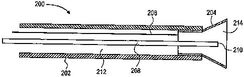
[0043] Figure 7 shows the distal end an electrode tool 200 for use with the
mapping device and method of this
invention. Tool 200 has a body 202 (formed, e.g., from reinforced silicone
tubing with a durometer of
approximately 50) supporting an electrode 204 at its distal end. A lightweight
metal coil may be added to the
electrode tool body to provide sufficient support. Electrode 204 may be formed
from a flared hypotube section. A
wire 206 extends proximally from electrode 204 to the mapping instnunent (not
shown), optionally through a
separate wire lumen. A marking lumen 208 extends proximally from a marking
port 210 to a source of a marking
agent (not shown). An annular suction lumen 212 surrounding marking lumen 208
and marking port 210 extends
proximally from suction port 214 within electrode 204 to a vacuum or suction
source (not shown).
[0044] In use, an endoscope is advanced transgastrically into the abdominal
cavity as described above, and the
electrode too1200 is advanced through a working channel of the endoscope to
place electrode 204 against the
patient's diaphragm at a stimulation site. Visualization from the endoscope
aids in placement. After placing the
electrode, suction is applied through suction lumen 212 to hold the electrode
in place, and a stimulus is applied (e.g.,
stimulus amplitude of 20 mA and pulse duration of 100 s). The magnitude of
the evoked muscle response, visual
confirmation of the contraction, and/or the change in pressure of the
abdominal cavity are noted and recorded. The
location of the stimulation site may then be marked by ejecting a marking
agent (such as gentian violet or india ink)
from marking port 210. Suction is then released, and the electrode is moved to
another stimulation site, where the
procedure is repeated. The response of the diaphragm to stimulation at the
multiple stimulation sites may be
mapped on a.grid overlying the endoscope monitor. The magnitude of the evoked
muscle response and the resultant
change in pressure of the abdominal cavity can then be used to identify the
optimal electrode implant site of each
hemidiaphragm. The optimal site, which is typically the phrenic nerve motor
point of the hemidiaphragm, is chosen
as the site that elicits a diffuse contraction and the greatest magnitude of
pressure change. Using the markings as a
guide, a stimulation electrode is then implanted under endoscopic
visualization at the optimal site in each
7
CA 02631915 2008-06-02
WO 2007/064847 PCT/US2006/045934
hemidiaplu-agm using, e.g., the implant tool described in U.S. Patent No.
5,797,923, or other technique as described
above. [0045] An alternative embodiment of an electrode too1300 is shown in
Figures 8 and 9. Tool 300 has a body 302
(form.ed, e.g., from reinforced silicone tubing with a durometer of
approximately 50) supporting an electrode 304 on
a side wall at its distal end. A wire 306 extends proximally from electrode
204 to the mapping instrument (not
shown), optionally through a wire lumen 307. A marking lumen 308 extends
proximally from a marking port 310 to
a source of a marking agent (not shown). A suction lumen 312 extends
proximally from suction ports 314, 316, and
318 within electrode 304 to a vacuum or suction source (not shown).
[0046] Use of the electrode too1300 of Figure 8 is similar to that of Figure
7. Tool 300 is advanced
transgastrically into the patient's abdominal cavity through an endoscope, and
electrode 304 is placed against the
patient's diaphragm. Suction is applied through suction lumen 312 to hold the
electrode in place, and a stimulus is
applied (e.g., stimulus amplitude of 20 mA and pulse duration of 100 s). The
magnitude of the evoked muscle
response, visual confirmation of the contraction, and/or the change in
pressure of the abdominal cavity are noted and
recorded. The location of the stimulation site is then marked by ejecting a
marking agent such as india ink from
marking port 310. Suction is then released, and the electrode is moved to
another stimulation site, where the
procedure is repeated.
[0047] Yet another embodiment of the electrode tool is shown in Figure 10.
Unlike the earlier embodiments, the
electrode too1400 of Figure 10 lacks a suction port. Electrode tool 400
therefore has a body 402 formed from a
higher durorrieter tubing than the embodiments of Figures 7 and 8 so that the
electrode 404 may be held in place on
the diaphragm without suction. A wire 406 extends proximally from the
electrode to the mapping instrument (not
shown), optionally through a wire lumen. Marking ink may be delivered tlirough
a marking lumen 408 and marking
port410.
[0048] Figure 11 shows a proximal handle for use with an electrode tool of
this invention. Handle 500 extends
proximally from the electrode tool body 502 and may be used to move and
otherwise manipulate the tool from
outside the patient. In addition, handle 500 has one or more actuators for
operating the electrode tool. As shown,
handle 500 has a suction actuator formed as a sliding piston 504 in sealed
communication with the tool's suction
lumen (not shown). Pulling piston 504 proximally (to the left, as shown in the
figure) creates suction in the suction
lumen. Ratchets, catches or other devices may be used to maintain the position
of the piston after actuation. Handle
500 may also have a suction release actuator, such as release button 506 that
releases the suction within the suction
lumen by venting the suction lumen and/or permitting piston 504 to return
toward its unactuated position. Handle
500 may also have a marking actuator, such as an ink reservoir 508 and ink
ejector 510 (such as a plunger or a CO2
charge) communicating with the tool's marking lumen (not shown). Handle 500
may also have an electrical
connector 512 to connect the tool's electrode with a stiinulus source (such as
a surgical stimulator, not shown) as
well as a switch 514 for operating the stimulus source.
8
CA 02631915 2008-06-02
WO 2007/064847 PCT/US2006/045934
Example 1
[0049] Metltods: Pigs were anesthetized and transgastric peritoneal access
with a flexible endoscope was obtained
using a guidewire, needle knife cautery and balloon dilatation. The diaphragm
was mapped to locate the motor point
(where stimulation provides complete contraction of the diaphragm) with an
endoscopic electrostimulation catheter.
An intramuscular electrode was then placed at the motor point with a
percutaneous needle. This was then attached to
the diaphragm pacing system. The gastrotomy was managed with a gastrostomy
tube.
[0050] Results: Four pigs were studied and the diaphragm could be mapped with
the endoscopic mapping
instrument to identify the motor point. In one animal, under trans-gastric
endoscopic visualization a percutaneous
electrode was placed into the motor point and the diaphragm could be paced in
conjunction with mechanical
ventilation.
[0051] Coficlusion: These animal studies support the concept that transgastric
mapping of the diaphragm and
implantation of a percutaneous electrode for therapeutic diaphragmatic
stimulation is feasible.
Exa[nule 2
[0052] Metl:ods: Four female pigs (25kg) were sedated and a single channel
gastroscope was passed
transgastrically into the peritoneal cavity. Pneumoperitoneum was achieved via
a pressure insufflator through a
percutaneous, intraperitoneal 14-gauge catheter. Three other pressures were
recorded via separate catheters. First, a
14-gauge percutaneous catheter passed intraperitoneally measured true intra-
abdominal pressure. The second
transducer was a 14-gauge tube attached to the endoscope used to measure
endoscope tip pressure. The third
pressure transducer was connected to the biopsy channel port of the endoscope.
The abdomen was insufflated to a
range (10-30 mmHg) of pressures, and simultaneous pressures were recorded from
all pressure sensors.
[0053] Results: Pressure correlation curves were developed for all animals
across all intraperitoneal pressures
(mean error -4.25 to -1 mmHg). Endoscope tip pressures correlated with biopsy
channel pressures (R2=0.99).
Biopsy channel and endoscope tip pressures fit a least-squares linear model to
predict actual intra-abdominal
pressure (R=0.99 for both). Both scope tip and biopsy channel port pressures
were strongly correlative with true
intra-abdominal pressures (R2 = 0.98, R2=0.99 respectively).
[0054] Conclusion: This study demonstrates that monitoring pressure through an
endoscope is reliable and
predictive of true intra-abdominal pressure.
[0055] While preferred embod'unents of the present invention have been shown
and described herein, it will be
obvious to thbse skilled in the art that such embodiments are provided by way
of example only. Numerous
variations, changes, and substitutions will now occur to those skilled in the
art without departing from the invention.
It should be understood that various alternatives to the embodiments of the
invention described herein may be
employed in practicing the invention. For example, the electrode tool body may
also be formed from PEEK or
PTFE. Also, otlier transvisceral approaches could be used, such as
transesophageal, transcolonic, transvaginal
approaches.
[0056] It is.intended that the following claims define the scope of the
invention and that methods and structures
within the scope of these claims and their equivalents be covered thereby.
9