Note: Descriptions are shown in the official language in which they were submitted.
CA 02635123 2008-07-22
~
ENDOVASCULARPROSTHESIS HAVING A
LAYER OF BIOLOGICAL TISSUE
Technical Field
The present invention relates to an endovascular
prosthesis and to a method of forming the erdovascular
prosthesis.
Background of the tnvention
Surgical procedures in which acardiovascu3ar
prosthesis is implanted into a patient's biood vessel
are common in treating many vascular =di-soorders. cor
example, one common type of car-diovasc.ular prosthesis
is an endovascular prosthesis that is used to
strengthen a blood vessel wa3.l in the location of an
aneurysm, or to open an occlusion in a bl.ood vessel.
A typical endovascular prosthesis includes a
flexible, tubular member, made of fab_ic or flTF~, that
may be anchored with sutures or carried by onee or more
support structures known as stents. 'Generally, each
stent is formed from a material having an elasticity
CA 02635123 2008-07-22
-2-
sufficient to permit radial expansion of the st-ent an-d
having a strength sufficient to prevent radial collapse
or burst. Such stents are typically formed from
stainless steel, titanium, Nitinol, or a suitable
plastic.
A common endeavor in the field of car-diovas-cular
prosthetics is to increase the patency rate of
prostheses. Thrombosis and platelet deposition on
surfaces of a cardiovascular prosthesis reduce the
patency rate of the prosthesis. For example,
thrombosis and platelet deposition within an
endovascular prosthesis may occlude the conduit defined
by the endovascular prosthesis.
Many factors contribute to thrombosis and platelet
deposition on the surfaces of known cardiovascular
prosthesis. The most common factors are dependent upon
the material or materials forming the inner surface -of
the conduit of the endovascular prosthesis. Typically,
thrombosis and platelet deposition begin to occlude the
conduit of the endovascular prosthesis when the
material or materials forming the conduit of the
endovascular prosthesis are foreign to the patient's
body. A thrombus begins to form on the inner surface
of the conduit of the endovascular prosthesis and
CA 02635123 2008-07-22
-3-
extends annularly about the inner surface of the
conduit. Eventually, the thrombus can sev-erely
restrict blood flow through the conduit defined by the
endovascular prosthesis and, if left untreated, can
completely occlude the conduit.
Additionally, thrombosis and platelet .depwsi.tion
may occur as a result of irregularities on the inner
surface of a cardiovascular prosthesis. The
irregularities may be formed by the structure of a'n
inner stent that is used to support t-he .~.a=rdiovasLula=r
prosthesis, or may be formed by the inner surface of
the flexible member used for the prosthesis.
SnmmarZ of the Invention
The present invention is an apparatus for grafting
of a blood vessel or other portion of the
cardiovascular system. The blood vessel has an inside
surface that defines a:cbnduit for directing blood
flow. The apparatus comprises an expandable support
member having inner and outer surfaces. The outer==
surface of the expandable support member is for
engaging and adhering to the inside surfa-ce of the
blood vessel. A layer of biological tis$ue is attached
to the inner surface of the support member. The layer
of biological tissue has an uninterrupted inwar=dly
CA 02635123 2008-07-22
- 4 -
facing surface for extending confluently with the
inside surface of the blood vessel to provide
resistance to thrombosis and platelet deposition as
blood flows through the conduit.
According to one aspect of the invention, the
layer of biological tissue is selected from the group
consisting of peritoneum, pleura, and pericardium.
In a further aspect of the invention, a graft for
a blood vessel is provided. The blood vessel has an
inside surface that defines a conduit for directing
blood flow. The graft comprises a layer of biological
tissue having an uninterrupted inwardly facing surface
for extending confluently with the inside surface of
the blood vessel to provide resistance to thrombosis
and platelet deposition as blood flows through the
conduit.
According to another aspect of the present
invention, the layer of biological tissue comprises an
inner lining of a serous membrane that is supported by
an outer lining of associated fascia. The outer lining
of associated fascia serves as a structural support for
the inner lining of serous membrane.
CA 02635123 2008-07-22
- 5 -
According to yet another aspect of the present
invention there is provided a method for forming a
graft for insertion in a blood vessel, the blood vessel
having an inside surface that at least partially
defines a conduit for directing blood flow, said method
comprising the steps of:
providing an expandable support member having a
mesh-like structure with inner and outer surfaces, the
outer surface for engaging and adhering to the inside
surface of the blood vessel;
providing a layer of peritoneal tissue comprising
an inner lining of a serous membrane having an
uninterrupted inwardly facing surface for extending
confluently with the inside surface of the blood vessel
to provide resistance to thrombosis and platelet
deposition as blood flows through the conduit;
forming the layer of peritoneal tissue into a
desired shape; and
attaching the layer of peritoneal tissue to the
inner surface of the support member.
CA 02635123 2008-07-22
- 6 -
In yet another aspect of the present invention, a
method for preparing a patch for insertion in a blood
vessel is provided. The blood vessel has an inside
surface that defines a conduit for directing blood
flow. According to the method, a layer of biological
tissue comprising an inner lining of a serous membrane
supported by an outer lining of associated fascia is
harvested. The inner lining of serous membrane has an
uninterrupted inwardly facing surface for extending
confluently with the inside surface of the blood vessel
to provide resistance to thrombosis and platelet
deposition as blood flows through the conduit. The
layer of biological tissue is molded into a desired
shape. The layer of biological tissue is packaged in a
sterile, biological medium and stored within a vacuum-
packed container.
CA 02635123 2008-07-22
- 6a -
Brief Description of the Drawings
The foregoing and other features of the present
invention will become apparent to those skilled in the
art to which the present invention relates upon reading
the following description with reference to the
accompanying drawings, in which:
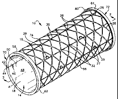
Fig. 1 is a perspective view of an apparatus
constructed in accordance with the present invention;
Fig. 2 is a view along line 2-2 in Fig. 1;
Fig. 3 is a view along line 3-3 in Fig. 2;
Figs. 4a-4f illustrate the method of forming the
apparatus of Fig. 1;
Fig. 5 is a sectional view illustrating the
apparatus of Fig. 1 implanted in a blood vessel;
Fig. 6 is a longitudinal sectional view of a
second embodiment of an apparatus constructed in
accordance with the present invention;
CA 02635123 2008-07-22
-7-
Fig. 7 is a?ongitudinal sectional view -of a third
embodiment of an apparatus constsucted in accordance
with the present invention;
Fig. 8 is a longitudinal sectional view of a
fourth embodiment of an apparatus constructed in
accordance with the present invention;
Fig. 9 is a longitudinal sectional view of a fifth
embodiment of an apparatus const-ructed in at.~-ortlanr.e
with the present invention;
Fig. 10 is a longitudinal sectional view of a
sixth embodiment of an apparatus constructed in
accordance with the present invention;
Fig. 11 is a perspective view of a seventh
embodiment of an apparatus constructed in accordance
with the present invention;
Fig. 12 is a perspective view of an eighth
embodiment of an apparatus constructed in .accordanre
with the presernt invention; and
Fig. 13 is a perspective view of a ninth
embodiment of an apparatus constructed in accordance
with the present invention.
Detailed Description of the Invention
Fig. 1 is a perspective view of an apparatus lfl
constructed in accordance with the present invention.
CA 02635123 2008-07-22
-8-
The apparatus 10 is a cardiovascular graft for grafting
of a blood vessel 12(Fig. 5). The apparatus 1-0
includes a layer of biological tissue 14 and an
expandable support member 16 or stent.
The layer of biologi-cal tissue 14 includes an
inner lining 18 and.an outer lining 2-0 ;Figs. 2 and 3).
The inner lining 18 is a serous membrane and the outer
lining 20 is fascia associated with the serous
membrane. The biological tissue 14 is autogenous
tissue. Alternatively, cadaveric tissue nr xenogeneic
tissue may be used. According to one embodiment, the
layer of biological tissue 14 is harvested from the
peritoneum. Alternatively, the biological tissue may
be harvested from the pericardium or from Lhe pleura.
As an alternative to a layer of biological tissue 14, a
layer of artificial tissue that mimics the
characteristics of peritoneal, pleural, or pericardial
membrane may be used. The artificial tissue may be
constructed from collagen scaffolding that is seeded-
with tissue cells, such as human keratinocytes. The
artificial tissue may also include a basement membrane.
The basement membrane may be a fascia lining or another
known artificial lining.
CA 02635123 2008-07-22
-9-
.The biological tissue 14 is harvested in sheets of
appropriate size. Conventional techniques are used for
harvesting the biological tissue"14. The sheet.nf
biological tissue 14 is fixed or preserved with
" alcohoT, glutaraldehyde, and/or another bi.ological
solution. After being fixed, the biological tissue 14.
is trimmed or cut into the -desired shape and size. it
is noted that the biological tissue 14 may shrink
slightly when fixed. Thus, the biological tissue 14
should be fixed prior to being trimrned to'the -desired
shape and size. Preferably, the-biologi-cal tissue 14
is trimmed into a rectangular shape. After being -
trimmed, the biological tissue may be bathed in t-hp
biological solution.
The expandable support member 16 is t3ibular and
extends axially from a first end 22 Mg. 2) to a
second end 24. The expandable support member 1fi
illustrated in Fig. 1 is a mesh structure that includes
a plurality of support beams. 2=6 and a plurality of
axially extending support rods 27.
Each support beam=26 has a generally sinusoi=dal
shape. The wavelength of each of the support beams 26
is identical or nearly identical to the wavelength of
adjacent support beams. Circumferentiallv adjacent
CA 02635123 2008-07-22
-ifl-
support beams 26 are 190 out of phase from one another.
Connector bars 28 (Fig. 1) connect the peaks 30 of each
support beam 26 to the associated troughs 32 (Fig. 1)
of the adjacent support beam. The amplitude (or
height) of each support beam 26 is designed so that a
whole number of support beams forms.the circumference
of the expandable support member 16.
Each of the axially extending support rods 27
extends parallel to axis A. The support rods 27 add
additional support to the expandable support member 16.
One embodiment of the apparatus 10 includes eight
support rods 27 that are equally spaced about the
circumference of the expandable sup,port member 16. In
the embodiment illustrated in Fig. 1, two support beams
26 are located between adjacent support rods 27.
The expandable support member 16 also includes a
plurality of eyelets 29, four of which are shown in
Fig. 1. Each eyelet 29 extends from one of the support
rods 27. The eyelets 29 illustrated in Fig. 1 are
circular, however other shapes may be used. The
eyelets 29 provide a means for suturing the layer of
biological tissue 14 to the outer support member 16.
The expandable support member 16 is formed from an
expandable metal, such as Nitinol. Alternatively, the
CA 02635123 2008-07-22
-11-
expandable support may be formed from-a fabric layer
such as Dacron or a plastic material such as
polytetraflouroethylene (PTFE).
The expandable support member 16 includes an outer
surface 34 and an inner surface 36 (Fig. 2). The outer
surface 34 is generally cylindrical and extends',axially
along axis A. The inner surface 3~6 is also genErally
cylindrical and is coaxial with the outer surface 34.-
Alternatively, the expandable support member 36
may include any known stent structure that is
expandable and that defines inner and out-er surfaces 36
and 34, respectively. Although the apparatus 10 is
illustrated as being -cylindrical with acircul:ar cross-
sectional shape, the cross-sectional shape of the
apparatus may alternatively be elliptical, polygonal,
or cone-shaped.
Figs. 4a-4f illustrate a meth-od for forming the
apparatus 10 of the present invention. The method
begins at Fig. 4a with a dowel 38 and a sheet of
biological tissue 14 that has been fixed and trimrned
into a rectangular shape. The dowe? 38 is formed from
glass. The dowel 38 illustrated in F3g. 4a is
cylindrical and has an outer surface 40 with a circular
cross-sectional shape. Alternatively, the dowel 38 may
CA 02635123 2008-07-22
-12-
be cone-shaped. A circumference of the outer surface
40 of the dowel 38 is equal to a width of the
biological tissue 14. The width of the biological
tissue 14 is defined as the distance between a first
side surface 42 and'a second side su-rface 44. Fig. 4a
illustrates the biological tissue 14 being wrapped or
rolled around the dowel 38.
Fig. 4b illustrates the bi43ogical tissue.14
completely wrapped around the dowel 38.. When
completely wrapped around the dowel 38, the first side
surface 42 of the biological tissue 14 abuts, rather
than overlaps, the second side surface 44 of the
biological tissue 14. An axially-extending seam 46 is
defined at the location where th.e first side surface 42
and the second side surface 44 meet. The sQam 46.
extends along an axial length of thie biological tissue
14. The axial length of the biological tissue 14 is
defined as a distance between a first axial end 58 and
a second axial end 60.
The first side surface 42 abuts the second side
surface 44 such that the inner surface 48 (Figs. 1-3)
of the apparatus 10, which is defined by an inner
surface 50 (Figs. 1-3) of the inner lining 18 of the
biological tissue 14, is smooth, continuous, and
CA 02635123 2008-07-22
-13-
uninterrupted. Since the inner surfa-ce 48 of th.e
apparatus 10 has no proj-ections or irregularities, such
as would be present if the biological tissue-14 were
overlapped, thrombosis and platelet deposition at the
seam 46 are resisted. An additional benefit of
abutting the first and second si-de surfaces 42 and. 44
of the biological tissue 14 together is that the
smooth, continuous, and uninterrupted inner surface 48
of the apparatus 10 does not create turbulent flow
through the apparatus.
In Fig. 4c, the first side surface 42 of the
biological tissue 14 is attached to the second sitle
surface 44 of the biological tissue 14 using sutur.es
52. The sutures 52 extend radially inwardly tbtrough
the biological tissue 14 and generally
circumferentially between areas adjacent the first an3
second side surfaces 42 and 44. The biological tissue
14 remains on the dowel 38 while the sutures 52 are
sewn in place. A layer of biological glue 54 may be
placed over the seam 46 on an outer surface St..of the
biological tissue 14. The biological glue 54 helps to
.ensure that the inner surface 48 of the apparatus 1-0
remains smooth, continuous, and uninterrupted. The
CA 02635123 2008-07-22
-14-
biological glue 54 also aids in completely sealing the
seam 46 to prevent any leakage through the seam 46.
Fig. 4d illustrates the expandable support member
16 being placed over the biological tissue 14. The
expandable support member 16 forms an outer support far
the biological tissue 14. The expandable support
member 16 forms the radially outermost component of the
apparatus 10. The radially innermost component of the
apparatus 10 is formed by the serous membrane lining 18
of the layer of biological tissue 14.
To place the expandable support member 16 over the
biological tissue 14, the expandable support member 16
is expanded. Any known method for expanding the
expandable support member 16 may be used, such as
heating or balloon dilation of the expandable support
member. The dowel 38 and the biological tissue 14 that
is being held on the dowel 38 are inserted into the
first end 22 of the expandable support member 16, as
shown in Fig. 4d. The expandable support member 16 and
the dowel 38 are moved relative to one another until an
equivalent amount of biological tissue 14 extends
axially outwardly of both the first and second ends 22
and 24 of the expandable support member 16.
CA 02635123 2008-07-22
-15-
The expandable support member 16 is then
constricted until the inner surface 36 of the
expandable support member 16 engages the outer surface
56 of,the biological tissue 14 equally ab-out the
circumference of the outeL surface 56 of the biological
tissue 14. Next, the biological tis-sue 14 is attached
to the expandable support member 116. Pr=eferably,
sutures (not shown) arQ used to attazh the biolo-gi,cal
tissue 14 to the expandable support member 16. Each
suture extends through the biological tissue 14 and a
portion of the suture is threadesl through one of the
eyelets 29 of the expandable support member 1-6. The
suture is then tied outside of the expandable support
member 16 and around the respective eyelet 29. The
suture holds the biological tissue 14 to the inner
surface 36 of the expandable support member 16. The
sutures are sufficiently small so that turbulent flow
will not result from the int-eraction of -bl+ood flow with
the sutures. Alternately, the ou-ter surfa:ce 56 of the
biological tissue 14 may be glued to the inner surface
36 of the expandable support member 16 using biological
glue. When biological glue is used to attach the
biological tissue 14 to the expandable support member
16, the support beams 26 and the support rods 27 must
CA 02635123 2008-07-22
-16-
have an inner surface area =large=enough for adhesion of
the biological tissue 14.
After the biological tissue 14 is attached to the
expandable support member 16, the first and second
axial ends 58 and 60 of the biological tissue 14 are
folded over the first and second ends 22 and'24,
respectively, of the expandable support member 16, as
is shown in Fig. 4e. The first axial end 58 of the
biological tissue 14 is stretched and folded over the
first end 22 of the expandable support member 1-6 to
form a first=folded portion 62. The first folded
portion 62 is then attached to the outer surface 34 of
the expandable support member 16 using sutures lnot
shown). A second axial end 60 of the biological tissue
14 is stretched and folded over the second end 24 of
the expandable support member 16 to form, a second
folded portion 64. The second folded portion 64 is
also attached to the expandable support member 16 using
sutures (not shown).
The apparatus 10, including the dowel 38, is
stored in a sterile environment until it is time for
implantation into a patient. Preferably, the apparatus
10 is submersed in a biological solution and is stored
in a sterile, vacuum-packed container (not shown).
CA 02635123 2008-07-22
-17-
Alternatively, the dowel 38 may be removed from the
apparatus 10 prior to storing the apparatus. Fig. 4f
illustrates the dowel 38 being removed from the
apparatus 10. Preferably, the clowel 38 and the
apparatus 10 are placed in biological or f ixing
solution to facilitate removal of the dow:e l 38 from
inside the apparatus 10. The solution will
sufficiently lubricate the dowel 38 and the biological
tissue 14 so that the dowel may be removed from the
apparatus 10 without tearing or weakening the
biological tissue 14. As a result, the i-nrier surface
48 of the apparatus 10 remains smooth, continuous, and
uninterrupted. Alternatively, the apparatus 10 may be
expanded and the dowel 38 =rAmoved from the expanded
apparatus 10.
Fig. 5 illustrates the apparatus 10 of the present
invention implanted in abiood vessel 12.. The blood
vessel 12 includes an outside surface fi6 and an insi-de
surface 68. The inside surface S8 of the =blood vessel
12 forms a conduit for directing blood flow. The
apparatus 10 is delivered and positioned in the blood
vessel 12 using methods that are known in the art.
Once the apparatus 10 is positioned in the -desired
location in the blood vessel 12, the -expandable support
CA 02635123 2008-07-22
-18-
member 16 is expanded, by a balloon (not shown) or
through self-expansion as is known in theart. When
the expandable support member 16 expands, a first end
70 of the apparatus 10 engages the blood vessel 12 such
that an interference fit is created between the~first
folded portion 62 and the inside surface 68 of the
blood vessel 12. Similarly, a second end 72 of the
appara-tus 10 engages the blood vessel 12 such that an
interference fit is created between the second folded
portion 64 and the inside surface 68-of the-blood
vessel 12. An interference fit is also created between
the expandable support member 16 and the inner surface
68 of the blood vessel 12 along the axial length of the
apparatus 10 that extends between the first and second
ends 70 and 72. In addition to the interference fit
between the expandable support member 16 and the blood
vessel 12, sutures can also used to anchor the
expandable support member 16 to the blood vessel 12.
When the apparatus 10 engages and adheres to the
inside surface 68 of the,blood vessel 12 in the above
manner, the inner lining 18 of serous membrane forms
the outermost surface at the first and second folded
portions 62 and 64. The inner lining 18 bonds to the
inside surface 68 of the blood vessel 12 in a normal
CA 02635123 2008-07-22
-19-
tissue-healing fashion and prevents the ingrowth of
inflammatory tissue. As a result, the bond between the
serous membrane of the inner lining 18 at the first and
second folded portions 62 and 64 and the inside surface
68 of the blood vessel 12 prevents restenosis or
occlusion. Additionally,, the healing bond between 'the
serous membrane of the inner lining 18 at the first and
second folded portions 62 and 64 and the inside surface
68 of the blood vessel 12 forms more quickly 'than a
bond between the fascia lining 20 and the insi-4e
surface 68 of the blood vessel 12.
When implanted in the blood vessel 12, the conduit
formed by the inner surface 50 of the biological tissue
14 is confluent with the inside su-rface 68 of the blood
vessel 12. The transition between the inside surface
68 of the blood vessel 12 and the -inner surface 50 of
the biological tissue 14 is smooth so that thrombosis
and platelet deposition is resisted and that blood flow
is not restricted when passing through the.apparatus
10. The expandable support member 1~6 provic3es
sufficient support against the internal pressure cau-sed
by the blood flow through the apparatus 10, and also
resists radial collapse of the blood vessel.
CA 02635123 2008-07-22
-2,0-
Fig. 6 is a longitudinal sectional view of a
second embodiment of an apparatus l0a constructed in
accordance with the present invention. Structures of
the embodiment shown in Fig. 6 that are similar to
structures of Figs. 1-3 have the same reference numbers
with the suffix "a" added. The apparatus 10a is
identical to apparatus 10 of Figs. 1-3 with the
exception that the layer of biological tissue 14a in
the embodiment of Fig. 6 includes only a layer 18a of
serous membrane.
The layer of biological tissue 14a is harvested to
include only the layer 18a of serous membrane. The
method for harvesting only a layer 18a of serous
membrane is known in the art
The assembly of apparatus 10a is identical to the
assembly of apparatus 10 that is illustrated in Figs.
4a-4f. When trimmed into the desired shape., the layer
of biological tissue 14a includes first and second side
surfaces 42a and 44a, respectively, and first and
2.0 second axial ends 58a and 60a, respectively.
The assembled apparatus includes a seam 46a that
is formed from abutting the first and second side
surfaces 42a and 44a. The assembled apparatus 1Oa also
includes first and second folded portions 62a and 64a.
CA 02635123 2008-07-22
-21-
The first folded portion E2a is fo-rmed by folding the
first axial end 58a of the layer of biological tissu.e
14a ove.r the first end 22a of the expandable support
member 16a. The second folded portion 64a is formed by
folding the second axial end 60a of the Iayer.of
biological tissue 14a over the second -end 24a of the
expandable support member 16a.
The inner surface 48a of the assembl-ed apparatus
l0a is defined by the inner surface 50a of the layer
18a of serous membrane. The inner surfa-ce 148a of the
apparatus l0a is smooth, continuous, and uninterrupted.
The smooth, continuous, and uninterrupted inner surface
48a of the apparatus 10a resists thrombosis and
platelet deposition.
Fig. 7 is a longitudinal sectional view of an
apparatus lOb constructed in accordance wit-h a tbir-d
embodiment of the present invention. Structures vf the
embodiment shown- in Fig. 7 that are similar to
structures of Figs. 1-3 have the same re-ference numbers
with the suffix "b" added.
The apparatus lOb illustrated in Fig. 7 i-n-cludes a
layer of biological tissue 14b and an ,expandabie
support member 16b. The layer of biological tis-sue.34b
includes a serous membrane lining 18b and associated
CA 02635123 2008-07-22
-22-
fascia lining 20b. The expandable support member 16b
has a structure similar to that illustrated in Fig. 1.
The layer of biological tissue 14b forms the innermost
component of the apparatus 10b.
The layer is biological tissue 14b is formed into
a tubular portion by abutting first and second sidQ
surfaces 42b and 44b of the biological tissue 14b at a
seam 46b:= Preferably, the first and second side
surfaces 42b and 44b are sutured together at the seam
46b and biological glue (not shown) is applied to an
outer surface 56b of the biological tissue 14b.
The outer surface 56b of the layer of biological
tissue 14b is attached to the inner surfa=ce 36b of the
expandable support member 1~6b. The expandable support
member 16b is placed over the biological tissue 14b
such that equal amounts of biological tissue 14b extend
from the first and second ends 22b and 24b of the
expandable support member 16b. Instead of folding the
first and second axial ends 58b and 60b of the
biological tissue 14b over the expandable support
member 16b as discussed above with regard to the
embodiment of Figs. 1-3, the first and second axial
ends 58b and, 60b of the biological tissue 14b extend
axially beyond the first and second ends 22b and 24b of
CA 02635123 2008-07-22
-23-
the expandable support member 16b. Thus, in assembling
the apparatus lOb, the step illustrated in Fig. 4e is
omitted.
When implanted into a blood vessel of a patient,
the first and second axial ends 58b and 50b of the
tissue .14b engage and. are adhered to the inside- surface
of the blood vessel by the expansion of the expandable
support member 16. The extension of the first and
second axial ends 58b and 60b of the biological tissue
14b axially beyond the first and sez:ond.ends 22b and
24b of the expandable support member 16b allows the
first and second axial ends of thebiological tissue to
be sutured directly to the inside surfaLe -of the blood
vessel.
Fig. 8 is a longitudinal sectional view of a
fourth embodiment of an apparatus lOc constructed in
accordance with the present invention. Structures of
the embodiment shown in Fig. 8 that are similar to
structures of Fig. 7 have the same reference numbers
with the suffix "c" replacing the suffix "b". The
apparatus 10c is identical to apparatus lOb af Fig. 7
with the exception that the layer of biological tissue
14c in the embodiment of Fig. 8 includes only a layer
18c of serous membrane.
CA 02635123 2008-07-22
-24-
The assembly of apparatus lOc is identical to th.e
assembly of apparatus 10b. When trimmed into the
desired shape, the layer of biological tissue 14c
includes first and second side surfaces 42c a-rid 44c,
respectively, and first and second axial ends 58c and
60c, respectively.
The assembled apparatus includes a seam 4:6c that
is formed from abutting the first and second side
surfaces 42c and 44c. The inner surface 48c of the
assembled apparatus 10c is defined by the inner surface
50c of the layer 18c of serous membrane. The inner
surface 48c of the apparatus 10c is smooth, continuous,
and uninterrupted. The smooth, continuous, and
uninterrupted inner surface 4.8c of the apparatus 10-c
resists thrombasis and platelet deposition.
Fig. 9 illustrates a longitudinal sectional view
of a fifth embodiment of an apparatus 10-d constructed
in accordance with the present inv.ention. Structures
of the embodiment shown in Fig. 9 that are simi-lar to
structures of Fig. 7 have the same referenr.e numbers
with the suffix "d" replacing the suffix "b".
The apparatus lOd of Fig. 9 is also a
cardiovascular graft. The apparatus lfld includes a
layer of biological tissue 14d that includes an inner
CA 02635123 2008-07-22
-25-
lining 18d of serous membrane and an outer lining 2{0d
of fascia associated with the serous membrane. The
layer ofbiological tissue 14d is rectangular and
includes first and second side surfa-ces 42d and 44d,
5- respectively, and first -and sec=ond axial ejnds 5$d and
.
60d, respectively. The inner lining 18d of =serrous'
membrane includes an inner surface 50d. The outer
lining 20d of fascia includ.es an outer surface 5fid.
The apparatus lUd illustrated in fi.g. 9 is .
cylindrical and is formed by the layer of -biological
tissue 14d. The first and second side surfaces 42d and
44d of the layer of =bioiugical tissue 14d are abutted
and secured together to define a seaan=96d. Sutures 52d
attach the first and second side surfaces 42d and 44-d
at the seam 46d. A layer of biological glue (cot
shown) is applied to the outer surface 56d of the outoer
lining 20d over the seam 46d. The biological glue ai=ds
in completely sealing the seam 46d to prev~ent any
leakage through the seam.
To form the apparatus lOd, the steps illustrated
in Figs. 4a to 4c and discussed in detail with regards
to apparatus 10 of Figs. 1-3 are followed. . After the =
step shown in. Fig. 4c, the apparatus 10d is stored in a
sterile environment until it is time for implantation
CA 02635123 2008-07-22
-2fi-
into a patient. Prior to implantation into the
patient, the dowel is removed from the apparatus.
The outer surface 56d of the outer lining 20d
forms the outermost component of the apparatus lOd.
The inner surface 50d of the inner lining 18d of serous
membrane forms the innermost component ,of the apparatus
lOd. The inner surface 540d of the inner lining 16d is
smooth, continuous, and uninterrupted. As a result,
the inner surface 48d of the apparatus lod is smooth,
continuous, and uninterrupted and resists thrombosis
and platelet deposition.
When surgically implante.d in a patient, the
apparatus lOd is attached using suturres. For example,
when used within a blood vessel, the apparatus 10d is
sutured to the'inside surface of the blood vessel. As
a result, the continuous and uninterrupted inner
surface 50d of the inner lining 1Bd is confluent with
the inside surface of the blood vessel.
Since the apparatus lOd includes no support
structures, the apparatus adapts or conforms to the
shape=of the blood vessel into which it is attached.
Thus, if the inside surface of the blood vessel has an
elliptical cross-sectional shape, the apparatus 1.0d,
CA 02635123 2008-07-22
-27-
when attached to the. inside surface of the blood
vessel, has an elliptical cross-sectional 'shape.
Fig. 10 is a longitudinal sectional view of a
sixth embodiment of an apparatus 10e constructed in
accordance with the present invention. Structures of
the embodiment shown in Fig: 10 that arae similar to
structures of Fig. 9 have the same r.eferen.ce numbers
with the suffix "e" replacing the suffix "d'=. The
apparatus 10e is identical to apparatus 10,d of Fig. 9
with the exception that the layer of biological tissue
14e in the embodiment of Fig. 1D includes only a layer
18e of serous membrane.
The assembly of apparatus 14e is identical t.o the
assembly of apparatus 10-e. When -t-rim,~ned into the
desired shape, the layer of bioiogi4cal tissue 14e
includes first and second side surfaces 42e and 44e,
respectively, and first and second axial ends 58e and
60e, respectively.
The assembled apparatus includes a seam 46e t7hat
is formed from abutting the first and second side
surfaces 42e and 44e. The inner surface 48e of th.e
assembled apparatus 10e is defined by the inner surface
50e of the layer 18e of serous membrane. The inner
surface 48e of the apparatus 10e is smooth, continuous,
CA 02635123 2008-07-22
-28-
and uninterrupted. The smooth, continuous, and
uninterrupted inner surface 48e of the apparatus 10e
resists thrombosis and platelet deposition.
Fig. 11 illustrates a perspective view of a
seventh embodiment of an apparatus 100 constructed in
accordance with the present invention. The apparatus
100 in Fig. 11 is a patch for r-epairing a grorti.on of a
blood vessel or other membrane within the
cardiovascular system of the human body.
The patch 100 includes a layer of biological
tissue 102 and an outer support member 104. The layer
of biological tissue 102 includes a serous membrane
lining 106 and associated fascia lining 108. The
serous membrane lining 106 forms an inner surfa.ce (not
shown) of the biological tissue 102 and the associated
fascia 108 forms an outer surface 110 of the biological
tissue 102. The layer of biological tissue 102 is
illustrated as being rectangular but may be of any
desired shape.
The outer support member 104 has the same shape as
the biological tissue 102 but is s'Lightly smaller is
size. The.outer support member 104 may have a curved
profile, as is illustrated in Fig. 11, for fitting to a
CA 02635123 2008-07-22
-29-
curved surface such as the inside or-outsi-de surfaces
of a blood vessel.
The outer support member 104 in Fig. -11-is
rectangular and includes an outer frame 112 and inner
support beams 114. The outer frame 112 defines the
shape of.the outer support member 104 and prova~d.es
support near the periphery of the biological tissue
102. The inner support beams 114 of the outer support
member 104 provide support for an interior portion of
the biol-ogical tissue 102. Eyelets 118 are provi=ded
through which sutures (not shown) may be tbreaded when
attaching the biological tissue 102 t-o the outer
support member 104.
The outer surface 110 of the biological tissue 102
is attached to the outer support member 104.
Preferably, the biological tissue 102 is suture.d to the
outer support member 104. The peripheral portion -of
the biological tissue 102 extends outwardly from the
outer support member 104. Alternatively, the
peripheral portion of the biological ti-ssue 102-may be
folded over the outer frame 112 of the outer-support
member 104.
When implanted in a blood vessel, an outer surface
116 of the outer support member 104 of the patch 1fl0 is
CA 02635123 2008-07-22
-30-
placed over an aneurysm or a weakened portion of the
blood vessel. The size of the outer support member 104
is preferably larger than the aneurysm or weakened
portion of the blood vessel such that the outer frame
112 of the outer support member 104 contacts bealthy
portions of the inside surface of the binod vessel.
The outer periphery of the biological tissue 102 is-
then attached to the inside surface of the blood
vessel, preferably by suturing. The patch 100 may
alternatively be placed over the-outside surface of the
blood vessel or be used on another membrane of the
cardiovascular system.
Fig. 12 is a view of an eighth embodiment of an
apparatus 100a constructed in accordance with the
present invention. Structures of the embodiment shown
in Fig. 12 that are similar to structures of Fig. 11
have the same reference numbers with the suffix "a"
added.
The apparatus 100a of Fig. 12 is also a patcb for
repairing a portion of a blood vessel or other membrane
within the cardiovascular system of the human body.
The patch 100a includes a layer of biological tissue
102a. The patch 100a of Fig. 12 does not include a
CA 02635123 2008-07-22
-31-
support structure such as the outer support structure
104 illustrated in Fig. 11.
The layer of biological tissue 102a includes a
serous membrane lining 106a and associated fascia
lining 108a. The serous membrane lining 106a forms an
inner surface {not shown) -of the biological tissue 102a
and the associated fascia 108a forms an outer surface
110a of the biological tissue 102a. The innier surface
of the biological tissue 102a is smooth, continuaus,
and uninterrupted. The layer of b3ologi:al tissue 1.02a
is illustrated as being rectanguiar-but may be of any
desired shape.
When implanted in a blood vessel, an oute=r surface
110a of the associated fascia 108a of the layer of
biological tissue 102a is placed over an a-neurysm or a
weakened portion of the blood vessel. The biological
tissue 102a is then attached to the inside surface =of
the blood vessel, preferably by suturin=g. Since the
patch 100a does not include structural support, the
patch 100a easily adapts to the shape of the blood
vessel or membrane to which it is attached to ensure a
sufficient area of contact between patch 100a and the
blood vessel or membrane. The patch 1O0a may
alternatively be placed over the outside surface of the
CA 02635123 2008-07-22
-32-
blood vessel or be used on another membrane of the
cardiovascular system.
Fig. 13 is a perspective view of a ninth
embodiment of an apparatus 100b constructed in
accordance with the present invention. Structur,es of
the embodiment shown in Fig. 13 that are similar to
structures of Fig. 12 have the same reference numbers
with the suffix "b" replacing the suffix "a".. The
apparatus 100b is identical to apparatus 100a of Fig.
12 with the exception that the layer of biological
tissue 102b in the embodiment of Fig. 13 includes only
a layer 106b of serous membrane.
The outer surface 110b of the aiol.ogioai tissue
102b is formed by an outer surface of the layer 106b of
serous membrane. The inner surf.a-ce (not shown) of the
biological tissue is formed by an inner surface of the
layer 106b of serous membrane and is smooth, continuous
and uninterrupted.
From the above description of the invention, those
skilled in the art will perceive improvements, changes.
and modifications. For example, a layer of artificial
tissue, which mimics the characte.ristics of the layer
of biological tissue, may be used in any of the
embodiments discussed above. Such improvements,
CA 02635123 2008-07-22
-33-
changes and modifications within the skill af the art
are intended to be covered by the appended claims.