Note: Descriptions are shown in the official language in which they were submitted.
CA 02636277 2008-07-04
WO 2007/081896
PCT/US2007/000433
1
PHARMACEUTICAL COMPOSITIONS AND METHODS TO VACCINATE
AGAINST DISSEMINATED CANDIDIASIS AND OTHER INFECTIOUS AGENTS
BACKGROUND OF THE INVENTION
This invention relates to Candida albicans surface adhesin proteins, to
antibodies
resulting from an immune response to vaccination with C. albicans surface
adhesion proteins
and to methods for the prevention and/or treatment of candidiasis and other
bacterial
infections with C. albicans surface adhesion proteins.
There has been a dramatic increase in the incidence of nosocomial infections
caused
by Candicla species in recent years. The incidence of hematogenously
disseminated candidal
infections increased 11-fold from 1980 to 1989. This increasing incidence has
continued into
the 1990s. Infections by Candida species are now the fourth most common cause
of
nosocomial septicemia, are equal to that of Escherichia coil, and surpass the
incidence caused
by Klebsiella species. Furthermore Candida species are the most common cause
of deep-
seated fungal infections in patients who have extensive bums. Up to 11% of
individuals
undergoing bone marrow transplantation and 13% of those having an orthotopic
liver
transplant will develop an invasive candidal infection.
Candida albicans, the major pathogen in this genus, can switch between two
morphologies: the blastospore (budding yeast) and filamentous (hyphae and
pseudohyphae)
phases. Candicla mutants that are defective in genes regulating filamentation
are reported to
have reduced virulence in animal models. This reduced virulence suggests that
the ability to
change from a blastospore to a filament is a key virulence factor of C.
albicans. To date, no
essential effectors of these filamentation pathways have been identified in C.
albicans. See
Caesar-TonThat, T.C. and J.E: Cutler, "A monoclonal antibody to Candicla
albicans
enhances mouse neutrophil candidacidal activity," Infect. Immun. 65:5354-5357,
1997.
Staphylococcus aureus infections also are common and increasingly result in
drug
resistance to antibiotics. For example, S. aureus is a common cause of skin
and skin structure
infections, endocarditis and bacteremia in the U.S. and throughout the world.
Formerly
community acquired S. aureus (CA-S. aureus) infections were nearly uniformly
susceptible
to penicillinase-resistant beta lactams such as cefazolin, oxacillin,
methicillin, penicillin and
amoxicillin. However, over the past decade, epidemics of beta-lactam resistant
S. aureus
(MRSA) infection have been seen in multiple locales throughout the world,
especially
CA 02636277 2008-07-04
WO 2007/081896 PCT/US2007/000433
2
community acquired MRSA (CA-MRSA). In many places MRSA has become the
predominant S. aztreus strain causing CA infections. A recent, prospective,
population-based
survey in three states in the U.S. estimated that the incidence of CA-MRSA
infections is 500
cases per 100,000 population, which translates to approximately 1.5 million
cases per year in =
the U.S. alone. The increasing frequency of drug-resistant S. aureirs
infections highlights the
need for new ways to prevent and treat these infections.
The identification of effectors in the regulatory pathways of the organism
that
contribute to virulence offers the opportunity for therapeutic intervention
with methods or
compositions that are superior to existing antifungal agents. The
identification of cell surface
proteins that affect a regulatory pathway involved in virulence is
particularly promising
because characterization of the protein enable immunotherapeutic techniques
that are superior
to existing antifungal agents when fighting a candidal infection.
The virulence of Candida albicans is.reg'ulated by several putative virulence
factors
of which adherence to host constituents and the ability to transform from
yeast-to-hyphae are
among the thost critical in determining pathogenicity. While potent antifungal
agents exist
that are microbicidal for Candida, the attributable mortality of candidemia is
approximately
386/0, even with treatment with potent anti-fungal agents such as amphotericin
B. Also,
existing agents such as amphotericin B tend to exhibit undesirable toxicity.
Although.
additional antifungals may be developed that are less toxic than amphotericin
B, it is unlikely
that agents will be developed that are more potent. Therefore, either passive
or active
immunotherapy to treat or prevent disseminated candidiasis is a promising
alternative to
standard antifungal therapy.
Thus, there exists a need for effective immunogens that will provide host
immune
protection and passive immunoprotection against Candida, S. aztreus and other
immunogenically related pathogens. The present invention satisfies this need
and provides
related advantages as well.
SUMMARY OF THE INVENTION
The invention provides a vaccine including an isolated Als protein family
member
having cell adhesion activity, or an immunogenic fragment thereof, with an
adjuvant in a
pharmaceutically acceptable medium. The invention also provides a method of
treating or
preventing disseminated candidiasis. The method includes administering an
immunogenic
CA 02636277 2016-11-10
CA 2636277
3
amount of a vaccine an isolated Als protein family member having cell adhesion
activity, or an
immunogenic fragment thereof, in a pharmaceutically acceptable medium. A
method of treating or
preventing disseminated candidiasis also is provided that includes
administering an effective amount of
an isolated Als protein family member having cell adhesion activity, or an
functional fragment thereof, to
inhibit the binding or invasion of Candida to a host cell or tissue. The Als
protein family member can be
derived from a Candida strain selected from the group consisting of Candida
albicans, Candida krusei,
Candida tropicalis, Candida glabrata and Candida parapsilosis and the Als
protein family member
includes Alsip, Als3p, Als5p, Als6p, Als7p or Als9p. Also provided is a
vaccine comprising an isolated
Als protein family member having cell adhesion activity, or an immunogenic
fragment thereof, for use in
treatment or prevention of vaginal candidiasis in a human or animal body. Also
provided is a method of
treating or preventing Staphylococcus aureus infections. The method includes
administering an
immunogenic amount of a vaccine an isolated Als protein family member having
cell adhesion activity,
or an immunogenic fragment thereof, in a pharmaceutically acceptable medium.
The claimed invention relates to a vaccine comprising an isolated Als protein
family member of
candidal origin having cell adhesion activity, or an immunogenic fragment
thereof, for use in treatment
or prevention of a Staphylococcus aureus infection in a human or animal body.
The claimed invention also relates to an isolated Als protein family member of
candidal origin
having cell adhesion activity, or an immunogenic fragment thereof, for use in
treatment or prevention of
a Staphylococcus aureus infection in a human or animal body.
The claimed invention also relates to an isolated Als protein family member of
candidal origin
having cell adhesion activity, or an immunogenic fragment thereof, for use in
preparation of a
medicament for treatment or prevention of a Staphylococcus aureus infection in
a human or animal
body.
CA 02636277 2013-06-04
CA 2636277
3a
BRIEF DESCRIPTION OF THE DRAWINGS
Figure 1A, 1B show the mediation of Alsip adherence of C. albicans to human
umbilical vein
endothelial cells. Values represent the mean SD of at least three
independent experiments, each
performed in triplicate. (A) Endothelial cell adherence of ALS1I/als2
als12/als1 and ALS/-complemented
mutants and wild-type CAI12(30((B) Endothelial cell adherence of PADH1-ALS/
mutant that overexpresses
ALSI, compared to wild type C. albicans. Statistical treatment was obtained by
Wilcoxon ran sum test and
corrected for multiple comparisons with the Bonferroni correction. *P<0.001
for all comparisons.
Figure 2A-D shows the cell surface localization of Als1P on filaments of C.
albicans indirect
immunofluorescence. Filamentation of C. albicans was induced by incubating
yeast cells in RPM( 1640
medium with glutamine for 1.5 hours at 37 C. Alsip was detected by incubating
organisms first with anti-
Alsip mouse mAb followed by FITC-labeled goat anti-mouse IgG. C. albicans cell
surface was also
stained with anti-C. albicans polyclonal Ab conjugated with Alexa 594
(Molecular Probes, Eugene, OR).
Areas with yellow staining represent Alsip localization. (A) C. albicans wild-
type. (B) als1/als1 mutant
strain. (C) als1/als1 complemented with wild type ALS1 (D) PADH1-ALS1
overexpression mutant.
Figure 3A, 3B show the mediation of Alsip on C. albicans filamentation on
solid medium. C.
albicans blastospores were spotted on Lee's agar plates and incubated at 37 C
for 4 days (A) or 3 days
(B).
CA 02636277 2008-07-04
WO 2007/081896 PCT/US2007/000433
4
Figure 4A, 4B show the control of ALS/ expression and the mediation of C.
alb/cans
filamentation by the EFGI filamentation regulatory pathway. (A) Northern blot
analysis
showing expression of ALS/ in (i) mutants deficient in different filamentation
regulatory
pathways. (ii) efgl/efgl mutant complemented with either EFGI or PADHI-ALS1.
Total
RNA was extracted from cells grown in RPM I 1640 + glutaine medium at 37 C for
90
minutes to induce filamentation. Blots were probed with ALS] and TEFI . (B)
Photomicrographs of the efgl/efgl mutant and efgl/efgl mutant complemented
with PADHI-
ALS1 grown on Lee's agar plates at 37 C. for 4 days.
Figure 5A, 5B show the reduction of virulence in the mouse model of
hematogenously disseminated candidiasis by (A) Male Balb/C mice (n = 30 for
each yeast
strain) were injected with stationary phase blastospores (106 per mouse in 0.5
ml of PBS).
Curves are the compiled results of three replicate experiments (n = 30 mice
for each strain).
The doubling times of all strains, grown in YPD at 30 C, ranged between 1.29
to 1.52 hours
and were not statistically different from each other. Southern blot analysis
of total
chromosomal DNA was used to match the identity of the genotype of C. albicans
strains
retrieved from infected organs with those of C. albicans strains used to
infect the mice.
Statistical analysis was obtained by Wilcoxon rank sum test and corrected for
multiple
comparisons with the Bonferroni correction. *P<0.002 for the alsl/als1 mutant
versus each
of the other strains. (B) Histological micrographs of kidneys infected with C.
a/a/can's wild-
type, homozygous alsl null mutant, or heterozygous ALS] complemented mutant.
Kidney
samples were retrieved 28 hours (a) or 40 (b) hours post infection, fixed in
paraformaldehyde
and sections were stained with silver (magnification X400). Arrows denote C.
albicans cells.
Figure 6 shows the prophylactic effect of anti-ALS antibody against
disseminated
candidiasis as a function of surviving animals over a 30-day period for
animals infused with
anti-Alslp polyserum.
Figure 7 is polypeptide sequence alignment of the N-terminal portion of select
ALS
polypeptides arranged by adherence phenotype. The top three lines are the
sequences from
ALSI, 3 and 5 polypeptides (SEQ ID NOS: 1-3, respectively), which bind
endothelial cells.
The bottom three are sequences from ALS6, 7 and 9 polypeptides (SEQ ID NOS; 4-
6,
respectively), which do not bind endothelial cells. The last line represents
the ALS
polypeptide family consensus sequence (SEQ ID NO:7).
CA 02636277 2008-07-04
WO 2007/081896 PCT/US2007/000433
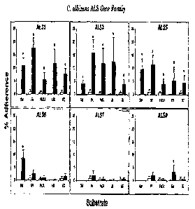
Figure 8 shows Als proteins confer substrate-specific adherence properties
when
heterologously expressed in Saccharonzyces cerevisiae. Each panel demonstrates
the
percentage adherence of one Alsp expression strain (filled bars) to a variety
of substrates to
which C. albicans is known to adhere. Adherence of S. cerevisiae transformed
with the
5 empty vector (empty bars) is included in each panel as a negative
control. Gel, gelatin; FN,
fibronectin; LN, laminin; FaDU, FaDU epithelial cells; EC, endothelial cells.
*, p < 0.01
when compared with empty plasmid control by single factor analysis of
variance. Results are
the mean S.D. of at least three experiments performed in triplicate.
Figure 9 shows domain swapping demonstrates that substrate-specific adherence
is
determined by the composition of the N-terminal domain of Als proteins. A
representation of
the ALS gene or construct being tested is depicted as a bar composed of
sequences from ALS5
(black) or ALS6 (white). Adherence properties of each mutant are displayed as
a
photomicrograph illustrating the adherence of transformed S. cerevisiae to
fibronectin-coated
beads and a graph demonstrating the adherence to gelatin (black bars) and
endothelial cells
(gray bars) as measured in the 6-well plate assay. Results are mean S.D. of
at least three
experiments, each performed in triplicate.
=
=
Figure 10 shows .a subset of Als proteins mediate endothelial cell invasion
when =
expressed in S. cerevisiae. A, endothelial cell adherence of S. cerevisiae
strains expressing
Als proteins or transformed with the empty plasmid (control). Data represent
the total number
of endothelial cell-associated organisms and are expressed as cells per high
power field. B,
degree of endothelial cell invasion of Alsp expressing S. cerevisiae strains
presented as the
number of intracellular organisms per high power field. *, p <0.01 when
compared with
empty plasm id control by single factor analysis of variance. Results are the
mean S.D. of at
least three experiments performed in triplicate.
Figure 11 shows an alignment of the N-terminal amino acid sequence of Als
proteins
of known function demonstrates an alternating pattern of CRs and HVRs. A,
percentage of
consensus identity among the N-terminal regions of Als proteins of known
function. Note
that the signal peptide region (amino acids 1-20) is not shown. Open boxes
indicate the
regions designated as HVRs 1-7. B, schematic alignment of Als proteins (SEQ ID
NOS:1-6,
respectively) showing the composition of the individual HVRs. The sequences
are arranged
to compare proteins with an affinity to multiple substrates with those that
bind few or no
CA 02636277 2008-07-04
WO 2007/081896 PCT/US2007/000433
6
identified substrates. The number of amino acids in each conserved region is
indicated in
parentheses.
Figure 12 shows CD and FTIR spectra of the Alsl protein N-terminal domain. A,
circular dichroism spectrum of 10 WV( Als I p in phosphate-buffered saline. B,
FTIR spectrum
of Alsl p self-film hydrated with D20 vapor.
Figure 13 shows a comparison of predicted physicochemical properties of N-
terminal
domains among the Als protein family. Hydrophobic, electrostatic, or hydrogen-
bonding
features are projected onto water-accessible surfaces of each domain.
Hydrophobics are
shown as follows: brown, most hydrophobic; blue, most hydrophilic.
Electrostatics (spectral
continuum) is shown as follows: red, most positive charge (+10 kcal/mol);
blue, most
negative charge (-10 kcal/rnol). Hydrogen-bonding potential (H-binding) is
shown as follows:
red, donor; blue, acceptor. Als proteins are distinguishable into three groups
based on the
composite of these properties. For example, note the similar hydrophobic,
electrostatic; and
hydrogen-bonding profiles among Als group A proteins, Alsip, Als3p, and Als5p.
In
contrast, Als group B members, Als6p and Als7p, display striking differences
in hydrophobic
and electrostatic features from those of Ms group A,. In addition to
biochemicafprofiles, note
the differences in predicted structure among these domains. =
==
Figure 14. Conceptual model of structural-functional relationships in Als
family
proteins. Als proteins are composed of three general components: an N-terminal
domain,
tandem repeats, and a serine/threonine-rich C-terminal domain containing a
glycosylphosphatidylinositol anchor that interfaces with the C. albicans cell
wall. As
illustrated, Als proteins contain multiple conserved anti-parallel 13-sheet
regions (CR1-n) that
are interposed by extended spans, characteristic of the immunoglobulin
superfamily.
Projecting from the 13-sheet domains are loop/coil structures containing the
HVRs. The three-
dimensional physicochemical properties of specific Als protein HVRs probably
govern
interactions with host substrates that confer adhesive and invasive functions
to Candida. For
illustrative purposes, only three N-terminal 13-sheet/coil domains and their
respective
CR/HVR components are shown. Note that this projection is viewed at right
angles to the
structural images shown in Fig. 13.
Figure 15. Immunization of mice (retired breeders) with rAls1p-N improves
survival
during subsequent disseminated candidiasis. Survival of mice immunized with
Alsip plus
CA 02636277 2008-07-04
WO 2007/081896
PCT/US2007/000433
7
adjuvant. N = 16 mice per group in duplicate experiments on different days;
Adj. = adjuvant.
*p <0.05 vs adjuvant.
Figure 16. Immunization with rAls1p-N improves the survival of both retired
breeder
and juvenile mice. Survival of retired breeder (A) and juvenile (B) mice
infected with a
rapidly fatal, 106 inoculum of C. olbicans. N = 16 mice per group in duplicate
experiments
on different days; Adj. = adjuvant. *p < 0.05 vs adjuvant control.
Figure 17. Anti-rAls1p-N titers do not correlate with survival. Titers of anti-
rAls1p-
N polyclonal antibodies raised in Balb/c mice immunized with varying doses of
rAls1p-N
with or without adjuvant. Adj. = adjuvant. * p 0.005 for 200 lug vs. all
others.
Figure 18. Only the protective dose of rAls1p-N induces an increase in C.
albicans-
stimulated Thl splenocytes. Induction of Thl (CD4+1FN-YEIL-4) and Th2
(CD4+IFN1-IL-
4.) splenoeytes by different doses of the rAls1p-N vaccine. Splenocytes from
immunized =
,
mice (n = 9 per group) were stimulated for 48 h with heat-killed pre-
germinated C. albicans
and then analyzed by 3-color flow cytometry. *p = 0.03 vs. adjuvant.
=
. .
Figure 19. Only the protective dose of rAls1p-N induces an increase in rAls1p-
N-
stimulated delayed type hypersensitivity. Delayed type hypersensitivity,
assessed by footpad
swelling, in mice (n = 9-12 per group) vaccinated with rAls1p-N or CFA alone.
Mice were
immunized with the indicated amount of rAls1p-N and then injected with 50 lig
of rAls1p-N
into the footpad. Footpad swelling was assessed 24 h later. *p < 0.05 versus
adjuvant, 0.2
gig, and 200 vtg.
Figure 20. The rAls1p-N vaccine requires T cells, but not B cells, to induce
protective immunity. Survival of B cell-deficient, T cell-deficient (nude),
and congenic wild-
type Balb/c control mice (n = 7 or 8 per group) was simultaneously assessed
after vaccination
with rAls1p-N + adjuvant or adjuvant alone. *p <0.04 versus adjuvant alone, ip
= 0.003
versus wild-type adjuvant-treated.
Figure 21. SQ vaccination with rAls1p-N induces an in vivo DTH response in
immunocompetent mice. Footpad swelling was assessed 24 h after injection of 50
lig of
rAls1p-N into the footpad in BALB/c mice (n = 10 per group). Median values are
displayed
as black bars. *p = 0.002 vs. control by Wilcoxon Rank Sum test.
CA 02636277 2008-07-04
WO 2007/081896 PCT/US2007/000433
8
Figure 22. The rAls1p-N Vaccine improves survival of immunocompetent mice with
hematogenously disseminated candidiasis and reduces tissue fungal burden. A)
Survival of
vaccinated or control BALB/c mice (n = 7 or 10 per group for 2.5 or 5 x 10⁵
inocula,
respectively) mice subsequently infected via the tail-vein with C. albicans.
Each experiment
was terminated at 30 days post-infection with all remaining mice appearing
well. *p < 0.05
vs. Control by Log Rank test. B) Kidney fungal burden in BALB/c mice (n = 7
per group)
infected via the tail vein with 5 x 10⁵ blastospores of C. albicans. They axis
reflects the
lower limit of detection of the assay. Median values are displayed as black
bars. *p = 0.01.
vs control by Wilxocon Rank Sum test.
Figure 23. The rAls1p-N vaccine induces a DTH reaction in neutropenic mice and
improves their survival during subsequent hematogenously disseminated
candidiasis. A)
Footpad swelling was assessed 24 h after injection of 50 i_tg of rAls1p-N into
the footpad in
BALB/c mice (n.= 10 for Control, n = 8 for rAls1p-N). * p = 0.006 vs Control
by Wilcoxon
Rank Sum test. B) Survival of neutropenic BALB/c mice (n = 16 per group from 2
experiments) infected with 2.5 x 10⁴ blastospores of C. albicans. *p = 0.007
vs adjuvant
control by Log Rank test.
Figure 24. The rAls1p-N vaccine diminishes the severity of histopathological
fungal
lesions on the tongues of mice with oropharyngeal candidiasis. N = 4 mice per
group.
Inflammatory score generated by a blinded observer as described in the text.
*p = 0.03 by
Wilcoxon Rank Sum test.
Figure 25 shows that rAls3p-N but not rAls1p-N vaccine diminishes fungal
colonization of vagina of mice inoculated with C. albicans (*p=0.01 vs mice
vaccinated with
CFA alone, by Wilcoxon Rank Sum test) N= Ii mice per group.
Figure 26 shows an Alsip homology map versus S. aureus clumping factor A
(c1n67A). Consensus functional sites from C. albicans Alsip and S. carrells
ClfA were
mapped onto the Alsip homology model. Numerous residues from the N-termini of
Alsip
and CllA map to a consensus cleft motif, which is where binding to substrate
is predicted to
occur for both adhcsins.
Figure 27 shows that rAls1p-N and rAls3p-N vaccines improve the survival of
staphylococcemic mice. (*p<0.003 vs mice vaccinated with CFA alone, by Log
Rank test).
N= 22 mice per group.
CA 02636277 2008-07-04
WO 2007/081896 PCT/US2007/000433
9
Figure 28 shows that antibody titers do not correlate with degree of
protection in
individual vaccinated mice, but they do distinguish unvaccinated from
vaccinated mice.
Titers of anti-rAls1p-N or anti-rAls3p-N polyclonal antibodies raised in
Balb/c mice
immunized with CFA alone, or CFA + 20 ug of rAls1p-N or rAls3p-N,
respectively. Overall
there is a significant correlation between antibody titers and survival (rho
=0.474, p=0.0057),
indicating that antibody titers can be used as a surrogate marker for vaccine
protection.
However, when data from mice receiving CFA alone are excluded, there is no
correlation
between antibody=titers and survival of mice vaccinated with rAls1p-N or
rAls3p-N (rho
0.041143, p=0.847), indicating that antibodies are likely not the predominant
mechanism of
protection of the vaccine.
Figure 29 shows that the rAls1p-N vaccine protects outbred, CD1 mice from
hematogenously disseminated candidiasis. A) CD1 mice (n = 8 per group) were
vaccinated
SQ with rAls1p-N (20 lag) + CFA, or CFA alone, and infected via the tail-vein
with C.
albicans SC5314 fourteen days after the boost. B) CD1 mice (n = 8 per group)
were
vaccinated SQ with rAls1p-N at various doses with alum, or with alum alone,
and infected
via the tail-vein with C. albicans SC5314 fourteen days after the boost. * p <
0.05 vs.
adjuvant control by Log Rank test.
Figure 30 shows that the rAls1p-N vaccine improves the survival of Balb/c mice
infected with one of several strains of C. albicans. Survival of Balb/c mice
immunized with
rAls1p-N plus CFA versus CFA alone and infected via the tail-vein with C.
albicans 15563
(7 x 10⁵ blastospores), 16240 (4 x 10⁵ blastospores), or 36082 (4 x 10⁵
blastospores) (n = 8
mice per group). *p < 0.05 vs adjuvant control by Log Rank test.
Figure 31 shows that the rAls1p-N vaccine reduces tissue fungal burden in
Balb/c
mice infected with several non-albicans species of Candia'a. Balb/c mice (n =
5 per group)
were vaccinated with CFA or CFA + rAls1p-N (20 l_tg) and infected via the tail-
vein with C.
glabrata, C. krusei, C. parapsilosis, or C. tropicalis. Infectious inocula are
shown in
parentheses below the species names. Kidney fungal burden was determined on
day five
post-infection. They axis reflects the lower limit of detection of the assay.
*p <0.05 vs.
adjuvant control by non-parametric Steel test for multiple comparisons.
Figure 32 shows that rAls3p-N-immunized mice generated antibodies that cross-
reacted against rAls1p-N. Titers of individual mice immunized with CFA alone,
CFA +
CA 02636277 2008-07-04
WO 2007/081896 PCT/US2007/000433
rAls1p-N, or CFA + rAls3p-N. N = 7 mice per group for CFA and CFA + rAls3p-N;
n = 6
mice for CFA + rAls1p-N. *p < 0.05 vs. CFA alone; **p <0.002 vs. CFA alone & p
< 0.011
vs. CFA + rAls1p-N by Mann Whitney U test. Bars denote medians.
Figure 33 shows that both rAls1p-N and rAls3p-N primed mice for in vivo
delayed
5 type hypersensitivity responses. Mice (n = 7 per groupfor CFA and CFA +
rAls3p-N; n = 6
for CFA + rAls1p-N) were vaccinated with CFA alone, CFA + rAls1p-N, or CFA +
rAls3p-
N. Delayed type hypersensitivity in vivo was measured by footpad swelling. *p
<0.05 vs.
CFA alone by Mann Whitney U test. Bars denote medians.
Figure 34 shows that the rAlslp-N and rAls3p-N vaccines mediated similar
efficacy
10 against murine hematogenously disseminated candidiasis. Survival of
Balb/c mice (n = 15
per group from 2 experiments for CFA and CFA + rAls3p-N, and n = 14 from 2
experiments
for CFA + rAls1p-N) infected via the tail vein with 5 x 10⁵ blastospores of C.
albicans. The
experiment was terminated at day 28 post-infection with all remaining mice
appearing well.
*p 5 0.0001 vs CFA control by Log Rank test.
Figure 3-5 shows that in vivo delaYed-type hypersensitivity correlated with
survival
during disseminated candidiasis. Anti-rAls1p-N or anti-rAls3p-N antibody
titers and footpad
swelling reactions were measured in mice (n = 7 per group for CFA or CFA +
rAls3p-N, n =
6 for CFA + rAls1p-N) two days prior to infection via the tail-vein with C.
albicans.
Correlations determined with the Spearman Rank sum test.
Figure 36 shows that the rAls3p-N vaccine significantly reduced tissue fungal
burden
during murine oropharyngeal candidiasis. Tongue fungal burden in mice (n = 7
for CFA and
8 for rAls1p-N or rAls3p-N vaccinated groups) with oropharyngeal candidiasis.
They axis
reflects the lower limit of detection of the assay. *p = 0.005 vs. CFA by Mann
Whitney 11
test.
Figure 37 shows that rAls3p-N reduced vaginal fungal burden compared to both
CFA
alone and CFA + rAls1p-N in murine candidal vaginitis. Vaginal fungal burden
in mice (n =
11 per group from 2 experiments) vaccinated with CFA, CFA + rAls1p-N, or CFA +
rAls3p-
N. The y axis reflects the lower limit of detection of the assay. *p <0.02 vs
CFA and CFA +
rAls1p-N by Steel test for multiple comparisons.
CA 02636277 2013-06-04
CA 2636277
11
DETAILED DESCRIPTION OF THE INVENTION
Candida albicans and Staphylococcus aureus are common pathogen in humans. For
example, C.
albicans, while normally a harmless commensal, this organism can cause a
variety of conditions ranging
from superficial mucocutaneous infection such as vaginal and/or oropharyngeal
candidiasis, to deep
organ involvement in disseminated candidiasis. Prior to causing disease, the
fungus colonizes the
gastrointestinal tract, and in some cases skin and mucous membranes. Adherence
to host mucosal
surfaces is a key prerequisite for this initial step. After colonization, C.
albicans enters the bloodstream via
infected intravascular devices or by transmigration through gastrointestinal
mucosa compromised by
chemotherapy or stress ulcerations. Organisms then disseminate via the
bloodstream, bind to and
penetrate the vascular endothelium to egress from the vascular tree, and
invade deep organs such as
liver, spleen, and kidney.
The identification and functional characterizations of a variety of exemplary
Als protein family
members described herein allow this family of proteins to be effectively
utilized in the treatment of
candidiasis. Specific binding activity to diverse substrates and other
selective cell adhesion functions can
be exploited in the production of vaccines for active or passive immunization,
in the production of peptide,
analogue of mimetic inhibitors of cell adhesion to reduce or prevent initial
infection by inhibiting binding,
adhesion or invasion of a host cell. Moreover, the differential binding and
invasion profiles allow design
and use of broad spectra or targeted inhibition of Als protein family member
activities. Additionally,
functional fragments that confer binding and/or invasive activity allow
elimination of unwanted foreign
protein sequences, thus, increasing the efficacy of the Als family protein
member vaccine or therapeutic
inhibitor.
The nature of the pathogenesis of C. albicans by adherence to endothelial
cells is discussed in
USP 5,578,309. For a description of the ALS1 gene and characteristics thereof,
including the
characterization of the gene product as an adhesin see, Fu, Y., G. Rieg, W.A.
Forizi, P.H. Belanger,
J.E.J. Edwards, and S. G. Filler. 1998. Expression of the Candida albicans
gene ALSI in Saccharomyces
cerevisiae induces adherence to endothelial and epithelial cells. Infect.
lmmun. 66:1783-1786; Hoyer, L.L.
1997. Fu Y, Ibrahim AS, Sheppard DC, Chen Y-C, French SW, Cutler JE, Filler
SG, Edwards, JE, Jr.
2002. Candida albicans Alsip: an adhesin that is a downstream effector of the
EFG1 filamentation
pathway. Molecular
CA 02636277 2008-07-04
WO 2007/081896 PCT/US2007/000433
12
Microbiology 44:61-72. Sheppard DC, Yuman MR, Welch WI-I, Phan QT, Fu Y,
Ibrahim
AS, Filler SG, Zhang M, Waring AJ, Edwards, Jr., JE 2004. Functional and
Structural
Diversity in the Als Protein Family of Candicia cub/cans. Journal Biological
Chemistry. 279:
30480-30489. The ALS gene family of Candida albicans. International Society
for Human
and Animal Mycology Salsimorge, Italy:(Abstract); Hoyer, L.L., S. Scherer,
A.R. Shatzman,
and G.P. Livi. 1995. Candida albicans ALSI: domains related to a
Saccharonzyces
cerevisiae sexual agglutinin separated by a repeating motif. Mol. Microbic].
15:39-54.
In this regard, the human fungal pathogen Candida albicans colonizes and
invades a
wide range of host tissues. Adherence to host constituents plays an important
role in this
process. Two members of the C. albicans Als protein family (Alsip and Als5p)
have been
found to mediate adherence and exemplify the binding, adhesion and cell
invasion activities
of Als protein family members. As described herein, members of the ALS gene
family were
cloned and expressed in S. cereviskte to characterize their individual
functions. Distinct Als
,
proteins conferred distinct adherence profiles to diverse host substrates.
Using chimeric
Als5p-Als6p constructs, the regions mediating substrate-specific adherence
were localized to
the N-terminal domains in Als proteins. In particular, a subset of Als
proteins also mediated
= = -
endothelial cell inVasion, a previously unknown function of this family.
CoriSistent with
these results, homology modeling revealed that Als members contain anti-
parallel 13-sheet =
motifs interposed by extended regions, homologous to adhesions or invasins of
the
immunoglobulin superfamily. This finding was confirmed using circular
dichroism and
Fourier transform infrared spectrometric analysis of the N-terminal domain of
Alsip.
Specific regions of amino acid hypervariability were found among the N-
terminal domains of
Als proteins, and energy-based models predicted similarities and differences
in the N-
terminal domains that probably govern the diverse function of Als family
members.
Collectively, these results indicate that the structural and functional
diversity within the Als
family provides C. albicans with an array of cell wall proteins capable of
recognizing and
interacting with a wide range of host constituents during infection.
The invention provides a vaccine having an isolated Als protein family member
having cell adhesion activity, or an immunogenic fragment thereof, and an
adjuvant in a
pharmaceutically acceptable medium. The vaccine can be an Als protein family
member
derived from a Candida species such as Candida albicans, Candida krusei,
Canclida
tropicalis, Canclida glabrata or Candida, parapsilosis. The Als protein family
member can
CA 02636277 2008-07-04
WO 2007/081896 PCT/US2007/000433
13
be, for example, Alsip, Als3p, Als5p, Als6p, Als7p and Als9p, or an
immunogenic fragment
thereof. All other Als protein family members within an Candi(la species can
similarly be
employed as a vaccine of the invention.
The present invention utilizes the gene product of C. albicans agglutinin like
sequence protein family member as a vaccine to treat, prevent, or alleviate
disseminated
candidiasis. The vaccine is effective against different strains of C. albicans
as well as against
different Caw-lida species. The Als protein family member can be, for example,
Alsip,
Als3p, Als5p, Als6p, Als7p and Als9p. The invention exploits the role of the
ALS gene
products in the adherence of and invasion by C. albicans to endothelial and/or
epithelial cells
and the susceptibility of the Ms protein family member-expressed surface
protein for use as a
vaccine to retard the pathogenesis of the organism.
Pursuant to this invention, an ALS family member gene encodes a surface
adhesin
that is selected as the target of an immunotherapeutic strategy against C.
albicans. A
demonstration that the expression product of the ALSI gene, the Alsip protein,
has structural
characteristics typical of surface proteins and is, in fact, expressed on the
cell surface of C.
albicans is one criterion for proteins that act as adhesins to host tissues.
The Als protein
family members can be structurally characterized as having a signal peptide-at
the N-
terminus, a glycosylphosphatidylinosine (GPI) anchorage sequence in the C-
terminus, and a -
central region comprising repeats rich in threonine and serine. Also, Als
protein family
members have N-, and 0- glycosylation sites, typical of proteins that are
expressed on the cell
surface. Indirect immunofluorescence using a monoclonal antibody directed
against the N-
terminus of ALs1p, for example, revealed that ALslp is expressed during the
log phase of
blastospores. This expression of ALsIp is increased during hyphal formation
and is localized
to the junction where the hyphal element extends from the blastospores as
indicated by the
diffused surface staining. Furthermore, this monoclonal antibody blocked the
enhanced
adherence of C. albicans overexpression mutant to endothelial cells, thereby
establishing the
principle for immunotherapy applications using ALsip. Functional
characteristics as they
relate to cell adhesion and invasion of other Als family members are described
further below
in Example VI.
Thus, according to one aspect, the invention provides an Als family member
surface
adhesion protein, designated, for example, Alsip, Als3p, Als5p, Als6p, Als7p
and Als9p, or a
functional fragment, conjugate or analogue thereof, having useful properties
when formulated
CA 02636277 2008-07-04
WO 2007/081896 PCT/US2007/000433
14
in a pharmaceutical composition and administered as a vaccine with or without
an adjuvant.
An Als protein family member, combination of two or more Als protein family
members or
one or more functional fragments, analogues, conjugates or derivatives
thereof, can be
obtained from, for example, Candida albicans. Similar adhesin or invasin
molecules or
analogues or derivatives thereof can be of candidal origin and can be
obtainable, for example,
from species belonging to the genera Candida, for example Canclida
pctrapsdosis, Canclida
krusei, Candida glabrata and Candida tropicalis. A surface adhesin or invasin
protein
according to the invention can be obtained in isolated or purified form, and
thus, according to
one embodiment of the invention a substantially pure Als protein family member
Candida
surface adhesin protein, or functional fragment, immunogenic fragment,
analogue, conjugate
or derivative thereof, is formulated as a vaccine to cause an immune response
in a patient to
elicit an immune response against Candida and/or to block adhesion of the
organism to the
endothelial cells. Fragments of Als protein family members that exhibit
similar binding,
adhesion or invasion activity as an intact As protein family member is
referred to herein as a
functional fragment. Fragments of Als protein family members that are
capable.of eliciting
an antibody or cellular immune response against a Canclida species are
referred to herein as
an immunogenic fragment. Exemplary functional fragments include
the,.1\17terminal
polypeptide regiOn pf the Als protein family member described .further below'
in Example VI.
Exemplarily immogenic fragments include the N-terminal Als polypeptide region,
the C-
terminal Als polypeptide region as well as any other Als fragment that is
sufficient to
generate an antibody, cellular or both an antibody and cellular immune
response. Such
immogenic fragments can be as small as about four amino acids and as large as
the intact
polypeptide as well as include all polypeptide lengths in between.
An analogue or derivative of the surface adhesion protein according to the
invention
can be identified and further characterized by the criteria described herein
for an ALS family
member gene and/or gene product. For example, a null mutant of the analogue or
derivative
would share markedly reduced adhesion to endothelial cells compared to
controls. Similarly,
over-expression of the analogue or derivative in an appropriate model would
show an
increased adherence to endothelial cells compared to controls and would be
confirmed as a
cell surface adhesin in accord with the criteria described above. Also,
antisera to an analogue
or derivative can cross-react with anti-Als protein family member antibodies
and can exhibit
increased survival times when administered in a mouse model of disseminated
candidiasis as
disclosed herein.
CA 02636277 2008-07-04
WO 2007/081896 PCT/US2007/000433
The invention also provides a method of treating or preventing disseminated
candidiasis. The method includes administering an immunogenic amount of a
vaccine an
isolated Als protein family member having cell adhesion or invasion activity,
or an
immunogenic fragment thereof, in a pharmaceutically acceptable medium. The
vaccine can
5 be administered with or without an adjuvant. The Als protein family
member can be derived
from different Candida strains as well as from different Candida species such
as Candida
tableaus, Candida krusei, Candida tropicalis, Candida glabrata and Candida,
parapsilosis.
An Als protein family member used in the method of treating or prevention
disseminated
candidias includes Alsip, Als3p, Als5p, Als6p, Als7p and Als9p.
10 The effectiveness of the vaccines of the invention against different
Candida strains,
different Candida species, other bacteria and infectious agents and their wide
range of
immune activity are described further below and exemplified in the Examples.
For example,
Example V shows that anti-ALS antibodies are effective against mucosa] and
hematogenously disseminated candidal infections. Example VII shows that
vaccination with
15 rAls1p-N improves survival during murine disseminated candidiasis by
enhancing cell-.
mediated immunity. Example VIII shows that the vaccines of the invention
reduce fungal
burden and improve survival in both immunocompetent and immunocompromised
mice: =
Example IX shows the effectiveness of the ALS vaccines of the invention
against S. aureits
infections. Example X exemplifies that the vaccines of the invention are
effective against
different strains of C. albicans and against different species such as C.
glabrata, C. krusei, C.
parapsilosis and C. tropicalis as well as effectiveness in different animal
models. Example
XI also exemplifies the effectiveness of the different vaccines of the
invention in different
animal models as well as provides a comparison of the different responses
elicited and
potency of two representative ALS vaccines.
The invention further provided is a method of treating or preventing
disseminated
candidiasis that includes administering an effective amount of an isolated Als
protein family
member having cell adhesion activity, or an functional fragment thereof, to
inhibit the
binding or invasion of Candida to a host cell or tissue. The Als protein
family member can
be derived from Catuticla albicans, Cam-lick krusei, Candida tropicalis,
Candida glabrata,
curd Candida, parapsilosis. An Als protein family member used in the method of
treating or
prevention disseminated candidias includes Alsip, Als3p, Als5p, Als6p, Als7p
and Als9p.
CA 02636277 2008-07-04
WO 2007/081896 PCT/US2007/000433
16
The cell adhesion activity includes binding to gelatin, fibronectin, laminin,
epithelial cells or
endothelial cells and/or promoting cell invasion.
In addition, the invention also provides a method of treating or preventing
Staphylococcus auretts infections using the Als protein family members
described herein. In
particular, the method of treating or preventing Staphylococcus attreus
infections includes
administering an immunogenic amount of a vaccine an isolated Als protein
family member
having cell adhesion activity, or an immunogenic fragment thereof, in a
pharmaceutically
acceptable medium.
Alsip and Als3p are particularly efficacious because of significant homology
to S.
aureus cell surface proteins. The sequence and structural homology of, for
example, Alsl p
and Als3p, are described further below in Example IX. Given the teachings and
guidance
provided herein, those skilled in the art will understand that the vaccines
and methods of the
.invention can be applied to the treatment of Ccindida and Staphylococcus
infections alike..
!-
Similarly, given the teachings and methods described herein, those skilled in
the art also will
understand that the vaccines and methods of the invention also can be applied
to other
pathogens having cell surface polypeptides with similar immunogenicity,
sequence and/or ,
.. =
structural homology to the Als protein family members described herein,
including fungus, .
bacteria and the like.
Immunotherapeutic and/or Als polypeptide inhibition of cell adhesion or
invasion .
strategies against Candida or Staphylococcus infection can operate at the
level of binding to
the vascular endothelial cells as well as through a downstream effector of the
filamentation
regulatory pathway. An immunotherapeutic strategy or inhibition of binding
using a soluble
Als protein family member or functional fragment is useful in this context
because: (i) the
morbidity and mortality associated with hematogenously disseminated
candidiasis and other
infectious pathogens remains unacceptably high, even with currently available
antifungal
therapy; (ii) a rising incidence of antifungal and antibiotic resistance is
associated with the .
increasing use of antifungal and antibacterial agents, iii) the population of
patients at risk for
serious Candida and Staphylococcus infections is well-defined and very large,
and includes
post-operative patients, transplant patients, cancer patients and low birth
weight infants; and
iv) a high percentage of the patients who develop serious Candida infections
are not
neutropenic, and thus may respond to a vaccine or a competitive polypeptide or
compound
inhibitor. For these reasons, Candida and Staphylococcus are attractive fungal
and bacterial
CA 02636277 2008-07-04
WO 2007/081896 PCT/US2007/000433
17
' targets for passive immunotherapy, active immunotherapy or a combination
of passive or
active immunotherapy. Additionally, Canalida also is attractive for
competitive inhibition
using an Als protein family member polypeptide, functional fragment thereof
and/or a
compound or mimetic thereof' that binds to one or more Als family members and
prevents
binding of Candi da to a host cell receptor.
Given the teachings and guidance provided herein, those skilled in the art
will
understand that immunotherapeutic methods well know in the art can be employed
with the
Als protein family members of the invention, immunogenic fragments, analogues,
conjugates,
and/or derivatives thereof, to use one or more of the molecule as an immunogen
in a
pharmaceutically acceptable composition administered as a vaccine with or
without an
adjuvant. For the purposes of this invention, the terms "pharmaceutical" or
"pharmaceutically acceptable" refer to compositions formulated by known
techniques to be
non-toxic and, when desired, used with carriers or additives that can be
safely administered to
humans. 'Administration can be performed using well known routes including,
for example,
intravenous, intramuscular, intraperitoneal or sub-cutaneous injection. Such
vaccines of the
inventions also can include buffers, salts or other solvents known to these
skilled in the art to
preserve the activity of the vaccine in solution. Similarly, any of a wide
range of adjuvants
well known in the art can be employed with the vaccine of the invention to
elicit, promote or
enhance a therapeutically effective immune response capable of reducing or
blocking
binding, invasion and/or infection of Candida or Staphylococcus to a
susceptible host cell.
Similarly, given the teachings and guidance provided herein, those skilled in
the art
also will understand that therapeutic methods well known in the art for
administering and
selectively blocking the binding of cell surface molecules to their cognate
receptors also can
be employed with the Als protein family members of the invention, functional
fragments,
analogues, conjugates and/or derivatives thereof, to use one or more of the
Als protein family
member as an inhibitor in a pharmaceutically acceptable composition. As with
vaccine
formulations, inhibitory formulations can similarly be administered using well
known method
in the art including, for example, intravenous intramuscular, intraperitoneal
or sub-cutaneous
injection. Such inhibitory compositions that bind Als family member receptors
and block an
Als protein family member binding also can include buffers, salts or other
solvents known to
these skilled in the art to preserve the activity of the vaccine in solution.
Further, any of a
wide range of formulations well known in the art can be employed with the
inhibitory
CA 02636277 2008-07-04
WO 2007/081896
PCT/US2007/000433
18
compositions of the invention to target and/or enhance delivery or uptake so
as to reduce or
inhibit binding, invasion and/or infection of Canclida or Staphylococcus to a
susceptible host
cell.
With respect to the molecule used as a therapeutic immunogen or receptor
binding
inhibitor pursuant to the present invention, those of skill in the art will
recognize that the Als
protein family member molecules can be truncated or fragmented without losing
the essential
qualities as an immunogenic vaccine or cell adhesion or invasion inhibitor.
For example, an
Als protein family member can be truncated to yield an N-terminal fragment by
truncation
from the C-terminal end with preservation of the functional properties
described above and
further below in the Examples. Similarly, C-terminal fragments can be
generated by
truncation from the N-terminal end with preservation of their functional
properties. Other
modifications in accord with the teachings and guidance provided herein can be
made =
pursuant to this invention to create other Als protein family member
functional fragments,
immunogenic fragments, analogs or derivatives thereof; to achieve the
therapeutically useful
properties described herein with the native protein.
One aspect of the..therapeutic effectiveness of Als protein family members and
methods of the invention achieves interference with regulation of
filamentation, to block
adherence of the organism to host constituents, and to enhance clearance of
the organism by
immunoeffector cells and other physiological mechanisms. Since endothelial
cells cover the
majority of the vasculature, strategies to block the adherence, invasion
and/or both of the
organism to endothelial cells using antibodies, Als family member proteins,
polypeptide or
peptides or any combination thereof include useful embodiment of the present
invention. As
described previously, such adherence and/or invasion blocking therapies
include active or
passive imnninotherapy or inhibitory binding directed against the candidal
adhesins, invasins,
or cognate receptors disclosed herein. Thus, for example, any suitable host
can be injected
with protein and the serum collected to yield the desired anti-adhesin
antibody after
appropriate purification and/or concentration. Prior to injection, the adhesin
or invasin
protein or a combination thereof, can be formulated in a suitable vehicle
preferably a known
immunostimulant such as a polysaccharide or delivery formulation such as
liposomes or
time-released compositions. Thus, according to a further aspect, invention
provides a
pharmaceutical composition comprising a candidal adhesin or invasin protein
together with
CA 02636277 2008-07-04
WO 2007/081896 PCT/US2007/000433
19
one or more pharmaceutically acceptable excipients in a formulation for use as
a vaccine or
Als receptor inhibitor.
The method of the invention is ameliorating and/or preventing candidal or
Staphylococcus infection by blocking the adherence of C. albicans to the
endothelial or
epithelial cells of a host constituent or by, for example, antibody binding to
the
Staphylococcus and allowing immune mechanisms remove the pathogen. Thus,
according to
one aspect of the invention, a pharmaceutical composition comprising an Als
protein family
member adhesin or invasin protein, functional or immunogenic fragment,
derivative,
analogue, or conjugate thereof is formulated as a vaccine or Als receptor
inhibitor in a
pharmaceutical composition containing a biocompatible carrier for injection or
infusion and
is administered to a patient. Also, direct administration of antiserum raised
against Als
family member protein or isolated or recombinant Als family member protein can
be used to
block the adherence of C. albicans to a mammalian host constituent or effect
the removal of a
Staphylococcus pathogen. Antiserumagainst adhesin protein can be obtained by
known
techniques, Kohler and Milstein, Nature 256: 495-499 (1975), and may be
humanized to
reduce antigenicity, see USP 5,693;762, or produced in transgenic mice leaving
an
unrearranged human immunoglobulin gene, see USP 5,877,397.,. Similarly,
isolated or
recombinant Als protein family member also can be produced using methods well
known to =
those skilled in the art including, for example, the recombinant production
described in the
Examples below.
A still further use of the invention, for example, is using the Als protein
family
member adhesin or invasin protein to develop vaccine strategies for the
prevention and/or
amelioration of candidal or Staphylococcus infections. Thus, according to one
aspect of the
invention, for example, standard immunology techniques can be employed to
construct a
multi-component vaccine strategy that can enhance and/or elicit immune
response from a
host constituent to bock adherence of C. albicans or to effect the elimination
of
Staphylococcus pathogens.
A still further use of the invention, for example, is developing DNA vaccine
strategies. Thus, according to one aspect of the invention, for example, the
ALS family
member polynucleotides encoding Als protein family member adhesin or invasin
or a
functional fragment thereof is administered according to a protocol designed
to yield an
immune response to the gene product. See e.g., Feigner USP 5,703,055.
CA 02636277 2008-07-04
WO 2007/081896 PCT/US2007/000433
A still further use of the invention, for example, is developing combination
vaccine
strategies. Thus, according to one aspect of the invention, for example, anti-
ALS protein
family member antibodies may be used with antibodies in treating and/or
preventing candidal
or Staphylococcus infections. See USP 5,578,309.
5 The following Examples illustrate the immunotherapeutic utility of the
ALS1 adhesin
as the basis for preventive measures or treatment of dissemiated candidiasis.
Example 1
describes the preparation of an ALS1 null mutant and a strain of C. albicans
characterized by
overexpression of ALS1 to confirm the mediation of adherence to endothelial
cells. Example
2 describes the localization of Alsip and the implication of the efg
filamentation regulatory
10 pathway. Example 3 describes the purification of ALS1 adhesin protein.
Example 4
describes the preparation of rabbit polyclonal antibodies raised against the
ALSI surface
adhesin protein to be used to demonstrate the blocking of the surface adhesin
protein.
Example 5, describes the blocking of adherence in vivo, using polyclonal
antibodies raised
against the ALS1 surface adhesion protein as described herein according to the
invention to
15 protect against disseminated candidiasis in a mouse model. Example VI
describes the
=
structural and functional characteristics of Ms protein family members.
It is understood that modifications which do not substantially affect the
activity of the
various embodiments of this invention are also included within the definition
of the invention
provided herein. Accordingly, the following examples are intended to
illustrate but not limit
20 the present invention.
EXAMPLE I
Alsl Mediates Adherence of C. albicans to Endothelial Cells
The URA blaster technique was used to construct a null mutant of C albicans
that
lacks express of the Alsip. The alsl/als1 mutant was constructed in C.
albicans strain CAILl
using a modification of the Ura-blaster methodology (Fonzi and Irwin, Genetics
134, 717
(1993)) as follows: Two separate alsl-hisG-IRA3-hisG-als1 constructs were
utilized to
disrupt the two different alleles of the gene. A 4.9 kb AsLS1 coding sequence
was generated
with high fidelity PCR (Boehringer Mannheim, Indianapolis, IN) using the
primers: 5'-
CCCTCGAGATGCTTCAACAATTTACATTGTTA-3' (SEQ ID NO:8) and 5-
CCGCTCGAGTCACTAAATGAACAAGGACAATA-3' (SEQ ID NO:9). Next, the PCR
fragment was cloned into pGEM-T vector (Promega, Madison, W1), thus obtaining
pGEM-T-
CA 02636277 2008-07-04
WO 2007/081896 PCT/US2007/000433
21
ALSI. The hisG-URA3-hisG construct was released from pMG-7 by digestion with
Kpn
and Hind3 and used to replace the portion of ALS] released by Kpnl and Hind3
digestion of
pGEM-T-ALS1. The final alsl-hisG-URA3-hisG-als1 construct was released from
the
plasmid by digestion with Xhol and used to disrupt the first allele of ALS1 by
transformation
of strain CAI-4.
A second alsl-hisG-URA3-hisG-als1 construct was generated in two steps. First,
a
Bg12-Hind3 hisG-URA3-hisG fragment of pMB7 was cloned into the Baml-Il-1-lind3
sites of
pUC19, thereby generating pYC2. PYC2 was then digested with Hind3, partially
filled in
with dATP and dGTP using T4 DNA polymerase, and then digested with Smal to
produce a
new hisGURA3-hisG fragment. Second, to generate ALS1 complementary flanking
regions,
pGEM-T-ALS1 was digested with Xbal and then partially filled in with dCTP and
dTTP.
This fragment was digested with Hpal to delete the central portion of ALS1 and
then ligated
to the hisG-URA3-hisG fragment generating pYC3. This plasmid was then digested
by Xhol
to release a construct that was used to disrupt the second allele of the ALS1.
Growth curves
were done throughout the experiment to ensure that the generated mutations had
no effect on
growth rates. All integrations were confirmed by Southern blot analysis using
a 0.9kb ALS1
specific probe generated by digestion of pYF5 with 3CbaI and HindIII.
The null mutant was compared to C. albie ans CAI-12 (a URA -1- revertant
strain) for
its ability to adhere in vitro to human umbilical vein endothelial cells. For
adherence studies,
yeast cells from YPD (2% glucose, 2% peptone, and 1 % yeast extract) overnight
culture,
were grown in RPM! with glutamine at 25 C for 1 hour to induce Alsip
expression. 3 x 10²
organisms in Hanks balanced salt solution (HBSS) (Irvine Scientific, Irvine,
CA) were added
to each well of endothelial cells, after which the plate was incubated at 37 C
for 30 minutes.
The inoculum size was confirmed by quantitative culturing in YPD agar. At the
end of
incubation period, the nonadherent organisms were aspirated and the
endothelial cell
monolayers were rinsed twice with HBSS in a standardized manner. The wells
were over
laid with YPD agar and the number of adherent organisms were determined by
colony
counting. Statistical treatment was obtained by Wilcoxon rank sum test and
corrected for
multiple comparisons with the Bonferroni correction. P<0.001.
Referring to Figure 1, a comparison of the ALS I/ALS1 and alsl/als1 strain
showed
that the ALS1 null mutant was 35% less adherent to endothelial cells than C.
albiectns CAL-
12. To reduce background adherence, the adherence of the wild-type strain
grown under non-
CA 02636277 2008-07-04
WO 2007/081896 PCT/US2007/000433
22
A LS 1 expressing conditions was compared with a mutant autonomously
expressing Alsip.
This mutant was constructed by integrating a third copy of ALS] under the
control of the
constitutive A D1-11 promoter into the wild-type C. albicans. To achieve
constitutive
expression of the ALS1 in C. albicans, a blunt-ended PCR generated URA3 gene
is ligated
into a blunt-edged Bg12 site of pOCUS-2 vector (Novagen, Madison, WI),
yielding p0U-2.
A 2.4 kb Notl -Stul fragment, which contained C. albicans alcohol
dehydrogenase gene
(ADI-11) promoter and terminator (isolated from pLI-I-ADHpt, and kindly
provided by A.
Brown, Aberdeen, UK), was cloned into p0U-2 after digestion with Notl and
Stul. The new
plasm id, named p0AU-3 had only one Bg12 site between the ADH1 promoter and
terminator. ALS1 coding sequence flanked by BamH1 restriction enzyme sites was
generated by high fidelity PCR using pYF-5 as a template and the following
primers: 5'-
CGGGATCCAGATGCTTCA-ACAATTTACATTG-3' (SEQ ID NO:10) and 5'-
CGGGATCCTCACTAATGAACAAGGACAATA-3' (SEQ ID NO:11). This PCR fragment
was digested with BamH1 and then cloned into the compatible Bg12 site of p0AU-
3 to
generate pAU-1. Finally, pAU-1 was linearized by Xbal prior to transforrning
C. albicans
CAI-4. The site-directed integration was confirmed by Southern Blot analysis.
Referring to'
Figure 1B, overexpressing ALS1 in this PADni-ALS1 strain resulted in a 76%
increase in
adherence to endothelial cells compared to the wild-type C. albicans. In
comparing
endothelial cell adherence of the wild-type to that of the oVerexpressing
mutant, yeast cells
were grown overnight in YPD at 25 C (non-inducing condition of Alsip). Alsip
expression
was not induced to reduce the background adherence of the wile-type, thus
magnifying the
role of Alsip in adherence through PADHI-ALS1 hybrid gene. The adherence assay
was , =
carried out as described above. Statistical treatment was obtained by Wilcoxon
rank sum test
and corrected for multiple comparisons with the Bonferroni correction.
P<0.001.
A monoclonal anti-Alsip murine IgG antibody was raised against a purified and
truncated N-terminus of Alsip (amino acid #17 to #432) expressed using
Clontech YEXpress
(TM) Yeast Expression System (Palo Alto, CA). The adherence blocking
capability of these
monoclonal anti-Alsip antibodies was assessed by incubating C. albicans cells
with either
anti-Als1 antibodies or mouse IgG (Sigma, St. Louis, MO) at a 1:50 dilution.
After which the
yeast cells were used in the adherence assay as described above. Statistical
treatment was
obtained by Wilcoxon rank sum test and corrected for multiple comparisons with
the
Bonferroni correction. P<0.001. The results revealed that the adherence of the
PADH l'ALS1
strain was reduced from 26.8% 3.5% to 14.7% 5.3%. Thus, the effects of ALS I
deletion
CA 02636277 2008-07-04
WO 2007/081896 PCT/US2007/000433
23
and overexpression demonstrate that Alsip mediates adherence of C. albicans to
endothelial
cells.
EXAMPLE II
Localization of Alsip
For Alsip to function as an adhesin, it must be located on the cell surface.
The cell
surface localization of Alsip was verified using indirect immunofluorescence
with the anti-
Alsip monoclonal antibody. Diffuse staining was detected on the surface of
blastospores
during exponential growth. This staining was undetectable on blastospores in
the stationary
phase. Referring to Figure 2A, when blastospores were induced to produce
filaments, intense
staining was observed that localized exclusively to the base of the emerging
filament. No
immunofluorescence was observed with the alsl/als1 mutant, confirming the
specificity of
this antibody for Alsip. See Figure 2B. These results establish that Alsip is
a cell surface
protein. =
=
The specific localization of Alsip to the blastospore-filatnent junction
implicates
Alsip in the filamentation process. To determine the mechanism, the
filamentation
phenotype of the C. albicans ALS1 mutants was analyzed. Referring to Figure
3A, the
a Isl/alsi mutant failed to form filaments after a 4 day incubation on Lee's
solid medium,
while both the ALS1/ALS1 AND ALSI/als1 strains as well as the ALS I-
complemented
mutant produced abundant filaments at this time point. The alsl/als1 mutant
was capable of
forming filaments after longer periods of incubation. Furthermore,
overexpressing ALS1
augmented filamentation: the PADm- ALS1 strain formed profuse filaments after
a 3 day
incubation, whereas the wild-type strain produced scant filaments at this time
point. See
Figure 3B. To further confirm the role of Alsip in filamentation, a negative
control was
provided using mutant similar to the ALS I overexpression mutant, except the
coding
sequence of the ALS1 was inserted in the opposite orientation. The
filamentation phenotype
or the resulting strain was shown to be similar to that of the wild-type
strain. The filament-
inducing properties of Alsip are specific to cells grown on solid media,
because all of the
strains described above filamented comparably in liquid media. The data
demonstrates that
Alsip promotes filamentation and implicates ALS! expression in the regulation
of
filamentation control pathways. Northern blot analysis of ALS1 expression in
mutants with
defects in each of these pathways, including efgl/efgl, cph I /cph I ,
efgl/efg cph 1 /cphl,
tupl/tup I , and cla4/c1a4 mutants were performed. Referring to Figure 4A,
mutants in which
CA 02636277 2015-11-12
CA 2636277
24
both alleles of EFG1 had been disrupted failed to express ALSI . Introduction
of a copy of wild-type
EFG1 into the efg1/efg1 mutant restored ALS 1 expression, though at a reduced
level. See Figure 4B.
Also, as seen in Figure 4A, none of the other filamentation regulatory
mutations significantly altered
ALS1 expression (Fig. 4A). Thus, Efg1p is required for ALS1 expression.
If Efg1p stimulates the expression of ALS1 , which in turn induces
filamentation, the expression
of ALS1 in the efg1/efg1 strain should restore filamentation. A functional
allele of ALS1 under the control
of the ADH1 promoter was integrated into the efg1/efg1 strain. To investigate
the possibility that ALS1
gene product might complement the filamentation defect in efg1 null mutant, an
Ura efg1 null mutant
was transformed with linearized pAU-1. Ura+ clones were selected and
integration of the third copy of
ALS1 was confirmed with PCR using the primers: 5 .-CCGTTTATACCATCCAATC-S' (SEQ
ID NO:12)
and 5'-CTACATCCTCCAATGATAT 1AAC-3' (SEQ ID NO:13). The resulting strain
expressed ALS1
autonomously and regained the ability to filament on Lee's agar. See Figures
4B and C. Therefore,
Efg1p induces filamentation through activation of ALS1 expression.
Because filamentation is a critical virulence factor in C. albicans
delineation of a pathway that
regulates filamentation has important implications for pathogenicity. Prior to
ALS1, no gene encoding a
downstream effector of these regulatory pathways had been identified.
Disruption of two other genes
encoding cell surface proteins, HWP1 AND INT1, results in mutants with
filamentation defects. Although
HWPI expression is also regulated by Efglp, the autonomous expression of HWP1
in the efg1/efg1
mutant fails to restore filamentation. Therefore Hwp1p alone does not function
as an effector of
filamentation downstream of EFG1. Also, the regulatory elements controlling
INT1 expression are not
known. Thus, Alsip is the first cell-surface protein identified that functions
as a downstream effector of
filamentation, thereby suggesting a pivotal role for this protein in the
virulence of C. albicans.
The contribution of Alsip to C. albicans virulence was tested in a model of
hematogenously
disseminated candidiasis, A.S. Ibrahim et al., Infect. Immun. 63, 1993 (1995).
Referring to Figure 5A,
mice infected with the als1/als1 null mutant survived significantly longer
than mice infected with the
ALS1/ALS1 strain, the ALS1/als1 mutant or the ALS1-complemented mutant. After
28 hours of infection,
the kidneys of mice infected with the als1/als1 mutant contained significantly
fewer organisms
(5.70 0.46 log10 CFU/g)
CA 02636277 2008-07-04
WO 2007/081896 PCT/US2007/000433
(P<0.0006 for both comparisons). No difference was detected in colony counts
of organisms
recovered from spleen, lungs, or liver of mice infected with either of the
strains at any of the
tested time points. These results indicate that Alsip is important for C.
albicans growth and
persistence in the kidney during the first 28 hours of infection. Referring to
Figure 5B,
5 examination of the kidneys of mice after 28 hours of infection revealed
that the alsl/als1
mutant produced significantly shorter filaments and elicited a weaker
inflammatory response
than did either the wild-type of ALS1-complemented strains. However, by 40
hours of
infection, the length of the filaments and the number of leukocytes
surrounding them were
similar for all three strains.
10 The filamentation defect of the als 1 /als1 mutant seen on
histopathology paralleled the
in vitro .filamentation assays on solid media. This mutant showed defective
filamentation at
early time points both in vivo and in vitro. This defect eventually resolved
with prolonged
infection/incubation. These results suggest that a filamentation regulatory
pathway that is
independent of ALS1 may become operative at later time points. The activation
of this
=
15 alternative filamentation pathway by 40 hours of infection is likely the
reason why mice
infected with the alsl/als1 mutant subsequently succumbed in the ensuing 2-3
days.
Collectively, these data demonstrate that C. albicans ALS1 encodes a cell
surface
protein that mediates both adherence to endothelial cells and filamentation.
Alslp is the only
identified downstream effector of any known filamentation regulatory pathway
in C.
20 albicans. Additionally, Alsip contributes to virulence in hematogenous
candidal infection.
The cell surface location and dual functionality of Alsip make it an
attractive target for both
drug and immune-based therapies.
EXAMPLE III
Purification of ALSI Adhesin Protein
25 The ALS1 protein synthesized by E. coll is adequate as an immunogen.
However
eukaryotic proteins synthesized by E. coli may not be functional due to
improper folding or
lack of glycosylation. Therefore, to determine if the ALS1 protein can block
the adherence
of C. albicans to endothelial cells, the protein is, preferably, purified from
genetically
engineered C. albicans.
PCR was used to amplify a fragment of ALS1, from nucleotides 52 to 1296. This
1246 bp fragment encompassed the N-terminus of the predicted ALS1 protein from
the end
CA 02636277 2008-07-04
WO 2007/081896 PCT/US2007/000433
26
of the signal peptide to the beginning of the tandem repeats. This region of
ALS I was
amplified because it likely encodes the binding site of the adhesin, based on
its homology to
the binding region of the S. cerevisiae Agal gene product. In addition, this
portion of the
predicted ALS1 protein has few glycosylation sites and its size is appropriate
for efficient
expression in E. coll.
The fragment of ALS! was ligated into pQE32 to produce pENS5. In this plasmid,
the
protein is expressed under control of the lac promoter and it has a 6-hits tag
fused to its N-
terminus so that it can be affinity purified. We transformed E. coli with
pINS5, grew it under
inducing conditions (in the presence of IPTG), and then lysed the cells. The
cell lysate was
passed through a Ni2+-agarose column to affinity purify the ALS1-61-Iis fusion
protein. This
procedure yielded substantial amounts of ALS1-6His. The fusion protein was
further
purified by SDS-PAGE. The band containing the protein was excised from the gel
so that
polyclonal rabbit antiserum can be raised against it. It will be appreciated
by one skilled in
the art that the surface adhesin protein according to the invention may be
prepared and
purified by a variety of known processes without departing,from the spirit of
the present
invention. The sequence of Alsip is listed in Figure 7.
EXAMPLE IV
Raising Polyclonal Antisera against ALS1 Protein
To determine whether antibodies against the ALS I protein block the adherence
of
Candiela albicans to endothelial and epithelial cells, and the selected host
constituent in vitro,
rabbits were inoculated with S. cerevisiae transformed with ALS I protein. The
immunization
protocol used was the dose and schedule used by Hasenclever and Mitchell for
production of
antisera that identified the antigenic relationship among various species of
Candida.
Hasenclever, H. F. and W. 0. Mitchell. 1960. Antigenic relationships of
Torulopsis glabrctta
and seven species of the genus Canclida. J. Bacteriol. 79:677-681. Control
antisera were also
raised against S. cerevisiae transformed with the empty plasmid. All yeast
cells were be
grown in galactose to induce expression of the ALS genes. Before being tested
in the
adherence experiments, the serum was heat-inactivated at 56 C to remove all
complement
activity.
Sera from immunized rabbits were absorbed with whole cells of S. cerevisiae
transformed with empty plasmid to remove antibodies that are reactive with
components of
CA 02636277 2008-07-04
WO 2007/081896 PCT/US2007/000433
27
the yeast other than ALS1 protein. The titer of the antisera was determined by
immunofluorescence using S. cerevisiae that express the ALS I gene. FITC-
labeled anti-
rabbit antibodies were purchased from commercial sources (Southern
Biotechnology, Inc).
Affinity-purified secondary antibodies were essential because many
commercially available
sera contain antibodies reactive with yeast glucan and mannan. The secondary
antibodies
were pretested using Candida cdbicans as well as S. cerevisiae transformed
with the plasmid
and were absorbed as needed to remove any anti-S. cerevisiae or anti-Candida
antibodies.
Negative controls were 1) preimmune serum 2) S. cerevisiae transformed with
the empty
plasmid, and 3) S. cerevisiae transformed with the ALS gene but grown under
conditions that
suppress expression of the ALS gene (glucose).
In addition to the above experiments, Western blotting was used to provide
further
confirmation that an antiserum binds specifically to the ALS protein against
which it was
raised. S. cerevisiae transformed with the ALS1 were grown under inducing
conditions and
their plasma membranes were isolated by standard methods. Panaretou R and P.
Piper. 1996.
Isolation of yeast plasma membranes. p. 117- 121. In I.H. Evans. (ed.), Yeast
Protocols.
Methods in Cell and Molecular Biology. Humana Press, Totowa, New Jersey.
Plasma
membranes were also prepared from S, cerevisiae transformed with the empty
plasmid and
grown under identical conditions. The membrane proteins were separated by SDS-
PAGE
and then transferred to PVDF membrane by electroblotting. Harlow, E. and D.
Lane. 1988.
Antibodies: a laboratory manual. Cold Spring Harbor Laboratory Press. After
being
blocked with nonfat milk, the blot was incubated with the ALS antiseru. The
preabsorbed
antiserum did not react with proteins extracted from S. cerevisiae containing
empty plasmid.
This antiserum blocked the adherence of S. cerevisiae pYF5 (a clone that
expresses Candida
albicans ALS1) to endothelial cells.
EXAMPLE V
Polyclonal Antibodies Against Specific ALS Proteins Prophylactically Protect
Mice
from Mucosa! and Hematogenously Disseminated Candidal Infections
Having identified the antisera that block the adherence of a clone of S.
cerevisiae
transformed with an ALS gene under the above conditions, these antisera were
demonstrated
to protect mice from intravenous challenge with Candida albicans.
CA 02636277 2008-07-04
WO 2007/081896 PCT/US2007/000433
28
The antisera against the ALS proteins were first tested in the murine model of
hematogenously disseminated candidiasis. Affinity-purified anti-ALS antibodies
are
effective in preventing adhesion of yeast cells to various substrates (see
EXAMPLE 3).
Affinity-purification is useful in this system because antibody doses can be
accurately
determined. Moreover, the unfractionated antisera will undoubtedly contain
large amounts of
antibody directed toward antigens on the S. cerevisiae carrier cells. Many of
these anti-
Saccharomyees antibodies would likely bind to C. albicans and make
interpretation of the
results impossible. Additionally, it is quite possible that the procedure used
to elute
antibodies from S. cerevisiae that express the ALS protein may also elute
small amounts of
yeast rnannan or glucan that could have adjuvant-like activity. The
immunoaffmity-purified
antibodies are further purified before use. They may also be preabsorbed with
mouse
splenocytes.
Antibody doses may be administered to cover the range that brackets the levels
of
serum antibody that can be expected in most active immunization protocols and
to cover the
range of antibody doses that are typically used for passive immunization in
murine models of
candidiasis. See Dromer, F., J. Charreire, A. Contrepois, C. Carbon, and P.
Yeni. 1987, -
Protection, of mice against experimental cryptococcosis by anti- Ciyptococcus
neofornwns
monoclonal antibody, Infect. Immun. 55:749-752; Han, Y. and I.E. Cutler. 1995,
Antibody
response that protects against disseminated candidiasis, Infect. Irnmun.
63:2714-2719;
Mukherjee, J., M.D. Scharff, and A. Casadevall. 1992, Protective murine
monoclonal
antibodies to Oyptococcus neofornwns, Infect. Immun. 60:4534-4541; Sanford,
J.E., D.M.
Lupan, A.M. Schlageter, and T.R. Kozel. 1990, Passive immunication against
Oyptococcus
neoformans with an isotype-switch family ofmonoclonal antibodies reactive with
cryptococcal polysaccharide, Infect. Immun. 58:1919-1923. BALB/c Mice (femal,
7 week
old, the NCI) were given anti-ALS that had been absorbed with mouse splenic
cells by an
intraperitoneal (i.p.) injection. Control mice received prebled serum that had
been absorbed
with mouse spenic cells, intact anti-ALS serum, or DPBS, respectively. For the
pre-
absorption, 2 ml of anti-ALS or prebled sera were mixed with 100,u/ of mouse
(BALD/c, 7
weeks old female, NCI) splenic cells (app. 9 x 10( cells per ml) at room
temperature for 20
minutes. The mixture was washed with warm sterile DPBS by centrifugation (@300
xg) for
3 minutes. This procedure was repeated three times. The volume of i.p.
injection was 0.4 ml
per mouse. Four hours later, the mice were challenged with C. albicans (strain
CA-1; 5 x 10⁵
CA 02636277 2008-07-04
WO 2007/081896
PCT/US2007/000433
29
hydrophilic yeast cells per mouse by i.v. injection. Then, their survival
times were measured.
See Figure 6.
Previous studies have shown that antibodies administered via the
intraperitoneal route
are rapidly (within minutes) and almost completely transferred to the serum
(Kozel and
Casadevall, unpublished observations). As a control for effects of
administering the antibody
preparations, a parallel group of mice were treated with antibodies isolated
from pre-immune
serum that has been absorbed with S. cerevisia transformed with the ALS gene.
The survival
time and numbers of yeast per gram of kidney were measured. Again, referring
to Figure 6,
mice infected intravenously with 106 blastopores of ALS1 null mutant.had a
longer median
survival time when compared to mice infected with Canclida albicans CAI-12 or
Candid
albicans in which one allele of the ALS1 had been deleted (p=0.003).
These results indicate that immunotherapeutic strategies using the ALS1
proteins as a
vaccine have a protective prophylactic effect against disseminated
candidiasis.
EXAMPLE VI
Functional and Structural Diversity in the Als.Protein Family of Catzdida
albicans
=
Isolation and characterized of the C. albicans ALS] gene by hetetplogous
complementation of nonadherent S. cerevisiae has been previously described (Fu
et al.,
Infect. Immun. 66:1783-1786 (1998)). ALSI encodes a cell surface protein that
mediates
adherence to endothelial and epithelial cells. Disruption of both copies of
this gene in C.
albicans is associated with a 35% reduction in adherence to endothelial cells,
and
overexpression of ALS./ increases adherence by 125% (Fu et al., Mol.
Microbiol. 44:61-72
(2002)).
ALS' is a member of a large C. albicans gene family consisting of at least
eight
members originally described by Hoyer et at. (Hoyer et al., Trends Microbiol.
9:176-180
(2001), Zhao et al., Microbiology 149:2947-2960 (2003)). These genes encode
cell surface
proteins that are characterized by three domains. The N-terminal region
contains a putative
signal peptide and is relatively conserved among Als proteins. This region is
predicted to be
poorly glycosylated (Zhao et al., Microbiology 149:2947-2960 (2003), Hoyer et
at., Genetics
157:1555-1567 (2001)). The central portion of these proteins consists of a
variable number
of tandem repeats (-36 amino acids in length) and is followed by a serine-
threonine-rich C-
terminal region that contains a glycosylphosphatidylinositol anchor sequence
(supra).
CA 02636277 2008-07-04
WO 2007/081896 PCT/US2007/000433
Whereas the proteins encoded by this gene family are known to be expressed
during infection
(Hoyer et at., Infect. Immun. 67:4251-4255 (1999), Zhang et at., Genome Res.
13:2005-2017
(2003)), the function of the different Als proteins has not been investigated
in detail.
Heterologous expression of Als proteins in nonadherent S. cerevisiac.- was
performed
5 to evaluate the function of Als proteins and to avoid the high background
adherence mediated
by the multiple other adhesins expressed by C. albicans. This heterologous
expression
system has been used extensively for the study of C. albicans genes, including
the isolation
and characterization of the adhesins ALS1, ALS5, and EAPI (Li et al., Eukaryot
Cell 2:1266-
1273 (2003), Fu et al, Infect. Immun. 66:1783-1786 (1998), Gaur et al.,
Infect. Immun.
10 65:5289-5294 (1997)). As described further below, using this model
system Als proteins
were demonstrated to have diverse adhesive and invasive functions. Consistent
with these
results, homology modeling indicated that Als proteins are closely related in
structure to
adhesin and invasin members of the immunoglobulin superfamily of proteins.
Structural
analyses using .CD and Fourier transform infrared (FTIR)1 spectrometry
confirmed that the
15 N-terminal domain of Alsip is composed of anti-parallel 13 sheet, turn,
a-helical, and
unstructured domains c9nsistent with the structutes of other members of the
immunoglobulin
superfamily. Finally, comparative energy-based models suggest differences in
key
physicochemical properties of the N-terminal domains among different Als
proteins,that may
govern their distinct adherence and invasive biological functiOns.
20 To clone ALS family members and express them in S. cerevisiae, ALSI , -
3, -5, -6, -7,
and -9 were successfully amplified and expressed as described below. Briefly,
for cloning
and other culture steps, S. cerevisiae strain S150-2B (leu2 his3 trpl ura3)
was used for
heterologous expression as has been described previously (Fu et at., Infect.
Immun. 66:2078-
2084 (1998)). C. albicans strain SC5314 was used for genomic cloning. All
strains were
25 grown in minimal defined medium (lx yeast nitrogen base broth (Difco),
2% glucose, and
0.5% ammonium sulfate, supplemented with 100 g/m1 L -leucine, - L tryptophan,
L-
histidine, and adenine sulfate) solidified with 1.5% bacto-agar (Difco) as
needed. Growth of
ura-strains was supported by the addition of 80 jig/m1 uridine (Sigma).
Plasmids pGK103,
containing ALS5, pYF5, containing ALS I, and pALSn, containing ALS9, have been
30 described previously (Fu et al., Infect. Immune. 66:1783-1786 (1998),
Gaur et at., Infect.
Immune. 65:5289-5297 (1997), Lucinod et al., Proceedings of the 102nd Annual
Meeting of
the American Societyibr Microbiology, pp. 204, American Society for
Microbiology, Salt
CA 02636277 2008-07-04
WO 2007/081896
PCT/US2007/000433
31
Lake City, Ut. (2002)). Plasmid pADH1, obtained from A. Brown (Aberdeen, UK)
contains
the C. crib/cans alcohol dehydrogenase gene (ADI-11) promoter and terminator,
which are
functional in S. cerevisicre (Bailey et al., J. Bacteriol. 178:5353-5360
(1996)). This plasmid
was used for constitutive expression of ALS genes in S. cerevisiae.
Human oral epithelial and vascular endothelial cells were obtained and
cultured as
follows. The FaDu oral epithelial cell line, isolated from a pharyngeal
carcinoma, was
purchased from the American Type Culture Collection (ATCC) and maintained as
per their
recommended protocol. Endothelial cells were isolated from umbilical cord
veins and
maintained by our previously described modification of the method of Jaffe et
al. (Fu et al.,
Mol. Microbiol. 44:61-72(2002), Jaffe et al., J. Clin. Invest. 52:2745-2756
(1973)). All cell
cultures were maintained at 37 C in a humidified environment containing 5%
CO2.
For cloning the ALS genes, genomic sequences of members of the ALS family were
identified by BLAST searching of the Stanford data base (available on the
World Wide Web
at URL: sequence_stanford.edu/group/candida/search.html). PCR primers were
generated to
specifically amplify each of the open reading frames that incorporated a 5'
BglI1 and a 3'
XhoI restriction enzyme site and are shown below in Table I (SEQ ID NOS: 14-19
(ALS 1, 3,
5, 6, 7 and 9 sense primers, respectively); SEQ ID NOS:20-25 ((ALS 1, 3, 5, 6,
7 and 9
antisense primers, respectively)). Each gene was cloned by PCR using. the
Expand High
Fidelity PCR system (Roche Applied Science). ALS3, ALS6, and A LS7 were
amplified from
C. cilbiccins SC5314 genomic DNA, whereas ALS1, ALS5, and ALS9 were amplified
from
plasmids that had been previously retrieved from C. cdbicans genomic libraries
(Fu et al., -
Infect. Immune. 66:1783-1786 (1998), Gaur et al., Infect. Immune. 65:5289-5297
(1997),
Lucinod et al., Proceedings of the 102nd Annual Meeting of the American
Society for
Microbiology, pp. 204, American Society for Microbiology, Salt Lake City, Ut.
(2002)).
PCR products were ligated into pGEM-T-Easy (Promega) for sequencing. Sequence-
verified
ALS open reading frames were then released from pGEM-T-Easy by BglII-Xhol co-
digestion
and ligated into pADH I , such that the ALS gene of interest was under the
control of the
ADH1 promoter. S. cereyisiae strain S150-2B was transformed with each of the
ALS
overexpression constructs as well as the empty pADHI construct using the
lithium acetate
method. Expression of each ALS gene in S. cerevisicte was verified by Northern
blot analysis
before phenotypic analyses were performed.
CA 02636277 2008-07-04
WO 2007/081896 PCT/US2007/000433
32
Table I
PCR primers used to amplify the coding regions of ALS gene for heterologous
expression in
S. cerevista
Sense (5' -3') Antisense (5' -3')
ALS
gene
AGATCTCAGATGCTTCAACAATTTACATT CTCGAGTCACTAAATGAACAAGGACAATA
ALS! G
GAAGATCTATGCTACAACAATATACATT CCGCTCGAGTTAAATAAACAAGGATAATA
A LS3 GTTACTC ATGTGATC
AGA TCTCAACTACCAACTGCTA ACA CTCGA G A CCATATTA 1 1 1
GGTACAATC
ALS5
AGATCTCATTCACCGACAATGAAGACA CTCGAGTTGGTACAATCCCGTTTGA
A LS6
AGATCTTCAACAGTCTAATACCTATGA CTCGAGAel 1 GATTGAATTATACCATATA
A LS7
AG ATCTCGAATGCTACCACAATTCCTA CTCGAGTCTiTAGCACCCTGACGTAGCT -
A LS9
ALS mRNA expression was detected by Northern blot analysis for each construct.
Despite the use of three sets of primers, amplification of ALS2 and ALS4 from
genomic
DNA of C. athicans SC5314 was unsuccessful. Given the difficulty of sequencing
and
assembling across the tandem repeats of ALS genes, it is possible that this
outcome reflects
errors in the sequence assembly currently available on the published genome
data base.
Flow cytometry confirmed that each of the Als proteins was expressed on the
surface
of their respective S. cerevisiae hosts. Briefly, confirmation of cell surface
expression for
each of the Als constructs was determined using indirect immunofluorescence
employing two
different polyclonal anti-Als antisera. Antiserum A consisted of anti-Alslp
antibodies,
generated by immunization of rabbits with a 417-amino acid N-terminal fragment
of Alsip.
Antiserum B was rabbit anti-C. albicans mannan factor 5 that recognizes C.
a/him/is cell
wall components but does not cross-react with S. cerevisiae (latron
Laboratories).
For each strain, 107 blastospores were isolated from overnight culture,
blocked with
100 I of goat serum, and then stained with either polyclonal antiserum A or B
at a 1:25
CA 02636277 2008-07-04
WO 2007/081896
PCT/US2007/000433
33
dilution, followed by fluorescein isothiocyanate-labeled goat anti-rabbit IgG
at 1:100. A
FACSCaliber (Becton Dickinson) instrument equipped with an argon laser
emitting at 488
nm was used for flow cytometric analyses. Fluorescence emission was detected
with a
515/40-nm bandpass filter. Fluorescence data for 10,000 events were collected,
and the
distribution of cells with fluorescence above base line (i.e. S. cerevisiae
transformed with the
empty plasmid) was analyzed for each strain using CELLQUEST software (Becton
Dickinson).
As shown in Table II, two distinct antisera demonstrated that all of the Alsp-
expressing strains exhibited at least a 4-fold increase in fluorescence when
compared with S.
cerevisiae transformed with the empty plasmid. Consistent with the predicted
structural
diversity among members of the Als family, the antisera displayed differences
in recognition
of individual Als expression strains. =
. Table II
Detection of Als proteins on the surface of S. cerevisiae by flow cytonzetric
analysis
Blastospores of each strain were stained using indirect immunofluorescence
with
either polyclonal anti-Alsip antiserum (A) or polyclonal anti-C. albicans cell
wall antiserum
(B) and then analyzed using flow cytometry. Restuls are expressed as
percentage of positive
cells above background (S. cerevisiae transformed with empty plasmid), with
¨fold increase
in parentheses.
Percentage of cells above background (-fold increase)
Als construct Antiserum A Antiserum B
Empty plasntid (1) (1)
Alsip 47.8 (17) 50.1 (19)
Als3p 24.5 (9) 54.0 (20)
Als5p 23.5(8) 28.2 (11)
Als6p 12.7(4) 16.2(6)
Als7p 22.1 (8) 15.7 (6)
Als9p 11.4(4) 33.9 (13)
S. cerevisiae clones that expressed the various Als proteins were examined for
their
ability to adhere to a variety of host substrates. As described below, the
results show that Als
proteins display different profiles of substrate-specific adherence.
CA 02636277 2008-07-04
WO 2007/081896 PCT/US2007/000433
34
Fungal adherence assays were preformed to determine the adherence properties
of
transformed S. cerevisiae strains. Briefly, a modification of previously
described adherence
assay (8) was employed as follows. Adherence plates were coated by adding 1
nil of a 0.01
mg/m1 solution of gelatin (Sigma), laminin (Sigma), or fibronectin (Becton
Dickinson) to
each well of a 6-well tissue culture plate (Costar) and incubating overnight
at 37 C. For
endothelial cells, second passage cells were grown to confluence in 6-well
tissue culture
plates coated with a 0.2% gelatin matrix, and for epithelial cells, FaDU cells
were grown to
confluence (3 days) in 6-well tissue culture plates coated with a 0.1%
fibronectin matrix..
Before adherence testing, wells were washed twice with 1 ml of warm Hanks'
balanced salt
solution (HBSS). S. cerevisiae strains to be tested were grown overnight in
minimal defined
media at 30 C and then harvested by centrifugation, washed with HBSS (Irvine
Scientific),
and enumerated using a hemacytometer. Three hundred organisms were added to
each well
of a 6-well tissue culture plate coated with the substrate of interest and
incubated for 30 min
at 37 C in CO2. Nonadherent organisms were removed by washing twice in a
standardized
manner with 10 ml of HBSS. The wells were overlaid with YPD agar (1% yeast
extract
(Difco), 2% bacto-peptone (Difco), 2% D-glucose, 1.5% agar), and the inoculum
was
confirmed by quantitative culture. Plates were incubated for 48 h at 30 C,
and the colonies
were counted. Adherence was expressed as a percentage of the initial inoculum.
Differences
in adherence were compared using a single factor analysis of variance test,
with p <0.01
considered significant.
There were striking differences in the adherence profiles of the S.
cerevisicte
transformants to the different substrates (Fig. 8). Whereas Alslp-, Als3p-,
and Als5p-
expressing strains bound to all substrates tested, Als6p-expressing S.
cerevisiae adhered only
to gelatin, and Als9p-expressing S. cerevisiae adhered above background levels
only to
laminin. Further, there were quantitative differences in adherence to the
various substrates.
For example, when compared with Als3p, Alsip conferred greater adherence to
gelatin but
less adherence to epithelial cells (p < 0.01, single factor analysis of
variance). Only S.
cerevisiae expressing Als7p adhered to none of the substrates tested. Whereas
small
differences in levels of Als protein expression cannot be ruled out by the
irnmunofluorescence studies shown in Table II, such differences are unlikely
to be
responsible for the substrate-specific binding patterns found in this study.
Such a global
increase or decrease in the amount of Als protein expressed on the cell
surface would be
CA 02636277 2008-07-04
WO 2007/081896 PCT/US2007/000433
expected to produce a commensurate increase or decrease in adherence across
all substrates
and not result in the substrate-specific differences that were observed.
As described below, the substrate binding specificity for Als proteins resides
in the N-
terminal sequences of Als Proteins. Briefly, Als5p expression in S. cerevisiae
confered
5 adherence to multiple substrates, including gelatin .and endothelial
cells, whereas Als6p
expression resulted in adherence to gelatin alone. Despite this marked
difference in function,
Als5p and Als6p are more than 80% identical at the amino acid level. The
tandem repeat and
C-terminal portions of these proteins are virtually identical, and the
majority of the sequence
differences are concentrated in the N termini of these two proteins. These
data indicate that
10 N-terminal sequence variability confers substrate specificity.
The above result was supported by the results of studies determining the
adherence
phenotypes of chimeric ALS5/ALS6 constructs. Briefly, chimeric Als5/A1s6
proteins were
constructed by exchanging the N termini of each protein. Chimeric ALS5/6 genes
were
constructed as follows. A BglII-HpaI fragment of ALS5 encompassing the 5' 2117
bp of the
15 =gene was isolated. pGEM-T-ALS6 was then digested with BglII and lipaI
to release the
corresponding 5' 2126 bp of ALS6, and the fragment consisting of pdEM-T-Easy
plus the 3:
sequences of ALS6 was isolated and,ligated to the 5' ALS5 fragment to generate
plasmid
pGEM-T-5N6C. An identical approach using the corresponding 5' fragment of ALS6
and 3'
fragment of ALS5 was used to generate plasmid p-GEM-T-6N5C. After sequence
20 confirmation, each chimeric ALS gene was released by Bg111-Xhol
digestion and subcloned
into pADH I as above. S. cerevisiae S150-2B was then transformed with these
constructs,
and expression was verified by Northern blot analysis before characterization
of their
adherence properties.
S. cerevisiae expressing a chimeric fusion of the N terminus of Als5p to the C
25 terminus of Als6p adhered to both gelatin and endothelial cells in a
manner similar to Als5p
(Fig. 9). Likewise, strains expressing the chimeric fusion of the A1s6 N
terminus to the C
terminus of Als5p adhered only to gelatin, as did S. cerevisiae expressing
Als6p (Fig. 9).
Further, strains expressing Als5p and chimeric Als5N6C protein agglutinated
fibronectin-
coated beads, whereas those expressing Als6p and chimeric Als6N5C protein had
little to no
30 affinity for these beads. Collectively, these data indicate that the
adherence profiles of these
transformed S. cerevisiae strains were governed by the N-terminal portion of
the Als protein.
CA 02636277 2008-07-04
WO 2007/081896 PCT/US2007/000433
36
In addition to the differences in substrate specificity demonstrated between
the Als
protein family members, differences in other biological functions also were
observed. For
example, a subset of Als proteins was shown to mediate endothelial cell
invasion by S.
cerevisiae. C. albicans invades endothelial cells by inducing its own
endocytosis (Filler et
al., Infect. lmmun. 63976-983 (1995), Belanger et al., Cell Microbiol., in
press (2002)). This
endocytosis occurs after the organism adheres to endothelial cells; however,
the C. albicans
ligands required for this process are unknown. Further, it is unclear if
distinct candidal
ligands are required for both adherence and endocytosis. In addition to being
nonadherent, S.
cerevisiae does not undergo significant endocytosis by endothelial cells.
Therefore, to test
whether Als proteins could serve as invasins as well as adhesins, the ability
of S. cerevisiae
strains expressing Als proteins to invade endothelial cells was determined.
The ability of Als proteins to mediate endothelial cell invasion was
determined using
a modification of a previously described differential fluorescence assay (Phan
et al., Infect.
Immun.68:348573490 (2000)). Briefly, endothelial cells were grown to
confluence on 12-
mm diameter glass coverslips coated with fibronectin and placed in a 24-well
tissue culture
= =
plate (Corning). Cells were then infected with 105 blastospores of each S.
cerevisiae strain- in
RPMI 1640 medium (Irvine Scientific). As a positive control, cells were
infected with a
similar number of C. albicans blastospores. After incubation for 90 min, the
cells were
rinsed twice with 0.5 ml of HBSS in a standardized manner and fixed with 3%
para formaldehyde. Organisms remaining adherent to the surface of the
endothelial cells were
stained for 1 h with the rabbit anti-C. albicans antiserum (Biodesign), which
had been
conjugated with Alexa 568 (Molecular Probes, Inc., Eugene, OR), which
fluoresces red. This
antiserum cross-reacts with S. cerevisiae at a 2-fold higher dilution. The
endothelial cells
were then permeabilized in 0.2% Triton X-100 in phosphate-buffered saline for
10 min, after
which the cell-associated organisms (the internalized plus adherent organisms)
were again
stained with the anti-C. albicans antiserum conjugated with Alexa 488, which
fluoresces
green. The coverslips were then observed under epifluorescence. The number of
organisms
that had been internalized by the endothelial cells was determined by
subtracting the number
of adherent organisms (fluorescing red) from the number of cell-associated
organisms
(fluorescing green). At least 100 organisms were counted on each coverslip,
and all
experiments were performed in triplicate on at least three separate occasions.
CA 02636277 2008-07-04
WO 2007/081896 PCT/US2007/000433
37
Fibronectin bead adherence assays also was performed to further characterize
the
binding characteristics of certain Als proteins. In this regard, Als5p was
originally identified
by virtue of the protein's ability to induce agglutination of fibronectin-
coated beads when
expressed on the surface of S. cerevisiae (Gaur et al., Infect. Immune.
65:5289-5297 (1997)).
Therefore, S. cerevisiae strains transformed with ALS5, ALS6, 5N6C, and 6N5C
for
fibronectin were tested for bead adherence using this Methodology (Gaur et
al., Infect.
Immune. 65:5289-5297 (1997), Gaur et al., Infect. lmmun. 67:6040-6047 (1999)).
Briefly,
tosylated magnetic beads (Dynal Biotech) were coated with fibronectin
following the
manufacturer's instructions. Next, 10 I of coated beads ( 106 beads) were
mixed with 1 x
108 transformed S. cerevisiae in 1 nil of lx Tris-EDTA (TE) buffer, pH 7.0,
and incubated
with gentle mixing for 45 min. The tubes were placed in a magnet to separate
beads and
adherent S. cerevisiae from nonadherent organisms. The supernatant containing
nonadherent
organisms was removed by aspiration, and the remaining beads were washed three
times by
resuspending in 1 ml of TE buffer, followed by magnetic separation and
aspiration of the
supernatant.' Finally, the washed beads and adherent organisms were
resuspended in 100 pi
of TE buffer and examined microscopicallyfor co-agglutination.
The results Show that S. cerevisiae expressing Alsip, Als3p, and Als5p
displayed a
significant increase in the percentage of cell-associated organisms,
reflecting their ability to
=
adhere to endothelial cells. In addition, organisms expressing Als3p and, to a
lesser extent,
Alsip and Als5p demonstrated significant endothelial cell invasion (Fig. 10).
In addition to the functional studies described above, Als proteins also were
found to
be homologous to adhesins and invasins of the immunoglobulin superfamily. As
an initial
step in the molecular modeling of Als proteins, a knowledge-based search
algorithm was used
to identify molecules that share significant structural similarity with Als
family members.
Briefly, homology and energy-based modeling was conducted to compare overall
physicochemical features of Als proteins. First, a knowledge-based method
(SWISS-
MODEL) (Guex et al., Electrophoresis 18:2714-2723 (1997), Schwede et al.,
Nucleic Acids
Res. 31:3381-3385 (2003)) was used to analyze and compare combinatorial
extension
structural alignments of structures in the Swiss and Brookhaven protein data
bases for
proteins with homologous conformation (Shindyalov et al., Protein Eng. 11:739-
747 (1998)).
This approach included the BLASTP2 algorithm (Altschul et al., Mol. Biol.
215:403-410
(1990)) to search for primary sequence similarities in the ExNRL-3D data base.
In parallel,
CA 02636277 2008-07-04
WO 2007/081896
PCT/US2007/000433
38
the dynamic sequence alignment algorithm SIM (Huang et al., Adv. Appl. Math.
12:337-367
(1991)) was used to select candidate templates with greatest sequence
identity. Subsequently,
ProModll was used to conduct primary and refined match analyses. Resulting
proteins were
used as templates for homology modeling of Als protein backbone trajectories.
Robust models of the N-terminal domains of Als proteins (e.g. amino acids 1-
480;
preceding initial tandem repeats) were generated through complementary
approaches. The
N-terminal domains of Als proteins were converted to putative solution
conformations by
sequence homology (Composer (Topham et al. Biochem. Soc. Symp. 57:1-9 (1990))
and
threading methods (Matchmaker (Godzik et al., J. Mol. Biol. 227:227-238
(1992)) and Gene-
Fold (Jaroszewski et al., Protein Sci. 7:1431-1440 (1998), Godzik etal.,
Protein Eng. 8:409-
416 (1995), Godzik et al., Proc. Natl. Acad. Sci. U.S.A. 89:12098-12102
(1992), Godzik et
al., J. Comput. Aided Mol. Des. 7:397-438 (1993)) using SYBYL 6.9.1 software
(Tripos
Associates) operating on Silicon Graphics workstations (SGI, Inc.). Resulting
conformers
and amino acid side chains of target Als domains were refined by molecular
dynamics, and
strain energies were minimized using the AMBER95 force field method (Duan et
al., J.
Comput. Chem. 24:1999-2012 (2003)) and the Powell minimizer (Powell et al.,
Math.
Program 12:241-254 (1977).
These approaches optimize side chain interactions where positions of the
peptide
backbone atoms are fixed. Preferred conformations were determined from
extended
molecular dynamics in aqueous solvent. Next, the torsion angles of all peptide
bonds were
adjusted to 180 15 , with minimal constraints. In some cases, molecular
dynamics were
executed, either with no constraints or with a-helical regions constrained by
applying a 0.4-kJ
penalty to the canonical Ramachandran o and tv angles. Final global energy
minimizations
were performed for each model after the removal of all constraints and
aggregates. Resulting
Als N-terminal domain models were prioritized based on three criteria: (i)
most favorable
strain energy (molecular mechanics); (ii) empirical positional energy
functions; and (iii)
preservation of the spatial arrangement of potential disulfide bridging
(Godzik et al., J. Mol.
Biol. 227:227-238 (1992), Bowie et al., Science 253:164-170 (1991), Eisenberg
et al.,
Methods Enzyrnol. 277:396-404 (1997), Fischer et al., FASEB J. 10:126-136
(1996), Luthy
et al., Nature 356:83-85 (1992)). Als models were assessed for validity in
relationship to
homology templates using standard measures (e-values (Welch et al.,
Biochemistry 35:7165-
7173 (1996), Welch et al., Biochemistry 33:6074-6085 (1994)). Finally, the
physicochemical
CA 02636277 2008-07-04
WO 2007/081896
PCT/US2007/000433
39
properties of the Als models were visualized by MOLCAD (Heiden et al., J.
Comput.
Chem.14:246-250 (1993)), as implemented in SYBYL and HINT platforms (Kellog et
al., J.
Comput. Aided Mol. Des. 5:545-552 (1991)), such that the physical properties
were projected
onto the water-accessible surface of the Als N-terminal domains.
These models indicate that the N-terminal domains of all Als proteins contain
multiple anti-parallel 13-sheet domains, consistent with members of the
immunoglobulin
superfamily. The results are summarized below in Table III. These proteins
typically consist
of complex seven-stranded anti-parallel 13-sheet domains, from which project
loop/coil
structures. The 13-sheet domains are separated from one another by interposing
regions. This
structure is often referred to as a beads-on-a-string motif. Particularly
noted is that virtually
all of the Als proteins modeled to known adhesin or invasin homologs (Table
III). Different
patterns of similarity were observed among the Als proteins analyzed. For
example, all Als
proteins examined, except Als7p, shared significant homology with collagen-
binding protein
of Staph.ylococcus aureus. However, the specific primary, secondary, and
tertiary homologs
varied for most family members (Table III). For example, Als2p and Als9p
shared an
identical primary, secondary, and tertiary homolog.
Table ill
Comparison of homologs among Als proteins
Homologes of each Als protein were identified by the knowledge-based algorithm
described and were ranked in descending order of structural correlation frail
1 to 3. NS, no
significant model identified for homology modeling (correlation coefficient
((r2) < 70%.
PDB, Protein Data Bank code per the National Center for Biotechnology
Information format.
Protein Homolog 1 Homolog 2 Homolog 3
Alsip Inyasin/integrin- Collagen-binding
Clumping factor S.
binding protein
protein Staphylococcus aureus (PDB 1n67A)h
Yersinia aureus (PDB 1d2p)u
pseuodtuberculosis
(PDBIcwv)'
Als2p Collagen-binding Inyasin/integrin-
Surface layer protein
protein S. aureus (PDB binding protein Y.
Ivfethanosarcina mazei
1d2p)a pseuodtuberculosis (PDB 1LOQA)"
(PDB lcwv)b
Als3p Collagen-binding Invasin/integrin-
Clumping factor S.
protein S. aureus (PDB binding protein Y.
aureus (PDB 1 n67A)C
CA 02636277 2008-07-04
WO 2007/081896
PCT/US2007/000433
Id2pr pseundmberculosis
(PDB I cµvv)h
Als4p Collagen-binding Invasin/integrin- NS
protein S. aureus (PDB binding protein Y.
Id2p)a psetrochuberculosis
(PDB I cwv)h
Als5p Invasin/integrin- Surface layer protein
Collagen-binding
binding protein M. mazei (PDB
protein S. aureus (PDB
Yersinia I LOQA)h 1d2p)`
pseuodtuberculosis
(PDB Icwv)h
Als6p Collagen-binding Invasin/integrin-
Neuraminidase
protein S. aureus (PDB binding protein Y. Influenza virus
type B
Id2p)b pseziodtuberculosis . (PDB I nsca)c
(PDB I cwv)h
Als7p Surface layer protein NS NS
M. mazei (PDB
1LOQA)h
Als9p Collagen-binding Invasin/integrin- Surface
layer protein
protein S. aureus (PDB = binding protein Y. M mazei (PDB
Id2p)a pseuodtuberculosis 1LOQA)"
. (PDB I cwv)h
Als proteins were also determined to contain N-terininal hypervariable regions
that
map to predicted loop/coil structures. In this regard, despite. the observed
differences in
substrate-specific adherence mediated by individual Als proteins, large
regions of sequence in
5 the N-terminal domains are conserved across this family. However, seven
regions of
significant divergence among Als proteins designated hypervariable regions
(HVRs) 1-7,
were found. These regions (composed of 8 or more amino acids) contained no
apparent
consensus identity across Als proteins and less than 50% consensus
conservation. In
contrast, the intervening conserved regions (CRs) 1-7, displayed more than 30%
consensus
10
identity and more than 50% consensus conservation across Als proteins. An
identity plot and
schematic alignment of these amino acid sequences comprising the N-terminal
domains
(residues 1-420) of Als proteins with known function is presented in Fig. 11,
A and B. In
particular, homology modeling revealed that the HVRs of different Als
proteins, while
distinguishable in sequence, are predicted to conform to similar loop/coil
structures that
15 project from the 13-sheet components of the CRs. Thus, the presence of
these conserved
HVRs indicate that they are available to interact with host constituents.
CA 02636277 2008-07-04
WO 2007/081896 PCT/US2007/000433
41
In addition to the homology modeling and related determinations described
above,
empirical determinations additionally confirm the predicted structure of the N-
terminal
domain of Alsip. To test the hypotheses generated by our homology modeling,
the structural
features of the N-terminal domain of Alsip was determined using the
complementary
approaches of CD and FTIR spectrometry. This protein, encompassing amino acids
17-432
of Alsl p, was produced in S. cerevisiae and has been described previously by
Fu, et al.,
Molecular Microbiology, 44:61-72 (2002).
Briefly, circular dichroic spectra were recorded with an AVIV 62DS
spectropolarimeter (Aviv Biomedical Inc.) fitted with a thermoelectric
temperature controller.
Aqueous solutions of Alsip (10 AM in phosphate-buffered saline) were scanned
using 0.1-
nun tight path demountable quartz cells at a rate of 10 nm/min from 260 to 185
nm and a
sample interval of 0.2 nm. Spectra from buffer lacking peptide were subtracted
from sample
solutions to minimize light scattering artifacts, and final spectra were an
average of 8 scans
recorded at 25 'C. The instrument was routinely calibrated with (+)-.10-
camphorsulfonic acid =
(1 mg/ml in a 1-mm path length cell) (Johnson et al., Proteins 7:205-
214(1990)), and
ellipticity was expressed as the mean residue ellipticity (1)MRE (degrees-cm2
dmo1-1). The
protein concentration was determined by absorbance at 280 nm based on aromatic
arnino,acid.
composition of the expressed Alsip domain (Pace et at, Protein Sci 4:2411-2423
(1995)).
The CD spectra were deconvoluted into helix, 0-sheet, turn, and disordered
structures using
Selcon (Sreerama et al, Protein Sci. 8:370-380 (1999)) through the internet-
based Diehroweb
(Lobley et al., Bioinformatics 18:211-212 (2002)) interface
(cryst.bbk.ac.uk/cdweb/html/home.html).
Infrared spectra of Alsip self-films were recorded at 25 C on a Bruker Vector
22
FTIR spectrometer (Bruker Optics) fitted with a deuterated triglycine sulfate
detector at a
gain of 4, averaged over 256 scans, and at a resolution of 2 cm-1. Fifty
micrograms of the
protein in 50 Al of phosphate-buffered saline were spread onto the surface of
a 50 x 20 x 2-
mm germanium attenuated total reflectance sample crystal (Pike Technologies)
and allowed
to dry. The dry protein self-film was then hydrated with D20 for 1 h prior to
recording the
infrared spectra. Amide I bands of the infrared spectra were analyzed for
secondary
conformations by area calculations of component peaks with curve-fitting
software
(GRAMS/32, Version 5; Galactic). The frequency limits for the various
conformations were
CA 02636277 2008-07-04
WO 2007/081896 PCT/US2007/000433
42
as follows: a-helix (1662-1645 cm-1), f3-sheet (1637-1613 and 1710-1682 cm-1),
(3-turn
loops (1682-1662 cm-]), and disordered structures (1645-1637 cm-1) (50-52).
Circular dichroism results of the N-terminal domain of Alsip are shown in
Figure
12A and reveal a dichroic minimum at 217 nm and strong positive dichroic
maximum near
200 nm. These features are characteristic of a protein having a dominant anti-
parallel 13 sheet
component. Deconvolution of the CD spectra indicated that the protein assumed
conformations of 50.1%13 sheet, whereas other structure class contributions
include
disordered structures (26.9%), turn structures (19.3%), and a-helix (3.7%).
As shown in Figure 12B, FTIR measurements of a self-film of the hydrated Alslp
strongly corroborated that the sample has a dominant 13-sheet conformation.
These spectra
revealed strong low frequency amide I bands with peaks centered at 1634 and
1628 cm-1 and
a weak high frequency band centered at 1685 cm-1. This frequency splitting of
the protein
amide I infrared spectra into high and low frequency components has been shown
to be
typical of the effect of transition dipole coupling between intermolecular
anti-parallel 13-
sheets (Halverson-et al., J. Am. Chem. Soc. 113:6701-6703 (1991)). Curve
fitting of the
spectra indicated that the protein construct is ¨57.2% antiparallel..13-sheet.
Other secondary
structural conformations from curve fitting of the IR spectra include
diSordered structures
(20.5%), turn components (13.3%), and a-helix (9.0%).
Taken together, the FTIR and CD data further corroborate that the N terminus
of
Alsip contains predominant domains of anti-parallel 13-sheet structure
containing minor a -
helical and turn components, interposed by less structured regions.
Three-dimensional models further indicate Physicochemical distinctions among
Als
N-terminal domains. In this regard, molecular models indicated differences in
predicted
physicochemical attributes of the N-terminal domains of Als proteins that
likely influence
their interactions with host cells and several substrates. As shown in Figure
13, Als proteins
are separable into three distinct groups based on surface distributions of
hydrophobicity,
charge, and hydrogen bonding potential. Alsip, Als3p, and Als5p each share
similar patterns
of these properties and thus are considered the Als group A. In contrast, the
predicted
physicochemical properties of Als6p and Als7p N-terminal domains (Als group B)
have
striking differences from those of the Als group A (Fig. 13). Whereas the
cationic potential
in Als group A members is typically segregated from their neutral or anionic
facets, positive
CA 02636277 2008-07-04
WO 2007/081896 PCT/US2007/000433
43
charge is broadly distributed across the entire surface of the Als group B
members Ms6p and
Als7p. Finally, the N terrnini of Als2p, Als4p, and Ms9p appear to constitute
a third group of
Als proteins (the Als group C) that differ structurally from either the Als
group A or B
proteins. The Als group C proteins would appear to be more similar to the Als
group A than
Ms group B proteins in terms of hydrophobic or electrostatic distribution.
Several proteins with adhesive function have been identified in C. albicans.
Hwpl p
has been shown to mediate adherence to buccal epithelial cells by acting as a
substrate for
mammalian transglutaminase (5). LAP] was recently identified by heterologous
expression
in S. cerevisiae and mediates adherence to polystyrene and renal epithelial
cells in vitro (7)_
Of the eight members of the Als protein family, only Alsip and Als5p have been
studied
from a functional perspective. Heterologous expression of Alsip has been shown
to mediate
binding to human vascular endothelial cells and epithelial cells, a finding
that has been =
confirmed in C. albicans through gene disruption studies (Fu et al., Mol.
Microbiol. 44:61-72
(2002), Fu et al., Infect. Immune. 66:1783-17.86 (1998)). Ileterologous
expression of ALS5 in
S. cerevisiae confers adherence to collagen, fibronectin, bovine serum
albumin, and larninin
(Gaur et al., Infect. Immune. 65:5289-5297 (1997), Gaur et al., Infect. Immun.
67:6040-6047
(1999), Gaur et al., Cell Commun. Adhes. 9:45-57 (2002)). No large scale
comparison of the
substrate specificities of C. albicans adhesins has been performed. In this
study, we
compared the adhesive properties of a structurally diverse group of Als
protein family
members. Our data demonstrate that the Als proteins comprise a diverse family
of surface
proteins with an overlapping spectrum of specificities for adherence to a
variety of human
substrates (Fig. 8). Further, results from the present domain exchange
experiments indicate
that the N-terminal domains of Als proteins confer the specificity of their
substrate adherence
profiles.
In addition to mediating adherence, our data suggest that Als proteins also
can
function as invasins. Interestingly, whereas both Alsip and Als3p expressing
S. cerevisiae
demonstrated similar endothelial cell adherence, A1s3p-expressing S.
cerevisiae underwent
internalization at a much higher rate. These results indicate that endocytosis
is not simply an
extension of adherence but rather a distinct process that can be influenced by
the ligand-
receptor interaction. It is likely that differences in N-terminal sequences in
Als proteins
mediate these distinct functions, as is the case with adherence.
CA 02636277 2008-07-04
WO 2007/081896 PCT/US2007/000433
44
The physicochemical properties of protein domains as distributed in three-
dimensional space are crucial structural features governing receptor-ligand
interactions
(Eisenberg et al, J. Mol. Biol. 179:125-142 (1984), Waring et at., Protein
Peptidew Lett.
3:177-184 (1996), Hancock et at., Lancet 349:418-422 (1997)). The Als proteins
share
conformational features characteristic of other adhesins and in vasins of the
immunoglobulin
superfamily. However, individual Als proteins differed in their primary
homolog, a finding
consistent with the experimental data indicating that members of the Als
family exhibit
different substrate-binding profiles. Collectively, these patterns of Als
homologies indicate
that, whereas Als protein members share a global similarity in structure and
predicted fold,
there exists structural differences among distinct Als proteins that are
responsible for their
differences in function.
The results described above relating to the Als family member structural
determinations corroborate the homology modeling, which indicates that the N-
tenninal
regions of Alsip are composed predominantly of anti-parallel P-Sheet domains
containing .
loop/coil structures, with lesser amounts of relatively unstructured regions.
These features
are indicative motifs of members of the immunoglobulin superfamily. These
results show
significant predictive correlation with circular dichroism studies of Als5p
(Hoyer et al., Yeast
18:49-60 (2001)), indicating that the N-terminal domain of Als5p is
characterized by a
relative predominance of anti-parallel p-sheet and loop/coil regions. Thus, it
is highly likely
that all members of the Als protein family exhibit this overall structure. In
particular, the
structural results above are also consistent with the homology models that
indicate that many
of the HVR.s correspond to the flexible loop/coil structures projecting from 3-
sheet domains
in the N termini of distinct Als proteins. Collectively, these results
indicate that these
structures are integral to substrate-specific binding by Als proteins (Fig.
14). Consistent with
the results above, analogous regions of mannose-binding lectin, a-agglutinin,
and other
members of the immunoglobulin superfamily appear to confer substrate binding
specificity
(Zhao et at., Hybrid Hybridomics 21:25-36 (2002), Wojciechowiez et at., Mol.
Cell.
Bio1.13:2554-2563 (1993)). Furthermore, mutations of these variable loop
regions
significantly alter substrate binding in these homologous proteins (Renz et
al., J. Cell Biol.
125-1395-1406 (1994), Viney et al., J. Immunol. 157:2488-2497 (1996)).
The three-dimensional modeling results further indicate that N-terminal
domains of
individual Als proteins possess distinctive molecular signatures that relate
to their adhesive
CA 02636277 2008-07-04
WO 2007/081896 PCT/US2007/000433
profiles. These signatures incorporate parameters such as surface area,
hydrophobicity, and
electrostatic charge, yielding configurations that distinguish structural
relationships among
Ms proteins. For example, Als proteins that bind to multiple substrates, such
as the Als
group A members (Alsip, Als3p, and Als5p), have similar predicted N-terminal
profiles in
5 terms of steric bulk, hydrophobic distribution, and electrostatic
potential. Yet, even within
this group, specific physicochemical distinctions exist that can govern
functional differences
within the group (Fig. 13). In contrast, Als proteins with reduced adhesive
capacity have
surface features predicted to be distinct from the Als group A proteins in
multiple
physicochemical properties, including hydrophobicity and electrostatic
potential. It is highly
10 likely that the aggregate effects of differences in these structural
features confer the specific
functional properties of distinct Als proteins.
Extensive genetic variability has been demonstrated within the ALS gene
family.
Sequence Variation in specific ALS genes of different isolates of C. albicans
has been
observed (Zhang et al., Genome Res. 13:2005-2017 (2003), 1-1oYer et al.,
Yeast 18:49-60
15 (2001)), and not all members of the ALS family are present in all
isolates. Even significant =
sequence-divergence between two different alleles in a single isolate have
been found (Zhao =
et al., Microbiology 149:2947-2960 (2003), Zhang et al., Genome Res. 13:2005-
2017
(20Q3)). This degree of genetic variability !would suggest that these proteins
may undergo
rearrangement or mutation at a relatively high frequency. Such a mechanism
would provide
20 the organism with the ability to generate the high degree of structural
and functional diversity
demonstrated in this study. Indirect support for this hypothesis is provided
by a recent study
of allelic variation of ALS7, which suggested both that this gene is both
hypermutable and
that these mutations are subject to selective pressure (Zhang et al., Genorne
Res. 13:2005-
2017 (2003)).
25 Collectively, the above results indicate an analogy between antibodies
and Als
proteins at both the structural and functional level. For example, the
homology modeling
underscores the similarities in structural configurations of these families,
with
hypervariability targeted to localized domains within an otherwise stable
framework (e.g.
1--1VRs of Als proteins and Fab regions in immunoglobulins). Further, as with
antibodies, the
30 genetic variability of the ALS gene family may provide the opportunity
for Candicia to
display a diverse array of proteins with a spectnim of specificity in
adherence and invasion.
The availability of such a group of related proteins is likely to improve the
ability of the
CA 02636277 2013-06-04
=
CA 2636277
46
organism to colonize and invade different anatomical and physiological niches
during
infection.
Throughout this application various publications have been referenced within
parentheses to
more fully describe the state of the art to which this invention pertains.
EXAMPLE VII
Vaccination with rAlsIp-N improves survival during murine disseminated
candidiasis
by enhancing cell-mediated, not humoral, immunity
This example shows that immunizing BALB/c mice with the recombinant N-terminus
of Alsip
(rAls1p-N) improved survival during subsequent challenge with a lethal
inoculum of C. albicans. The
protective 20 pg dose of rAlsIp-N significantly increased Candida-stimul of
Th1 splenocytes and increased
in vivo delayed type hypersensitivity. In contrast, antibody titers did not
correlate with protection. Finally,
the vaccine was not protective in T cell-deficient mice but was protective in
B cell-deficient mice. These
data indicate that the mechanism of action of the rAlsIp-N vaccine is
stimulation of cell mediated, rather
than humoral, immunity against C. albicans.
The C. albicans used in the study was SC5314, a well-characterized clinical
isolate that is highly
virulent in animal models (Spellberg et al., Infect Immun. 71:5756-5764
(2003)) was supplied by W. Fonzi
(Georgetown University). The organism was serially passaged three times in
yeast peptone dextrose
broth (Difco) prior to infection.
The mice strains used in the study were female BALB/c mice obtained from the
National Cancer
Institute (Bethesda, MD). To explore the impact of age on vaccine efficacy,
both juvenile mice (8-10
weeks) and retired breeders (>6 months) were utilized. Female B cell-deficient
mice bearing a
homozygous deletion of the igh loci (C. 129B65-1gH-
CA 02636277 2008-07-04
WO 2007/081896 PCT/US2007/000433
47
Mdtm I Dim), T cell-deficient nude mice (C.Cg/AnBomTac-FoxnlnuN20), and
congenic
wild-type BA LB/c littermates were obtained from Taconic Farms (Germantown,
NY). Mice
were housed in filtered cages with irradiated food and autoclaved water ad
libitum. For
survival experiments, mice were immunized with varying doses of antigen (see
below) and
subsequently infected via the tail vein with the appropriate inoculum of C.
albicans SC5314
blastospores, or PBS (Irvine Scientific, Irvine, CA) control. Results of
replicate survival
studies were combined if the individual datasets demonstrated no statistical
heterogeneity
(see below). All procedures involving mice were approved by the institutional
animal.use
and care committee, following the National Institutes of Health guidelines for
animal housing
and care.
The rAls1p-N immunization procedures described below were performed as
follows.
Briefly, rAls1p-N (amino acids 17 to 432 of Alsip) was produced in S.
cerevisiae and
purified by gel filtration and Ni-NTA matrix affinity purification (Fu et al.,
Molec. Microbiol.
44:61-72 (2002)). The amount of protein was quantified by modified Lowry
assay. A high
degree of purity (-=-90%) was confirmed by SDS-polyacrylamide gel
electrophoresis as well
as circular dichroism and FTIR, as described above. Mice were immunized by
intraperitoneal (ip) injection of rAls1 p-N Mixed with complete Freund's
adjuvant (CFA,
Sigma-Aldrich) at day 0, boosted with another dose of the antigen with
incomplete Freund's
adjuvant (IFA, Sigma-Aldrich) at day 21, and infected two weeks following the
boost.
Resultant Antibody titers were determined by ELISA in 96 well plates. Briefly,
wells =
were coated with 100 1.11 per well of 5 pg/m1rAls1p-N in PBS. Mouse sera were
incubated
for 1 h at room temperature following a blocking step with tris buffer saline
(TBS) (0.01 M
TrisHC1, pH 7.4, 0.15 M NaC1) containing 3% bovine serum albumin. The wells
were
washed 3 times with TBS containing 0.05% Tween 20, followed by another 3
washes with
TBS. Goat anti-mouse secondary antibody conjugated with horseradish peroxidase
(Sigma)
was added at a final dilution of 1:5000 and the plate was further incubated
for 1 h at room
temperature. Wells were washed with TBS and incubated with substrate
containing 0.1 M
citrate buffer (pH 5.0), 50 mg/ml of o-phenylenediamine (Sigma), and 10 pl of
30% H202.
The color was allowed to develop for 30 min after which the reaction was
terminated by
adding 10% H2SO4 and the optical density (OD) was determined at 490 nm in a
microtiter =
plate reader. Negative control wells received only diluent, and background
absorbance was
subtracted from the test wells to obtain final OD readings. The ELISA titer
was taken as the
CA 02636277 2008-07-04
WO 2007/081896 PCT/US2007/000433
48
reciprocal of the last serum dilution that gave a positive OD reading (i.e. >
mean OD of
negative control samples + 2standard deviation).
Other methods described below were performed as follows. Briefly, C. albicans-
induced cytokine profiles were performed to determine the effect of the rAls1p-
N vaccine on
cell-mediated immunity and in vivo cytokine profiles. Mice were immunized as
described
above. Two weeks after the final boost, splenocytes were harvested and
cultured in complete
media at a density of 4 x 10⁶ cells/ml as previously described (Spellberg et
al., Infect. Immun.
71:5756-5764 (2003)). To stimulate cytokine production, splenocytes were co-
cultured with
heat-killed C. albicans SC5314 germ tubes. We used heat-killed C. othicans in
lieu of
rAls1p-N to stimulate the splenocytes to mimic the in vivo situation during
infection. The C.
alble.ons cells were pre-germinated in RPMI-1640 with glutamine (Gibco BRL)
for 90
minutes to induce expression of Alsip (Fu et al., Molec. Microbiol. 44:61-72
(2002)). The
resulting C. alb/cans germ tubes were heat-killed by incubation for 90 minutes
at 60 C
(Ibrahim et al., Infect. Immun. 63:4368-74 (1995)). The heat-killed fungi were
added to the
15, splenocyte cultures at a density of 2 x 10⁷ Pseudohyphae/ml (ratio of
five fungi to one
leukocyte). After 48 h, splenocytes were profiled for Thl (CD4+IFN-- +I L-4-),
Th2 =
z (CD4+IFN- -IL-4+), or CD4+IL-10+ frequencies by intracellular'cytokine
detectiorrand
flow cytometry, as previously described (Spellberg et al., Infect.
Immun.=71:5756-5764
(2003)). Three-color flow cytometry Was performed on a Becton-Dickinson
FACScan
instrument calibrated with CaliBRITE beads (Becton Dickinson, San Jose, CA)
using
FACSComp software as per the manufacturer's recommendations. During data
acquisition,
CD4+ lymphocytes were gated by concatenation of forward and side scatter, and
FITC-anti-
CD4 antibody fluorescence properties. Data for each sample were acquired until
10,000
CD4+ lymphocytes were analyzed. Results are presented as the median 25th and
75th
quartiles of the percentage of all gated lymphocytes that were Thl or Th2
cells.
Footpad swelling was determined by the method of Oomura et al (41). Briefly,
mice
were immunized with the appropriate dose of rAls1p-N or CFA alone as described
above.
Two weeks following the boost, baseline footpad sizes of immunized mice were
measured
using an electronic digital caliper. Fifty mg of rAls1p-N in 25 p.1 of PBS was
injected into the
right footpads, and PBS alone injected into the left footpads of the immunized
mice. Twenty-
four hours later the foot-pads were again measured. Antigen-specific footpad
swelling was
CA 02636277 2008-07-04
WO 2007/081896
PCT/US2007/000433
49
calculated as: (right footpad thickness at 24 h ¨ right footpad thickness at
baseline) ¨ (left
footpad thickness at 24 h ¨ left footpad thickness at baseline).
The non-parametric Log Rank test was utilized to determine differences in
survival
times of the mice. Titers of antibody, frequency of Th I or Th2 lymphocytes,
and footpad
swelling were compared by the Steel test for non-parametric multiple
comparisons (Rhyne et
al., Biometrics 23:539-49 (1967) or the Mann Whitney U test for unpaired
comparisons, as
appropriate. Correlations were calculated with the Spearman Rank sum test. To
determine if
heterogeneity existed in replicate survival studies, the Kolmogorov-Smirnov
test was utilized.
P values < 0.05 were considered significant.
To determine the most effective dose of the rAls1p-N immunogen, a 107-fold
dose
range was evaluated (20 pg to 200 p.g per mouse). Female retired breeder
BALB/c mice were
immunized with rAls1p-N plus adjuvant (CFA/IFA) or adjuvant alone. Immunized
mice
were bled 2 weeks after boosting to determine anti-rAls1p-N antibody titers
(see below). The
mice were subsequently infected with a lethal inoculum of C. albicans (2 x 10⁵
blastospores).
The survival data from repeat experiments were combined since the individual
experiments
demonstrated no statistical heterogeneity (p.> 0.05 by Kolmogorov-Smirnov
test). The 20 jag
dose of rAls lp-N resulted in long-term survival of 25% of the infected mice,
and a significant
increase in overall survival compared to adjuvant alone (p = 0.044 by Log Rank
test, Figure
1). Neither 10-fold higher (Figure 15) nor lower (data not shown) doses
significantly
increased survival compared to adjuvant alone. These results indicate that an
intermediate
dose of the rAls1p-N vaccine induces protection against murine disseminated
candidiasis.
The above findings established a protective dose for the rAlstp-N vaccine.
Next the
efficacy of the vaccine was evaluated in a more rapidly lethal model of mice
infected with
106 blastospores (median survival 3 vs. 11 days for 106 vs. 2 x 10⁵ inocula in
unvaccinated
mice, respectively). Again the data from repeat studies were combined as the
results of the
individual experiments demonstrated no statistical heterogeneity (p > 0.05 by
Kolmogorov-
Srnimov test). When administered as a 20 jig dose + CFA to Balb/c mice
infected with 106
C. albicans blastospores, the rAls1p-N vaccine more than doubled the median
survival and
resulted in a significant increase in overall survival versus unvaccinated
controls (p = 0.001
by Log Rank test, Fig. 16A). To determine if the age of the mice influenced
their response to
the rAls1p-N vaccine, we tested it in juvenile mice. A similar survival
benefit was found
CA 02636277 2008-07-04
WO 2007/081896 PCT/US2007/000433
when juvenile mice were vaccinated and infected with the same high inoculurn
(p = 0.02 by
Log Rank test, Fig, 16B).
Although the 200 j.tg dose of rAlsIp-N resulted in inferior protection
compared to the
20 j.ig dose, only the 200 .t.g dose of antigen induced a significant increase
in serum anti-
5 Als 1 p antibody titers (p 0.005 for 200 i_tg dose vs. all other groups,
Figure 17). No
significant increases in anti-Alsip antibody titers were detected at the
intermediate, protective
antigen dose (p = 0.1 for 20 1.tg vs. adjuvant),. When the serum anti-Alsip
antibody titers of
individual mice were plotted against the survival time of each mouse, no
correlation between
antibody titer and survival was found (R2= 0.03, p> 0.05 by the Spearman rank
sum test).
10 Indeed, mice immunized with the highest dose of antigen (200 ug) had
anti-rAls1 p-N
antibody titers in excess of 1:100,000, but had survival durations no
different from mice
immunized with lower doses of antigen whose titers were at the lower limit of
detection (-
1:100). These results indicate that protection induced. by the rAls1p-N
vaccine does not
appear to correlate with antibody titers.
15 Since humoral immunity did not correlate with rAls1p-N-induced
protection, we
,. examined the cell-mediated immune response induced by protective and hon-
protective doses
of rAls lp-N. Mice were immunized with 0.2 20, or 200 lig of rAls1p-N, or
adjuvant alone,
as above. Two weeks after the boost, splenocytes were harvested and cultured
in the
presence of heat-killed, pre-germinated C. albicans, which are known to
express Alsip (Fu et
20 al., Molec. Microbiol. 44:61-72 (2002)). Following 48 h of culture,
splenocytes were
harvested for intracellular cytokine detection by flow cytometry. Only the
lymphocytes from
mice immunized with the protective 201.1.g dose of antigen developed a
significantly
increased frequency of Thl cells compared to mice given adjuvant alone (p =
0.03, Fig. 18).
No significant differences in Th2 frequency (Fig. 18) or in the frequency of
IL-10+ T
25 lymphocytes (data not shown) were detected between mice immunized with
adjuvant or any
of the doses of antigen.
To confirm that Type 1 immunity was stimulated by r-Alslp-N in vivo, delayed
type
hypersensitivity was tested by footpad swelling. Only mice vaccinated with the
protective 20
1.1.g dose of rAls1p-N developed a significantly increased delayed type
hypersensitivity
30 reaction compared to control, and this response was also significantly
greater than that
induced by the non-protective 0.2 and 200 pg doses (Fig. 19, p <0.05 for all
comparisons
CA 02636277 2008-07-04
WO 2007/081896 PCT/US2007/000433
51
versus 20 1.tg dose, by the non-parametric Steel test). Collectively, these
results indicate that
a protective dose of the rAls1p-N antigen induced significant Thl polarization
and delayed
type hypersensitivity reaction.
To define the role of antibody and T-cells in vaccine-mediated protection, B
cell-
deficient, T-cell deficient nude, or congenic BALB/c wild-type control mice
were immunized
with 20 jig of rAls1p-N plus adjuvant or adjuvant alone, and infected with a
lethal inoculum
(8 x 10⁵ blastospores) of C. a/Net-tits. 13 cell-deficient mice trended to
being more resistant to
infection, whereas T cell-deficient mice were more susceptible, than were wild-
type control
mice given adjuvant alone (p = 0,065 and 0.01 for B cell-deficient and T cell-
deficient mice
versus wild-type adjuvant-treated, respectively, Fig. 20). Finally, the rAls1p-
N vaccine
maintained its efficacy in B cell-deficient mice (p = 0.04 for rAls1p-N
vaccinated versus
adjuvant alone, Fig. 6) but was ineffective in T cell-deficient mice (p = 0.4
for rAls1p-N
vaccinated versus adjuvant alone, Fig. 20). These results indicate that the
Alsip vaccine is
effective in B cell-deficient mice but not in T-cell deficient nude mice.
Described above are the results showing that immunization with the N-termiims
of
this protein improved survival of both juvenile and mature,BALB/c mice during
subsequent .
-
hematogenously disseminated candidiasis. In particular, an intermediate dose
of rAls1p-N =
(20 1.ig) provided superior protection compared to both lower doses and a
higher dose (200
jig). Nevertheless, the non-protective 200 t_ig dose of rAls1p-N was
immunogenic, as it
induced 100-fold higher titers of antibody than did the protective 20 jig
dose.
The inverted U-shaped dose-response efficacy curve, with lower protection at
the
highest dose of rAls1p-N, is reminiscent of the classical studies of Parish et
al., who first
described the inverse relationship between the induction of humoral and cell-
mediated
immunity by a given dose of antigen. In the context of Parish's seminal data,
an inverted U-
shaped dose-response efficacy curve could be explained if: 1) vaccine efficacy
depended on
cell-mediated immunity and, 2) intermediate doses of rAls1p-N stimulated
superior cell-
mediated immunity compared to the high, antibody-stimulating dose. We
therefore
hypothesized that the inverted U-shaped dose response efficacy curve seen with
the rAls1p-N
vaccine was due to superior induction of cell-mediated immunity by the
protective,
intermediate doses of antigen.
CA 02636277 2008-07-04
WO 2007/081896 PCT/US2007/000433
52
To test this hypothesis, the ability of high, intermediate, and low doses of
antigen to
stimulate Thl cells and delayed-type hypersensitivity were determined. To
stimulate
cytokine-production from splenocytes, we specifically activated the cells by
exposure to heat-
killed C. tableaus, instead of rAls1p-N, to mimic the in vivo situation during
infection. Only
the protective 20 ug dose significantly increased the frequency of C.
crib/cans-stimulated,
splenic Thl lymphocytes. The frequency of Thl cells seen in ex vivo C.
edbiewts-stimulated
splenocytes was similar to that detected in vivo during disseminated
candidiasis in mice (59),
underscoring the.relevance of this approach.
To determine if the detected ex vivo Thl cells were of functional significance
in vivo,
we compared the delayed type hypersensitivity induced by different doses of
rAls1p-N
immunization. Concordant with the frequency of Thl cells, only the protective
20 us dose of
rAls1p-N stimulated a significant in vivo delayed type hypersensitivity
reaction. These
results are consistent with the hypothesis that vaccine-induced protection was
due to
induction of Type I, cell mediated immunity. Surprisingly, despite induction
of markedly
elevated antibody titers by the 200 us dose of rAls lp-N, we did not find an
increase in
s =
splenic Th2 lymphocytes in mice vaccinated with this dose.. One possible
explanation is that
=Th2 cells' were activated in peripheral lymph nodes rather than the spleen:
Alternatively,
other T cell populations (e.g. NKT cells) may have been responsible for
inducing the high
antibody titers seen in response to the 200 us dose of rAls1p-N.
The lack of correlation between antibody titer and protection did not
completely
exclude a role of antibodies in mediating vaccine-induced protection. For
example, ELISA
titers are the result of enumeration of antibodies with a variety of
specificities and affinities.
Therefore, the possibility that small subsets of antibodies were generated
that did participate
in vaccine-mediated protection could not be excluded by measuring antibody
titer. To
confirm the role of cell-mediated and not humoral immunity in rAls1p-N vaccine-
mediated
protection, we tested the efficacy of the vaccine in B cell- and T cell-
deficient mice. B cell-
deficient mice trended to being more resistant to disseminated candidiasis
than wild-type
controls, and the efficacy of the vaccine was not abrogated in B cell-
deficient mice. In
contrast, T cell-deficient mice were more susceptible to disseminated
candidiasis than were
wild-type controls, and the efficacy of the vaccine was lost in T cell-
deficient mice. Our
findings therefore confirm that the efficacy of the rAls1p-N vaccine is
dependent of induction
of T-cell mediated, and not primarily humoral, immunity. As well, because B
cell-deficient
CA 02636277 2008-07-04
WO 2007/081896 PCT/US2007/000433
53
mice were not more susceptible to disseminated candidiasis than congenic wild
type
littermates, antibody is not a dominant effector against disseminated
candidiasis in this
model.
In sum, we report that the novel rAls1p-N vaccine mediates protection against
experimental disseminated candidiasis by inducing cell-mediated rather than
humoral
immunity. Enhancement of the modest protective effect of the rAls1p-N vaccine
may
therefore be accomplished with additional priming of cell-mediated immunity
using
optimized adjuvants and/or cytokines, or an alternate route of immunization.
Indeed, in our
ongoing studies we have already found a marked increase in efficacy by
administering
rAls1p-N subcutaneously as compared to intraperitoneally.
EXAMPLE VIII
The anti-Candida albicans rAls1p-N vaccine reduces fungal burden and improves
survival in both immunocompetent and immunocompromised mice
This example describes enhancement of the efficacy of the rAls1p-N Vaccine
described in example VII when administered by a subcutaneous (SQ) route in
both
immunoCompetent and"immunocompromiSed mice. Initially, the efficacy of the
rAls1p-N =
vaccine in immunocompetent mice. rAls1p-N, encompassing amino acids 19-433 of
the full
length protein, was produced in S. cerevisiae and purified as described above.
Control
preparation was similarly purified from S. cerevisiae transformed with an
empty plasmid.
BAL.13/c retired breeder mice (25-30 g) were immunized by SQ injection of
rAls1p-N (20 lig)
or control preparation mixed with Complete Freund's Adjuvant (CFA) at day 0,
followed by
a booster dose in Incomplete Freund's Adjuvant (IFA) at day 21. Two weeks
following the
boost, the immunogenicity of the vaccine was confirmed by evaluating the
intensity of the
footpad swelling reaction as a marker of delayed type hypersensitivity (DTH),
as previously
described. Vaccinated mice had marked increases in rAls1p-N specific DTH (Fig.
21).
The efficacy of the rAls1p-N vaccine was evaluated by determining the impact
of
rA1s1p-N vaccination on survival in infected BALB/c mice (Fig. 22A).
Vaccinated or control
mice were infected via the tail-vein with rapidly lethal inocula (2.5-5 x 10⁵
blastospores) of
C. cdhicans. We have previously shown that mice infected such inocula die of
overwhelming
septic shock (Spellberg et al., J. Infect. Dis. In press (2005)). Vaccination
markedly
CA 02636277 2008-07-04
WO 2007/081896 PCT/US2007/000433
54
prolonged time to death (p < 0.05 for both inocula by Log Rank test) and
improved 30 day
survival (50-57% vs. 0%, p < 0.05 for both inocula by Fisher's Exact test).
The impact of vaccination on tissue fungal burden during hematogenously
disseminated candidiasis was then determined. Fourteen days following the
boost,
vaccinated and control BALB/c mice were infected with via the tail-vein with 5
x 105
blastospores of C. albicans SC5314. Six days following infection, prior to
onset of the first
deaths in the control arm, kidneys were harvested, homogenized, and
quantitatively cultured
in Sabouraud dextrose agar (Difco) (18). SQ vaccination with rAls1p-N resulted
in a median
1.5 log CFU/g decrease in kidney fungal burden compared to control (p = 0.01
by Wilcoxon
Rank Sum test, Fig. 22B).
The efficacy of the rAls1p-N vaccine also was assessed in immunocompromised
mice. Having demonstrated efficacy in immunocompetent mice, the potential for
the rAls1p-
N vaccine to induce immunity in and protect neutropenic mice from disseminated
candidiasis
also was evaluated. Vaccinated BALB/c mice were made neutropenic by
administration of
cyclophosphamide (200 mg/kg ij on day ¨2, and lOo mg/kg ip on day +9 relative
to'=
infection, resulting in approximately 12 days of neutropenia, as described
(Sheppard et al.,
, .
Antimi6rob. Agents. Chemother. 48:1908-11 (2004)). Footpad swelling reaction
was =
performed 2 days after the first dose of cyclophosphamide. Vaccinated
neutropenic mice .
developed DTH reactions of similar magnitude to immunocompetent mice (Fig. 23A
vs. 1,
experiments performed in parallel). In neutropenic mice infected via the tail-
vein with 2.5 x
104 blastospores of C. albicans, vaccination also resulted in significant
improvements in time
to death (p = 0.007 by Log Rank test vs. Control), median survival time (>21
vs 12 d, p =
0.008 by Wilcoxo.n Rank Sum Test), and overall survival (88% vs. 38%, p =
0.005 by
Fisher's Exact test) (Fig. 238).
To determine the efficacy of rAlsIp-N vaccination in mucosal infection, the
vaccine
was tested in a murine oropharyngeal candidiasis (OPC) model (Kamai et al.,
Infect. Immun.
70:5256-8 (2002) and Kamai et al., Antimicrob. Agents Chemother. 45:3195-97
(2001))
Vaccinated mice were treated with cortisone acetate (225 mg/kg SQ on days -1,
I, and 3
relative to infection) and infected sublingually as described. Tongues were
excised on day 5
post-infection. Because colony forming units of homogenized tongues cannot
distinguish
between invasive infection and surface-adherent colonization, we evaluated
extent of
invasion by histopathology. A blinded observer (B.IS) scored each section by
scanning along
CA 02636277 2008-07-04
WO 2007/081896 PCT/US2007/000433
the entire length of the tongues and quantifying the severity of fungal
lesions per 40x high-
powered field (0 = no lesion, 1+ = mild mucosal inflammation, 2+ = significant
inflammation
restricted to the epithelium, 3+ = inflammation extending through the entire
epithelial layer,
4+ = inflammation extending into the subepithelium). To avoid sampling bias,
two sections
5 of each tongue, separated by at least five intervening tissue sections,
were scored. All control
mice developed marked fungal invasion of their tongues in numerous locations,
while only
two vaccinated mouse developed any tongue lesions. In total, the median number
(75th, 25th
quartile) of lesions per tongue in control mice was 6.5 (8, 5.75) as compared
to I (2.5, 0) for -
vaccinated mice (p = 0.03 by Wilcoxon Rank Sum test). Semi-quantitative
evaluation of the
10 severity of infection demonstrated a significant reduction in vaccinated
mice compared to
controls (Fig. 24, p = 0.03 by Wilcoxon Rank Sum test).
To determine the efficacy of rAls1p-N or rAls3-p-N vaccination in mucosa]
infection,
these two* vaccines in a murine model of vaginal colonization (Clemons et al.,
Infect. Immun.
-
72: 4818-80 (2004); Fidel. Int Rev Immunol. 21: 515-48 (2002) and Wozniak et
al., Infect
15 Immun. 70: 5790-9 (2002)). Vaccinated mice were treated with estrogen
(30 us, given SQ)
-
on day -3 relative to infection and then challenged in the vagina with 104.t1
phosphate ,
buffered saline containing 106 blastospores of C albicans. Vaginas were
excised on day 3
post-inoculation, homogenized and serial dilutions were plated on YPD plates.
Colony =
forming units (CFU) were enumerated 24-48 h following incubation of plates at
30-35 C.
20 Vaginas collected from mice vaccinated with rAls3p-N but not those
collected from mice
vaccinated with rAls1p-N had lower CFU than vaginas collected from control
mice (i.e mice
vaccinated with CFA alone) (Fig 25, p=0.01 by Wilcoxon Rank Sum test).
In light of the increasing incidence of candidemia and its continuing high
mortality
rate, development of a vaccine against Canelicicc spp. is of great importance.
The results
25 described above show that SQ vaccination with rAls1p-N resulted in
marked improvement in
survival and significant reductions in fungal burden during otherwise rapidly
fatal
hernatogenously disseminated candid iasis in both immunocompetent and
immunocornpromised mice. Of interest are the kidney fimgal burden results from
individual
vaccinated mice, demonstrating that approximately half the mice had kidney
fungal burdens
30 under 5 log CFU/g. We have previously found that the threshold of kidney
fungal burden
indicative of a fatal infection is 5 log CFU/g; mice with kidney fungal
burdens above this
level typically die from infection, whereas mice with kidney fungal burdens
below this
CA 02636277 2008-07-04
WO 2007/081896 PCT/US2007/000433
56
burden survive the infection (Spellberg et al., J. Infect. Dis. In press
(2005) and (Spellberg et
al., Infect. Immun. 71:5756-5764 (2003)). Therefore, breakthrough deaths in
the vaccinated
group likely reflect high fungal burden in spite of vaccination. The mouse to
mouse
variations in tissue fungal burden may reflect the complexities of host-
pathogen interactions
and/or variable vaccine responsiveness.
In summary, the rAls1p-N vaccine can be used for the treatment, reduction in
severity
and/or prevention of increasingly common and highly lethal disseminated
candidiasis. The
vaccine is efficacious in immunocompetent mice, and efficacy is retained even
in neutropenic
and corticosteroid-treated hosts. Finally, the vaccine can protect against
mucocutaneous
candidiasis including vaginal and oropharyngeal candidiasis
EXAMPLE TX
Effectiveness of ALS Vaccines Against S. attrerts Infections
This Example shows that Als proteins from C. cab/cans improves survival of
animal =
models infected with S. aureus.
Als adhesins of C. albicans were identifiedlo be significantly homologous to
adhesins on.S. aureus.. ,This characteristic was used to design and implement
an effective.
vaccine against S. aureus using Als adhesins. Briefly, the C. albicans ALS
family is
comprised of at least 9 genes (Hoyer et al., Genetics 157:1555-67 (2001);
Hoyer LL., Trends
Microbial. 9:176-80 (2001)). As described previously, Als proteins function as
adhesins to
biologically relevant substrates (Fu et al., Malec. Microbial. 44:61-72
(2002); Gaur and
Klotz, Infect. humun. 65:5289-94 (1997); Zhao et al., Microbiology 150:2415-28
(2004); Oh
et at., Microbialogy151:673-81 (2005); Zhao et al. Microbiology 151:1619-30
(2005)); Hoyer
et al., Mol. Microbiol. 15:39-54 (1995)). In particular, the N-termini of
Alsip and Als3p are
significantly homologous to surface proteins expressed by pathogenic S.
aureus, including
collagen binding protein and clumping factor (Table IV; Sheppard et al. J.
Biol. Chem.
279:30480-89 (2004)).
CA 02636277 2008-07-04
WO 2007/081896 PCT/US2007/000433
57
Table IV. Homology of Als proteins to various pathogenic
adhesins and invasions
Protein Homologue 1 Homologue 2
Alsip Collagen binding protein Clumping factor of S.
of S. aureus: 95% aureus: 90% homology
homology
Als3p Collagen binding protein Clumping factor of S.
of S. aureus:_. 95% aureus: 80% homology
homology
Als5p Invasin/integrin-binding Surface layer protein M.
protein Y. mazei
pseuodtuberculosis
The homology calculation provided above in Table IV takes into account both
features of sequence alignment and 3-dimensional surface structure. Homology
of Alslp was
calculated to be greater than 95% or 90% compared to collagen binding protein
or clumping
' =
factor of S. aureus (r2 90%; Sheppard.et al., supra). Similarly, homology of
Als3p was
calculated to be greater than 95% or 80% compared to collagen binding
protein,or clumping
factor of S. aureus (r2 90%).
To corroborate the above findings, homology and threading methods were
employed
=
to model structure-function congruence between Alsip and S. aureus clumping
factor A
(C1fA ¨ PDB code: cln67A). These methods assessed specific homologies in
primary
structure, 3-D conformation and pattern analyses were conducted to seek
analogous
functional motifs. For example, BLASTP, PROSITE and JALVIEW methods were
employed to analyze similarities and differences in ALS versus ClfA primary
sequences
(Yount et al. Antimicrob. Agents Chemother. 48:4395-4404 (2004) and Yount and
Yeaman. Proc.
Natl. Acad. Sci. USA 101:7363-7368 (2004)). Internet-based applications
including 3-D PSSM
= 20 were then used to prioritize potential ALS homologues for further
analysis (Sheppard, et al. 3.
Biol. Chem. 279:30480-30489 (2004)). Along with resulting data, the PHYRE
application
(Kelley, L., R. Bennett-Lovsey, A. Herbert, and K. Fleming; website is as
follows:
http://www.sbg.bio.ic.ac.uld¨phyref) was used to conduct topology mapping and
to identify 3-
dimensional motifs shared by proteins with greatest structural or functional
homology to
selected ALS proteins for the purpose of identifying putative shared
functional motifs. The
above methods are widely available in public domain and used in a variety of
proteomic and
structural biology applications. Based on the above homology and threading
method results a
CA 02636277 2008-07-04
WO 2007/081896 PCT/US2007/000433
58
consensus of functional site homologies between Alsip and ClfA was generated
and mapped
to specific residues of the Alslp model constructed on ClfA. Several
particular findings
emanated from these modeling analyses as set forth below.
First, significant homology was identified between the N-terminal regions of
AsIlp
and ClfA in secondaiy structure and amino acid conservation, particularly in
the region
encompassed by amino acids 30¨ 300 (i.e. the N-termini of both proteins).
Second, consensus mapping of homologous functional sites based on established
ClfA
adhesin determinants converged on a specific topological motif in Alsip. This
topological
motif is shown in Figure 26 as a cleft formed by the inflection of adjacent
facets of two 13/-
sheet domains.
Third, consistent with primary structure homology, the predicted functional
cleft
motif in Alsip maps to specific residues originating from hypervariable
regions in the N-
terminal region encompassing amino acid residues 30 ¨ 300.
These results provided a Structural basis for congruent biological functions,
as well as
inmninological responses to Alsip and ClfA. These results also further
corroborate our /
overall model of Alsip structure-activitY, and further facilitate
targeted'approaches to
mutational analyses and epitope mapping. Finally, these results indicate that
Alsip and ClfA
are adhesins of analogous structure and function present on diverse microbial
pathogens.
A monoclonal antibody against S. aureus also was identified that may reduce
infections caused by C. albicans. As with the above structural findings, this
characteristic
also was used to design and implement an effective vaccine against S. aureus
using Als
adhesins.
Briefly, a humanized anti-staphylococcal monoclonal antibody (Aurexis ) that
is
known to recognize surface adhesins on S. aureus is currently in clinical
trials. This
monoclonal antibody also cross reacts with Als family members. Favorable
results of a phase
11 clinical trial of Aurexis for the treatment of stap. hylococectl
bloodstream infections have
been reported (Inhibitex Inc., 2005; accessed September 19, 2005, at
http://phx.corporate-
innet/phoenix.zhtml?c=176944&wrirol-newsArticle&ID-707322&highlight----).
Briefly, in
this report, patients with known S. aureus in the blood were administered the
Aurexis
antibody as treatment for active infection (i.e., this is not an active
vaccine strategy or a
CA 02636277 2008-07-04
WO 2007/081896 PCT/US2007/000433
59
prophylaxis study). Nine patients receiving placebo experienced breakthrough
bloodstream
infections caused by Candida, while only three patients in the Aurexis arm
experienced
Candida bloodstream infections. Recognizing the decrease in Candida blood
infection for
those patients treated with an antibody to S. aureus combined with the above
homology and
structural findings indicate that immunogenic epitopes are shared between
Cwidida and S.
aureus and that these immunogenic epitopes can be targeted for therapeutic
benefit using
immune responses, antibodies or effector mechanisms raised against one species
for
treatment of the other species. Therefore, the above data together provide for
immune =
responses to surface adhesins on S. aureus to cross react with Candida spp.
Following the above strategy, exemplary Als adhesin vaccines were designed and
shown to improve survival of mice infected with S. aureus. The exemplary Als
adhesins
used to vaccinate were rAls1p-N or rAls3p-N, which were produced and used as
described
above. Briefly, to determine if these Als vaccines against Candida, rAls1p-N
and rAls3p-N,
can mediate cross-species protection against S. aureus, female Balb/c mice
were vaccinated
with the previously described regimen (Complete Freund's Adjuvant + 20 p.g of
rAls1p-N or
rAls3p-N on day 0, followed by a booster dose in' Incomplete Freund's
Adjuvant' at 3 weeks,
both administered subcutaneously). Two week's following vaCcination,
mice.µ'kere infected
via the tail-vein with a lethal dose of S. ciurezic strain 67-0, which is
methicillin-resistant and
known to be virulent in animal models. The results showing mice survival are
shown in
Figure 26. As indicated, both the rAls1p-N and rAls3p-N vaccines mediated
improved long-
term survival in these infected mice (Figure 27). Additionally, the mechanism
of protection
likely to be an enhancement of Thl rather than Th2 since no correlation
between Ab titers
and survival of mice vaccinated with either rAls1p-N or rAls3p-N was observed
(Figure 28).
EXAMPLE X
The Anti-Candida rAisluo-N Vaccine Mediates a Broad Range of Protection
Against
Disseminated Candidiasis
This Example show that the rAls1p-N vaccine protects outbred mice from
disseminated candidiasis, and protects Balb/c mice against other virulent
strains of C.
albicans and non-albicans Candida.
The current studies were perfonned to illustrate the breadth of protection
induced by
rAls1p-N by specifically evaluating its efficacy in outbred mice, in
combination with a
CA 02636277 2008-07-04
WO 2007/081896 PCT/US2007/000433
second adjuvant other than Freund's adjuvant, against other strains of
albicans, and
against non-albiatns species of Cann'ida,
Vaccination with rAls1p-N protected outbred mice from disseminated
candidiasis.
Briefly, outbred CD I mice were obtained from the National Cancer Institute
(Bethesda, MD).
5 An procedures involving mice were approved by the institutional animal
use and care
committee, following the National Institutes of Health guidelines for animal
housing and
care. The mice were vaccinated with rAls1p-N + Freund's adjuvant as previously
described
above and in, for example, Ibrahim et al., Infect. Imrnun. 73:999-1005 (2005);
Spellberg et
al., Infect. unman. 73:6191-93 (2005). rAls1p-N (amino acids 17 to 432 of
Alsip) was
10 produced in S. eerevisiae and purified by gel filtration and Ni-NTA
matrix affinity
purification. A high degree of purity (.--.90%) was confirmed by SDS-
polyacrylamide gel
electrophoresis as well as circular dichroism and FTIR, as described above and
in, for
example, Sheppard et al., J Biol Chem 279:30480-89 (2004). Mice were immunized
by SQ
injection of rAls1p-N (20 pg) mixed with Complete Freund's Adjuvant (CFA;
Sigma-
15 Aldrich, St. Louis; MO) at day 0, followed by a booster dose. in
Incomplete Freund's
Adjuvant (IFA; Sigma-Aldrich) at day 21. Control mice Were immunized with
CFA/1FA
alone. .Fourteen days following the boost, immunized mice were infected via
the tail-vein .
with C. albicans SC5314, as we have.described previously Ibrahim et al.,
(2005) supra; and '
Spellberg et al. (2005), supra. Similar to our previous findings in Balb/c
mice, the rAls1p-N
20 vaccine markedly improved the survival of infected CDI mice (Figure
29A).
Because Freund's adjuvant is considered to be too toxic for use in humans, we
performed a dose response of rAls1p-N vaccine in alum (2% Alhydrogel, Brenntag
Biosector,
Frederikssund, Denmark), the only vaccine adjuvant currently approved by the
US Food and
Drug Administration (FDA) for use in humans. Vaccination with alum was
performed on an
25 identical schedule as Freund's adjuvant, with immunization on day 1,
boost on day 21, and
infection 2 weeks later. We found that higher doses of rAls1p-N combined with
alum
resulted in significant improvements in survival of mice with disseminated
candidiasis
(Figure 29B). There are also appeared to be a dose response relationship, with
trends to
improved survival at higher doses of rAls1p-N when combined with alum.
30 The rAls1p-N vaccine also was shown to improve the survival of Balb/c
mice infected
with several strains of C:. albicans. Particularly useful vaccines utilize an
immunogen that
can prime the immune system to recognize multiple strains of the target
pathogen. By DNA
CA 02636277 2008-07-04
WO 2007/081896 PCT/US2007/000433
61
sequence analysis, we found that the predicted amino acid sequence of the N-
terminal region
of Alsip was 99.9% conserved amongst a diverse group of clinical C. a/bicalis
isolates from
bloodstream (5 strains), urine (5 strains) and oropharyngeal (10 strains)
infections (data not ,
shown). These results indicated that the rAlslp-N vaccine can be effective
against a broad
array of C. uthicans strains. To confirm the breadth of protection of the
rAls1p-N vaccine
against other strains of C. albicans, mice were vaccinated with rAls1p-N +
Freund's adjuvant
as above, and infected with one of several clinical isolates of C. alb/cans
(Ibrahim et al.,
Wect 1111111111163:1993-98 (1995)). As shown in Figure 30, the rAls1p,N
vaccine significantly
improved the survival of mice infected with each of these strains.
The rAls1p-N vaccine also was shown to reduce tissue fungal burden in mice
infected
with several non-albicans species of Canclida. Briefly, the ALS gene family is
present in
other Cundida species, including C. dubliniensis and C. tropicalis (Hoyer et
al., Genetics
157:1555-67 (2001)). Similarly, an adhesin analogous to Als2family members has
been
described in C. glabrata (Cormack et al., Science. 285:5.78-82 (1999); Frieman
et al., ilio/
Microbiol 46:479-92 (2002)). To confirm the efficacy of the rAls1p-N against
non-albicans
species, Balb/c mice were vaccinated withyAls1p-N Freund's adjuvant as above,
and
infected, viq the tail-vein with C. glabrata 31028 (a clinical bloodstream
isolate from the
microbiology laboratory at Harbor-UCLA Medical Center), C.,4-ru.s.ei 91-11.59,
(generously
provided by Michael Rinaldi, San Antonio, Texas), C. pa rap.s-ilosi.s. 22019
(clinical
bloodstream isolate from Harbor-UCLA Medical Center), or C. tropicalis.4243
(clinical
bloodstream isolate from Harbor-UCLA Medical Center). As shown in Figure 31,
the
rAlsip-N vaccine reduced the kidney fungal burden of mice infected with each
of these
species.
In summary, the rAls1p-N vaccine is able to prevent and/or reduce the severity
of an
increasingly common and highly lethal disseminated candidiasis. The vaccine is
efficacious
in both inbred and outbred mice, when mixed with alum as an adjuvant, against
multiple
strains of C. albicans, and against several non-albicans species of Candicia.
These results
further corroborate that the ALS vaccines of the invention are effective
against a wide variety
of candidal and other infections.
CA 02636277 2008-07-04
WO 2007/081896 PCT/US2007/000433
62
EXAMPLE X1
The Anti-Candida rAls3p-N Vaccine is Equally Effective as rAls1p-N Against
Disseminated and More Efficacious Against Mucosal Candidiasis
This Example compares the efficacy of rAls3p-N to rAls1p-N vaccines in murine
models of hematogenously disseminated, oropharyngeal, and vaginal candidiasis.
Of the ALS family members, the ALS/ and ALS3_genes encode adhesins with the
broadest array of substrate affinity. When compared to one another, Alsip
mediated greater
adherence to endothelial cells and gelatin, but inferior adherence to
epithelial cells (Sheppard
et a)., J Biol Chem 279:30480-89 (2004)). Their differences in adherence
qualities suggested
that rAls3p-N may have different efficacy as a vaccine immunogen compared to
rAls1p-N.
The vaccines and vaccinations were performed as described above. Briefly,
rAls1p-N
and rAls3p-N (amino acids 17 to 432 of Alsip or Als3p) were produced in S.
cerevisiae and .
purified by gel filtration and Ni-NTA matrix affinity purification, as
described above, and in
Ibrahim et al., (2005), supra; Spellberg et al., (2905), supra). The amount
ofprotein was
quantified bxmodified Lowry assay., A high degree of purity (,--:,-90%) was
'confirmed by.
SDS-polyacrylamide gel electrophoresis as well as circular dichroism and FTIR,
as described
above and in Ibrahim et al., (2005), supra; Spellberg et al., (2005), supra).
Mice were
immunized by subcutaneous (SQ) injection of 20 p.g of rAls1p-N or rAls3p-N
mixed with
Complete Freund's adjuvant (CFA, Sigma-Aldrich, St. Louis, MO) at day 0,
boosted with
another dose of the antigen with Incomplete Freund's adjuvant (IFA, Sigma-
Aldrich) at day
21, and infected two weeks following the boost.
Statistical analyses were performed as follows. The non-parametric Log Rank
test
was utilized to determine differences in survival times of the mice. Antibody
titers and
footpad swelling were compared by the Steel test for non-parametric multiple
comparisons
Rhyne et al., Biometrics 23:539-49 (1967), or the Mann Whitney U test for
unpaired
comparisons, as appropriate. Correlations were calculated with the Spearman
Rank test. To
determine if heterogeneity existed in replicate survival studies, the
K.olmogorov-Smirnov test
was utilized. P values < 0.05 were considered significant.
Vaccination with rAls3p-N was shown to stimulate a broader array of antibody
responses in comparison with rAls1p-N. In this regard, the results shown in
Figure 32 show
CA 02636277 2008-07-04
WO 2007/081896 PCT/US2007/000433
63
mice vaccinated with CFA + rA Islp-N or rAls3p-N developed antibody titers
significantly
greater than mice receiving CFA alone. Of note, mice vaccinated with rAls3p-N
generated
anti-rAls1p-N antibodies at equivalent titers to mice vaccinated with rAls1p-N
(Fig. 32, top).
In contrast, mice vaccinated with rAls1p-N generated smaller titers against
rAls3p-N than did
mice vaccinated with rAls3p-N (Fig. 32, bottom). However, both rAls1p-N and
rAls3p-N
resulted in similar delayed type hypersensitivity responses in vivo as shown
in Figure 33.
The rAls1p-N and rAls3p-N vaccines also were shown to mediate similar efficacy
against disseminated candidiasis. Briefly, to further corroborate that the
rAls3p-N vaccine
was as effective as rAls1p-N against hematogenously disseminated candidiasis,
mice were
vaccinated with CFA, CFA + rAls1p-N, or CFA + rAls3p-N, and subsequently
infected via
the tail-vein with C. athicans. The results shown in Figure 34 demonstrate
that both the
rAls1p-N and rAls3p-N vaccines resulted in significant improvement in
survival.
Correlation of anti-Alsp antibody titers and delayed type hypersensitivity
reactions
with survival in vaccinated mice subsequently infected with C. albicans was
also determined.
Briefly, antibody titers were determined by EL1SA in 96 well plates, as we
have siescijbed
previously and in Ibrahim et al., (2005), supra; Spellberg et al., (2005),
supra. Wells were
=
coated with 100 ul per well of 5 lag/m1 rAls1p-N or rAls3p-N in PBS. Mouse
sera were
incubated for 1 h at room temperature following a blocking step with tris
buffer saline (TBS)
(0.01 M TrisHC1, pH 7.4, 0.15 M NaCI) containing 3% bovine serum albumin. The
wells
were washed 3 times with TBS containing 0.05% Tween 20, followed by another 3
washes
with TBS without Tween. Goat anti-mouse IgG secondary antibody conjugated with
horseradish peroxiclase (Sigma-Aldrich) was added at a final dilution of
1:5000 and the plate
was further incubated for 1 h at room temperature. Wells were washed with TBS
and
incubated with substrate containing 0.1 M citrate buffer (pH 5.0); 50 mg/ml of
o-
(Sigma), and 10 }Al of 30% H202. The color was allowed to develop for 30
min after which the reaction was terminated by adding 10% H2SO4 and the
optical density
(OD) was determined at 490 nm in a microtiter plate reader. Negative control
wells received
irrelevant antibody, and background absorbance was subtracted from the test
wells to obtain
final OD readings. The ELISA titer was taken as the reciprocal of the last
serum dilution that
gave a positive OD reading (i.e. > mean OD of negative control samples +
(standard
deviation 2)).
CA 02636277 2008-07-04
WO 2007/081896 PCT/US2007/000433
64
Delayed type hypersensitivity reactions were assessed by measuring the footpad
swelling tests. Briefly, mice were immunized with rAls1p-N, rAls3p-N, or CFA
alone. Two
weeks following the boost, baseline footpad sizes of immunized mice were
measured using
an electronic digital caliper. Fifty ..tg of rAls1p-N or rAls3p-N in 25 p.1 of
PBS were injected
into the right footpads, and PBS alone injected into the left footpads of the
immunized mice.
Twenty-four hours later the footpads were again measured. Antigen-specific
footpad
swelling was calculated as: (right footpad thickness at 24 h ¨ right footpad
thickness at
baseline) ¨ (left footpad thickness at 24 h ¨ left footpad thickness at
baseline). =
Vaccinated mice were bled for titer determinations and underwent footpad
swelling
tests two days prior to infection. Vaccinated mice that did not survive the
infection
nevertheless had a broad range of antibody titers as shown in Figure 35. Many
such mice had
anti-rAls1p-N and anti-rAls3p-N antibody titers of? 1:50,000 (?4.5 logio). As
a result,
antibody titers did not significantly correlate with survival. In contrast,
the intensity of
footpad swelling reactions did correlate with survival (Fig. 35, p = 0.6 & p =
0.009 by
=
Spearman Rank correlation test).
= The rAls3p-N_ vaccine also demonstrated more efficacy than rAls1p-N in
two models
of mucosal candidiasis. Because Als3p mediated superior adhesion to epithelial
cells
compared to Als1p,.this observation indicates that rAls3p-N can exhibit unique
efficacy in
mucosal models of infection. The efficacy of rAls1p-N compared to rAls3p-N
assessed in a
steroid-treated, oropharyngeal model of infection and in a model of candidal
vaginitis.
Briefly, vaccine studies in the above murine oropharyngeal candidiasis (OPC)
model
were performed as previously described and as described in Spellberg et al.,
(2005), supra;
Kamai et al., Antinlicrob Agents Chenzother 45:3195-57 (2001), and Kamai et
al., Infect
11771771411 70:5256-58 (2002). Vaccinated mice were immunocompromised by
treatment with
cortisone acetate (225 mg/kg SQ on days -1, 1, and 3 relative to infection).
On the day of
infection, the mice were anesthetized by intraperitoneal injection with 8 mg
xylazine and 110
mg ketamine per kg. Calcium alginate urethral swabs were saturated with C.
albicans by
placing them in a suspension of 106 organisms per ml in HBSS at 30 C. The
saturated swabs
were placed sublingually in the oral cavity of the mice for 75 min. After 5
days of infection,
the tongue and hypoglossal tissue were excised, weighed, homogenized, and then
quantitatively cultured to determine the oral fungal burden.
CA 02636277 2008-07-04
WO 2007/081896
PCT/US2007/000433
Effectiveness of the vaccine against murine vaginal candidiasis was performed
by
vaccinating female Ball* mice were treated with 30 lag of subcutaneous
estradiol valerate
dissolved in peanut oil (both from Sigma-Aldrich) on day ¨3 relative to
infection to induce
pseudoestrus. On the day of infection, mice were sedated by ip administration
of 100 mg/kg
5 of ketamine. Sedated mice were infected intravaginally with 106
blastospores of C. albicans
in 101.11 of HBSS. On day 3 post-infection, vaginas and approximately one
centimeter of
each uterine horn were dissected en block, homogenized, and quantitatively
cultured.
As shown in Figure 36, in cortisone-treated mice with oropharyngeal
candidiasis, the
rAls1p-N vaccine mediated a strong trend towards reduced tongue fungal burden
(p = 0.054).
10 The overall magnitude of the benefit was < 0.3 log CFU/gram (Fig. 36).
In comparison, the
rAls3p-N vaccine mediated a> 0.6 log CFU/gram decrease in tongue fungal burden
that was
statistically significant (p = 0.005, Fig. 36). Similarly, in a non-
immunocornpromised model
of candidal vaginitis, the rAls3p-N vaccine mediated a 0.7 log CFU/gram
decrease in vaginal
= .
fungal burden compared to CFA alone (p = 0.02) as shown in Figure 37. In
comparison,
15 rAls1p-N mediated no benefit atall in the vaginitis model, and rAls3p-N
was significantly
more effective than rAls1p-N (p ¨0.01).
The above results indicate that a vaccine based on rAls3p-N, which is 85%
homologous to rAls1p-N at the amino acid level, was equally effective against
disseminated
candidiasis, but was more effective than rAls1p-N against mucosal infection.
The increased
20 effectiveness of rAls3p-N was seen in both a steroid-treated model of
oropharyngeal
candidiasis and an immunocompetent model of candidal vaginitis. The above
results also
show achievement of 50% long-term survival in a murine model of candidal
septic shock
with no adjunctive anti-fungal therapy is encouraging, and further
corroborates the
therapeutic benefit all ALS vaccines of the invention.
25
Antibody titers did not correlate with the protective effect of either vaccine
during
disseminated candidiasis, but induction of delayed type hypersensitivity in
vivo did correlate
with protection. These data also further corroborate the mechanism of vaccine-
induced
protection was induction of Type 1, cell-mediated immunity to the fungus. Both
rAls1p-N
and rAls3p-N induced equivalent titers of antibody against rAls1p-N, but that
rAls3p-N
30 induced significantly higher titers of anti-rAls3p-N antibodies than did
rAls1p-N. These data
indicated that, despite their high degree of amino acid sequence homology
(85%), the
humoral immune system can distinguish between rAls1p-N and rAls3p-N. The above
results
CA 02636277 2015-11-12
CA 2636277
66
further corroborate that, regardless of differences in Alsip and Als3p
epithelial cell adherence
characteristics, the rAls1p-N and rAls3p-N vaccines were equally effective in
protecting against
hematogenously disseminated (i.e. endovascular) candidiasis.
In sum, the anti-candidal rAls3p-N vaccine induced equivalent cell-mediated
but broader
antibody-based responses than did the rAls1p-N vaccine. The immunogens
resulted in an equivalent
degree of protection against hematogenously disseminated candidiasis, but
rAls3p-N mediated greater
protection against both oropharyngeal and vaginal candidiasis.
Although the invention has been described with reference to the disclosed
embodiments, those
skilled in the art will readily appreciate that the specific examples and
studies detailed above are only
illustrative of the invention. It should be understood that various
modifications can be made without
departing from the scope of the invention.
SEQUENCE LISTING
This description contains a sequence listing in electronic form in ASCII text
format. A copy of
the sequence listing is available from the Canadian Intellectual Property
Office. The sequences of SEA
ID NO:1-7 are reproduced in the following table.
SEQUENCE TABLE
<210> 1
<211> 534
<212> PRT
<213> Candida albicans
<400> 1
Met Leu Gin Gin Phe Thr Leu Leu Phe Leu Tyr Leu Ser Ile Ala Ser
1 5 10 15
Ala Lys Thr Ile Thr Gly Val Phe Asp Ser Phe Asn Ser Leu Thr Trp
20 25 30
Ser Asn Ala Ala Asn Tyr Ala Phe Lys Gly Pro Gly Tyr Pro Thr Trp
35 40 45
Asn Ala Val Leu Gly Trp Ser Leu Asp Gly Thr Ser Ala Asn Pro Gly
50 55 60
Asp Thr Phe Thr Leu Asn Met Pro Cys Val Phe Lys Tyr Thr Thr Ser
65 70 75 80
Gin Thr Ser Val Asp Leu Thr Ala Asp Gly Val Lys Tyr Ala Thr Cys
85 90 95
Gln Phe Tyr Ser Gly Glu Glu Phe Thr Thr Phe Ser Thr Leu Thr Cys
100 105 110
Thr Val Asn Asp Ala Leu Lys Ser Ser Ile Lys Ala Phe Gly Thr Val
115 120 125
Thr Leu Pro Ile Ala Phe Asn Val Gly Gly Thr Gly Ser Ser Thr Asp
130 135 140
CA 02636277 2015-11-12
=
CA 2636277
67
Leu Glu Asp Ser Lys Cys Phe Thr Ala Gly Thr Asn Thr Val Thr Phe
145 150 155 160
Asn Asp Gly Asp Lys Asp Ile Ser Ile Asp Val Glu Phe Glu Lys Ser
165 170 175
Thr Val Asp Pro Ser Ala Tyr Leu Tyr Ala Ser Arg Val Met Pro Ser
180 185 190
Leu Asn Lys Val Thr Thr Leu Phe Val Ala Pro Gin Cys Glu Asn Gly
195 200 205
Tyr Thr Ser Gly Thr Met Gly Phe Ser Ser Ser Asn Gly Asp Val Ala
210 215 220
Ile Asp Cys Ser Asn Ile His Ile Gly Ile Thr Lys Gly Leu Asn Asp
225 230 235 240
Trp Asn Tyr Pro Val Ser Ser Glu Ser Phe Ser Tyr Thr Lys Thr Cys
245 250 255
Thr Ser Asn Gly Ile Gin Ile Lys Tyr Gin Asn Val Pro Ala Gly Tyr
260 265 270
Arg Pro Phe Ile Asp Ala Tyr Ile Ser Ala Thr Asp Val Asn Gin Tyr
275 280 285
Thr Leu Ala Tyr Thr Asn Asp Tyr Thr Cys Ala Gly Ser Arg Ser Gin
290 295 300
Ser Lys Pro Phe Thr Leu Arg Trp Thr Gly Tyr Lys Asn Ser Asp Ala
305 310 315 320
Gly Ser Asn Gly Ile Val Ile Val Ala Thr Thr Arg Thr Val Thr Asp
325 330 335
Ser Thr Thr Ala Val Thr Thr Leu Pro Phe Asn Pro Ser Val Asp Lys
340 345 350
Thr Lys Thr Ile Glu Ile Leu Gin Pro Ile Pro Thr Thr Thr Ile Thr
355 360 365
Thr Ser Tyr Val Gly Val Thr Thr Ser Tyr Leu Thr Lys Thr Ala Pro
370 375 380
Ile Gly Glu Thr Ala Thr Val Ile Val Asp Val Pro Tyr His Thr Thr
385 390 395 400
Thr Thr Val Thr Ser Glu Trp Thr Gly Thr Ile Thr Thr Thr Thr Thr
405 410 415
Arg Thr Asn Pro Thr Asp Ser Ile Asp Thr Val Val Val Gin Val Pro
420 425 430
Ser Pro Asn Pro Thr Val Ser Thr Thr Glu Tyr Trp Ser Gin Ser Phe
435 440 445
Ala Thr Thr Thr Thr Val Thr Ala Pro Pro Gly Gly Thr Asp Thr Val
450 455 460
Ile Ile Arg Glu Pro Pro Asn His Thr Val Thr Thr Thr Glu Tyr Trp
465 470 475 480
Ser Gin Ser Phe Ala Thr Thr Thr Thr Val Thr Ala Pro Pro Gly Gly
485 490 495
Thr Asp Ser Val Ile Ile Arg Glu Pro Pro Asn Pro Thr Val Thr Thr
500 505 510
Thr Glu Tyr Trp Ser Gin Ser Phe Ala Thr Thr Thr Thr Val Thr Ala
515 520 525
Pro Pro Gly Gly Thr Asp
530
<210> 2
<211> 534
<212> PRT
<213> Candida albicans
CA 02636277 2015-11-12
CA 2636277
68
<400> 2
Met Leu Gin Gin Tyr Thr Leu Leu Leu Ile Tyr Leu Ser Val Ala Thr
1 5 10 15
Ala Lys Thr Ile Thr Gly Val Phe Asn Ser Phe Asn Ser Leu Thr Trp
20 25 30
Ser Asn Ala Ala Thr Tyr Asn Tyr Lys Gly Pro Gly Thr Pro Thr Trp
35 40 45
Asn Ala Val Leu Gly Trp Ser Leu Asp Gly Thr Ser Ala Ser Pro Gly
50 55 60
Asp Thr Phe Thr Leu Asn Met Pro Cys Val Phe Lys Phe Thr Thr Ser
65 70 75 80
Gin Thr Ser Val Asp Leu Thr Ala His Gly Val Lys Tyr Ala Thr Cys
85 90 95
Gin Phe Gin Ala Gly Glu Glu Phe Met Thr Phe Ser Thr Leu Thr Cys
100 105 110
Thr Val Ser Asn Thr Leu Thr Pro Ser Ile Lys Ala Leu Gly Thr Val
115 120 125
Thr Leu Pro Leu Ala Phe Asn Val Gly Gly Thr Gly Ser Ser Val Asp
130 135 140
Leu Glu Asp Ser Lys Cys Phe Thr Ala Gly Thr Asn Thr Val Thr Phe
145 150 155 160
Asn Asp Gly Gly Lys Lys Ile Ser Ile Asn Val Asp Phe Glu Arg Ser
165 170 175
Asn Val Asp Pro Lys Gly Tyr Leu Thr Asp Ser Arg Val Ile Pro Ser
180 185 190
Leu Asn Lys Val Ser Thr Leu Phe Val Ala Pro Gin Cys Ala Asn Gly
195 200 205
Tyr Thr Ser Gly Thr Met Gly Phe Ala Asn Thr Tyr Gly Asp Val Gin
210 215 220
Ile Asp Cys Ser Asn Ile His Val Gly Ile Thr Lys Gly Leu Asn Asp
225 230 235 240
Trp Asn Tyr Pro Val Ser Ser Glu Ser Phe Ser Tyr Thr Lys Thr Cys
245 250 255
Ser Ser Asn Gly Ile Phe Ile Thr Tyr Lys Asn Val Pro Ala Gly Tyr
260 265 270
Arg Pro Phe Val Asp Ala Tyr Ile Ser Ala Thr Asp Val Asn Ser Tyr
275 280 285
Thr Leu Ser Tyr Ala Asn Glu Tyr Thr Cys Ala Gly Gly Tyr Trp Gin
290 295 300
Arg Ala Pro Phe Thr Leu Arg Trp Thr Gly Tyr Arg Asn Ser Asp Ala
305 310 315 320
Gly Ser Asn Gly Ile Val Ile Val Ala Thr Thr Arg Thr Val Thr Asp
325 330 335
Ser Thr Thr Ala Val Thr Thr Leu Pro Phe Asp Pro Asn Arg Asp Lys
340 345 350
Thr Lys Thr Ile Glu Ile Leu Lys Pro Ile Pro Thr Thr Thr Ile Thr
355 360 365
Thr Ser Tyr Val Gly Val Thr Thr Ser Tyr Leu Thr Lys Thr Ala Pro
370 375 380
Ile Gly Glu Thr Ala Thr Val Ile Val Asp Ile Pro Tyr His Thr Thr
385 390 395 400
Thr Thr Val Thr Ser Lys Trp Thr Gly Thr Ile Thr Ser Thr Thr Thr
405 410 415
CA 02636277 2015-11-12
CA 2636277
69
His Thr Asn Pro Thr Asp Ser Ile Asp Thr Val Ile Val Gin Val Pro
420 425 430
Ser Pro Asn Pro Thr Val Thr Thr Thr Glu Tyr Trp Ser Gin Ser Phe
435 440 445
Ala Thr Thr Thr Thr Ile Thr Gly Pro Pro Gly Asn Thr Asp Thr Val
450 455 460
Leu Ile Arg Glu Pro Pro Asn His Thr Val Thr Thr Thr Glu Tyr Trp
465 470 475 480
Ser Glu Ser Tyr Thr Thr Thr Ser Thr Phe Thr Ala Pro Pro Gly Gly
485 490 495
Thr Asp Ser Val Ile Ile Lys Glu Pro Pro Asn Pro Thr Val Thr Thr
500 505 510
Thr Glu Tyr Trp Her Glu Ser Tyr Thr Thr Thr Ser Thr Phe Thr Ala
515 520 525
Pro Pro Gly Gly Thr Asp
530
<210> 3
<211> 534
<212> PRT
<213> Candida albicans
<400> 3
Met Ile Gin Gin Phe Thr Leu Leu Phe Leu Tyr Leu Ser Phe Ala Thr
1 5 10 15
Ala Lys Ala Ile Thr Gly Ile Phe Asn Ser Ile Asp Ser Leu Thr Tyr
20 25 30
Ser Asn Ala Gly Asn Tyr Ala Phe Lys Gly Pro Gly Tyr Pro Thr Tyr
35 40 45
Asn Ala Val Leu Gly Trp Ser Leu Asp Gly Thr Ser Ala Asn Pro Gly
50 55 60
Asp Thr Phe Ile Leu Asn Met Pro Cys Val Phe Lys Phe Thr Ala Ser
65 70 75 80
Gin Lys Ser Val Asp Leu Thr Ala Asp Gly Val Lys Tyr Ala Thr Cys
85 90 95
Gin Phe Tyr Ser Gly Glu Glu Phe Thr Thr Phe Ser Thr Leu Thr Cys
100 105 110
Thr Val Asn Asp Ala Leu Lys Ser Ser Ile Lys Ala Phe Gly Thr Val
115 120 125
Thr Leu Pro Ile Ala Phe Asn Val Gly Gly Thr Gly Ser Ser Thr Asp
130 135 140
Leu Glu Asp Ser Lys Cys Phe Thr Ala Gly Ile Asn Thr Val Thr Phe
145 150 155 160
Asn Asp Gly Ser Lys Lys Leu Ser Ile Ala Val Asn Phe Glu Lys Ser
165 170 175
Thr Val Asp Arg Ser Gly Tyr Leu Thr Thr Ser Arg Phe Met Pro Ser
180 185 190
Leu Asn Lys Ile Ala Thr Leu Tyr Val Ala Pro Gin Cys Glu Asn Gly
195 200 205
Tyr Thr Ser Gly Thr Met Gly Phe Ser Thr Ser Tyr Gly Asp Val Ala
210 215 220
Ile Asp Cys Ser Asn Val His Ile Gly Ile Ser Lys Gly Val Asn Asp
225 230 235 240
Trp Asn His Pro Val Thr Ser Glu Ser Phe Ser Tyr Thr Lys Ser Cys
245 250 255
CA 02636277 2015-11-12
CA 2636277
Ser Ser Phe Gly Ile Ser Ile Thr Tyr Gin Asn Val Pro Ala Gly Tyr
260 265 270
Arg Pro Phe Ile Asp Ala Tyr Ile Ser Pro Ser Asp Asn Asn Gin Tyr
275 280 285
Gin Leu Ser Tyr Lys Asn Asp Tyr Thr Cys Val Asp Asp Tyr Trp Gin
290 295 300
His Ala Pro Phe Thr Leu Lys Trp Thr Gly Tyr Lys Asn Ser Asp Ala
305 310 315 320
Gly Ser Asn Gly Ile Val Ile Val Ala Thr Thr Arg Thr Val Thr Asp
325 330 335
Ser Thr Thr Ala Val Thr Thr Leu Pro Phe Asn Pro Ser Val Asp Lys
340 345 350
Thr Lys Thr Ile Glu Ile Leu Gin Pro Ile Pro Thr Thr Thr Ile Thr
355 360 365
Thr Ser Tyr Val Gly Val Thr Thr Ser Tyr Leu Thr Lys Thr Ala Pro
370 375 380
Ile Gly Glu Thr Ala Thr Leu Ile Val Asp Val Pro Tyr His Thr Thr
385 390 395 400
Thr Thr Val Thr Ser Glu Trp Ile Gly Thr Thr Thr Thr Thr Thr Thr
405 410 415
Arg Thr Asn Pro Thr Asp Ser Ile Asp Thr Val Val Val Gin Val Pro
420 425 430
Leu Pro Asn Pro Thr Thr Thr Thr Thr Gin Phe Trp Ser Glu Ser Phe
435 440 445
Thr Ser Thr Thr Thr Ile Thr Asn Ser Leu Lys Gly Thr Asp Ser Val
450 455 460
Ile Val Arg Glu Pro His Asn Pro Thr Val Thr Thr Thr Glu Phe Ser
465 470 475 480
Ser Glu Ser Phe Ala Thr Thr Glu Thr Ile Thr Ser Lys Pro Glu Gly
485 490 495
Thr Asp Ser Val Ile Val Arg Glu Pro His Asn Pro Thr Val Thr Thr
500 505 510
Thr Glu Phe Trp Ser Glu Ser Tyr Ala Thr Thr Glu Thr Ile Thr Asn
515 520 525
Gly Pro Glu Gly Thr Asp
530
<210> 4
<211> 537
<212> PRT
<213> Candida albicans
<400> 4
Met Lys Thr Val Ile Leu Leu His Leu Phe Phe Tyr Cys Thr Ile Ala
1 5 10 15
Met Ala Lys Thr Ile Ser Gly Val She Thr Ser She Asn Ser Leu Thr
20 25 30
Tyr Thr Asn Thr Gly Asn Tyr Pro Tyr Gly Gly Pro Gly Tyr Pro Thr
35 40 45
Tyr Thr Ala Val Leu Gly Trp Ser Leu Asp Gly Thr Leu Ala Ser Pro
50 55 60
Gly Asp Thr Phe Thr Leu Val Met Pro Cys Val Phe Lys Phe Ile Thr
65 70 75 80
Thr Gin Thr Ser Val Asp Leu Thr Ala Asn Gly Val Lys Tyr Ala Thr
85 90 95
CA 02636277 2015-11-12
CA 2636277
71
Cys Thr Phe His Ala Gly Glu Asp Phe Thr Thr Phe Ser Ser Met Ser
100 105 110
Cys Val Val Asn Asn Gly Leu Ser Ser Asn Ile Arg Ala Phe Gly Thr
115 120 125
Val Arg Leu Pro Ile Ser Phe Asn Val Gly Gly Thr Gly Ser Ser Val
130 135 140
Asn Ile Gin Asp Ser Lys Cys Phe Thr Ala Gly Thr Asn Thr Val Thr
145 150 155 160
Phe Thr Asp Gly Asp His Lys Ile Ser Thr Thr Val Asn Phe Pro Lys
165 170 175
Thr Pro Gin Ser Ser Ser Ser Leu Val Tyr Phe Ala Arg Val Ile Pro
180 185 190
Ser Leu Asp Lys Leu Ser Ser Leu Val Val Ala Ser Gin Cys Thr Ala
195 200 205
Gly Tyr Ala Ser Gly Val Leu Gly Phe Ser Ala Thr Lys Asp Asp Val
210 215 220
Thr Ile Asp Cys Ser Thr Ile His Val Gly Ile Thr Asn Gly Leu Asn
225 230 235 240
Ser Trp Asn Met Pro Val Ser Ser Glu Ser Phe Ser Tyr Thr Lys Thr
245 250 255
Cys Thr Pro Asn Ser Phe Ile Ile Thr Tyr Glu Asn Val Pro Ala Gly
260 265 270
Tyr Arg Pro Phe Ile Asp Ser Tyr Val Lys Lys Ser Ala Thr Ala Thr
275 280 285
Asn Gly Phe Asn Leu Asn Tyr Thr Asn Ile Tyr Asn Cys Met Asp Gly
290 295 300
Lys Lys Gly Asn Asp Pro Leu Ile Tyr Phe Trp Thr Ser Tyr Thr Asn
305 310 315 320
Ser Asp Ala Gly Ser Asn Gly Ala Ala Val Val Val Thr Thr Arg Thr
325 330 335
Val Thr Asp Ser Thr Thr Ala Ile Thr Thr Leu Pro Phe Asp Pro Thr
340 345 350
Val Asp Lys Thr Lys Thr Ile Glu Val Ile Glu Pro Ile Pro Thr Thr
355 360 365
Thr Ile Thr Thr Ser Tyr Val Gly Ile Ser Thr Ser Leu Ser Thr Lys
370 375 380
Thr Ala Thr Ile Gly Gly Thr Ala Thr Val Val Val Asp Val Pro Tyr
385 390 395 400
His Thr Thr Thr Thr Ile Thr Ser Ile Tyr Thr Gly Ser Ala Thr Thr
405 410 415
Ser Ser Thr Tyr Thr Asn Pro Thr Asp Ser Ile Asp Thr Val Val Val
420 425 430
Gin Val Pro Ser Pro Asn Pro Thr Val Thr Thr Thr Gin Phe Trp Ser
435 440 445
Gly Ser Val Pro Thr Thr Glu Thr Val Thr Thr Gly Pro Gin Gly Thr
450 455 460
Asp Ser Val Ile Ile Lys Glu Pro His Asn Pro Thr Val Thr Thr Thr
465 470 475 480
Giu Phe Ser Ser Glu Ser Phe Ala Thr Thr Glu Thr Val Thr Asn Gly
485 490 495
Pro Glu Gly Thr Asp Ser Val Ile Val Arg Glu Pro His Asn Pro Thr
500 505 510
Val Thr Thr Thr Glu Phe Trp Ser Glu Ser She Ala Thr Thr Giu Thr
515 520 525
,
CA 02636277 2015-11-12
CA 2636277
72
Val Thr Asn Gly Pro Glu Gly Thr Asp
530 535
<210> 5
<211> 536
<212> PRT
<213> Candida albicans
<400> 5
Met Lys Lys Leu Tyr Leu Leu Tyr Leu Leu Ala Ser Phe Thr Thr Val
1 5 10 15
Ile Ser Lys Glu Val Thr Gly Val Phe Asn Gin Phe Asn Ser Leu Ile
20 25 30
Trp Ser Tyr Thr Tyr Arg Ala Arg Tyr Glu Glu Ile Ser Thr Leu Thr
35 40 45
Ala Lys Ala Gin Leu Glu Trp Ala Leu Asp Gly Thr Ile Ala Ser Pro
50 55 60
Gly Asp Thr Phe Thr Leu Val Met Pro Cys Val Tyr Lys Phe Met Thr
65 70 75 80
Tyr Glu Thr Ser Val Gin Leu Thr Ala Asn Ser Ile Ala Tyr Ala Thr
85 90 95
Cys Asp Phe Asp Ala Gly Glu Asp Thr Lys Ser Phe Ser Ser Leu Lys
100 105 110
Cys Thr Val Thr Asp Glu Leu Thr Glu Asp Thr Ser Val Phe Gly Ser
115 120 125
Val Ile Leu Pro Ile Ala Phe Asn Val Gly Gly Ser Gly Ser Lys Ser
130 135 140
Thr Ile Thr Asp Ser Lys Cys Phe Ser Ser Gly Tyr Asn Thr Val Thr
145 150 155 160
Phe Phe Asp Gly Asn Asn Gin Leu Ser Thr Thr Ala Asn Phe Leu Pro
165 170 175
Arg Arg Glu Leu Ala Phe Gly Leu Val Val Ser Gin Arg Leu Ser Met
180 185 190
Ser Leu Asp Thr Met Thr Asn Phe Val Met Ser Thr Pro Cys Phe Met
195 200 205
Gly Tyr Gin Ser Gly Lys Leu Gly Phe Thr Ser Asn Asp Asp Asp Phe
210 215 220
Glu Ile Asp Cys Ser Ser Ile His Val Gly Ile Thr Asn Glu Ile Asn
225 230 235 240
Asp Trp Ser Met Pro Val Ser Ser Val Pro Phe Asp His Thr Ile Arg
245 250 255
Cys Thr Ser Arg Ala Leu Tyr Ile Glu Phe Lys Thr Ile Pro Ala Gly
260 265 270
Tyr Arg Pro Phe Val Asp Ala Ile Val Gin Ile Pro Thr Thr Glu Pro
275 280 285
Phe She Val Lys Tyr Thr Asn Glu Phe Ala Cys Val Asn Gly Ile Tyr
290 295 300
Thr Ser Ile Pro Phe Thr Ser Phe Phe Ser Gin Pro Ile Leu Tyr Asp
305 310 315 320
Glu Ala Leu Ala Ile Gly Ala Asp Leu Val Arg Thr Thr Ser Thr Val
325 330 335
Ile Gly Ser Ile Thr Arg Thr Thr Thr Leu Pro Phe Ile Ser Arg Leu
340 345 350
Gin Lys Thr Lys Thr Ile Leu Val Leu Glu Pro Ile Pro Thr Thr Thr
355 360 365
CA 02636277 2015-11-12
CA 2636277
73
Val Thr Thr Ser His His Gly Phe Asp Thr Trp Tyr Tyr Thr Lys Lys
370 375 380
Ala Thr Ile Gly Asp Thr Ala Thr Val Phe Ile Asp Val Pro Gin His
385 390 395 400
Thr Ala Thr Thr Leu Thr Thr Tyr Tyr Gin Glu Ser Ser Thr Ala Thr
405 410 415
Thr Thr Tyr Phe Asp Asp Ile Asp Leu Val Asp Thr Val Ile Val Lys
420 425 430
Ile Pro Tyr Pro Asn Pro Thr Val Ile Thr Thr Lys Phe Trp Ser Glu
435 440 445
Ser Phe Ala Thr Thr Glu Thr Val Thr Asn Gly Pro Glu Gly Thr Asp
450 455 460
Gly Val Ile Ile Lys Glu Pro His Asn Pro Thr Val Thr Thr Thr Lys
465 470 475 480
Phe Ser Ser Glu Ser Phe Ala Thr Thr Glu Thr Val Thr Asn Gly Pro
485 490 495
Glu Gly Thr Asp Ser Val Ile Ile Lys Glu Pro His Asn Pro Thr Val
500 505 510
Thr Thr Thr Lys Phe Trp Ser Glu Ser Phe Ala Thr Thr Glu Thr Val
515 520 525
Thr Asn Gly Pro Glu Gly Thr Asp
530 535
<210> 6
<211> 532
<212> PRT
<213> Candida albicans
<400> 6
Met Leu Pro Gin Phe Leu Leu Leu Leu Leu Tyr Leu Thr Val Ser Thr
1 5 10 15
Ala Lys Thr Ile Thr Gly Val Phe Asn Ser Phe Asn Ser Leu Thr Trp
20 25 30
Ala Asn Ala Ala Asn Tyr Gly Tyr Gin Ile Pro Glu Thr Pro Thr Trp
35 40 45
Thr Ala Val Leu Gly Trp Ser Leu Asn Ser Thr Thr Ala Asp Ala Gly
50 55 60
Asp Thr Phe Thr Leu Ile Met Pro Cys Val Phe Lys Phe Ile Thr Ser
65 70 75 80
Gin Thr Ser Val Asp Leu Thr Ala Asp Gly Val Ser Tyr Ala Thr Cys
85 90 95
Asp Phe Asn Ala Gly Glu Glu Phe Thr Thr Phe Ser Ser Leu Ser Cys
100 105 110
Thr Val Asn Ser Val Ser Val Ser Tyr Asp Lys Ala Ser Gly Thr Val
115 120 125
Lys Leu Pro She Ser She Asn Val Gly Gly Thr Gly Ser Ser Val Asp
130 135 140
Leu Thr Asp Ser Lys Cys Phe Thr Ala Gly Lys Asn Thr Val Thr Phe
145 150 155 160
Thr Asp Gly Asp Thr Glu Ile Ser Thr Ser Val Asp Phe Gin Ala Ser
165 170 175
Pro Ile Ser Ser Ser Gly Tyr Ile Ala Ser Ala Arg Val Val Pro Ser
180 185 190
Leu Asn Lys Ala Ser Ser Leu Phe Val Leu Pro Gin Cys Glu Asn Gly
195 200 205
. .
CA 02636277 2015-11-12
CA 2636277
74
Tyr Thr Ser Gly Ile Met Gly Phe Val Thr Ser Gin Gly Ala Thr Ile
210 215 220
Asp Cys Ser Asn Ile Asn Ile Gly Ile Ser Lys Gly Leu Asn Asp Trp
225 230 235 240
Asn Phe Pro Val Ser Ser Glu Ser Phe Thr Tyr Thr Lys Thr Cys Ser
245 250 255
Ser Ser Gly Ile Ile Val Glu Tyr Glu Asn Val Pro Ala Gly Tyr Arg
260 265 270
Pro Phe Val Asp Ala Tyr Ile Ser Ser Glu Asn Val Glu Gin Tyr Thr
275 280 285
Leu Thr Tyr Ala Asn Glu Tyr Thr Cys Lys Asn Gly Asn Thr Val Val
290 295 300
Asp Pro Phe Thr Leu Thr Trp Ile Gly Tyr Lys Asn Ser Glu Ala Asp
305 310 315 320
Ser Asn Gly Asp Ile Ile Val Val Thr Thr Lys Thr Val Thr Ala Ser
325 330 335
Thr Thr Ala Val Thr Thr Leu Pro Phe Asn Pro Thr Val Asp Lys Thr
340 345 350
Glu Thr Ile Glu Val Ile Gin Pro Ile Pro Thr Thr Thr Thr Thr Thr
355 360 365
Ser Tyr Val Gly Val Thr Thr Ser Tyr Glu Thr Phe Thr Ala Thr Ile
370 375 380
Gly Gly Thr Ala Thr Val Ile Val Asp Thr Pro Tyr His Ile Thr Thr
385 390 395 400
Thr Val Thr Thr Phe Trp Ile Gly Ser Val Thr Thr Thr Thr Thr Tyr
405 410 415
Ser Asn Pro Thr Gly Ser Val Asp Thr Val Ile Val Glu Leu Pro Leu
420 425 430
Pro Ala Pro Thr Val Thr His Glu Phe Trp Ser Glu Ser Phe Ala Ser
435 440 445
Thr Thr Thr Val Thr Asn Pro Pro Asp Gly Thr Asn Ser Val Ile Ile
450 455 460
Lys Glu Pro Tyr Asn Pro Thr Val Thr Thr Thr Glu Phe Ser Ser Glu
465 470 475 480
Ser Phe Ala Ser Thr Thr Thr Val Thr Asn Pro Pro Asp Gly Thr Asn
485 490 495
Ser Val Ile Val Lys Glu Pro Tyr Asn Pro Thr Val Thr Thr Thr Glu
500 505 510
Phe Trp Ser Glu Ser Phe Ala Ser Thr Thr Thr Val Thr Asn Pro Pro
515 520 525
Asp Gly Thr Asn
530
<210> 7
<211> 201
<212> PRT
<213> Cadida albicans
<400> 7
Leu Lys Gly Phe Ser Leu Thr Ala Leu Trp Leu Thr Ala Gly Asp Thr
1 5 10 15
Phe Leu Met Pro Cys Val Lys Ser Val Leu Thr Ala Tyr Ala Thr Cys
20 25 30
Phe Gly Glu Phe Ser Cys Val Gly Val Leu Pro Phe Asn Val Gly Gly
35 40 45
CA 02636277 2015-11-12
CA 2636277
Gly Ser Asp Ser Lys Cys Phe Gly Asn Thr Val Thr Phe Asp Gly Ser
50 55 60
Phe Arg Ser Leu Cys Gly Tyr Ser Gly Gly Phe Ile Asp Cys Ser Gly
65 70 75 80
Ile Asn Trp Pro Val Ser Phe Thr Cys Pro Ala Gly Tyr Arg Pro Phe
90 95
Asp Tyr Asn Cys Pro Ala Gly Val Thr Thr Thr Val Ser Thr Thr Thr
100 105 110
Leu Pro Phe Lys Thr Thr Ile Pro Ile Pro Thr Thr Thr Thr Thr Ser
115 120 125
Gly Thr Thr Ala Ile Gly Thr Ala Thr Val Asp Pro His Thr Thr Thr
130 135 140
Trp Thr Thr Asp Thr Val Val Pro Pro Pro Thr Trp Ser Ser Thr Thr
145 150 155 160
Thr Thr Val Glu Pro Asn Thr Val Thr Thr Thr Trp Ser Ser Thr Thr
165 170 175
Thr Pro Gly Thr Ser Val Ile Glu Pro Asn Pro Thr Val Thr Thr Thr
180 185 190
Trp Ser Ser Thr Thr Thr Pro Gly Thr
195 200