Note: Descriptions are shown in the official language in which they were submitted.
CA 02637559 2008-07-15
SYSTEMS AND METHODS FOR THERMALLY PROFILING
RADIOFREOUENCY ELECTRODES
BACKGROUND
1. Technical Field
[0002] The present disclosure relates to systems and methods for
providing radiofrequency ("RF") energy to biological tissue and, more
particularly to systems and methods for thermally profiling radiofrequency
electrodes used in surgical procedures using RF energy.
2. Background of Related Art
[0003] The use of radiofrequency energy ("RF energy") and, in particular,
radiofrequency electrodes ("RF electrodes") for ablation of tissue in the body
or
for the treatment of pain is known. Generally, such RF electrodes (e.g.,
probes,
resistive heating elements and the like) include an elongated cylindrical
configuration for insertion into the body to target tissue which is to be
treated or
ablated. The RF electrodes can further include an exposed conductive tip
portion
and an insulated portion. Accordingly, when the RF electrode is connected to
an
external source of radiofrequency power (e.g., an electrosurgical generator),
heating of tissue occurs near and around the exposed conductive tip portion
thereof, whereby therapeutic changes in the target tissue, near the conductive
tip,
are created by the elevation of temperature of the tissue.
1
CA 02637559 2008-07-15
[0004] The use of thermal therapy in and around the spinal column is also
known. It is desirable to treat the posterior or posterior/lateral portion of
the
intervertebral disc for the indication of mechanical degeneration of the disc
and
discogenic back pain. Pain can be derived from degeneration or compression of
the intervertebral disc in its posterior or posterior/lateral portions. There
is some
innervation of the intervertebral disc near the surface of the disc and also
within
its outer portion known as the annulus fibrosis. Mechanical damage such as
fissures or cracks within the disc caused by age or mechanical trauma may
result
in disc innervation which is believed to be associated with painful symptoms.
[0005] Heating in an intervertebral disc to relieve such painful symptoms
is described in U.S. Pat. No. 5,433,739 and U.S. Pat. No. 5,571,147, both to
Sluijter et al.
In these patents, electrodes are described in either radiofrequency or
resistive thermal heating of all or a portion of the intervertebral disc. '
Straight,
curved, and flexible-tipped electrodes are described for this purpose.
[0006] In U.S. Pat. No. 6,007,570 to Sharkey there is disclosed an
intervertebral disc apparatus for the treatment of an intervertebral disc. The
apparatus includes a catheter having an intradiscal section in the form of a
conventional helical coil. In use, the intradiscal section is advanced through
the
nucleus pulposus and is manipulated to navigate within the nucleus along the
inner wall of the annulus fibrosis. An energy delivering member incorporated
into the apparatus adjacent the intradiscal section supplies energy to treat
the disc
area.
[0007] A continuing need exists for improved electrosurgical and
particularly RF energy procedures which utilize thermal profiling of
radiofrequency electrodes for placement of the radiofrequency electrode and
the
visualization of the area and/or zone of treatment of the radiofrequency
electrode.
A continuing need also exists for improved systems for thermally profiling
radiofrequency electrodes used in surgical procedures using RF energy.
2
CA 02637559 2008-07-15
SUMMARY
[00081 The present disclosure is directed to novel and/or improved
systems and methods for thermally profiling radiofrequency electrodes.
[0009] A system for thermal or electroiimagnetic treatment of a target
surgical site, according to one particular embodiment of the present
disclosure,
includes a cannula having proximal and distal ends and a probe for energy
delivery having proximal and distal ends. The probe is selectively advanceable
within the cannula to expose the distal end of the probe from the distal end
of the
cannula. A library is also included having a plurality of overlays. Each
overlay
includes an image depicting a treatment profile for a particular probe. The
treatment profile estimates a depth of therapeutic treatment upon activation
of the
probe.
[0010] The image of each overlay depicts a particular thermal profile
which surrounds the exposed distal end of the probe. In one embodiment, the
overlay desirably is a digital representation which can be scaled according to
the
size of the cannula.
[0011] In another embodiment, the system includes an imaging system
for imaging the target surgical site. The imaging system includes a monitor
for
displaying the image of the target surgical site and is configured to
operatively
communicate with the library of overlays to allow selective superimposed
imaging of a particular profile over the probe. Desirably, each overlay is
superimposable on the image of the target surgical site.
[0012] The probe is adapted to be connected to a power source which is
selectively adjustable to vary at least one operative setting. The operative
settings may include temperature, impedance, RF power, RF current, RF voltage,
mode of operation and/or duration of application.
[0013] In another embodiment, the system includes one or more overlays
corresponding to the relative overlay exposure of the distal end of the probe
from
3
CA 02637559 2008-07-15
the distal end of the cannula. Additional overlays may include overlays for
each
operative setting of the power source.
[0014] According to another aspect of the present disclosure, a system for
thermally treating target tissue having a graphical user interface is
provided. The
system includes a surgical device connected to a surgical generator, an
imaging
device displaying an image of the surgical device in situ and a database
module
connected to a library of thermal images. The system also includes a querying
algorithm configured to select simulated thermal images. The thermal images
are
selected according to a desired thermal image for the surgical device based on
at
least one electrical settings, e.g., tip configuration of the surgical device;
depth of
the penetration of the surgical device; activation time of the surgical
device; and
combinations thereof. The system further includes a graphical user interface
module configured to overlay a selected simulated image on the imaging device
over the surgical device in situ.
[0015] In another embodiment, the system may include a medical image
which may be a digital representation of a real time image or an archived
image.
The surgical device described in the system may be a probe, e.g., an
electrode,
microwave antenna, optical fiber and cryoablation probe. The probe may further
include proximal and distal ends, the distal end of the probe being
selectively
advanceable to expose the distal end of the probe from the distal end of the
cannula. The library of thermal images in the system may include images of an
actual thermal profile of the surgical device and/or a thermal profile derived
from
computer simulating techniques. The querying algorithm may be configured to
locate, orient and scale the thermal images according to the surgical device
in
situ. The graphical user interface may consist of an electronic pointing
device,
which facilitates identification, manipulation and/or highlighting of the
medical
image of the surgical device in situ. The graphical user interface may include
a
monitor, keyboard and electronic pointing device. The system may fnrther
include an image system which images the target surgical site on a display to
operatively associate the surgical site with the library of thermal images to
allow
selectable positioning of a thermal image within the target surgical site.
4
CA 02637559 2008-07-15
[0016] A method of creating an overlay for performing surgical
procedures is also disclosed. The method includes the steps of: providing a
thermal acquisition system having a bath containing a quantity of a test gel;
at
least one sheet of a thermally reactive paper; a probe which is connectable to
a
power source and capable of delivering energy; and an image/data acquisition
system operatively couplable to the power source and directed toward the bath.
[0017] The method further includes the steps of: stabilizing the
temperature of the bath; placing a piece of the thermally reactive paper into
the
bath; placing the probe into the bath such that the probe is disposed between
the
thermally reactive paper and the image/data acquisition system; activating the
source of power; and recording the image created on the thermally reactive
paper
and the parameters associated with the power source with the image/data
acquisition system. The parameters recorded include temperature, impedance,
RF power, RF current, RF voltage, mode of operation, amount of exposure of the
probe from a distal end of the cannula, and/or duration of activation of the
source
of power. The method may further include the step of storing the overlay
having
the image and the parameters in a library accessible by the user selectively.
[0018] The method may further include the step of creating a plurality of
overlays by repeating the method for each parameter and recording the image
and
associated parameters in the liking.
[0019] According to another aspect of the present disclosure, a method of
treating a target surgical site, is provided. The method includes the steps of
providing one or more overlays including an image depicting a treatment
profile
of a probe, the treatment profile providing an estimation of a depth of a
therapeutic treatment upon activation of a probe corresponding to the probe of
the
respective overlay; and superimposing the overlay(s) on an image scan of the
target surgical site in order to visualize the depth of the therapeutic
treatment
deliverable with a probe configured according to the treatment profile of the
respective overlay.
[0020] The method may further include the step of: providing a plurality
of overlays, each overlay depicting a treatment profile corresponding to one
of a
CA 02637559 2008-07-15
plurality of unique probe configurations and intensity settings. The method
may
also include the step of providing a probe capable of delivering energy. The
probe is selectively advanceable within a cannula to expose a distal end of
the
probe from a distal end of the cannula. The method further includes the step
of
providing a source of electrosurgical energy connectable to the probe.
[0021] The method may further include the steps of: imaging the target
surgical site; and superimposing at least one of the overlays on the image of
the
target surgical site. The method may also include the step of selecting an
overlay
depicting a treatment profile corresponding to the therapeutic treatment and
resulting effect desired.
[0022] In one particular embodiment, the method further includes the
steps of: introducing the probe into the target surgical site according to the
treatment profile of the selected overlay; and activating the probe according
to the
treatment profile of the selected overlay.
[0023] According to another aspect of the present disclosure, a method of
treating a target surgical site having a graphical user interface, is
provided. The
method includes the initial steps of: selecting a surgical device adapted to
connect
to a surgical generator; displaying an image of the surgical device in situ;
and
connecting to a database module adapted to connect to a library of thermal
images. The method further includes the steps of: querying simulated thermal
images according to a desired thermal image profile for the surgical device
based
on one or more electrical settings of the generator, e.g., tip configuration,
depth
of penetration, activation time and combination thereof; and superimposing a
selected simulated image atop the image of the surgical device and displaying
both the image of the surgical device in situ and the simulated image on an
imaging device over the surgical device in situ.
[0024] The method may further include the step of identifying,
manipulating and highlighting a simulated image on the imaging device. Also,
the method may include the step of querying computer generated predicted
overlays according to at least one of the size, shape and type of procedure
and
surgical device selected.
6
CA 02637559 2008-07-15
[0025] These and other aspects and advantages of the disclosure will
become apparent from the following detailed description and the accompanying
drawings, which illustrate by way of example the features of the disclosure.
BRIEF DESCRIPTION OF THE DRAWINGS
[0026] The features of the system and method of the present disclosure
will become more readily apparent and may be better understood by referring to
the following detailed descriptions of illustrative embodiments of the present
disclosure, taken in conjunction with the accompanying drawings, wherein:
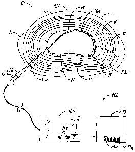
[0027] FIG. 1 is a cross-sectional view of an intervertebral disc with a
portion of the intervertebral apparatus of the present disclosure inserted
into the
intervertebral disc;
[00281 FIGS. 2a, 2b, and 2c show a system of components for the
intervertebral apparatus of FIG. I for RF intervertebral disc heating or any
other
RF heating, thermal ablation, or cryogenic denervation, the apparatus
including a
cannula, impedance stylet, and electrode;
[0029] FIG. 3 is a flow chart illustrating a method of creating a database
of thermal profile overlays;
[0030] FIG. 4 is a schematic view of a thermal acquisition system for
creating a thermal profile overlay in accordance with the present disclosure;
[0031] FIG. 5 is an enlarged schematic illustration depicting the creation
of a thermal profile image;
[0032] FIG. 6 is an exemplary thermal profile image produced by the
thermal acquisition system of FIG. 4;
[0033] FIG. 7 is a schematic illustration of a system for performing
surgical procedures using thermal profiling;
[0034] FIG. 8 is a flow chart illustrating an exemplary methods of
performing surgical procedures using thermally profiled electrodes;
7
CA 02637559 2008-07-15
[0035] FIG. 9 is a schematic illustration of a step of the method of FIG. 8;
[0036] FIG. 10 is a schematic illustration of another step of the method of
FIG. 8;
[0037] FIG. 11 is a schematic illustration of yet another step of the
method of FIG. 8;
[0038] FIG. 12 is an enlarged schematic illustration of the indicated area
of FIG. 11;
[0039] FIG. 13 is a fluoroscopic image of a spine illustrating a spinal
needle being inserted from the left into the nucleus pulposus of a vertebral
disc,
and an introducer cannula and electrode being inserted from the right into the
annulus fibrosus of the vertebral disc;
[0040] FIG. 14 is a fluoroscopic image of the spine of FIG. 13 illustrating
an overlay, in accordance with the present disclosure, superimposed over the
electrode to provide visualization of the predicted area of thermal effect;
[0041] FIG. 15 is an enlarged fluoroscopic image illustrating the
electrode/thermal profile of the overlay of FIG. 14;
[0042] FIG. 16 is an overlay illustrating the tissue histology together with
the actual thermal effects produced by the treatment;
[0043] FIG. 17 is a schematic illustration of a system for performing
surgical procedures using thermal profiling having a graphical user interface;
[0044] FIG. 18 is a schematic illustration of a system for thennally
treating target tissue having a graphical user interface which analyzes data
based
on physician-user outlined area on the target site;
[0045] FIG. 19 is a flow chart illustrating a method of performing a
surgical treatment with a user interface combined with a querying algorithm
and
recommender algorithm;
8
CA 02637559 2008-07-15
[0046] FIG. 20 is an illustration of a generator with a graphical user
interface; and
[0047] FIG. 21 is an illustration of a surgical instrument with a graphical
user interface.
DETAILED DESCRIPTION
[0048] The systems and methods of the present disclosure provide for a
more precise controlled positioning of a thermal probe in an intervertebral
disc
targeted for treatment. Moreover, the systems and methods of the present
disclosure provide for an improved ability to predict and/or visualize the
depth of
treatment possible by the thermal probe when set to various operative
parameters.
[0049] It will be readily apparent to a person skilled in the art that the
systems and methods of use of the systems can be used to treat/destroy body
tissues in any body cavity or tissue locations that are accessible by
percutaneous
or endoscopic catheters or open surgical techniques, and is not limited to the
disc
and/or spinal area. Applications of the systems and methods in all of these
organs and tissues are intended to be included within the scope of the present
disclosure.
[0050] Prior to a detailed discussion of the system and methods of use of
the systems and method of the present disclosure, a brief overview of the
anatomy of the intervertebral disc is presented. With reference to FIG. 1, an
intervertebral disc "D" is comprised of an annulus fibrosis "A" and a nucleus
pulposus "N" disposed within annulus fibrosis "A". Annulus fibrosis "A"
includes a tough fibrous material which is arranged to define a plurality of
annular cartilaginous rings "R" forming the natural striata of the annulus.
Nucleus pulposus "N" consists primarily of an amorphous gel having a softer
consistency than annulus fibrosis "A". Nucleus pulposus "N" usually contains
70% - 90% water by weight and mechanically functions similar to an
incompressible hydrostatic material. The juncture or transition area of the
annulus fibrosis "A" and nucleus pulposus "N" generally defines, for
discussion
purposes, an inner wall "W" of annulus fibrosis "A". Disc cortex "C" surrounds
9
CA 02637559 2008-07-15
annulus fibrosis "A". The posterior, anterior and lateral aspects of
intervertebral
disc "D" are identified as "P", "AN" and "L", respectively, with the opposed
posterior-lateral aspects identified as "PL".
[0051] When mechanical stress is put upon an intervertebral disc or when
an intervertebral disc degenerates with age, fissures, (illustrated by cracks
"F" in
FIG. 1), may occur in the posterior or posterior/lateral portions of the disc
"D".
Problems with the nerves, fissures "F" and degenerative discs can give rise to
various patient problems, such as back or leg pain originating from the
irritation
or occurrence of these abnormalities. Moreover, these conditions may
ultimately
result in conditions such as bulging or herniated discs. Heating and/or
electromagnetic field (EMF) therapy of intervertebral disc "D", for example,
annulus fibrosis "A" in the posterior "P" or posterior-lateral "PL" portions,
will
result in denervation of nerves and/or alterations and thermal ablation of
disc
structures, which will, in turn, produce alleviation of pain and healing of
the disc.
Thus, it is desirable, to insert and place a thermal or electromagnetic probe
in
posterior "P" and/or posterior-lateral "PL" portion of intervertebral disc "D"
where these neural and aberrant structures occur for the relief of pain and
other
disc related problems.
1. System for Thermally ProfilingSurgical Electrode
[0052] In the drawings and in the description which follows, the term
"proximal", as is traditional, will refer to the end of the system, or
component
thereof, which is closest to the operator, and the term "distal" will refer to
the end
of the system, or component thereof, which is more remote from the operator.
[0053] With reference to FIG. 1, in accordance with an embodiment of
the present disclosure, a system of using RF energy and thermal profiling in
surgical procedures is generally designated as 100. System 100 includes an
outer
insertion or introducer cannula 102, a probe for energy delivery (e.g.,
electrode,
thermal probe, EMF probe, electrosurgical probe, etc.) 104 which is
positionable
within cannula 102, an electrosurgical generator, power source or the like 106
connected to probe 104. Optionally, system 100 can include an impedance stylet
108 which is also positionable within cannula 102.
CA 02637559 2008-07-15
100541 As seen in FIGS. 1 and 2a, introducer cannula 102, typically is a
rigid tubular shaft 110 defining a longitudinal axis "X". Tubular shaft 110
which
may include a beveled tip 112 adjacent the distal end 114 and angled with
respect
to the longitudinal "X" axis. Beveled tip 112 may be angled from about 15 to
about 45 . Shaft I 10 may be composed of a conductive material such as
stainless
steel or other suitable composition and is insulated with insulation 116 along
at
least a portion, of the length thereof. Alternatively, shaft 110 may be
fabricated
from a suitable polymeric material and formed by conventional injection
molding
techniques. Distal end 114 of shaft 110 may be left un-insulated or exposed to
allow electrical communication with the tissue as cannula 102 is placed in the
tissue. (e.g., for impedance measuring, etc.) A handle or housing 118 is
connected to a proximal end of cannula 102 and may include an index marker
120 to indicate the direction of beveled tip 112 such that when probe 104 is
introduced within cannula 102, the surgeon may determine in which azimuthal
rotational direction beveled tip 112 is oriented.
[0055] Shaft 110 may have a diameter ranging from a fraction of a
millimeter to several millimeters and a length of a few centimeters up to
about 20
centimeters or more. Alternatively, shaft 110 may be fabricated from an MRI
(Magnetic Resonance Imaging) compatible material, including cobalt alloys,
titanium, copper, Nitinol, etc.
[0056] Power source or generator 106 may be, for example, a
radiofrequency generator providing energy at frequencies between several
kilohertz to several hundred megahertz. Generator 106 may have a power output
ranging from several watts to several hundred watts, depending on the clinical
need. Generator 106 typically includes control devices to increase or modulate
power output as well as readout and display devices to monitor energy
parameters such as voltage, current, power, frequency, temperature, impedance,
etc., as appreciated by one skilled in the art. Other types of power sources
and/or
generators are contemplated, e.g., including and not limited to resistive
heating
units, laser sources, or microwave generators.
11
CA 02637559 2008-07-15
[0057] With continued reference to FIGS. 1 and 2a-2c, probe (e.g.,
thermal or EMF probe) 104 of system 100 will be discussed. As seen in FIGS. 1
and 2c, electrode 104 is positionable within cannula 102 and is adapted for
reciprocal movement therewithin. When used as a radiofrequency probe, probe
104 is a monopolar system and is used in conjunction with an extended surface
area grounding pad 134 (see FIG. 13) which contacts the patient's skin over a
very large surface area relative to the exposed surface area of the electrode
tip. In
addition, when used as a radiofrequency probe, electrode 104 may be insulated
except for a distal portion thereof which may be left un-insulated for
transmission
of energy. Alternatively, and in one particular embodiment, probe 104 may be
entirely un-insulated while cannula 102 functions as the insulating element of
the
apparatus. In this arrangement, the degree of extension of the distal end
portion
of probe 104 beyond beveled tip 112 determines the heating capability of
electrode 104. Probe 104 includes a handle 130 and an elongated member or rod
132 extending distally from handle 130. An exemplary embodiment of a thermal
or EMF probe is provided in U.S. Patent 6,604,003 to Fredricks et al.
[0058] As seen in FIGS. 1 and 2b, impedance stylet 108 is positionable
within the lumen of cannula 102 and occludes the front opening of cannula 102
to
prevent entry of tissue, fluids, etc., during introduction of cannula 102
within
intervertebral disc "D". Stylet 108 may include a proximally positioned hub
140
which mates with housing 118 of cannula 102 into which stylet 108 is
introduced
to monitor impedance of the tissue adjacent the distal end of cannula 102.
Once
the combination of stylet 108 and cannula 102 are inserted into the body,
impedance monitoring assists in determining the position of beveled tip 112 of
cannula 102 with respect to the patient's skin, cortex "C", annulus fibrosis
"A",
and/or nucleus "N" of intervertebral disc "D". Each of these regions will have
different impedance levels which are readily quantifiable.
[0059] For example, for a fully insulated electrode or cannula with an
exposed area of a few millimeters at the cannula end, the impedance will
change
significantly from the position of the tip near to or contacting cortex "C" of
intervertebral disc "D" to the region where the tip is within annulus fibrosis
"A"
12
CA 02637559 2008-07-15
and further where the tip is within nucleus "N" of intervertebral disc "D".
Differences in impedance can range from a few hundred ohms outside
intervertebral disc "D", to 200 to 300 ohms in annulus fibrosis "A", to
approximately 100 to 200 ohms in nucleus "N".
[0060] This variation can be detected by the surgeon by visualizing
impedance on meters or by hearing an audio tone whose frequency is
proportional to impedance. Such a tone can be generated by a monitor (not
shown). In this way, an independent means is provided for detecting placement
of cannula 102 within intervertebral disc "D". Thus, for example, in an
application where an electrode 104 in the form of an EMF probe is to be
inserted
between adjacent layers of annular tissue, undesired penetration of the tip of
EMF
probe 104, extending from cannula 102, through inner wall "W" of annulus "A"
and into nucleus pulposus "N" can be detected via the impedance monitoring
means.
[0061] As seen in FIGS. 1, 4 and 7, system 100 further includes a library
200 including a plurality of thermal profiles/overlays 202. As used herein,
the
term library is understood to include and is not limited to repository,
databank,
database, cache, storage unit and the like. Each overlay 202 includes a
thermal
profile which is characteristic of and/or specific to a particular
configuration of
cannula/electrode assembly or amount of exposure (i.e., specific to the amount
of
probe 104 extending from the distal tip of cannula 102) of the
cannula/electrode
assembly. In addition, for each amount of exposure or configuration of the
cannula/electrode assembly, a plurality of overlays 202 is provided which
includes a thermal profile which relates to, for example, the amount of time
probe
104 is activated, the temperature to which probe 104 is heated, the frequency
of
the probe, etc.
[0062] As seen in FIG. 7, system 100 further includes an imaging system
300 configured and adapted to image and display a target surgical site.
Imaging
system 300 includes an imaging device 302, in the form of an x-ray imager, a
CT
scanner, an MRI device, a fluoroscopic imager and the like, and a monitor 304
13
CA 02637559 2008-07-15
for displaying the image produced by imaging device 302. The library 200 is
configured to operatively communicate with imaging system 300.
2. Method of Creating Thermal Overlay
[0063] Turning now to FIGS. 3-6, a method of creating a thermal overlay
202 (of a plurality of thermal overlays 202), is illustrated and described.
Creation
of a thermal overlay 202 includes the initial step of providing an acquisition
system 400, which may be a thermal acquisition system. Thermal acquisition
system 400 includes a bath 402 containing a quantity of a transparent test gel
404
(e.g., SMK/RFK formulation conductive polymer), a fixture 406 configured and
adapted to support a cannula/ probe assembly 102/104 and a piece of thermally
reactive paper, for example, thermal liquid crystal (LC) paper 408. The system
also includes an electrosurgical generator 410 operatively connected to
cannula/electrode assembly 102/104, and an image/data acquisition system 412
operatively connected to electrosurgical generator 410 and oriented toward
bath
402.
[0064] The method of creating thermal overlay 202 further includes the
steps of:
= stabilizing the temperature of test gel 404 in bath 402 to
approximately 30 C;
= coupling cannula/probe assembly 102/104 and LC paper 408 to
fixture 406 such that cannula/probe assembly 102/104 and LC paper 408
are placed in close proximity to one another, at a predetermined distance;
= placing (e.g., submerging) cannula/probe assembly 102/104 and
LC paper 408 into bath 402 such that cannula/probe assembly 102/104 is
disposed between LC paper 408 and image/data acquisition system 412;
= setting electrosurgical generator 410 to a predetermined setting
"lesion" or continuous mode at a temperature of about 42 C or about
80 C;
14
CA 02637559 2008-07-15
= activating and/or stimulating electrosurgical generator 410 such
that thermal radiation emanating from probe 104 impinges LC paper 408
to create a thermal image "TI"; and
= recording, with image/data acquisition system 412, the image (i.e.,
temperature gradients or "halos" 150 around cannula/probe assembly
102/104) created on LC paper 408 and recording the input parameters
(e.g., temperature, impedance, RF power, RF current, RF voltage, mode
of operation, exposure of probe 104 from distal end of cannula 102,
duration of application of the electrosurgical energy, etc.) associated with
the creation of the image on LC paper 408.
[0065] As can be appreciated from FIG. 5, temperature gradients or
"halos" 150 formed on LC paper 408 include a plurality of "halos" 150 of
differing color with each color representing a different temperature. The
temperature at which electrosurgical generator 410 is set will determine the
LC
paper that is used, for example, for a temperature setting of 42 C, LC paper
having a range of 35-40 C is used and for a temperature setting of 80 C LC
paper
having a range of 55-60 C is used. A thermal imaging camera or the like is
used
to record the temperature gradients produced on LC paper 408.
[0066] Thermal image "TI" and the data provided by thermal image "TI"
are recorded digitally. Accordingly, the method of creating one or more
thermal
overlays 202 can further include the step of storing, for example, digitally,
the
image and the data in library 200.
[0067] The process is repeated to create an overlay 202 for each
configuration of cannula/probe assembly 102/104 and each setting. In this
manner, a plurality of overlays 202 is created and stored in library 200. For
example, a series of overlays 200 can be created for each temperature setting
of
electrosurgical generator 410 (e.g., 42 C and 80 C). For each temperature
setting
of electrosurgical generator 410, a series of overlays 200 can be created for
each
tip exposure dimension (e.g., 3, 4 and 6mm) of cannula 102. For each exposure
dimension of cannula 102, a series of overlays 200 can be created for each
offset
CA 02637559 2008-07-15
position of probe 104 relative to cannula 102. Offset positions are referenced
to
the "flush" condition (i.e., 0 mm) which is obtained by placing a flat surface
flush
against the bevel of cannula 102 and inserting probe 104 into cannula 102
until
probe 104 contacts the flat surface.
[00681 As seen in FIG. 6, the shapes of thermal images "TI" are usually
elliptical and are centered on the exposed tip of probe 104. The major axis of
the
ellipse can be measured using an image analysis software program. Thermal
image "TI" is represented by a series of gradations or rings 150, each ring
150
representing a different temperature intensity.
[0069] Creation of the thermal overlays according to the present method
provides visual information that will assist in comparing the performance
between different electrodes and different electrosurgical generators.
[0070] While the above-described method is a particular method of
creating a thermal overlay, it is envisioned that other methods are also
possible.
For example, it is envisioned that a thermally responsive gel or paint (e.g.,
a
composition containing quantities of a thermally responsive substance therein)
may be applied to the surface of a sample tissue (e.g., human cadaver tissue,
porcine tissue and the like). The cannula/probe assembly 102/104 may then by
introduced into the sample tissue and electrosurgical generator 410 activated
in
accordance with the method described above in order to create a thermal
profile
on the surface of the sample tissue. The thermal profile may be recorded in a
manner similar to the method described above. This procedure may be repeated
as many times as necessary in order to produce thermal profiles for various
insertion depths of cannula/probe assembly 102/104 into the sample tissue, for
various settings of electrosurgical generator 410, and/or for various
configurations of cannula/probe assembly 102/104. In this manner, the effects
of
cannula/probe assembly 102/104 may be easily mapped on tissue.
3. Method of Performing Surgical Procedures
[00711 Prior to a detailed discussion of the methods of performing
surgical procedures in accordance with the present disclosure, a brief
overview of
16
CA 02637559 2008-07-15
a general method of performing thermal treatment of an intervertebral disc is
discussed. With reference to FIG. 1, the targeted intervertebral disc "D" is
identified during a preoperative phase of surgery. Access to the
intervertebral
disc area is then ascertained, through percutaneous techniques or, less
desirably,
through open surgical techniques. Cannula 102, with stylet108 positioned and
secured therein, is introduced within intervertebral disc "D" from a posterior
or
posterior-lateral location. Alternatively, cannula 102 may be utilized without
stylet 108.
[0072] During introduction of the cannula/stylet assembly 102/108, the
impedance of the tissue adjacent the distal end of cannula 102 is monitored.
Impedance monitoring may be utilized to determine the position of the tip of
cannula 102 with respect to the patient's skin, the cortex "C", the annulus
fibrosis
"A" and/or the nucleus pulposus "N" of the intervertebral disc "D". As
discussed
above, these regions have different and quantifiable impedance levels thereby
providing an indication to the user of the position of the tip of cannula 102
in the
tissue. Monitoring of the location of the tip of cannula 102 may also be
confirmed with use of imaging system 300. Typically, the tip of cannula 102 is
positioned within annulus fibrosis "A" of intervertebral disc "D" at a
posterior
lateral "PL" location of intervertebral disc "D" without penetrating through
inner
wall "W" and into nucleus "N".
[0073] With cannula 102 in the desired position, stylet 108 is removed
and probe 104 is positioned within cannula 102 and advanced therethrough.
Probe 104 is advanced an amount sufficient to at least partially expose a
distal
portion thereof from the tip of cannula 102. The degree of exposure of the
distal
end portion of probe 104 from the tip of cannula 102 may be indicated by
distance or indexing markings provided on rod 132 of probe 104.
[0074] Once probe 104 is positioned within annulus fibrosis "A" as
desired, power source 106 is activated whereby probe 104 delivers thermal
energy and/or creates an electromagnetic field adjacent intervertebral disc
"D" to
produce the thermal and/or EMF therapy desired. Appropriate amounts of power,
current, or thermal heat may be monitored from power source 106 and delivered
17
CA 02637559 2008-07-15
for a certain amount of time as determined appropriate for clinical needs. For
example, if denervation of nerves surrounding intervertebral disc "D" is the
objective, the tissue adjacent the exposed end of probe 104 is heated to a
temperature from about 45 C to about 60 C. If healing of fissures in
intervertebral disc "D" is the surgical objective, the temperature in the
tissue is
raised to about 60 C-75 C.
[0075] As can be appreciated by one of skill in the art, the degree and/or
amount of exposure of the distal portion of probe 104 from the tip of cannula
102
controls the volume of disc tissue heated by probe 104. Sensors (not shown)
can
be used to provide information concerning the temperature of tissue adjacent
probe 104. Alternatively, impedance means (not shown), associated with, e.g.,
probe 104, can provide impedance measurements of the tissue thereby providing
an indication of the degree of desiccation, power rise or charring, that may
be
taking place near the exposed distal portion of probe 104. This indicates the
effectiveness of the treatment.
[0076] Turning now to FIGS. 7-16, in accordance with the present
disclosure, a method of performing a surgical procedure using thermally
profiled
electrode overlays is illustrated and described. The method includes the
initial
step of providing a system 100 for using RF energy and thermal profiling in
surgical procedures. As described above, system 100 includes an introducer
cannula 102, at least one probe 104 positionable within cannula 102, a library
200
including a plurality of thermal overlays 202. An imaging system 300 is also
included which is configured and adapted to take images of a target surgical
site
and display the images of the target surgical site to the operator.
[0077] The method of performing the surgical procedure further includes
the steps of:
= imaging the target surgical site with imaging system 300 in order
to display the target surgical site on monitor 304, see FIGS. 7 and 10;
18
CA 02637559 2008-07-15
= selecting an overlay 202 from the plurality of overlays 202 stored
in library 200 relating to the desired treatment effect of a particularly
shaped probe;
= superimposing the selected overlay 202 over the imaged target
surgical site, see FIGS. 7, 11, 12, 14 and 15;
= evaluating the scope, degree and/or depth of treatment provided to
the target surgical site by using and/or configuring system 100 to the
parameters corresponding to and/or associated with the selected
overlay 202;
= inserting a cannula/probe assembly 102/104, including a probe
104 corresponding to the electrode parameters of the selected overlay
202, into the target surgical site, see FIGS. 11 and 15; and
= activating and/or stimulati.ng probe 104 according to the
parameters corresponding to and/or associated with the selected
overlay 202.
[0078] In an alternative method, probe 104 is inserted into the target
surgical site (see FIGS. 11 and 13) prior to superimposing overlay 202
thereon.
With probe 104 in position, various overlays 202 are superimposed over probe
104 in order to illustrate the various depths of thermal penetration possible
and in
order to determine the desired and/or appropriate operative and/or activation
parameters for probe 104, see FIGS. 12, 14 and 15. An overlay 200 is selected
which corresponds to a surgical effect desired. Probe 104 is then activated in
accordance with the parameters of selected overlay 202.
[0079] In either of these methods, the selected overlay 202 provides the
operator with a visual representation of the depth of thermal penetration
produced
by probe 104 when set to the parameters of the selected overlay 202. In
addition,
the selected overlay 202 enables the operator to better visualize the desired
placement of probe 104 and/or enables the operator to guide probe 104 into the
target surgical site along a path corresponding to the direction of the
thermal
19
CA 02637559 2008-07-15
profile of the selected overlay 202. Moreover, the thermal visualizations
offered
by overlays 202 can assist in identifying mechanisms of action and optimizing
the desired effects. In addition, the operator may compare the various effects
of a
variety of differently-shaped electrodes to optimize surgical outcome.
[0080] FIGS. 13-16 are fluoroscopic images of the spine illustrating the
steps of the methods described above.
[0081] The implementation and use of overlays 202 on monitors 304 can
range from simple systems where the operator manually places overlay 202 on
monitor 304 or to more sophisticates pattern recognition systems. The pattern
recognition systems could be used to identify the treatment parameters
selected
by the operator, select the appropriate overlay 202 from library 200, and
project
and/or display overlay 202, at appropriate scale and placement, on monitor
304.
As can be appreciated, this enables the surgeon to visualize and estimate the
and
result of the treatment and overall tissue effect (e.g., thermal spread)
before
energizing the electrode.
4. System for thermally treating target tissue having a graphical user
interface
[0082] FIG. 17 illustrates another embodiment according to the present
disclosure which includes a system 500 for thermally treating target tissue
having
a graphical user interface. More particularly, system 500 includes a surgical
device 104 adapted to connect to a power source (e.g., surgical generator 106)
and an imaging device 302 which displays an image 510' of the surgical device
in situ (at the target surgical site) 510. A database module 225 is
operatively
coupled to a library 200 of thermal overlays 202 or a plurality of computer-
simulated thermal overlays 508. For the purposes herein, the thermal overlays
202 are actual overlays created by the aforementioned method utilizing thermal
paper and digital photography wherein computer-simulated overlays are overlays
which may be manipulated by the user based on different surgical parameters,
tip
configurations, depth of penetration, different surgical techniques,
previously-
recorded surgeries, etc.
CA 02637559 2008-07-15
[0083] A querying algorithm 502 (not shown) may be included which is
configured to enable a user to select one or more simulated thermal overlays
202
or computer simulated thermal overlays 508 according to a desired thermal
image
for the surgical device 104 used in a particular surgery or for a particular
surgical
purpose. For example, the querying algorithm 502 enables a user to select one
or
more thermal overlays 202 or computer simulated thermal overlays 508 based on
various electrical settings of the power source 106, tip configuration of the
surgical device 104, depth of penetration of the surgical device 104,
activation
time of the surgical device 104 and combinations thereof. A graphical user
interface system 516 (e.g., a monitor 304) is connected to the system 500 to
facilitate selection of various thermal or computer-simulated overlays 202 and
508, respectively. It is envisioned that graphical user interface or monitor
516
may be connected to a group of devices which include a power source 106, image
display 304 and querying algorithm 502.
[0084] It is also envisioned that a particular medical image 510' may be
selected from a group (not shown) consisting of real time images and archived
images of a particular patient or surgery. Real time images are images that
appear live, whereas archived images are pre-recorded. It is also envisioned
that
archived images may be a digital representation, for example DICOM format,
which may be stored in a library or the like. A physician-user may retrieve an
archived medical images, for example similar to their patient or study, and
simulate a treatment plan. Archived medical images may be stored in database
modules, for example archival systems such as PACS or the like.
[0085] As seen in FIG. 17, system 500 further includes imaging system
300 configured and adapted to display a surgical device in situ 510' as well
as a
selected overlay 202 or 508. Imaging system 300 may include one or a
combination of the following known imaging devices 302: an x-ray imager, a CT
scanner, an MRI device, a fluoroscopic imager or the like for scanning or
reading
a surgical site 510 and a monitor 304 for displaying the surgical site image
510'
produced by imaging device 302. As mentioned above, library 200 is operatively
coupled to imaging system 300 to allow a surgeon to superimpose overlays 202
or 508 on a monitor 304.
21
CA 02637559 2008-07-15
[0086] Thermal overlay 202 may be a thermal image depicting a thermal
profile of the surgical device 104. It is envisioned thermal overlay 202 may
have
certain stored data inputs which may include input parameters for a power
source
106. When the suggested input parameters of the overlay are entered, the power
source 106 delivers the energy to surgical device 104 and creates the energy
of
the thermal overlay 202 depicted on the image display 304. A method of
creating
a thermal overlay 202 is described more in detail above. Alternatively (or in
addition to), the surgeon may select a computer-simulated image 508 and
overlay
the computer-simulated image 508 atop the active tip of the surgical device to
visualize "what if' scenarios when adjusting various surgical parameters such
as,
generator controls, depth of penetration, type of tip being utilized, etc. In
one
envisioned embodiment, one or more thermal images 202 are overlayed on the
surgical device and displayed on the monitor and one or more computer-
simulated overlays are superimposed atop the thermal images 202.
[0087] Querying algorithm 502 may be utilized to facilitate or enhance
the ability of the user to search the library 200 of thermal overlays 202 or
computer simulated thermal overlays 508 and locate one or more relevant
overlays 202 or 508 for a particular surgical purpose. Each overlay 202 or 508
may be oriented and scaled to correspond with the scale of the surgical device
in
situ 510 as displayed on the monitor 304. It is envisioned that part of the
querying process may be searching the library 200 of thermal overlays 202 or
computer simulated thermal overlays 508 according to the type of surgical
device
104 attached to system 500 or displayed on an archived image (not shown) if a
surgical device 104 is not connected. Thermal overlays 202 or computer
simulated thermal overlays 508 may be based on various electrical settings of
the
generator 106, tip configuration of the surgical device 104, depth of
penetration
of the surgical device 104, activation time of the surgical device 104 and
combinations thereof. The querying system 502 may also be configured to
determine similarities of the thermal overlays 202 or computer simulated
thermal
overlays 508 and the displayed image 510' displayed. Each surgical device 104
may have a different thermal 202 image and therefore the thermal overlay 202
will portray a different treatment effect in surgical site 510.
22
CA 02637559 2008-07-15
[0088] The graphical user interface or monitor 516 may include an
electronic pointing device 518 to facilitate selection or manipulation of a
particular overlay 202 or 508 on the displayed image 510'. Alternatively; the
graphical user interface system 516 may include touch screen capabilities, a
mouse, a stylet or interact with the user using voice-activated commands.
[0089] System 500 is also envisioned to include the capability of
predicting the thermal treatment of a device in a computer simulation or
utilizing
a computer simulated overlay. In other words, the user may select a particular
thermal overlay 202 and the system may be able to provide a computer-generated
simulation of the thermal treatment of tissue from activation to a preset time
period based on the input parameters by the user. Moreover, if the particular
actual thermal image 202 for a particular instrument or tip configuration is
not
archived in the library based on the input parameters, the system 500 may be
configured to utilized one or more computer-simulated overlays 508 to
extrapolate a thermal profile of the treatment zone. For example, the querying
algorithm 502 (or a separate algorithm) may be designed to locate and select a
predicted computer generated overlay 508 from the library 200 of images that
most closely corresponds to the target surgical site. Predicted computer
generated overlay 508 may be a computer-generated image of the target surgical
site after treatment. The predicted overlay 508 may be part of library 200 of
a
plurality of predicted overlays 508.
[0090] As best shown in Fig. 18, the present disclosure also relates to a
system 600 and method of thermal imagery which analyzes data based on a
physician-user outlined area on the target site. For example, it is envisioned
that
the user or physician may mark up the desired treatment zone on the monitor
304
and the querying algorithm 502 may then be activated to match the outlined
treatment zone 512 with a particular surgical device, tip configuration, depth
of
penetration, etc. to satisfy the desired treatment zone 512 indicated by the
physician.
[0091] System 600 may also include imaging tools 302, e.g., a magnetic
resonance imager (MRI), which provides thermal imagery in real time or near
23
CA 02637559 2008-07-15
real time on monitor 304 to allow a physician to monitor thermal surgical
procedures in situ during the operation. Other types of imaging devices are
also
contemplated as is known in the art.
[0092] The present disclosure may also relate to a method for thermally
treating target tissue utilizing a graphical user interface utilizing system
600. The
method includes the initial steps of selecting a surgical device 104 adapted
to
connect to a surgical generator 106 and displaying the treatment site in situ
510'.
The method would also include connecting to a database module 225 adapted to
connect to a library 200 of thermal images 202, 508. The method also includes
the steps of introducing the surgical device 104 to the treatment site 510 and
querying a library 200 of thermal overlays 202, 508; superimposing a selected
simulated image 514 atop the image of the surgical device 104' and displaying
both the image of the surgical device in situ 104' and the simulated image
510'
on monitor 304 by an imaging device 302 over the surgical device 104 in situ
510.
[0093] The method may also include superimposing one or more overlays
atop the tissue to ascertain a desired treatment area 512 for the tissue based
on
tissue type, generator settings, type of surgical device, tip configuration of
the
surgical device and/or depth of penetration of the surgical device. The method
also includes the step of selecting a desired overlay and configuring the
generator
and surgical instrument accordingly to treat tissue.
[0094] The method may also include the steps of providing an interactive
device 518 such as a mouse, pencil, stylet, touch screen, voice command for
identifying and outlining a desired treatment zone 512 across the tissue. The
method would also include the step of utilizing the querying algorithm 502
(not
shown) to select or recommend one or more surgical instruments, general
settings, tip configurations and depth of penetration for achieving the
desired
tissue treatment.
[0095] It is also envisioned that the physician may initially choose a
particular instrument, tip configuration, power setting or penetration depth
and
then utilize the querying algorithm to display a range or treatment options to
24
CA 02637559 2008-07-15
achieve the desired treatment result, according to one or more variable, e.g..
size,
shape and type of procedure and surgical device selected. If no appropriate
overlay is available in the library of images 200, the querying algorithm may
be
configured to extrapolate or scale one or more overlays 202, 508 to achieve an
approximate desired result. The querying algorithm may be utilized to
automatically select one or more recommended overlays 202, 508 based on the
tissue type, generator setting, tip configuration, type of instrument or
desired
depth of penetration and display such recommendation on the monitor for
interactive comrnunication with the physician through one or more of the above
interactive devices. The physician may also be able to scroll through the
library
200 of images and pick a desired image 202, 508 manually if desired.
[0096] As seen in FIG. 18, it is also envisioned that the physician may
utilize the overlay during the course of tissue treatment and intermittently
display
the overlay atop the actual treatment zone for verification purposes. The
overlay
may also include one or more overlay modes which allow the physician to treat
the tissue without the overlay interfering with the physician's view of the
treatment area. For example, a phantom mode (or ghost mode) 514, an outline
mode or a strobe-like mode may be employed for this purpose which displays the
overlay 202, 508 in a particular fashion (e.g., lighter contrasting color for
phantom mode) so not to impede visualization of the treatment zone 512.
[0097] As can be appreciated, utilizing the overlays 202, 508 prior to
activation allows a surgeon to select the most appropriate generator setting,
surgical instrument, tip configuration and depth of penetration for a
particularly-
sized treatment area without activating of the surgical device 102. Once the
physician determines whether the area to be treated is appropriately covered
by
the selected thermal overlay 202, 508 based on the generator settings,
instrument,
tip configuration and/or depth of penetration, the physician is free to
activate the
instrument to treat the tissue.
5. Method of Performinga Surgical Treatment with a User Interface
[0098] Turning now to FIG. 19, steps 700 - 790 illustrate and describe a
method of performing a surgical treatment with a user interface combined with
a
CA 02637559 2008-07-15
querying algorithm and recommender algorithm. In step 700, the generator 106,
user interface 516 and the surgical instrument 104 are activated. Activation
may
be executed manually or automatically. The user may manually activate each
component by interacting with the user interface or by remote control.
Alternatively, the generator 106 may be configured to activate automatically,
for
example, when specific or all the components of the surgical operation are
connected and ready for use. A feedback loop may be used for this purpose.
[0099] In step 710, the components, for example, the imaging device,
generator and surgical instrument are synchronized. Synchronization may be
required for tuning the surgical instrument, for example ultrasonic surgical
instruments. Synchronization may also be required for compatibility purposes
since certain types of power generators and surgical instruments may need to
be
programmed to work together. In the event any of the components are not
compatible with each other, an indicator may be provided to alert the user via
an
audible alarm, a visual alarm and/or an error message displayed on the
graphical
user interface of the generator 106.
[00100] In step 720, the user views the entire surgical site 510' via screen
304 sent by an imaging device, not shown, (e.g., a CT scanner, an MRI, or the
like), as illustrated in FIG. 20. The user then selects target tissue, for
example a
tumor, that may require ablation.
[00101] Further in step 730, the querying algorithm 502 searches the
database, described in detail above, for a substantially similar stored image
of a
tissue volume from a library 200 as discussed above. As mentioned above, the
library 200 or database 225 may contain overlays, computer generated
simulations, or video of previously performed surgical procedures on similar
tissue volumes.
[00102] Step 740 provides a recommender algorithm to search through the
database, library or look-up tables for actual or computer-generated
simulations
of recommended treatments for the tumor volume based on this pre-recorded
data. The treatment may include electrode shape, size, orientation, actuation
levels and actuation times. In addition, the database, library or look-up
table may
26
CA 02637559 2008-07-15
be searched for recommended instrument types, models, and/or sizes for use
with
specific treatment procedures. Other examples may include, the recommender
algorithm fmding a video of a previous surgical procedure that used an antenna
to
ablate a similarly sized tumor. In this manner, the user may view the
recommended instrument and procedure and compare it to a pre-planned
treatment or procedure. At any time the user may follow the recommended
procedure or proceed manually. The user may also decide to alter the
recommended surgical procedure and adjust the settings on the user interface
if
applicable.
[00103] At step 750, the user places the surgical instrument recommended
by the recommender algorithm or user's choice into the patient and proceeds
with
the surgical procedure. Further at step 760, when the user has initiated the
surgical procedure an instrument locating algorithm may scan the entire
surgical
site and display on the graphical user interface where the surgical instrument
is
located. The recommender algorithm may recommend a location for the user to
place the surgical instrument or a cluster of locations for placement of the
surgical instrament. For example, it is contemplated that the recommender
algorithm may recommend a particular ablation electrode being activated in a
series of steps to properly ablate the tissue.
[00104] The recommender algorithm may create an electrosurgical
protocol based on voltage, current and activation time, model, and size of the
surgical instrument. The electrosurgical protocol provides for a more accurate
recommendation as to which surgical instrument may be used for a particular
surgical procedure.
[00105] At any one of these steps an alarm may activate if a particular
surgical instrument is misplaced relative to the recommended placement area.
Also, at any one of these steps an alarm may activate if healthy tissue is
being
affected and/or if the target tissue volume should no longer be treated, e.g.,
a
complete alarm. Also in the present disclosure, at any time the surgical
procedure may be modified and/or altered by user.
27
CA 02637559 2008-07-15
[00106] In step 770 the user interface displays an option for the user to
vary the size of target surgical site area. FIGS. 20 and 21 illustrate a
graphical
user interface where the generator 106 and/or the surgical instrument 104 have
a
graphical user interface to control the size of the desired tissue volume 512.
[00107] FIG. 20 shows a generator 106 that has a touch screen 304
integrated therein which may be utilized in an interactive fashion depending
on
user preference. The user is able to adjust the overall size of the desired
tissue
volume 512, via the user interface, by using up and down arrows 602, 604 on
the
touch screen or possibly an interactive tool. Also, as shown in FIG. 21, the
user
may vary the size of the target tissue volume 512 to be treated through
various
input devices 606 (for example, a mouse, a pen, or a wand) that may be on the
surgical instrument 104, a handheld electrode or antenna. The input device 606
may have up and down arrows to vary the size of the desired target tissue
volume. The surgical instrument 104 and input device 606 may also have a
display 608, e.g., an LCD, that displays the size of the target tissue volume
and
the desired treatment.
[00108] Alternatively, a user may adjust the size of the area to be treated
by drawing or selecting the area displayed on the screen. The user interface
on
the generator 106 may comprise a touch screen 304 that indicates an image of
the
surgical site 510'. The image is produced by an imaging device (not shown), as
described above. The user may indicate a target surgical site 512 where the
surgical procedure should take place. The user may select the target surgical
site
512 with a mouse, stylet, touch screen or the like. The recommender algorithm
may be configured to utilize this information and recommend a particular
insttument electrode, placement, ablation series and/or activation energy and
time.
[00109] In addition, a plurality of overlays may be placed directly on the
surgical site image 510' and manually compared. The group of overlays may
comprise an overlay of a particular surgical instrument thermal plumes, a
target
surgical site, or a predicted end result target surgical site. The overlays
may be
displayed during surgery or for consultation of the surgical procedure.
28
CA 02637559 2008-07-15
[00110] The user proceeds with the surgical procedure (e.g., ablation,
cauterization, etc.) and the graphical user interface may display thermal
arrays in
order to characterize the difference between temperature gradients.
[00111] In addition, the thermal temperature may be used to monitor the
surgical site, e.g., the target volume. The target surgical site may be mapped
out
as a graph and each location would be mapped at a specific coordinate. The
graphical user interface may display the surgical site in a two-dimensional
array
or a three-dimensional array.
[00112] At step 780, a thermal sensor feedback loop may be utilized to
monitor and/or control the surgical device and/or the generator. For example,
the
thermal sensor feedback loop may be configured to control the energy output of
the surgical instrument according to the size and location of surrounding
tissue of
the target surgical site. Each location on the screen may have a different
temperature or appearance. The user may select or exclude certain
characteristics
from being monitored by the thermal sensor feedback loop (for warning/alarm
purposes).
[00113] In step 790 the system systematically shuts down the surgical
instrument energy power source and may be configured to provide a complete
alarm. Other areas of the surgical site may also be monitored.
[00114] While the above description contains many specific examples,
these specific should not be construed as limitations on the scope of the
disclosure, but merely as exemplifications of particular embodiments thereof.
Those skilled in the art will envision many other possible variations that are
within the scope and spirit of the disclosure as defmed by the claims appended
hereto.
29