Note: Descriptions are shown in the official language in which they were submitted.
CA 02639416 2008-09-08
DIAGNOSTIC TEST FOR SUSCEPTIBILITY TO B-RAF KINASE INHIBITORS
BACKGROUND OF THE INVENTION
The BRAF gene encodes B-Raf, a serine/threonine kinase, which couples
signaling from
activated RAS to downstream MAPK kinases (Wellbrock et al., Cancer Res.
64:2338-
2342, 2004). Oncogenic mutations in B-Raf result in gain-of-kinase-function,
rendering
the Raf-MEK-ERK pathway constitutively active in the absence of the typical
growth
factors. This constitutive activation correlates with poor prognosis in
metastatic
melanoma (Houben et al., 2004, supra). Activating mutations in BRAF have been
reported in a variety of malignancies. For example, mutations in BRAF are
found in as
many as 70% of human melanoma cases. A single-base mutation (T>A) at
nucleotide
position 1799 in codon 600 of exon 15 leads to a valine-to-glutamate
substitution
(V600E), which is present in more than 85% of melanomas with a mutation in B-
Raf
(Davies et al., Nature 417: 949-954, 2002; Garnett and Marais, Cancer Cell 6:
313-319,
2004; Gray-Schopfer et al., Cancer Metastasis Rev. 24:165-183, 2005; Houben et
al.,
2004). Less commonly, V600E results from the two-base mutation TG>AA at
nucleotide positions 1799-1800 (Houben et al., J Carcinog. 3:6, 2004).
A number of kinase inhibitors are known, including those that inhibit B-Raf.
Such
inhibitors include PLX4032, which selectively inhibit B-Raf V600E kinase
activity. The
current invention provides methods of identifying V600E-positive patients to
select for
treatment using a B-Raf kinase inhibitor, e.g., PLX4032.
BRIEF SUMMARY OF THE INVENTION
The invention provides methods and compositions for the detection of patients
who are
candidates for treatment with a B-Raf kinase inhibitor. Thus, in one aspect,
the
invention provides a method of determining sensitivity of cancer cells to a B-
Raf
inhibitor, the method comprising: (i) providing a nucleic acid sample from
cancer cells
from a patient that has a cancer; (ii) amplifying a target polynucleotide
sequence in the
nucleic acid sample using a primer pair that amplifies the target
polynucleotide
sequence, wherein the target polynucleotide sequence comprises a V600E
mutation site
in BRAF and amplification is performed in the presence of a labeled
oligonucleotide
1
CA 02639416 2008-09-08
probe that comprises at least 15 contiguous nucleotides of the sequence set
forth in
SEQ ID NO:1 and detects the presence of a mutated sequence at the V600E
mutation
site in BRAF; and (iii) detecting the presence or absence of a V600E mutation
in
BRAF; thereby determining the sensitivity of the cancer to the B-Raf
inhibitor. In some
embodiments, the probe has the nucleotide sequence set forth in SEQ ID NO: 1.
The
amplification can be performed in the presence of a second probe that detects
the
presence of a wild type sequence at the V600E mutation site. In some
embodiments, the
second probe comprises at least 15 nucleotides of SEQ ID NO:2. In some
embodiments
the second probe has the nucleotide sequence set forth in SEQ ID NO:2. In
certain
embodiments the probe can be labeled with a fluorescent label. The probe can
also be
labeled with two labels, where one of the labels is a fluorescent dye and the
other label
is a quenching moiety.
In some embodiments, both primers of the primer pair used in the amplification
reaction
hybridizes to exon 15 of BRAF. In some embodiments, one of the primers of the
primer
pair used in the amplification reactions comprises at least 15 contiguous
nucleotides of
the nucleotide sequence set forth in SEQ ID NO:3, e.g., one of the primers in
the primer
pair can have the nucleotide sequence set forth in SEQ ID NO:3. In further
embodiments, one of the primers in the primer pairs comprises at t least 15
contiguous
nucleotides of the nucleotide sequence set forth in SEQ ID NO:4. For example,
the
primer can have the nucleotide sequence set forth in SEQ ID NO:4. In further
embodiments, the primer pair used for the amplification comprises primers
having the
nucleotide sequences set forth in SEQ ID NO:3 and SEQ ID NO:4.
In some embodiments, the step of amplifying the reaction comprises an RT-PCR.
The method can be employed to detect cancers that have a mutation at amino
acid
position 600 of B-Raf, e.g., a V600E mutation. In some embodiments, the cancer
is
melanoma. In other embodiments, the cancer is colon cancer or thyroid cancer.
In some
embodiments, the nucleic acid sample used in the methods of the invention to
detect the
mutation can be from a skin biopsy. In other embodiments, the sample is from a
colon
biopsy. The sample can also be from paraffin-embedded tissue. The method of
the
invention can further comprise recording a diagnosis that the patient is
sensitive to a
B-Raf inhibitor, such as a mutant -specific B-Raf inhibitor, e.g., PLX4032.
2
CA 02639416 2008-09-08
In some embodiments, the method further comprises administering a B-Raf
inhibitor to
the patient. The B-Raf inhibitor can be a mutant specific B-Raf inhibitor such
as,
PLX4032.
In another aspect, the invention provides a method of identifying a PLX4032
treatment
candidate, the method comprising: providing a sample from a subject; and
detecting a
biomolecule comprising a BRAF V600E mutation from the sample, thereby
identifying
the PLX4032 treatment candidate. In some embodiments, the biomolecule is a
nucleic
acid. In other embodiments, the biomolecule is a polypeptide. The polypeptide
can be
obtained, e.g., from a sample comprising cancer cells from the patient. In
some
.. embodiments, the polypeptide may be detected using an immunoassay. The
method can
also comprise administering PLX4032 to the subject.
In another aspect, the invention provides a kit for detecting a patient that
is a candidate
for treatment with a B-Raf inhibitor, wherein the kit comprises a first allele-
specific
probe, wherein the first probe detects a V600E mutation in BRAF and comprises
at least
.. 15 contiguous nucleotides of the sequence set forth in SEQ ID NO:l. In some
embodiments, probe has the nucleotide sequence set forth in SEQ ID NO:l. A kit
of the
invention may further comprise a second allele-specific probe, wherein the
second
probe detects the wild type BRAF sequence and comprises at least 15 contiguous
of the
nucleotide sequence set forth in SEQ ID NO:2. In some embodiments, the second
probe
has the nucleotide sequence set forth in SEQ ID NO:2.
In further embodiments, a kit of the invention further comprises a primer pair
that
amplifies a target region of BRAF that comprises a V600E mutation site. For
example,
the primer pair can comprise a primer that comprises at least 15 contiguous
nucleotides
of the nucleotide sequence set forth in SEQ ID NO:3. In certain aspect the
primer has
.. the nucleotide sequence set forth in SEQ ID NO:3. In other embodiments the
primer
pair can comprise primers that comprise at least 15 contiguous nucleotides of
the
nucleotide sequence set forth in SEQ ID NO:3 and SEQ ID NO:4. In some
embodiments, the primer pair comprises primers that have the nucleotide
sequences set
forth in SEQ ID NO:3 and SEQ ID NO:4.
.. Thus, in some embodiments, a kit of the invention can comprise: a probe
that has the
nucleotide sequence set forth in SEQ ID NO:1; a probe that has the nucleotide
sequence
3
CA 02639416 2008-09-08
set forth in SEQ ID NO:2; a primer that has the nucleotide sequence set forth
in SEQ ID
NO:3; and a primer that has the nucleotide sequence set forth in SEQ ID NO:4.
BRIEF DESCRIPTION OF THE DRAWINGS
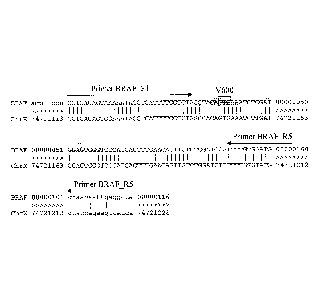
Figure 1 shows an alignment of a B-Raf V600E amplicon (SEQ ID NO:5) with a
corresponding region of Chromosome X (SEQ ID NO:6). The B-Raf V600E primer
sites are shown with arrows. The amplicon includes portions of exon 15
(uppercase) and
intron 15 (lowercase). Vertical bars denote positions of identity between the
BRAF and
chromosome X sequences. Codon 600 is boxed; GTG corresponds to valine (V). The
probe binding region is highlighted by shading; the mutant (MU) probe is
longer than
the wild type (WT) probe by two nucleotides (5'-CT in highlighted region) and
both
probes bind to the complement of the sequence shown here.
Figure 2 shows an alignment of a B-Raf exon 15 region surrounding codon 600
(arrow)
(SEQ ID NO:?) with homologous regions of A-Raf (SEQ ID NO:8) and C-Raf (SEQ ID
NO:9). Asterisks mark amino acid differences relative to the B-Raf sequence
(e.g.,
Mercer & Pritchard, Biochim Biophys Acta 1653:25-40, 2003).
Figure 3 shows an alignment of the a B-Raf V600E amplicon (SEQ ID NO:5) with
the
corresponding sequences from the ARAF (encodes A-Raf) (SEQ ID NO:11) and RAF]
(encodes C-Rat) (SEQ ID NO:10) genes. Nucleotides in lowercase differ from the
BRAF sequence.
DETAILED DESCRIPTION OF THE INVENTION
Definitions
In the context of this application, a "V600E" refers to a mutation in BRAF
(T>A at
nucleotide position 1799) that results in substitution of a glutamine for a
valine at amino
acid position 600 of B-Raf "V600E" is also known as "V599E" (1796T>A) under a
previous numbering system (Kumar et al., Clin. Cancer Res. 9:3362-3368, 2003).
In the context of this invention, a "B-Raf kinase inhibitor" inhibits activity
of a B-Raf
kinase. Such an inhibitor may also inhibit the activity of other kinases,
including other
raf kinases.
4
CA 02639416 2008-09-08
A "mutant-specific B-Raf kinase inhibitor" as used herein refers to a B-Raf
inhibitor
that has selectivity for a mutant B-Raf such as B-Raf having a mutation at the
valine
residue at amino acid position 600, e.g., a V600E mutation, compared to wild
type B-
Raf. Such an inhibitor is at least two times, more often at least three times,
or more as
potent an inhibitor of mutant B-Raf, e.g., a B-Raf having a V600E mutation, in
comparison to wild type. For example, in some embodiments, a "mutant-specific
B-Raf
inhibitor" may have an IC50 for kinase inhibition activity (biochemical assay)
of about
30 nM for V600E B-Raf whereas the corresponding IC50 for wild type B-Raf is
about
100 nM. The potency can also be compared in terms of IC50 values for cellular
assays,
e.g., cellular assays that measure growth inhibition. A "mutant-specific B-Raf
inhibitor"
in the context of this invention may also inhibit kinases other than B-Raf,
e.g., other raf
kinases.
The term "hybridization" refers to the formation of a duplex structure by two
single
stranded nucleic acids due to complementary base pairing. Hybridization can
occur
between exactly complementary nucleic acid strands or between nucleic acid
strands
that contain minor regions of mismatch. As used herein, the term
"substantially
complementary" refers to sequences that are complementary except for minor
regions of
mismatch. Typically, the total number of mismatched nucleotides over a
hybridizing
region is not more than 3 nucleotides for sequences about 15 nucleotides in
length.
.. Conditions under which only exactly complementary nucleic acid strands will
hybridize
are referred to as "stringent" or "sequence-specific" hybridization
conditions. Stable
duplexes of substantially complementary nucleic acids can be achieved under
less
stringent hybridization conditions. Those skilled in the art of nucleic acid
technology
can determine duplex stability empirically considering a number of variables
including,
for example, the length and base pair concentration of the oligonucleotides,
ionic
strength, and incidence of mismatched base pairs. For example, computer
software for
calculating duplex stability is commercially available from National
Biosciences, Inc.
(Plymouth, Minn.); e.g., OLIGO version 5, or from DNA Software (Ann Arbor,
Michigan), e.g., Visual OMP 6.
Stringent, sequence-specific hybridization conditions, under which an
oligonucleotide
will hybridize only to the exactly complementary target sequence, are well
known in the
art (see, e.g., the general references provided in the section on detecting
polymorphisms
5
CA 02639416 2008-09-08
in nucleic acid sequences). Stringent conditions are sequence-dependent and
will be
different in different circumstances. Generally, stringent conditions are
selected to be
about 5 C lower than the thermal melting point (Tm) for the specific sequence
at a
defined ionic strength and pH. The Tm is the temperature (under defined ionic
strength
and pH) at which 50% of the base pairs have dissociated. Relaxing the
stringency of the
hybridizing conditions will allow sequence mismatches to be tolerated; the
degree of
mismatch tolerated can be controlled by suitable adjustment of the
hybridization
conditions.
The term "primer" refers to an oligonucleotide that acts as a point of
initiation of DNA
synthesis under conditions in which synthesis of a primer extension product
complementary to a nucleic acid strand is induced, i.e., in the presence of
four different
nucleoside triphosphates and an agent for polymerization (i.e., DNA polymerase
or
reverse transcriptase) in an appropriate buffer and at a suitable temperature.
A primer is
preferably a single-stranded oligodeoxyribonucleotide. The primer includes a
"hybridizing region" exactly or substantially complementary to the target
sequence,
preferably about 15 to about 35 nucleotides in length. A primer
oligonucleotide can
either consist entirely of the hybridizing region or can contain additional
features which
allow for the detection, immobilization, or manipulation of the amplified
product, but
which do not alter the ability of the primer to serve as a starting reagent
for DNA
synthesis. For example, a nucleic acid sequence tail can be included at the 5'
end of the
primer that hybridizes to a capture oligonucleotide.
An "allele-specific" primer, as used herein, is a primer that hybridizes to a
target
sequence such that the 3' end, usually the 3' nucleotide, of the primer aligns
with a site
of interest, e.g., nucleotide 1799, which is the second position within codon
600 of
BRAF, and is exactly complementary to either the wild type allele or mutant
allele at the
position of interest. As used herein, the primer is "specific for" the allele
to which it is
exactly complementary at the 3' end, e.g., at the 3' nucleotide or penultimate
nucleotide.
In general, primer extension is inhibited when a mismatch is present at the 3'
end of the
primer. An allele-specific primer, when hybridized to the exactly
complementary allele,
is extendable at a greater efficiency. The same primer, when hybridized to the
other
allele, is not as readily extendable because of the mismatch at the 3' end,
e.g., the 3'
nucleotide or penultimate nucleotide at the 3' end, of the primer in the
hybridization
6
CA 02639416 2008-09-08
duplex. Thus, the use of an allele-specific primer provides allelic
discrimination based
on differential formation of an extension product.
The term "probe" refers to an oligonucleotide that selectively hybridizes to a
target
nucleic acid under suitable conditions.
An "allele-specific" probe contains a "hybridizing region" exactly or
substantially
complementary to the target sequence, and is exactly complementary to the
target
sequence at the site of interest, e.g., nucleotide 1799 in codon 600 of BRAF.
Thus, for
example, a V600E allele-specific probe selectively detects a V600E mutation
sequence,
whereas a wild type BRAF allele-specific probe selectively detects the wild
type
sequence. A hybridization assay carried out using the probe under sufficiently
stringent
hybridization conditions enables the selective detection of a specific target
sequence
comprising the site of interest. The probe hybridizing region is preferably
from about 10
to about 35 nucleotides in length, more preferably from about 15 to about 35
nucleotides in length. The use of modified bases or base analogues which
affect the
hybridization stability, which are well known in the art, may enable the use
of shorter or
longer probes with comparable stability. A probe oligonucleotide can either
consist
entirely of the hybridizing region or can contain additional features which
allow for the
detection or immobilization of the probe, but which do not significantly alter
the
hybridization characteristics of the hybridizing region.
The term "target sequence" or "target region" refers to a region of a nucleic
acid that is
to be analyzed and comprises the polymorphic site of interest.
As used herein, the terms "nucleic acid," "polynucleotide" and
"oligonucleotide" refer
to primers, probes, and oligomer fragments. The terms are not limited by
length and are
generic to linear polymers of polydeoxyribonucleotides (containing 2-deoxy-D-
ribose),
polyribonucleotides (containing D-ribose), and any other N-glycoside of a
purine or
pyrimidine base, or modified purine or pyrimidine bases. These terms include
double-
and single-stranded DNA, as well as double- and single-stranded RNA.
Oligonucleotides of the invention may be used as primers and/or probes.
A nucleic acid, polynucleotide or oligonucleotide can comprise phosphodiester
linkages
or modified linkages including, but not limited to phosphotriester,
phosphoramidate,
siloxane, carbonate, carboxymethylester, acetamidate, carbamate, thioether,
bridged
phosphoramidate, bridged methylene phosphonate, phosphorothioate,
7
CA 02639416 2008-09-08
methylphosphonate, phosphorodithioate, bridged phosphorothioate or sulfone
linkages,
and combinations of such linkages.
A nucleic acid, polynucleotide or oligonucleotide can comprise the five
biologically
occurring bases (adenine, guanine, thymine, cytosine and uracil) and/or bases
other than
the five biologically occurring bases. These bases may serve a number of
purposes, e.g.,
to stabilize or destabilize hybridization; to promote or inhibit probe
degradation; or as
attachment points for detectable moieties or quencher moieties. For example, a
polynucleotide of the invention can contain one or more modified, non-
standard, or
derivatized base moieties, including, but not limited to, N6-methyl-adenine,
N6-tert-
butyl-benzyl-adenine, imidazole, substituted imidazoles, 5-fluorouracil, 5
bromouracil,
5-chlorouracil, 5-iodouracil, hypoxanthine, xanthine, 4-acetylcytosine, 5
(carboxyhydroxymethyl)uracil, 5 carboxymethylaminomethy1-2-thiouridine, 5
carboxymethylaminomethyluracil, dihydrouracil, beta-D-galactosylqueosine,
inosine,
N6 isopentenyladenine, 1-methylguanine, 1-methylinosine, 2,2-dimethylguanine,
2-
methyladenine, 2-methylguanine, 3-methylcytosine, 5-methylcytosine, N6-
methyladenine, 7-methylguanine, 5-methylaminomethyluracil, 5-
methoxyaminomethy1-
2-thiouracil, beta-D mannosylqueosine, 5'-methoxycarboxymethyluracil, 5-
methoxyuracil, 2-methylthio-N6- isopentenyladenine, uracil-5-oxyacetic acid
(v),
wybutoxosine, pseudouracil, queosine, 2 thiocytosine, 5-methyl-2-thiouracil, 2-
thiouracil, 4-thiouracil, 5-methyluracil, uracil-5- oxyacetic acidmethylester,
3-(3-amino-
3-N-2-carboxypropyl) uracil, (acp3)w, 2,6- diaminopurine, and 5-propynyl
pyrimidine.
Other examples of modified, non-standard, or derivatized base moieties may be
found in
U.S. Patent Nos. 6,001,611; 5,955,589; 5,844,106; 5,789,562; 5,750,343;
5,728,525;
and 5,679,785.
Furthermore, a nucleic acid, polynucleotide or oligonucleotide can comprise
one or
more modified sugar moieties including, but not limited to, arabinose, 2-
fluoroarabinose, xylulose, and a hexose.
Introduction
The invention provides a V600E diagnostic assay for use in selecting cancer
patients,
e.g., melanoma cancer patients, colon cancer patients, thyroid cancer
patients, and
patients having low-grade serous ovarian cancers, who are candidates for
treatment with
a B-Raf inhibitor, such as a selective mutant B-Raf inhibitor, e.g., PLX4032.
Thus, the
8
CA 02639416 2008-09-08
diagnostic test can be used to classify patients according to their
probability of
responding to treatment with PLX4032.
Typically, the V600E mutation (also known as V599E (1796T>A)) is detected
using a
method that comprises determining the presence of a single-base mutation (T>A)
at
nucleotide position 1799 in codon 600 of exon 15. This mutation can also
result from
the two-base mutation TG>AA at nucleotide positions 1799-1800. The two-base
mutation can also be detected by evaluating position 1799. In some
embodiments, a
nucleic acid may also be evaluated for the presence of a substitution at
position 1800.
Other mutations also can occur at codon 600. These include V600K, V600D, and
V600R. In some embodiments, a probe that detects a V600E mutation can also
detect
other codon 600 mutations, e.g., V600D, V600K and/or V600R. In some
embodiments,
a probe may also detect a mutation at codon 601.
The presence of a V600E mutation is typically determined by assessing nucleic
acid,
e.g., genomic DNA or mRNA, for the presence of a base substitution at position
1799.
.. A wide variety of assays are available. In some embodiments, the assay is a
5' nuclease
assay.
V600E can also be detected by detecting the presence of a mutant V600E B-Raf,
e.g.,
using an immunoassay.
The presence of V600E indicates that the patient is a candidate for treatment
of a B-Raf
inhibitor, such as a mutant-specific B-Raf inhibitor. Therefore, the invention
also
comprises a method wherein a patient that is determined to have a V600E
mutation is
treated with a B-Raf inhibitor, such as a mutant-specific B-Raf inhibitor,
e.g., PLX4032.
Biological sample
The V600E mutation can be detected in various kinds of cancer, including
melanoma;
colorectal cancer; thyroid cancer, e.g., papillary thyroid cancer; and ovarian
cancer, e.g.,
low-grade serous carcinoma. In some embodiments, V600E mutations are detected
in
lung cancer, gliomas, prostate cancer, breast cancer, sarcoma, liver cancer,
pituitary
cancer, and cancers that occur in the large intestine, biliary tract, eye,
pancreas,
stomach, central nervous system, hematopoetic and lymphoid tissue.
A V600E mutation is detected in a biological sample from a patient. The
biological
sample typically comprises a cancer cell. For example, the sample can be a
tumor
9
CA 02639416 2008-09-08
biopsy, e.g., of a malignant melanocytic neoplasm, a colorectal tumor, or a
thyroid
tumor; or from a tissue sample from a metastatic site. In other embodiments,
the
biological sample can be blood or another fluid, where the fluid comprises a
cancer cell.
In other embodiments, the biological sample can comprise circulating (cell-
free) nucleic
acids.
The mutation is often detected in nucleic acids that are obtained from the
biological
sample. The nucleic acid that is evaluated for the presence of a mutation can
be RNA or
DNA. In some embodiments, the mutation is detected in genomic DNA.
A biological sample can be obtained using any of a number of methods in the
art.
Further, a biological sample can be treated with a fixative such as
formaldehyde and
embedded in paraffin and sectioned for use. Alternatively, fresh or frozen
tissue can be
employed. In other embodiments, fine-needle aspirates may be used.
Detection of a V600E mutation in a nucleic acid sequence
Detection techniques for evaluating nucleic acids for the presence of a V600E
mutation
involve procedures well known in the field of molecular genetics. Further,
many of the
methods involve amplification of nucleic acids. Ample guidance for performing
such
procedures is provided in the art. Exemplary references include manuals such
as PCR
Technology: Principles and Applications for DNA Amplification (ed. H. A.
Erlich,
Freeman Press, NY, N.Y., 1992); PCR Protocols: A Guide to Methods and
Applications
(eds. Innis, et al., Academic Press, San Diego, Calif., 1990); Current
Protocols in
Molecular Biology, Ausubel, 1994-1999, including supplemental updates through
April
2004; Sambrook & Russell, Molecular Cloning, A Laboratory Manual (3rd Ed,
2001).
Although the methods typically employ PCR steps, other amplification protocols
may
also be used. Suitable amplification methods include ligase chain reaction
(see, e.g., Wu
& Wallace, Genomics 4:560-569, 1988); strand displacement assay (see, e.g.,
Walker et
al., Proc. Natl. Acad. Sci. USA 89:392-396, 1992; U.S. Pat. No. 5,455,166);
and several
transcription-based amplification systems, including the methods described in
U.S. Pat.
Nos. 5,437,990; 5,409,818; and 5,399,491; the transcription amplification
system (TAS)
(Kwoh etal., Proc. Natl. Acad. Sci. USA 86:1173-1177, 1989); and self-
sustained
.. sequence replication (3SR) (Guatelli et al., Proc. Natl. Acad. Sci. USA
87:1874-1878,
1990; WO 92/08800). Alternatively, methods that amplify the probe to
detectable levels
CA 02639416 2008-09-08
can be used, such as QP-replicase amplification (Kramer & Lizardi, Nature
339:401-
402, 1989; Lomeli et al., Clin. Chem. 35:1826-1831, 1989). A review of known
amplification methods is provided, for example, by Abramson and Myers in
Current
Opinion in Biotechnology 4:41-47, 1993.
.. Typically, the detection of V600E is performed by assessing nucleic acids
from the
patient in an assay comprising oligonucleotide primers and/or probes.
Oligonucleotides
can be prepared by any suitable method, usually chemical synthesis.
Oligonucleotides
can be synthesized using commercially available reagents and instruments.
Alternatively, they can be purchased through commercial sources. Methods of
synthesizing oligonucleotides are well known in the art (see, e.g, Narang et
al., Meth.
Enzymol. 68:90-99, 1979; Brown et al., Meth. Enzymol. 68:109-151, 1979;
Beaucage et
al., Tetrahedron Lett. 22:1859-1862, 1981; and the solid support method of
U.S. Pat.
No. 4,458,066). In addition, modifications to the above-described methods of
synthesis
may be used to desirably impact enzyme behavior with respect to the
synthesized
oligonucleotides. For example, incorporation of modified phosphodiester
linkages (e.g.,
phosphorothioate, methylphosphonates, phosphoamidate, or boranophosphate) or
linkages other than a phosphorous acid derivative into an oligonucleotide may
be used
to prevent cleavage at a selected site. In addition, the use of 2'-amino
modified sugars
tends to favor displacement over digestion of the oligonucleotide when
hybridized to a
nucleic acid that is also the template for synthesis of a new nucleic acid
strand.
Most assays for detecting a V600E mutation on the nucleic acid level entail
one of
several general protocols: hybridization using allele-specific
oligonucleotides, primer
extension, allele-specific ligation, sequencing, or electrophoretic separation
techniques,
e.g., singled-stranded conformational polymorphism (SSCP) and heteroduplex
analysis.
Exemplary assays include 5' nuclease assays, template-directed dye-terminator
incorporation, molecular beacon allele-specific oligonucleotide assays, single-
base
extension assays, and mutations analysis using real-time pyrophosphate
sequencing.
Analysis of amplified sequences can be performed using various technologies
such as
microchips, fluorescence polarization assays, and matrix-assisted laser
desorption
ionization (MALDI) mass spectrometry. Two additional methods that can be used
are
assays based on invasive cleavage with Flap nucleases and methodologies
employing
padlock probes.
11
CA 02639416 2008-09-08
Determination of the presence or absence of a V600E allele is generally
performed by
analyzing a nucleic acid sample that is obtained from a biological sample
comprising
cancer cells from a patient to be analyzed. Often, the nucleic acid sample
comprises
genomic DNA. The genomic DNA is typically obtained from tumor samples, but may
also be obtained from other cells or tissues, e.g., from metastatic site or
blood.
The nucleic acid sample to be analyzed can also be RNA. For example, mRNA from
a
biological sample can be analyzed to determine the presence or absence of a
V600E
mutation. The nucleic acid sample is obtained from cells in which the target
nucleic acid
is expressed, e.g., a primary tumor or tissue from a metastatic site. Such an
analysis can
be performed by first reverse-transcribing the target RNA using, for example,
a viral
reverse transcriptase, and then amplifying the resulting cDNA; or using a
combined
high-temperature reverse-transcription-polymerase chain reaction (RT-PCR), as
described in U.S. Pat. Nos. 5,310,652; 5,322,770; 5,561,058; 5,641,864; and
5,693,517.
Frequently used methodologies for analysis of nucleic acid samples to detect
nucleotide
substitutions are briefly described. However, any method known in the art can
be used
in the invention to detect the presence of nucleotide substitutions.
Allele Specific Probes
Allele-specific hybridization relies on distinguishing between two DNA
molecules
differing by at least one base by hybridizing an oligonucleotide that is
specific for one
of the variant sequences to an amplified product obtained from amplifying the
nucleic
acid sample. An allele-specific assay may also comprise two allele-specific
oligonucleotides, e.g., an allele-specific probe for the first variant and an
allele-specific
probe to the second variant where the probes differentially hybridize to one
variant
versus the other. Allele-specific hybridization typically employs short
oligonucleotides,
e.g., 15-35 nucleotides in length. Principles and guidance for designing such
probe is
available in the art, e.g., in the references cited herein. Hybridization
conditions should
be sufficiently stringent that there is a significant difference in
hybridization intensity
between alleles, and preferably an essentially binary response, whereby a
probe
hybridizes to only one of the alleles. Some probes are designed to hybridize
to a
segment of target DNA such that the site of interest, which is nucleotide
position 1799
in codon 600 of exon 15 of BRAF, aligns with a central position (e.g., in a 15-
base
12
CA 02639416 2008-09-08
oligonucleotide at the 7 position; in a 16-based oligonucleotide at either the
8 or 9
position) of the probe, but this design is not required.
The amount and/or presence of an allele is determined by measuring the amount
of
allele-specific probe that is hybridized to the sample. Typically, the
oligonucleotide is
labeled with a label such as a fluorescent label. For example, an allele-
specific probe
that is specific for a V600E nucleotide substitution is hybridized to nucleic
acids
obtained from a biological sample under hybridization conditions that result
in
preferential hybridization to an allele. Fluorescence intensity is measured to
determine if
specific oligonucleotide has hybridized.
In one embodiment, the nucleotide present at the V600E position is identified
by
hybridization under sequence-specific hybridization conditions with an
oligonucleotide
probe exactly complementary to the mutant (or wild type) sequence of BRAF that
comprises the V600E mutant site. The probe hybridizing sequence and sequence-
specific hybridization conditions are selected such that a single mismatch at
the
mutation site destabilizes the hybridization duplex sufficiently so that it is
effectively
not formed. Thus, under sequence-specific hybridization conditions, stable
duplexes
will form only between the probe and the exactly complementary allelic
sequence.
Thus, oligonucleotides from about 10 to about 35 nucleotides in length,
preferably from
about 15 to about 35 nucleotides in length, which are exactly complementary to
an
allele sequence in a region which encompasses the mutation site are within the
scope of
the invention.
In an alternative embodiment, the nucleotide present at the mutation site is
identified by
hybridization under sufficiently stringent hybridization conditions with an
oligonucleotide substantially complementary to the mutant or wild type allele
in a
region encompassing the mutation site, and exactly complementary to the allele
at the
mutation site. Because mismatches which occur at the sites that are not
mutated, there
are mismatches with both allele sequences, the difference in the number of
mismatches
in a duplex formed with the target allele sequence and in a duplex formed with
the
corresponding non-target allele sequence is the same as when an
oligonucleotide exactly
complementary to the target allele sequence is used. In this embodiment, the
hybridization conditions are relaxed sufficiently to allow the formation of
stable
duplexes with the target sequence, while maintaining sufficient stringency to
preclude
13
CA 02639416 2008-09-08
the formation of stable duplexes with non-target sequences. Under such
sufficiently
stringent hybridization conditions, stable duplexes will form only between the
probe and
the target allele. Thus, oligonucleotides from about 10 to about 35
nucleotides in length,
preferably from about 15 to about 35 nucleotides in length, which are
substantially
complementary to an allele sequence in a region which encompasses the mutation
site,
and are exactly complementary to the allele sequence at the mutation site, are
within the
scope of the invention.
The use of substantially, rather than exactly, complementary oligonucleotides
may be
desirable in assay formats in which optimization of hybridization conditions
is limited.
.. For example, in a multi-target immobilized-probe assay format, probes for
each target
are immobilized on a single solid support. Hybridizations are carried out
simultaneously
by contacting the solid support with a solution containing target DNA. As all
hybridizations are carried out under identical conditions, the hybridization
conditions
cannot be separately optimized for each probe. The incorporation of mismatches
into a
probe can be used to adjust duplex stability when the assay format precludes
adjusting
the hybridization conditions. The effect of a particular introduced mismatch
on duplex
stability is well known, and the duplex stability can be routinely both
estimated and
empirically determined, as described above. Suitable hybridization conditions,
which
depend on the exact size and sequence of the probe, can be selected
empirically using
the guidance provided herein and well known in the art. The use of
oligonucleotide
probes to detect single base pair differences in sequence is described in, for
example,
Conner et al., 1983, Proc. Natl. Acad. Sci. USA 80:278-282, and U.S. Pat. Nos.
5,468,613 and 5,604,099.
The proportional change in stability between a perfectly matched and a single-
base
mismatched hybridization duplex depends on the length of the hybridized
oligonucleotides. Duplexes formed with shorter probe sequences are
destabilized
proportionally more by the presence of a mismatch. In practice,
oligonucleotides
between about 15 and about 35 nucleotides in length are preferred for sequence-
specific
detection. Furthermore, because the ends of a hybridized oligonucleotide
undergo
continuous random dissociation and re-annealing due to thermal energy, a
mismatch at
either end destabilizes the hybridization duplex less than a mismatch
occurring
internally. Preferably, for discrimination of a single base pair change in
target sequence,
14
CA 02639416 2008-09-08
the probe sequence is selected which hybridizes to the target sequence such
that the
mutation site occurs in the interior region of the probe.
The above criteria for selecting a probe sequence that hybridizes to BRAF
apply to the
hybridizing region of the probe, i.e., that part of the probe which is
involved in
hybridization with the target sequence. A probe may be bound to an additional
nucleic
acid sequence, such as a poly-T tail used to immobilize the probe, without
significantly
altering the hybridization characteristics of the probe. One of skill in the
art will
recognize that for use in the present methods, a probe bound to an additional
nucleic
acid sequence which is not complementary to the target sequence and, thus, is
not
involved in the hybridization, is essentially equivalent to the unbound probe.
5'-nuclease assay
In some embodiments, the nucleic acid samples are assessed for the presence of
a
V600E mutation using a TaqMan or "5'-nuclease assay", as described in U.S.
Pat.
Nos. 5,210,015; 5,487,972; and 5,804,375; and Holland et al., 1988, Proc.
Natl. Acad.
Sci. USA 88:7276-7280. In the TaqMan assay, labeled detection probes that
hybridize
within the amplified region are present during the amplification reaction. The
probes are
modified so as to prevent the probes from acting as primers for DNA synthesis.
The
amplification is performed using a DNA polymerase having 5' to 3' exonuclease
activity. During each synthesis step of the amplification, any probe which
hybridizes to
the target nucleic acid downstream from the primer being extended is degraded
by the 5'
to 3' exonuclease activity of the DNA polymerase. Thus, the synthesis of a new
target
strand also results in the degradation of a probe, and the accumulation of
degradation
product provides a measure of the synthesis of target sequences.
The hybridization probe employed in the assay can be an allele-specific probe
that
discriminates between the mutant and wild type alleles of BRAF at the V600E
mutation
site. Alternatively, the method can be performed using an allele-specific
primer and a
labeled probe that binds to amplified product.
Any method suitable for detecting degradation product can be used in a 5'
nuclease
assay. Often, the detection probe is labeled with two fluorescent dyes, one of
which is
capable of quenching the fluorescence of the other dye. The dyes are attached
to the
probe, preferably one attached to the 5' terminus and the other is attached to
an internal
site, such that quenching occurs when the probe is in an unhybridized state
and such that
CA 02639416 2008-09-08
cleavage of the probe by the 5' to 3' exonuclease activity of the DNA
polymerase occurs
in between the two dyes. Amplification results in cleavage of the probe
between the
dyes with a concomitant elimination of quenching and an increase in the
fluorescence
observable from the initially quenched dye. The accumulation of degradation
product is
monitored by measuring the increase in reaction fluorescence. U.S. Pat. Nos.
5,491,063
and 5,571,673 describe alternative methods for detecting the degradation of
probe
which occurs concomitant with amplification.
In one embodiment, a 5' nuclease assay to evaluate patient samples for the
presence of
the V600E mutation in BRAF can be performed using the following primers, or
sequences that are substantially identical to the primers:
TTS068-BRAF Fl: 5' CCTCACAGTAAAAATAGGTGATTTTGGTCTE 3'
(E= t-butyl benzyl dA) (SEQ ID NO:25)
RL BRAF R5: 5' TAGCCTCAATTCTTACCATCCACAAAA 3' (SEQ ID
NO:4).
Sequences that are substantially identical to the primer sequences include
those that
hybridize to the same complementary sequence. Thus, in some embodiments,
primer
sequences for use in the invention comprise at least 15 contiguous
nucleotides,
sometimes at least 16, 17, 18, 19, 20, 21, 22, 23, 24, 25, or 26 contiguous
nucleotides of
TTS068-BRAF _ Fl or RL _ BRAF _R5. In some embodiments, a primer has at least
27,
28, 29, or 30 contiguous nucleotide of Fl.TTS068-BRAF_ In other
embodiments,
primers for use in the invention have at least 80% identity, in some
embodiments at
least 85% identity, and in other embodiments at least 90% or greater identity
to
TTS068-BRAF _ Fl or RL_ BRAF_R5. In some embodiments, the forward primer is
TTS068-BRAF-F1 and the reverse primer is a primer that allows for
discrimination of
the X chromosome pseudo gene, but does not overlap with RL BRAF R5, or has
less
than a 10 base pair over lap with RL BRAF R5. In some embodiments, the reverse
primer can bind to a site that includes 20 nucleotides or less downstream of
the binding
site for RL_ BRAF _R5. For example, reverse primers of the following sequences
may
also be employed:
5' A AAT AGC CTC AAT TCT TAC CAT CCA CAA AA 3' (SEQ ID NO:12)
5' TAG CCT CAA TTC TTA CCA TCC ACA AAA 3' (SEQ ID NO:13)
16
CA 02639416 2008-09-08
5' TAG CCT CAA TTC TTA CCA TCC ACA AAE 3' (SEQ ID NO:14)
5' AGG GCC AAA AAT TTA ATC AGT GGA AAA A 3' (SEQ ID NO:15)
5' CAG TGG AAA AAT AGC CTC AAT TCT TAC CA 3' (SEQ ID NO:16)
In some embodiments, the forward primer can bind to a site that includes 20
bases
upstream of the binding site of BRAF-Fl. In some embodiments, the forward
primer
can be:
5' TTTCTTCATGAAGACCTCACAGTAAAAATE 3' (SEQ ID NO:17); or
5' ATATATTTCTTCATGAAGACCTCACAGTAAE 3' (SEQ ID NO:18).
A V600E mutation can also be detected where RNA is the starting template. Such
an
assay can comprise a reverse primer, e.g., a primer comprising a sequence:
5' ATG ACT TCT GGT GCC ATC CAC AA 3' (SEQ ID NO:19).
Other reverse primers that can be employed include:
5' AAA AAT AGC CTC AAT TCT TAC CAT CCA CAA AA 3' (SEQ ID
NO:20),
5' GCC ATC CAC AAA ATG GAT CCA GAC A 3' (SEQ ID NO:21); or
5' CAA AAT GGA TCC AGA CAA CTG Trc AAA 3' (SEQ ID NO:22).
In one embodiment of the invention, a 5' nuclease assay is performed using one
or both
of the following allele-specific probes, which detect either a mutant (TTS155-
BRAF MU) or wild type (TTS148-BRAF_WTs) sequence:
TTS155-BRAF MU 5' QCTACAIAIFAAATCTCGATGGAGTGGGTCCCAP 3' (SEQ
ID NO:23)
TTS148-BRAF WT 5' QACAITGEAAATCTCGATGGAGTGGGTCCCAP 3' (SEQ
ID NO:24)
(E = HEX Reporter Dye, F= FAM Reporter Dye, I = deoxyinosine, Q = BHQ2
Quencher Dye, P= 3'-Phosphate). The dye (F or E) is inserted between either dl
and dA
(TTS155-BRAF_MU) or between dG and dA (TTS148-BRAF_WT).
17
CA 02639416 2008-09-08
As understood in the art, a TTS155-BRAF MU or TTS148-BRAF WT probe may also
include modifications, e.g., the particular fluorescent dye, the quencher,
and/or the
positions of the dye, that are different from those depicted above.
In some embodiments, the probe that detects V600E, e.g., TTS155-BRAF MU, also
detects V600D (1799 1800TG>AT) and V600K (1798 1799GT>AA). In some
embodiments, a probe that detects a V600E mutation also detects K601E
(1801A>G)
and V600R (1798 1799GT>AG).
In some embodiments, a sequence substantially identical to a probe sequence
can be
used. Sequences that are substantially identical to the probe sequences
include those that
hybridize to the same complementary sequence as the probe. Thus, in some
embodiments, probe sequences for use in the invention comprise at least 15
contiguous
nucleotides, sometimes at least 16, 17, 18, 19, 20, 21, 22, 23, 24, 25, 26,
27, 28, 29, or
30 contiguous nucleotides of the TTS155-BRAF MU or TTS148-BRAF WT. In some
embodiments, a primer has at least 27, 28, 29, or 30 contiguous nucleotide of
TTS155-
BRAF MU or TTS148-BRAF WT. In other embodiments, primers for use in the
invention have at least 80% identity, in some embodiments at least 85%
identity, and in
other embodiments at least 90% or greater identity to TTS155-BRAF_MU or TTS148-
BRAF WT.
A 5' nuclease assay for the detection of a V600E mutation in BRAF can employ
any
polymerase that has a 5' to 3' exonuclease activity. Thus, in some
embodiments, the
polymerases with 5'-nuclease activity are thermostable and thermoactive
nucleic acid
polymerases. Such thermostable polymerases include, but are not limited to,
native and
recombinant forms of polymerases from a variety of species of the eubacterial
genera
Thermus, Thermatoga, and Thermosipho, as well as chimeric forms thereof. For
example, Thermus species polymerases that can be used in the methods of the
invention
include Thermus aquaticus (Taq) DNA polymerase, Thermus thermophilus (Tth) DNA
polymerase, Thermus species Z05 (Z05) DNA polymerase, Thermus species sps17
(sps17), and Thermus species Z05 (e.g., described in U.S. Pat. Nos. 5,405,774;
5,352,600; 5,079,352; 4,889,818; 5,466,591; 5,618,711; 5,674,738, and
5,795,762).
Thermatoga polymerases that can be used in the methods of the invention
include, for
example, Thermatoga maritima DNA polymerase and Thermatoga neapolitana DNA
polymerase, while an example of a Thermosipho polymerase that can be used is
18
CA 02639416 2008-09-08
Thermosipho africanus DNA polymerase. The sequences of Thermatoga maritima and
Thermosipho africanus DNA polymerases are published in International Patent
Publication WO 92/06200. The sequence of Thermatoga neapolitana may be found
in
International Patent Publication No. WO 97/09451.
In the 5' nuclease assay, the amplification detection is typically concurrent
with
amplification (i.e., "real-time"). In some embodiments the amplification
detection is
quantitative, and the amplification detection is real-time. In some
embodiments, the
amplification detection is qualitative (e.g., end-point detection of the
presence or
absence of a target nucleic acid). In some embodiments, the amplification
detection is
subsequent to amplification. In some embodiments, the amplification detection
is
qualitative, and the amplification detection is subsequent to amplification.
The probe can be labeled with any number of labels, but is typically a
fluorescent label.
In some embodiments, the fluorophore moiety is selected from the group
consisting of
fluorescein-family dyes, polyhalofluorescein-family dyes,
hexachlorofluorescein-family
dyes, coumarin-family dyes, rhodamine-family dyes, cyanine-family dyes,
oxazine-
family dyes, thiazine-family dyes, squaraine-family dyes, chelated lanthanide-
family
dyes, azo-family dyes, triphenylmethane-family dyes, and BODIPYO-family dyes.
The assay often comprises a probe labeled with a fluorescent label and a
quencher
moiety. In some embodiments, the quencher moiety is selected from the group
consisting of fluorescein-family dyes, polyhalofluorescein-family dyes,
hexachlorofluorescein-family dyes, coumarin-family dyes, rhodamine-family
dyes,
cyanine-family dyes, oxazine-family dyes, thiazine-family dyes, squaraine-
family dyes,
chelated lanthanide-family dyes, BODIPY -family dyes, azo-family dyes,
triphenylmethane-family dyes, low-fluorescent quencher moieties (i.e., "dim
donors")
and non-fluorescent quencher moieties (e.g., so-called "dark quenchers"
including
Black Hole QuenchersTM (BHQ)).
In one embodiment, the fluorophore moiety is a fluorescein-family dye and the
quencher moiety is a cyanine-family dye. In one embodiment, the fluorophore
moiety is
a fluorescein-family dye and the quencher moiety is a hexachlorofluorescein-
family
dye. In one embodiment, the fluorophore moiety is a hexachlorofluorescein-
family dye
and the quencher moiety is a cyanine-family dye. In one embodiment, the
quencher is a
dark quencher, for example, a BHQ-1, a BHQ-2 or a BHQ-3 (Biosearch
Technologies,
19
CA 02639416 2008-09-08
Novato, CA). In one embodiment, the fluorophore moiety is a fluorescein-family
dye or
a cyanine-family dye and the quencher moiety is a dark quencher.
Immobilized supports
In some embodiments, allele-specific hybridization is performed in an assay
format
using an immobilized target or immobilized probe. Such formats are known in
the art
and include, e.g., dot-blot formats and reverse dot blot assay formats that
are described
in U.S. Pat. Nos. 5,310,893; 5,451,512; 5,468,613; and 5,604,099.
Allele-Specific Primers
The presence or absence of a V600E mutation can be detected using allele-
specific
amplification or primer extension methods. These reactions typically involve
use of
primers that are designed to specifically target the mutant (or wild type)
site via a
mismatch at the 3' end of a primer, e.g., at the 3' nucleotide or penultimate
3' nucleotide.
The presence of a mismatch effects the ability of a polymerase to extend a
primer when
the polymerase lacks error-correcting activity. For example, to detect a V600E
mutant
sequence using an allele-specific amplification- or extension-based method, a
primer
complementary to the mutant A allele at nucleotide position 1799 in codon 600
of
BRAF is designed such that the 3' terminal nucleotide hybridizes at the mutant
position.
The presence of the mutant allele can be determined by the ability of the
primer to
initiate extension. If the 3' terminus is mismatched, the extension is
impeded. Thus, for
.. example, if a primer matches the mutant allele nucleotide at the 3' end,
the primer will
be efficiently extended. Amplification may also be performed using an allele-
specific
primer that is specific from the BRAF wild type sequence at position 1799.
Typically, the primer is used in conjunction with a second primer in an
amplification
reaction. The second primer hybridizes at a site unrelated to the mutant
position.
Amplification proceeds from the two primers leading to a detectable product
signifying
the particular allelic form is present. Allele-specific amplification- or
extension-based
methods are described in, for example, WO 93/22456; U.S. Pat. Nos. 5,137,806;
5,595,890; 5,639,611; and U.S. Pat. No. 4,851,331.
Using allele-specific amplification-based genotyping, identification of the
alleles
requires only detection of the presence or absence of amplified target
sequences.
Methods for the detection of amplified target sequences are well known in the
art. For
CA 02639416 2008-09-08
example, gel electrophoresis and probe hybridization assays described are
often used to
detect the presence of nucleic acids.
In an alternative probe-less method, the amplified nucleic acid is detected by
monitoring
the increase in the total amount of double-stranded DNA in the reaction
mixture, is
described, e.g., in U.S. Pat. No. 5,994,056; and European Patent Publication
Nos.
487,218 and 512,334. The detection of double-stranded target DNA relies on the
increased fluorescence various DNA-binding dyes, e.g., SYBR Green, exhibit
when
bound to double-stranded DNA.
As appreciated by one in the art, allele-specific amplification methods can be
performed
in reaction that employ multiple allele-specific primers to target particular
alleles.
Primers for such multiplex applications are generally labeled with
distinguishable labels
or are selected such that the amplification products produced from the alleles
are
distinguishable by size. Thus, for example, both wild type and mutant V600E
alleles in
a single sample can be identified using a single amplification reaction by gel
analysis of
the amplification product.
An allele-specific oligonucleotide primer may be exactly complementary to one
of the
alleles in the hybridizing region or may have some mismatches at positions
other than
the 3' terminus of the oligonucleotide. For example the penultimate 3'
nucleotide may be
mismatched in an allele-specific oligonucleotide. In other embodiments,
mismatches
may occur at (non-mutant) sites in both allele sequences.
Additional V600E BRAF nucleic acid mutation detection methods
The presence (or absence) of a V600E mutation can also be detected by direct
sequencing. Methods include dideoxy sequencing-based methods and methods such
as
PyrosequencingTM of oligonucleotide-length products. Such methods often employ
amplification techniques such as PCR. Another similar method for sequencing
does not
require use of a complete PCR, but typically uses only the extension of a
primer by a
single, fluorescence-labeled dideoxyribonucleic acid molecule (ddNTP) that is
complementary to the nucleotide to be investigated. The nucleotide at the
polymorphic
site can be identified via detection of a primer that has been extended by one
base and is
fluorescently labeled (e.g., Kobayashi eta!, Mol. Cell. Probes, 9:175-182,
1995).
21
CA 02639416 2008-09-08
Amplification products generated using an amplification reaction can also be
analyzed
by the use of denaturing gradient gel electrophoresis. Different alleles can
be identified
based on the different sequence-dependent melting properties and
electrophoretic
migration of DNA in solution (see, e.g., Erlich, ed., PCR Technology,
Principles and
Applications for DNA Amplification, W. H. Freeman and Co, New York, 1992,
Chapter 7).
In other embodiments, alleles of target sequences can be differentiated using
single-
strand conformation polymorphism analysis, which identifies base differences
by
alteration in electrophoretic migration of single stranded PCR products, as
described,
.. e.g, in Orita et al., Proc. Nat. Acad. Sci. 86, 2766-2770 (1989). Amplified
PCR products
can be generated as described above, and heated or otherwise denatured, to
form single
stranded amplification products. Single-stranded nucleic acids may refold or
form
secondary structures which are partially dependent on the base sequence. The
different
electrophoretic mobilities of single-stranded amplification products can be
related to
sequence differences between alleles of target regions.
The methods used to detect the presence of a V600E mutation in BRAF, typically
employ labeled oligonucleotides, e.g., fluorescent labels, as described above.
Oligonucleotides can be labeled by incorporating a label detectable by
spectroscopic,
photochemical, biochemical, immunochemical, or chemical means. Useful labels
include fluorescent dyes, radioactive labels, e.g., 32P, electron-dense
reagents, enzyme,
such as peroxidase or alkaline phosphatase, biotin, or haptens and proteins
for which
antisera or monoclonal antibodies are available. Labeling techniques are well
known in
the art (see, e.g., Current Protocols in Molecular Biology, supra; Sambrook &
Russell,
supra).
Detection of protein variants
A V600E mutation in B-Raf can also be detected by methods that discriminate
between
wild type and mutant B-Raf. Often these methods employ an antibody specific to
mutant B-Raf.
A general overview of the applicable technology can be found in Harlow & Lane,
Antibodies: A Laboratory Manual (1988) and Harlow & Lane, Using Antibodies
(1999).
Methods of producing polyclonal and monoclonal antibodies that react
specifically with
an allelic variant are known to those of skill in the art (see, e.g., Coligan,
Current
22
CA 02639416 2008-09-08
Protocols in Immunology (1991); Harlow & Lane, supra; Goding, Monoclonal
Antibodies: Principles and Practice (2d ed. 1986); and Kohler & Milstein,
Nature
256:495-497 (1975)). Such techniques include antibody preparation by selection
of
antibodies from libraries of recombinant antibodies in phage or similar
vectors, as well
as preparation of polyclonal and monoclonal antibodies by immunizing rabbits
or mice
(see, e.g., Huse et al., Science 246:1275-1281 (1989); Ward et al., Nature
341:544-546
(1989)).
The mutant B-Raf can be detected by a variety of immunoassay methods. For a
review
of immunological and immunoassay procedures, see Basic and Clinical Immunology
(Stites & Ten eds., 7th ed. 1991). Moreover, the immunoassays of the present
invention
can be performed in any of several configurations, which are reviewed
extensively in
Enzyme Immunoassay (Maggio, ed., 1980); and Harlow & Lane, supra. For a review
of
the general immunoassays, see also Methods in Cell Biology: Antibodies in Cell
Biology, volume 37 (Asai, ed. 1993); Basic and Clinical Immunology (Stites &
Ten,
eds., 7th ed. 1991).
Commonly used assays include noncompetitive assays, e.g., sandwich assays, and
competitive assays. Typically, an assay such as an ELISA assay can be used.
The
amount of the polypeptide variant can be determined by performing quantitative
analyses.
Other detection techniques, e.g., MALDI, may be used to directly detect the
presence of
V600E in B-Raf.
A sample for analysis is obtained from cancer cells, e.g., tumor tissue, from
the patient.
Treatment
The mutation detection method can include the use of data analysis software
that will
inform the user whether the patient should be treated or not with a B-Raf
inhibitor, such
as a mutation-specific B-Raf inhibitor, e.g., PLX4032, based on the presence
or
absence, respectively, of the B-Raf V600E mutation.
A patient that is determined to be positive for the presence of a mutation at
amino acid
position 600, e.g., a V600E mutation, is a candidate for treatment with a B-
Raf kinase
inhibitor, e.g., a mutant specific B-Raf kinase inhibitors such as PLX4032.
Various B-
Raf kinase inhibitors, including mutant-specific B-Raf kinase inhibitors, are
described,
23
CA 02639416 2008-09-08
e.g., in WO 2007/002325 and WO 2007/002433. PLX4032 is referred to in
WO 2007/002433 and WO 2007/002325 as "P-0956".
Direction for administration of B-Raf kinase inhibitors, e.g., a mutant-
specific B-Raf
kinase inhibitor, can be found, e.g., in WO/2007/002325, and WO/2007/002433.
Suitable dosage forms, in part, depend upon the use or the route of
administration, for
example, oral, transdermal, transmucosal, inhalant or by injection
(parenteral). Such
dosage forms should allow the compound to reach target cells. Other factors
are well
known in the art, and include considerations such as toxicity and dosage forms
that
retard the compound or composition from exerting its effects. Techniques and
formulations generally may be found in The Science and Practice of Pharmacy,
21st
edition, Lippincott, Williams and Wilkins, Philadelphia, PA, 2005.
Pharmaceutical compositions can include carriers or excipients. The carriers
or
excipients can be chosen to facilitate administration of the compound.
Examples of
carriers include calcium carbonate, calcium phosphate, various sugars such as
lactose,
glucose, or sucrose, or types of starch, cellulose derivatives, gelatin,
vegetable oils,
polyethylene glycols and physiologically compatible solvents. Examples of
physiologically compatible solvents include sterile solutions of water for
injection
(WFI), saline solution, and dextrose.
The compounds can be administered by different routes including intravenous,
intraperitoneal, subcutaneous, intramuscular, oral, transmucosal, rectal,
transdermal, or
inhalant. In some embodiments, the specific B-Raf kinase inhibitor is
administered
orally. For oral administration, for example, the compounds can be formulated
into
conventional oral dosage forms such as capsules, tablets, and liquid
preparations such as
syrups, elixirs, and concentrated drops.
For inhalants, B-Raf inhibitors may be formulated as dry powder or a suitable
solution,
suspension, or aerosol. Powders and solutions may be formulated with suitable
additives
known in the art. For example, powders may include a suitable powder base such
as
lactose or starch, and solutions may comprise propylene glycol, sterile water,
ethanol,
sodium chloride and other additives, such as acid, alkali and buffer salts.
Such solutions
or suspensions may be administered by inhaling via spray, pump, atomizer, or
nebulizer,
and the like.
24
CA 02639416 2008-09-08
Alternatively, B-Raf inhibitors can be injected (parenteral administration)
may be used,
e.g., intramuscular, intravenous, intraperitoneal, and/or subcutaneous. For
injection, the
inhibitors are formulated in sterile liquid solutions, preferably in
physiologically
compatible buffers or solutions, such as saline solution, Hank's solution, or
Ringer's
solution. In addition, the compounds may be formulated in solid form and
redissolved
or suspended immediately prior to use. Lyophilized forms can also be produced.
Administration can also be by transmucosal, topical, or transdermal means. For
transmucosal, topical or transdermal administration, penetrants appropriate to
the barrier
to be permeated are used in the formulation. Such penetrants are generally
known in the
.. art, and include, for example, for transmucosal administration, bile salts
and fusidic acid
derivatives. In addition, detergents may be used to facilitate permeation.
Transmucosal
administration, for example, may be through nasal sprays or suppositories
(rectal or
vaginal). The topical compositions of this invention are formulated preferably
as oils,
creams, lotions, ointments and the like by choice of appropriate carriers
known in the
.. art. Suitable carriers include vegetable or mineral oils, white petrolatum
(white soft
paraffin), branched chain fats or oils, animal fats and high molecular weight
alcohol.
The preferred carriers are those in which the active ingredient is soluble.
Emusifiers,
stabilizers, humectants and antioxidants may also be included as well as
agents
imparting color or fragrance, if desired. Creams for topical application can
be
formulated from a mixture of mineral oil, self-emulsifying beeswax and water
in which
mixture the active ingredient, dissolved in a small amount solvent (e.g., an
oil), is
admixed. Additionally, administration by transdermal means may comprise a
transdermal patch or dressing such as a bandage impregnated with an active
ingredient
and optionally one or more carriers or diluents known in the art. To be
administered in
the form of a transdermal delivery system, the dosage administration will, of
course, be
continuous rather than intermittent throughout the dosage regimen.
The amounts of various compounds to be administered can be determined by
standard
procedures taking into account factors such as the compound IC50, the
biological half-
life of the compound, the age, size, and weight of the subject, and various
other
.. parameters associated with the subject. The importance of these and other
factors are
well known to those of ordinary skill in the art. Generally, a dose will be
between about
CA 02639416 2008-09-08
0.01 and 50 mg/kg, preferably 0.1 and 20 mg/kg of the subject being treated.
Multiple
doses may be used.
B-Raf inhibitors can also be used in conjunction with other cancer therapies.
Kits
The invention also provides kits comprising useful components for practicing
the
methods. In some embodiments, the kit may comprise one or more
oligonucleotides
probes that are specific for the mutant or V600E allele. Such a kit can also
contain
amplification primers for amplifying a region of BRAF locus that encompasses
the
V600E mutation site. Thus, in some embodiments, a kit comprises the primer set
TTS068-BRAF Fl: 5' CCTCACAGTAAAAATAGGTGATTTTGGTCTE 3' (E= t-
butyl benzyl dA) (SEQ ID NO:25) and
RL BRAF R5: 5' TAGCCTCAATTCTTACCATCCACAAAA 3' (SEQ ID NO:4),
which amplify a target region of BRAF . The kit can further comprises probes,
such as
the allele-specific probes. Examples of allele-specific probes that can be
used in a kit of
the invention are:
TTS155-BRAF MU 5' QCTACAIAIFAAATCTCGATGGAGTGGGTCCCAP 3' (SEQ
ID NO:23)
TT5148-BRAF WT 5' QACAITGEAAATCTCGATGGAGTGGGTCCCAP 3' (SEQ
ID NO:24).
A kit can also comprise primers and/or probes that are substantially identical
to the
noted oligonucleotides. In some embodiments, a kit for use in the invention
comprises
one or more oligonucleotides that comprise at least 15 contiguous nucleotides
of the
nucleic acid sequence of SEQ ID NO:1, SEQ ID NO:2, SEQ ID NO:3, and/or SEQ ID
NO:4.
Other optional components of the kits include additional reagents for
determining the
presence of nucleotide substitutions. For example, a kit can contain a
polymerase,
substrate nucleoside triphosphates and appropriate buffers for amplification
reactions,
and instructions for carrying out the present method. Other kit components can
include
DNA extraction reagents and protocol and divalent ion cofactor. The buffer can
include,
e.g., UNG. In some embodiments, the buffer can include aptamer.
26
CA 02639416 2008-09-08
A kit may also include controls, such as V600E DNA controls.
EXAMPLES
Detection of B-Raf V600E mutation
This example demonstrates a real-time PCR (polymerase chain reaction)-based
diagnostic test for the measurement of the B-Raf V600E (BRAF nucleotide 1799 T
>A)
mutation in genomic DNA extracted from formalin-fixed paraffin embedded (FFPE)
cancer tumor tissues. In this example, genomic DNA extracted from FFPE tissue
was
tested by TaqMan PCR analysis. For the PCR reaction, a Master Mix was made by
combining TaqMan RNA Reaction Mix, Primer-Probe Mix and magnesium acetate
(Mg(0Ac)2) cofactor. Amplification was performed using 125 ng of genomic DNA.
Amplification products were detected using a COBAS TaqMan 48 Analyzer.
The genomic region containing the B-Raf V600E site (BRAF 1799 T>A) in exon 15
was amplified, producing a 116-base pair double-stranded DNA (amplicon). The
amplification reaction was performed for 55 cycles. The amplification and
detection
system measured, in real-time, the amounts of PCR products generated at each
cycle of
amplification by measuring the fluorescent signal resulting from cleavage of
the target-
specific probes. The target-specific B-Raf wild-type (WT; V600) and B-Raf
V600E
mutation probes were each labeled with a different reporter dye. Wavelength-
specific
measurement of the fluorescence emitted by these reporter dyes during each
amplification cycle permitted identification of B-Raf WT and V600E amplicons
in a
single reaction tube. Once fluorescent signal from each reporter dye reached a
predefined threshold level, cycle-to-threshold (Ct) values were calculated for
both the
WT and V600E mutation allele in the multiplex reaction.
BRAF primers were designed to amplify a BRAF exon 15 region of 115 base pairs
(bp).
Using the UCSC Genome Browser's BLAT tool (http://www.genome.ucsc.edu/cgi-
bin/hgBlat?command=start) on the Human Genome March 2006 Assembly, BRAF exon
15 has 93.4% homology with a chromosome X (chrX) sequence chrX:74721094-
74721213, an apparent pseudo gene. A reverse primer that includes a portion of
BRAF
intron 15 was used to specifically amplify the intended BRAF sequence. As
shown in
Figure 1, the reverse primer has only 55.6% (15 of 27 nucleotides) homology
with the
27
CA 02639416 2008-09-08
chromosome X sequence. The probes in the BRAF Primer-Probe Mix are
complementary to the amplicon strand resulting from extension of this reverse
primer,
further ensuring that the test result is specific to the BRAF sequence.
Primers were synthesized on an ABI 394 DNA Synthesizer (Applied Biosystems
Inc.,
Foster City, CA) using standard deoxynucleotide phosphoramidites,
dicyanoimidazole
(DCI) as activator, and standard synthesis cycles (with minor modifications)
in the
conventional 3' to 5' orientation on a solid phase controlled pore glass (CPG)
support.
The 3'-t-Butyl Benzyl dA was introduced as a modified nucleoside, as part of
the CPG (
Roche Applied Sciences, Penzberg, Germany) used as the solid support for the
DNA
synthesis. Following the addition of the last base, the 5'-dimethoxytrityl
(DMT)
protecting group was removed on the synthesizer and the oligonucleotide was
deprotected with ammonium hydroxide at 55 C for 16 hours. The crude
oligonucleotide
was evaporated to remove the ammonia and purified using Mono-Q HR 16/10 strong
anion exchange high pressure liquid chromatography (HPLC) column (Amersham
Biosciences, Piscataway, NJ) with a linear gradient of sodium chloride, at
high pH.
Fractions were analyzed using a DNAPac PA100 (Dionex Inc., Sunnyvale, CA) ion
exchange column and pooled using a minimum purity criterion of >90%. The
pooled
fractions were desalted and formulated in 10 mM Tris, pH 8Ø The following
primers
were synthesized for the PCR reaction:
TT5068-BRAF_F1: 5' CCTCACAGTAAAAATAGGTGATTTTGGTCTE 3' (E= t-
butyl benzyl dA) (SEQ ID NO:25) and
RL BRAF R5: 5' TAGCCTCAATTCTTACCATCCACAAAA 3' (SEQ ID NO:4).
TaqMan probes were synthesized on an ABI 394 DNA Synthesizer (Applied
Biosystems Inc.) using ultramild deoxynucleotide phosphoramidites (Pierce
Biotechnology, Milwaukee, WI), DCI as activator, and standard synthesis cycles
(with
minor modifications) in the conventional 3' to 5' orientation on a solid phase
CPG
support. Fluorescent label, cx-FAM (6-carboxyfluorescein, Biogenex Inc.) or cx-
HEX
(6-carboxyhexachlorofluorescein (Biogenex Inc., San Ramon, CA), and Black Hole
Quencher (BHQ-2) (Biosearch Inc., Novato, CA) quencher were incorporated using
phosphoramidite reagents and 10-minute coupling cycles. The 3'-phosphate was
introduced by means of 3'-Extension Blocker CPG (Clontech Inc., Mountain View,
CA). Following synthesis, the DMT protecting group was removed on the
synthesizer
28
CA 02639416 2008-09-08
and the oligonucleotide was deprotected with ammonium hydroxide at ambient
temperature for 16 hours. The crude oligonucleotide was purified using Mono-Q
HR
16/10 strong anion exchange HPLC column (Amersham Biosciences) with a linear
gradient of sodium chloride. Fractions were analyzed using a DNAPac PA100
(Dionex
Inc., Sunnyvale, CA) ion exchange column and pooled using a minimum purity
criterion of >90%. The pooled fractions were desalted and formulated in probe
storage
buffer comprising 80 mM Tricine, pH 8.2, 240 mM potassium acetate, 0.1 mM
EDTA,
and 0.09% sodium azide. The Taqman probes are:
V600E (TTS155-BRAF MU): 5'
QCTACAIAIFAAATCTCGATGGAGTGGGTCCCAP 3' (SEQ ID NO:23)
wild type (TTS148-BRAF WT): 5'
QACAITGEAAATCTCGATGGAGTGGGTCCCAP 3' (SEQ ID NO:24)
(E = HEX reporter eye, F= FAM reporter eye, I = deoxyinosine, Q = BHQ2
quencher
dye, P= 3'-phosphate).
The amplification reaction also included AmpErase (uracil-N-glycosylase, UNG)
and
deoxyuridine triphosphate (dUTP). AmpErase recognizes and catalyzes the
destruction
of DNA strands containing deoxyuridine, but not DNA containing thymidine.
Deoxyuridine is not present in naturally occurring DNA, but is always present
in
amplicon due to the use of deoxyuridine triphosphate in place of thymidine
triphosphate
as one of the dNTPs in the PCR Reaction Mix reagent. The incorporation of LING
in the
Reaction Mix minimizes contamination caused by amplicon carryover. AmpErase
will
not degrade target amplicon if and probes, and test kits.
Detection of PCR Products
The BRAF primer-probe mix employed in this analysis contained two dual-labeled
fluorescent probes that are specific for the nucleotide sequences encoding B-
Raf WT or
V600E, respectively, enabling real-time detection of specific PCR product
accumulation
through the release of fluorescent signal during the amplification process.
The B-Raf
WT and V600E probes are labeled with different fluorescent reporter dyes and
the same
quencher dye. When the probes are intact, the reporter dye fluorescence is
suppressed
by the quencher dye due to Forster-type energy transfer. During PCR, each
probe
hybridizes in a sequence-dependent manner to its target sequence and is
cleaved by the
29
CA 02639416 2008-09-08
5'-nuclease activity of the Z05 DNA polymerase, separating the two dyes. Once
the
reporter and quencher dyes are separated, fluorescence from the reporter dye
is
detectable. With each PCR cycle generating more amplicon, the cumulative
signal from
the reporter dye is effectively increased. Amplification of the B-Raf WT and
V600E
sequences was monitored independently by measuring each probe at its
corresponding
specific emission wavelength range during each cycle.
Each genomic DNA sample was amplified in a 100 I.LL reaction comprising 50 tL
of a
working master mix, which was made by adding 17 ).11., of primer-probe mix and
13 pL
of 25 mM Mg(0Ae)2 cofactor to 201AL of TaqMan RNA reaction mix per sample. A
genomic DNA sample (125ng) was added in a volume of 50 !IL to the working
master
mix. Each sample was amplified in duplicate. The amplification reactions were
performed under the conditions shown in Table 1. Each run included a positive
control
reaction for V600E and a wild type positive control reaction, as well as a
negative
control reaction.
Table 1: Thermal Cycling Profile
Number
Step Description Temperature Time
of Cycles
1 UNG Decontamination 50 C 5 min. 1X
2 Pre-Cycling Denaturation 95 C 1 min. 1X
Denaturation 1 95 C 15 sec.
3 2X
Annealing/Extension 1 61 C 30sec.
Denaturation 2 92 C 15 sec.
4 53X
Annealing/Extension 2 61 C 30 sec.
5 Post-Cycling Hold 40 C 2 min. 1X
Data analysis
Ct values for B-Raf V600E (channel 1) and WT (channel 2) were calculated by
AmpliLink 3.1 software on the COBAS TaqMan 48 Analyzer workstation. The Ct
values from the negative and positive control reactions were used to determine
if a run
was valid. Table 2 lists the acceptable Ct values for each control reaction.
CA 02639416 2008-09-08
=
Table 2: Acceptable Ct Values for Control Reactions
Channel 1 Channel 2
(FAM, V600E probe)* (HEX, WT probe)*
Negative Control Ct = -1 Ct = -1
Positive Control #1
Ct between 29 and 33 Ct = -1
(V600E Plasmid)
Positive Control #2
Ct = -1 Ct between 28 and 32
(WT Plasmid)
* A Ct value of -1 indicates that no significant amplification was detected.
For valid runs, the Ct value for each sample was evaluated against acceptable
ranges for
each channel that have been defined by analytical performance studies. Table 3
provides
an example of how each pair of Ct values was translated into a B-Raf V600E-
mutation
status call.
Table 3: Table of Ct Values for Assigning V600E Status
Channel 1
Channel 2
(FAM, V600E conditional V600E Status
(HEX, WT probe)*
probe)*
Ct = -1 and Ct between 15 and 40
Negative
< Ct < 50 and 15 <Ct<40 Positive
Ct <15 or >50 or Ct <15 or >40 Indeterminate
* A Ct value of -1 indicates that no significant amplification was detected.
Key: E = HEX Reporter Dye, F= FAM Reporter Dye, I = deoxyinosine, Q = BHQ2
10 Quencher Dye, P= 3'-Phosphate
The following samples were evaluated. The TaqMan PCR results were validated
by
sequencing.
Table 4. Clinical Sample Characteristics
Sample ID Diagnosis % Tumor
PRIMARY MELANOMA VERTICAL GROWTH
M1 PHASE 60
M2 MELANOMA 95
M3 75
PRIMARY MELANOMA VERTICAL GROWTH
31
CA 02639416 2008-09-08
Sample ID Diagnosis % Tumor
PHASE
M4 MELANOMA, 50
PRIMARY MELANOMA VERTICAL GROWTH
M5 PHASE 85
PRIMARY MELANOMA VERTICAL GROWTH
M6 PHASE 90
M7 MELANOMA 90
PRIMARY MELANOMA VERTICAL GROWTH
M8 PHASE 90
PRIMARY MELANOMA VERTICAL GROWTH
M9 PHASE 90
M10 PRIMARY MELANOMA RADIAL GROWTH PHASE 95
RM1 Metastatic melanoma (LN) 10
RM2 Metastatic melanoma (LN) 45
RM3 Metastatic melanoma (LN) 30
RM4 Metastatic melanoma (LN) 10
RM5 Metastatic melanoma (LN) 50
RM6 Metastatic melanoma (LN) 30
RM7 Metastatic melanoma (LN) 50
RM8 Metastatic melanoma (LN) 40
RM9 Metastatic melanoma (LN) 40
RM10 Metastatic melanoma (LN)? 10
RM11 Metastatic melanoma (LN) 75
RM12 Metastatic melanoma (LN) 55
RM13 Metastatic melanoma (LN) 30
RM14 Metastatic melanoma 50
RM15 Metastatic melanoma (LN) 80
RM16 Metastatic melanoma (LN) 45
RM17 Metastatic melanoma (LN) 50
RM18 Metastatic melanoma (LN) 50
RM19 Metastatic melanoma (LN) 50
RM20 Metastatic melanoma (LN) 35
DNA was extracted from thirty FFPE melanoma specimens (Table 4). The resulting
samples contained a mixture of DNA from both tumor and normal cells. The
mutation
detection results from the TaqMan assay in comparison with pyrosequencing and
GS20 sequencing are shown in Table 5.
32
CA 02639416 2008-09-08
Table 5 V600E Status as Determined by Three Independent Methods
Sample ID TaqMan Pyrosequencing GS20 Sequencing
M1 negative negative negative
M2 negative negative negative
M3 negative failed negative
M4 positive positive positive
M5 positive positive positive
M6 positive positive positive
M7 positive positive positive
M8 negative negative negative
M9 negative negative negative
M10 negative negative negative
RM1 negative failed negative
RM2 negative negative negative
RM3 positive negative positive
RM4 negative negative negative
RM5 positive positive positive
RM6 positive negative positive
RM7 negative negative negative
RM8 positive negative positive
RM9 negative negative negative
RM10 negative negative negative
RM11 negative negative negative
RM12 negative negative* negative
RM13 negative negative* negative
RM14 positive positive* positive
RM15 positive positive positive
RM16 positive positive positive
RM17 positive negative* positive
RM18 negative negative negative
RM19 negative negative negative
RM20 negative negative negative
* Manual interpretation.
Samples M3 and RM1 failed to amplify for pyrosequencing, even after a second
extraction; both samples were called V600E-negative (WT) by the TaqMan and
GS20
methods. Four samples required manual interpretation for a pyrosequencing
call: For
RM17, the pyrosequencing call was V600E-negative whereas both the TaqMan and
GS20 results indicated that the sample was V600E-positive; in the remaining
three
cases, the three methods gave concordant results. Three additional samples
(RM3, RM6,
33
CA 02639416 2008-09-08
RM8) called V600E-negative by pyrosequencing were V600E-positive by both the
TaqMan and GS20 methods. These results are consistent with an insufficient
yield of
mutation-containing DNA from RM3, RM6, RM8 and RM17 for mutation detection by
pyrosequencing. Sample RM8 was V600E-positive in 4% of the 4300 GS20
Sequencing
reads (-2000 each direction), the lowest percentage of mutated BRAF observed
among
all of the V600E-positive samples.
Due to the greater sensitivity of GS20 Sequencing compared with
pyrosequencing, the
GS20 method was used as the reference method for the TaqMan test. The level
of
agreement between the COBAS TaqMan B-Raf V600E Test and GS20 Sequencing is
12/12 = 100% for V600E-positive and 18/18 = 100% for V600E-negative (WT)
samples.
Exemplary Sequences:
SEQ ID NO:1 BRAF_MU (I = deoxyinosine P= 3'-Phosphate)
5' CTACAIAIAAATCTCGATGGAGTGGGTCCCAP 3'
SEQ ID NO:2 BRAF_WT (I = deoxyinosine 13= 3'-Phosphate)
5' ACAITGAAATCTCGATGGAGTGGGTCCCAP 3'
SEQ ID NO:3 BRAF_Fl:
.. 5' CCTCACAGTAAAAATAGGTGATTTTGGTCT 3'
SEQ ID NO:4 BRAF_R5:
5' TAGCCTCAATTCTTACCATCCACAAAA 3'
Although the foregoing invention has been described in some detail by way of
illustration and example for purposes of clarity of understanding, it will be
readily
apparent to those of ordinary skill in the art in light of the teachings of
this invention
that certain changes and modifications may be made thereto without departing
from the
spirit or scope of the appended claims.
34