Note: Descriptions are shown in the official language in which they were submitted.
CA 02641733 2008-08-07
WO 2007/095193 PCT/US2007/003709
SCAFFOLDS FOR ORGAN RECONSTRUCTION AND AUGMENTATION
FIELD OF THE INVENTION
The invention is directed to neo-organ constructs and methods for tissue and
organ
reconstruction, repair, augmentation and replacement and particularly to use
of these neo-
organ constructs in patients having a defect in urogenital tissues or organs
or both, such as
the bladder. The invention is directed also to methods and materials for
attachment of
vessels and other tubular elements to neo-organ constructs for tissue
reconstruction, repair,
augmentation and replacement.
BACKGROUND OF THE INVENTION
The medical community has directed considerable attention and effort to the
substitution of defective organs with operationally effective replacements.
The
replacements have ranged from completely synthetic devices such as artificial
hearts to
completely natural organs from another mammalian donor. The field of heart
transplants
has been especially successful with the use of both synthetic hearts and
natural hearts from
living donors. Equal success has not been achieved in many other organ fields
particularly
in the field of bladder reconstruction.
The human urinary bladder is a musculomembranous sac, situated in the anterior
part of the pelvic cavity, that serves as a reservoir for urine, which it
receives through the
ureters and discharges through the urethra. In a human the bladder is found in
the pelvis
behind the pelvic bone (pubic symphysis) and is above and posterior to a
drainage tube,
called the urethra, that exits to the outside of the body. The bladder,
ureters, and urethra
are all similarly structured in that they comprise muscular structures lined
with a
membrane comprising urothelial cells coated with mucus that is impermeable to
the
normal soluble substances of the urine. The trigone of the bladder, also
called the
1
CA 02641733 2008-08-07
WO 2007/095193 PCT/US2007/003709
trigonum vesicae, is a smooth triangular portion of the mucous membrane at the
base of
the bladder. The bladder tissue is elastic and compliant. That is, the bladder
changes
shape and size according to the amount of urine it contains. A bladder
resembles a
deflated balloon when empty but becomes somewhat pear-shaped and rises into
the
abdominal cavity when the amount of urine in it increases.
The bladder wall has three main layers of tissues: the mucosa, submucosa, and
detrusor. The mucosa, comprising urothelial cells, is the innermost layer and
is composed
of transitional cell epithelium. The submucosa lies immediately beneath the
mucosa and
its basement membrane. It is composed of blood vessels which supply the mucosa
with
nutrients and the lymph nodes which aid in the removal of waste products. The
detrusor is
a layer of smooth muscle cells which expands to store urine and contracts to
expel urine.
The urinary bladder is subject to numerous maladies and injuries which cause
deterioration of the urinary bladder in patients. For example, bladder
deterioration may
result from infectious diseases, neoplasms and developmental abnormalities.
Further,
bladder deterioration may also occur as a result of trauma such as, for
example, car
accidents and sports injury.
Although a large number of bio-materials, including synthetic and naturally-
derived polymers, have been employed for tissue reconstruction or augmentation
(see,
e.g., "Textbook of Tissue Engineering" Eds. Lanza, R., Langer, R., and Chick,
W, ACM
Press, Colorado (1996) and references cited therein), many materials have
proven to be
unsatisfactory for use in bladder reconstruction. For example, synthetic
biomaterials such
as polyvinyl and gelatin sponges, polytetrafluoroethylene (Teflon) felt, and
silastic patches
have been relatively unsuccessful, generally due to foreign body reactions
(see, e.g.,
Kudish, H. G., J. Urol. 78:232 (1957); Ashkar, L. and Heller, E., J. Urol.
98:91(1967);
Kelami, A. et al., J. Urol. 104:693 (1970)). Other attempts have usually
failed due to
mechanical, structural, fimctional, or biocompatibility problems. Permanent
synthetic
materials have been associated with mechanical failure and calculus formation.
Naturally-derived materials such as lyophilized dura, deepithelialized bowel
segments, and small intestinal submucosa (SIS) have also been proposed for
bladder
replacement (for a general review, see Mooney, D. et al., "Tissue Engineering:
Urogenital
System" in "Textbook of Tissue Engineering" Eds. Lanza, R., Langer, R., and
Chick, W.,
ACM Press, Colorado (1996)). However, it has been reported that bladders
augmented
2
CA 02641733 2008-08-07
WO 2007/095193 PCT/US2007/003709
with dura, peritoneum, placenta and fascia contract over time (Kelami, A. et
al., J. Urol.
105:518 (1971)). De-epithelized bowel segments demonstrated an adequate
urothelial
covering for use in bladder reconstruction, but difficulties remain with
either mucosal
regrowth, segment fibrosis, or both. It has been shown that de-epithelization
of the
intestinal segments may lead to mucosal regrowth, whereas removal of the
mucosa and
submucosa may lead to retraction of the intestinal segment (see, e.g., Atala,
A., J. Urol.
156:338 (1996)).
Other problems have been reported with the use of certain gastrointestinal
segments for bladder surgery including stone formation, increased mucus
production,
neoplasia, infection, metabolic disturbances, long term contracture and
resorption. These
attempts with natural or synthetic materials have shown that bladder tissue,
with its
specific muscular elastic properties and urothelial impermeability functions,
cannot be
easily replaced.
Due to the multiple complications associated with the use of gastrointestinal
segments for bladder reconstruction, investigators have sought alternate
solutions. Recent
surgical approaches have relied on native urological tissue for
reconstruction, including
auto-augmentation and ureterocystoplasty. However, auto-augmentation has been
associated with disappointing long-term results and ureterocystoplasty is
limited to cases
in which a dilated ureter is already present. A system of progressive dilation
for ureters
and bladders has been proposed, however, this has not yet been attempted
clinically. Sero-
muscular grafts and de-epithelialized bowel segments, either alone or over a
native
urothelium, have also been attempted. However, graft shrinkage and re-
epithelialization of
initially de-epithelialized bowel segments has been a recurring problem.
One significant limitation besetting bladder reconstruction is directly
related to the
availability of donor tissue. The limited availability of bladder tissue
prohibits the frequent
routine reconstruction of bladder using normal bladder tissue. The bladder
tissue that is
available, and considered usable, may itself include inherent imperfections
and disease.
For example, in a patient suffering from bladder cancer, the remaining bladder
tissue may
be contaminated with metastasis. Accordingly, the patient is predestined to
less than
perfect bladder function.
Accordingly, there exists a need for methods and devices for the
reconstruction,
repair, augmentation or replacement of organs or tissue structures in a
patient in need of
3
CA 02641733 2008-08-07
WO 2007/095193 PCT/US2007/003709
such treatment. In addition, there is a need for artificial organ constructs
with improved
biomechanical properties.
BRIEF SUMMARY OF THE INVENTION
Biocompatible synthetic or natural scaffolds are provided for the
reconstruction,
repair, augmentation or replacement of organs or tissue structures in a
patient in need of
such treatment.
The scaffolds are shaped to conform to at least a part of the organ or tissue
structure and may be seeded with one or more cell populations. The seeded
scaffolds are
implanted into the patient at the site in need of treatment to form an
organized organ or
tissue structure. The scaffolds may be used to form organs or tissues, such as
a bladder.
The constructs described herein for the reconstruction, repair, augmentation
or
replacement of laminarily organized lumina] organs or tissue structures
include an
implantable, biocompatible, synthetic or natural polymeric matrix or scaffold
having at
least two separate surfaces and shaped to conform to at least a part of the
huninal organ or
tissue structure in need of the treatment, at least one receptacle or port
adapted to receive a
tubular vessel or insert; and at least one cell population deposited on or in
a first surface of
the polymeric matrix, a second surface of the polymeric matrix, or both, to
form a
construct of matrix plus cells, wherein the at least one cell population
comprises at least
one cell population that is substantially a muscle cell population. The muscle
cell
population is, e.g., a smooth muscle cell population. Optionally, a second
cell population
may be deposited on or in a first surface of the polymeric matrix, a second
surface of the
polymeric matrix, or both, wherein the second cell population comprises a
urothelial cell
population.
The constructs described herein for the reconstruction, repair, augmentation
or
replacement of laminarily organized luminal organs or tissue structures also
comprise a
first implantable, biocompatible, synthetic or natural polymeric matrix or
scaffold having
at least two separate surfaces, and a second implantable, biocompatible,
synthetic or
natural polymeric matrix or scaffold having at least two separate surfaces,
which are
adapted to mate to each other and shaped to conform to at least a part of the
lumina' organ
or tissue structure in need of the treatment when mated. The first and second
polymeric
4
CA 02641733 2008-08-07
WO 2007/095193 PCT/US2007/003709
matrices may be formed from one integral unit subdivided into two or more
distinct parts,
or from two or more distinct parts, adapted to mate.
In some embodiments, the first and second polymeric matrices are symmetrical,
while in other embodiments, the first and second polymeric matrices are
asymmetrical. In
one embodiment, the first polymeric matrix or scaffold has a hemispherical or
quasi-
hemispherical shape having a closed, domed end and an open, equatorial border,
and the
second polymeric matrix or scaffold is a collar adapted to mate with the
equatorial border
of the first polymeric matrix. In another embodiment, the first and second
polymeric
matrices are each hemispherical or quasi-hemispherical in shape, having a
closed, domed
end and an open, equatorial border. In yet another embodiment, the first and
second
polymeric matrices each comprise a circular or semi-circular base and at least
2 petals
radially extending from each base. In this embodiment, the bases and petal
shaped
portions of the first and the second polymeric matrices are mated to create a
hollow
spherical or quasi-spherical matrix or scaffold such that a flanged
longitudinal, elliptical
opening is created on one side of the mated polymeric matrices, and a circular
opening is
created on the side opposite the longitudinal opening. In another embodiment,
the first
and second polymeric matrices are made from 3 parts comprising a top, a front
and a
sidepiece, adapted to mate. In this embodiment, the 3 distinct parts are mated
using at least
3, preferably four vertical seams, thereby forming a crown shaped neo-bladder
construct.
The crown shaped constructs are preferably used alone as a device for organ
repair or
augmentation.
The first polymeric matrix or the second polymeric matrix, if any, or both,
comprise at least one cell population deposited on or in a first surface of
the first
polymeric matrix, a first surface of the second polymeric matrix, or both, to
form a
construct of matrix or scaffold plus cells, wherein at least one cell
population comprises
substantially a muscle cell population. The muscle cell population is, e.g., a
smooth
muscle cell population. Optionally, a second cell population may be deposited
on or in a
second surface of the first polymeric matrix or a second surface of the second
polymeric
matrix, or both, wherein the second cell population comprises a urothelial
cell population.
Additionally, the first polymeric matrix, the second polymeric matrix, or
both, may
contain at least one receptacle or port adapted to receive a tubular vessel or
insert where
the connection of the construct to a native vessel or tube is necessary.
5
CA 02641733 2008-08-07
WO 2007/095193 PCT/US2007/003709
The biocompatible material used for these constructs is, for example,
biodegradable. In some constructs, the biocompatible material is polyglycolic
acid. The
vessels or inserts are themselves, for example, cylindrical or tubular shaped
polymer
matrices, each having at least one flange located at a first end of the
cylindrical polymer.
The vessels or inserts are, preferably, composed of the same biocompatible
material as the
first or second polymeric matrices described above. In some embodiments, the
vessel or
insert also contains a washer adapted to fit around the cylindrical or tubular
vessel or insert
polymer matrix. For example, the washer is a hydrogel. The cylindrical or
tubular vessel
or insert may optionally contain a washer. The washer may be hydrogel.
Additionally,
the cylindrical or tubular insert may be self-stabilizing.
These constructs are used to treat, repair, augment or replace luminal organ
or
tissue structures such as genitourinary organs, including for example, the
urinary bladder,
ureters and urethra. For example, the luminal organ or tissue structure is a
bladder or
bladder segment, and the polymeric matrix or scaffold has smooth muscle cells
deposited
on a surface of the matrix.
In one embodiment, the methods described herein for the reconstruction,
repair,
augmentation or replacement of laminarily organized luminal organs or tissue
structures in
a patient in need of such treatment include the following steps: providing a
biocompatible
synthetic or natural polymeric matrix or scaffold shaped to conform to at
least a part of the
luminal organ or tissue structure in need of the treatment; depositing at
least a first cell
population on or in a first surface of the polymeric matrix or a second
surface of the
polymeric matrix or both, the first cell population being substantially a
muscle cell
population; and implanting the shaped polymeric matrix-cell construct into the
patient at
the site of the treatment for the regeneration of a luminal organ or tissue
structure.
Optionally, the polymeric matrix or scaffold contains at least one receptacle
or port
adapted to receive a tubular or cylindrical vessel or insert. Optionally, the
methods
described herein further include the step of depositing a second cell
population on or in a
first surface of the polymeric matrix or a second surface of the polymeric
matrix or both,
wherein the second cell population comprises a urothelial cell population.
In another embodiment, the methods described herein for the reconstruction,
repair, augmentation or replacement of laminarily organized luminal organs or
tissue
structures in a patient in need of such treatment include the following steps:
providing a
6
CA 02641733 2008-08-07
WO 2007/095193 PCT/US2007/003709
first implantable, biocompatible, synthetic or natural polymeric matrix or
scaffold having
at least two separate surfaces, and a second implantable, biocompatible,
synthetic or
natural polymeric matrix or scaffold having at least two separate surfaces,
which are
adapted to mate to each other and shaped to conform to at least a part of the
luminal organ
or tissue structure in need of the treatment when mated; depositing at least a
first cell
population on or in a first surface of the first polymeric matrix or a first
surface of the
second polymeric matrix, or both, the first cell population being
substantially a muscle cell
population; and implanting the shaped polymeric matrix or scaffold cell
construct into the
patient at the site of the treatment for the regeneration of a luminal organ
or tissue
IO structure. Optionally, the first polymeric matrix or the second
polymeric matrix, or both,
contain at least one receptacle or port adapted to receive a cylindrical or
tubular vessel or
port. Optionally, the methods described herein further include the step of
depositing a
second cell population on or in a first surface of the first polymeric matrix
or a second
surface of the second polymeric matrix, or both, wherein the second cell
population
comprises a urothelial cell population.
In another embodiment, more than two separate biocompatible polymeric
matrices,
one or more of which may be seeded with one or more cell populations and one
or more of
which may contain at least one receptacle or port adapted to receive a
cylindrical or
tubular vessel or insert may be provided and implanted in a patient at a site
of the
treatment for the regeneration of a luminal organ or tissue structure.
The biocompatible material used in these methods is, for example,
biodegradable.
In some methods, the biocompatible material is polyglycolic acid. The vessels
or inserts
are, for example, cylindrical or tubular shaped polymer matrices having at
least one flange
located at a first end of the cylindrical or tubular matrix. The vessels or
inserts are,
preferably, composed of the same biocompatible material as the matrices into
which they
are inserted. In some embodiments, the vessel or insert also contains a washer
adapted to
fit around the cylindrical polymer. For exaniple, the washer is a hydrogel.
These methods are used to treat, repair, replace or augment luminal organ or
tissue
structures such as genitourinary organs, including for example, the urinary
bladder, ureters
and urethra. For example, the luminal organ or tissue structure is a bladder
or bladder
segment , and the polymeric matrix or scaffold (or matrices) have smooth
muscle cells
deposited on a surface thereof. These methods are also used to treat, repair,
replace or
7
CA 02641733 2008-08-07
WO 2007/095193 PCT/US2007/003709
augment other organs and tissue structures, such as, for example, kidneys,
blood vessels
and reproductive organs such as the uterus.
BRIEF DESCRIPTION OF THE DRAWINGS
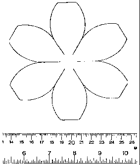
Figure 1 is an illustration depicting a template for a multi-petal-shaped neo-
organ
matrix or scaffold. The edges of the petals are mated to form a quasi-
spherical shaped
hollow matrix.
Figure 2 is an illustration depicting a two-part neo-organ matrix or scaffold
for
organ augmentation that includes a dome-shaped piece with a flanged equatorial
border
having flaps, this first piece being seeded with cells, and a second piece
comprising a ring
with a flanged collar having flaps, the flanged collar designed to mate with
the flanged
equatorial border of the dome piece.
Figure 3 is an illustration depicting a two-part neo-organ matrix or scaffold
for
organ replacement, each part having a hemispherical or quasi-hemispherical
shape and
each with a flanged equatorial border for mating the two parts.
Figure 4 is an illustration depicting the two-part neo-organ matrix or
scaffold
portions of the two-part scaffold shown in Figure 3 with flanges ready to be
joined.
Figure 5 is an illustration depicting the joined neo-organ matrix or scaffold
portions of the scaffold shown in Figure 4 with trimmed flanges.
Figure 6 is an illustration depicting a bisected neo-organ matrix or scaffold
design
for organ replacement in which the neo-organ matrix or scaffold is bisected
along a non-
equatorial axis so that the non-equatorial borders of each bisected portion
are closer to the
tubular structures or vessels to be attached such as the urethral tube.
Figure 7 depicts a two-part neo-organ matrix or scaffold template designed to
create, when the two parts are mated, a hollow, quasi-spherical matrix or
scaffold with a
flanged, longitudinal, elliptical opening on one side, and a circular opening
in the surface
opposite the longitudinal openingõ both openings to allow access to the
interior of the
matrix or scaffold and to allow for the attachment of tubular vessels to the
matrix.
Figure 8 depicts the top-view of the neo-organ matrix or scaffold constructed
from
the two-part hollow scaffold template depicted in Figure 7, showing a
longitudinal,
elliptical opening at the dome of the scaffold with tabs or flanges on the
lips of the
opening.
8
CA 02641733 2008-08-07
WO 2007/095193 PCT/US2007/003709
Figure 9 depicts a side view of the hollow neo-organ matrix or scaffold
constructed
from the two-part template depicted in Figure 7.
Figure 10 depicts a bottom view of the hollow neo-organ matrix or scaffold
constructed from the two-part scaffold template depicted in Figure 7, showing
the circular
opening opposite the longitudinal, elliptical opening depicted in Figure 8.
Figures 11A-11C is an illustration depicting a three-part template design for
constructing a quasi-hemispherical crown shaped neo-organ matrix or scaffold.
Figure
11A depicts the top piece of the crown shaped scaffold; Figure 11B depicts the
front piece
of the crown shaped scaffold; Figure 11C depicts the side piece of the crown
shaped
scaffold. =
Figures 12A-12D depicts the quasi-hemispherical crown shaped neo-organ matrix
or scaffold constructed from the three-part template shown in Figures 11A-11C.
Figure
12A is a side view of the crown shaped scaffold; Figure 12B depicts the front
view of the
crown shaped scaffold; Figure 12C depicts a top view of the crown shaped
scaffold;
Figure 12D depicts the bottom view of the crown shaped scaffold.
Figures 13 and 14 are illustrations depicting the initial seeding vessel and
bioreactor for use in seeding and growing neo-organ matrices or scaffolds.
Note that the
bioreactor must be opened completely to seed and change medium.
Figure 15 is an illustration the presence of smooth muscle cells on and in the
polymeric matrix of a neobladder scaffold.
Figure 16 is an illustration depicting the presence of urothelial cells on and
in the
polymeric matrix of a neobladder scaffold.
Figures 17-20 are illustrations depicting containers for packing and shipping
cell-
seeded neo-organ scaffolds. Note that the neo-bladder must be removed from the
seeding
bioreactor and manipulated with hemostats and forceps for attachment to the
inner basket
of the shipping container.
Figure 17 depicts a shipping container with a screw-cap lid for packing and
transporting cell-seeded neo-organ constructs.
Figure 18 depicts an aerial view of the shipping container depicted in Figure
17,
without the screw-cap lid, showing an inner basket supporting a cell-seeded
neo-organ
construct.
9
CA 02641733 2008-08-07
WO 2007/095193
PCT/US2007/003709
Figure 19 depicts the inner support basket shown in Figure 18 with a cell-
seeded
neo-organ construct inside the basket.
Figure 20 depicts a temperature controlled, insulated box used to ship the neo-
organ construct shipping container depicted in Figure 17.
Figures 21A-21D are a series of illustrations depicting a joined two-part,
hollow
neo-organ polymeric matrix or scaffold for organ replacement, with receptacles
or ports
for the attachment of tubular vessels or inserts such as the ureters and the
urethra. Panels
A and C provide a solid view of the assembled neo-organ construct for organ
replacement,
while Panels B and D provide a cross-sectional view of the assembled
construct. Each of
these panels also depicts the polymeric flanged tubular vessel or insert
matrices, which are
to be inserted into the receptacles or ports joined two-part hollow matrix.
Figure 22 depicts a tubular vessel with a flange at the end after being
inserted
through a receptacle or port of a neo-organ matrix or scaffold wall (shown in
cross
section) and a washer.
Figure 23 depicts a flanged tubular vessel or insert for the attachment of
tubular
vessels to a neo-organ matrix or scaffold prior to insertion into the scaffold
wall, shown
with washer near the flanged end with a dehydrated hydrogel located on the
side of the
washer proximal to the flange.
Figure 24 depicts the insert shown in Figure 23 after the flanged end has been
inserted through the wall of a neo-organ matrix or scaffold. The remainder of
the insert
stays on the other side of the scaffold wall.
Figure 25 depicts the insert of Figure 24 after the hydrogel has been swollen,
=
thereby filling the space between the outer flange and the neo-organ scaffold
wall.
Figure 26 is an illustration depicting a two-part neo-organ matrix or scaffold
for
bladder replacement. Each scaffold portion includes one or more unseeded tabs,
a flange,
and at least one receptacle or port to accept a flanged insert for attachment
of a tubular
vessel.
Figure 27 is an illustration depicting the scaffold of Figure 26 after the two
hemispherical neo-organ matrix or scaffolds have been joined.
Figure 28 is an illustration depicting the scaffold of Figure 27 after the two
joined
hemispherical scaffold portions have been sutured together and the tabs have
been
removed. The joined flange surfaces may be trimmed at this stage.
=
CA 02641733 2008-08-07
WO 2007/095193 PCT/US2007/003709
Figures 29A-29B are a series of illustrations depicting trigone-sparing
bladder
augmentation surgery. Figure 29A is an illustration of bladder augmentation
surgery using
a previous neo-organ augmentation construct design while Figure 29B is an
illustration of
bladder augmentation using a modified neo-organ construct design with flaps
and an outer
rim.
Figure 30 is an illustration depicting non-trigone sparing bladder replacement
surgery using a previous neo-organ replacement construct design.
Figure 31 is an illustration depicting non-trigone sparing bladder replacement
surgery using a modified neo-organ replacement construct design that includes
receptacles
or ports adapted to receive a tubular vessel or insert, for the attachment of
the ureters and
the urethra.
DETAILED DESCRIPTION OF THE INVENTION
Constructs and methods useful in the reconstruction, repair, augmentation or
replacement of organs or tissues structures are provided.
In its broadest form, the constructs and methods of the present invention are
useful
in the reconstruction, repair, augmentation or replacement of organs or
tissues structures
that comprise multilayer cellular organization and particularly those organs
or tissue
structures that are lurninal in nature. More particularly, the present
invention provides
constructs and methods that facilitate the reconstruction, repair,
augmentation or
replacement of shaped hollow organs or tissue structures that exhibit a
laminar segregation
of different cell types and that have a need to retain a general luminal
shape. Luminal
organs or tissue structures that contain a smooth muscle cell (SMC) layer to
impart
compliant or contractible properties to the organ or structure are
particularly well suited to
the constructs and methods of the present invention.
In an example of one preferred embodiment of the invention, the luminal organ
is
the bladder, which has an inner layer of a first cell population that
comprises urothelial
cells and an outer layer of a second cell population that comprises smooth
muscle cells.
This organization is also present in other genitourinary organs and tissue
structures such as
the ureters and urethra. Laminarily organized organs or tissues refer to any
organ or tissue
11
CA 02641733 2008-08-07
WO 2007/095193 PCT/US2007/003709
made up of, or arranged in laminae including ductal tissue. Other suitable la-
minarily
organized luminal organs, tissue structure, or ductal tissues to which the
present invention
is directed include vas deferens, fallopian tubes, lacrimal ducts, trachea,
stomach,
intestines, vasculature, biliary duct, ductus ejaculatorius, ductus
epididymidis, ductus
parotideus, and surgically created shunts. Other suitable organs and tissue
structures
include, for example, kidneys, blood vessels and reproductive organs such as
the uterus.
The neo-organ constructs and methods of the present invention comprise a
biocompatible synthetic or natural polymeric matrix or scaffold, and one or
more cell
populations seeded on one or more surfaces of the matrix or scaffold. The
method of the
present invention in its broadest aspect encompasses as a first step providing
a
biocompatible synthetic or natural polymeric matrix or scaffold that is shaped
to conform
to its use as a part or all of the luminal organ or tissue structure to be
repaired,
reconstructed, augmented or replaced. Hereinafter, the terms matrix and
scaffold may be
used interchangeably. A biocompatible material is any substance not having
toxic or
injurious effects on biological function. The shaped matrix or scaffold is
preferably porous
to allow for cell deposition both on and in the pores of the matrix. The
shaped matrix or
scaffold may then be contacted with one or more cell populations to seed the
cell
populations on or into (or both) the matrix or scaffold. The cell-seeded
matrix scaffold
(i.e., the construct) is then implanted in the body of the recipient where the
construct
facilitates the regeneration of neo-organs or tissue structures. The
constructs may be used
to reconstruct, repair, augment or replace any organ, and may especially be
utilized in
patients having a defect in urogenital tissues such as the bladder.
In a preferred embodiment, the materials and methods of the invention are
useful
for the reconstruction, replacement or augmentation of bladder tissue. Thus,
the invention
provides treatments for such conditions as neurogenic bladder, bladder
exstrophy, bladder
volume insufficiency, bladder non-compliance, reconstruction of bladder
following partial
or total cystectomy, repair of bladders damaged by trauma, and the like.
One issue that can face the surgeon during the implantation of a neo-organ
construct or neo-vessel construct, such as a bladder, kidney or blood vessel,
is the
attachment of vessels, such as the urethra, ureters, and renal blood vessels.
Currently, one
method to achieve this is for the resected end of the urethra or ureter to be
fed through a
hole in the wall of the neo-bladder construct and splatulated and sutured into
the interior of
12
CA 02641733 2008-08-07
WO 2007/095193 PCT/US2007/003709
the construct. Limitations with this method include extended working time
during which
the neo-bladder construct is out of medium (which negatively impacts the
viability of the
cells contains on the construct), cumbersomeness of working with neo-bladder
construct
during splatulation and suturing with resulting damage to the neo-bladder
construct, and
the requirement for the surgeon to be suturing in the tight spaces in the
bottom of the
bladder "bowl".
The constructs and methods described herein are designed to improve the ease
with, and reduce the surgical time in which vessels and other tubular
structures, such as
the urethra and ureter, are surgically connected to a neo-organ construct such
as a neo-
bladder construct. The current invention provides for the use of a flanged
tubular matrix
to address this issue. The methods described herein are also used to improve
the ease with
which vessels and other tubular structures, such as blood vessels, are
surgically connected
to a neo-organ, to a neo-vessel structure or to another blood vessel.
According to one
method, the neo-organ construct is a neo-bladder construct, and the urethra is
first attached
to a tubular element which is flanged at one end, referred to herein as an
insert, then the
flanged end of the insert is placed into the interior of the neo-bladder. In
contrast to
current methods, the insert is not initially attached to the neo-bladder
construct. Insert
design variations alleviating the need for suturing to the neo-bladder
construct, and the use
of a hydrogel to facilitate seating of the insert, are also disclosed. Matrix
or scaffold
design variations, include tabs on the scaffold to ease positioning during
implantation, a
flanged collar to help attach the cut edge of the native bladder trigone to
the flanged cell
seeded neo-bladder construct, and an approach of forming two-part neo-organ
constructs
with a geometry that allows for easier access to elements inside the
constructs prior to
joining them, and an approach to join two halves of neo-organs, are also
presented
While reference is made herein to reconstructions, replacements or
augmentation
of the bladder and methods of attaching vessels such as the urethra or ureter
to a neo-
bladder construct, it will be understood that the methods and materials
described herein
are useful for tissue reconstruction, replacement or augmentation of a variety
of tissues
and organs in a subject. Thus, for example, organs or tissues such as bladder,
ureter,
urethra, renal pelvis, and the like, can be augmented or repaired with
polymeric matrixes
seeded with cells. The materials and methods of the invention further can be
applied to the
reconstruction, replacement or augmentation of vascular tissue (see, e.g.,
Zdrahala, R. J., J
13
CA 02641733 2008-08-07
WO 2007/095193 PCT/US2007/003709
Biomater. App!. 10 (4): 309-29 (1996)), intestinal tissues, stomach (see,
e.g., Laurencin,
C. T. et al., J Biomed Mater. Res. 30 (2): 133-8 1996), and the like. The
patient to be
treated may be of any species of mammals such as a dog, cat, pig, horse, cow,
or human,
in need of reconstruction, repair, replacement or augmentation of a tissue.
Neo-Organ matrix or scaffolds
Biocompatible material and especially biodegradable material is the preferred
material for the construction of the matrix.
Biocompatible refers to materials which do not have toxic or injurious effects
on
biological functions. Biodegradable refers to material that can be absorbed or
degraded in
a patient's body. Representative materials for forming the biodegradable
matrix or
scaffold include natural or synthetic polymers, such as, for example,
collagen, poly(alpha
esters) such as poly(lactate acid) and poly(glycolic acid), polyorthoesters
and
polyanhydrides and their copolymers, which degrade by hydrolysis at a
controlled rate and
are reabsorbed. These materials provide the maximum control of degradability,
manageability, size and configuration. Preferred biodegradable polymer
material includes
polyglycolic acid and polyglactin, developed as absorbable synthetic material.
Polyglycolic acid and polyglactin fibers may be used as supplied by the
manufacturer.
Other biodegradable materials include cellulose ether, cellulose, cellulosic
ester,
fluorinated polyethylene, phenolic, poly-4-methylpentene, polyacrylonitrile,
polyamide,
polyamideimide, polyadrylate, polybenzoxazole, polycarbonate,
polycyanoarylether,
polyester, polyestercarbonate, polyether, polyetheretherketone,
polyetherimide,
polyetherketone, polyethersulfone, polyethylene, polyfluoroolefin, polyimide,
polyolefin,
polyoxadiazole, polyphenylene oxide, polyphenylene sulfide, polypropylene,
polystyrene,
polycaprolactone, polysulfide, polysulfone, polytetrafluoroethylene,
polythioether,
polytriazole, polyurethane, polyvinyl, polyvinylidene fluoride, regenerated
cellulose,
silicone, urea-formaldehyde, or copolymers or physical blends of these
materials. The
material may be impregnated with suitable antimicrobial agents and may be
colored by a
color additive to improve visibility and to aid in surgical procedures.
Other biocompatible materials include synthetic suture material manufactured
by
Ethicon Co. (Ethicon Co., Somerville, N.J.), such as MONOCRYL (copolymer of
glycolide and epsilon-caprolactone), VICRYL or Polyglactin 910 (copolymer of
lactide
and glycolide coated with Polyglactin 370 and calcium stearate), and PANACRYL
14
CA 02641733 2008-08-07
WO 2007/095193
PCT/US2007/003709
(copolymer of lactide and glycolide coated with a polymer of caprolactone and
glycolide).
= (Craig P. H., Williams J. A., Davis K. W., et al.: A Biological
Comparison of Polyglactin
910 and Polyglycolic Acid Synthetic Absorbable Sutures. Surg. 141; 1010,
(1975)) and
polyglycolic acid. These materials can be used as supplied by the
manufacturer.
In yet another embodiment, the matrix or scaffold can be created using parts
of a
natural decellularized organ. Biostructures, or parts of organs can be
decellularized by
removing the entire cellular and tissue content from the organ. The
decellularization
process comprises a series of sequential extractions. One key feature of this
extraction
process is that harsh extraction that may disturb or destroy the complex infra-
structure of
the biostructure, be avoided. The first step involves removal of cellular
debris and
solubilization of the cell membrane. This is followed by solubilization of the
nuclear
cytoplasmic components and the nuclear components.
Preferably, the biostructure, e.g., part of an organ is decellularized by
removing the
cell membrane and cellular debris surrounding the part of the organ using
gentle
mechanical disruption methods. The gentle mechanical disruption methods must
be
sufficient to disrupt the cellular membrane. However, the process of
decellularization
should avoid damage or disturbance of the biostructure's complex infra-
structure. Gentle
mechanical disruption methods include scraping the surface of the organ part,
agitating the
organ part, or stirring the organ in a suitable volume of fluid, e.g.,
distilled water. In one
preferred embodiment, the gentle mechanical disruption method includes
stirring the organ
part in a suitable volume of distilled water until the cell membrane is
disrupted and the
cellular debris has been removed from the organ.
After the cell membrane has been removed, the nuclear and cytoplasmic
components of the biostructure are removed. This can be performed by
solubilizing the
cellular and nuclear components without disrupting the infra-structure. To
solubilize the
nuclear components, non-ionic detergents or surfactants may be used. Examples
of
nonionic detergents or surfactants include, but are not limited to, the Triton
series,
available from Rohm and Haas of Philadelphia, Pa., which includes Triton X-
100, Triton
N-101, Triton X-114, Triton X-405, Triton X-705, and Triton DF-16, available
commercially from many vendors; the Tween series, such as monolaurate (Tween
20),
monopalmitate (Tween 40), monooleate (Tween 80), and polyoxethylene-23-lauryl
ether
(Brij. 35), polyoxyethylene ether W-1 (Polyox), and the like, sodium cholate,
CA 02641733 2008-08-07
WO 2007/095193
PCT/US2007/003709
deoxycholates, CHAPS, saponin, n-Decyl-D-glucopuranoside, n-heptyl-D-
glucopyranoside, n-Octyl-D-glucopyranoside and Nonidet P-40.
One skilled in the art will appreciate that a description of compounds
belonging to
the foregoing classifications, and vendors may be commercially obtained and
may be
found in "Chemical Classification, Emulsifiers and Detergents", McCutcheon's,
Emulsifiers and Detergents, 1986, North American and International Editions,
McCutcheon Division, MC Publishing Co., Glen Rock, N.J., U.S.A. and Judith
Neugebauer, A Guide to the Properties and Uses of Detergents in Biology and
Biochemistry, Calbiochem. R., Hoechst Celanese Corp., 1987. In one preferred
embodiment, the non-ionic surfactant is the Triton. series, preferably, Triton
X-100.
The concentration of the non-ionic detergent may be altered depending on the
type
of biostructure being decellularized. For example, for delicate tissues, e.g.,
blood vessels,
the concentration of the detergent should be decreased. Preferred
concentration ranges of
non-ionic detergent can be from about 0.001 to about 2.0% (w/v). More
preferably, about
0.05 to about 1.0% (w/v). Even more preferably, about, 0.1% (w/v) to about
0.8% (w/v).
Preferred concentrations of these range from about 0.001 to about 0.2% (w/v),
with about
0.05 to about 0.1% (w/v) particular preferred.
The cytoskeletal component, which includes the dense cytoplasmic filament
networks, intercellular complexes and apical microcellular structures, may be
solubilized
using alkaline solution, such as, ammonium hydroxide. Other alkaline solution
consisting
of ammonium salts or their derivatives may also be used to solubiliz,e the
cytoskeletal
components. Examples of other suitable ammonium solutions include ammonium
sulphate, ammonium acetate and ammonium hydroxide. In a preferred embodiment,
ammonium hydroxide is used.
The concentration of the alkaline solutions, e.g., ammonium hydroxide, may be
altered depending on the type of biostructure being decellularized. For
example, for
delicate tissues, e.g., blood vessels, the concentration of the detergent
should be decreased.
Preferred concentrations ranges can be from about 0.001 to about 2.0% (w/v).
More
preferably, about 0.005 to about 0.1% (w/v). Even more preferably, about,
0.01% (w/v) to
about 0.08% (w/v).
The decellularized, lyophilized structure may be stored at a suitable
temperature
until required for use. Prior to use, the decellularized structure can be
equilibrated in
16
CA 02641733 2013-11-15
suitable isotonic buffer or cell culture medium. Suitable buffers include, but
are not
limited to, phosphate buffered saline (PBS), saline, MOPS, HEPES, Hank's
Balanced Salt
Solution, and the like. Suitable cell culture medium includes, but is not
limited to, RPMI
1640, Fisher's, Iscove's, McCoy's, Dulbecco's medium, and the like.
Still other biocompatible materials that may be used include stainless steel,
titanium, silicone, gold and silastic.
The biocompatible polymer may be shaped using methods such as, for example,
solvent casting, compression molding, filament drawing, meshing, leaching,
weaving and
coating. In solvent casting, a solution of one or more polymers in an
appropriate solvent,
such as methylene chloride, is cast as a branching pattern relief structure.
After solvent
evaporation, a thin film is obtained. In compression molding, a polymer is
pressed at
pressures up to 30,000 pounds per square inch into an appropriate pattern.
Filament
drawing involves drawing from the molten polymer and meshing involves forming
a mesh
by compressing fibers into a felt-like material. In leaching, a solution
containing two
materials is spread into a shape close to the final form of the construct.
Next a solvent is
used to dissolve away one of the components, resulting in pore formation. (See
Miloas,
U.S. Pat. No. 5,514,378 .) In nucleation, thin films
in
the shape of a RUG are exposed to radioactive fission products that create
tracks of
radiation damaged material. Next the polycarbonate sheets are etched with acid
or base,
turning the tracks of radiation-damaged material into pores. Finally, a laser
may be used to
shape and bum individual holes through many materials to form a structure with
uniform
pore sizes. Coating refers to coating or permeating a polymeric structure with
a material
such as, for example liquefied copolymers (poly-DL-lactide co-glycolide 50:50
80 mg/nil
methylene chloride) to alter its mechanical properties. Coating may be
performed in one
layer, or multiple layers until the desired mechanical properties are
achieved. These
shaping techniques may be employed in combination, for example, a polymeric
matrix or
scaffold may be weaved, compression molded and glued together. Furthermore
different
polymeric materials shaped by different processes may be joined together to
form 'a
composite shape. The composite shape may be a laminar structure. For example,
a
polymeric matrix or scaffold may be attached to one or more polymeric matrixes
to form a
multilayer polymeric matrix or scaffold structure. The attachment may be
performed by
gluing with a liquid polymer or by suturing. In addition, the polymeric matrix
or scaffold
17
=
CA 02641733 2008-08-07
WO 2007/095193 PCT/US2007/003709
may be formed as a solid block and shaped by laser or other standard machining
techniques to its desired final form. Laser shaping refers to the process of
removing
materials using a laser.
The polymeric matrix or scaffold can be reinforced. For example, reinforcing
materials may be added during the formation of a synthetic matrix or scaffold
or attached
to the natural or synthetic matrix prior to implantation. Representative
materials for
forming the reinforcement include natural or synthetic polymers, such as, for
example,
collagen, poly(alpha esters) such as poly(lactate acid), poly(glycolic acid),
polyorthoesters
and polyanhydrides and their copolymers, which degraded by hydrolysis at a
controlled
rate and are reabsorbed. These materials provide the maximum control of
degradability,
manageability, size and configuration.
The biodegradable polymers can be characterized with respect to mechanical
properties, such as tensile strength using an Instron tester, for polymer
molecular weight
by gel permeation chromatography (GPC), glass, transition temperature by
differential
scanning calorimetry (DSC) and bond structure by infrared (IR) spectroscopy;
with respect
to toxicology by initial screening tests involving Ames assays and in vitro
teratogenicity
assays and implantation studies in animals for immunogenicity, inflammation,
release and
degradation studies. In vitro cell attachment and viability can be assessed
using scanning
electron microscopy, histology and quantitative assessment with radioisotopes.
The
biodegradable material may also be characterized with respect to the amount of
time
necessary for the material to degrade when implanted in a patient. By varying
the
construction, such as, for example, the thickness and mesh size, the
biodegradable material
may substantially biodegrade between about 2 years or about 2 months,
preferably =
between about 18 months and about 4 months, most preferably between about 15
months
and about 8 months and most preferably between about 12 months and about 10
months. If
necessary, the biodegradable material may be constructed so as not to degrade
substantially within about 3 years, or about 4 years or about five or more
years.
The polymeric matrix or scaffold may be fabricated with controlled pore
structure
as described above. The size of the pores may be used to determine the cell
distribution.
For example, the pores on the polymeric matrix or scaffold may be large to
enable cells to
migrate from one surface to the opposite surface. Alternatively, the pores may
be small
such that there is fluid communication between the two sides of the polymeric
matrix or
18
CA 02641733 2008-08-07
WO 2007/095193
PCT/US2007/003709
scaffold but cells cannot pass through. Suitable pore size to accomplish this
objective may
be about 0.04 micron to about 10 microns in diameter, preferably between about
0.4
micron to about 4 microns in diameter. In some embodiments, the surface of the
polymeric
matrix or scaffold may comprise pores sufficiently large to allow attachment
and
migration of a first population of cells into the pores. The pore size may be
reduced in the
interior of the polymeric matrix or scaffold to prevent cells from migrating
from one side
of the polymeric matrix or scaffold to the opposite side. On the opposite side
of the
polymeric matrix, the pores may again enlarge to allow the attachment and
establishment
of a second population of cells. Because of the reduced pore size in the
interior of the
polymeric matrix, the first cell population and the second cell population
initially cannot
mix. One embodiment of a polymeric matrix or scaffold with reduced pore size
is a
laminated structure of a small pore material sandwiched between two large pore
material.
Alternatively, a large pore material laminated to a small pore material may
also allow cells
to establish growth on both sides without any intermixing of cells.
Polycarbonate
membranes are especially suitable because they can be fabricated in very
controlled pore
sizes such as, for example, about 0.01 microns, about 0.05 micron, about 0.1
micron,
about 0.2 micron, about 0.45 micron, about 0.6 micron, about 1.0 micron, about
2.0
microns and about 4.0 microns. At the submicron level the polymeric matrix or
scaffold
may be impermeable to bacteria, viruses and other microbes.
The following characteristics or criteria, among others, are taken into
account in
the design of each discrete matrix, or part thereof: (i) shape, (ii) strength,
(iii) stiffness and
rigidity, and (iv) suturability (the degree to which the matrix, or part
thereof, is readily
sutured or otherwise attached to adjacent tissue). As used herein, the
stiffness of a given
matrix or scaffold is defined by the modulus of elasticity, a coefficient
expressing the ratio
between stress per unit area acting to deform the scaffold and the amount of
deformation
that results from it. (See e.g., Handbook of Biomaterials evaluation,
Scientific, Technical,
and Clinical Testing of Implant Materials, 2nd edition, edited by Andreas F.
von Recum,
(1999); Ratner, etal., Biomaterials Science: An Introduction to Materials in
Medicine,
Academic Press (1996)). The rigidity of a scaffold refers to the degree of
flexibility (or
lack thereof) exhibited by a given scaffold.
Each of these criteria is a variable that can be changed (through, among other
things, the choice of material and the manufacturing process) to allow the
matrix, or part
19
CA 02641733 2008-08-07
WO 2007/095193
PCT/US2007/003709
=
=
thereof to best placed and modified to address the medical indication and the
physiological
function for which it is intended. For example, the material comprising the
matrix or
scaffold for bladder replacement, reconstruction and/or augmentation must be
sufficiently
strong to support sutures without tearing, while being sufficient compliant so
as to
accommodate fluctuating volumes of urine.
Optimally, the matrix or scaffold should be shaped such that after its
biodegradation, the resulting reconstructed bladder is collapsible when empty
in a fashion
similar to a natural bladder and the ureters will not be obstructed while the
urinary catheter
has been removed from the tissue engineered bladder without leaving a leak
point from the
dome. The bioengineered bladder construct can be produced as one piece or each
part can
be individually produced or combinations of the sections can be produced as
specific
parts. Each specific matrix or scaffold part may be produced to have a
specific function.
Otherwise specific parts may be produced for manufacturing ease. Specific
parts may be
constructed of specific materials and may be designed to deliver specific
properties.
Specific part properties may include tensile strength similar to the native
tissue (e.g.
ureters) of 0.5 to 1.5 MPa2 and an ultimate elongation of 30 to 100% or the
tensile strength
may range from 0.5 to 28 MPa2 , ultimate elongations may range from 10-200%
and
compression strength may be <12.
A mesh-like structure formed of fibers, which may be round, scalloped,
flattened,
star shaped, solitary or entwined with other fibers is preferred. The use of
branching fibers
is based upon the same principles which nature has used to solve the problem
of increasing
surface area proportionate to volume increases. All multicellular organisms
utilize this
repeating branching structure. Branching systems represent communication
networks
between organs, as well as the functional units of individual organs. Seeding
and
implanting this configuration with cells allows implantation of large numbers
of cells,
each of which is exposed to the environment of the host, providing for free
exchange of
nutrients and waste while neovascularization is achieved. The polymeric matrix
or
scaffold may be made flexible or rigid, depending on the desired final form,
structure and
function.
In one preferred embodiment, the polymeric matrix or scaffold is formed with a
polyglycolic acid with an average fiber diameter of 15 um and configured into
a bladder
shaped mold using 4-0 polyglactin 910 sutures. The resulting structure is
coated with a
CA 02641733 2008-08-07
WO 2007/095193 PCT/US2007/003709
liquefied copolymer, such as, for example, pol-DL-lactide-co-glycolide 50:50,
80
milligram per milliliter methylene chloride, in order to achieve adequate
mechanical
characteristics and to set its shape.
Polymeric matrixes can be treated with additives or drugs prior to
implantation
(before or after the polymeric matrix or scaffold is seeded with cells, if the
optional seeded
cells are employed), e.g., to promote the regeneration of new tissue after
implantation.
Thus, for example, growth factors, cytokines, extracellular matrix or scaffold
components,
and other bioactive materials can be added to the polymeric matrix or scaffold
to promote
graft healing and regeneration of new tissue. Such additives will in general
be selected
according to the tissue or organ being reconstructed, replaced or augmented,
to ensure that
appropriate new tissue is formed in the engrafted organ or tissue (for
examples of such
additives for use in promoting bone healing, see, e.g., Kirker-Head, C. A.
Vet. Surg. 24
(5): 408-19 (1995)). For example, when polymeric matrices (optionally seeded
with
endothelial cells) are used to augment vascular tissue, vascular endothelial
growth factor
(VEGF), (see, e.g., U.S. Pat. No. 5,654,273) can be employed to promote the
regeneration
of new vascular tissue. Growth factors and other additives (e.g., epidermal
growth factor
(EGF), heparin-binding epidermal-like growth factor (HBGF), fibroblast growth
factor
(F'GF), cytokines, genes, proteins, and the like) can be added in amounts in
excess of any
amount of such growth factors (if any) which may be produced by the cells
seeded on the
polymeric matrix, if added cells are employed. Such additives are preferably
provided in
an amount sufficient to promote the regeneration of new tissue of a type
appropriate to the
tissue or organ, which is to be repaired, replaced or augmented (e.g., by
causing or
accelerating infiltration of host cells into the graft). Other useful
additives include
antibacterial agents such as antibiotics.
One preferred supporting matrix or scaffold is composed of crossing filaments
which can allow cell survival by diffusion of nutrients across short distances
once the cell
support is implanted. The cell support matrix or scaffold becomes vascularized
in concert
with expansion of the cell mass following implantation.
The building of three-dimensional structure constructs in vitro, prior to
implantation, facilitates the eventual terminal differentiation of the cells
after implantation
in vivo, and minimizes the risk of an inflammatory response towards the
matrix, thus
avoiding graft contracture and shrinkage.
21
CA 02641733 2008-08-07
WO 2007/095193 PCT/US2007/003709
The polymeric matrix or scaffold may be sterilized using any known method
before use. The method used depend on the material used in the polymeric
matrix.
Examples of sterilization methods include steam, dry heat, radiation, gases
such as
ethylene oxide, gas and boiling.
Method for Forming Neo-Organ Matrices or Scaffolds
The biocompatible scaffold may be shaped using methods such as, for example,
solvent casting, compression molding, filament drawing, meshing, leaching,
weaving,
foaming, electrospinning and coating. In solvent casting, a solution of one or
more
polymers in an appropriate solvent, such as methylene chloride, is cast as a
branching
pattern relief structure. After solvent evaporation, a thin film is obtained.
In compression
molding, a polymer is pressed at pressures up to 30,000 pounds per square inch
into an
appropriate pattern. Filament drawing involves drawing from the molten polymer
and
meshing involves forming a mesh by compressing fibers into a felt-like
material. In
leaching, a solution containing two materials is spread into a shape close to
the final form
of the artificial organ. Next a solvent is used to dissolve away one of the
components,
resulting in pore formation. (See U.S. Patent No. 5,514,378 to Mikos).
In nucleation, thin films in the shape of an artificial organ are exposed to
radioactive fission products that create tracks of radiation damaged material.
Next the
polycarbonate sheets are etched with acid or base, turning the tracks of
radiation-damaged
material into pores. Finally, a laser may be used to shape and bum individual
holes
through many materials to form a scaffold structure with uniform pore sizes.
Coating
refers to coating or permeating a structure with a material such as, for
example liquefied
copolymers (poly-DL-lactide co-glycolide 50:50 80 mg/ml methylene chloride) to
alter its
mechanical properties. Coating may be performed in one layer, or multiple
layers until the
desired mechanical properties are achieved. These shaping techniques may be
employed in
combination, for example, a scaffold may be weaved, compression molded and
glued
together. Furthermore different materials shaped by different processes may be
joined
together to form a composite shape. The composite shape may be a laminar
structure. For
example, a matrix or scaffold may be attached to one or more matrices to form
a
multilayer scaffold structure. The attachment may be performed by gluing with
a liquid
polymer or by suturing. In addition, the matrix or scaffold may be formed as a
solid block
and shaped by laser or other standard machining techniques to its desired
final form. Laser
22
CA 02641733 2008-08-07
WO 2007/095193 PCT/US2007/003709
shaping refers to the process of removing materials using a laser.
The scaffold may be shaped into any number of desirable configurations to
satisfy
any number of overall system, geometry or space restrictions. For example, in
the use of
the scaffold for bladder, urethra, valve, or blood vessel reconstruction, the
matrix or
scaffold may be shaped to conform to the dimensions and shapes of the whole or
a part of
the tissue. Naturally, the scaffold may be shaped in different sizes and
shapes to conform
to the organs of differently sized patients. For bladders, the scaffold should
be shaped such
that after its biodegradation, the resulting reconstructed bladder may be
collapsible when
empty in a fashion similar to a natural bladder. The matrix or scaffold may
also be shaped
in other fashions to accommodate the special needs of the patient.
Cells for Organ Reconstruction
In one embodiment, the scaffolds are seeded with one or more populations of
cells
to form an artificial organ construct. The artificial organ construct can be
autologous,
where the cell populations are derived from the subject's own tissue, or
allogenic, where
the cell populations are derived from another subject within the same species
as the
patient. The artificial organ construct can also be xenogenic, where the
different cell
populations are derived form a mammalian species that is different from the
subject. For
example the cells can be derived from organs of mammals such as humans,
monkeys,
dogs, cats, mice, rats, cows, horses, pigs, goats and sheep.
The process for isolating cell is described generally herein, and specific
procedures
are presented in the examples provided below. Cells can be isolated from a
number of
sources, including, for example, biopsies from living subjects and whole-organ
recover
from cadavers. The isolated cells are preferably autologous cells, obtained by
biopsy from
the subject intended to be the recipient. For example, a biopsy of skeletal
muscle from the
arm, forearm, or lower extremities, or smooth muscle from the area treated
with local
anesthetic with a small amount of lidocaine injected subcutaneously, and
expanded in
culture. The biopsy can be obtained using a biopsy needle, a rapid action
needle which
makes the procedure quick and simple. The small biopsy core of either skeletal
or smooth
muscle can then be expanded and cultured, as described by Atala, et al.,
(1992) J. Urol.
148, 658-62; Atala, et al. (1993) J. Urol. 150: 608-12. Cells from relatives
or other donors
of the same species can also be used with appropriate inununosuppression.
23
CA 02641733 2013-11-15
Methods for the isolation and culture of cells are discussed in Fauza et al.
(1998) J.
Ped. Surg. 33,7-12. Cells may be isolated using
techniques known to those skilled in the art. For example, the tissue or organ
can be
disaggregated mechanically and/or treated with digestive enzymes and/or
chelating agents
that weaken the connections between neighboring cells making it possible to
disperse the
tissue into a suspension of individual cells without appreciable cell
breakage. Enzymatic
dissociation can be accomplished by mincing the tissue and treating the minced
tissue with
any of a number of digestive enzymes either alone or in combination. These
include but
are not limited to ttypsin, chymotrypsin, collagenase, elastase, and/or
hyalu.ronidase,
DNase, pronase and dispase. Mechanical disruption can also be accomplished by
a number
of methods including, but not limited to, scraping the surface of the organ,
the use of
grinders, blenders, sieves, homogenizers, pressure cells, or insonicators. For
a review of
tissue disaggregation techniques, see Freshney, (1987), Culture of Animal
Cells. A
Manual of Basic Technique, 2d Ed., A. R. Liss, Inc., New York, Ch. 9, pp. 107-
126.
Preferred cell types include, but are not limited to, urothelial cells,
mesenchymal
cells, especially smooth or skeletal muscle cells, myocytes (muscle stem
cells), fibroblasts,
chondrocytes, adipocytes, fibromyoblasts, and ectodermal cells, including
ductile and skin
cells, hepotocytes, Islet cells, cells present in the intestine, and other
parenchymal cells,
osteoblasts and other cells forming bone or cartilage. In some cases, it may
also be
desirable to include nerve cells.
Once the tissue has been reduced to a suspension of individual cells, the
suspension can be fractionated into subpopulations from which the cells
elements can be
obtained. This also may be accomplished using standard techniques for cell
separation
including, but not limited to, cloning and selection of specific cell types,
selective
destruction of unwanted cells (negative selection), separation based upon
differential cell
agglutinability in the mixed population, freeze-thaw procedures, differential
adherence
properties of the cells in the mixed population, filtration, conventional and
zonal
centrifugation, centrifugal elutriation (counterstreaming centrifugation),
unit gravity
separation, countercurrent distribution, electrophoresis and fluorescence-
activated cell
sorting. For a review of clonal selection and cell separation techniques, see
Freshney,
(1987), Culture of Animal Cells. A Manual of Basic Techniques, 2d Ed., A. R.
Liss, Inc.,
New York, Ch. 11 and 12, pp. 137-168. For example, one cell type may be
enriched by
24
CA 02641733 2008-08-07
WO 2007/095193 PCT/US2007/003709
magnetic-activated and fluorescence-activated cell sorting, and other cell
types may be
reduced for collection of a specific cell type.
Cell fractionation may also be desirable, for example, when the donor has
diseases
such as cancer or metastasis of other tumors to the desired tissue. A cell
population may be
sorted to separate malignant cells or other tumor cells from normal
noncancerous cells.
The normal noncancerous cells, isolated from one or more sorting techniques,
may then be
used for organ reconstruction.
Isolated cells can be cultured in vitro to increase the number of cells
available for
coating the biocompatible scaffold. The use of allogenic cells, and more
preferably
autologous cells, is preferred to prevent tissue rejection. However, if an
immunological
response does occur in the subject after implantation of the artificial organ,
the subject
may be treated with immunosuppressive agents such as, cyclosporin or FK506, to
reduce
the likelihood of rejection. In certain embodiments, chimeric cells, or cells
from a
transgenic animal, can be coated onto the biocompatible scaffold.
Isolated cells may be transfected prior to coating with genetic material.
Useful
genetic material may be, for example, genetic sequences which are capable of
reducing or
eliminating an immune response in the host. For example, the expression of
cell surface
antigens such as class I and class II histocompatibility antigens may be
suppressed. This
may allow the transplanted cells to have reduced chance of rejection by the
host. In
addition, transfection could also be used for gene delivery.
Isolated cells can be normal or genetically engineered to provide additional
or
normal function. Methods for genetically engineering cells with retroviral
vectors,
polyethylene glycol, or other methods known to those skilled in the art can be
used. These
include using expression vectors which transport and express nucleic acid
molecules in the
cells. (See Goeddel; Gene Expression Technology: Methods in Enzymology 185,
Academic Press, San Diego, Calif. (1990).
Vector DNA is introduced into prokaryotic or cells via conventional
transformation or transfection techniques. Suitable methods for transforming
or
transfecting host cells can be found in Sambrook et al. (Molecular Cloning: A
Laboratory
Manual, 2nd Edition, Cold Spring Harbor Laboratory press (1989)), and other
laboratory
textbooks.
CA 02641733 2008-08-07
WO 2007/095193 PCT/US2007/003709
Seeding of the Neo-Organ Matrix or scaffolds
Seeding of cells onto the matrix or scaffold can be performed according to
standard
methods. For example, the seeding of cells onto polymeric substrates for use
in tissue
repair has been reported (see, e.g., Atala, A. etal., J. Urol. 148(2 Pt 2):
658-62 (1992);
Atala, A., et al. J. Urol. 150 (2 Pt 2): 608-12 (1993)). Cells grown in
culture can be
trypsinized to separate the cells, and the separated cells can be seeded on
the matrix.
Alternatively, cells obtained from cell culture can be lifted from a culture
plate as a cell
layer, and the cell layer can be directly seeded onto the scaffold without
prior separation of
the cells.
In a preferred embodiment, in the range of 1 million to 700 50 million cells
are
suspended in medium and applied to each square centimeter of a surface of a
scaffold.
Preferably, between 1 million and 50 million cells, and more preferably,
between 1 million
and 10 million cells are suspended in media and applied to each square
centimeter of a
surface of a scaffold. The matrix or scaffold is incubated under standard
culturing
conditions, such as, for example, 37 C, 5% CO2, for a period of time until the
cells
attached. However, it will be appreciated that the density of cells seeded
onto the scaffold
can be varied. For example, greater cell densities promote greater tissue
regeneration by
the seeded cells, while lesser densities may permit relatively greater
regeneration of tissue
by cells infiltrating the graft from the host. Other seeding techniques may
also be used
depending on the matrix or scaffold and the cells. For example, the cells may
be applied to
the matrix or scaffold by vacuum filtration. Selection of cell types, and
seeding of cells
onto a scaffold, will be routine to one of ordinary skill in the art in light
of the teachings =
herein.
In one embodiment, the scaffold is seeded with one population of cells to form
an
artificial organ construct. In another embodiment, the matrix or scaffold is
seeded on two
sides with two different populations of cells. This may be performed by first
seeding one
side of the matrix or scaffold and then seeding the other side. For example,
the scaffold
may be placed with one side on top and seeded. Then the matrix or scaffold may
be
repositioned so that a second side is on top. The second side may then be
seeded with a
second population of cells. Alternatively, both sides of the matrix or
scaffold may be
seeded at the same time. For example, two cell chambers may be positioned on
both sides
(i.e., a sandwich) of the scaffold. The two chambers may be filled with
different cell
26
CA 02641733 2008-08-07
WO 2007/095193 PCT/US2007/003709
populations to seed both sides of the matrix or scaffold simultaneously. The
sandwiched
scaffold may be rotated, or flipped frequently to allow equal attachment
opportunity for
both cell populations. Simultaneous seeding may be preferred when the pores of
the
matrix or scaffold are sufficiently large for cell passage from one side to
the other side.
Seeding the scaffold on both sides simultaneously will reduce the likelihood
that the cells
would migrate to the opposite side. =
In another embodiment, two separate scaffolds may be seeded with different
cell
populations. After seeding, the two matrices may be attached together to form
a single
matrix or scaffold with two different cell populations on the two sides.
Attachment of the
scaffolds to each other may be performed using standard procedures such as
fibrin glue,
liquid co-polymers, sutures and the like.
Surgical Reconstruction
Grafting of scaffolds to an organ or tissue to be augmented can be performed
according to the methods described in the Examples or according to art-
recognized
methods. The matrix or scaffold can be grafted to an organ or tissue of the
subject by
suturing the graft material to the target organ. Implanting a neo-organ
construct for total
organ replacement can be performed according to the methods described in the
Examples
or according to art-recognized surgical methods.
The described techniques may also be used to treat cancer in an organ or
tissue.
For example, a normal tissue sample may be excised from a patient suffering
from cancer.
Cell populations from the tissue sample may be cultured for a period of time
in vitro and
expanded. The cells may be sorted using a florescent activated cell sorter to
remove
cancerous or precancerous cells. The sorted cells may be used to construct a
seeded
scaffold. At the same time, the patient may be treated for cancer. Cancer
treatment may
involve excision of the cancerous part of the organ in addition to
chemotherapy or
radiation treatment. After the cancer treatment, the seeded scaffold may be
used to
reconstruct the tissue or organ.
While a method for bladder reconstruction is disclosed in the Examples, other
methods for attaching a graft to an organ or tissue of the subject (e.g., by
use of surgical
staples) may also be employed. Such surgical procedures can be performed by
one of
ordinary skill in the art according to known procedures.
The present invention will be further understood by reference to the following
non-
27
CA 02641733 2008-08-07
WO 2007/095193 PCT/US2007/003709
limiting examples.
Example 1: Creation of Bladder-Shaped Polymeric Matrices or Scaffolds
The neo-organ constructs described herein are presented using neo-bladder
constructs as an example. While reference is made here to neo-bladder
constructs, it will
be understood that the methods and materials described herein are useful for
creating a
variety of neo-organs and neo-vessel augmentation constructs, including, for
example,
neo-kidney augmentation constructs.
Manufacture of neo-bladder matrix or scaffold. The neo-bladder matrices or
scaffolds for tissue reconstruction, repair, augmentation, or replacement are
constructed
using polyglycolic acid (PGA) non-woven felt (BMS 2.5 mm thick, 58 mg/cc or 99
mg/ml). The PGA non-woven felt has an average fiber diameter of about 15 gm,
an
interfiber distance between about 0 to about 200 pm, and dimensions of about
10 cm by
about 10 cm. The starting material for constructing the non-woven felt is PGA
or PLGA
10:90 or 15:85, having a molecular weight MW of 100 kDa. The starting material
has a
2.5 mm thickness foam, a porosity of approximately 95% and an average pore
size of
approximately 150 microns.
The PGA non-woven felt is cut using a neo-bladder pattern as a template. The
neo-bladder pattern is for example, spherical, quasi-spherical, hemispherical,
or quasi-
hemispherical in shape, such that bladder repair, or augmentation procedures
require one
hemispherical or quasi-hemispherical neo-bladder construct, while total
bladder
reconstruction may require one spherical or quasi-spherical neo-bladder
construct, or two
hemispherical or quasi-hemispherical neo-bladder constructs joined together to
create a
spherical or quasi-spherical construct.
To create spherical, quasi-spherical, hemispherical or quasi-hemispherical neo-
bladder constructs for repair, augmentation, or replacement, the PGA non-woven
felt is cut
using a neo-bladder template. The neo-bladder template is a single piece of
PGA non-
woven felt or multiple pieces that are joined together, e.g., two or more
pieces, three or
more pieces, or four or more pieces. The template is then assembled, for
example, by
joining distinct areas of a single template together, or by joining two or
more pieces of a
multi-piece template together. In one embodiment, a single distinct template
is used to
form a spherical or quasi-spherical neo-bladder construct. In another
embodiment, a single
distinct template is used to form two hemispherical or quasi-hemispherical neo-
bladder
28
CA 02641733 2008-08-07
WO 2007/095193 PCT/US2007/003709
constructs, such that a two-part construct is initially formed from one
integral part. In
another embodiment, two or more distinct templates are used to create
hemispherical or
quasi-hemispherical neo-bladder constructs which are adapted to mate to each
other, such
that each half of the neo-bladder construct is formed from two or more
distinct parts. In
some embodiments, the two or more distinct templates or parts used to create a
hemispherical or quasi-hemispherical parts adapted to mate are symmetrical,
while in
other embodiments, the two or more distinct templates or parts are
asymmetrical.
Augmentation Construct Designs
Single neo-bladder template designs, when assembled, produce a spherical or
quasi-spherical construct for use in bladder augmentation. Regardless of the
template
used, the assembled construct is designed to fit within the geometry of the
intended site of
implantation, e.g., within a human subject.
An example of an initial, single neo-bladder template used to create a quasi-
spherical neo-bladder construct is shown in Figure 1. The neo-bladder template
of Figure
1, when assembled, creates a unitary construct that is spherical or quasi-
spherical. After
the pattern shown in Figure 1 is cut using a die press or manually, the petal
portions are
mated together. The petal portions can be mated using glue, staples, sutures
or other
technique known to one of ordinary skill in the art. Preferably, the petal
portions are
assembled such that at least a portion of each petal overlaps with at least a
portion of the
adjacent petal, thereby forming a tulip or tulip-like shape. For example, a 4-
0 vicryl suture
is used to suture each petal together from the inside out, using a simple
uninterrupted
stitch or "blanket stitch" with a knot every third or fourth stitch. Once two
petals are
sutured together, loops of suture, e.g., a 1.5 inch loop or a 3 inch loop, are
made at the end
of every other petal. Preferably, there are six loops per scaffold, one at the
end of each
petal. Another loop of suture, e.g., a three inch loop, is made at the apex of
the scaffold to
finish the suturing. These loops form handles for increased ease of
manipulation and
implantation for the neo-bladder constructs described herein. For example, the
surgeon
uses these loops as handles to hold onto the neo-bladder construct during
implantation.
In other embodiments, the neo-bladder matrix or scaffold is formed using any
of a
variety of techniques known in the art. The neo-bladder matrix or scaffold is,
for example,
molded, foamed or electrospun.
Neo-bladder constructs for augmentation may also be formed as a multi-part
29
CA 02641733 2008-08-07
WO 2007/095193 PCT/US2007/003709
design, for example, a two-part design or a three-part design. The multi-part
constructs use
distinct scaffold parts adapted to mate to form a single hemispherical or
quasi-
hemispherical construct. As used herein, the term hemi-shape denotes one half
of any
= geometrical shape. The hemi-shapes adapted to mate may be symmetrical or
asymmetrical. The distinct scaffold parts can be initially formed from a
single template as
one integral part, or formed from two or more templates as distinct parts
adapted to mate
to each other.
In the construction of neo-organs, at times it is desirable to access the
interior of
the construct. For example, access to the interior may be necessary to seed
cells on the
interior surface of the scaffold, to attach vessels to the interior of the
organ, or other
manipulation in surgery. After such manipulation, the scaffold parts must be
joined to
form the neo-bladder construct. The construct design and methods described
herein allow
for rapid, reproducible, aseptic joining of the scaffold parts.
An example of an initial, asymmetrical two-part design used to create a quasi-
spherical neo-bladder construct is shown in Figure 2. In one embodiment, the
two-part
neo-bladder construct design includes unseeded flaps for increased ease of
manipulation,
an outer rim or brim where sutures and adhesives can be used to seal the
apical dome and
a flanged collar, as shown in Figure 2. The flaps can be integrally formed
with the neo-
bladder construct, or they can be made as discrete parts that are stapled in
place.
The initial two-part neo-bladder construct design shown in Figure 2
incorporates
tabs that allow the surgeon to maneuver the construct without touching cell
seeded areas.
This design also includes a flanged collar to help attach the cut edge of the
native bladder
trigone to the flanged cell seeded neo-bladder scaffold. The flanged collar is
constructed
from the same scaffold material, but is not seeded with cells. This procedure
helps speed
up the surgery time for implantation. Briefly, the surgeon will be able to
first suture the
angled collar to the trigone portion of the remaining bladder, and then be
able to remove
the neo-bladder construct from the shipping vessel and suture the two flanged
edges
together to complete the procedure. This two-part system should decrease the
amount of
time the seeded neo-bladder construct is exposed, and will decrease the stress
on the
patient. The flanged edges can be sutured together using a continuous simple
uninterrupted stitch, rather than multiple stitches to connect the trigone
directly to the un-
flanged neo-bladder construct. In addition, the surgeon can handle and
manipulate the
=
CA 02641733 2008-08-07
WO 2007/095193 PCT/US2007/003709
unseeded collar via the tabs during attachment to the trigone portion, and
then remove the
tabs after the two halves have been joined.
The multi-part neo-bladder construct design was ultimately modified to adapt
to
the unique geometry of the intended site of implantation. In particular, the
multi-part
augmentation template was modify such that, when assembled, the construct is
crown-
shaped or generally crown-shaped to accommodate the geometry of the site of
implantation. This can be accomplished using a three-part design to form
distinct quasi-
hemispherical matrix or scaffold parts. One three-part quasi-hemispherical
shape may be
used for bladder augmentation or repair. Alternatively, two three-part quasi-
hemispherical
shapes may be mated to create a quasi-spherical shape for total bladder
replacement. An
example of three distinct templates used to create a quasi-hemi-spherical neo-
bladder
construct which can accommodate the geometry of the site of implantation is
shown in
Figures 11A-11C. In one embodiment, the three-part design used to create quasi-
hemispherical neo-bladder constructs includes unseeded flaps for increased
ease of
manipulation, an outer rim or brim where sutures and adhesives can be used to
mate the
three parts together. The flaps can be integrally formed with the neo-bladder
construct, or -
they can be made as discrete parts that are stapled or sewn in place.
One advantage of using a three-template design to create distinct quasi-
hemispherical scaffold parts is scalability. One of ordinary skill in the art
could calculate
the appropriate length, width, and height of each of the three templates to
scale the
patterns up or down, depending on the desired volume of the organ to be
augmented or
replaced. In one embodiment, the three patterned template shown in Figures 11A-
11C has
an equator length of 8.6 cm, equator width of 8.4 cm, and a height of 7.2 cm
is used to
create a 250 mL neobladder scaffold. In another embodiment, an equator length
of 9.6 cm,
equator width of 8.7 cm, and a height of 7.3 cm is used to create a 350 mL neo-
bladder
scaffold. In yet another embodiment, an equator length of 10.9 cm, equator
width of 10.5
cm and a height of 8.3 cm is used to create a 450 mL scaffold.
PGA non-woven felt is cut using the three-part neo-bladder template shown in
Figure 11A-11C. Each of the three parts are then sutured together using three,
preferably
four, or more than four vertical seams to create a crown shaped construct. An
example of
the three distinct scaffold parts sutured together using four vertical seams
is shown in
Figures 12A-12D. Preferably, the seams are sutured in such a way as to be cut
without
31
CA 02641733 2008-08-07
WO 2007/095193 PCT/US2007/003709
unraveling. For example, knotting every stitch in the vertical seams would
allow a surgeon
to cut and sew without the seams unraveling. Thus, in addition to scalability,
this three
part-part neo-bladder construct design is advantageous over previous designs
in that it also
allows flexibility for the surgeon to customize the shape of the neobladder
construct to the
individual anatomy of the patient at the time of surgery.
Replacement Construct Designs
The two-part constructs for bladder replacement use distinct scaffold parts,
preferably at least two hemi-shapes, adapted to mate to form a single
spherical or semi-
spherical construct. As used herein, the term hemi-shape denotes one half of
any
geometrical shape. The hemi-shapes adapted to mate may be symmetrical or
asymmetrical. The distinct scaffold parts can be initially formed from a
single template as
one integral part, or formed from two or more templates as distinct parts
adapted to mate
to each other.
An example of an initial, symmetrical two-part design used to create a
spherical
neo-bladder construct for use in bladder replacement is shown in Figures 3-5.
In one
embodiment, the hemispherical distinct scaffold parts are fabricated with
exterior flanges
on the seams that need to be joined (Figures 3-5). The unseeded flanges are
used as
handles for manipulation prior to or during surgery. The flanges are used to
seal the
halves together following manipulation. The lower hemisphere is, for example,
placed on
a ring which supports the hemisphere by its flange. The upper hemisphere is
then placed
on top of the lower and its flange is covered with another ring. The rings are
squeezed
together. Energy, such as heat, RE, and/or ultrasound, is applied to the
flanges, joining
them together and forming a seal. The energy and pressure soften the molecules
at the
interface (increasing their mobility, temporarily raising them above the glass
transition
temperature (Tg)). This allows the molecules from the different flanges to
form an
interpenetrating network, which results in a rigid seal when the molecules
return to their
glassy state. The rings may be metal or any sterilizable material.
In addition, the apparatus can be constructed to trim off extra flange
material.
Preferably, the configuration would leave tabs or handles on the construct for
the surgeon
to use in manipulating the construct. This type of construct could be set up
sterilely in the
laminar flow hood to seal the construct immediately following cell seeding,
and could be
portable and brought into the operating suite for the surgeons to join the
construct parts in
32
CA 02641733 2008-08-07
WO 2007/095193 PCT/US2007/003709
surgery.
Another example of an initial symmetrical two-part neo-bladder construct
design is
shown in Figure 6. In this alternative design, instead of splitting the neo-
bladder into its
two parts along an equatorial plane, which is a long distance from the area of
urethral
attachment, the neo-bladder is bisected next to the structures what allow for
urethral
attachment. This gives the surgeon easy access to the connections points. This
is
especially useful when a less spherical - more elongated shape is employed. In
this
configuration, the halves could be made of at least two separate pieces of
scaffold
material, adapted to mate. Alternatively, the halves could each be made from
one seamless
piece of scaffold material, instead of pieces with multiple petals that need
to be joined.
This system also could be used to provide unseeded flaps or handles that would
allow for
manipulation prior to and during surgery, and could be removed by the surgeon
of left in
place (see Figure 6).
Ultimately, the replacement bladder construct design was modified. In
particular,
a two-part neo-bladder construct was designed to contain a flanged
longitudinal, elliptical
opening on one side of construct, and a circular or quasi circular opening the
side opposite
the longitudinal, elliptical opening, each to allow access to the interior of
the construct.
The circular and longitudinal, elliptical openings provide access to the
interior of the
construct, and provide attachment points of urogenital tubes such as the
ureters and urethra
that optimize the regeneration and proper functioning of the construct-tube
junctions.
The template design for a two-part neo-bladder construct with a circular or
quasi-
circular opening opposite an elliptical, longitudinal opening comprises a base
with petals
radially extending from the base. The two distinct scaffold parts could be
made from one
continuous piece of scaffold material which is then cut to form two-distinct
parts. In this
configuration, the template comprises a base and at least 4 petals, preferably
5 petals, more
preferably 6 petals, or more than six petals, radially extending from the
base. Once cut,
additional scaffold material is optionally added to the base of each half to
create unseeded
flaps, tabs, or handles that would allow for manipulation prior to and during
surgery, and
could be removed by the surgeon or left in place. For example, the neo-bladder
template is
designed such that at least one pair of opposing petals is shorter in length
than the other
petals. In this design, the construct that is formed when the template is
assembled contains
an opening at one end of the construct.
33
CA 02641733 2008-08-07
WO 2007/095193 PCT/US2007/003709
Alternatively, the two distinct scaffold parts could be made from multiple
pieces of
scaffold material. In a preferred embodiment, the two halves are made from 2
separate
pieces of scaffold material adapted to mate. In this configuration, the
template for each
half comprises a base, and at least 2 petals radially extending from the base.
Flaps, tabs or
handles which are cut from the same scaffold material, may be integrated into
the base
design to allow for ease of manipulation prior to and during surgery, thereby
eliminating
the need to attach additional materials to the base to create flaps, tabs, or
handles, as
shown in Figure 7.
One of ordinary skill in the art would be able to determine the appropriate
circumference or diameter of the base, the appropriate shape, width, and
length of each
petal, and the appropriate number of petals depending on the shape and size of
the organ to
. be repaired, replaced, or augmented. The size, shape and number of the
petals can be
scaled up or down to accommodate the geometric site of implantation once the
scaffold
parts are sutured together or otherwise mated and to control the length of the
elliptical,
longitudinal, flanged opening and the area of the opening on the side opposite
the
longitudinal opening. In a preferred embodiment, each half of the template
comprises
three petals, wherein the first and third petal on each half template are
longer than the
second petal such that the formation of a circular or semi-circular opening is
created where
the tips of the petals come together when mated.
For example, to construct a two-part neo-bladder construct with a circular
opening
and a longitudinal opening opposite the circular opening, scaffold or matrix
material such
as PGA non-woven felt is die pressed or manually cut using a neo-bladder
template, such
as the template shown in Figure 7. Scaffold or matrix material, such as PGA
non-woven
felt, is die-pressed or manually cut to create two distinct scaffold parts
without a need for
additional cutting.
Once the scaffold template is cut, the scaffold is constructed by mating the
petal
portions together. The petal portions may be mated using glue, staples,
sutures, or other
technique known to one of ordinary skill in the art. For example, a 4-0 vicryl
suture, and a
simple uninterrupted stitch or "blanket stitch" with a knot every third or
fourth stitch, can
be used to suture each petal together from the inside out. Once the petals are
sutured
together, a quasi-spherical shape having 6 vertical seams, with a flanged
longitudinal
opening on one end (Figure 8) and a circular or quasi circular opening
opposite the
34
CA 02641733 2008-08-07
WO 2007/095193 PCT/US2007/003709
longitudinal opening (Figure 10) is achieved. Depending on the template design
used, the
longitudinal opening may contain flaps, tabs, or handles on the lip of the
opening. For
example, in the template design shown in Figure 7, the two semi-circular bases
that face
each other in Figure 7 become the flanged elliptical, longitudinal opening
(shown in
=
Figure 8 and Figure 9), and tips of the mated petals form the circular opening
(shown in
Figure 10) when the two halves of the scaffold parts are mated. The scaffold
is then
coated with PLGA, seeded with cells and cultured, packaged and shipped to the
surgeon.
This two-part neo-bladder design allows a surgeon to attach the scaffold to
the
urethra using the urethral opening, and access the interior of the construct
to attach the
ureters using the longitudinal opening on the dome of the scaffold, and using
the flaps,
tabs, or handles on the lips of the longitudinal opening (if incorporated).
The construct is
then sutured closed, any flaps, tabs, or handles removed, resulting in a neo-
bladder that is
essentially equivalent to the one piece unit described in Figure 1, with no
latitudinal seams
to interfere with cell migration from the urethra.
This two-part neo-bladder construct containing a first opening at one end and
a
longitudinal opening opposite the first opening is advantageous over previous
designs in
that it also allows each of the ureters to be attached at each distal end of
the longitudinal
opening. Previous neo-bladder scaffold designs which require attachment of the
ureters
through openings at the bottom (recapitulating a trigone-like structure), or
through
openings nearer the top of the construct risk the two ureters merging with the
urethra or
merging into one another as the bladder regenerates. This risk is avoided when
the ureters
are attached at each distal end of the longitudinal opening, as far from each
other as
possible within the geometry of the neo-bladder construct. Additionally, the
longitudinal
opening enables the ureters to be attached at an angle, recapitulating
ureteral valves as the
bladder regenerates.
In some embodiments, the two-part neo-bladder replacement constructs described
herein are designed such that one or more of the distinct scaffold parts
contain holes,
receptacles or ports adapted to receive one or more flanged tube inserts, with
washers or
without washers, to facilitate attachment of utogenital tubes to the neo-
bladder construct,
as described in Example 9. In some embodiments, the two-part neo-organ
constructs
described herein contain holes, ports, or receptacles adapted to receive one
or more self-
stabilizing inserts, as described below in Example 9.
CA 02641733 2008-08-07
WO 2007/095193 PCT/US2007/003709
The two-part neo-bladder constructs for replacement, augmentation and/or
repair
provide a variety of advantages. For example, fabricating the halves of the
two-part neo-
organ constructs described herein from seamless pieces reduces fabrication
steps and
complexity. Placing the bisecting plane and/or creating a longitudinal opening
near the
area of greatest surgical manipulation allows the surgeon easier access to
features which
need to be sutured or otherwise manipulated during surgery. TJnseeded flaps or
handles
that are integral to the construct provide the surgeon with another method of
easily
manipulating the construct. In embodiments where the halves are pre-joined,
the surgeon
is sent an intact sphere, thereby eliminating the need for suturing the halves
together
during surgery, which, in turn, reduces surgery time and gives a more
consistent product.
For instance, pre-joining the halves allows for rapid joining of the parts in
surgery, after
suturing vessels to the interior of the construct. In other embodiments where
the halves
are joined after coating and forming, but prior to seeding, the interior can
be examined and
manipulated during the coating process. The presence of the flange material
creates
handles that could be trimmed off during the sealing process. Alternatively,
the flange
material can be trimmed to leave small handles for additional manipulation.
Neo-bladder matrix or scaffold prewetting, coating, and sterilization. A 5%
solution of Poly-DL-lactide-glycolide (5% PLGA) is prepared by weighing out 5g
of Poly-
DL-lactide-glycolide 50:50 beads and placing the beads into 500 ml glass
bottle. 100 ml
dichloromethane is added to the bottle, and the solution is stirred at room
temperature for
at least one hour to allow the beads to dissolve. After 5% PLGA is dissolved,
a pre-
wetting solution is prepared by adding 100 ml dichloromethane to a 250 ml
beaker. Using
either hemostats or forceps to manipulate the scaffold and using the scaffold
suture
handles, the shaped scaffold undergoes pre-wetting by submerging the scaffold
in a 250
ml beaker containing 100 ml dichloromethane. Once the scaffold is completely
wet, it is
removed from the beaker, and the excess liquid is allowed to drain. The
scaffold is coated
by submerging the scaffold in the 250 ml beaker containing 5% PLGA for two
seconds.
The coated scaffold is removed from the solution and shaped into the form of a
parachute
by holding the suture handles with either a hemostat or forceps, while drying
the scaffold
using a stream of cool air, e.g., by placing the scaffold under a blow drier
set on "cool."
Once dry, the scaffold is placed in the fume hood for an additional 2 hours to
allow further
solvent evaporation. The process described above is repeated for a second
coating. The
36
CA 02641733 2008-08-07
WO 2007/095193 PCT/US2007/003709
coated scaffold is then placed under vacuum for 2 days. At the end of this
time, the coated
scaffold is placed into foil bag, sealed, and sterilized at 30 C using
ethylene oxide. Prior
to sterilizing the coated scaffold, a reinforcing natural or synthetic
material can be added
to the coated polymer construct through physical attachment using suture
material or other
physical and/or chemical means to attach the reinforcing material to the
bladder polymer.
Pre-wetting of neo-bladder matrix or scaffold prior to cell harvesting. Prior
to
cell harvesting, e.g., one day prior to harvesting cells, a sterilized
scaffold undergoes a pre-
wetting procedure. For example, in neo-bladder constructs seeded with smooth
muscles
and urothelial cells, the coated scaffold undergoes pre-wetting one day prior
to harvesting
the smooth muscle cells (SMC). The scaffold is pre-wet by adding 500m1 of SMC
growth
medium (described below) to a pre-wetting container, such as a sterilized 1
liter Nalgene
polypropylene jar with a screw cap lid with a Teflon seal. The pre-wetting
container is
placed in a vacuum chamber, and vacuum is applied to the chamber. When air
bubbles are
no longer observed and the scaffold is completely wet, remove the pre-wetting
container
from the vacuum chamber. The pre-wetting container is closed and 'allowed to
sit
overnight.
For watertight constructs, the lining of the scaffolds is fragile and cannot
withstand
much mechanical force. Therefore, prewetting with ethanol is the preferred
method. (See
Ishaug et al., J Biomed Mater Res.; 36(1):17-28 (1997)). Use of ethanol as a
wetting
agent involves minimal mechanical stress put on the construct in contrast to
previously
used methodologies that involve pipetting medium through the scaffold to
ensure adequate
wetting.
The procedure for wetting with ethanol is as follows. In the biosafety cabinet
(B SC), 100% ethanol is placed in a sterile container in an amount sufficient
to cover the
scaffold. The same amount of 75% ethanol, 25% PBS; 50% ethanol, 50% PBS, 25%
ethanol, 75% PBS, and 100% PBS is placed in sequential containers. In the BSC,
the
sterile scaffold is removed from its packaging and placed into the first
container with
forceps. Alternatively, the scaffold, which is attached to the insert, is
removed from
sterilization container and placed into the ethanol. After 20 minutes, the
scaffold is moved
to the 75% ethanol container. Every twenty minutes, the scaffold is moved to
the next
higher PBS concentration. After 20 minutes in 100% PBS, the scaffoldis placed
in
DMEM medium supplanted with gentamicin (50 ug/ml final concentration)
overnight.
37
CA 02641733 2008-08-07
WO 2007/095193 PCT/US2007/003709
Vacuum can be applied at any stage of the process to facilitate wetting, or
the solutions
can be gently agitating in the containers, either with stir bar or rocker.
Example 2: Cell Harvest and Culture
Biopsy procurement. In contrast to previous studies in which a lx1 cm biopsy
was
taken from the side of the bladder using a scalpel to dissociate the tissue,
the tissue
samples used to create the neo-bladder constructs described in this Example
were obtained
by taking a lx1 cm biopsy from the bladder apex, using a staple method.
Previous biopsy
procedures, such as the methods described in U.S. Patent No. 6,576,019 by
Atala et al.,
removed tissue from the vesical dome in general. In contrast, the biopsy
procedures used
herein remove tissue from a specific portion of the vesical dome, the bladder
apex.
Removing tissue from the bladder apex has been shown to provide a greater
yield of useful
cells. Useful cells refers to viable cells that are capable of expansion and
seeding on the
neo-bladder scaffolds described herein
The staple method used. herein involves making a loop in the apex of the
bladder,
stapling the base of the loop, and excising the loop. The staple biopsy
provides several
advantages over a scalpel biopsy, including, for example, an increase in the
amount of
tissue safely removed and a concomitant decrease in deleterious effects for
the animal.
Cells isolated from biopsy material procured in this manner demonstrated
superior in vitro
attachment and proliferation compared to cells isolated from biopsies obtained
from the
bladder side using a scalpel. Unlike previous studies where the biopsy
material is
transported in a standard culture medium such as DMEM supplemented with the
standard
antibiotic penicillin/streptomycin, the biopsy samples used to create the neo-
bladder
constructs described herein are transported in a transport medium in which
DMEM
medium has been supplemented with the broader-spectrum antibiotic gentamicin
(50ug/m1
final concentration) to decrease the incidence of receiving contaminated
biopsy specimens.
This broader-spectrum antibiotic gentamicin has been approved for use in a
variety of
subjects, including humans. All subsequent manipulations on the biopsy sample
are
performed under aseptic conditions, e.g., within the confines of a biosafety
cabinet (BSC).
Urothelial and smooth muscle cell populations, dissociated the bladder
biopsies, are
routinely expanded and passaged separately as described below.
Urothelial cell extraction and plating : The biopsy is placed into a sterile
plastic
tissue culture dish containing 3 ml of medium, referred to herein as
Urothelial Cell (UC)
38
CA 02641733 2008-08-07
WO 2007/095193 PCT/US2007/003709
medium, which contains 500 ml KSFM supplemented with 25 ug/ml bovine pituitary
extract (BPE), 0.1-0.2 ng/m1 of recombinant epidermal growth factor (rEGF),
and 5 ug/m1
gentamicin. Using microsurgical forceps and a scalpel, the urpthelial surface
of the biopsy
is scraped repeatedly into the medium. This method results in the liberation
of more cells,
with increased viability and vitality, compared to previously used methods
involving long
periods of biopsy incubation with proteolytic enzymes. The urothelial cells
(UC) are then
collected by pipetting, and the viability and total cell number are determined
by
microscopic counting using a hemcytometer. Appropriate dilutions are then made
and the
cells are plated. The cultures are maintained in a humidified 37 C incubator
in 5% CO2.
Urothelial cell expansion. When cells are nearly confluent (Le., the surface
of the
culture vessel is 75-90% covered with cells), the dishes are washed with PBS
containing
0.5 mm EDTA (PBS/EDTA). The cells are incubated in PBS/EDTA for 5-15 min. at
room temperature in the BSC. The PBS/EDTA is aspirated from culture dishes.
0.25%
Trypsin/EDTA is added to the dishes, which are then incubated for 5-10 minutes
at room
temperature in the BSC. The PBS/EDTA incubation step reduces the amount of
time the
cells need to incubate in the harsher 0.25% Trypsin/EDTA solution. This
results in less
damage to the cell membranes and is reflected in a greater population of
viable cells
capable of attaching to the culture dishes in subsequent passages. The cells
are then
collected from plate and combined into sterile 50 ml conical tube(s) or 225 ml
conical
bottle(s). Trypsin is neutralized by adding FBS to a final concentration of
0.5%: The cell
suspension is centrifuged at room temperature for 5 minutes at 300 g. The
aqueous layer
is aspirated from cell pellet. The pellet is then resuspended for cell
counting in UC growth
medium. Viable and non-viable cells are counted. The cells are plated so as to
achieve a
plating density of between 4,000 ¨ 10,000 viable cells/cm2. The culture
dishes, e.g., P150
culture dishes, are placed into a humidified 37 C incubator in 5% CO2: This
process is
repeated until final harvest of cells, which occurs at passages 4-6 (P4-P6).
Smooth Muscle cell extraction and plating. After extracting UC, the biopsy
sample is placed in a P100 dish, and the mucosal layer is trimmed into
approximately 1
mm diameter pieces using microsurgical scissors, forceps and/or scalpel. The
minced
tissue pieces are distributed evenly on the bottom of a labeled sterile tissue
culture dish.
10 ml (for P100s) or 25 ml (for P150s) SMC growth medium (DMEM supplemented
with
10% FBS and 5 ug/ml gentamicin) is added to gently moisten and submerge tissue
39
CA 02641733 2008-08-07
WO 2007/095193 PCT/US2007/003709
fragments without dislodging them from the tissue culture dish, and the dish
is placed into
humidified 37 C incubator in 5% CO2.
Smooth Muscle cell expansion. When smooth muscle cells (SMC) are nearly
confluent (i.e., the surface of the culture vessel is 75-90% covered with
cells), the dishes
are washed with phosphate buffered saline (PBS). Cells are incubated in PBS
for 2-5
minutes at room temperature in the BSC. PBS is aspirated from culture dishes.
0.05%
Trypsin/EDTA is added to dishes, which are incubated for 5-10 minutes at room
temperature in the BSC. Cells are collected from the plate and combined into
sterile 50 ml
conical tube(s) or 225 ml conical bottle(s). Trypsin is neutralized by adding
a volume of
SMC growth medium that equals approximately 10-20% of the recovered cell
suspension
volume. The cell suspension is centrifuged at room temperature for 5 minutes
at 300 g.
The aqueous layer is aspirated from cell pellet, and the pellet is resuspended
for cell
counting in SMC growth medium. Viable and non-viable cells are counted. Cells
are
plated so as to achieve a plating density of 4,000¨ 10,000 viable cells/cm2.
The flasks or
cell factories are placed into a humidified 37 C incubator in 5% CO2. This
process is
repeated until final harvest of cells, at passages 4-6 (P4-P6).
Example 3: Cell Seeding on a Polymeric Matrix or Scaffold
Neo-bladder matrix or scaffold seeding with SMC. After the smooth muscle
cells (SMC) are harvested and expanded as described above in Example 2, the
cell pellet is
resuspended in 6 ml of SMC growth medium. The matrix or scaffold is removed
from the
pre-wetting container using forceps and is placed in an empty sterile cell-
seeding container
(see Figures 13 and 14, originally designed and manufactured by Tengion Inc.).
In one
embodiment, the cell-seeding container utilizes a Rubbermaid plastic three
quart container
as a seeding vessel and bioreactor for the culture period prior to shipping.
The container is
wider than it is tall which is useful when seeding the matrix or scaffold with
cells. The lid
of the container can be removed when the seeding of the neo-bladder takes
place. The lid
can then be closed and sealed utilizing a PALL acro-0.2um PTFE filter disc for
gas
exchange. This seeding container can hold up to two liters of medium which is
changed
daily for 6 days prior to shipping. The volume of culture medium is enough to
maintain
cells during the culture period. The sealed container can be moved between the
BSC and
incubator in order to change medium and seed bladder cells. The design of the
seeding
container/bioreactor facilitates manipulation of the scaffold in any
orientation to better
CA 02641733 2008-08-07
WO 2007/095193
PCT/US2007/003709
distribute the bladder cells evenly.
In one embodiment of the methods of the present invention, the neo-bladder
matrix
or scaffold formed by the template depicted in Figure 1 is seeded as follows.
Using a 50
ml pipette, SMC growth medium is added to any scaffold surface that appears
dry. All
medium is then removed from the cell-seeding container. Using a 10 ml pipette,
approximately 6-7 ml of medium is aspirated from the matrix or scaffold, as
possible.
This aspiration step provides a defined, minimum volume of cell suspension
which may be
taken up by the scaffold. Alternatively, medium is absorbed from the scaffold
by blotting
with sterile gauze. Using a P1000 pipettor, one sixth of the cell suspension
is taken and
seeded on one petal of the scaffold on the outside surface. The cells are
distributed evenly
on the petal. To prevent cell loss, care is taken not to allow any fluid to
drip out of the
matrix or scaffold. The remaining five petals are seeded using the procedure
described
above. Adding the cell suspension drop-wise on each petal ensures a more
consistent and
even distribution of cells over the scaffold surface, thereby promoting the
regenerative
process and function of the neo-bladder construct. The drop-wise addition is
an
improvement over previous methods whereby the cell suspension was washed over
the
scaffold surface, or the scaffold was dipped into the cell suspension. Once
seeding of the
petals has been completed, 10-15 ml of SMC growth medium are added around the
outer
edges of the cell seeding container to create a humidity chamber without the
medium
touching the seeded construct. The cell-seeding container is sealed and placed
in the
incubator. After 2-4 hours, the cell-seeding container is removed and placed
in the BSC.
The cell-seeding container is carefully opened, and 150 ml of SMC growth
medium is
added into the cell-seeding container. The cell-seeding container is closed
again and
placed in the incubator overnight. On Day 2 (i.e., the next day after seeding
scaffold), the
cell-seeding container is removed and placed in the BSC. The cell-seeding
container is
carefully opened, and 1.5 L of SMC Growth medium is added to the container (or
to the
top of container). The cell-seeding container is closed again and placed in
the incubator.
Bright field microscopy (Figure 15) confirmed that SMC do indeed take up
residence within scaffolds seeded using the procedures described above.
Neo-bladder scaffold seeding with Urothelial Cells. After the urothelial cells
(UC) are harvested and expanded as described above in Example 2, the cell
pellet is
resuspended in 6 ml of Construct Growth Medium 1:1 mixture of DMEM/10% FBS :
41
CA 02641733 2008-08-07
WO 2007/095193 PCT/US2007/003709
KSFM). On Day 3 (i.e., the next day after feeding the SMC-seeded scaffold),
the cell-
seeding container with the SMC-seeded scaffold in SMC growth medium is removed
from
the incubator. The medium is removed from the cell-seeding container. A 10 ml
pipette is
used to aspirate 6 ml medium from the scaffold. As mentioned above, this step
provides
for a defined, minimum volume of cell suspension which may be taken up by a
neo-
bladder scaffold formed from the template design depicted in Figure 1. A
sterile 5 ml
pipette is used to take one sixth of the cell suspension and seed the cell
suspension on a
pellet of the scaffold on the inside surface. The cells are distributed evenly
on the pellet,
no fluid is allowed to drip out of the scaffold to prevent cell loss. The
remaining five
petals are seeded using the procedure described above. The drop-wise addition
of the cell
suspension on each petal ensures a more consistent and even distribution of
cells over the
scaffold surface, which is an improvement over previous methods whereby the
cell
suspension was washed over the scaffold surface, or the scaffold dipped into
the cell
suspension. Once completed, 10-15 ml of Construct Growth Medium are added to
the
outer edges of the cell-seeding container to create a humidity chamber without
medium
touching the construct. The cell-seeding container is sealed and placed in the
incubator.
After 2 hours, the cell-seeding container is removed and placed in the BSC.
The cell-
seeding container is carefully opened, and 150 ml of Construct Growth Medium
is added
into the container. The cell-seeding container is closed and placed in the
incubator
overnight. On Day 4 (i.e., the next day after UC cell seeding), the cell-
seeding container is
removed from the incubator and placed in the BSC. Construct Growth Medium is
added
to fill the cell-seeding container (-4.5L). The cell-seeding container is
closed and placed
in the incubator. On Day 5 (the next day), the container is removed from the
incubator
and placed in the BSC. Medium is aspirated from cell-seeding container, and
Construct
Growth Medium is added to fill the cell-seeding container (-1.5L). The cell-
seeding
container is closed and placed in the incubator. Electron microscopy (Figure
16),
following a 6 day incubation, confirmed that scaffolds seed with UC using the
procedures
described herein do indeed take up residence within the scaffold.
Example 4: Packaging and Shipping of Cell Seeded Neo-bladder Constructs
Once the neo-bladder has incubated in the bioreactor for 6 days, it is
transported to
the shipping container. In the studies described herein, the shipping
container is a 1 liter
42
CA 02641733 2008-08-07
WO 2007/095193 PCT/US2007/003709
NALGENE polypropylene jar with a screw cap lid with a Teflon seal (Figure
17). The
NALGENE jar contains an inner plastic basket which supports the neo-bladder
during
transport (Figure 18). The neo-bladder can be secured to the inner support
basket to
prohibit movement during the shipping process (Figure 19). The inner basket
can also be
removed at time of surgery. This enables the surgical team to remove the neo-
bladder from
the outside container, drain the medium, and then place the sterile neo-organ
basket onto
the surgical field. This shipping container is an original design and is
manufactured by
Tengion Inc. The NALGENE shipping container was chosen for its size and
volume
requirements necessary for shipping. During shipping, the container is sealed
and two
layers of parafilm are wrapped around the edge of the lid to prohibit leakage.
The
shipping container is labeled and placed in a temperature controlled insulated
box, sealed,
and shipped (Figure 20).
Example 5: Bladder Reconstruction
Following pretreatment with intramuscular injection of 6.1 mg of acepromazine
for
every kilogram of body weight, surgery is performed under inhalation
anesthesia
(flurothane) of about 25 to about 35 mg per kilogram of body weight with
endotracheal
aeration. About 500 mg of Cefazolin sodium is administered intravenously both
preoperatively and intraoperatively. Additional treatment of subcutaneously
Cefazolin
sodium is administered for 5 postoperative days at a dose of about 30
milligrams per
kilogram body weight per day. Postoperative analgesic treatment is managed
with
subcutaneous injections of about 0.1 to about 0.6 milligrams of butorphanol
per kilogram
of body weight.
A midline laparotomy is performed, the bladder is exposed and both ureters are
identified. The bladder wall is incised ventrally and both ureteric junctions
are visualized
and temporarily intubated with 4 F stents. A subtotal cystectomy is performed,
sparing the
trigone area bearing the urethra and ureteral junctions. The animals can
receive either a
bladder shaped polymer alone or a bladder shaped polymer coated with cells. A
10 F
silicone catheter is inserted into the urethra from the trigone in a
retrograde fashion. An 8
F suprapubic catheter is brought into the bladder lumen passing through a
short
submucosal tunnel in the trigonal region. The suprapubic catheter is secured
to the bladder
serosa with a pursestring suture of 4-0 chromic. The anastomosis between
trigone and
43
CA 02641733 2008-08-07
WO 2007/095193 PCT/US2007/003709
graft is marked at each quadrant with permanent polypropylene sutures for
future graft site
identification. To ensure adherence between the cell-seeded neo-bladder
construct and the
surrounding omentum tissue at the site of implantation and to ensure adherence
within the
omentum itself, fibrin glue is applied to the surrounding omentum.
Alternatively, or in
addition, the neo-bladder is covered with fibrin glue (Vitex Technologies
Inc., New York,
N.Y.). The omentum is wrapped and secured around the neo-reservoir. The
abdomen is
closed with three layers of 3-0 vicryl. After recovery from anesthesia, all
animals wear
restraint collars to avoid wound and catheter manipulation during the early
postoperative
period. The transurethral catheters are removed between postoperative days 4
and 7.
Cystograms are performed about four weeks postoperatively, immediately prior
to the
suprapubic catheter removal. Cystograms and urodynamic studies are serially
performed at
about 1, about 2, about 3, about 4, about 6 and about 11 months after surgery.
=
Example 6: Analysis of Reconstructed Bladder
Urodynamic studies and radiographic cystograms are performed preoperatively
and postoperatively at about 1, about 2, about 3, about 4, about 6, and about
11 months
after surgery. Animals are sacrificed at about 1, about 2, about 3, about 4,
about 6 and
about 11 months after surgery. Bladders are retrieved for gross, histological
and
immunocytochemical analyses.
Uro dynamic studies are performed using a 7 F double-lumen transurethral
catheter.
The bladders are emptied and intravesical pressures are recorded during
instillation of
prewarmed saline solution at constant rates. Recordings are continued until
leak point
pressures (LPP) were reached. Bladder volume at capacity (Volmax), LPP and
bladder
compliance (Volmaõ /LPP) are documented. Bladder compliance, also called
bladder
elastance, denotes the quality of yielding to pressure or force without
disruption. Bladder
compliance is also an expression of the measure of the ability to yield to
pressure or force
without disruption, as an expression of the distensibility of the bladder. It
is usually
measured in units of volume change per unit of pressure change. Subsequently,
radiographic cystograms are performed. The bladders are emptied and contrast
medium is
intravesically under fluoroscopic control.
Example 7: Gross Findings
At the intended time points, the animals are euthanized by intravenous
44
CA 02641733 2008-08-07
WO 2007/095193 PCT/US2007/003709
pentobarbital administration The internal organs and the urogenital tract are
inspected for
gross abnormalities. The bladder is retrieved and the marking sutures
identifying the
transition zone between native trigone and graft were exposed. Cross sections
are taken
from within the native trigone, the outlined transition zone and the
proximally located neo-
bladder.
Example 8: Histological and Immunocytochemical Findings
Specimens are fixed in 10% buffered formalin and processed. Tissue sections
are
cut at about 4 to about 6 microns for routine staining with Hematoxylin and
Eosin (H&E)
and Masson's trichrome. Immunocytochemical staining methods are employed with
several specific primary antibodies in order to characterize urothelial and
smooth muscle
cell differentiation in the retrieved bladders. Anti-Desmin antibody
(monoclonal NCL-
DES-DERII, clone DE-R-11, Novocastrao, Newcastle UK), which reacts with parts
of the
intermediate filament muscle cell protein desmin, and Anti-Alpha Smooth Muscle
Actin
antibody (monoclonal NCL-SMA, clone asm-1, Novocastra , Newcastle UK), which
labels bladder smooth muscle actin, are used as general markers for smooth
muscle
differentiation. Anti-Pancytokeratins AE1 /AE3 antibody (monoclonal, Cat. No.
1124 161,
Boehringer Mannheim ) and Anti-Cytokeratin 7 antibody (NCL-CK7, Clone LP5K,
IgG2b, NovocastraCii), New Castle, UK) which react against intermediate
filaments that
form part of the cytoskeletal complex in epithelial tissues, are used to
identify urothelium.
Anti-Asymmetric Unit Membrane (AUM) staining, using polyclonaI antibodies, is
used to
investigate the presence of mammalian uroplakins, which form the apical
plaques in
mammalian urothelium and play an important functional role during advanced
stages of
urothelial differentiation. Anti S-100 antibody (Sigma , St. Louis Mo., No.
IMMH-9),
reacting with the acidic calcium-binding protein S-100, mainly present in
Schwann cells
and glial elements in the nervous system, is used to identify neural tissues.
Specimens are fixed in Camoy's solution or other acceptable fixative for
immunohistochemical staining and routinely processed for immunostaining. High
temperature antigen unmasking pretreatment with about 0.1% trypsin is
performed using a
commercially available kit according to the manufacturer's recommendations
(Sigma , St.
Louis Mo., T-8 128). Antigen-specific primary antibodies are applied to the
deparaffinized
and hydrate tissue sections. Negative controls are treated with plain serum
instead of the
CA 02641733 2008-08-07
WO 2007/095193 PCT/US2007/003709
primary antibody. Positive controls consist of normal bladder tissue. After
washing with
phosphate buffered saline, the tissue sections are incubated with a
biotinylated secondary
antibody and washed again. A peroxidase reagent is added and upon substrate
addition, the
sites of antibody deposition are visualized by a brown precipitate.
Counterstaining is
performed with Gill's hematoxylin.
Example 9: Inserts for Vessel Attachments to Neo-Organ Scaffold
Flanged-tube inserts for vessel attachment to neo-organ. To facilitate
attachment of a tubular vessel to a neo-organ scaffold or construct or the
attachment of a
neo-vessel scaffold or construct to a neo-organ scaffold or construct or to
another tubular
structure (e.g., blood vessel or another neo-vessel scaffold or construct),
the scaffold
material is formed in the shape of one or more flanged tubes. For example, in
one
embodiment of a neo-bladder construct, the urethra is fed through the interior
of the tube,
splatulated, and sutured to the leading surface of the flange. The insert is
uncoated,
partially coated, or coated, and may be reinforced with a woven mesh or
suture, or other
common reinforcing method. The insert is exemplified below using a neo-bladder
scaffold design. However, it will be understood that the inserts described
herein are useful
in conjunction with a variety of neo-organ and neo-vessel scaffolds,
including, for
example, a neo-kidney scaffold, a neo-vessel scaffold and a neo-uterus
scaffold.
Once the vessel, e.g., urethra, is attached to the insert, the flanged end is
inserted
into the interior of the organ or neo-organ construct, e.g., bladder or neo-
bladder, through
a hole, receptacle, or port, adapted to receive a tubular vessel or insert,
(e.g., a neo-bladder
construct of the type illustrated in Figures 21, 26 and 27) or at the distal
ends of the
elliptical, longitudinal opening, (e.g., a neo-bladder construct on the type
illustrated in
Figures 8-10), while the tubular portion of the insert remains outside the
organ or neo-
organ construct. The edges of the hole, receptacle, or port in the neo-bladder
construct
surface (e.g., a neo-bladder construct of the type illustrated in Figure 21)
are notched to
form flaps, allowing the larger flange portion to enter, and then close the
hole around the
insert. In one embodiment, the hole, receptacle, or port is exactly the outer
diameter of the
tubular section of the insert, so after the insert is in place, the closed
flaps lay flat on the
side of the flange. Figures 21A-21D depict an assembled two-part neo-bladder
construct
with ports adapted to receive tubular inserts and the flanged inserts located
therein.
46
CA 02641733 2008-08-07
WO 2007/095193
PCT/US2007/003709
In another embodiment, the flaps are lifted and pulled out from the neo-bl
adder
construct, the insert is placed, and the flaps allowed to rest against the
side tube section of
the insert. The flaps are then sutured closed. The neo-bladder construct is
shipped with
the flaps loosely sutured, so that the surgeon only needs to pull the sutures
tight to close
and tighten the flaps. In addition, the flaps could be sutured to the tube
section of the
insert in order to increase stability of the insert-urethra element.
The inserts described herein have several advantages over current methods for
attaching vessels to tissue-engineered cell matrix constructs. For example, in
the case of
the bladder, when inserts are used, the surgeon only needs to suture (or
otherwise attach)
the urethra or ureters to the insert, not to the neo-bladder construct
directly. The inserts
are much smaller and easier to work with than the neo-bladder construct, thus
decreasing
the length of surgery time. Also, if the insert is not seeded with cells, it
can be handled
extensively without fear of damaging cells.
In addition, the use of inserts allows the cells on the neo-bladder construct
to
remain in the medium and be exposed for a much shorter time. Without inserts,
the neo-
bladder construct is typically removed from the medium and remains exposed to
the
atmosphere while all of the vessels are sutured onto it. With this system, the
neo-bladder
construct will stay in the liquid medium until all of the suturing of the
vessels to the inserts
is completed.
Furthermore, the use of inserts allows the neo-bladder construct to be shipped
to
the surgeon as an intact sphere, quasi-sphere, hemisphere, or quasi-
hemisphere, with
holes, receptacles, or ports adapted to receive the tubular vessel or insert,
and flaps for the
vessels or inserts. In this case, the surgeon would not need to join the two
halves of the
neo-bladder construct together in the operating suite. In addition, since the
flanged tubes
are not attached to the neo-bladder construct prior to the time of
implantation, they will
produce no torque or strain on the construct during culture and shipping.
Moreover,
several inserts in a range of sizes could be supplied to the surgeon to
account for inter-
patient variation.
Flanged-tube inserts with washers. The neo-organ scaffolds and constructs
described in this embodiment use preformed insets, holes, receptacles, or
ports, adapted to
receive a tubular vessel or insert, to which the tubular vessels or inserts
are attached prior
to implantation of the neo-organ scaffold or construct., which will take place
after the neo-
47
CA 02641733 2008-08-07
WO 2007/095193 PCT/US2007/003709
bladder construct is set into place. This insert has a flange at the end
closest to the neo-
bladder scaffold or construct and a washer (Figure 22). The washer can be any
shape or
. size that is suitable for the site of implantation. The washer can be
made from any
material suitable for use at the site of implantation. The vessel is run
through the insert,
which has a washer positioned on the insert tube proximal to the flange. The
forward end
of the vessel is splayed, and sutured or glued onto the leading face of the
flange. The
flanged, tubular vessel construct will be inserted into the neo-bladder
scaffold or construct,
through a hole, receptacle, or port adapted to receive the inert, so that the
neo-bladder
scaffold or construct is between the flange and the washer. The flange is then
brought into
contact with the interior surface of the scaffold or construct wall and the
washer is brought
into contact with the external surface of the scaffold or construct wall, the
flange and
washer thus "sandwiching" the scaffold or construct wall. This sandwiched area
may then
be sutured to the washer, giving added strength to this joint.
Self-stabilizing inserts for attachment of vessels to a neo-organ construct.
The
inserts described herein use "gaskets" of a swellable, biodegradable material
(hydrogel) to
firmly attach inserts into a hollow neo-organ scaffold or construct. This
insert has two
flanges instead of one at the end closest to the neo-organ. The dehydrated
hydrogel is
located between the flanges. The flanged, tubular insert construct is inserted
into the neo-
organ scaffold or construct, so that the scaffold or construct wall is between
the two
flanges. The hydrogel then swells, forming a tight seal, thereby attaching the
insert to the
neo-organ scaffold or construct and preventing leakage around the insert,
which, in turn,
eliminates the need for suturing and shortens operating time. As depicted in
Figures 23-
25, the insert would have two flanges at one end, with the dehydrated hydrogel
in
between. The hydrogel could be in the form of a washer, two or more washers, a
coating
on one flange, or coating on both flanges. After the vessel is attached to the
insert as
described above, the first flange is inserted through the wall of the neo-
organ scaffold or
construct. With the wall between the two flanges, the hydrogel is swollen,
filling the gap
between the flanges and pulling them tight against the neo-organ scaffold or
construct
wall. This will prevent movement of the interior flange with respect to the
wall, allowing
for cell migration between them and tissue regeneration across the boundary.
In addition,
the swollen hydrogel prevents leakage through the hole in the wall around the
outside of
the tube. Thus, the swollen hydrogel eliminates the need for suturing the wall
closed and
48
CA 02641733 2008-08-07
WO 2007/095193 PCT/US2007/003709
for suturing or otherwise fixing the insert in place. Over time, as the tissue
regenerates
and the vessel attaches to the neo-organ construct, the hydrogel will degrade
in a manner
similar to the other scaffold material.
The swellable, biodegradable hydrogel could consist of a variety of materials,
including, but not limited to: cellulosics, starches, gelatins, collagen,
chitosan, crosslinked
proteins, poly(ethylene oxide) (PEO), copolymers of PEO with other
biodegradable
polymers, such as polyglycolic acid, polylactic acid, polylactic-co-glycolic
acid, acrylates,
polyesters, etc., acrylates modified to be biodegradable, interpenetrating
networks and
semi-interpenetrating networks. In some embodiments, the hydrogel swells
simply by
exposure to water of body fluid. Alternatively, or in addition, the hydrogel
could swell in
response to a stimulus, such as a particular ionic concentration, pH,
osmololality, or
temperature change. The rate at which the hydrogel swells can be controlled by
means of
chemical composition, current hydration state, ion concentration, and other
means. The
hydrogel could be contained in a non-hydrogel membrane which possesses
appropriate
material properties, such as strength, toughness and pliability.
Assembled two-part neo-bladder replacement implant with inserts. The two-
part hollow neo-bladder replacement scaffold or construct shown in Figures 26-
28
incorporates the flanged inserts, unseeded tabs and flanged rims described
above, and
additionally, each half is comprised of a single-piece each. The separate
pieces or
hemispheres may be sutured together to form a single spherical neo-organ
scaffold prior to
or after coating and cell seeding. The unseeded tabs are used for maneuvering
the
construct parts during implantation, and the unseeded flanges are used to
increase the ease
of securing the two halves together. Vessels are attached to the inserts prior
to
implantation of the scaffold or construct. Once the neo-organ scaffold or
construct is
secured, the vessels are 'plugged' into place, thus completing the neo-bladder
construct in
vivo.
Example 10: Use of the Two-Part Neo-Bladder Construct in Trigone-Sparing
Augmentation
The illustration shown in Figure 29A depicts the use of an initial
augmentation
construct design consisting of a one-piece, non-flanged construct design
during
experimental surgical manipulations at the time of trigone-sparing surgical
augmentation.
49
CA 02641733 2008-08-07
WO 2007/095193
PCT/US2007/003709
Figure 29B, in contrast, depicts the use of a two-part construct comprising a
cell seeded
dome shaped, flanged construct and a separate flanged collar, as described
above in
Example 1 and depicted in Figure 2.
Surgical protocol for the implantation of a neo-bladder construct requires the
use
of the patient's omental tissue. The omental tissue mass and volume reflects
individual
variability and is affected by disease processes. Therefore, an early, but
critical, surgical
manipulation in the implantation of a neo-bladder construct is the extraction
of the
abdominal mesenteric omentum. Once the neo-bladder construct is taken out of
the
transport media, to protect the cells, it is critical for the surgeon not to
touch the surfaces
of the neo-bladder to be implanted. Therefore, the original neo-bladder design
shown in
Figure 29A caused problems for the surgeon; requiring additional surgical
tools and
surgical time (see Figure 29C). The two-part neo-bladder augmentation
construct shown
in Figure 29B alleviates these problems. As described above, the two-part
augmentation
design includes unseeded flaps or tabs and an outer ring or brim for ease of
manipulation.
The two part neo-bladder augmentation construct shown in Figures 7-10 was
designed to
further alleviate these problems.
Example 11: Use of the Two-Part Neo-Bladder Construct in Non-Trigone-Sparing
Augmentation (full Bladder Replacement)
Full bladder replacement pilot test results. The illustration presented in
Figure
30 depicts the experimental surgical manipulation at the time of the non-
Trigone-sparing
surgical replacement with an initial neo-bladder construct design, using
tunnels to guide
ureters and urethra into place, for spatulation.
Previous studies involving bladder replacement surgery using the initial two-
piece
construct design shown in Figure 30 were unsuccessful. Accordingly, the neo-
bladder
replacement implant design was modified to include tubes instead of tunnels
for ureter
attachment, as illustrated in figures 21, 26 and 27. Figure 31 depicts the
modified neo-
bladder replacement implant design and the inclusion of stents used in the non-
Trigone-
sparing surgical replacement.
Additional bladder replacement studies were also unsuccessful. However, it was
determined that the modified ureteral attachments to the neo-bladder, plus the
use of the
stents, prevented any block of the renal outflow into the neo-bladder. The
replacement
= CA 02641733 2013-11-15
construct was then redesigned to produce a scaffold such as the assembled
construct
shown in Figures 8-10.
It is understood that the disclosed methods are not limited to the particular
methodology, protocols, and reagents described as these may vary. It is also
to be
understood that the terminology used herein is for the purpose of describing
particular
embodiments only, and is not intended to limit the scope of the present
invention which
will be limited only by the appended claims.
Unless defined otherwise, all technical and scientific terms used herein have
the
same meanings as commonly understood by one of skill in the art to which the
disclosed
invention belongs.
Those skilled in the art will recognize, or be able to ascertain using no more
than
routine experimentation, many equivalents to the specific embodiments of the
invention
described herein.
51