Note: Descriptions are shown in the official language in which they were submitted.
CA 02645142 2013-12-09
REGULATING STEM CELLS
CROSS-REFERENCES TO RELATED APPLICATIONS
The present application claims priority from US Provisional Patent
Application 60/780,781 to Porat et at., filed March 8, 2006, entitled,
"Regulating
stem cells," which is assigned to the assignee of the present invention.
FIELD OF THE INVENTION
The present invention generally relates to regulating stem cells.
Specifically,
the present invention relates to the induction of migration and
differentiation of stem
BACKGROUND OF THE INVENTION
Since the discovery of stem cells, it has been understood that they have
significant potential to effectively treat many diseases [1]. Pluripotcnt stem
cells
derived from embryos and fetal tissue have the potential to produce more than
200
different known cell types, and thus can potentially replace dying or damaged
cells
of any specific tissue [2, 3]. Stem cells differ from other types of cells in
the body,
and, regardless of their source, have three general properties: (a) they are
capable of
dividing and renewing themselves for long periods, (b) they are
undifferentiated, and
(c) they can give rise to specialized cell types.
Stem cells have been identified in most organs and tissues, and can be found
in adult animals and humans. Committed adult stem cells (also referred as
somatic,
" stem cells) were identified long ago in bone marrow. In the past decade,
committed
adult stem cells have also been identified in tissues that were previously not
thought
to contain them, such as brain tissue, skin tissue, and skeletal muscle tissue
[8, 9, 10,
11, 12, 13]. It was initially believed that adult stem cells are tissue-
committed cells
that can only differentiate into cells of the same tissue and thus regenerate
the
damaged tissue [1, 4, 5, 6, 7]. However, recent work suggests that adult
organ-specific stem cells are capable of differentiating into cells of
different tissues
[8, 9, 10, 11, 14, 16]. Transplantation of cells derived from brain, muscle,
skin and
fat tissue has been shown to result in a detectable contribution in several
lineages
CA 02645142 2008-09-08
WO 2007/102162
PCT/1L2007/000308
distinct from their tissue of origin [8, 9, 10, 11]. For example, recent
reports support
the view that cells derived from hematopoietic stern cells (HSCs) can
differentiate
into cells native to the adult brain [14], providing additional evidence for
the
plasticity of such stem cells.
The HSC is the best characterized stem cell. This cell, which originates in
bone marrow, peripheral blood, cord blood, the fetal liver, and the yolk sac,
generates blood cells and gives rise to multiple hematopoietic lineages. As
early as
1998, researchers reported that pluripotent stem cells from bone marrow can,
under
certain conditions, develop into several cell types different from known
hematopoietic cells [13, 17, 18, 19, 20, 21, 22, 23, 24, 25, 26, 27]. Such an
ability to
change lineage is referred to as cellular transdifferentiation or cell
plasticity. Bone
marrow-derived stem cells (BMSCs) have already been shown to have the ability
to
differentiate into adipocytes, chondrocytes, osteocytes, hepatocytes,
endothelial cells,
skeletal muscle cells, and neurons [28, 29, 30, 31, 32].
The process of stem cell differentiation is controlled by internal signals,
which are activated by genes within the cell, and by external signals for cell
differentiation that include chemicals secreted by other cells, physical
contact with
neighboring cells, and certain molecules in the microenvironment [33, 34]. For
example, if embryonic stem cells are allowed to aggregate to form embryoid
bodies,
they begin to differentiate spontaneously. Embryonic cells of embryoid bodies
can
form muscle cells, nerve cells, and many other cell types [35, 36]. Although
spontaneous differentiation is a good indication that a culture of embryonic
stem
cells is healthy, it is not an efficient way to produce cultures of specific
cell types. In
order to generate cultures of specific types of differentiated cells, e.g.,
myocytes,
blood cells, or nerve cells, scientists must control the multiplication and
the
differentiation of stem cells by regulating the chemical composition of the
culture
medium, altering the surface of the culture dish, and/or by inserting specific
genes.
Successful attempts have been made in vitro to induce differentiation of adult
stem cells into other cells by co-culturing with other adult cells. For
example, recent
work has shown that co-culturing adult mouse BMSCs and embryonic heart tissue
causes the BMSCs to both integrate into cardiac tissue and differentiate into
cardiomyocytes (CMCs). Other work has shown that mesenchymal stem cells
2
CA 02645142 2008-09-08
WO 2007/102162
PCT/1L2007/000308
acquire characteristics of cells in the periodontal ligament when co-cultured
with
periodontal ligament tissue [37, 38].
Tissue injury may be one of the stimulants for the recruitment of stem cells
to
an injured site, by causing changes in the tissue environment, thereby drawing
stem
cells from peripheral blood, as well as triggering tissue replacement by
locally
resident stem cells. Some reports of elevated levels of chemokines and
chemoldne
receptors such as CXCR4-SDF explain some of this in vivo stem cell recruitment
[39]. Other reports suggest an important role of the chemokine CXCR8 (IL-8) as
an
anti-apoptotic agent which promotes tissue survival and induces recruitment of
endogenous stem/progenitor cells [M, N, 0]. An example of this mechanism can
be
seen in recent work showing that stem cells differentiate into liver cells
when
co-cultured with injured liver cells separated from the stem cells by a
barrier [30].
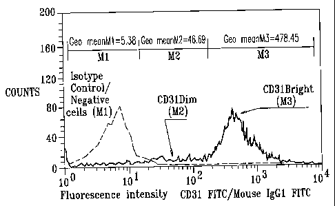
CD31, the platelet endothelial cell adhesion molecule-1 (PECAM-1), is
widely used as a marker during the development of endothelial cell
progenitors,
vasculogenesis and angiogenesis (A, B, C, D, E, F, Hi). CD31 is constitutively
expressed on the surface of adult and embryonic endothelial cells, is a major
constituent of the endothelial cell intercellular junction (where up to 10^6
PECAM-1
molecules are concentrated) and is weakly expressed on many peripheral
leukocytes
and platelets (E, G, 121). With a few minor exceptions, CD31 is not present on
fibroblasts, epithelium, muscle, or other nonvascular cells. Independently of
CD31
expression, endothelial cells and their progenitors are typically
characterized by
binding of Ulex-lectin in combination with the ability to uptake Acetylated-
Low
Density Lipoprotein (Ac-LDL) (/).
Regenerative medicine is an emerging scientific field with implications for
both basic and practical research. Stem and progenitor cells are applied in a
form of
cellular therapy for local tissue repair and regeneration [41, 42]. These
treatments
aim to treat disorders in practically all tissues and organs, such as the
bladder,
intestine, kidney, trachea, eye, heart valves, and bones [43, 44]. Intensive
studies are
being conducted worldwide in order to generate stem cell-based tissue
engineering
therapies. These studies include experiments for the regeneration of blood
vessels
[13], bone [35, 45], cartilage, cornea, dentin, heart muscle [46], liver,
pancreas [47],
nervous tissue, skeletal muscle, and skin [18, 34, 48, 49]. Stem cell-based
therapies
can use cells from various organs in order to generate different tissues. For
example,
3
CA 02645142 2013-12-09
epithelial surfaces (taken from various tissues such as the skin, cornea and
mucosal
membrane) may be used as a source for corneal and skeletal tissues [50, 51].
Additionally, in a more widespread application, blood marrow-derived stem
cells are
used for regeneration of several different tissues such as bone, cartilage,
adipocytes,
neurons, and cells of the hematopoietic system [33, 42].
Stem cells can be administrated systemically or locally using injections to
the
injured site. However, other potential administration routes and usage of
different
medical devices are being developed and tested. Different medical devices such
as
chemical, metal or biodegradable based devices have been described for the
administration of stem cells into the heart and blood vessels (I, K).
US Patent Application Publication 2004/0228847 to Goldschmidt-Clermont
et al., describes stem/progenitor cells and, in particular, therapeutic
strategies based
on the use of such cells to effect vascular rejuvenation and/or to serve as
delivery
vehicles.
PCT Patent Publication WO 2005/120090 to Pulga et al., describes a method
for use with extracted blood, including (a) applying blood to a first gradient
suitable
for selecting first-pass cells having a density less than 1.077 g/m1; (b)
applying the
first-pass cells to a second gradient suitable for selecting 20 second-pass
cells having
a density between 1.055 and 1.074 g/m1; (c) increasing the number of cells
having a
density between 1.055 and 1.074 g/ml, by culturing the second-pass cells for a
period
lasting between 3 and 30 days; and (d) identifying endothelial progenitor
cells in the
cultured cells. Other embodiments are also described.
United States Patent Application Publication 2004-0228897 to Zhang et al.,
describes a medical device for use to assist stem cell and/or stem cell
derivatives in
repopulating, repairing and/or replacing the heart tissue in a failing heart
muscle, in
order to restore the heart's ability to pump blood. The medical device is made
of
biocompatible materials. The specific design of the device is described as
facilitating
the stem cells coated in the device to repopulate heart muscles inside the
heart. Stem
cells are attached to the coated device, proliferated and/or differentiated on
the device
in a bioreactor before implantation. The device also contains bioactive
components
that diminish rejection by the host's immune system. The device may be
directly
4
CA 02645142 2013-12-09
implanted into the failing heart muscle area to assist stern cells to repair
failing heart
muscles via surgical and/or percutaneous catheter based procedures. In another
embodiment, the device may be implanted to the surgical site where abnormal
heart
muscles are removed, to assist stem cells to repopulate heart muscles, to
replace the
failing heart muscles.
US Patent Application Publication 2005/0209556 to Tresco et al., describes a
device and method for the delivery of cells, tissues, enzymes and/or
pharmacological
agents for the treatment or prevention of diseases, disorders or deficiencies.
The
device is placed intravascularly and includes a chamber that houses living
cells
delimited by a membrane on either side that physically separates the cells
from the
blood stream and the central lumen of the catheter. The device can be inserted
over a
guidewire and permits flushing and reloading of the central lumen with
viability
supporting factors that sustain the cells in the outer chamber for long
indwelling
times without removing it from the body. In addition, the central lumen can be
used
to deliver therapeutic substances or withdraw blood. The new intravascular
catheter is
described as being able to be used for the treatment or prevention of a
variety of
diseases and disorders, and may use the implantation of living cells, tissues,
enzymes
or pharmacological agents. The device is described as being used, for example,
for
non-therapeutic purposes that may involve sustained intravascular release of
biological factors as, for example, in stimulating growth of farm animals to
augment
the production of meat. Placement of cells within the device for release of
angiogenesis, cytokines, enzymes, and other factors is described. The use of
stem
cells within the device is also described.
US Patent 6,810,286 to Donovan et al., describes a stimulatory device for the
controlled production of angiogenic growth factors. More specifically, a
subthreshold
pulse generator is used for the local production of vascular endothelial
growth factor.
The following references may be of interest:
1. Leblond= G.P. (1964),. "Classification of cell populations on the basis
of their proliferative behaviour," Natl. Cancer Inst. Monogr. 14:119-150
5
CA 02645142 2008-09-08
WO 2007/102162 PCT/1L2007/000308
2. Evans M.J. and Kaufman M.H. (1981), "Establishment in culture of
pluripotential cells from mouse embryos," Nature 292:154-156
3. Donovan P.J. and Gearhart J. (2001), "The end of the beginning for - =
pluripotent stem cells," Nature 414:92-97
4. Spradling A. et al. (2001), "Stem cells find their niche," Nature 414:98-
104
5. Weissman I.L. et al. (2001), "Stem and progenitor cells: origins,
phenotypes, lineage commitments, and transdifferentiations," Annu. Rev. Cell.
Dev.
Biol. 17:387-403
6. Weissman I.L. (2000), "Stem cells: units of development, units of
regeneration, and units in evolution," Cell 100:157-68
7. Cheng A, Wang S, Cai J, Rao MS, Mattson MP (2003), "Nitric oxide acts
in a positive feedback loop with BDNF to regulate neural progenitor cell
proliferation and differentiation in the mammalian brain," Dev Biol.
258(2):319-33
8. Cousin B, Andre M, Arnaud E, Penicaud L, Casteilla L (2003),
"Reconstitution of lethally irradiated mice by cells isolated from adipose
tissue,"
Biochem Biophys Res Commun. 301(4):1016-22
9. Anderson D.J., Gage, F.H., and Weissman, I.L. (2001), "Can stem cells
cross lineage boundaries?" Nat. Med. 7:393-395
10. Robey P.G. (2000), "Stem cells near the century mark," J. Clin. Invest.
105:1489-1491
11. Eisenberg LM, Burns L, Eisenberg CA (2003), "Hematopoietic cells from
bone marrow have the potential to differentiate into cardiomyocytes in vitro,"
Anat
Rec. 274A(1):870-82
12. Karl J.L., Fernandes Ian A. McKenzie, Pleasantine Mill et al. (2004), "A
dermal niche for multipotent adult skin-derived precursor cells," Nature Cell
Biology
Published online: 1 November 2004, DOI: 10.1038/ncb1181
13. Jackson KA, Mi T, Goodell MA (1999), "Hematopoietic potential of stem
cells isolated from murine skeletal muscle," Proc Natl Acad Sci U S A
96(25):14482-6
6
CA 02645142 2008-09-08
WO 2007/102162 PCT/1L2007/000308
14. Brazelton TR, Rossi FM, Keshet GI, Blau HM (2000), "From marrow to
brain: expression of neuronal phenotypes in adult mice," Science
290(5497):1775-9
- 15. Bjornson CR, Rietze RL, Reynolds BA, Magli MC, Vescovi AL
(1999), -
"Turning brain into blood: a hematopoietic fate adopted by adult neural stem
cells in
vivo," Science 283(5400:534-7
16. Slack, J.M. (2000), "Stem cells in epithelial tissues," Science
287:1431-1433
17. Ferrari G., Cusella-De Angelis G., Coletta M., Paolucci E., Stornaiuolo
A., Cossu G., and Mavilio F. (1998), "Muscle regeneration by bone marrow-
derived
myogenic progenitors," Science 279:528-30
18. Lagasse E, Connors H, Al-Dhalimy M, Reitsma M, Dohse M, Osborne L,
Wang X, Finegold M, Weissman IL, Grompe M (2000), "Purified hematopoietic
stem cells can differentiate into hepatocytes in vivo," Nat Med. 6:1229-34
19. Hirschi, K. K., and Goodell, M. A. (2002), "Hematopoietic, vascular and
cardiac fates of bone marrow-derived stem cells," Gene Ther. 9:648-652
20. Theise N.D. et al. (2000), "Liver from bone marrow in humans,"
Hepatology 32:11-16
21. Kleeberger W. et al. (2002), "High frequency of epithelial chimerism in
liver transplants demonstrated by microdissection and STR-analysis,"
Hepatology
35:110-116
22. Weimann J.M. et al. (2003), "Contribution of transplanted bone marrow
cells to Purkinje neurons in human adult brains," Proc. Natl. Acad. Sci. USA
100:2088-2093
23. Quaini F. et al. (2002), "Chimerism of the transplanted heart," N. Engl.
Med 346:5-15
24. Blau H.M. et al. (2001), "The evolving concept of a stem cell: entity or
function?" Cell 105:829-841
25. Goodell M.A. et al. (2001), "Stein cell plasticity in muscle and bone
marrow," Ann. NY Acad. Sci. 938:208-218
7
CA 02645142 2008-09-08
WO 2007/102162
PCT/1L2007/000308
26. Krause D.S. (2002), "Plasticity of marrow-derived stem cells," Gene
Ther. 9:754-758
27. Wulf G.G. et al. (2001), "Somatic stein cell plasticity," Exp Hematol.
29:1361-1370
28. Pittenger M.F. et al. (1999), "Multilineage potential of adult human
mesenchymal stem cells," Science 284:143-147
29. Liechty K.W. et al. (2000), "Human mesenchymal stem cells engraft and
demonstrate site-specific differentiation after in utero transplantation in
sheep,"
Nature Med. 6:1282-1286
XIII. Li A. et al. (2003), "IL-8 directly enhanced endothelial cell survival,
proliferation, and matrix metalloproteinases production and regulated
angiogenesis",
Journal of immunol. 170:3369-3376.
XIV. Laterveer L. et al. (1995), "Interleukin-8 induces rapid mobilization of
hematopoietic stem cells with radioprotective capacity and long-term
myelolymphoid repopulating ability", Blood 85:2269-75.
XV. Scho-mig K. et al. (2006), "Interleukin-8 is associated with
circulating
CD133+ progenitor cells in acute myocardial infarction", European Heart
Journal 27:
1032-1037
30. Sang YY, Collector MI, Baylin SB, Diehl AM, Sharkis SJ (2004),
"Hematopoietic stem cells convert into liver cells within days without
fusion," Nat
Cell Biol. 6(6):532-9. Epub 2004 May 09
31. Bittner R.E., Schofer C., Weipoltshammer K., Ivanova S., Streubel B.,
Hauser E., Freilinger M., Hoger H., Elbe-Burger A., and Wachtler F. (1999),
"Recruitment of bone-marrow-derived cells by skeletal and cardiac muscle in
adult
dystrophic mdx mice," Anat. Embryol. (Berl) 199:391-396
32. Mezey E, Chandross KJ, Harta G, Maki RA, McKercher SR (2000),
"Turning blood into brain: cells bearing neuronal antigens generated in vivo
from
bone marrow," Science. 290(5497):1779-82
8
CA 02645142 2008-09-08
WO 2007/102162
PCT/1L2007/000308
33. Douglas W.L., Dimmeler S. (2004), "Therapeutic angiogenesis and
vasculogenesis for ischemic diseases. Part I: Angiogenic cytokines,"
Circulation
109:2487-2491
34. Douglas W.L., Dimmeler S. (2004), "Therapeutic angiogenesis and
vasculogenesis for ischemic diseases. Part II: Cell-based therapy,"
Circulation
109:2692-2697
35. Rodda SJ, Kavanagh SJ, Rathjen J, Rathjen PD (2002), "Embryonic stern
cell differentiation and the analysis of mammalian development," Int J Dev
Biol.
46(4):449-58
36. Amit M., Carpenter M.K., Inokuma M.S., Chiu C.P., Harris C.P., Waknitz
M.A., Itskovitz-Eldor J., and Thomson J.A. (2000), "Clonally derived human
embryonic stern cell lines maintain pluripotency and proliferative potential
for
prolonged periods of culture," Dev Biol. 227(2):271-8
37. Aoki S, Toda S, Sakemi T, Sugihara H (2003), "Coculture of endothelial
cells and mature adipocytes actively promotes immature preadipocyte
development
in vitro," Cell Struct Funct. 28(1):55-60
38. Wan H, An Y, Zhang Z, Zhang Y, Wang Z (2003), "Differentiation of rat
embryonic neural stem cells promoted by co-cultured Schwann cells," Chin Med J
(Engl). 116(3):428-31
39. Kollet 0, Shivtiel S, Chen YQ. et al. (2003), "HGF, SDF-1, and MMP-9
are involved in stress-induced human CD34+ stem cell recruitment to the
liver," J
Clin Invest. 112(2):160-9
40. Badorff C, Brandes RP, Popp R, Rupp S, Urbich C, Aicher A, Fleming I,
Busse R, Zeiher AM, Dimmeler S (2003), "Transdifferentiation of blood-derived
human adult endothelial progenitor cells into functionally active
cardionvocytes,"
Circulation 107(7):1024-32
41. Bianco, P. and Robey P.G. (2001), "Stem cells in tissue engineering,"
Nature 414:118-121
42. Lagasse E. et al. (2001), "Toward regenerative medicine," Immunity
14:425-436
9
CA 02645142 2008-09-08
WO 2007/102162
PCT/1L2007/000308
43. Stock U.A., Vacanti J.P. (2001), "Tissue engineering: current state and
prospects," Ann. Rev. Med 52:443-451
44. Kim W.S. et al. (1994), "Bone defect repair with tissue-engineered
cartilage," Plast. Recontr. Surg. 94:580-584
45. Petite H. et al. (2000), "Tissue-engineered bone regeneration," Nature
Biotechnol. 18:959-963
46. Jackson KA, Majka SM, Wang H, Pocius J, Hartley CJ, Majesky MW,
Entman ML, Michael LH, Hirschi KK, Goodell MA (2001), "Regeneration of
ischemic cardiac muscle and vascular endothelium by adult stem cells," J Clin
Invest.
107(10:1395-402
47. Ramiya V.K. et al. (2000), "Reversal of insulin-dependent diabetes using
islets generated in vitro from pancreatic stem cells," Nature Medicine 6:278-
282
48. Rafii S., Lyden D. (2003), "Therapeutic stern and progenitor cell
transplantation for organ vascularization and regeneration," Nature Medicine
9:702-712
49. Gussoni E., Soneoka Y., Strickland C., Buzney E., Khan M., Flint A.,
Kunkel L., and Mulligan R. (1999), "Dystrophin expression in the mdx mouse
restored by stem cell transplantation," Nature 401:390-4
50. Zhao Y et al. (2003), "A human peripheral blood monocyte-derived
subset acts as pluripotent stem cells," Proc. Natl. Acad. Sci. USA 100:2426-
2431
51. Kohji N, Masayuki Y, Yasutaka H. et al. (2004), "Corneal reconstruction
with tissue-engineered cell sheets composed of autologous oral mucosal
epithelium,"
N Engl J Med 351:1187-96
52. Kayisli U.A., Luk J, Guzeloglu-Kayisli 0. et al. (2005), "Regulation of
angiogenic activity of human endometrial endothelial cells in culture by
ovarian
steroids," J Clin Endocrinol Metab 89:5794-5802
53. Dimmeler S. (2005), "Circulating endothelial precursors: Identification of
functional subpopulations," Blood 106(7):2231-2232
54. Urbich C. et al. (2004), "Endothelial progenitor cells: Characterization
and role in vascular biology," Circulation Research 95:343-353
CA 02645142 2008-09-08
WO 2007/102162
PCT/1L2007/000308
A. Asahara T, Murohara T, Sullivan A, et al. (1997), "Isolation of putative
progenitor endothelial cells for angiogenesis," Science 275:964 ¨967.
Kalka C, Masuda H, Takahashi T, et al. (2000), "Transplantation of ex
vivo expanded endothelial progenitor cells for therapeutic
neovascularization," Proc
Nati Acad Sci USA. 97:3422-3427.
C. Assmus B, Schachinger V, Teupe C, et al. (2002), "Transplantation of
Progenitor Cells and Regeneration Enhancement in Acute Myocardial Infarction
(TOPCARE-AMI)," Circulation. 106:3009 ¨3017.
D. Yoon C.H, Hur J., Park KW, et al. (2005), "Synergistic
Neovascularization by Mixed Transplantation of Early Endothelial Progenitor
Cells
and Late Outgrowth Endothelial Cells," Circulation. 112:1618-1627.
K DeLisser, H.M., Christofidou-Solomidou, R.M. Strieter, M.D. et al.
(1997), "Involvement of endothelial PECAM-1/CD31 in angiogenesis," Am J
Pathol.
151: 671 -677.
F. Kawamoto A., Tkebuchava T., Yamaguchi J.I., et al. (2003),
"Intramyocardial Transplantation of Autologous Endothelial Progenitor Cells
for
Therapeutic Neovascularization of Myocardial Ischemia," Circulation. 107:461-
468
G. Newman P.J. (1997), "The Biology of PECAM-1," J Clin Invest. 99:3-8.
H Vecchi, A., C. Garlanda, M.G. Lampugnani, M. Resnati, et al. (1994),
"Monoclonal antibodies specific for endothelial cells of mouse blood vessels.
Their
application in the identification of adult and embryonic endothelium," Eur J
Cell
Biol. 63: 247 - 254.
Hi. Kanayasu-Toyoda T., Yamaguchi T., Oshizawa T., et al. (2003),
"CD31 (PECAM-1)-Bright Cells Derived From AC133-Positive Cells in Human
Peripheral Blood as Endothelial-Precursor Cells," Journal of Cell. Physiol.
195:119-
129.
I. Yamamoto K., Takahashi T., Asahara T., et al. (2003), "Proliferation,
differentiation, and tube formation by endothelial progenitor cells in
response to
shear stress," J Appl Physiol. 95: 2081-2088.
J. Cohen S., and Leor J. (2004), "Rebuilding Broken Hearts," Scientific
American, November: 45-51.
K US Patent Application Publication 2004/0228897 to Zhang et al.
11
CA 02645142 2013-12-09
L. US Patent Application Publication 2005/0272152 to Xu et al.
SUMMARY OF THE INVENTION
In the context of the present patent application and in the claims, a "core
cell
population" (CCP) is a population of at least 5 million cells which have a
density of
less than 1.072 g/ml, and at least 1% of which are CD34+CD45-/Dim (i.e., at
least
50,000 ofthe cells are both (a) CD34 positive and (b) CD45 negative or CD45
Dim).
A CCP is typically, but not necessarily, generated from a hematopoietic
source.
For most applications, at least 40% of the CCP is CD31Bright (i.e., at least 2
million cells out of the 5 million cells are CD31 Bright).
While not being limited to any method of detection, cells expressing increased
amounts of CD31 relative to isotype control are termed "CD31Bright" cells,
because
these cells bear more CD31 molecules relative to other cells, and thus tend to
fluoresce
brightly when stained with fluorescently-labeled antibodies. In this context,
in the
specification and in the claims, "bright" means that the fluorescence
intensity of the
labeled cellular marker of interest is at least 50 times higher (if measured
using flow
cytometry) than the isotype control intensity.
In accordance with an embodiment of the present invention, a method for
producing a progenitor/precursor cell population (PCP) is provided, comprising
(a) 20
processing cells extracted from a cell donor to yield a CCP, and (b)
stimulating the
CCP to differentiate into the progenitor/precursor cell population. In the
context of the
specification and in the claims, "progenitor/precursor" cells are partially
differentiated
cells that are able to divide and give rise to differentiated cells.
In accordance with an embodiment of the present invention, a composition of
matter, comprising a population of cultured cells that comprises a sub-
population of
cells that stain as CD31Bright demonstrate uptake of Ac-LDL+ and secrete
interleukin-
8 and angiogenin, wherein the sub-population of cells comprises at least 10%
of the
cells in the population of cultured cells.
12
CA 02645142 2013-12-09
While for some applications described herein, the density of the cells in the
CCP is typically less than 1.072 g/ml (as described), for some applications,
the CCP
has at least 5 million cells having a density of less than 1.062 g/ml.
In the context of the specification and in the claims, an "elemental cell
population" (ECP) is a population of at least 5 million cells which have a
density of
less than 1.072 g/ml, at least 1.0% of which are CD34+CD45-/Dim, and at least
30%
30 ofwhich are CD31Bright. Typically, but not necessarily, at least 40% of the
cells in
12a
CA 02645142 2008-09-08
WO 2007/102162
PCT/1L2007/000308
the ECP are CD31Bright. Typically, but not necessarily, at least 30% of the
cells in
the ECP are CD14+. Typically, but not necessarily, at least 1.5% or at least
2% of
the cells in the ECP are CD34+CD45-/Dim. For some applications, the ECP has at
least 5 million cells having a density of less than 1.062 g/ml. It is
typically but not
necessarily the case that a CCP is also an ECP. It is noted that, although for
simplicity, embodiments of the present invention are described herein with
respect to
procedures relating to a CCP, the scope of the present invention includes, in
each
instance, performing the same procedure in relation to an ECP.
An "initiating cell population" (ICP), in the context of the specification and
in
the claims, is a cell population that can differentiate into a PCP. CCPs and
ECPs are
both examples of an ICP. An ICP is typically but not necessarily created by a
process that comprises separating lower density cells (that are included in
the ICP)
from higher density cells. Such a separation may be accomplished, for example,
by
use of one or more gradients.
For some applications, the CCP-derived progenitor cells are used as a
therapeutic cell product (e.g., for cancer therapy, for tissue regeneration,
for tissue
engineering, and/or for tissue replacement), as a research tool (e.g., for
research of
signal transduction, or for screening of growth factors), and/or as a
diagnostic tool.
When the CCP-derived progenitor cells are used as a therapeutic cell product,
they
are typically administered to a patient, in whom the progenitor cells mature
into the
desired cells (e.g., endothelial cells, retinal cells, etc.).
In an embodiment, at least one result of at least one stage in a process
described herein is used as a diagnostic indicator. For example, pathology of
a
patient may be indicated if an in vitro procedure performed on extracted blood
of the
patient does not produce a CCP, when the same procedure performed on cells
extracted from a healthy volunteer would result in production of the CCP.
Alternatively or additionally, a pathology of a patient may be indicated if an
in vitro
stimulation procedure perfauned on an autologous CCP does not produce a
desired
number of progenitor cells of a particular class, when the same procedure
would
produce the desired number of progenitor cells of a particular class from a
CCP
derived from cells of a healthy volunteer. Further alternatively or
additionally, a
pathology of a patient may be indicated if one or more in vitro protocols used
to
assess a PCP do not yield the same results as a PCP originated from a healthy
13
CA 02645142 2008-09-08
WO 2007/102162
PCT/1L2007/000308
volunteer. Still further alternatively or additionally, a pathology of a
patient may be
indicated if one or more protocols used to assess a PCP following implantation
within a patient do not perform as expected (e.g., like a PCP implanted in a
healthy
animal or human volunteer, or in an animal model of a similar disease).
When hematopoietic stem cells are used as a source to create the CCP, the
resultant CCP is typically but not necessarily characterized by at least 40%
of the
cells in the CCP being CD31Bright, and at least 2.2% or at least 2.5% of the
cells
being CD34+CD45-/Dim.
Typically, the process of stimulating the CCP takes between about 2 and
about 15 days (e.g., between about 3 and about 15 days), or between about 15
and
about 120 days (e.g., between about 15 and about 30 days). Alternatively,
stimulating the CCP takes less than 2 days, or more than 120 days.
The mammalian cell donor may be human or non-human, as appropriate. For
some applications, the mammalian cell donor ultimately receives an
administration
of a product derived from the CCP, while for other applications, the mammalian
cell
donor does not receive such a product. Stem cells that can be used to produce
the
CCP are typically but not necessarily derived from one or more of the
following
source tissues: embryonic tissue, umbilical cord blood or tissue, neonatal
tissue, adult
tissue, bone marrow, mobilized blood, peripheral blood, peripheral blood
mononuclear cells, skin cells, and other stem-cell-containing tissue. It is
noted that
the stem cells may be obtained from fresh samples of these sources or from
frozen
and then thawed cells from these source tissues.
The CCP is typically prepared by generating or obtaining a single cell
suspension from one of the abovementioned source tissues. For example,
mobilized
blood mononuclear cells may be extracted using a 1.077 g/ml density gradient,
e.g., a
Ficoll (TM) gradient, including copolymers of sucrose and epichlorohydrin. It
is to
be noted that such a gradient is not used for all applications, e.g., for
applications in
which a single cell suspension is generated from a non-hematopoietic source
(e.g.,
mucosa' or skin cells). The output of this gradient is then typically passed
through a
second gradient (e.g., a Percoll (TM) gradient, including
polyvinylpyrrolidone-coated silica colloids), suitable for selecting cells
having a
density less than 1.072 g/ml or less than 1.062 g/ml. These selected cells
then
14
CA 02645142 2008-09-08
WO 2007/102162
PCT/1L2007/000308
typically propagate, in vitro, until they become a CCP. As appropriate, other
density
gradients may be used, independently of or in combination with those cited
above in
order to enrich the designated cells of the CCP. For example, an OptiPrep (TM)
gradient, including an aqueous solution of Iodixanol, and/or a Nycodenz (TM)
gradient may also be used.
The CCP is typically stimulated to generate progenitor cells of one or more of
the following cell classes:
Blood cells (e.g., red blood cells and/or white blood cells (such as T cells
or B
cells));
Neural lineage cells (e.g., CNS neurons, oligodendrocytes, astrocytes,
peripheral nervous system (PNS) neurons, and retinal cells (including, but not
limited
to, photoreceptors, pigment epithelium cells or retinal ganglion cells).
Endothelial cells;
Pericytes;
Smooth muscle cells;
Cardiomyocytes;
0 steoblasts;
Pancreatic endocrine or exocrine cells (e.g., beta cells or alpha cells);
Hepatic tissue (e.g., hepatocytes); and
Kidney cells.
For some applications, the CCP is transfected with a gene prior to the
stimulation of the CCP, whereupon the CCP differentiates into a population of
desired progenitor cells containing the transfected gene. Typically, these
progenitor
cells are then administered to a patient. For some applications, the PCP is
transfected with a gene. Typically, these PCP cells are then administered to a
patient.
In order to stimulate the CCP to differentiate into a desired class of
progenitor
cells, or in association with stimulation of the CCP to differentiate into a
desired
class of progenitor cells, the CCP is typically directly or indirectly co-
cultured with
"tnrget tissue." The "target tissue" typically but not necessarily includes
tissue from
an organ whose cells represent a desired final state of the progenitor cells.
For
example, the target tissue may include brain or similar tissue, or heart or
similar
CA 02645142 2008-09-08
WO 2007/102162
PCT/1L2007/000308
tissue, if it is desired for the progenitor cells to differentiate into brain
tissue or into
heart tissue, respectively. Other examples include:
(a) co-culturing the CCP with peripheral nerves (and/or culturing the CCP in
conditioned medium derived therefrom), to induce differentiation of the CCP
into
peripheral neurons; =
(b) co-culturing the CCP with central nervous system (CNS) nerves (and/or
culturing the CCP in conditioned medium derived therefrom), to induce
differentiation of the CCP into CNS neurons;
(c) co-culturing the CCP with retinal tissue (and/or culturing the CCP in
conditioned medium derived therefrom), to induce differentiation of the CCP
into
retinal tissue. The retinal tissue may include, for example, one or more of:
pigment
epithelium, or photoreceptors. As appropriate, the retinal tissue may comprise
fetal
retinal tissue, embryonic retinal tissue, or mature retinal tissue;
(d) co-culturing the CCP with blood vessel tissue (and/or culturing the CCP
in conditioned medium derived therefrom), to induce differentiation of the CCP
into
angiogenic lineage tissue and/or cardiomyocytes (CMCs);
(e) co-culturing the CCP with cardiac tissue (and/or culturing the CCP in
conditioned medium derived therefrom), to induce differentiation of the CCP
into
CMCs;
(f) co-culturing the CCP with pancreatic endocrine or exocrine tissue (and/or
culturing the CCP in conditioned medium derived therefrom), to induce
differentiation of the CCP into pancreatic endocrine or exocrine cells; and
(g) co-culturing the CCP with smooth muscle tissue (and/or culturing the
CCP in conditioned medium derived therefrom), to induce differentiation of the
CCP
into smooth muscle cells.
Techniques described herein with respect to use of a target tissue may be used
with any "sample" tissue, regardless of whether it is desired for the CCP to
differentiate into a PCP having cells like those in the sample tissue.
For some applications, slices or a homogenate of the target tissue are used
for
co-culturing, although other techniques for preparing the target tissue will
be
16
CA 02645142 2008-09-08
WO 2007/102162
PCT/1L2007/000308
apparent to a person of ordinary skill in the art who has read the disclosure
of the
present patent application.
- The target tissue may be in essentially direct contact with the CCP, or
separated therefrom by a semi-permeable membrane. As appropriate, the target
tissue may be autologous, syngeneic, allogeneic, or xenogeneic with respect to
the
source tissue from which the CCP was produced. Alternatively or additionally,
the
CCP is cultured in a conditioned medium made using target , tissue (e.g., a
target
tissue described hereinabove), that is autologous, syngeneic, allogeneic, or
xenogeneic with respect to the source tissue from which the CCP was produced.
For
some applications, the target tissue and the CCP are co-cultured in the
conditioned
medium. It is to be noted that the source of the target tissue may also be
tissue from
a cadaver, and/or may be lyophilized, fresh, or frozen.
Alternatively or additionally, for some applications, to produce a desired
class
of progenitor cells, cells from the CCP are cultured in the presence of
stimulation
caused by "stimulation factors," e.g., one or more antibodies, cytokines,
growth
factors, tissue-derived extra cellular matrix, and/or other molecules, such
as: IL-8,
anti-IL-8, anti-CD34, anti-Tie-2, anti-CD133, anti-CD117, LIF, EPO, IGF, b-
FGF,
M-CSF, GM-CSF, TGF alpha, TGF beta, VEGF, BHA, BDNF, NGF, NT3, NT4/5,
GDNF, S-100, CNTF, EGF, NGF3, CFN, ADMIF, estrogen, cortisone,
dexamethasone, or any other molecule from the steroid family, prolactin, an
adrenocorticoid hormone, ACTH, glutamate, serotonin, acetylcholine, NO,
retinoic
acid (RA), heparin, insulin, forskolin, a statin, an anti-diabetic drug (e.g.,
a
thiazolidinedione such as rosiglitazone), NO, MCDB-201, MCT-165, glatiramer
acetate (L-glutamic acid, L-alanine, L-tyrosine, L-lysine), a glatiramer
acetate-like
molecule, IFN alpha, IFN beta, or any other immunoregulatory agent, sodium
selenite, linoleic acid, ascorbic acid, transferrin, 5-azacytidine, PDGF,
VEGF,
cardiotrophin, and thrombin.
In the context of the specification and in the claims, a "glatiramer acetate-
like
molecule" means a copolymer comprising:
(a) the same four amino acids as in glatiramer acetate, but in different
ratios,
(e.g., within 5%, 10%, or 25% of their current values of L-glutamic acid: L-
alanine :
L-tyro sine : L-lysine = 0.141 : 0.427 : 0.095 : 0.338);
17
CA 02645142 2008-09-08
WO 2007/102162
PCT/1L2007/000308
(b) three of the four amino acids in glatiramer acetate, but the fourth amino
acid
is replaced by a different naturally-occurring or synthetic amino acid;
= (c) four amino acids, in which at least one of the amino acids is an
enantiomer
of the corresponding amino acid in glatiramer acetate, and the remainder of
the
amino acids (if any) are the corresponding L- amino acids that are in
glatiramer
acetate; or
(d) a combination one or more of (a), (b), and (c).
It is to be appreciated that the particular stimulation factors described
herein
are by way of illustration and not limitation, and the scope of the present
invention
includes the use of other stimulation factors. As appropriate, these may be
utilized in
a concentration of between about 100 pg/ml and about 100 ig/m1 (or molar
equivalents). Typically, particular stimulation factors are selected in
accordance
with the particular class of progenitor cells desired. For example, to induce
neural
progenitor cells, one or more of the following stimulation factors are used:
BHA,
BDNF, NGF, NT3, NT4/5, GDNF, MCT-165, glatiramer acetate, a glatiramer
acetate-like molecule, IFN alpha, IFN beta or any other immunoregulatory
agent,
S-100, CNTF, EGF, NGF3, CFN, ADMIF, and acetylcholine. In another example,
to induce CMC progenitors, one or more of the following stimulation factors
are
used: bFGF, cortisone, estrogen, progesterone, or any other molecule form the
steroid family, NO, sodium selenite, linoleic acid, ascorbic acid, retinoic
acid (RA)
or any other derivative of vitamin D, transferrin, 5-azacytidine, MCT-165,
glatiramer
acetate, a glatiramer acetate-like molecule, IFN alpha, IFN beta, or any other
immunoregulatory agent, TGF-beta, insulin, EGF, IGF, PDGF, VEGF,
cardiotrophin, MCDB201, and thrombin.
For some applications, the stimulation factors are introduced to the CCP in a
soluble form, and/or in an aggregated form, and/or attached to a surface of a
culture
dish. In an embodiment, the CCP is incubated on a surface comprising a
growth-enhancing molecule other than collagen or fibronectin. The
growth-enhancing molecule may comprise, for example, VEGF or another suitable
antibody or factor described herein. As appropriate, the growth-enhancing
molecule
may be mixed with collagen or fibronectin or plasma, or may be coated on the
surface in a layer separate from a layer on the surface that comprises
collagen or
18
CA 02645142 2008-09-08
WO 2007/102162
PCT/1L2007/000308
fibronectin or plasma. Alternatively, the only growth-enhancing molecule(s) on
the
surface of the culture dish is collagen and/or fibronectin and/or plasma.
In the context of the present patent application= and in the claims, a surface
"comprising" or "including" a molecule means that the molecule is coated on
the
surface, attached to the surface, or otherwise integrated into the surface.
Following stimulation of the CCP, the resultant product is typically tested to
verify that it has differentiated into a desired form. Characterization of the
differentiated cells is performed according to the cells' phenotypical,
genotypical and
physiological features. In accordance with an embodiment of the present
invention,
the cells are characterized by assessing functional/physiological activity
thereof, in
combination with or in place of evaluating the presence or absence of certain
cellular
markers. Evaluating functional/physiological activity of the cells following
the
stimulation of the CCP helps increase the likelihood that the product obtained
and
designated for in vivo use will perform as expected.
For example, when angiogenic cell precursors (ACPs) (which also include
endothelial progenitor cells (EPCs)) are the desired product, the product is
typically
positive for the generation and/or expression of one or more of: CD34, CD117,
CD133, Tie-2, CD31, CD34+CD133+, KDR, CD34+KDR+, CD144, von Willebrand
Factor, SH2 (CD105), SH3, fibronectin, collagen (types I, III and/or IV), ICAM
(type 1 or 2), VCAM1, Vimentin, BMP-R IA, BMP-RII, CD44, integrin bl,
aSM-actin, and MUC18, CXCR4. Additionally, the ACP product typically
functionally demonstrates uptake of Acetylated-Low Density Lipoprotein (Ac-
LDL)
(i.e., the product is Ac-LDL+) and/or secretes one or more of the following
molecules: Interleukin-8 (IL-8), VEGF, Angiogenin, Matrix metalloproteinase 2
(MMP-2), or Matrix metalloproteinase 9 (MMP-9). Alternatively or additionally,
the ACP product generates tube-like structures on a semi-solid matrix, and/or
migrates towards chemoattractants (such as SDF-1 or VEGF), and/or proliferates
in
response to cell activation, and/or generates typical cell colonies/clusters.
For some
applications, in order to further characterize the cells, CD31Bright cells
that
demonstrate uptake of Ac-LDL are examined.
Typically, greater than 1.5% of the core cell population that was stimulated
demonstrates one or more of the abovementioned characteristics. Alternatively,
if
19
CA 02645142 2008-09-08
WO 2007/102162
PCT/1L2007/000308
neural progenitor cells are the desired product, then the product is typically
positive
for the generation and/or the expression of one or more of: Nestin, NSF,
Notch,
numb, Musashi-1, presenilin, FGFR4, Fz9, SOX 2, CD133, CD15, GD2, rhodopsin,
recoverin, calretinin, PAX6, RX, Chx10, 04, and GFAP. Further alternatively,
if
cardiomyocyte (CMC) progenitors are the desired product, then the product is
typically positive for the generation and/or the expression of one or more of:
CD31,
CD117, sarcomeric alpha-actin, beta-actin, alpha-actinin, desmin, cardiac
troponin
T, conrxexin43, alphaketa-MHC, sarcomeric alpha-tropomyosin, Troponin I,
GATA-4, Nkx2.5/Csx,and MEF-2.
For some applications, the time duration between collecting cells from the
cell donor and using the CCP-derived progenitor cells (e.g., for
administration into a
patient), is reduced in order to effect almost immediate use thereof.
Alternatively,
the cells are preserved at one or more points in the process. For example, the
CCP
may be frozen prior to the stimulation thereof that generates progenitor
cells.
Alternatively, the CCP is stimulated in order to generate desired progenitor
cells, and
these progenitor cells are frozen. In either of these cases, the frozen cells
may be
stored and/or transported, for subsequent thawing and use.
"Transport," in the
context of the specification and the claims, means transport to a remote site,
e.g., a
site greater than 10 km or 100 km away from a site where the CCP is first
created.
It is noted that certain applications are suitable for large-scale
commercialization, including freezing and transport, such as (a) generation of
stores
of CCPs, (b) generation of stores of PCPs, (such as hematopoietic stern cells
able to
mature into CMCs), and (c) stem cell banks where individuals may store a CCP
or
differentiated progenitor cells, for possible later use. Other applications
(such as
acute post-stroke autologous administration of neuronal stem cells) may not
benefit,
or may not benefit as greatly, from the time delays provided by freezing of
cells,
although the technique may be useful for some purposes.
For some applications, the CCP is cultured for a period lasting between about
1 and about 20 days (e.g., between about 1 and 5 days) in a culture medium
comprising less than about 5% serum. Alternatively, the CCP is cultured for a
period
lasting between about 1 and about 20 days (e.g., between about 1 and about 5
days)
in a culture medium comprising greater than about 10% serum. In an embodiment,
one of these periods follows the other of these periods.
CA 02645142 2008-09-08
WO 2007/102162
PCT/1L2007/000308
For some applications, the CCP is cultured, during a low-serum time period,
in a culture medium comprising less than about 10% serum, and, during a
high-serum time period, in a culture medium comprising greater than or equal
to
about 10% serum. In an embodiment, culturing the CCP during the low-serum time
period comprises culturing the CCP for a duration of between about 1 and about
20
days (e.g., between about 1 and about 5 days). Alternatively or additionally,
culturing the CCP during the high-serum time period comprises culturing the
CCP
for a duration of between about 1 and about 120 days (e.g., between about 1
and
about 30 days). Typically, culturing the CCP during the low-serum time period
is
performed prior to culturing the CCP during the high-serum time period.
Alternatively, culturing the CCP during the low-serum time period is performed
following culturing the CCP during the high-serum time period.
For some applications, during a hypoxic time period lasting at least about 2
hours, the CCP is cultured under hypoxic conditions, and, during a non-hypoxic
time
period lasting at least about 1 day, the CCP is cultured under non-hypoxic
conditions.
Culturing the CCP under hypoxic conditions may be performed prior to or
following
culturing the CCP under non-hypoxic conditions. Typically, but not
necessarily, the
hypoxic and non-hypoxic time-periods are within a culturing time period
lasting less
than about 120 days (e.g., less than about 30 days), and culturing the CCP
under
hypoxic conditions comprises culturing the CCP under hypoxic conditions during
the
first about two days of the culturing time period. Alternatively or
additionally,
culturing the CCP under hypoxic conditions comprises culturing the CCP under
hypoxic conditions during the last about two days of the culturing time
period.
Further alternatively or additionally, culturing the CCP under hypoxic
conditions
comprises culturing the CCP under hypoxic conditions for at least about 2
hours
between a first two days and a last two days of the culturing time period.
For some applications, the CCP is cultured in a culture medium comprising at
least one of the following: erythropoietin, a statin, and an antidiabetic
agent (e.g., a
thiazolidinedione such as rosiglitazone). Alternatively or additionally, the
CCP is
cultured in the presence of one or more proliferation-differentiation-
enhancing
agents, such as, anti-CD34, anti-Tie-2, anti-CD133, anti-CD117, LIF, EPO, IGF,
b-FGF, M-CSF, GM-CSF, TGF alpha, TGF beta, VEGF, BHA, BDNF, NGF, NT3,
NT4/5, GDNF, S-100, CNTF, EGF, NGF3, CFN, ADMIF, estrogen, prolactin, an
21
CA 02645142 2013-12-09
adrenocorticoid hormone, ACTH, glutamate, serotonin, acetylcholine, NO,
retinoic
acid (RA) or any other vitamin D derivative, heparin, insulin, forskolin,
cortisone,
cortisol, dexamethasone, progesterone, or any other molecule from the steroid
family, a statin, or an anti-diabetic drug (e.g., a thiazolidinedione such as
rosiglitazone), MCDB-201, MCT-165, glatiramer acetate, a glatiramer acetate-
like
molecule, IFN alpha, 1FN beta or any other inununoregulatory agent, sodium
selenite, linoleic acid, ascorbic acid, transferrin, 5-azacytidine, PDGF,
VEGF,
cudiotrophin, and thrombin.
In an embodiment, techniques described herein are practiced in combination
with (a) techniques described in one or more of the references cited herein,
(b)
techniques described in US Provisional Patent Application 60/576,266, filed
June 1,
2004, (c) techniques described in US Provisional Patent Application
60/588,520,
filed July 15, 2004, (d) techniques described in US Provisional Patent
Application
60/668,739, filed April 5, 2005, (e) techniques described in US Provisional
Patent
Application 60/636,391, filed December 14, 2004, (1) techniques described in
PCT
Patent Application PCT/IL2005/001345, filed December 14, 2005, and/or PCT
Patent Application PCT/11,2005/001348, filed December 14, 2005. Each of these
patent applications is assigned to the assignee of the present patent
application
and the scope of the present invention includes embodiments described therein.
In an embodiment, a method is provided comprising culturing the CCP in a
first container during a first portion of a culturing period; removing all or
at least
some cells of the COP from the first container at the end of the first portion
of the
period; and culturing, in a second container during a second portion of the
period, the
cells removed from the first container. For example, removing at least some of
the
COP cells may comprise selecting for removal cells that adhere to a surface of
the
first container.
When the cells from a progenitor/precursor cell population (PCP) derived
from a COP are designated for implantation into a human, they should be
generally
free from any bacterial or viral contamination. Additionally, in the case of a
PCP of
angiogenic cell precursors (ACPs), one or more of the following phenotypical,
genotypical and physiological conditions should typically be met:
22
CA 02645142 2008-09-08
WO 2007/102162
PCT/1L2007/000308
(I) Cells should be morphologically characterized as (a) larger in size than
20
uM and/or (b) elongated, spindle-shaped or irregular-shaped and/or (c)
granulated or
dark nucleated and/or (d) with flagella-like structures or pseudopodia and/or
(e)
fibroblast-like or polygonal in shape.
(II) Final cell suspension should typically contain at least 1 million cells
expressing one or more of the following markers: CD31Bright, CD34, CD117,
CD133, Tie-2, CD34+CD133+, KDR, CD34+KDR+, CD144, von Willebrand
Factor, SH2 (CD105), SH3, fibronectin, collagen (types I, III and/or IV),
ICA1VI
(type or 2), VCAM1, Vimentin, BMP-R IA, BMP-RII, CD44, integrin b 1 ,
aSM-actin, and MUC18, CXCR4
(III) Cells should be able to uptake Ac-LDL.
(IV) Cells expressing CD31Bright should also demonstrate the ability to
uptake Ac-LDL (e.g., at least about 10% or about 25% of cells that are
CD31Bright
also are able to uptake Ac-LDL).
(V) Cells should generally secrete one or more of the following molecules:
IL-8, Angiogenin, VEGF, MMP2, and MMP9.
(VI) Cells should generally form tube-like structures when cultured on a
semi-solid matrix containing growth factors.
(VII) Cells should generally migrate chemotactically towards different
chemoattractants, such as SDF-1 and VEGF.
(VIII) Cells should generally form typical colonies and/or clusters when
cultured in medium supplemented with growth factors such as VEGF and GM-SCF.
It is noted that the cells in CCPs generated from various tissues typically
can
be characterized as having greater than 75% viability.
It is noted that CCPs generated from blood, bone marrow, and umbilical cord
blood, typically have greater than 70% of their cells being CD45+.
In some embodiments of the present invention, a novel composition of matter
is provided, comprising (a) a cell population, or (b) a mixture comprising a
cell
population and molecules produced by the cell population, wherein (a) or (b)
are
produced by a method described herein (for example, in one of the methods set
forth
23
CA 02645142 2008-09-08
WO 2007/102162
PCT/1L2007/000308
in the following paragraphs preceding the Brief Description section of the
present
patent application, or in one of the methods described in the Detailed
Description
section of the present patent application).
There is therefore provided, in accordance with an embodiment of the
invention, a composition of matter, including a population of cultured cells
that
includes a sub-population of cells that both stain as CD31Bright and
demonstrate
uptake of Ac-LDL+.
In an embodiment, the sub-population includes at least 10%, 25%, or 50% of
the cells in the population.
In an embodiment, at least 1.5% of the cells of the population include at
least
one morphological feature selected from the group consisting of: a cell size
larger
than 20 um, an elongated cell, a spindle-shaped cell, an irregularly-shaped
cell, a
granulated cell, a cell including an enlarged dark nucleus, a multinuclear
cell, a cell
including flagella-like structures, a cell including pseudopodia, and a cell
having a
polygonal shape.
In an embodiment, at least 1.5% of the cells of the population include at
least
one feature selected from the group consisting of: CD34, CD117, CD133, Tie-2,
CD34+CD133+, KDR, CD34+KDR+, CD144, von Willebrand Factor, SH2
(CD105), SH3, fibronectin, collagen type I, collagen type III, collagen type
IV,
ICAM type 1, ICAM type 2, VCAM1, vimentin, BMP-R IA, BMP-RII, CD44,
integrin bl, aSM-actin, MUC18, and CXCR4.
In an embodiment, at least 1.5% of the cells of the population secrete at
least
one molecule selected from the group consisting of: IL-8, angiogenin, VEGF,
MMP2, and MMP9.
In an embodiment, at least 1.5% of the cells of the population include at
least
one feature selected from the group consisting of: a tube-like structure, a
tendency to
form a colony, a tendency to form a cluster, and a tendency to migrate towards
a
chemoattractant.
24
CA 02645142 2008-09-08
WO 2007/102162
PCT/1L2007/000308
There is further provided, in accordance with an embodiment of the
invention, a method including in vitro stimulating an initiating cell
population (ICP)
of at least 5 million cells that have a density of less than 1.072 g/ml, at
least 1% of
which are CD34+CD45-/Dim, and at least 25% of which are CD31Bright, to
differentiate into a progenitor/precursor cell population (PCP).
There is still further provided, in accordance with an embodiment of the
invention, a method including in vitro stimulating an initiating cell
population (ICP)
of at least ten thousand cells that have a density of less than 1.072 g/ml and
at least
25% of which are CD31Bright = to differentiate into a progenitor/precursor
cell
population (PCP).
There is yet further provided, in accordance with an embodiment of the
invention, a method including separating lower density cells from higher
density
cells, the lower density cells defining an initiating cell population (ICP) at
least 40%
of which are CD31Bright, and in vitro stimulating the ICP to differentiate
into a
progenitor/precursor cell population (PCP).
In an embodiment, stimulating the ICP includes culturing the ICP for a period
lasting between 1 and 5 days in a culture medium including less than or equal
to 10%
serum.
In an embodiment, stimulating the ICP includes culturing the ICP for a period
lasting between 1 and 5 days in a culture medium including less than or equal
to 5%
serum.
In an embodiment, stimulating the ICP includes culturing the ICP for a period
lasting between 1 and 5 days in a culture medium including 5-10% serum.
In an embodiment, stimulating the ICP includes culturing the ICP for a period
lasting between 1 and 5 days in a culture medium including less than or equal
to 5%
serum.
In an embodiment, stimulating the ICP includes culturing the ICP for a period
lasting between 1 and 5 days in a culture medium including at least 10% serum.
CA 02645142 2008-09-08
WO 2007/102162
PCT/1L2007/000308
In an embodiment, stimulating the ICP includes culturing the ICP in a culture
medium including a factor selected from the group consisting of: anti-Tie-2,
anti-CD133, and anti-CD117.
In an embodiment, stimulating the ICP includes culturing the ICP in a culture
medium including a factor selected from the group consisting of: anti-Tie-2,
anti-CD133, and anti-CD117, anti-IL-8, anti IL-8 receptor, IL-8-antagonist,
VEGF,
anti-VEGF, and anti-VEGF receptor.
In an embodiment, stimulating the ICP includes culturing the ICP in a culture
medium including IL-8.
In an embodiment, stimulating the ICP includes culturing the ICP in the
presence of a factor selected from the group consisting of: anti IL-8
receptor,
IL-8-antagonist, VEGF, anti-VEGF, and anti-VEGF receptor.
In an embodiment, stimulating the ICP includes culturing the ICP in the
presence of IL-8.
In an embodiment, characterizing the PCP includes characterizing the PCP in
response to an identification in the PCP of CXCR8.
In an embodiment, characterizing the PCP includes identifying that at least
1.5% of cells of the PCP include CXCR8.
In an embodiment, characterizing the PCP includes culturing a portion of the
PCP on a semi-solid extracellular matrix (ECM), and identifying in the
cultured
portion a feature selected from the group consisting of: a tube-like
structure, a
colony, a cluster, and a tendency to migrate towards a chemoattractant.
In an embodiment, characterizing the PCP includes culturing at least a
portion of the PCP on a membrane, and identifying a tendency of the at least a
portion of the PCP to migrate toward IL-8.
In an embodiment, the ICP includes at least 5 million cells, and stimulating
the ICP includes stimulating the ICP that includes the at least 5 million
cells.
In an embodiment, at least 1.5% of the cells of the ICP are
CD34+CD45-/Dim, and stimulating the ICP includes stimulating the ICP of which
at
least 1.5% of the cells are CD34+CD45-/Dim.
26
CA 02645142 2008-09-08
WO 2007/102162
PCT/1L2007/000308
In an embodiment, at least 2% of the cells of the ICP are CD34+CD454Dim,
and stimulating the ICP includes stimulating the ICP of which at least 2% of
the cells
are CD34+CD45-/Dim.
In an embodiment, at least 30% of the cells of the ICP are CD31Bright, and
stimulating the ICP includes stimulating the ICP of which at least 30% of the
cells
are CD31Bright.
In an embodiment, the ICP includes at least 5 million cells that have a
density
of less than 1.062 g/ml, at least 1% of which are CD34+CD45-/Dim, and
stimulating
the ICP includes stimulating the ICP that has the at least 5 million cells
that have a
density of less than 1.062 g/ml.
In an. embodiment, at least 50% of cells in the ICP are CD31Bright, and
stimulating the ICP includes stimulating the ICP of which at least 50% of
cells
therein are CD31Bright.
In an embodiment, the method includes preparing the PCP as a product for
administration to a patient. Alternatively or additionally, the method
includes
preparing the PCP as a research tool.
In an embodiment, stimulating the ICP includes only stimulating the ICP if
the ICP is derived from a mammalian donor.
In an embodiment, the method includes applying cells extracted from a
mammalian donor to one or more gradients suitable for selecting cells having a
density less than 1.072 g/ml, and deriving the ICP from the cells applied to
the
gradient.
In an embodiment, the ICP is characterized by at least 2.5% of the ICP being
CD34+CD45-/Dim, and stimulating the ICP includes stimulating the ICP having
the
at least 2.5% of the ICP that are CD34+CD45-/Dim.
In an embodiment, the ICP is characterized by at least 40% of the ICP being
CD31Bright, and stimulating the ICP includes stimulating the ICP having the at
least
40% of the ICP that are CD31Bright.
In an embodiment, stimulating the ICP includes stimulating the ICP to
differentiate into a pre-designated, desired class of progenitor cells.
27
CA 02645142 2008-09-08
WO 2007/102162
PCT/1L2007/000308
In an embodiment, the method includes deriving the ICP from at least one
source selected from the group consisting of: embryonic tissue, fetal tissue,
umbilical
cord blood, umbilical cord tissue, neonatal tissue, adult tissue, bone marrow,
mobilized blood, peripheral blood, peripheral blood mononuclear cells, skin
cells,
and plant tissue.
In an embodiment, the method includes deriving the ICP from at least one
source selected from the group consisting of: fresh tissue and frozen tissue.
In an embodiment, the method includes identifying an intended recipient of
the PCP, and deriving the ICP from at least one source selected from the group
consisting of: tissue autologous to tissue of the intended recipient, tissue
syngeneic to
tissue of the intended recipient, tissue allogeneic to tissue of the intended
recipient,
and tissue xenogeneic to tissue of the intended recipient.
In an embodiment, stimulating the ICP includes culturing the ICP for a period
lasting between 1 and 5 days in a culture medium including less than 5% serum.
In an embodiment, stimulating the ICP includes culturing the ICP for a period
lasting between 1 and 5 days in a culture medium including at least 10% serum.
In an embodiment, stimulating the ICP includes culturing the ICP in a culture
medium including a factor selected from the group consisting of:
erythropoietin, a
statin, and an antidiabetic agent.
In an embodiment, stimulating the ICP includes culturing the ICP in a culture
medium including a factor selected from the group consisting of: estrogen,
prolactin,
progestin, an adrenocorticoid hormone, ACTH, and cortisone.
In an embodiment, stimulating the ICP includes culturing the ICP in a culture
medium including a factor selected from the group consisting of: anti-Tie-2,
anti-CD133, and anti-CD117.
In an embodiment, stimulating the ICP includes culturing the ICP in the
presence of a factor selected from the group consisting of: erythropoietin, a
statin, an
antidiabetic agent, a thiazolidinedione, rosiglitazone, a
proliferation-differentiation-enhancing agent, anti-CD34, anti-Tie-2, anti-
CD133,
anti-CD117, LIF, EPO, IGF, b-FGF, M-CSF, GM-CSF, TGF alpha, TGF beta,
VEGF, BHA, BDNF, GDNF, NGF, NT3, NT4/5, S-100, CNTF, EGF, NGF3, CFN,
28
CA 02645142 2008-09-08
WO 2007/102162
PCT/1L2007/000308
ADMIF, estrogen, prolactin, an adrenocorticoid hormone, ACTH, MCT-165,
glatiramer acetate, a glatiramer acetate-like molecule, IFN alpha, IFN beta,
glutamate, serotonin, acetylcholine, NO, retinoic acid (RA), heparin, insulin,
cortisone, and forskolin.
In an embodiment, the method includes preparing the ICP, and facilitating a
diagnosis responsive to a characteristic of the preparation of the ICP.
In an embodiment, the method includes freezing the ICP prior to stimulating
the ICP.
In an embodiment, the method includes freezing the PCP.
In an embodiment, the method includes transporting the ICP to a site at least
10 km from a site where the ICP is first created, and stimulating the ICP at
the
remote site.
In an embodiment, the method includes transporting the PCP to a site at least
10 km from a site where the PCP is first created.
In an embodiment, the method includes identifying the PCP as being suitable
for therapeutic implantation in response to an assessment that the PCP
includes at
least 1 million PCP cells.
In an embodiment, the method includes identifying the PCP as being suitable
for therapeutic implantation in response to an assessment that at least 1.5%
of cells of
the PCP demonstrate a feature selected from the group consisting of: a desired
morphology, a desired cellular marker, a desired cellular component, a desired
enzyme, a desired receptor, a desired genotypic feature, and a desired
physiological
feature.
In an embodiment, the method includes identifying the PCP as being suitable
for therapeutic implantation in response to an assessment that the PCP
includes at
least 1 million angiogenic cell precursors (ACPs).
In an embodiment, the method includes identifying the PCP as being suitable
for therapeutic implantation in response to an assessment that the PCP
includes at
least 1 million cardiomyocyte progenitors.
29
CA 02645142 2008-09-08
WO 2007/102162
PCT/1L2007/000308
In an embodiment, the method includes identifying the PCP as being suitable
for therapeutic implantation in response to an assessment that the PCP
includes at
least 1 million neural cell progenitors.
In an embodiment, the method includes transfecting into the PCP a gene
identified as suitable for gene therapy.
In an embodiment, the method includes transfecting a gene into the PCP, and
subsequently assessing a level of expression of the gene.
In an embodiment, the method includes transfecting a gene into the ICP, and
subsequently assessing a level of expression of the gene.
In an embodiment, stimulating the ICP includes culturing the ICP during a
period of between 2 and 120 days.
In an embodiment, stimulating the ICP includes culturing the ICP during a
period of between 3 and 60 days.
In an embodiment, stimulating the ICP includes culturing the ICP in a culture
medium including less than 10% serum, for a duration of between 1 and 120
days.
In an embodiment, stimulating the ICP includes culturing the ICP in a culture
medium including at least 10% serum, for a duration of between 1 and 120 days.
In an embodiment, the method includes characterizing the PCP as including
angiogenic cell precursors (ACPs), in response to an evaluation of at least a
feature
selected from the group consisting of: a phenotypical feature of cells in the
PCP, a
genotypical feature of cells in the PCP, and a physiological feature of cells
in the
PCP.
In an embodiment, characterizing the PCP includes characterizing the PCP in
response to an evaluation of at least two of the features.
In an embodiment, characterizing the PCP includes characterizing the PCP in
response to an evaluation of each of the features.
In an embodiment:
the phenotypical feature includes a morphological feature selected from the
group consisting of: a cell size larger than 20 1..tm, an elongated cell, a
spindle-shaped
cell, an irregularly-shaped cell, a granulated cell, a cell including an
enlarged dark
CA 02645142 2008-09-08
WO 2007/102162
PCT/1L2007/000308
nucleus, a multinuclear cell, a cell including flagella-like structures, a
cell including
pseudopodia, and a cell having a polygonal shape; and
characterizing the PCP includes characterizing the PCP in response to an
evaluation of the selected morphological feature.
In an embodiment, characterizing the PCP includes identifying that at least
1.5% of cells of the PCP have the selected feature.
In an embodiment, characterizing the PCP includes characterizing the PCP in
response to an identification in the PCP of a feature selected from the group
consisting of: CD31, CD31Bright, CD34, CD117, CD133, Tie-2, CD34+CD133+,
KDR, CD34+KDR+, CD144, von Willebrand Factor, SH2 (CD105), SH3,
fibronectin, collagen type I, collagen type III, collagen type IV, ICAM type
1, ICAM
type 2, VCAM1, vimentin, BMP-R IA, BMP-RII, CD44, integrin bl, aSM-actin,
MUC18, and CXCR4.
In an embodiment, characterizing the PCP includes identifying that at least
1.5% of cells of the PCP have the selected feature.
In an embodiment, characterizing the PCP includes characterizing the PCP in
response to an assessment of uptake by the PCP of Ac-LDL.
In an embodiment, characterizing the PCP includes identifying that at least
1.5% of cells of the PCP demonstrate uptake of Ac-LDL.
In an embodiment, the PCP includes CD31Bright PCP cells, and
characterizing the PCP includes identifying that at least 10% of the
CD31Bright PCP
cells demonstrate uptake of Ac-LDL.
In an embodiment, characterizing the PCP includes assessing secretion by the
PCP of a molecule selected from the group consisting of: IL-8, angiogenin,
VEGF,
MMP2, and MMP9.
In an embodiment, characterizing the PCP includes identifying that at least
1.5% of cells of the PCP secrete the selected molecule.
In an embodiment, characterizing the PCP includes culturing a portion of the
PCP on a semi-solid extracellular matrix (ECM), and identifying in the
cultured
portion a feature selected from the group consisting of: a tube-like
structure, a
colony, a cluster, and a tendency to migrate towards a chemoattractant.
31
CA 02645142 2008-09-08
WO 2007/102162
PCT/1L2007/000308
In an embodiment, characterizing the PCP includes identifying that at least
1.5% of cells in the cultured portion have a property selected from the group
consisting of: formation of a tube-like structure, an ability to form a
colony, a cluster,
and a tendency to migrate towards a chemoattractant.
In an embodiment, the method includes identifying the PCP as being suitable
for therapeutic implantation in response to an assessment that the PCP
includes at
least 1 million ACPs.
In an embodiment, the method includes characterizing the PCP as including a
cardiomyocyte (CMC) PCP in response to an evaluation of a feature selected
from
the group consisting of: a phenotypic feature of cells in the PCP, a genotypic
feature
on the cells in the PCP, and a physiological feature of cells in the PCP.
In an embodiment, characterizing the PCP includes characterizing the PCP in
response to an evaluation of at least two of the features.
In an embodiment, characterizing the PCP includes characterizing the PCP in
response to an evaluation of each of the features.
In an embodiment, the phenotypic feature includes a morphological feature
selected from the group consisting of: a cell size larger than 20 um, an
elongated cell,
an irregularly-shaped cell, a granulated cell, a cell including an enlarged
dark
nucleus, a multinuclear cell, a cell with dark cytoplasm, and cells arranged
in parallel
to each other; and
characterizing the PCP includes characterizing the PCP in response to an
evaluation of the selected morphological feature.
In an embodiment, characterizing the PCP includes characterizing the PCP in
response to an identification in the PCP of a feature selected from the group
consisting of: CD31, CD117, sarcomeric alpha-actin, beta-actin, alpha-actinin,
desmin, cardiac troponin T, Connexin-43, alpha/beta-MHC, sarcomeric
alpha-tropomyosin, Troponin I, GATA-4, Nkx2.5/Csx, MLC-2, and MEF-2.
In an embodiment, characterizing the PCP includes characterizing the PCP in
response to an identification of the PCP . as expressing a gene for a factor
selected
from the group consisting of: sarcomeric alpha-actin, beta-actin, alpha-
actinin,
32
CA 02645142 2008-09-08
WO 2007/102162
PCT/1L2007/000308
desmin, cardiac troponin T, Connexin-43, alpha/beta-MHC, sarcomeric
alpha-tropomyosin, Troponin I, GATA-4, Nkx2.5/Csx, MLC-2 and MEF-2.
In an embodiment, the method includes identifying the PCP as being suitable
for therapeutic implantation in response to an assessment that the PCP
includes at
least 1 million CMC progenitors.
In an embodiment, characterizing the PCP includes identifying that at least
1.5% of cells of the PCP have a characteristic selected from the group
consisting of:
a CMC-progenitor morphological characteristic, expression of a CMC-associated
cellular marker, expression of a CMC-progenitor gene product, and expression
of a
CMC-progenitor physiological feature.
In an embodiment, characterizing the PCP includes characterizing the PCP in
response to an identification in the PCP of an action in response to
activation of the
PCP, the action selected from the group consisting of: increasing
intracellular Ca2+
level, generating membranal electrophysiological action potentials, and
mechanical
cellular contraction in vitro.
In an embodiment, activating the PCP to produce the selected action, using a
technique selected from the group consisting of: electrical activation of the
PCP, and
chemical activation of the PCP.
In an embodiment, the method includes:
assessing a phenotypic aspect of the PCP and a genotypic aspect of the PCP
and a physiological aspect of the PCP; and
designating the PCP as being suitable for implantation in a patient in
response
to each of the assessments.
In an embodiment, assessing the phenotypic aspect of the PCP includes
assessing an aspect of the PCP selected from the group consisting of:
morphology of
the PCP, a cellular marker of cells of the PCP, an enzyme of the PCP, a
coenzyme of
the PCP, and presence of a designated cellular receptor on cells of the PCP.
In an embodiment, assessing the genotypic aspect of the PCP includes
assessing an aspect of the PCP selected from.the group consisting of:
production of a
gene by cells of the PCP, expression of a gene by cells of the PCP, and
generation of
a gene product by cells of the PCP.
33
CA 02645142 2008-09-08
WO 2007/102162
PCT/1L2007/000308
In an embodiment, assessing the physiological aspect of the PCP includes
assessing an aspect of the PCP selected from the group consisting of:
secretion of
soluble molecules by cells of the PCP, generation of signals by cells of the
PCP,
response by cells of the PCP to signals, and an enzymatic reaction performed
by cells
of the PCP.
In an embodiment, the method includes facilitating a diagnosis responsive to
stimulating the ICP to differentiate into the PCP.
In an embodiment, facilitating the diagnosis includes assessing an extent to
which the stimulation of the ICP produces a particular characteristic of the
PCP.
In an embodiment, the method includes transfecting a gene into the ICP prior
to stimulating the ICP.
In an embodiment, transfecting the gene includes transfecting into the ICP a
gene identified as suitable for gene therapy.
In an embodiment, the method includes preparing, as a product for
administration to a patient, the PCP generated by differentiation of the ICP
into
which the gene has been transfected.
In an embodiment, stimulating the ICP includes incubating the ICP in a
container having a surface including a growth-enhancing factor.
In an embodiment, the growth-enhancing factor is selected from the group
consisting of: collagen, plasma, fibronectin, a growth factor, tissue-derived
extra
cellular matrix, and an antibody to a stem cell surface receptor.
In an embodiment, stimulating the ICP includes incubating the ICP in a
container with a surface including a growth-enhancing molecule other than
collagen
or fibronectin.
In an embodiment, incubating the ICP includes incubating the ICP in a
container having a surface that includes, in addition to the growth-enhancing
molecule, at least one of: collagen and fibronectin.
In an embodiment, the method includes mixing the growth-enhancing
molecule with the at least one of: collagen and fibronectin.
34
CA 02645142 2008-09-08
WO 2007/102162
PCT/1L2007/000308
In an embodiment, the method includes applying to the surface a layer that
includes the growth-enhancing molecule and a separate layer that includes the
at
least one of: collagen and fibronectin.
In an embodiment, stimulating the ICP includes:
during a low-serum time period, culturing the ICP in a culture medium
including less than 10% serum; and
during a high-serum time period, culturing the ICP in a culture medium
including greater than or equal to 10% serum.
In an embodiment, culturing the ICP during the low-serum time period
includes culturing the ICP for a duration of between 1 and 60 days.
In an embodiment, culturing the ICP during the low-serum time period
includes culturing the ICP for a duration of between 1 and 5 days.
In an embodiment, culturing the ICP during the high-serum time period
includes culturing the ICP for a duration of between 1 and 120 days.
In an embodiment, culturing the ICP during the high-serum time period
includes culturing the ICP for a duration of between 1 and 60 days.
In an embodiment, culturing the ICP during the low-serum time period is
performed prior to culturing the ICP during the high-serum time period.
In an embodiment, culturing the ICP during the low-serum time period is
performed following culturing the ICP during the high-serum time period.
In an embodiment, the method includes:
during a hypoxic time period lasting at least 2 hours, culturing the ICP under
hypoxic conditions; and
during a non-hypoxic time period lasting at least 1 day, culturing the ICP
under non-hypoxic conditions.
In an embodiment, the hypoxic and non-hypoxic time-periods are within a
culturing time period lasting less than 30 days, and culturing the ICP under
hypoxic
conditions includes culturing the cells under hypoxic conditions during a
first two
days of the culturing time period.
CA 02645142 2008-09-08
WO 2007/102162
PCT/1L2007/000308
In an embodiment, the hypoxic and non-hypoxic time-periods are within a
culturing time period lasting less than 30 days, and culturing the ICP under
hypoxic
conditions includes culturing the ICP under hypoxic conditions during a last
two
days of the culturing time period.
In an embodiment, the hypoxic and non-hypoxic time-periods are within a
culturing time period lasting less than 30 days, and culturing the ICP under
hypoxic
conditions includes culturing the ICP under hypoxic conditions for at least 2
hours
between a first two days and a last two days of the culturing time period.
In an embodiment, culturing the ICP under hypoxic conditions is performed
prior to culturing the ICP under non-hypoxic conditions.
In an embodiment, culturing the ICP under hypoxic conditions is performed
following culturing the ICP under non-hypoxic conditions.
In an embodiment, stimulating the ICP includes:
culturing the ICP in a first container during a first portion of a culturing
period;
removing at least some cells of the ICP from the first container at the end of
the first portion of the period; and
culturing, in a second container during a second portion of the period, the
cells removed from the first container.
In an embodiment, the method includes subsequently to the step of culturing
in the second container:
culturing the ICP in a primary container during a first portion of an
additional
culturing period;
removing at least some cells of the ICP from the primary container at the end
of the first portion of the additional period; and
culturing, in a secondary container during a second portion of the additional
period, the cells removed from the primary container.
In an embodiment, stimulating the ICP includes:
culturing the ICP in a first container during a first portion of a culturing
period;
removing cells of the ICP from the first container at the end of the first
portion of the period; and
36
CA 02645142 2008-09-08
WO 2007/102162
PCT/1L2007/000308
culturing, in a second container during a second portion of the period, the
- cells removed from the first container.
In an embodiment, removing at least some cells of the ICP includes selecting
for removal cells that adhere to a surface of the first container.
In an embodiment, removing at least some cells of the ICP includes selecting
for removal cells that do not adhere to a surface of the first container.
In an embodiment, the first container includes on a surface thereof a
growth-enhancing molecule, and culturing the ICP in the first container
includes
culturing the ICP in the first container that includes the growth-enhancing
molecule.
In an embodiment, the growth-enhancing molecule is selected from the group
consisting of: collagen, plasma, fibronectin, a growth factor, tissue-derived
extra
cellular matrix and an antibody to a stem cell surface receptor.
In an embodiment, the second container includes on a surface thereof a
growth-enhancing molecule, and culturing the ICP in the second container
includes
culturing the ICP in the second container that includes the growth-enhancing
molecule.
In an embodiment, the growth-enhancing molecule is selected from the group
consisting of: collagen, fibronectin, a growth factor, and an antibody to a
stem cell
surface receptor.
In an embodiment, stimulating includes culturing the ICP with at least one
factor derived from a sample tissue.
In an embodiment, the method includes preparing a conditioned medium for
culturing the ICP therein, the conditioned medium including the factor, the
factor
being derived from the tissue, the tissue being selected from the group
consisting of:
peripheral nerve tissue, central nervous system (CNS) tissue, retinal tissue,
pigment
epithelial tissue, photoreceptor tissue, fetal retinal tissue, embryonic
retinal tissue,
mature retinal tissue, blood vessel tissue, cardiac tissue, pancreatic
endocrine tissue,
pancreatic exocrine tissue, smooth muscle tissue, lymphatic tissue, hepatic
tissue,
lung tissue, skin tissue, exocrine glandular tissue, mammary gland tissue,
endocrine
glandular tissue, thyroid gland tissue, pituitary gland tissue, and plant
tissue.
37
CA 02645142 2008-09-08
WO 2007/102162
PCT/1L2007/000308
In an embodiment, stimulating includes co-culturing the ICP with a sample
tissue.
- In an embodiment, co-culturing includes preparing the = sample
tissue by a -
method selected from the group consisting of: slicing the sample tissue, and
homogenizing the sample issue.
In an embodiment, co-culturing includes:
utilizing the sample tissue to produce a conditioned medium; and
co-culturing the ICP with the sample tissue in the conditioned medium.
In an embodiment, co-culturing includes separating the sample tissue from
the ICP by a semi-permeable membrane.
In an embodiment, designating the sample tissue to include a tissue selected
from the group consisting of: peripheral nerve tissue, central nervous system
(CNS)
tissue, retinal tissue, pigment epithelial tissue, photoreceptor tissue, fetal
retinal
tissue, embryonic retinal tissue, mature retinal tissue, blood vessel tissue,
cardiac
tissue, pancreatic endocrine tissue, pancreatic exocrine tissue, smooth muscle
tissue,
lymphatic tissue, hepatic tissue, lung tissue, skin tissue, exocrine glandular
tissue,
mammary gland tissue, endocrine glandular tissue, thyroid gland tissue,
pituitary
gland tissue, and plant tissue.
In an embodiment, the method includes systemically administering the PCP
to a patient.
In an embodiment, the method includes locally administering the PCP to a
site of the patient including injured tissue.
In an embodiment, locally administering the PCP includes implanting at the
site a device including the PCP.
In an embodiment, the device includes at least one item selected from the
group consisting of: a metal, a plastic, a glass, and a biodegradable element,
and
implanting the device includes implanting the device including the selected
item.
In an embodiment, the method includes using the device to enable increased
survival of PCP in injured tissue
38
CA 02645142 2008-09-08
WO 2007/102162
PCT/1L2007/000308
In an embodiment, the method includes configuring the device for slow
release of cells of the PCP into the injured tissue.
In an embodiment, the method includes secreting, from the PCP, therapeutic=
molecules to the tissue.
In an embodiment, the method includes secreting, from the device, soluble
molecules that support the PCP.
There is also provided, in accordance with an embodiment of the invention,
apparatus for implantation in a patient, including a medical device including
a PCP
produced according to any of the procedures described herein for producing a
PCP.
In an embodiment, the medical device includes a chamber having the PCP
disposed therein, and a membrane, through which molecules generated by the PCP
are able to pass.
There is further provided, in accordance with an embodiment of the
invention, a method including in vitro stimulating an initiating cell
population (ICP)
of at least 5 million cells that have a density of less than 1.072 g/ml, and
at least 1%
of which are CD34+CD45-/Dim, to differentiate into a progenitor/precursor cell
population (PCP).
There is also provided, in accordance with an embodiment of the invention, a
method including in vitro stimulating an initiating cell population (ICP) of
at least
ten thousand cells that have a density of less than 1.072 g/m1 to
differentiate into a
progenitor/precursor cell population (PCP).
There is further provided, in accordance with an embodiment of the
invention, a method including separating lower density cells from higher
density
cells, the lower density cells defining an initiating cell population (ICP),
and in vitro
stimulating the ICP to differentiate into a progenitor/precursor cell
population (PCP).
In an embodiment, the ICP includes at least 5 million cells, and wherein
stimulating the ICP includes stimulating the ICP that includes the at least 5
million
cells.
39
CA 02645142 2008-09-08
WO 2007/102162
PCT/1L2007/000308
In an embodiment, at least 1.5% of the cells of the ICP are
CD34+CD45-/Dim, and wherein stimulating the ICP includes stimulating the ICP
of
which at least 1.5% of the cells are CD34+CD45-/Dim.
In an embodiment, at least 2% of the cells of the ICP are CD34+CD45-/Dim,
and wherein stimulating the ICP includes stimulating the ICP of which at least
2% of
the cells are CD34+CD45-/Dim.
In an embodiment, at least 30% of the cells of the ICP are CD31Bright, and
wherein stimulating the ICP includes stimulating the ICP of which at least 30%
of
the cells are CD34+CD45-/Dim.
In an embodiment, the ICP includes at least 5 million cells that have a
density
of less than 1.062 g/ml, at least 1% of which are CD34+CD45-/Dim, and wherein
stimulating the ICP includes stimulating the ICP that has the at least 5
million cells
that have a density of less than 1.062 g/ml.
In an embodiment, at least 50% of cells in the ICP are CD31Bright, and
wherein stimulating the ICP includes stimulating the ICP of which at least 50%
of
cells therein are CD31Bright.
In an embodiment, the method includes preparing the PCP as a product for
administration to a patient.
In an embodiment, the method includes preparing the PCP as a research tool.
In an embodiment, stimulating the ICP includes only stimulating the ICP if
the ICP is derived from a mammalian donor.
In an embodiment, the method includes applying cells extracted from a
mammalian donor to one or more gradients suitable for selecting cells having a
density less than 1.072 g/ml, and deriving the ICP from the cells applied to
the
gradient.
In an embodiment, the ICP is characterized by at least 2.5% of the ICP being
CD34+CD45-/Dim, and wherein stimulating the ICP includes stimulating the ICP
having the at least 2.5% of the ICP that are CD34+CD45-/Dim.
CA 02645142 2008-09-08
WO 2007/102162
PCT/1L2007/000308
In an embodiment, the ICP is characterized by at least 50% of the ICP being
CD31Bright, and wherein stimulating the ICP includes stimulating the ICP
having
the at least 50% of the ICP that are CD31Bright.
In an embodiment, the ICP is characterized by at least 40% of the ICP being
CD31Bright, and wherein stimulating the ICP includes stimulating the ICP
having
the at least 40% of the ICP that are CD31Bright.
In an embodiment, stimulating the ICP includes stimulating the ICP to
differentiate into a pre-designated, desired class of progenitor cells.
In an embodiment, the method includes deriving the ICP from at least one
source selected from the group consisting of: embryonic tissue, fetal tissue,
umbilical
cord blood, umbilical cord tissue, neonatal tissue, adult tissue, bone marrow,
mobilized blood, peripheral blood, peripheral blood mononuclear cells, skin
cells,
and plant tissue.
In an embodiment, the method includes deriving the ICP from at least one
source selected from the group consisting of: fresh tissue and frozen tissue.
In an embodiment, the method includes identifying an intended recipient of
the PCP, and deriving the ICP from at least one source selected from the group
consisting of: tissue autologous to tissue of the intended recipient, tissue
syngeneic to
tissue of the intended recipient, tissue allogeneic to tissue of the intended
recipient,
and tissue xenogeneic to tissue of the intended recipient.
In an embodiment, stimulating the ICP includes culturing the ICP for a period
lasting between 1 and 5 days in a culture medium including less than 5% serum.
In an embodiment, stimulating the ICP includes culturing the ICP for a period
lasting between 1 and 5 days in a culture medium including at least 10% serum.
In an embodiment, stimulating the ICP includes culturing the ICP in a culture
medium including a factor selected from the group consisting of:
erythropoietin, a
statin, and an antidiabetic agent.
In an embodiment, stimulating the ICP includes culturing the ICP in a culture
medium including a factor selected from the group consisting of: estrogen,
prolactin,
progestin, an adrenocorticoid hormone, ACTH, and cortisone.
41
CA 02645142 2008-09-08
WO 2007/102162
PCT/1L2007/000308
In an embodiment, stimulating the ICP includes culturing the ICP ma culture
medium including a factor selected from the group consisting of: anti-Tie-2,
anti-CD133, and anti-CD117.
In an embodiment, stimulating the ICP includes culturing the ICP in the
presence of a factor selected from the group consisting of: erythropoietin, a
statin, an
antidiabetic agent, a thiazolidinedione, rosiglitazone, a
proliferation-differentiation-enhancing agent, anti-CD34, anti-Tie-2, anti-
CD133,
anti-CD117, LIF, EPO, IGF, b-FGF, M-CSF, GM-CSF, TGF alpha, TGF beta,
VEGF, BHA, BDNF, GDNF, NGF, NT3, NT4/5, S-100, CNTF, EGF, NGF3, CFN,
ADMIF, estrogen, prolactin, an adrenocorticoid hormone, ACTH, MCT-165,
glatiramer acetate, a glatiramer acetate-like molecule, IFN alpha, IFN beta or
any
other immunoregulatory agent, glutamate, serotonin, acetylcholine, NO,
retinoic acid
(RA) or any other vitamin D derivative, heparin, insulin, and forskolin,
cortisone.
In an embodiment, the method includes preparing the ICP, and facilitating a
diagnosis responsive to a characteristic of the preparation of the ICP.
In an embodiment, the method includes freezing the ICP prior to stimulating
the ICP.
In an embodiment, the method includes freezing the PCP.
In an embodiment, the method includes transporting the ICP to a site at least
10 km from a site where the ICP is first created, and stimulating the ICP at
the
remote site.
In an embodiment, the method includes transporting the PCP to a site at least
10 km from a site where the PCP is first created.
In an embodiment, the method includes identifying the PCP as being suitable
for therapeutic implantation in response to an assessment that the PCP
includes at
least 1 million PCP cells.
In an embodiment, the method includes identifying the PCP as being suitable
for therapeutic implantation in response to an assessment that at least 1.5%
of cells of
the PCP demonstrate a feature selected from the group consisting of: a desired
morphology, a desired cellular marker, a desired cellular component, a desired
42
CA 02645142 2008-09-08
WO 2007/102162
PCT/1L2007/000308
enzyme, a desired receptor, a desired genotypic feature, and a desired
physiological
feature.
In an embodiment, the method includes identifying the PCP as being suitable
for therapeutic implantation in response to an assessment that the PCP
includes at
least 1 million angiogenic cell precursors (ACPs).
In an embodiment, the method includes identifying the PCP as being suitable
for therapeutic implantation in response to an assessment that the PCP
includes at
least 1 million cardiomyocyte progenitors.
In an embodiment, the method includes identifying the PCP as being suitable
for therapeutic implantation in response to an assessment that the PCP
includes at
least 1 million neural cell progenitors.
In an embodiment, the method includes transfecting into the PCP a gene
identified as suitable for gene therapy.
In an embodiment, the method includes transfecting a gene into the PCP, and
subsequently assessing a level of expression of the gene.
In an embodiment, the method includes transfecting a gene into the ICP, and
subsequently assessing a level of expression of the gene.
In an embodiment, stimulating the ICP includes culturing the ICP during a
period of between 2 and 120 days.
In an embodiment, stimulating the ICP includes culturing the ICP during a
period of between 3 and 60 days.
In an embodiment, stimulating the ICP includes culturing the ICP in a culture
medium including less than 10% serum, for a duration of between 1 and 120
days.
In an embodiment, stimulating the ICP includes culturing the ICP in a culture
medium including at least 10% serum, for a duration of between 1 and 120 days.
In an embodiment, the method includes characterizing the PCP as including
angiogenic cell precursors (ACPs), in response to an evaluation of at least a
feature
selected from the group consisting of: a phenotypical feature of cells in the-
PCP, a
genotypical feature of cells in the PCP, and a physiological feature of cells
in the
PCP.
43
CA 02645142 2008-09-08
WO 2007/102162
PCT/1L2007/000308
In an embodiment, characterizing the PCP includes characterizing the PCP in
response to an evaluation of at least two of the features.
In an embodiment, characterizing the PCP includes characterizing the PCP in
response to an evaluation of each of the features.
In an embodiment:
the phenotypical feature includes a morphological feature selected from the
group consisting of: a cell size larger than 201AM, an elongated cell, a
spindle-shaped
cell, an irregularly-shaped cell, a granulated cell, a cell including an
enlarged dark
nucleus, a multinuclear cell, a cell including flagella-like structures, a
cell including
pseudopodia, and a cell having a polygonal shape; and
characterizing the PCP includes characterizing the PCP in response to an
evaluation of the selected morphological feature.
In an embodiment, characterizing the PCP includes identifying that at least
1.5% of cells of the PCP have the selected feature.
In an embodiment, characterizing the PCP includes characterizing the PCP in
response to an identification in the PCP of a feature selected from the group
consisting of: CD31Bright, CD34, CD117, CD133, Tie-2, CD34+CD133+, KDR,
CD34+KDR+, CD144, von Willebrand Factor, SH2 (CD105), SH3, fibronectin,
collagen type I, collagen type III, collagen type IV, ICAM type 1, ICAM type
2,
VCAM1, virnentin, BMP-R IA, BMP-RIL 0D44, integrin bl, aSM-actin, MUC18,
and CXCR4.
In an embodiment, characterizing the PCP includes identifying that at least
1.5% of cells of the PCP have the selected feature.
In an embodiment, characterizing the PCP includes characterizing the PCP in
response to an assessment of uptake by the PCP of Ac-LDL.
In an embodiment, characterizing the PCP includes identifying that at least
1.5% of cells of the PCP demonstrate uptake of Ac-LDL.
In an embodiment, characterizing the PCP includes identifying that at least
1.5% of cells that are CD31Bright demonstrate uptake of Ac-LDL.
44
CA 02645142 2008-09-08
WO 2007/102162
PCT/1L2007/000308
In an embodiment, characterizing the PCP includes assessing secretion by the
PCP of a molecule selected from the group consisting of: IL-8, angiogenin,
VEGF,
MMP2, and MMP9.
In an embodiment, characterizing the PCP includes identifying that at least
1.5% of cells of the PCP secrete the selected molecule.
In an embodiment, characterizing the PCP includes culturing a portion of the
PCP on a semi-solid extracellular matrix (ECM), and identifying in the
cultured
portion a feature selected from the group consisting of: a tube-like
structure, a
colony, a cluster, and a tendency to migrate towards a chemoattractant.
In an embodiment, characterizing the PCP includes identifying that at least
1.5% of cells in the cultured portion have a property selected from the group
consisting of: formation of a tube-like structure, an ability to form a
colony, a cluster,
and a tendency to migrate towards a chemoattractant.
In an embodiment, the method includes including identifying the PCP as
being suitable for therapeutic implantation in response to an assessment that
the PCP
includes at least 1 million ACPs.
In an embodiment, the method includes characterizing the PCP as including a
cardiomyocyte (CMC) PCP in response to an evaluation of a feature selected
from
the group consisting of: a phenotypic feature of cells in the PCP, a genotypic
feature
on the cells in the PCP, and a physiological feature of cells in the PCP.
In an embodiment, characterizing the PCP includes characterizing the PCP in
response to an evaluation of at least two of the features.
In an embodiment, the method includes characterizing the PCP includes
characterizing the PCP in response to an evaluation of each of the features.
In an embodiment, the phenotypic feature includes a morphological feature
selected from the group consisting of: a cell size larger than 20 gm, an
elongated
cell, an irregularly-shaped cell, a granulated cell, a cell including an
enlarged dark
nucleus, a multinuclear cell, a cell with dark cytoplasm, and cells arranged
in parallel
to each other; and
wherein characterizing the PCP includes characterizing the PCP in response
to an evaluation of the selected morphological feature.
CA 02645142 2008-09-08
WO 2007/102162
PCT/1L2007/000308
In an embodiment, characterizing the PCP includes characterizing the PCP in
response to an identification in the PCP of a feature selected from the group
consisting of: CD31, CD117, sarcomeric alpha-actin, beta-actin, alpha-actinin,
desmin, cardiac troponin T, Connexin-43, alpha/beta-MHC, sarcomeric
alpha-tropomyosin, Troponin I, GATA-4, Nkx2.5/Csx, MLC-2, and MEF-2.
In an embodiment, characterizing the PCP includes characterizing the PCP in
response to an identification of the PCP as expressing a gene for a factor
selected
from the group consisting of: sarcomeric alpha-actin, beta-actin, alpha-
actinin,
desmin, cardiac troponin T, Connexin-43, alpha/beta-MHC, sarcomeric,
alpha-tropomyosin, Troponin I, GATA-4, Nkx2.5/Csx, MLC-2 and MEF-2.
In an embodiment, the method includes identifying the PCP as being suitable
for therapeutic implantation in response to an assessment that the PCP
includes at
least 1 million CMC progenitors.
In an embodiment, characterizing the PCP includes identifying that at least
1.5% of cells of the PCP have a characteristic selected from the group
consisting of:
a CMC-progenitor morphological characteristic, expression of a CMC-associated
cellular marker, expression of a CMC-progenitor gene product, and expression
of a
CMC-progenitor physiological feature.
In an embodiment, characterizing the PCP includes characterizing the PCP in
response to an identification in the PCP of an action in response to
activation of the
PCP, the action selected from the group consisting of: increasing
intracellular Ca2+
level, generating membranal electrophysiological action potentials, and
mechanical
cellular contraction in vitro.
In an embodiment, the method includes activating the PCP to produce the
selected action, using a technique selected from the group consisting of:
electrical
activation of the PCP, and chemical activation of the PCP.
In an embodiment, the method includes:
assessing a phenotypic aspect of the PCP and a genotypic aspect of the PCP
and a physiological aspect of the PCP; and
designating the PCP as being suitable for implantation in a patient in
response
to each of the assessments.
46
CA 02645142 2008-09-08
WO 2007/102162
PCT/1L2007/000308
In an embodiment, assessing the phenotypic aspect of the PCP includes
assessing an aspect of the PCP selected from the group consisting of:
morphology of
the PCP, a cellular marker of cells of the PCP, an enzyme of the PCP, a
coenzyme of
the PCP, and presence of a designated cellular receptor on cells of the PCP.
In an embodiment, assessing the genotypic aspect of the PCP includes
assessing an aspect of the PCP selected from the group consisting of:
production of a
gene by cells of the PCP, expression of a gene by cells of the PCP, and
generation of
a gene product by cells of the PCP.
In an embodiment, assessing the physiological aspect of the PCP includes
assessing an aspect of the PCP selected from the group consisting of:
secretion of
soluble molecules by cells of the PCP, generation of signals by cells of the
PCP,
response by cells of the PCP to signals, and an enzymatic reaction performed
by cells
of the PCP.
In an embodiment, the method includes facilitating a diagnosis responsive to
stimulating the ICP to differentiate into the PCP.
In an embodiment, facilitating the diagnosis includes assessing an extent to
which the stimulation of the ICP produces a particular characteristic of the
PCP.
In an embodiment, the method includes transfecting a gene into the ICP prior
to stimulating the ICP.
In an embodiment, transfecting the gene includes transfecting into the ICP a
gene identified as suitable for gene therapy.
In an embodiment, the method includes preparing, as a product for
administration to a patient, the PCP generated by differentiation of the ICP
into
which the gene has been transfected.
In an embodiment, the method includes stimulating the ICP includes
incubating the ICP in a container having a surface including a growth-
enhancing
factor.
In an embodiment, the method includes the growth-enhancing factor is
selected from the group consisting of: collagen, plasma, fibronectin, a growth
factor,
tissue-derived extra cellular matrix, and an antibody to a stem cell surface
receptor.
47
CA 02645142 2008-09-08
WO 2007/102162
PCT/1L2007/000308
In an embodiment, stimulating the ICP includes incubating the ICP in a
container with a surface including a growth-enhancing molecule other than
collagen
or fibronectin.
In an embodiment, incubating the ICP includes incubating the ICP in a
container having a surface that includes, in addition to the growth-enhancing
molecule, at least one of: collagen and fibronectin.
In an embodiment, the method includes mixing the growth-enhancing
molecule with the at least one of: collagen and fibronectin.
In an embodiment, the method includes applying to the surface a layer that
includes the growth-enhancing molecule and a separate layer that includes the
at
least one of: collagen and fibronectin.
In an embodiment, stimulating the ICP includes:
during a low-serum time period, culturing the ICP in a culture medium
including less than 10% serum; and
during a high-serum time period, culturing the ICP in a culture medium
including greater than or equal to 10% serum.
In an embodiment, culturing the ICP during the low-serum time period
includes culturing the ICP for a duration of between 1 and 60 days.
In an embodiment, culturing the ICP during the low-serum time period
includes culturing the ICP for a duration of between 1 and 5 days.
In an embodiment, culturing the ICP during the high-serum time period
includes culturing the ICP for a duration of between 1 and 120 days.
In an embodiment, culturing the ICP during the high-serum time period
includes culturing the ICP for a duration of between 1 and 60 days.
In an embodiment, culturing the ICP during the low-serum time period is
performed prior to culturing the ICP during the high-serum time period.
In an embodiment, culturing the ICP during the low-serum time period is
performed following culturing the ICP during the high-serum time period.
In an embodiment, the method includes:
48
CA 02645142 2008-09-08
WO 2007/102162
PCT/1L2007/000308
during a hypoxic time period lasting at least 2, hours, culturing the ICP
under
hypoxic conditions; and
during a non-hypoxic time period lasting at least 1 day, culturing the ICP
under non-hypoxic conditions.
In an embodiment, the hypoxic and non-hypoxic time-periods are within a
culturing time period lasting less than 30 days, and wherein culturing the ICP
under
hypoxic conditions includes culturing the cells under hypoxic conditions
during a
first two days of the culturing time period.
In an embodiment, the hypoxic and non-hypoxic time-periods are within a
culturing time period lasting less than 30 days, and wherein culturing the ICP
under
hypoxic conditions includes culturing the ICP under hypoxic conditions during
a last
two days of the culturing time period.
In an embodiment, the hypoxic and non-hypoxic time-periods are within a
culturing time period lasting less than 30 days, and wherein culturing the ICP
under
hypoxic conditions includes culturing the ICP under hypoxic conditions for at
least 2
hours between a first two days and a last two days of the culturing time
period.
In an embodiment, culturing the ICP under hypoxic conditions is performed
prior to culturing the ICP under non-hypoxic conditions.
In an embodiment, culturing the ICP under hypoxic conditions is performed
following culturing the ICP under non-hypoxic conditions.
In an embodiment, stimulating the ICP includes:
culturing the ICP in a first container during a first portion of a culturing
period;
removing at least some cells of the ICP from the first container at the end of
the first portion of the period; and
culturing, in a second container during a second portion of the period, the
cells removed from the first container.
In an embodiment, the method includes, subsequently to the step of culturing
in the second container:
culturing the ICP in a primary container during a first portion of an
additional
culturing period;
49
CA 02645142 2008-09-08
WO 2007/102162
PCT/1L2007/000308
removing at least some cells of the ICP from the primary container at the end
of the first portion of the additional period; and
culturing, in a secondary container during a second portion of the additional
period, the cells removed from the primary container.
In an embodiment, stimulating the ICP includes:
culturing the ICP in a first container during a first portion of a culturing
period;
removing cells of the ICP from the first container at the end of the first
portion of the period; and
culturing, in a second container during a second portion of the period, the
cells removed from the first container.
In an embodiment, removing at least some cells of the ICP includes selecting
for removal cells that adhere to a surface of the first container.
In an embodiment, removing at least some cells of the ICP includes selecting
for removal cells that do not adhere to a surface of the first container.
In an embodiment, the first container includes on a surface thereof a
growth-enhancing molecule, and wherein culturing the ICP in the first
container
includes culturing the ICP in the first container that includes the growth-
enhancing
molecule.
In an embodiment, the growth-enhancing molecule is selected from the group
consisting of: collagen, plasma, fibronectin, a growth factor, tissue-derived
extra
cellular matrix and an antibody to a stem cell surface receptor.
In an embodiment, the second container includes on a surface thereof a
growth-enhancing molecule, and wherein culturing the ICP in the second
container
includes culturing the ICP in the second container that includes the
growth-enhancing molecule.
In an embodiment, the growth-enhancing molecule is selected from the group
consisting of: collagen, fibronectin, a growth factor, and an antibody to a
stem cell
surface receptor.
In an embodiment, stimulating includes culturing the ICP with at least one
factor derived from a sample tissue.
CA 02645142 2008-09-08
WO 2007/102162
PCT/1L2007/000308
In an embodiment, the method includes preparing a conditioned medium for
culturing the ICP therein, the conditioned medium including the factor, the
factor
being derived from the tissue, the tissue being selected from the group
consisting of:
peripheral nerve tissue, central nervous system (CNS) tissue, retinal tissue,
pigment
epithelial tissue, photoreceptor tissue, fetal retinal tissue, embryonic
retinal tissue,
mature retinal tissue, blood vessel tissue, cardiac tissue, pancreatic
endocrine tissue,
pancreatic exocrine tissue, smooth muscle tissue, lymphatic tissue, hepatic
tissue,
lung tissue, skin tissue, exocrine glandular tissue, mammary gland tissue,
endocrine
glandular tissue, thyroid gland tissue, pituitary gland tissue, and plant
tissue.
In an embodiment, stimulating includes co-culturing the ICP with a sample
tissue.
In an embodiment, co-culturing includes preparing the sample tissue by a
method selected from the group consisting of: slicing the sample tissue, and
homogenizing the sample issue.
In an embodiment, co-culturing includes:
utilizing the sample tissue to produce a conditioned medium; and
co-culturing the ICP with the sample tissue in the conditioned medium.
In an embodiment, co-culturing includes separating the sample tissue from
the ICP by a semi-permeable membrane.
In an embodiment, the method includes designating the sample tissue to
include a tissue selected from the group consisting of: peripheral nerve
tissue, central
nervous system (CNS) tissue, retinal tissue, pigment epithelial tissue,
photoreceptor
tissue, fetal retinal tissue, embryonic retinal tissue, mature retinal tissue,
blood vessel
tissue, cardiac tissue, pancreatic endocrine tissue, pancreatic exocrine
tissue, smooth
muscle tissue, lymphatic tissue, hepatic tissue, lung tissue, skin tissue,
exocrine
glandular tissue, mammary gland tissue, endocrine glandular tissue, thyroid
gland
tissue, pituitary gland tissue, and plant tissue.
There is further provided, in accordance with an embodiment of the
invention, a method for treating a patient, including:
identifying a patient having a sexual dysfunction; and
51
CA 02645142 2008-09-08
WO 2007/102162
PCT/1L2007/000308
administering angiogenic cell precursors to the patient, in order to treat the
dysfunction.
There is also provided, in accordance with an embodiment of the present
invention, a method including in vitro stimulating a core cell population
(CCP) of at
least 5 million cells that have a density of less than 1.072 g/ml, and at
least 1% or at
least 2% of which are CD34+CD45-/Dim, to differentiate into a
progenitor/precursor
cell population (PCP).
For some applications, the CCP includes at least 5 million cells that have a
density of less than 1.062 g/ml, at least 2% of which are CD34+CD45-/Dim, and
stimulating the CCP includes stimulating the CCP that has the at least 5
million cells
that have a density of less than 1.062 g/ml.
For some applications, the method includes preparing the PCP as a product
for administration to a patient. Alternatively, the method includes preparing
the PCP
as a research tool or a diagnostic tool.
For some applications, stimulating the CCP includes only stimulating the
CCP if the CCP is derived from a mammalian donor. For some applications, the
method includes applying cells extracted from a mammalian donor to one or more
gradients suitable for selecting cells having a density less than 1.072 g/ml,
and
deriving the CCP from the cells applied to the gradient.
For some applications, the CCP is characterized by at least 2.5% of the CCP
being CD34+CD45-/Dim, and stimulating the CCP includes stimulating the CCP
having the at least 2.5% of the CCP that are CD34+CD45-/Dim. For some
applications, the CCP is characterized by at least 50% of the CCP being
CD31Bright,
and stimulating the CCP includes stimulating the CCP having the at least 50%
of the
CCP that are CDC31Bright+. For some applications, the CCP is characterized by
at
least 40% of the CCP being CD31Bright, and stimulating the CCP includes
stimulating the CCP having the at least 40% of the CCP that are CD31Bright.
For some applications, stimulating the CCP includes stimulating the CCP to
differentiate into a pre-designated, desired class of progenitor cells.
For some applications, stimulating the CCP includes culturing the CCP
during a period of between 3 and 30, 60, or 120 days.
52
CA 02645142 2008-09-08
WO 2007/102162
PCT/1L2007/000308
For some applications, the method includes deriving the CCP from at least
one source selected from the group consisting of: embryonic tissue, fetal
tissue,
umbilical cord blood, umbilical cord tissue, neonatal tissue, adult tissue,
bone
marrow, mobilized blood, peripheral blood, peripheral blood mononuclear cells,
skin
cells, and plant tissue. Alternatively, the method includes deriving the CCP
from at
least one source selected from the group consisting of: fresh tissue and
frozen tissue.
For some applications, the method includes identifying an intended recipient
of the
PCP, and deriving the CCP from at least one source selected from the group
consisting of: tissue autologous to tissue of the intended recipient, tissue
syngeneic to
tissue of the intended recipient, tissue allogeneic to tissue of the intended
recipient,
and tissue xenogeneic to tissue of the intended recipient.
For some applications, stimulating the CCP includes incubating the CCP in a
container having a surface including an antibody.
For some applications, stimulating the CCP includes incubating the CCP in a
container having a surface including a plasma.
For some applications, stimulating the CCP includes culturing the CCP for a
period lasting between 1 and 5, 10, or 20 days in a culture medium including
less
than 5% serum. For some applications, stimulating the CCP includes culturing
the
CCP for a period lasting between 1 and 5, 10, or 20 days in a culture medium
including at least 10% serum.
For some applications, stimulating the CCP includes culturing the CCP in the
presence of at least one of the following: erythropoietin, a statin, an
antidiabetic
agent, a thiazolidinedione, rosiglitazone, a proliferation-differentiation-
enhancing
agent, anti-CD34, anti-Tie-2, anti-CD133, anti-CD117, LIF, EPO, IGF, b-FGF,
M-CSF, GM-CSF, TGF alpha, TGF beta, VEGF, BHA, BDNF, NGF, NT3, NT4/5,
GDNF, S-100, CNTF, EGF, NGF3, CFN, ADMIF, prolactin, an adrenocorticoid
hormone, ACTH, glutamate, serotonin, acetylcholine, NO, retinoic acid (RA) or
any
other vitamin D derivative, heparin, insulin, forskolin, cortisone, cortisol,
dexamethasone, estrogen, a steroid, MCDB-201, MCT-165, glatiramer acetate, a
glatiramer acetate-like molecule, IFN
alpha, IFN beta or any other
immunoregulatory agent, sodium selenite, linoleic acid, ascorbic acid,
transferrin,
5-azacytidine, PDGF, VEGF, cardiotrophin, and thrombin.
53
CA 02645142 2008-09-08
WO 2007/102162 PCT/1L2007/000308
For some applications, the method includes preparing the CCP, and
facilitating a diagnosis responsive to a characteristic of the preparation of
the CCP.
For some applications, the method includes freezing the CCP prior to - -
stimulating the CCP. For some applications, the method includes freezing the
PCP.
For some applications, the method includes transporting the CCP to a site at
least 10 km from a site where the CCP is first created, and stimulating the
CCP at the
remote site. For some applications, the method includes transporting the PCP
to a
site at least 10 km from a site where the PCP is first created.
In an embodiment, the method includes facilitating a diagnosis responsive to
stimulating the CCP to differentiate into the PCP. For some applications,
facilitating
the diagnosis includes assessing an extent to which the stimulation of the CCP
produces a particular characteristic of the PCP.
In an embodiment, the method includes transfecting a gene into the CCP prior
to stimulating the CCP. For some applications, the method includes preparing,
as a
product for administration to a patient, the PCP generated by differentiation
of the
CCP into which the gene has been transfected.
In an embodiment, the method includes transfecting a gene into the PCP prior
to administration of the PCP to a patient.
In an embodiment, stimulating the CCP includes incubating the CCP in a
container with a surface including a growth-enhancing molecule other than
collagen
or fibronectin. For some applications, incubating the CCP cells includes
incubating
the CCP in a container having a surface that includes, in addition to the
growth-enhancing molecule, at least one of: collagen, plasma and fibronectin.
For
some applications, the method includes mixing the growth-enhancing molecule
with
the at least one of: collagen, plasma and fibronectin. For some applications,
the
method includes applying to the surface a layer that includes the growth-
enhancing
molecule and a separate layer that includes the at least one of: collagen,
plasma and
fibronectin.
In an embodiment, stimulating the CCP includes:
during a low-serum time period, culturing the CCP in a culture medium
including less than 10% serum; and
54
CA 02645142 2008-09-08
WO 2007/102162 PCT/1L2007/000308
during a high-serum time period, culturing the CCP in a culture medium
including greater than or equal to 10% serum.
For some applications, culturing the CCP during the low-serum time period
includes culturing the CCP for a duration of between 1 and 5 or 20 days. For
some
applications, culturing the CCP during the high-serum time period includes
culturing
the CCP for a duration of between 1 and 30, 60, or 120 days. For some
applications,
culturing the CCP during the low-serum time period is performed prior to
culturing
the CCP during the high-serum time period. For some applications, culturing
the
CCP during the low-serum time period is performed following culturing the CCP
during the high-serum time period.
In an embodiment, the method includes:
during a hypoxic time period lasting at least 2 hours, culturing the CCP under
hypoxic conditions; and
during a non-hypoxic time period lasting at least 1 day, culturing the CCP
under non-hypoxic conditions.
For some applications, the hypoxic and non-hypoxic time-periods are within
a culturing time period lasting less than 120 days (e.g., less than 30 days),
and
culturing the CCP under hypoxic conditions includes culturing the cells under
hypoxic conditions during a first two days of the culturing time period. For
some
applications, the hypoxic and non-hypoxic time-periods are within a culturing
time
period lasting less than 120 days (e.g., less than 30 days), and culturing the
CCP
under hypoxic conditions includes culturing the CCP under hypoxic conditions
during a last two days of the culturing time period. For some applications,
the
hypoxic and non-hypoxic time-periods are within a culturing time period
lasting less
than 120 days (e.g., less than 30 days), and culturing the CCP under hypoxic
conditions includes culturing the CCP under hypoxic conditions for at least 2
hours
between a first two days and a last two days of the culturing time period.
For some applications, culturing the CCP under hypoxic conditions is
performed prior to culturing the CCP under non-hypoxic conditions.
Alternatively,
culturing the CCP under hypoxic conditions is performed following culturing
the - -
CCP under non-hypoxic conditions.
In an embodiment, stimulating the CCP includes:
CA 02645142 2008-09-08
WO 2007/102162
PCT/1L2007/000308
culturing the CCP in a first container during a first portion of a culturing
period;
removing all or at least some cells of the CCP from the first container at the
end of the first portion of the period; and
culturing, in a second container during a second portion of the period, the
cells removed from the first container.
For some applications, removing at least some cells of the CCP includes
selecting for removal cells that adhere to a surface of the first container.
For some
applications, removing at least some cells of the CCP includes selecting for
removal
cells that do not adhere to a surface of the first container.
For some applications, the first container includes on a surface thereof a
growth-enhancing molecule, and culturing the CCP in the first container
includes
culturing the CCP in the first container that includes the growth-enhancing
molecule.
For some applications, the growth-enhancing molecule is selected from the
group consisting of: collagen, plasma, fibronectin, a growth factor, tissue-
derived
extra cellular matrix and an antibody to a stem cell surface receptor.
For some applications, the second container includes on a surface thereof a
growth-enhancing molecule, and culturing the CCP in the second container
includes
culturing the CCP in the second container that includes the growth-enhancing
molecule.
For some applications, the growth-enhancing molecule is selected from the
group consisting of: collagen, plasma, fibronectin, a growth factor, tissue-
derived
extra cellular matrix and an antibody to a stern cell surface receptor.
In an embodiment, stimulating includes culturing the CCP with at least one
factor derived from a target tissue. For some applications, the method
includes
preparing a conditioned medium for culturing the CCP therein, the conditioned
medium including the factor, the factor being derived from a tissue selected
from the
group consisting of: peripheral nerve tissue, central nervous system (CNS)
tissue,
retinal tissue, pigment epithelial tissue, photoreceptor tissue, fetal retinal
tissue,
embryonic retinal tissue, mature retinal tissue, blood vessel tissue, cardiac
tissue,
pancreatic endocrine tissue, pancreatic exocrine tissue, smooth muscle tissue,
lymphatic tissue, hepatic tissue, lung tissue, skin tissue, exocrine glandular
tissue,
56
CA 02645142 2008-09-08
WO 2007/102162
PCT/1L2007/000308
mammary gland tissue, endocrine glandular tissue, thyroid gland tissue,
pituitary
gland tissue, and plant tissue.
In an embodiment, stimulating includes co-culturing the CCP with a tissue.
For some applications, co-culturing includes preparing a target tissue by a
method
selected from the group consisting of: slicing the target tissue, and
homogenizing the
target issue. For some applications, co-culturing includes utilizing the
target tissue to
produce a conditioned medium, and co-culturing the CCP with the target tissue
in the
conditioned medium. For some applications, co-culturing includes separating
the
target tissue from the CCP by a semi-permeable membrane.
For some applications, the method includes designating a tissue for co-culture
purposes to include a tissue selected from the group consisting of: peripheral
nerve
tissue, central nervous system (CNS) tissue, retinal tissue, pigment
epithelial tissue,
photoreceptor tissue, fetal retinal tissue, embryonic retinal tissue, mature
retinal
tissue, blood vessel tissue, cardiac tissue, pancreatic endocrine tissue,
pancreatic
exocrine tissue, smooth muscle tissue, lymphatic tissue, hepatic tissue, lung
tissue,
skin tissue, exocrine glandular tissue, mammary gland tissue, endocrine
glandular
tissue, thyroid gland tissue, pituitary gland tissue, and plant tissue.
There is also provided, in accordance with an embodiment of the present
invention, a method including in vitro stimulating an elemental cell
population (ECP)
of at least 5 million cells that have a density of less than 1.072 g/ml, at
least 1.5% of
which are CD34+CD45-/Dim, and at least 30% of which are CD31Bright, to
differentiate into a progenitor/precursor cell population (PCP).
For some applications, the present invention includes treating a patient with
a
PCP administrated systemically.
For some applications, the present invention includes treating a patient with
a
PCP administrated locally to injured tissue.
There is also provided, in accordance with an embodiment of the present
invention, a method for treating a patient including administering a PCP using
an
implantable medical device, which, in an embodiment, includes metal, plastic,
glass,
or another material, and which for some applications is biodegradable. As
57
CA 02645142 2008-09-08
WO 2007/102162
PCT/1L2007/000308
appropriate for a given application, the medical device may include a stent,
microparticles, or microcapsules.
There is also provided, in accordance with an embodiment of the present
invention, a method comprising implanting, at a site including injured tissue,
a
medical device including a PCP, to enable increased survival at the site of
the PCP.
There is also provided, in accordance with an embodiment of the present
invention, a method including a medical device that provides slow release of a
PCP
into injured tissue.
There is also provided, in accordance with an embodiment of the present
invention, a method including coupling a PCP to a medical device, wherein the
PCP
is adapted to be a source of therapeutic soluble molecules to a subject in
whom the
medical device is implanted. For some applications, apparatus comprises a
chamber
having disposed therein a population of stem cells (e.g., a PCP produced using
techniques described herein), the chamber being surrounded by a semi-permeable
membrane. Therapeutic molecules leave the chamber through the membrane, and
treat the patient. As appropriate, techniques and apparatus described in the
above-referenced US Patent Application Publication 2005/0209556 to Tresco and
article by Rehman may be practiced in combination with this embodiment,
mutatis
mutandis.
There is also provided, in accordance with an embodiment of the present
invention, a method including attaching a PCP to a medical device, wherein the
medical device is a source of soluble molecules that support the PCP.
There is additionally provided, in accordance with an embodiment of the
present invention, a composition of matter, including a population of cultured
cells
that includes a sub-population of cells that both stain as CD31Bright and
demonstrate
uptake of Ac-LDL+.
In an embodiment, the sub-population secretes 1L-8.
In an embodiment, the sub-population secretes at least 50 pg IL-8 per 106
cells/ml over a period of at least 24 hours.
58
CA 02645142 2008-09-08
WO 2007/102162
PCT/1L2007/000308
In an embodiment, the sub-population secretes at least 150 pg IL-8 per 106
cells/ml over a period of at least 24 hours.
In an embodiment, the sub-population secretes at least 1000 pg per 106
cells/ml over a period of at least 24 hours.
In an embodiment, at least 1.5% of the cells of the population secrete a
molecule selected from the group consisting of: IL-8, angiogenin, VEGF, MMP2,
and MMP9.
In an embodiment, at least 1.5% of the cells of the population have a
tendency to migrate toward a chemoattractant selected from the group
consisting of:
bFGF, VEGF, SCF, G-CSF, GM-CSF, SDF-1, and IL-8.
There is further provided, in accordance with an embodiment of the present
invention, a composition of matter, including a population of cultured cells
that
includes a sub-population of cells that stain as CD31Bright, demonstrate
uptake of
Ac-LDL+ and secrete interleukin-8.
In an embodiment, the sub-population includes at least 10% of the cells in the
population.
In an embodiment, the sub-population includes at least 25% of the cells in the
population.
In an embodiment, the sub-population includes at least 50% of the cells in the
population.
In an embodiment, the sub-population secretes at least 50 pg IL-8 per 106
cells/ml over a period of at least 24 hours.
In an embodiment, the sub-population secretes at least 150 pg IL-8 per 106
cells/ml over a period of at least 24 hours.
In an embodiment, the sub-population secretes at least 1000 pg IL-8 per 106
cells/ml over a period of at least 24 hours.
59
CA 02645142 2008-09-08
WO 2007/102162
PCT/1L2007/000308
In an embodiment, at least 1.5% of the cells of the population include a
morphological feature selected from the group consisting of: a cell size
larger than 20
um, an elongated cell, a spindle-shaped cell, an irregularly-shaped cell, a
granulated
cell, a cell including an enlarged dark nucleus, a multinuclear cell, a cell
including
flagella-like structures, a cell including pseudopodia, and a cell having a
polygonal
shape.
In an embodiment, at least 1.5% of the cells of the population include a
feature selected from the group consisting of: CD34, CD117, CD133, Tie-2,
CD34+CD133+, KDR, CD34+KDR+, CD144, von Willebrand Factor, SH2
(CD105), SH3, fibronectin, collagen type I, collagen type III, collagen type
IV,
ICAM type 1, ICAM type 2, VCAM1, vimentin, BMP-R IA, BMP-RII, CD44,
integrin bl, aSM-actin, MUC18, CXCR4 and CXCR8.
In an embodiment, at least 1.5% of the cells of the population secrete a
molecule selected from the group consisting of: angiogenin, VEGF, MMP2, and
MMP9.
In an embodiment, at least 1.5% of the cells of the population include a
feature selected from the group consisting of: a tube-like structure, a
tendency to
form a colony, a tendency to form a cluster, and a tendency to migrate towards
chemoattractants selected from the group consisting of: bFGF, VEGF, SCF, G-
CSF,
GM-CSF, SDF-1, and 1L-8.
In an embodiment, characterizing the PCP includes culturing at least a
portion of the PCP on a surface, and identifying a tendency of the at least a
portion of
the PCP to migrate toward IL-8.
The present invention will be more fully understood from the following
detailed description of embodiments thereof, taken together with the drawings,
in
which:
CA 02645142 2008-09-08
WO 2007/102162
PCT/1L2007/000308
BRIEF DESCRIPTION OF THE DRAWINGS
Fig 1 =is a = graph showing results obtained from CCP cells in one
representative experiment, in accordance with an embodiment of the present
invention;
Fig. 2 is a photograph showing morphology of angiogenic cell precursor
cells, produced in accordance with an embodiment of the present invention;
Fig. 3 is a photograph characterizing uptake of Ulex-Lectin and staining with
CD31 stain of an ACP-rich PCP, produced in accordance with an embodiment of
the
present invention;
Fig. 4 is a photograph characterizing uptake of Ac-LDL and staining with
CD31 stain of an ACP-rich PCP, produced in accordance with an embodiment of
the
present invention;
Fig. 5 is a photograph showing tube formation in an ACP-rich PCP, produced
in accordance with an embodiment of the present invention;
Figs. 6A and 6B are graphs showing migration of Ac-LDL-Di0 pre-labeled
ACPs in response to hIL-8, in accordance with an embodiment of the present
invention;
Fig. 7 is a graph showing migration of Ac-LDL-Di0 pre-labeled ACPs in
response to a cultured medium, in accordance with an embodiment of the present
invention;
Fig. 8 is a graph of migration of PBMC cells in response to hIL-8, in
accordance with an embodiment of the present invention;
Figs. 9A and 9B are graphs showing experimental results of improved
ejection fraction and reduced necrosis in response to injection of ACP cells
in
accordance with an embodiment of the present invention;
Figs. 9C, 9D, and 9E are photographs showing sections taken from a rat's
heart after injection of ACPs derived from a human-PBMC-derived CCP, produced
in accordance with an embodiment of the present invention;
61
CA 02645142 2008-09-08
WO 2007/102162
PCT/1L2007/000308
Fig. 10 is a photograph showing the morphology of cardiomyocytes derived
from the CCP and produced in accordance with an embodiment of the present
invention; .
Figs. 11A, 11B, and 11C are photographs showing immunostaining of
CCP-derived cardiomyocytes, in accordance with an embodiment of the present
invention;
Figs. 12A and 12B are graphs showing flow cytometry analysis results,
obtained from immunostaining of a cardiomyocyte-rich PCP, in accordance with
an
embodiment of the present invention; and
Fig. 13 is a graph showing experimental results of improved ejection fraction
in a rat model of acute myocardial infarction following injection of the CCP-
derived
cardiomyocytes, in accordance with an embodiment of the present invention.
DETAILED DESCRIPTION OF EMBODIMENTS
EXAMPLE 1:
A test was carried out in accordance with an embodiment of the present
invention, and results are shown in Table 1 below. Peripheral blood was
extracted
from ten human volunteers for use in ten respective experiments. In each
experiment, cells were fractioned from the blood using a Ficoll (TM) gradient
in
order to generate a population of peripheral blood mononuclear (PBMC) cells as
source cells ("S. cells"). Subsequently, a CCP was generated in accordance
with
protocols described herein for Percoll (TM) based enrichment. Results in Table
1
show enrichment of the percentages of CD34+CD45-/Dim cells in the CCP
compared to the source cells. Enrichment is defined as the percentage of cells
having
a given characteristic in the CCP, divided by the percentage of cells having
that
characteristic in the source cells.
Table 1
Exp %CD34+
%Viability %CD45
No CD45-/Dim
62
CA 02645142 2008-09-08
WO 2007/102162 PCT/1L2007/000308
S S Enrich-
. .
CCP CCP S. cells CCP ment
cells cells
factor
1 97.56 97.86 94.00 93.46 1.4 = 4.07 2.9
2 98.49 97.61 92.09 87.10 0.77 3.48 4.5
3 94.28 100 94.72 96.44 0.72 2.31 3.2
4 98.82 98.18 93.11 92.77 0.24 2.69 11.2
98.10 98.53 63.15 84.30 1.78 2.77 1.6
6 98.54 98.33 91.58 76.16 0.69 2.37 3.4
7 98.18 97.78 95.58 94.46 0.88 3.7 4.2
8 99.49 97.93 96.11 92.39 0.83 6.14 7.4
9 99.09 97.64 96.75 96.55 0.39 2.24 5.7
97.53 99.37 84.46 98.44 0.52 1.67 3.2
AVG 98.01 98.32 90.58 91.41 0.82 3.14 4.7
EXAMPLE 2:
In a separate set of experiments, in accordance with an embodiment of the
present invention, results were obtained as shown in Fig. 1 and Table 2 below.
5 Peripheral blood was extracted from ten human volunteers for use in ten
experiments. A CCP was generated in accordance with protocols described herein
(see Example 1). Results in Fig. 1 and in Table 2 show the fluorescent
intensity of
CD31Bright cells in the CCP. CD31 brightness (Dim or Bright) is defined as the
ratio between intensity resulting from staining using anti-CD31 FITC-
conjugated
10 monoclonal antibodies and intensity resulting from staining using
isotype control
FITC-conjugated antibodies.
Fig. 1 is a graph showing results obtained from CCP cells in one
representative experiment, in accordance with an embodiment of the present
invention. CCP cells stained using isotype control FITC-conjugated antibodies
are
represented by the dashed line and CCP cells stained using FITC-conjugated
anti-CD31 antibodies are represented by the black line. Three different
intensity
areas were marked:
63
CA 02645142 2008-09-08
WO 2007/102162
PCT/1L2007/000308
(a) M1 - low intensity corresponding to non-specific staining of isotype
control or cells that do not express CD31, located at geometric mean intensity
of
5.38;
(b) M2 - dim intensity corresponding to cells expressing CD31 at a geometric
mean intensity of 46.69; and
(c) M3 - bright intensity corresponding to cells expressing CD31 at a
geometric mean intensity of 478.45.
64
CA 02645142 2008-09-08
WO 2007/102162 PCT/1L2007/000308
Table 2 is a numerical summary of intensities Ml, M2 and M3 and their
respective ratios resulting from ten independent experiments.
Table 2
- - - - - - - -
CD31 Intensity Geo Mean Intensity Ratio
EXP Isotype dim bright
No. Control M2 / M1 M3 / MI M3 / M2
MI M2 M3
1 5.25 42.90 299.92 8 57 7
2 5.38 46.69 478.45 9 89 10
3 5.52 30.37 340.24 6 62 11
4 4.9 28.41 266.46 6 54 9
5 4.57 33.19 456.80 7 100 14
6 5.31 34.94 384.76 7 72 11
7 2.91 25.45 318.20 9 109 13
8 2.19 27.43 361.86 13 165 13
9 3.86 33.57 310.46 9 80 9
10 5.3 42.68 400.03 8 75 9
AVG 4.52 34.56 361.72 8.00 86.50
10.69
SE 0.37 2.30 21.74 0.63 10.42 0.66
.. _
CD31Bright cells' (M3) mean intensity is 86.5 (SE-10.42) times greater than
the
negative control intensity (M1) and 10.69 (SE-0.66) times more than CD31Dim
cells
(M2) (which themselves have an intensity 8.00 (SE= 0.63) times more than M1).
Thus, results indicate that the CCP was enriched to provide CD31+ cells.
EXAMPLE 3:
In a separate set of experiments, in accordance with an embodiment of the
present invention, results were obtained as shown in Table 3 below. Peripheral
blood
was extracted from nine human volunteers for use in nine experiments. A CCP
was
generated in accordance with protocols described hereinabove with reference to
Example 1.
CA 02645142 2008-09-08
WO 2007/102162
PCT/1L2007/000308
Table 3
%CD3113right
Exp No. Enrichment
S. Cells CCP
Factor
1 10.1 60.4 6.0
2 25.4 80.85 3.2
3 19.1 76.85 4.0
4 25.1 77.3 3.1
16.1 75.8 4.7
6 12.7 75.0 5.9
7 17.5 53.3 3.1
8 21.9 80.96 3.7
9 18.6 64.58 3.5
AVG 18.5 71.67 4.13
Results in Table 3 indicate percentage enrichment of CD31Bright cells in the
CCP as
compared to the source cells.
EXAMPLE 4:
5 In a separate set of experiments, a human-PBMC-derived CCP was cultured
in order to generate an ACP-rich PCP; the CCP was grown on fibronectin or
plasma-coated T75 flasks in the presence of medium containing autologous serum
(>= 10%), 2 ng/ml VEGF, and 5 IU/ml Heparin.
Fig. 2 is a photograph showing the morphology of a typical angiogenic cell
precursor (ACP) population, produced in the experiments of Example 3, in
accordance with an embodiment of the present invention. Typically, elongated
and
spindle-shaped cells are observed in cultures of ACPs. This image was obtained
from x200 magnification of cultured ACPs.
EXAMPLE 5:
In a separate set of experiments, a human-PBMC-derived CCP was cultured
in order to generate an ACP-rich PCP, as described hereinabove with respect to
Example 4. The CCP was grown on fibronectin or plasma-coated T75 flasks in the
66
CA 02645142 2008-09-08
WO 2007/102162
PCT/1L2007/000308
presence of medium containing autologous serum (>= 10%), 2 ng/ml VEGF, and 5
IU/ml Heparin.
Fig. 3 contains photographs of a typical angiogenic cell precursor (ACP)
population, produced in the experiments of Example 2, in accordance with an
embodiment of the present invention. Harvested cells were loaded on a glass
slide
and fixed prior to their specific staining. Stained cells were mounted using a
fluorescent mounting solution containing the nuclear stain DAFT. Figures A1-A3
are
a series of photographs from cells stained with FITC-conjugated Ulex-Lectin,
cells
stained with PE-conjugated anti-CD31, or cells that stained with both Ulex-
Lectin
and anti-CD31, in accordance with respective embodiments of the present
invention.
Al is a photograph of cells stained with the nuclear marker DAPI. A2 is a
photograph showing green emission resulting from staining of the same cells
with
FITC-conjugated Ulex-lectin. A3 is a photograph showing red emission resulting
from staining of the same cells with PE-conjugated anti-CD31 antibodies.
Figures
B1-B3 are a series of photographs from cells stained with isotype control
antibodies,
in accordance with respective embodiments of the present invention. B1 is a
photograph of cells stained with the nuclear marker DAFT, B2 is a photograph
showing green emission resulting from staining of the same cells with
FITC-conjugated mouse IgG antibodies, and B3 is a photograph showing red
emission resulting from staining of the same cells with PE-conjugated mouse
IgG
Antibodies.
Typically, ACP cells fluoresce both red and green indicating adhesion of both
Ulex-Lectin and anti-CD31 thereto. Images were obtained from x200
magnification.
EXAMPLE 6:
In a separate set of experiments, a human-PBMC-derived CCP was cultured
in order to generate an ACP-rich PCP, as described hereinabove with reference
to
Example 4. The CCP was grown on fibronectin or plasma-coated T75 flasks in the
presence of medium containing autologous serum (>= 10%), 2 ng/ml VEGF, and 5
IU/ml Heparin.
Fig. 4 contains photographs of a typical angiogenic cell precursor (ACP)
population, produced in the experiments of Example 4, in accordance with an
67
CA 02645142 2008-09-08
WO 2007/102162
PCT/1L2007/000308
embodiment of the present invention. Harvested cells were loaded on a glass
slide
and fixed prior to their specific staining. Stained cells were mounted with a
fluorescent mounting solution containing the nuclear stain DAPI. Figures A2-A3
are
a series of photographs demonstrating uptake of Ac-LDL, cells stained with
anti-CD31, or cells that show both uptake of Ac-LDL and staining with anti-
CD31,
in accordance with respective embodiments of the present invention. Al is a
photograph of cells stained with the nuclear marker DAN, A2 is a photograph
showing green emission resulting from uptake of FITC labeled-Ac-LDL by the
same
cells, and A3 is a photograph showing red emission resulting from staining of
the
same cells with PE-conjugated anti-CD31 antibodies. Figures B 1 -B3 are a
series of
photographs from cells stained with isotype control antibodies, in accordance
with an
embodiment of the present invention. B1 is a photograph of cells stained with
the
nuclear marker DAPI, B2 is a photograph showing green emission resulting from
staining of the same cells with FITC-conjugated mouse IgG antibodies, and A3
is a
photograph showing red emission resulting from staining of the same cells with
PE-conjugated mouse IgG Antibodies.
Typically, ACP cells fluoresce both green and red indicating that ACPs
uptake Ac-LDL as well as comprise CD31. Images were obtained from x200
magnification.
EXAMPLE 7:
In the same set of experiments, the human-PBMC-derived CCP was cultured
in order to generate an ACP-rich PCP as described hereinabove with respect to
Example 4. Flow-cytometry percentage staining results from nine independent
experiments are summarized in Table 4, and show the average staining results
obtained on day 5 of culturing.
Table 4
- - - - - - - - ¨ - - - -
Number
Average on Standard
experiments
day 5 Error
0_1)
%CD34 9 53.1 6.9
%KDR 9 2.3 1.1
%Tie-2 9 6.6 1.6
68
CA 02645142 2008-09-08
WO 2007/102162
PCT/1L2007/000308
%Ac-LDL x CD31Bright I 9 60.7 4.7
Results using such a protocol typically yield a PCP having an average of 60.7%
of
cells that both demonstrate uptake of Ac-LDL and stain for CD31Bright.
EXAMPLE 8:
In a separate set of experiments, a human-PBMC-derived CCP was cultured
in order to generate an ACP-rich PCP, as described hereinabove with respect to
Example 4. Harvested ACP-rich PCP cells were washed from culture medium and
incubated for 24 hours in a serum-free medium. Average secretion levels
(pg/ml) of
1L-8, VEGF, and angiogenin as obtained from four independent experiments are
summarized in Table 5.
Table 5
Group IL-8 pg/ml VEGF pg/ml Angiogenin
pg/ml
Control Medium <20 _<_20
ACP derived
10107 165 615
medium
EXAMPLE 9:
In the same set of experiments, a human-PBMC-derived CCP was cultured in
order to generate an ACP-rich PCP, as described hereinabove with reference to
Example 4. Angiogenic pattern and vascular tube formation of ACP-rich PCP
cells
were examined microscopically following plating of the cells on an
extracellular
matrix gel (ECM). Typically, semi-closed and closed polygons of capillaries
and
complex mesh-like capillary structures were observed and scored according to a
scale published by Kayisli et al. (52) as grade 4-5, indicating the angiogenic-
inducing
properties of the ACP-rich PCP.
Fig. 5 is a photograph showing tube formation in an ACPs, produced in the
experiments of Example 6, in accordance with an embodiment of the present
invention. Typical mesh-like capillary structures generated from a harvested
ACPs
are present in the culture and are suitable for administration to a human.
69
CA 02645142 2008-09-08
WO 2007/102162
PCT/1L2007/000308
EXAMPLE 10:
In a separate set of experiments, a human-PBMC-derived CCP was cultured
in order to generate an ACP-rich PCP; the CCP was grown on fibronectin or
plasma-coated 175 flasks in the presence of medium containing autologous serum
(>= 10%), 2-10 ng/ml VEGF, and 5 IU/ml Heparin. At the end of the culturing
period, ACP cells were harvested and labeled with 0.8 ug/ml Ac-LDL-Di0 for 15
min at 37 C and placed in inserts which were placed in wells. One million
labeled
ACPs were placed on microporous membrane inserts with a pore size of 8
micrometer. 200 ul medium was placed at the bottom of each of the wells.
Negative
control (M199), positive control (e.g., 20 ng/ml VEGF, 20 ng/ml bFGF, and 20
ng/ml SCF) and 0.08-60 ng/ml human recombinant Interleukin-8 (hIL-8) diluted
in
M199 medium were plated in respective wells and the ACP cells were allowed to
migrate toward each respective medium. Following 1 hour incubation in the
presence of the negative control medium, the positive control medium, and the
IL-8
containing media, labeled migrating cells from 10-15 random microscopic fields
were evaluated using fluorescent microscope and automated counting software
(NTH
ImageJ). Calculation of cell number per 1 mmA2 was based on area of counting
field
(x20) which equals 0.178 mtnA2, and each mmA2 contains 5.62 fields. Assessment
of ACP migratory potential indicated that ACPs migrate toward chemokines such
as
VEGF, bFGF, SCF, and hIL-8 in a manner dependent on respective concentrations
thereof, e.g., hIL-8 concentration of typically higher than 6.7 ng/ml induces
substantial migration of ACPs, and hIL-8 concentrations of about 7-20 ng/ml
typically induce substantial migration of ACPs.
Figs. 6A and 6B are graphs showing results obtained in five experiments of
Example 10, in accordance with an embodiment of the present invention. Fig. 6A
shows migration toward negative and positive control media of Ac-LDL-Di0
pre-labeled ACPs. Fig. 6B shows a dosage-dependence curve reflecting migration
of
Ac-LDL-Di0 pre-labeled ACPs in response to increasing concentrations of hIL-8.
ACPs derived from the human-PBMC-derived CCP show statistically significant
migration toward the positive control samples. Moreover, ACP migration
corresponding to increasing hIL-8 doses was observed. Dose-dependent ACP
migration peaked at 6.7-20 ng/ml of hIL-8.
CA 02645142 2008-09-08
WO 2007/102162
PCT/1L2007/000308
EXAMPLE 11:
In a separate set of experiments, the human-PBMC-derived CCP was cultured
in order to generate an ACP-rich PCP, as described hereinabove with reference
to
Examples 4 or 10. In some embodiments, generation of the ACP-rich PCP is
attributed to migration of ACP cells to a specific chemokine, in combination
with the
differentiation of CCP cells. Migratory potential of ACP-rich PCP was measured
as
described hereinabove with respect to Example 10. In this example, a
conditioned
medium (CM) was generated using the patients' cells which secrete chemokines
into
the medium. The patients' cells were then extracted from the medium, leaving a
chemokine-rich medium for subsequent plating of ACP therein. The potential for
ACP migration in response to chemokines was then assessed when the ACPs were
incubated for 1 hour with conditioned medium.
Following 1 hour incubation in the presence of negative control (M199); 20
ng/ml hIL-8; or CM (at concentrations of 2-20 ng/ml), migration of labeled
cells
from 10-15 random microscopic fields was evaluated using a fluorescent
microscope
and automated counting software (NIH ImageJ). Calculation of cell number per 1
mmA2 is based on area of counting field (x20) = 0.148 mmA2 and thus each
square
millimeter contains 6.7 fields. It was determined that ACPs migrate toward
chemokines secreted during the production of the ACP-rich PCP.
For some applications, the generated ACP-rich PCP batches were used to
treat cardiovascular patients. All patients treated with these batches showed
more
than 10% improvement in left-ventricular-ejection fraction both 3 and 6 months
following treatment. It is hypothesized that this improvement was enabled at
least in
part by the migration of ACPs to the vicinity.
Fig. 7 is a graph showing results obtained in four experiments of Example 11,
in accordance with an embodiment of the present invention. The ACPs derived
from
the human-PBMC-derived CCP show statistically significant migration toward the
conditioned medium containing secreted chemokines; this medium was generated
in
the process of the production of ACP-rich PCP.
71
CA 02645142 2008-09-08
WO 2007/102162
PCT/1L2007/000308
EXAMPLE 12:
In a separate set of experiments, migratory potential of human-PBMC toward
hIL-8 was measured. In vitro assessment of PBMC migratory capability in
response
to hIL-8 was used to determine the potential of IL-8 to mobilize blood derived
stem/progenitor cells from peripheral blood to locations in which high
concentrations
of IL-8 are expressed in vivo. Peripheral blood was extracted from six human
volunteers for use in six respective experiments. In each experiment, a Ficoll
(TM)
gradient was used to generate a population of PBMCs. One million PBMCs were
placed on 3 urn pore size microporous membrane inserts which were placed in
wells.
200 ul medium was placed at the bottom of each of the wells. Negative control
(M199) and positive control (20 ng/ml hIL-8) diluted in M199 medium were
plated
in respective wells and the PBMCs cells were allowed to migrate toward each
respective medium. Following 1 hour incubation in the presence of the negative
control medium and the positive control medium, migration of cells from 10-15
random microscopic fields was evaluated using a fluorescent microscope and
automated counting software (NIH ImageJ). Calculation of cell number per 1
mmA2
is based on area of counting field (x20) which equals 0.148 mmA2, and each
square
millimeter contains 6.7 fields. It was determined that hIL-8 induced
mobilization of
only a small fraction of the PBMCs, probably the stem/progenitor cells.
Fig. 8 is a graph of migration of PBMCs in response to hIL-8, in accordance
with an embodiment of the present invention. The results were obtained from
six
experiments (Example 12), and show that stem/progenitor cells derived from
human-PBMCs migrate toward hIL-8.
72
CA 02645142 2008-09-08
WO 2007/102162
PCT/1L2007/000308
EXAMPLE 13:
Reference is now made to Figs. 9A and 9B which are graphs showing results
obtained in the experiments following injection into rats of ACP-rich PCPs
derived
from a human-PBMC-derived CCP (as described hereinabove with respect to
Example 4) following acute myocardial infarction, in accordance with an
embodiment of the present invention.
The human-PBMC-derived CCP was cultured in order to generate an
ACP-rich PCP as described in Example 4. ACP-rich PCP therapeutic potential was
then assessed in a rat model of acute myocardial infarction which was induced
in 15
male nude rats (200-225 g) by ligation of the left anterior descending (LAD)
artery.
Six days after myocardial infarction, 10 rats were injected with 1.5x10^6
ACP-enriched cells (ACP, n=-10), while 5 rats were injected with the culture
medium
(Control, n=5), via the aortic arch. Cardiac function (ejection fraction) and
the ratio
of necrotic scar area to left ventricular free wall area were measured 28 days
following the ACP-rich PCP and the culture medium administrations. It is to be
noted that the percentage of ejection fraction of the ACP-administered rats,
as
represented by Fig. 9A, increased substantially in comparison to the decreased
percentage ejection fraction of the control rats. Additionally, a percentage
reduction
of necrotic tissue was observed in the ACP-administered rats in comparison to
the
percentage of necrotic tissue observed in the control rats. Paraffin fixed
tissue
sections obtained from the 10 ACP-administered rats were stained in order to
trace
engrafted human cells and cardiomyocyte (CMC) markers in the border area of
the
scar tissue.
Figures 9C, 9D and 9E are photographs showing typical sections taken from a
heart of one of the 10 rats 28 days after the injection of the ACPs derived
from a
human-PBMC-derived CCP in the experiments of the present example (Example 13)
in accordance with an embodiment of the present invention. Fig. 9C shows
staining
of the rat's heart cells with anti-human mitochondria. Fig 9D shows the cells
stained
for CMC markers (myosin heavy chain (MHC)), Fig. 9E shows the rat heart cells
stained for cardiac Troponin I. (Reference is again made to Fig. 9C-E. The
stained
cells are marked by arrows). These results depicted in Figs. 9C-D demonstrate
that
the human ACPs, derived in accordance with an embodiment of the present
invention
73
CA 02645142 2008-09-08
WO 2007/102162
PCT/1L2007/000308
from the hPBMC-derived CCP, homed to damaged cardiac tissues, engrafted, and
is
hypothesized to have transdifferentiated into cells expressing cardiomyocyte
markers.
It is to be noted that ACPs typically improve systemic endothelial
functioning, as expressed by improved ejection fraction and reduced necrosis.
Particular examples of improvement due to administration of ACPs, derived in
accordance with an embodiment of the present invention, include improved
cardiovascular functioning and improved sexual functioning. The scope of the
present invention includes identifying a patient having cardiovascular
dysfunction or
sexual dysfunction, and administering ACPs to the patient in order to treat
the
dysfunction.
EXAMPLE 14:
In a production procedure, individual autologous human-PBMC-derived
CCPs were cultured in order to generate an ACP-rich PCP, as described
hereinabove.
The CCPs were grown. on autologous plasma-coated T75 flasks in the presence of
medium containing autologous serum (>= 10%), 2-10 ng/ml VEGF, and 5 IU/ml
Heparin. Harvested cells, approved by Quality Control for clinical use, were
administrated to patients. The therapeutic potential of ACP-rich PCP is
summarized
in results of administration thereof to 14 patients suffering from end-stage
heart
failure. Left ventricular ejection fraction (EF) and disease severity score
(Score)
were assessed prior to and 1-8 months after the ACP cell administration.
Improvement of these parameters was calculated relative to each patient's
baseline
evaluation according to the following equation:
%Improvement = (Test result after treatment ¨ Baseline test result) / Baseline
test result.
Results show statistically significant improvement (p<0.0001; tested using
two-tailed, paired t test analysis) in both parameters following treatment by
administering ACP-rich PCP.
Table 6 shows the number of treated patients, averages and individual results
relating to EF and disease severity score, as well as the calculated percent
improvement thereof.
74
CA 02645142 2008-09-08
WO 2007/102162
PCT/1L2007/000308
Table 6
SCORE
%EF %EF * EF SCORE* %
Batch No. Baselin At 1-8 % SCORE
Baseline At 1-8
e Months Improvement Months Improv
ement
N 14 14 14 14.0 14.0 14.0
Average 24.1 34.8 49.8 2.9 1.6 45.6
14.9-36.
Range 20.0-50.0 11.1-133 2.0-4.0 1.0-3.0 29.0-67.0
0
SE 2.1 2.8 10.9 0.1 0.1 4.1
PCEPC066 30.0 40.0 33.3 2.00 1.00 50.00
PCEPC081 23.0 50.0 117.4 3.00 1.00 66.67
PCEPC083 27.5 32.5 18.2 3.00 2.00 33.33
PCEPC091 14.9 20.0 34.2 3.00 2.00 33.33
PCEPC092 35.0 41.0 17.1 3.00 2.00 33.33
PCEPC094 36.0 40.0 11.1 3.00 2.00 33.33
PCEPC097 15.0 27.5 83.3 3.00 1.00 66.67
PCEPC099 15.0 35.0 133.3 3.50 2.00 42.86
PCEPC103 18.3 20.9 14.2 3.00 2.00 33.33
PCEPC106 15.0 20.0 33.3 3.00 1.00 66.67
PCEPC110 25.0 50.0 100.0 3.00 2.00 33.33
,
PCEPC114 22.0 30.0 36.4 2.00 1.00 50.00
PCEPC121 25.0 32.0 28.0 3.50 2.50 28.57
PCEPC137 35.0 48.0 37.1 3.00 1.00 66.67
* Significant improvement p < 0.0001
CA 02645142 2008-09-08
WO 2007/102162
PCT/1L2007/000308
EXAMPLE 15:
=
In a separate set of experiments, a human-PBMC-derived CCP was cultured
in order to generate a cardiomyocyte (CMC)-rich PCP; the CCP was grown on
fibronectin or plasma-coated T75 flasks in accordance with protocols described
herein (see medium preparation).
Fig. 10 is a photograph of a typical CMC-rich PCP from the experiments of
the current example (Example 15), derived in accordance with an embodiment of
the
present invention. Typically, these cells appear elongated with dark
cytoplasm,
which may indicate high protein content. This image was obtained from x200
magnification of cultured CMC-rich PCP cells.
Figs. 11A, 11B, and 11C are photographs showing immunostaining of
CCP-derived cardiomyocytes in the experiments of the current example (Example
15), in accordance with an embodiment of the present invention. Slide-fixed
CMC
PCP cells were stained with:
Fig. 11A - anti-cardiac Troponin detected by anti-mouse Cy-3;
Fig. 11B - anti-alpha-actin detected by anti-mouse IgG-FITC; and
Fig. 11C - anti-connexin 43 detected by anti-mouse IgG-FITC.
Cells stained with non-specific mouse IgG were detected by anti-mouse
IgG-FITC or by anti-mouse IgG-Cy3 and were used as negative controls.
Figs. 11A-C show that CMC-rich PCP cells expressed the typical
cardiomyocyte cellular markers: cardiac Troponin T (Figure 11A), alpha-actin
(Figure 11B), as well as the functionally important GAP junction marker
connexin-43 (Figure 11C). The images were obtained from x100 magnification of
slide-fixed cells.
EXAMPLE 16:
In the same set of experiments that produced the results shown in Figs.
10-11C, a human-PBMC-derived CCP was cultured in order to generate a CMC-rich
PCP; the CCP was grown on fibronectin or plasma-coated T75 flasks in
accordance
with protocols described herein (see medium preparation).
76
CA 02645142 2008-09-08
WO 2007/102162
PCT/1L2007/000308
Figs. 12A and 12B are graphs showing flow cytometry analysis results
obtained from immunostaining of a CMC-rich PCP in the experiments of the
current
example (Example 16), in accordance with respective embodiments of the present
invention. In Figs. 12A-B, lines describing control, e.g., non-specific
staining, are
marked as "Control"; specific immunostaining with the cardiac cellular markers
desmin and troponin T are marked as Desmin (Figure 12A) and Troponin T (Figure
12B), respectively. The M1 line represents the statistical marker area in
which the
cells are positively stained for the respective marker.
EXAMPLE 17:
In a separate set of experiments, a human-PBMC-derived CCP was cultured
in order to generate a CMC-rich PCP, as described hereinabove. The CMC-rich
PCP
cells' therapeutic potential was assessed in a rat model of acute myocardial
infarction.
CMC-rich PCP cells were used for implantation into a rat model of induced
acute
myocardial infarction as described hereinabove with respect to Example 13
(with the
exception that CMC-rich PCP cells were used for implantation into the rat
model in
the current example, whereas in Example 13, ACP-rich PCP cells were used for
implantation). Six days after myocardial infarction, heart muscle of 9 rats
were
injected with 1.5x10^6 CMC PCP cells (CMC, n=9), while heart muscle of 5 rats
were injected with culture medium (Control, n=5). Cardiac function (ejection
fraction) was evaluated 14 days following the administration of the CMC-rich
PCP
cells or culture medium.
Figure 13 is a graph showing experimental results obtained in the experiments
of Example 13, in accordance with another embodiment of the present invention.
It
is to be noted that the percentage of ejection fraction of the CMC-
administered rats
increased substantially in comparison to the decreased percentage ejection
fraction of
the control rats.
A series of protocols are described hereinbelow which may be used, as
appropriate, separately or in combination with Examples 1-17, in accordance
with
embodiments of the present invention. It is to be appreciated that numerical
values
are provided by way of illustration and not limitation. Typically, but not
necessarily,
protocols may be derived using values selected from a range of values that is
within
20% of the value shown. Similarly, although certain steps are described herein
with
77
CA 02645142 2008-09-08
WO 2007/102162
PCT/1L2007/000308
a high level of specificity, a person of ordinary skill in the art will
appreciate that
additional or other steps may be performed, mutatis mutandis.
In accordance with an embodiment of the present invention, generation of a
single-cell suspension is carried out using the following protocols:
PROTOCOL 1: Extraction of peripheral blood mononuclear cells (PBMC).
Receive blood bag and sterilize it with 70% alcohol.
Load blood cells onto a Ficoll (TM) gradient.
Spin the tubes for 20 minutes at 1050 g at room temperature (RT), with no
brake.
Collect most of the plasma from the supernatant.
Collect the white blood cell fraction from every tube.
Transfer the collected cells to a new 50 ml tube, adjust volume to 30 ml per
tube using PBS.
Spin tubes for 15 minutes at 580 g, RT, and discard supernatant.
Count cells in Trypan Blue.
Re-suspend in culture medium comprising, for example, X-vivo 15 (TM).
PROTOCOL 2: Extraction of cells from umbilical cord.
Isolate 10 cm umbilical cord.
Wash thoroughly with sterile PBS.
Identify the major vein of the cord, and clamp one end of the vein.
Wash twice with 30 ml sterile PBS.
Fill vein with 0.15% collagenase (about 5 ml of 0.15% collagenase solution).
Clamp the second end of the vein.
Incubate at 37 C for 15 min.
Wash outer side of the cord with 70% ethanol.
78
CA 02645142 2008-09-08
WO 2007/102162
PCT/1L2007/000308
Release the clamp from one end of the vein and collect the cell suspension.
Centrifuge for 10 min at 580 g, 21 C.
Re-suspend the cells in culture medium comprising, for example, X-vivo 15
(TM), 10% autologous serum, 5 IU/ml heparin, and one or more
growth factors.
PROTOCOL 3: Extraction of cells from bone marrow.
Get bone marrow aspiration from surgical room.
Re-suspend in culture medium comprising, for example, X-vivo 15 (TM),
10% autologous serum, 5 IU/ml heparin, and one or more growth
factors.
Pass suspension through a 200 urn mesh.
In accordance with an embodiment of the present invention, generation of a
CCP is carried out using the following protocols:
PROTOCOL 1: Generation of a human CCP from PBMCs using a Percoll (TM)
gradient.
Prepare gradient by mixing a ratio of 5.55 Percoll (TM) (1.13 g/m1) : 3.6
ddH20 : 1 PBSx10.
For every 50 ml tube of Percoll: mix 20 ml of Percoll (TM) stock, 13 ml of
ddH20 and 3.6 ml of PBSx10.
Mix vigorously, by vortexing, for at least 1 min.
Load 34 ml mix into each 50 ml tube.
Centrifuge tubes, in a fixed angle rotor, for 30 mm at 17,000 g, 21 C, with no
brake.
Gently layer 3.0 ml of cell suspension of 150 million - 400 million PBMCs
on top of the gradient.
79
CA 02645142 2008-09-08
WO 2007/102162
PCT/1L2007/000308
Prepare a second tube with density marker beads: gently layer 3.0 ml of
medium on top of the gradient.
Gently load density marker beads - 10 ul from each bead type.
Centrifuge tubes, in a swinging bucket rotor, for 30 mm at 1260 g at 13 C,
with no brake.
Gently collect all bands located above the red beads, and transfer to tube
with
ml medium.
Centrifuge cells for 15 min at 580 g at 21 C.
Discard supernatant and re-suspend pellet in medium.
10 Count cells in Trypan blue.
Centrifuge cells for 10 min at 390 g, 21 C.
Discard supernatant and re-suspend pellet in medium.
Take CCP cells for FACS staining.
PROTOCOL 2: Generation of human CCP from PBMCs using an OptiPrep (TM)
gradient.
Take up to 130 million cells for each enrichment tube.
Spin cells for 10 mm at 394 g, 21 C.
Suspend cell pellet in 10 ml of donor serum.
Prepare a 1.068 g/ml OptiPrep (TM) gradient by mixing a ratio of 1 OptiPrep
(TM): 4.1 PBS.
For every 50 ml enrichment tube:
Mix 10 ml of cell suspension with 4 ml OptiPrep (TM).
For preparation of a 1.068 g/m1 OptiPrep (TM) gradient, mix 5 ml of
OptiPrep (TM) and 20.5 ml of PBS.
Gently layer 20 ml of the 1.068. g/m1 gradient on top .of the cell suspension.
Gently layer 1.5 ml Hank's buffered saline (HBS) on top of the gradient layer.
Centrifuge for 30 min at 700 g at 4 C, with no brake.
CA 02645142 2008-09-08
WO 2007/102162
PCT/1L2007/000308
Gently collect the layer of cells that floats to the top of the 1.068 g/ml
OptiPrep (TM) gradient into a 50 ml tube pre-filled with PBS.
Centrifuge for 10 min at 394 g, 21 C.
Discard supernatant and re-suspend pellet in medium.
Count cells in Trypan Blue.
It is to be noted that culture containers are typically either un-coated or
coated
with one or a combination of ACP-enhancing materials such as collagen,
fibronectin,
CD34, CD133, Tie-2, or anti-CD117.
In accordance with an embodiment of the present invention, the coating of a
tissue culture container is carried out using the following protocols:
PROTOCOL 1: Coating T75 flasks with 25 ug/ml fibronectin.
For 20 T75 flasks - Prepare up to seven days before, or on day of PBMC
preparation.
Prepare 50 ml of 25 ug/ml fibronectin solution in PBS.
Fill every flask with 2-5 ml fibronectin 25 ug/ml.
Incubate at 37 C for at least 30 min.
Collect fibronectin solution.
Wash flask twice in PBS.
Dry flasks
Keep dry flasks at room temperature.
Dried flasks can be saved for one week at room temperature (RT).
PROTOCOL 2: Coating_T75 flasks with 25 ug/ml fibronectin and 5 ng/ml BDNF
Coat flasks with Fibronectin 25 jag/ml, as described in Protocol 1.
Prepare 50 ml of 5 ng/ml BDNF solution in PBS.
81
CA 02645142 2008-09-08
WO 2007/102162
PCT/1L2007/000308
After washing off Fibronectin, fill every flask with 2-5 ml BDNF 10 ng/ml.
Incubate at 37 C for 1 hour.
Collect the solution.
Wash flask twice in 10 ml PBS.
Keep dry flasks at room temperature until use.
In accordance with an embodiment of the present invention, serum
preparation is carried out using the following protocol: (Serum can be
obtained
directly or prepared from plasma).
PROTOCOL: Preparation of serum from human plasma.
Take 100 ml of undiluted blood.
Spin at 1100 g (2500 rpm) for 10 min.
Transfer the upper layer (plasma) to a new 50 ml tube.
Add 1.0 ml 0.8M CaC12-2H20 for every 40 ml plasma.
Incubate for 0.5 - 3 hours at 37 C.
Spin coagulated plasma 5 min at 2500 g.
Collect the serum in a new tube, avoiding clotting.
Aliquot collected serum and save at -20 C until use.
In accordance with an embodiment of the present invention, medium
preparation is carried out using the following protocols:
Medium should contain 1-20% autologous serum and/or 1-20% conditioned
medium.
Medium can contain one or more additives, such as LIF, EPO, IGF, b-FGF,
M-CSF, GM-CSF, TGF alpha, TGF beta, VEGF, BHA, BDNF, NGF, EGF, NT3,
NT4/5, GDNF, S-100, CNTF, NGF3, CFN, ADMIF, estrogen, progesterone,
cortisone, cortisol, dexamethasone, or any other molecule from the steroid
family,
prolactin, an adrenocorticoid hormone, ACTH, glutamate, serotonin,
acetylcholine,
82
CA 02645142 2008-09-08
WO 2007/102162
PCT/1L2007/000308
NO, retinoic acid (RA) or any other vitamin D derivative, Heparin, insulin,
forskolin,
Simvastatin, MCDB-201, MCT-165, glatiramer acetate, a glatiramer acetate-like
molecule, IFN alpha, IFN beta or any other immunoregulatory agent sodium
selenite,
linoleic acid, ascorbic acid, transferrin, 5-azacytidine, PDGF, VEGF,
cardiotrophin,
and thrombin or Rosiglitazone in various concentrations, typically ranging
from
about 100 pg/ml to about 100 p,g/m1 (or molar equivalents).
Typically, medium should not be used more than 10 days from its preparation
date.
PROTOCOL 1: Medium for enhancement of CCP-derived angiogenic cell precursors
(ACPs).
Serum-free medium (e.g., X-vivo 15 (TM))
10% autologous serum
5 IU/ml Heparin
5 ng/ml VEGF
1 ng/ml EPO
PROTOCOL 2: Medium for enhancement of CCP-derived neuronal progenitor cells.
Serum-free medium (e.g., X-vivo 15 (TM))
ng/ml bFGF
50 ng/ml NGF
20 200 uM BHA (this is added during the last 24 hours of culturing)
10 ng/ml IFN beta
10 ug/ml glatiramer acetate
10 uM forskolin
1 uM cortisone
1 ug/ml insulin
83
CA 02645142 2008-09-08
WO 2007/102162
PCT/1L2007/000308
PROTOCOL 2.1: Medium for enhancement of CCP-derived neuronal progenitor
cells.
Serum-free medium (e.g., X-vivo 15 (TM))
20 ng/ml bFGF
50 ng/ml NGF
25 ng/ml BDNF
200 uM BHA (this is added during the last 24 hours of culturing)
PROTOCOL 3: Medium for enhancement of CCP- derived retinal cells.
Serum-free medium (e.g., X-vivo 15 (TM))
10% autologous serum
5 IU/ml Heparin
10 ng/ml EGF
ng/ml bFGF
10 ug/ml glatiramer acetate
15 50 ng/ml NGF3
PROTOCOL 4a. Medium for enhancement of CCP-derived cardiomyocyte (CMC)
progenitor cells.
Step I
Serum-free medium (e.g., X-vivo 15 TM)
20 10% autologous serum
20 ng/ml bFGF
20 ug/ml IFN beta
5 IU heparin.
84
CA 02645142 2008-09-08
WO 2007/102162
PCT/1L2007/000308
Step II
Five to ten days after culture onset, add 3 uM 5-azacytidine for 24 hours.
PROTOCOL 4h: Medium for enhancement of CCP-derived CMC progenitor cells.
Serum free medium DMEM-Low glucose
20% autologous serum
10% MCDB-201
2 ug/ml Insulin
2 ug/ml Transferin
ng/ml Sodium Selenite
10 50 mg/ml BSA
1 nM Dexamethasone
ug/ml Glatiramer acetate
0.47 ug/ml Linoleic acid
0.1 mM Ascorbic Acid
15 100 U/ml penicillin
In accordance with an embodiment of the present invention, conditioned
medium preparation is carried out using the following protocol:
PROTOCOL 1: Preparation of 100 ml enriched medium containing 10% autologous
20 conditioned medium.
Thaw 10 ml conditioned medium in an incubator.
When thawed, add it to culture medium using pipette.
Extraction of tissue pieces for co-culture:
Dissection of rat blood vessels (other non-human or human tissues may also be
used):
CA 02645142 2008-09-08
WO 2007/102162
PCT/1L2007/000308
Anesthetize animal using anesthetic reagents (e.g., 60-70% CO2, isoflurane,
benzocaine, etc.).
Lay animal on its back and fix it to an operating table.
Using sterile scissors, cut animal's skin and expose the inner dermis.
Using a second set of sterile scissors, cut the dermis, cut chest bones, and
expose the heart and aorta.
Cut small pieces, 0.2 - 1 cm long, from the aorta and other blood vessels, and
place them in a container pre-filled with 50 ml cold culture medium
(e.g. RPMI, X-vivo 15 (TM), or any other growth medium).
Using forceps and scissors, clean tissue sections, to remove outer layers such
as muscle, fat, and connective tissue.
Using forceps and scalpel, cut each blood vessel along its length, and expose
the inner layer of endothelial cells.
Using forceps and scalpel, cut small pieces of up to 0.1 cm2 from the tissue.
It is to be understood that whereas this technique is in accordance with one
embodiment of the present invention, the scope of the present invention
includes
extracting a blood vessel from a human, as well. For example, an incision may
be
made over the saphenous vein, in order to facilitate dissection of a distal 1
cm
portion of the vein. Tributary veins thereto are tied and transected. Distal
and
proximal ends of the 1 cm portion of the saphenous vein are tied, and the vein
is
harvested.
Use the dissected tissue for direct and/or indirect co-culturing with the CCP
and/or to generate conditioned medium.
Generation of conditioned medium:
Lay dissected pieces in culture containers, for example in T75 flasks, or 50
ml
tubes.
Optionally, fill with cell culture medium containing 0.1 - 3 ug/ml or 3 - 100
ug/ml apoptotic reagent (such as valinomycin, etoposide or
Staurosporine), until all pieces are covered.
86
CA 02645142 2008-09-08
WO 2007/102162
PCT/1L2007/000308
Refresh culture medium every 2 days.
Collect this medium (now conditioned medium) into 50 ml tubes.
Spin collected conditioned medium at 450 g for 10 min, at room temperature.
Collect supernatant in a new sterile container.
Details regarding preservation of the conditioned medium, in accordance with
an embodiment of the present invention, are described hereinbelow.
In accordance with an embodiment of the present invention, culturing of a
CCP to produce a PCP is carried out using the following protocols:
PROTOCOL 1: Culturing of CCP cell suspension in T75 Flasks.
Spin suspension for 15 minutes at 450 g, 21 C.
Discard the supernatant.
Gently, mix cell pellet and re-suspend the CCP cells.
Re-suspend pellet to 10 million CCP cells / ml.
Fill T75 flask with 15 ml enriched medium, and add 5 ml of 10 million CCP
cells / ml to attain a final concentration of 50 million CCP cells/flask.
Incubate T75 flasks, plates and slides at 37 C, 5% CO2.
PROTOCOL 2: Applied hypoxia.
For some applications, increased expansion and/or differentiation of the CCP
may be obtained by exposure of the cell culture to oxygen starvation, e.g.,
0.1-5% or
5-15% oxygen (hypoxia), for 2-12 or 12-48 hours. This is typically done one or
more times, at different points during cell culturing.
Incubate T75 flasks in an oxygen-controlled incubator.
Set the oxygen pressure at 0.1%, and maintain it at this level for 24 hours.
Remove the flasks from the incubator and examine the culture.
Take a sample of CCP cells and test viability by Trypan blue exclusion
method.
87
CA 02645142 2008-09-08
WO 2007/102162
PCT/1L2007/000308
Set the oxygen pressure of the incubator at 20%.
Re-insert the flasks into the incubator and continue incubation for the rest
of
the period. This procedure can be repeated, for example, once a week
during the culture period and/or within 24, 48, or 72 hours before
termination of the culture.
PROTOCOL 3: Reseeding of adherent and/or detached and/or floating cells.
For some applications, increased expansion and differentiation of the CCP
may be achieved by re-seeding collected cells on new pre-coated dishes in
culture
medium.
Collect all cultured CCP in tubes.
Spin tubes for 10 minutes at 450 g, 21 C.
Discard the supernatant.
Gently mix pellet and re-suspend cells in 10 ml fresh medium per T75 flask.
Seed suspended cells in new pre-coated T75 flasks.
Continue culturing the cells, and perform all other activities (e.g., medium
refreshment, visual inspection, and/or flow cytometry), as appropriate,
as described herein.
This procedure can be performed weekly during the culture period and/or
within 24, 48, or 72 hours before termination of the culture.
In accordance with an embodiment of the present invention, co-culturing of
CCP with tissue-derived conditioned medium is carried out using the following
protocol:
PROTOCOL 1: Culturing of CCP in the presence of conditioned medium derived
from a blood vessel culture.
Spin CCP cells for 15 minutes at 500 g, 21 C.
Discard the supernatant.
88
CA 02645142 2008-09-08
WO 2007/102162
PCT/1L2007/000308
Gently mix cell pellet and re-suspend cells to 5-50 million/m1 in autologous
medium containing 1-20% autologous serum and/or 1-20%
conditioned medium.
Seed flasks with 2-5 million CCP cells/ml.
Incubate flasks at 37 C, 5% CO2.
After first three days of culture, non-adherent cells can be removed from the
culture.
In accordance with an embodiment of the present invention, refreshing of the
media in ongoing growing CCP cultures is carried out using the following
protocol:
Refreshing of the media in ongoing growing flasks should occur every 3-4
days.
PROTOCOL 1: Refreshing of medium in T-75 Flasks.
Collect non-adherent cells in 50 ml tubes.
Fill every flask with 10 ml fresh culture medium enriched with conditioned
medium.
Spin tubes for 10 minutes at 450 g, RT; discard the supernatant.
Gently mix cell pellet and re-suspend cells in 10 ml/flask fresh culture
medium enriched with condition medium.
Return 5 ml of cell suspension to every flask.
In accordance with an embodiment of the present invention, indirect
co-culture of CCP cells with tissue dissection is carried out using the
following
protocol:
PROTOCOL 1: Indirect co-culture of dissected blood vessel and CCP cells in a
semi-permeable membrane apparatus.
Lay dissected tissue pieces in the upper chamber of the apparatus on top of
the semi-permeable membrane.
89
CA 02645142 2008-09-08
WO 2007/102162
PCT/1L2007/000308
Implant CCP cells in lower chamber.
Lower chamber can be pre-coated with growth-enhancing molecules such as
collagen, plasma, fibronectin, a growth factor, tissue-derived extra
cellular matrix and an antibody.
Refresh culture medium in the upper chamber - aspirate conditioned medium
into 50 ml tubes and add autologous culture medium.
Preserve collected conditioned medium at -20 C.
Remove upper chamber after four days of co-culture.
Refresh culture medium of the CCP cells with culture medium containing
1-20% autologous serum and/or 1-20% conditioned medium.
Continue growing and harvesting as described herein.
Co-culture in separate chambers within a culture container
In accordance with an embodiment of the present invention, co-culturing
within a culture container is carried out using the following protocol:
PROTOCOL 1: Direct co-culturing of autologous dissected blood vessel and CCP
cells.
Lay dissected tissue pieces in pre-coated flasks.
Implant CCP cells in pre coated second chamber.
Using forceps, take out tissue pieces after four days of co-culture.
Refresh culture medium of the CCP cells with culture medium containing
1-20% autologous serum and/or 1-20% condition medium.
Continue growing and harvesting as described herein.
In accordance with an embodiment of the present invention, harvesting of the
cellular product is carried out using the following protocal:
CA 02645142 2008-09-08
WO 2007/102162
PCT/1L2007/000308
PROTOCOL 1: Collection of resulting ACP cultures.
Collect cells in 50 ml tubes.
Carefully wash flask surface by pipetting with cold PBS to detach adherent
cells.
Collect washed adherent cells to 50 ml tubes.
Add 5 ml of cold PBS.
Detach remaining adherent cells using gentle movements with cell scraper.
Collect the detached cells and add them to the tubes
Optionally, add 5 ml EDTA to each flask and incubate at 37 C for 5 min.
Collect the detached cells and add them to the tubes Spin tubes for 5 min, at
450 g, room temperature.
Re-suspend the pellets in 2-5 ml PBS.
Count the cells in Trypan blue.
In accordance with an embodiment of the present invention, cellular product
preservation is carried out using the following protocols:
Cellular product can be kept in preservation media or frozen in freezing
buffer until use for transplantation into a patient.
PROTOCOL 1: Cryopreservation of cellular product.
Prepare freezing buffer containing 90% human autologous serum and 10%
DMSO.
Suspend cellular product in freezing buffer and freeze in liquid nitrogen.
PROTOCOL 2: Short-period preservation of cellular product.
Prepare preservation medium including growth medium containing 1-20%
autologous serum, with few or no other additives. Maintain preservation medium
with cellular product at 2-12 C
91
CA 02645142 2008-09-08
WO 2007/102162
PCT/1L2007/000308
In accordance with an embodiment of the present invention, conditioned
medium collection and preservation is carried out using the following
protocol:
Conditioned medium can be kept until use for growth medium preparation.
Conditioned medium should be collected under sterile conditions.
Spin collected conditioned medium for 10 min at 450 g, 21 C.
Collect supernatant in a new sterile container.
Filter supernatant through a 22 urn membrane.
Aliquot conditioned medium to 10 and/or 50 ml sterile tubes, pre-marked
with donor details.
Keep at -20 C until use.
In accordance with an embodiment of the present invention, FACS staining is
carried out using the following protocol:
92
CA 02645142 2008-09-08
WO 2007/102162
PCT/1L2007/000308
PROTOCOL 1: Staining of ACP enriched population.
FACS staining protocol:
Tube No. Staining Aim of staining
1. Cells Un-stained control
2. CD45 (IgG1)-FITC
Single staining for PMT and
3. CD14-PE (IgG2a)
compensation settings
4. CD45 (IgG1)-APC
mIgGl-FITC
5. mIgGl-PE Isotype control
mIgGl-APC
CD45-FITC (IgG1)
6. KDR-PE (IgG2a)
CD34-APC (IgG1)
Ac-LDL-FITC
7.
CD31-PE (IgG1)
Ulex-Lectin-FITC
8.
CD31-PE (IgG1)
mIgGl-FITC
9. mIgG2a-PE Isotype control
mIgGl-APC
CD45-FITC (IgG1)
10. CD133-PE (IgG2a)
CD34-APC (IgG1)
93
CA 02645142 2008-09-08
WO 2007/102162
PCT/1L2007/000308
PROTOCOL 2: Staining of CMC progenitors.
FACS staining protocol for fixed permeabilized cells:
Staining Staining
Tube No. Aim of
staining
1st step 2nd step
1 Cells Un-
stained control
2 CD45-FITC (IgG1) Single
staining for
PMT and
3 CD14-PE (IgG2a)
compensation
settings
mIgG1 Anti mouse -PE Isotype control
6 Desmin Anti mouse -PE
7 Troponin T Anti mouse -PE Isotype
control
- - - - - - - - - -
94
CA 02645142 2008-09-08
WO 2007/102162
PCT/1L2007/000308
In accordance with an embodiment of the present invention,
immunohistochemistry staining (IHC) is carried out using the following
protocols:
PROTOCOL 1: IHC staining protocol for ACPs .
Slide Staining
Aim of staining
No. 1st step
1. mIgG1 Isotype control
2. mIgGl-PE Isotype control
3. CD34-APC Specific Staining
4. CD144-FITC Specific Staining
5. CD133-PE Specific Staining
Ac-LDL-FITC
6. Specific Staining
CD31-PE
Ulex-Lectin-FITC
7. Specific Staining
CD31-PE
PROTOCOL 2: IHC staining protocol for CMC progenitors.
Slide Staining Staining
Aim of staining
No. 1st step 2nd step
1. mIgGl-FITC Isotype control
2. mIgG1 Anti mouse-Cy-3
Isotype control
3. Connexin 43 Anti mouse-FITC
Specific Staining
4. Alfa actin Anti mouse-FITC
Specific Staining
5. Troponin Anti mouse-
PE Specific Staining
CA 02645142 2013-12-09
In accordance with an embodiment of the present invention, a tube formation
assay is carried out using the following protocol:
Tube formation was tested using the ECM625(Chemicon) in vitro
angiogenesis assay kit.
Angiogenic pattern and vascular tube formation was numerically scored as
described by Kayisli U.A. et al. 2005 (52).
In accordance with an embodiment of the present invention, secretion of
cytokines from harvested cells is assessed using the following protocols:
Culture 0.5-1x10^6 cells/m1 over night in 24 well plates in serum-free
medium (e.g., X-vivo 15)
Collect culture supernatant and spin at 1400 rpm for 5 minutes
TM
Transfer supernatant to an eppendorf tube and freeze at -80 C until ready to
test cytokine secretion.
PROTOCOL 1: ELISA for IL-8.
A conmiercial DuoSet CXCr8/IL-8 (R&D Systems) was used for the
detection of IL-8 secretion.
PROTOCOL 2: Cytometric Bead Array.
A commercial cytornetric bead array (CBA) kit for human nngiogenesis (BD
558014) was used for the detection of IL-8, VEGF, TNF and
Angiogenin secretion.
It is to be noted that the scope of the present invention includes injecting
IL-8
into a human patient in order to recruit ACP cells to a given destination
within a
given patient, in accordance with the needs of the patient.
For some applications, techniques described herein are practiced in
combination with techniques described in one or more of the references cited
in the
Background section and Cross-References section of the present patent
application.
96
CA 02645142 2013-12-09
It is to be appreciated that by way of illustration and not limitation,
techniques are described herein with respect to cells derived from an animal
source.
The scope of the present invention includes performing the techniques
described
herein using a CCP derived from non-animal cells (e.g., plant cells), mutatis
177utandis.
It will be appreciated by persons skilled in the art that the present
invention is
not limited to what has been particularly shown and described hereinabove.
Rather,
the scope of the present invention includes both combinations and
subcombinations
of the various features described hereinabove, as well as variations and
modifications
thereof that are not in the prior art, which would occur to persons skilled in
the art
upon reading the foregoing description.
97