Note: Descriptions are shown in the official language in which they were submitted.
CA 02647055 2008-09-19
SPECIFICATION
IONTOPHORESIS DEVICE
FIELD OF THE INVENTION
[0001] The present invention relates to an iontophoresis device
for applying an electrical field to a drug dissociated into cations
or anions in a solution to thereby percutaneously drive the ions
(anions and cations) to a living body.
BACKGROUND OF THE INVENTION
[0002] Iontophoresis is a technique of applying an electric field
to a drug dissociated into cations or anions in a solution to
thereby allow the ions (anions and cations) to percutaneously
transfer into a living body. This technique is considered one of
promising administration methods in terms of little pain on a
patient and high dosage controllability. Nowadays iontophoresis
is applied to administration of various drugs.
[0003] However, an ion mobility based on the electric field
application tends to decrease in inverse proportion to a molecular
weight of an ion. In addition, a higher-molecular-weight ion is
more difficult to permeate through the skin (especially, stratum
corneum). Hence, it has been said that a drug coritaining
macromolecules such as protein or peptide molecules is hardly
delivered through iontophoresis.
[0004] JP 10-5101`75 A discloses an iontophoresis device as shown
in FIG. 6, as a device capable of delivering such a drug cor.ttaining
macromolecules.
[0005] As shown in FIG. 6, the device is structured such that a
skin contact member (transferring means) 215 is interposed between
a drug holding part (reservoir layer) 214 and a skin 240, the skin
contact member having a substrate (supporting layer) on which
plural needle-like members 252 to be punctured into the skin 240
are formed, and a voltage applied from an electrode 211 allows drug
ions in the drug holding part 214 to pass through holes (fl(Dw path)
253 formed inside the needle-like members 252 and migrate into the
skin 240.
[0006] As described in JP 10-5101'75 A, the needle-like members
252 are formed into the lengths enough for the needle to pass
completely or halfway through the stratum corneum 241 with
substantially or absolutely no damage on an underlying skin
surface 242. More specifically, the length (LN) of the needle 252
is set to 1, 000 pm (maximum) or shorter, particularly preferably,
1 pm to 500 pm. Hence, it is possible to eliminate pains on a patient
at the time of delivering a drug. In addition, a porosity of the
skin contact member 215 is set to 30% (maximum) or smaller. More
specifically, the holes 253 or the needle-like members 252 are
formed in the skin contact member at the density of about 2,500
(holes or needle-like members)Jcm`. The holes 253 each have a
1
CA 02647055 2008-09-19
length (LK) of 1 pm to 3,000 pm, particularly preferably, 10 pm
to 1, 000 pm, and the diameter of 0. 03 pm to 300 pm, particularly
preferably 0.1 pm to 100 pm. Hence, a drug can be delivered in
sufficient amounts.
[0007] However, as a result of studies made by the inventors of
the present invention, it was revealed that a delivery speed of
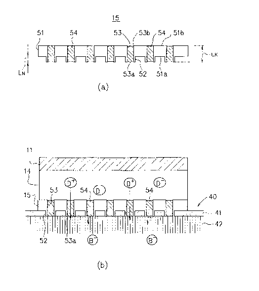
a high-molecular-weight drug (drug containing macromolecules such
as protein or peptide molecules) is far from sufficient even with
the use of the device disclosed in JP 10-510175 A. In particular,
the device faces a problem in that under such current or voltage
conditions that cause no damage on the skin, it is impossible to
deliver an effective amount of drug within a period allowable as
a drug delivery period.
[0008] Patent Document 1:JP 10-510175 A
Patent Document 2:US 6256533 A
Patent Document 3:JP 2005-503194 A
DISCLOSURE OF INVENTION
PROBLEMS TO BE SOLVED
[0009] The present invention has been made in view of the
above-described problem, and it is therefore an object of the
present invention to provide an iontophoresis device capable of
delivering to a living body, an ionizable drug (drug whose active
ingredients dissociate into cations or anions when dissolved) of
high-molecular-weight containing macromolecules such as protein
or peptide molecules at high speed or with high efficiency.
[0010] It is another object of the present invention to provide
an iontophoresis device capable of delivering a
high-molecular-weight ionizable drug containing macromolecules
such as protein or peptide molecules with high efficiency under
lower current or voltage conditions.
[0011] It is still another object of the present invention to
provide an iontophoresis device capable of delivering an ionizable
drug with an efficiency or speed much higher than conventional
iontophoresis devices including the device disclosed in JP
10-510175 A, irrespective of a molecular weight of the ionizable
drug.
[0012] It is yet still another object of the present invention
to provide an iontophoresis device capable of delivering an
ionizable drug with high efficiency under low current or voltage
conditions as compared with conventional iontophoresis devices
including the device disclosed in JP 10-510175 A, irrespective of
a molecular weight of the ionizalbe drug.
MEANS FOR SOLVING PROBLEMS
[001.3] The present invention is on an iontophoresis device for
administering drug ions dissociated into a first conductivitytype
by way of a plurality of needle-like members to be punctured into
skin, wherein an element having a function for selectively passing
2
CA 02647055 2008-09-19
ions of the first conductivity type is placed between a drug
holding part for holding the drug ions and the skin to which the
drug ions are administered, and the above mentioned problems are
solved by the iontophoresis device above. In concrete, the above
mentioned problems are solved by the inventions in the first to
fifth embodiments below.
[0014} That is, according to the first embodiment of the present
invention, the above mentioned problems are solved by an
iontophoresis device having a working electrode structure, said
working electrode structure comprising:
a first electrode;
a skin contact member including a substrate having a front
surface and a rear surface, and a plurality of needle-like members
that protrude from the front surface of the substrate and can be
punctured into skin; and
a drug holding part to be applied with voltage through the
first electrode and holding a drug solution containing drug ions
charged in the first conductivity type, the drug holding part being
arranged on the rear side of the substrate, wherein:
a hole communicating between a tip end of each of the
needle-like members and the rear surface of the substrate is formed
in the needle-like members, and the hole of the needle is filled
with an ion-exchange resin which is introduced second polarity or
conductivity type ion exchange groups.
[0015} According to the f:irst embodiment, a
f irst-conductivity-type voltage is applied to the f irst electrode
to deliver drug ions in the drug holding part into the living body
through the needle-like members punctured into the skin which have
penetrated through the stratum corneum, as in Patent Document 1.
Further, second polarity or conductivity type ion exchange groups
are introduced into the ion exchange resin filling the holes. The
ion exchange groups prevent backflow of biological counter ions
from the living body to the drug holding part.
[0016] Thus, it is possible to avoid a situation in which most
of current supplied to the first electrode is dissipated by
transference of biological counter ion (particularly, biological
counter ion having a low molecular weight, i.e. highmobility, such
as Na+ or C1 ) to the iontophoresis device. Therefore, a larger
amount of current supplied to the first electrode can be used for
delivery of the drug ions to the living body, thereby considerably
enhancing drug delivery efficiency and delivery speed.
[0017] The iontophoresis device according to the present
invention attains the above effects and thus makes it possible to
improve drug ion delivery efficiency or speed regardless of a
molecular weight of a drug ion, or to further lower current or
voltage conditions for the drug delivery, and deliver even drug
ions of macromolecules such as protein or peptide molecules under
lower current or voltage conditions at higher speed or higher
efficiency.
3
CA 02647055 2008-09-19
[0018] For the ion-exchange resin introducing second pol.arity or
conductivity type ion exchange groups in the present invention,
any known ion-exchange resin can be used. Examples thereof include
an ion-exchange resin prepared by introducing a cation-exchange
group (a group whose counter ion is cation) such as a sulforiic group,
a carboxylic group, and a phosphonic group, or an anion-exchange
group (a group whose counter ion is anion) such as primary to
tertiary amino groups, a quaternary ammonium group, a pyridyl
group, an imidazole group, a quaternary pyridinium group, or a
quaternary imidazolium group, to a polymer having a
three-dimensional network structure such as hydrocarbon-based
resins such as a polystyrene resin or acrylic acid resin or
fluororesin-based resins having a perfluorocarbon backbone.
[0019] The ion-exchange resin may be filled into the hole of the
needle-like members by using any method, for example, by
infiltrating or impregnating a monomer forming the hydrocarbon
resin and blended with a cross-linking agent into the holes to
cause a cross-linking reaction, or infiltrating or impregnating
a powdery ion-exchange resin blended with a given binder polymer
into the hole, and optionally curing the binder polymer.
[0020] Each of the needle-like members of the present invention
preferably protrude from the substrate by a length enough for the
needle-like member to pass through all or most of a stratum corneum
which is regarded as a main barrier against percutaneous delivery
of a drug. The length of the needle-like member is preferably l, 000
pm or shorter, particularly preferably, 1}.im to 300 pm. The inner
diameter of each of the holes formed in needle-like members can
be set, for example, to 0.03 um to 300 um, particularly preferably,
0.1 pm to 100 pm. The length of each of the holes from the rear
surface of the substrate to the tip ends of the needle-like members
is preferably set, for example, to 1 um to 3, 000 pm, particularly
preferably, 10 pm to 500 pm.
[0021] The needle-like members or the skin contact member of the
present invention can be formed of an organic material such as hard
plastics or an inorganic material such as silicon by utilizing
known methods such as lithography, molding, andlaserirradiation.
[0022] According to the second embodiment of the present
invention, the above mentioned problems are solved by an
iontophoresis device having a working electrode structure, said
working electrode structure comprising:
a first electrode;
a skin contact member including a substrate having a front
surface and a rear surface, and a plurality of needle-like members
that protrude from the front surface of the substrate and can be
punctured into skin; and
a drug holding part applied with voltage through the first
electrode and holding a drug solution containing drug ions charged
in the first conductivity type, the drug holding part being
arranged on the rear side of the substrate, wherein:
4
CA 02647055 2008-09-19
a hole communicating between a tip end of each of the
needle-like members and the rear surface of the substrate is formed
in the needle-like members, and the skin contact member further
includes a first ion-exchange membrane that is interposed between
the drug holding part and the substrate and allows selective
permeation of the ions of the first conductivity type.
[0023] In the second embodiment of the present invention, the
first-conductivity-type voltage is applied to the first electrode,
whereby drug ions in the drug holding part are delivered through
the holes formed in the needle-like members into the skin. Further,
the biological counter ions cannot pass through the first
ion-exchange membrane by the function of the first ion-exchange
membrane and thus are accumulated in the holes of the needle-like
members or a space between the skin contact member and the first
ion-exchange membrane. As a result, the transference of the
biological counter ion is substantially blocked. Therefore, the
drug ions can be delivered with delivery efficiency or speed
comparable or approximate to that of the first embodiment of the
invention.
[ 0024 ] The needle-like members or the skin contact member of the
second embodiment may have the same constituent as that of the
first embodiment.
[0025] Further, any ion-exchange membrane having a function of
allowing selective passage of ions of a first conductivity type
and blocking or suppressing the passage of ions of a second
conductivity type can be used as the first ion-exchange membrane
of the second embodiment. An ion-exchange membrane, pores of a
porous film of which are partially or completely filled with an
ion-exchange resin introduced with a second polarity or
conductivity type ion exchange groups, can preferably used as the
first ion-exchange membrane of the second embodiment.
[0026] The iontophoresis device according to the second
embodiment of the present invention can be manufactured through
such a simple manufacturing process that interposes an
ion-exchange membrane easily available on the market etc. between
the drug holding part and the substrate of the skin contact member.
Thus an advantage in terms of a lower manufacturing cost can be
obtained.
[0027] According to the third embodiment of the present invention,
the above mentioned problems are solved by an iontophoresis device
having a working electrode structure, said working electrode
structure comprising:
a first electrode;
a skin contact member including a substrate having a front
surface and a rear surface, and a plurality of columnar members
embedded in the substrate and made of an ion-exchange resin
introduced with a second polarity or conductivity type ion
exchange groups; and
a drug holding part applied with voltage through the first
CA 02647055 2008-09-19
electrode and holding a drug solution containing drug ions charged
in the first conductivity type, the drug holding part being
arranged on the rear side of the substrate, wherein:
each of the columnar members is exposed to the rear surface
of the substrate at one end and protrudes by a predetermined length
from the front surface of the substrate at the other end to form
a needle-like member puncturable into the skin.
[0028] According to the third embodiment of the present invention,
the columnar member made of an ion-exchange resin has the functions
as both the needle-like member puncturable into the skin and the
member that allows selective passage of the ions of the first
conductivity type, thereby attaining the operational effect as
those of the first or second embodiments.
[0029] That is, the drug ions in the drug holding part are
delivered into the living body through the columnar members by the
application of the first-conductivity-type voltage to the first
electrode delivers. Further, the columnar members are formed of
an ion-exchange resin introduced with an ion-exchange group whose
counter ion is the first conductivity type, whereby backflow of
the biological counter ion to the drug holding part through the
columnar members is prevented. As a result, it is possible to
improve drug ion delivery efficiency or speed, or to further lower
current or voltage conditions for the drug delivery, or deliver
even drug ions containing macromolecules such as protein or
peptide molecules under lower current or voltage conditions with
higher efficiency or speed, as in the first or second embodiment.
[0030] The resins described for the first embodiment can be used
as the ion-exchange resin introduced with the second polarity or
conductivity type ion exchange groups, for forming the columnar
members of the third embodiment. Examples of a method of forming
the ion-exchange resin into a columnar shape includes a method of
molding hydrocarbon-based resins or a fluororesin forming the
ion-exchange membrane into a linear shape through
extrusion-molding, and then cutting the resultant into a
predetermined size.
[0031] Note that the sectional shape of the columnar member may
be arbitrarily set, for example, as a circle or rectangle. The
length of the columnar member is preferably 1 to 3,000 .im, more
preferably, 10 um to 500 pm. The diameter of the columnar member
is preferably 0.03 to 300 pm, more preferably, 0.1 pm to 100 pm.
Further, the length of the needle made up of the columnar member
(protrusion length of the columnar member from the front surface
of the substrate) is preferably 1,000 pm or shorter, more
preferably 1 pm to 300 pm.
[0032] According to the fourth embodiment of the present
invention, the above mentioned problems are solved by an
iontophoresis device having a working electrode structure, said
working electrode structure comprising:
a first electrode;
6
CA 02647055 2008-09-19
a skin contact member including a substrate having a front
surface and a rear surface, and a multi-needle member having a
plurality of needle-like projections radially protruding
therefrom and made of an ion-exchange resin introduced with a
second polarity or conductivity type ion exchange groups; and
a drug holding part applied with voltage through the first
electrode and holding a drug solution containing drug ions charged
in the first conductivity type, the drug holding part being
arranged on the rear side of the substrate, wherein:
at least a part of a surface of the multi-needle member
is exposed to the rear surface of the substrate, and any one or
more of the needle-like projections of the multi-needle member
protrudes by a predetermined length from the front surface of the
substrate to form a needle puncturable into the skin.
[0033] In this fourth embodiment of the present invention, the
same effect as in the first to third embodiments can be attained
by the fact that the multi-needle member made of an ion-exchange
resin have both the function as the needle being able to be
punctured into skin and the function of the member for selectively
passing the first ions.
[0034] That is, the drug ions in the drug holding part are
delivered into the living body through the multi-needle member by
the application of the first-conductivity-type voltage to the
first electrode. Further, since, the multi-needle member is
formed of an ion-exchange resin introduced with a second polarity
or conductivity type ion exchange groups, the ion exchange groups
prevent backflow of biological counter ions from the living body
to the drug holding part. As a result, it is possible to improve
drug ion delivery efficiency or speed, or to further ease current
or voltage conditions for the drug delivery, or deliver even drug
ions containing macromolecules such as protein or peptide
molecules under lower current or voltage conditions with higher
efficiency or speed.
[0035] The resins described for the first embodiment can be used
as the ion-exchange resin introduced with a second polarity or
conductivity type ion exchange groups, for forming the
multi-needle member of the fourth embodiment.
[0036] The needle-like projection of the multi-needle of the
fourth embodiment is preferably formed into the length of 1,000
pm or shorter, more preferably 1 pm to 300 pm through
micromachining, for example.
[0037] According to the fifth embodiment of the present invention,
the above mentioned problems are solved by an iontophoresis device
having a working electrode structure, said working electrode
structure comprising:
a first electrode;
a skin contact member including a substrate having a front
surface and a rear surface, and a plurality of needle-like members
that protrude from the front surface of the substrate and can be
7
CA 02647055 2008-09-19
punctured into skin, the skin contact member being formed of an
ion-exchange membrane allowing selective passage of ions of a
first conductivity type; and
a drug holding part applied with voltage through the first
electrode and holding a drug solution containing drug ions charged
in the first conductivity type, the drug holding part being
arranged on the rear side of the substrate.
[0038] In this fifth embodiment, as in the third or fourth
embodiment, drug ions in the drug holdi.ng part are delivered into
the skin through the needle-like members by the application of the
first-conductivity-type voltage to the first electrode. Further,
the skin contact member is formed of the ion-exchange membrane
allowing selective passage of the ions of the first conductivity
type, so the backflow of the biological counter ion into the drug
holding part is blocked or suppressed. Therefore, it is possible
to improve drug ion delivery efficiency or speed, or to further
ease current or voltage conditions for the drug delivery, or
deliver even drug ions containing macromolecules such as protein
or peptide molecules under lower current or voltage conditions
with higher efficiency or speed as in any of the first to fourth
embodiments.
[0039] Here, the ion-exchange membrane of the fifth embodiment
may be made of the materials described for the second embodiment.
The needle-like members may be formed on the front surface of the
ion-exchange membrane by press molding the substrate constituting
the ion-exchange membrane, for example.
[0040] In theiontophoresis device accordingto thethirdtofifth
embodiments of the present invention, the holes are preferably
formed in the columnar members, the multi-needle member, or the
needle-like members, the holes communicating with the opening at
the rear surface of the substrate, whereby the drug ion delivery
speed or efficiency can further be enhanced.
[0041] In the iontophoresis device according to the first --ofifth
embodiments of the present invention, the working electrode
structure may further include: a first electrolyte holding part
for holding an electrolyte that is in contact with the first
electrode; and a second ion-exchange membrane that is interposed
between the first electrolyte holding part and the drug holding
part and allows selective permeation of ions of a second
conductivity type. With this arrangement, it is possible to avoid
ion decomposition of the drug ions around the first electrode,
transference of H+ ions or 0H ions generated at the first electrode
to the drug holding part, or resultant fluctuation in a pH value
at the drug holding part and in turn, at the interface between the
skin and the skin contact member, and inflammation caused on the
skin contacting the skin contact member in some cases, and to
achieve more stable, safe drug delivery.
[0042] The iontophoresis device of the first to fifth embodiments
of the present invention may further include a nonworking
8
CA 02647055 2008-09-19
electrode structure comprising: a second electrode; a second
eiectrolyte holding part that is in contact with the second
electrode; a third ion-exchange membrane that is arranged on a
front side of the second electrolyte holding part and allows
selective passage of the ions of the first conductivity type; a
third electrolyte holding part that is arranged on a front side
of the third ion-exchange membrane and holds an electrolyte; and
a fourth ion-exchange membrane that is arranged on a front side
of the third ion-exchange membrane and allows selective passage
of ions of a second conductivity type. With this arrangement, it
is possible to avoid fluctuation in a pH value at the interface
between the skin and the nonworking electrode structure, and
inflammation caused on the skin contacting the nonworking
electrode structure in some cases, and to achieve more stable, safe
drug delivery.
BREIF DESCRIPTION OF THE DRAWINGS
[0093] [FIG. I] Fig.l illustrates a structure of an iontophoresis
device according to an embodiment of the present invention.
[FIG. 2] FIG. 2(a) illustrates a embodiment of a
skin contact member used in the iontophoresis device according to
the present invention, and FIG. 2(b) illustrates how ions migrate
in the iontophoresis device according to the present invention.
[FIG. 3] FIGS. 3(a) to (g) each illustrates another
embodiment of the skin contact member.
[FIG. 4] FIG. 4 illustrates an example of a
manufacturing method for a skin contact member.
[FIG. 5] FIG. 5 illustrates a structure of an
iontophoresis device according to another embodiment of the
present invention.
[FIG. 6] FIG. 6 illustrates an example of a
conventional iontophoresis device.
BEST MODE FOR CARRYING OUT THE INVENTION
[0044] Hereinafter, embodiments of the present invention will be
described.
[0045] As a matter of practical convenience for explanation,
description is given of an embodiment of an iontophoresis device
for delivering drugs whose active ingredients dissociate into
positive drug ions (for example, lidocaine hydrochloride as an
anesthetic, carnitine chloride as a remedy for gastrointestinal
disorder, pancuronium bromide as muscle relaxants, and morphine
hydrochloride as an anesthetic) by way of example. However, as
regards an iontophoresis device for delivering drugs whose active
ingredients dissociate into negative drug ions (for example,
ascorbic acids as vitamins, and Lipid A used as a vaccine adjuvant)
the polarity (plus or minus) of the electrodes of the power source
and the ion-exchange group introduced to the ion-exchange membrane
or the ion-exchange resin has only to be reversed. In addition,
9
CA 02647055 2008-09-19
proteins and peptides are amphoteric electrolytes, which are
dissociable into either cations or anions depending on pH of a drug
solution. Thus, either one of the two is used depending on pH.
[0046] FIG. 1 is a schematic sectional diagram showing a basic
structure of an iontophoresis device 1 according to the present
invention.
[0047] As illustrated in FIG. 1, the iontophoresis device 1 of
the present invention includes a working electrode structure 10,
and a nonworking electrode structure 20, a power source 30 as main
components (members).
[0048] The working electrode structure 10 includes: an electrode
member 11 connected with a positive electrode of the power source
30; a drug holding part 14 that holds a drug solution that is in
contact with the electrode member 11 and applied with voltage
through the electrode member 11; a skin contact member 15 arranged
on a front side of the drug holding part 14; and a cover or container
16 that houses those members.
[0049] The nonworking electrode structure 20 includes: an
electrode member 21 connected with a negative electrode of the
power source 30; an electrolyte holding part 22 that holds an
electrolyte that is in contact with the electrode member 21 and
applied with voltage through the electrode member 21 and a cover
or container 26 that houses those members.
[0050] As the electrode members 11 and 21, electrodes made of any
conductive materials can be used with no particular limitations,
and it is particularly preferable to use an active electrode such
as silver/silver halide coupled electrode, which can suppress
electrolytically-generated H+ ions and 0H- ions from water.
[0051] The drug holding part 14 holds an aqueous solution of a
drug whose active ingredients dissociate into cations when
dissolved (for example, proteins and peptides having positive
charges in total in the solution, lidocaine, carnitine crloride,
pancuronium bromide, and morphine hydrochloride) as a drug
solution.
[0052] The electrolyte holding part 22 holds an electrolyte that
enables current to flow. As the electrolyte, a phosphate buffered
saline or physiological saline can be used. Alternatively, it is
possible to use an electrolyte susceptible to oxidation or
reduction (oxidation at the positive electrode and reduction at
the negative electrode) as compared with an electrolytic reaction
of water, examples of which include: inorganic compounds such as
ferrous sulfate and ferric sulfate; agents such as an ascorbic acid
(vitamin C) and sodium ascorbate; organic acids such as a lactic
acid, an oxalic acid, a malic acid, a succinic acid, and fumaric
acid and/or salts thereof; and mixtures thereof. The use thereof
makes it possible to avoid fluctuation in pH value or gas
generation due to the electrolytic reaction of water, and any
resulting increase in ion conduction resistance.
[0053] The drug holding part 14 and the electrolyte holding part
CA 02647055 2008-09-19
22 may respectively retain the drug solution and electrolyte in
a liquid form, or retain the drug solution and electrolyte in the
form of being impregnated into a carrier made of any material
having a water retentivity such as a fibrous sheet such as gauze
or filter paper, or a polymer gel sheet made of an acrylic resin
hydrogel (acrylic hydrogel) or segmented polyurethane-based gel.
This facilitates, for example, handling thereof.
[0054] In this case, the impregnation rate of the drug solution
or electrolyte into the carrier needs to be set to such a value
as to ensure sufficient current supply and high transport rate.
The impregnation rate of the drug solution is set to 20 to 60% for
the drug holding part 14, whereby a transport rate (drug delivering
property) as high as 70 to 80% can be attained, for example.
[0055] Note that the impregnation rate is represented by weight
percent, and derived from the expression of 100 x (W-D)/D (%),
wherein said D represents a pre-impregnation (dry) weight and W
represents a post-impregnation (wet) weight. The transport rate
indicates a ratio of current used for drug ion delivery to total
current supplied to the working electrode structure.
[0056] FIG. 2(a) is a conceptual explanatory diagram showing a
detailed structure of the skin contact member 15 in the
iontophoresis device 1.
[0057] As illustrated in FIG. 2(a), the skin contact member 15
includes a substrate 51 having a front surface 51a and a rear
surface 51b, and needle-like members 52 each protruding from the
front surface 51a and having a size, shape, and strength enough
for the puncture into the skin. In each needle-like member 52 has
a hole 53 communicating between an opening 53a at the tip end of
the needle-like member and an opening 53b at the rear surface of
the substrate.
[0058] As a method of manufacturing the skin contact member 15,
there are a variety of known manufacturing methods. For example,
the skin contact member can be manufactured by molding organic
materials, such as plastics in line with the method disclosed in
US 6256533 or by etching inorganic materials such as silicon in
line with the method disclosed in JP 2005-503194 A.
[0059] Here, the length (LN) of each of the needle-like members
52 of the skin contact member 15 is set to 1,000 pm or shorter,
preferably, 1 um to 300 um. Hence, it is possible to relieve pains
on a patient at the time of delivering a drug. In addit--on, the
length (LK) of each of the holes 53 extending from the opening 53a
at the tip end of the needle-like members to the opening 53b at
the rear surface of the substrate is set to 1 to 3, 000 pm, preferably,
pm to 500 pm. The inner diameter of each of the holes 53 is
set to 0.03 pm to 300 pm, preferably, 0.1 pm to 100 pm. Hence,
it is possible to secure a flow path large enough for smooth
delivery of drug ions. The needle-like members 52 or holes 53 of
the skin contact member 15 may be formed at the density of several
to 5,000 (holes or needle-like members)/cmG, for example.
11
CA 02647055 2008-09-19
[0060] Note that each of the needle-like members 52 and/or the
holes 53 may have any sectional shape such as a circular shape,
an elliptical shape, or a rectangular shape. Besides, they may
be formed into such a shape that has a uniform sectional area in
a longitudinal direction of the needle-like members 52 as shown
in FIG. 2(a) or a tapered shape as shown in FIG. 6, which facilitates
insertion into the skin.
[0061] In addition, a cation-exchange resin (ion-exchange resin
introduced with an anion group) 54 fills the inside of the holes
53 of the needle-like members 52.
[0062] As the above cation-exchange resin 54, usable is a resin
prepared by introducing a cation-exchange group such as a sulfonic
group, a carboxylic group, or a phosphonic group into a polymer
substrate having a three-dimensional network structure such as
hydrocarbon-based resins such as a polystyrene resin or acrylic
acid resin or fluororesin-based resins having a perfluorocarbon
backbone.
[0063] The holes 53 may be filled with the cation-exchange resin
54 by using any method, for example, by soaking the tip ends of
the needle-like members 52 or the entire skin contact member 15
in a solution prepared by mixing a cross-linking monomer forming
the polymer substrate such as styrene-divinylbenzene or
chloromethylstyrene-divinylbenzene with a polymerization
initiator; by charging the solution from the rear surface 51b of
the substrate 51 using a spatula member so that the solution is
infiltrated or impregnated into the holes 53, followed by
polymerization and then introduction of the cation-exchange
group; or by infiltrating or impregnating into the holes 53, a
binder polymer such as a phenol resin or methyl methacrylate into
which a fine powder of a cation-exchange resin is dispersed, in
place of the above solution in the above way, and then curing the
resultant binder polymer.
[0064] The cation-exchange resin 54 can be filled to the full
length of the holes 53 as shown in FIG. 2(a) , or may be partially
filled, for example, filled into only a portion of the holes 53
around the openings 53a at the tip ends of the needle-like members
52.
[0065] FIG. 2(b) schematically illustrates how ions deliver in
the drug holding part 14 and a skin 40 when the voltage is applied
through the electrode member 11 (and electrode member 21) with the
skin contact member 15 brought into contact with the skin 40. In
FIG. 2(b), D+ represents a positively charged drug ion, D
represents a counter ion thereof (drug counter ion) , and B
represents a negative ion in the living body (or at the surface
of the skin 40) . In addition, reference numerals 41 and 42 denote
a stratum corneum covering the skin surface and a subcutaneous
tissue underlying the stratum corneum, respectively.
[0066] The drug ions D+ in the drug holding part 14 are driven
through the application of a positive voltage to the electrode
12
CA 02647055 2008-09-19
member 11, and delivered to the skin 40 through the holes 53. At
this time, the drug ions D+ can pass through the cation-exchange
resin 54 filled in the holes 53 because of its positive polarity.
[0067] In addition, the punctured needle-like members 52
penetrate the stratum corneum 41 which is a barrier against the
delivery of the drug ions D', so the drug ions D+ having reached
the opening 53a can be delivered into the subcutaneous tissue 42
without being blocked by the stratum corneum 41. Note that it is
most preferable that all the needle-like members 52 completely
penetrate the stratum corneum 41 like the illustrated example, but
the drug delivery may be performed while all or some of the
needle-like members 52 are halfway punctured into the stratum
corneum 41. In this case as well, the drug delivery efficiency
can be improved according as the thickness of the stratum corneum
41 from the opening 53a to the subcutaneous tissue 42 is reduced.
[0068] In contrast, the biological counter ions B present in the
living body (or at the surface of the skin) are moved to the drug
holding part 14 side through the application of a positive voltage
to the electrode member 11, but the movement of the biological
counter ion B- is completely blocked or suppressed to an allowable
level owing to the cation-exchange resin 54 filled in the holes
53.
[0069] Accordingly, a ratio of current consumed for the movement
of the biological counter ion B to the drug holding part 14 to
total current supplied to the electrode member 11 is reduced or
minimized substantially to zero, which increases a ratio of
current consumable for the delivery of the drug ions D' to the
living body. As a result, delivery speed and efficiency of the
drug ions D+ improve, or the drug ions D+ can be delivered efficiency
under lower current or voltage conditions.
[00'70] Note that the ion mobility tends to reduce in reverse
proportion to its molecular weight. Hence, when the
cation-exchange resin 54 does not fill the hole 53, the biological
counter ion B_ consumes more current upon movement to the drug
holding part 14 in the case of delivering the drug ion D' having
a higher molecular weight. Therefore, an effect of improving the
drug ion delivery speed and efficiency of the present invention
can be greatly exerted in the case of using a drug ion of a higher
molecular weight, which was hardly delivered with the conventional
iontophoresis device.
[0071] A battery, a constant voltage generator, a constant
current source, a constant voltage/current source, and the like
can be used as the power source 30 in the iontophoresis device of
the present invention. It is preferable to use a constant current
source operable under stable voltage conditions that enable
arbitrary current adjustment in a range of 0.01 to 1.0 mA/cm2,
preferably 0.01 to 0.5 mA/cm2, more specifically, voltage
conditions of 50 V or lower, preferably 30 V or lower.
[0072] In the above described iontophoresis device, a liner may
13
CA 02647055 2008-09-19
be attached to the front side of the skin contact member 1.5 and/or
the electrolyte holding part 22 for the purpose of preverting the
drug holding part 14 or the electrolyte holding part 22 from drying
or preventing foreign substances from mixing into the drug holding
part 14 or the electrolyte holding part 22, or an adhesive layer
for improving adhesion between the working electrode structure 10
and/or the nonworking electrode structure 20, and the skin may be
laminated on a bottom "b" of the cover or container 16 and/or the
cover or container 26.
[0073] FIG. 3(a) to 3(g) illustrate structures of skin contact
members 15a to 15g as other embodiments of the skin contact member
15, each of which can replace the skin contact member 15.
[0074] The skin contact member 15a of FIG. 3(a) has the substrate
51, the needle-like members 52, and the holes 53 as in the skin
contact member 15. However, instead of filling the
cation-exchange resin 54 into the holes 53, a cation-exchange
membrane (ion-exchange membrane allowing selective passage of
cations) 55 is provided on the rear side of the substrate 51 and
the front side of the drug holding part 14.
[0075] With an iontophoresis device using the above skin contact
member 15a, the drug ions D+ pass through the cation-exchange
membrane 55 and the holes 53, and are then delivered into the living
body through the opening at the tip end of the needle 52 due to
the voltage applied to the electrode member 11 as in the
iontophoresis device 1.
[0076] In contrast, the cation-exchange membrane 55 blocks or
suppresses the migration of the biological counter ion B_ to the
drug holding part 14, so the biological counter ions B- are
accumulated in the hole 53, and migration is substantially
inhibited.
[0077] Accordingly, a larger amount of supplied current can be
used for delivery of the drug ions D+ into the living body,
improving the delivery speed and efficiency of the drug ions D`
or enabling the drug delivery under lower current or voltage
conditions.
[0078] Note that as the cation-exchange membrane 55 used herein,
any cation-exchange membrane having a function of allowing
selective passage of cations can be used, examples of which include
NEOSEPTA CM-l, CM-2, CMX, CMS, and CMB (available from Tokuyama
Co., Ltd.). Particularlypreferable is a cation-exchangemembrane
prepared by completely or partially filling a cation-exchange
resin into pores of a porous film made of a polyolefin resin,
vinylchloride-based resins, fluororesin-based resins, a
polyamide resin, a polyimide resin, or the like. The
cation-exchange resin may be filled by, for example, impregnating
into the pores of the porous film, a solution prepared by mixing
a cross-linking monomer such as styrene-divinylbenzene or
chloromethylstyrene-divinylbenzene with a polymerization
initiator, followed by polymerization, and then introducing
14
CA 02647055 2008-09-19
cation-exchange groups such as a sulfonic group, a carboxylic
group, and a phosphonic group into the polymer.
[0079] In addition, it is preferable to bond the cation-exchange
merribrane 55 and the substrate 51 at the interface by an appropriate
method such as bonding by use of an adhesive or ultrasonic bonding.
This overcomes a problem in that a gap is left at the interface
to increase the migration amount of the biological counter ion B-
or bubbles are generated to lower the conductivity.
[0080] The skin contact member 15b of FIG. 3(b) includes: the
substrate 51 as in the skin contact member 15; and a number of
columnar members 52 embedded into the substrate 51 and made of a
cation-exchange resin. Each columnar member 56 is exposed to the
rear surface of the substrate 51 at one end, and protrudes by a
predetermined length from the front surface of the substrate 51
at the other end to constitute the needle 52.
[0081] With respect to an iontophoresis device using the above
skin contact member 15b, the drug ions D+ pass through the columnar
member 56 to be delivered into the living body through the
application of a positive voltage to the electrode member 11
similarly to the iontophoresis device 1. Further, the
cation-exchange resin forming the columnar member 56 blocks or
suppresses migration of the biological counter ion B- to the drug
holding part 14, so a larger amount of supplied current cari be used
for derivery of the drug ions D+ into the living body, improving
the delivery speed or efficiency of the drug ions D+ or enabling
the drug delivery under lower current or voltage conditions.
[0082] The described the cation-exchange resin 54 of the skin
contact member 15 can be used as the cation-exchange resin forming
the columnar member 56. The columnar member can be formed into
a columnar shape through machining such as micromachining or
through extrusion-molding of hydrocarbon-based resins or a
fluororesin forming the cation-exchange resin into a linear shape,
followed by cutting into a predetermined length with the
cation-exchange groups being introduced before or after the
cutting.
[0083] Regarding the size of the columnar member 56, the length
(LK) of the columnar member 56 is set, for example, to 1 to 3,000
um, preferably, 10 pm to 500 pm. If the columnar member 56 is
circular in section, its diameter may be set, for example, to 0.03
to 300 pm, preferably, 0.1 pm to 100 pm. Further, in embedding
the columnar member into the substrate 51, the protrusiori length
(LN) from the front surface of the substrate 51 is preferably set,
for example, to 1,000 pm or less, more preferably 1 pm to 300 pm.
[0084] Note that the sectional shape of the columnar member 56
is not limited to a circle but may be any shape such as an ellipse
or rectangle. In addition, the member can be tapered towards its
tip end to facilitate the puncture to the skin.
[0085] The skin contact member 15c of FIG. 3(c) has the same
CA 02647055 2008-09-19
structure as the skin contact member 15b except that recesses 56a
are formed in the columnar member 56 to open at the rear surface
of the substrate 51.
[0086] Accordingly, with an iontophoresis device using the skin
contact member 15c, the drug ions D+ are delivered in the same way
as in the case of using the skin contact member 15b. However, the
drug solution from the drug holding part 14 can be infiltrated into
the recesses 56a, making it possible to deliver drug ions with
higher efficiency than the case of using the skin contact member
15b.
[0087] Note that the recesses 56a can be formed through machining
such as micromachining of the columnar member 56 manufactured by
the above method.
[0088] The skin contact member 15d of FIG. 3(d) includes: the
substrate 51 as in the skin contact member 15; and a plural
multi-needle members 57 embedded into the substrate 51 and made
of a cation-exchange resin. As shown in FIG. 3(d), the
multi-needle members 57 each have plural needle-like projections
that radially protrude. Any of the needle-like projections
protrude from the front surface of the substrate 51 and serve as
the needle-like members 52. Further, the multi-needle members 57
are embedded into the substrate 51 in such a form that at least
a part thereof is exposed to the rear surface of the substrate 51.
[0089] A resin similar to that for the columnar members 56 can
be used as the cation-exchange resin of the multi-needle members
57. The multi-needle members 57 can be shaped through
micromachining or the like.
[0090] With respect to an iontophoresis device using the above
skin contact member 15d, the drug ions D+ pass through the
multi-needle members 57, and are then delivered into the living
body due to the positive voltage applied to the electrode member
11 as in the iontophoresis device 1. Further, the cation-exchange
resin forming the multi-needle members 57 blocks or suppresses
migration of the biological counter ion B- to the drug holding part
14. Accordingly, a larger amount of supplied current can be used
for delivery of the drug ions D+ into the living body, improving
the delivery speed or efficiency of the drug ions D+ or enabling
the drug delivery under lower current or voltage conditions.
[0091] Note that the skin contact member 15d is advantageous in
that a manufacturing process can be simplified as compared with
the skin contact member l5b because the multi-needle menibers 57
havi.ng an appropriate number of needle-like projections with an
appropriate length enables any of the needle-like projections to
protrude to the front surface 51a of the substrate 51 even when
embedded into the substrate 51 regardless of an embedding
direction (orientation) of the multi-needle members, and enables
at least a part of the multi-needle members 57 to be exposed to
the rear surface 51b of the substrate 51.
[0092] The skin contact member 15e of FIG. 3(e) has the same
16
CA 02647055 2008-09-19
structure as the skin contact member 15d except that recesses 57a
are formed in the substrate 51 and the multi-needle members 57 to
open on the rear surface 51b of the substrate 51.
[0093] Accordingly, in the iontophoresis device using the skin
contact member 15e, the drug ion delivery is carried out in a way
similar to that using the skin contact member 15d. However, the
drug solution from the drug holding part 14 can be infiltrated into
the recesses 57a, whereby the drug ion can be delivered with higher
efficiency than that in the case of using the skin contact member
15d.
[0094] The recesses 57a can be formed through micromachining or
the like.
[0095] The skin contact member 15f of FIG. 3(f) includes: the
substrate 51; and the needle-like members 52 protruding from the
front surface of the substrate 51, and the skin contact member 15f
is made up of a cation-exchange membrane in its entirety.
[0096] The above skin contact member 15f can be molded by using
molds 61a and 62a as shown in FIG. 4(a), for example.
[0097] More specifically, a porous film 63 made of a thermoplastic
resin such as a polyolefin resin, vinylchloride-based resins,
fluororesin-based resins, a polyamide resin, or a polyimide resin
is press-molded between the molds 61a and 62a. After that, a
cation-exchange resin is filled into the pores of the porous film
63 based on the same manner as that for the cation-exchange
membrane 55, or the porous film 63 whose pores are previously
filled with the cation-exchange resin is press-molded between the
molds 61a and 62a to thereby form the skin contact member 15f.
[0098] Alternatively, the above skin contact member 15f can be
produced by molding a binder polymer such as polyethylene,
polystyrene, a phenol resin, or methyl methacrylate into which a
fine powder of a cation-exchange resin is dispersed, between the
molds 61a and 62a into a membrane.
[0099] With respect to an iontophoresis device using the skin
contact member 15f, the drug ions D+ pass through the skin contact
member 15f, and are then delivered into the living body from the
tip ends of the needle-like members 52 due to the positive voltage
applied to the electrode member 11 as in the iontophoresis device
1. Further, the skin contact member 15f as a cation-exchange
membrane blocks or suppresses migration of the biological counter
ion B to the drug holding part 14 . Accordingly, a larger amount
of supplied current can be used for delivery of the drug ions D+
into the living body, improving the delivery speed or efficiency
of the drug ions D' or enabling the drug delivery under lower
current or voltage conditions.
[0100] In addition, with respect to this iontophoresis device,
the drug ions D+ can deliver into the living body from other
portions than the needle-like members 52 of the substrate 51 in
contact with the skin 40 albeit delivery by way of the stratum
corneum 41. Thus, especially in the case where a drug ion having
17
CA 02647055 2008-09-19
a relatively low molecular weight is used, or a drag ion a
significant quantity of which can be delivered through the stratum
corneum 41 is used, the drug delivery speed or efficiency can
further be enhanced.
[0101] The skin contact member 15g of FIG. 3(g) has the same
structure as the skin contact member 15f except that recesses 58
extend from the rear surface of the substrate 51 to the inside of
the needle-like members 52 in the skin contact member 15g.
[0102] The skin contact member 15g can be molded using molds 61b
and 62b as shown in FIG. 4(b) by the similar ways as those for the
skin contact member 15f.
[0103] An iontophoresis device using the skin contact member 15g
delivers drug ions in a similar way to that in the case the skin
contact member 15f is used. However, the drug solution from the
drug holding part 14 can be infiltrated into the recesses 58,
whereby the drug ion can be delivered with a higher efficiency than
that in the case of using the skin contact member 15g.
[0104] FIG. 5 illustrates a structure of an iontophoresis device
101 according to another embodiment of the present invention.
[0105] An iontophoresis device 101 of the present invention
includes a working electrode structure 110, a nonworkingelectrode
structure 120, and a power source 130 like the iontophoresis device
1.
[0106] The working electrode structure110 includes: an electrode
member 111 connected with a positive electrode of the power source
130; an electrolyte holding part 112 that holds an electrolyte that
is in contact with the electrode member 111 and is applied with
voltage through the electrode member 111; an anion-exchange
membrane 113 arranged on a front side of the electrolyte holding
part 112; a drug holding part 114 that is placed on a front side
of the anion-exchange membrane 113 and holds a drug solution that
is applied with voltage from the electrode member 111 through the
electrolyte holding part 112 and the anion-exchange membrane 113;
a skin contact member 115 arranged on a front side of the drug
holding part 114; and a cover or container 116 that houses those
members.
[0107] The nonworking electrode structure 120 includes: an
electrode member 121 connected with a negative electrode of the
power source 130; an electrolyte holding part 122 that holds an
electrolyte that is in contact with the electrode member 121 and
is applied with voltage through the electrode member 121; a
cati.on- exchange membrane 123 arranged on a front side of the
electrolyte holding part 122; an electrolyte holding part 124 that
is placed on a front side of the cation-exchange membrane 123 and
holds an electrolyte that is applied with voltage from the
electrode member 121 through the electrolyte holding part 122 and
the cation-exchange membrane 123; an anion-exchange membrane 125
arranged on a front side of the electrolyte holding part 124; and
a cover or container 126 that houses those members.
18
CA 02647055 2008-09-19
0108] Here, the electrode members 111 and 121, the drug holding
part 114, and the electrolyte holding parts 112, 122, and 124 have
the same structures as the electrode members 11 and 21, the drug
holding part 14, and the electrolyte holding part 22, respectively,
and membranes similar to the above-described membranes for the
cation-exchange membrane 55 can be used for the cation-exchange
membrane 123.
[0109] As the anion-exchange membranes 113 and 125, for example,
any anion-exchange membrane having a function of allowing
selective passage of anions can be used, examples of which include
NEOSEPTA AM-1, AM-3, AMX, AHA, ACH, and ACS (available from
Tokuyama Co., Ltd.). Particularly preferable is an anion-exchange
membrane prepared by filling an anion-exchange resin into pores
of a porous film similar to that for the cation-exchange inembrane
55. In this case, the anion-exchange resin may be filled by, for
example, impregnating into the pores of the porous film, a solution
prepared by mixing a cross-linking monomer such as
styrene-divinylbenzene or chloromethylstyrene-divinylbenzene
with a polymerization initiator, followed by polymerization and
then introduction of anion-exchange groups to the polymer.
[0110] Further, usable as the skin contact member 115 are members
similar to the skin contact member 15 or the skin contact members
15a to 15f.
[0111] The iontophoresis device 101 achieves an operational
effect equivalent to the above effect of the iontophoresis device
1 or the devices replacing the skin contact member 15 of the
iontophoresis device 1 with any of the skin contact members 15a
to 15f, and further attains the following additional operational
effect.
[0112] That is, the anion-exchange membrane 1.13 or
cation-exchange membrane 123 blocks or suppresses migration of H+
ions or 0H- ions generated at the electrode members 11l and 121
due to the voltage application from the electrode members 111 and
121 to the drug holding part 114 and the electrolyte holding part
124 . Thus, it is possible to suppress fluctuation in pH value in
the drug holding part 114 and the electrolyte holding part 124 and
in turn, at the contact surfaces of the working electrode structure
110 and the nonworking electrode structure 120 to the skin. As
a result, damage to the skin is reduced to enhance a safety level
of the drug delivery.
[0113] Further, as discussed above, the migration of H' or OH ions
to the drug holding part 114 and the electrolyte holding part 124
is blocked or suppressed, whereby a carbon electrode as an inactive
electrode can be used for the electrode members 111 and 121 i_n place
of the active electrode such as a silver/silver halide coupled
electrode. Consequently, it is possible to attain an
iontophoresis device without a problem where metal ions eluting
from the electrode migrate into the living body, causing damage
to health.
19
CA 02647055 2008-09-19
[0114] Further, the drug holding part 114 is separated from the
electrode member 111 by the anion-exchange membrane 113, which
prevents a problem in that ion decomposition of drug ions takes
place around the electrode member 111 to generate any hazardous
substance.
[0115] The present invention has been described so far based on
several embodiments. Thepresent invention is not limitedtothose
embodiments but allows any addition, change, and deletion of the
components in the embodiments within the scope of claims.