Note: Descriptions are shown in the official language in which they were submitted.
CA 02648021 2008-09-30
WO 2007/114896 PCT/US2007/008088
PROGNOSTIC AND DIAGNOSTIC METHOD FOR CANCER THERAPY
Field of the invention
The invention relates to diagnostic and prognostic methods and kits for cancer
therapy.
Background
A wicle variety of cancer treatment protocols have been developed in recent
years. Often,
very aggressive cancer therapy is reserved for late stage cancers due to
unwanted side effects
produced by such therapy. However, even such aggressive therapy commonly fails
at such a late
stage. The ability to identify cancers responsive only to the most aggressive
therapies at an
earlier stage could greatly improve the prognosis for patients having such
cancers.
Only very recently, however, have markers predictive of such outcomes been
identified.
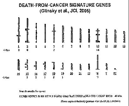
Glinsky, G.V. et al., J. Clin. Invest. 113: 913-923 (2004) teaches that gene
expression profiling
predicts clinical outcomes of prostate cancer. van `t Veer et al., Nature 415:
530-536 (2002)
teaches that gene expression profiling predicts clinical outcomes of breast
cancer. Glinsky et al.,
J. Clin. Invest. 115: 1503-1521 (2005) teaches that altered expression of the
BMIl oncogene is
functionally linked with self-renewal
state of normal and leukemic stem cells as well as a poor prognosis profile of
an 11-gene
death-from-cancer signature predicting therapy failure in patients with
multiple types of
cancer. These studies utilized the microarray gene expression analysis
approach.
There is, therefore, a need for methods for early diagnosis of cancer and for
prognostic assays for cancer therapy that are readily adaptable to the
clinical setting.
Such methods should utilize technologies that can be readily carried out in
clinical
laboratories, and should accurately predict the resistance of various cancers
to be applied
to standard therapeutic regimens.
Summary Of The Invention
The present invention is directed to novel methods and kits for diagnosing the
presence of cancer within a patient, and for determining whether a subject who
has
CA 02648021 2008-09-30
WO 2007/114896 PCT/US2007/008088
cancer is susceptible to different types of treatment regimens. The cancers to
be tested
include, but are not limited to, prostate, breast, lung, gastric, ovarian,
bladder,
lymphoma, mesothelioma, medullablastoma, glioma, and AML.
One embodiment of the present invention is directed to a method for diagnosing
cancer or predicting cancer-therapy outcome by detecting the expression levels
of
multiple markers in the same cell at the same time, and scoring their
expression as being
above a certain threshold, wherein the markers are from a particular pathway
related to
cancer, with the score being indicative or a cancer diagnosis or a prognosis
for cancer-
therapy failure. This method can be used to diagnose cancer or predict cancer-
therapy
outcomes for a variety of cancers. The simultaneous co-expression of at least
two
markers in the same cell from a subject is a diagnostic for cancer and a
predictor for the
subject to be resistant to standard cancer therapy. The markers can come from
any
pathway involved in the regulation of cancer, including specifically the PcG
pathway and
the "steinness" pathway. The markers can be inRNA, DNA, or protein.
These and other embodiments of the present invention rely at least in part
upon
the novel finding that the expression of inultiple markers above a threshold
level in the
same cell at the same time, wherein the markers are found within pathways
related to
cancer, can be used as an assay to diagnose cancer disorders and to predict
whether a
patient already diagnosed with cancer will be therapy-responsive or therapy-
resistant. An
element of the assay is that two or more markers are detected simultaneously
within the
same cell. Marker detection can be made through a variety of detection means,
including
bar-coding through immunofluorescence. The markers detected can be a variety
of
products, including mRNA, DNA, and protein. For mRNA based markers, PCR can be
used as a detection means. Additionally, protein products or gene copy number
can be
identified through detection means known in the art. The markers detected can
be from a
variety of pathways related to cancer. Suitable pathways for markers within
the scope of
the present invention include any pathways related to oncogenesis and
metastasis, and
more specifically include the Polycomb group (PcG) chromatin silencing pathway
and
the "stemness" pathway.
2
CA 02648021 2008-09-30
WO 2007/114896 PCT/US2007/008088
In another embodiment, the invention is directed to a method for diagnosing
cancer or predicting cancer-therapy outcome in a subject, said method
comprising the
steps of:
a) obtaining a sample from the subject,
b) selecting a marker from a pathway related to cancer,
c) screening for a simultaneous aberrant expression level of two or more
markers
in the same cell from the sample, and
d) scoring their expression level as being aberrant when the expression level
detected is above or below a certain detection threshold coefficient, wherein
the detection
threshold coefficient is determined by comparing the expression levels of the
samples
obtained from the subjects to values in a reference database of sample
phenotypes
obtained from subjects with either a known diagnosis or known clinical outcome
after
therapy, wherein the presence of an aberrant expression level of two or more
markers in
individual cells and presence of cells aberrantly expressing two or more such
markers is
indicative of a cancer diagnosis or a prognosis for cancer-therapy failure in
the subject.
The subset of markers to be used within the methods of the present invention
include any
markers associated with cancer pathways.
In preferred embodiments, the markers can' be selected from the genes
identified
in Figures 27-38. The markers can comprise anywhere ranging from two markers
listed
within each table up to the whole set of genes listed within each of these
tables. The
markers can comprise any percentage of genes selected from each of these
tables,
including 90%, 80%, 70%, 60%, or 50% of the genes identified in Figures 27-38.
In this method, an aberrant co-expression level of the markers can be
indicative of
the presence of cancer in the subject, or predictive of cancer-therapy failure
in the
subject. The markers can be selected from any suitable cancer pathway,
including in
preferred embodiments markers from the Polycomb or "stemness" pathway. These
markers can be genes selected from the group consisting of ADA, AMACR+p63,
ANK3,
BCL2L1, BIRC5, BMI-1, BUB I, CCNB1, CCND1, CES1, CHAF1A, CRIPI, CRYAB,
ESM1, EZH2, FGFR2, FOS, Gbx2, HCFC1, IER3, ITPR1, JUNB, KLF6, K167, KNTC2,
MGC5466, Phcl, RNF2, Suz12, TCF2, TRAP100, USP22, Wnt5A and ZFP36. In
preferred embodiments, the mai-kers are selected from the group consisting of
BMI1,
3
CA 02648021 2008-09-30
WO 2007/114896 PCT/US2007/008088
Ezh2, H2A, H3, transcription factors, and methylation patterns. In one
preferred
embodiment, the aberrant co-expression level detected is of BMII and Ezh2, and
in
another preferred embodiment the aberrant co-expression level detected is of
H2A and
H3. The markers being detected are in the form of either mRNA, DNA, or
protein.
In a preferred embodiment, the sample phenotypes are selected from the group
consisting of cancer, non-cancer, recurrence, non-recurrence, relapse, non-
relapse,
invasiveness, non-invasiveness, metastatic, non-metastatic, localized, tumor
size, tumor
grade, Gleason score, survival prognosis, lymph node status, tumor stage,
degree of
differentiation, age, hormone receptor status, PSA level, histologic type, and
disease free
survival.
The aberrant expression level of two or more markers can be detected by any
detection means known in the art, including, but not limited to, subjecting
the cells to an
analysis selected from the group consisting of inulticolor quantitative
immunofluorescence co-localization analysis, fluorescence in situ
hybridization, and
quantitative RT-PCR analysis.
In another embodiment, the present invention is directed to a method of
determining a detection threshold coefficient for classifying a sample
phenotype from a
subject, the method comprising the steps of:
a) obtaining a sample from the subject,
b) selecting two or more markers from a pathway related to cancer,
c) screening for a simultaneous aberrant expression level of the two or more
markers in the same cell from the sample;
d) scoring the marker expression in the cells by comparing the expression
levels
of the samples obtained from the subjects to values in a reference database of
samples
obtained from subjects with either a known diagnosis or known clinical outcome
after
therapy, and
e) determining the detection threshold coefficient for the sample
classification
accuracy at different detection thresholds using reference database of samples
from
subjects with known phenotypes.
Detection threshold coefficients which are indicative of a cancer diagnosis or
a
prognosis for cancer-therapy failure have an absolute value within the range
of
4
CA 02648021 2008-09-30
WO 2007/114896 PCT/US2007/008088
(YtoreqØ5. to .gtoreqØ999. Preferred levels of detection threshold
coefficients which
are indicative of a cancer diagnosis or a prognosis for cancer-therapy failure
have an
absolute value of .gtoreqØ5,.gtoreqØ6,.gtoreqØ7,.gtoreqØ8,.gtoreqØ9,
.gtoreqØ95, .gtoreqØ99, .gtoreqØ995., and .gtoreqØ999.
In another embodiment, the method further comprises determining the best
performing magnitude of said detection threshold and using said magnitude to
assess the
reliability of said established detection threshold in classifying a sample
phenotype. In
another embodiment, the method further comprises using the best performing
magnitude
of said detection threshold to score an unclassified sample and assign a
sample phenotype
to said sample.
In another embodiment, the present invention is directed to a method for
simultaneously detecting an aberrant co-expression level of two or more
markers a single
cell, said method comprising the steps of:
a) obtaining a sample of tissue,
b) selecting a marker defined by a pathway,
c) screening for a simultaneous aberrant expression level of the two or more
markers, and
d) scoring their expression level as being aberrant when the expression level
detected is above or below a certain detection threshold coefficient, wherein
the detection
threshold coefficient is deterinined by comparing the expression levels of the
samples
obtained from the subjects to values in a reference database of sample
phenotypes
obtained from subjects with either a known diagnosis or known clinical outcome
after
therapy.
The present invention is also directed to kits useful in detecting the
simultaneous
aberrant co-expression levels of two or more markers in a single cell.
Brief Description Of The Drawinp-s
Figure 1 shows HapMap analysis revealing population-specific profiles of
genotype and allele frequencies of SNPs associated with cancer therapy outcome
predictor (CTOP) genes comprising an 11-gene death-from-cancer signature.
CA 02648021 2008-09-30
WO 2007/114896 PCT/US2007/008088
A. Chromosomal locations of genes encoding transcripts comprising an 11-gene
death-
from-cancer signature
B, E. Annotated haplotypes associated with the BMII (B) and BUBl (E) genes in
CEU,
YRI, CHB, and JPT HapMap populations. Arrows indicate SNPs with population-
specific profiles of genotype and allele frequencies.
C, D, F - H. Bar graph plots demonstrating population-specific profiles of
genotype and
allele frequencies in different HapMap populations for individual SNPs
associated with
genes comprising an 11-gene death-from-cancer signature. For each SNP the
frequencies
shown within each set of bar graphs in the following order (from left to
right): CEU,
CHB, JPT, YRI.
B - D, BMl1 gene; F - H, CCNBI, KNTC2, HCFC1, FGFR2, and BUBI genes.
Figure 2 shows HapMap analysis revealing population-specific profiles of
genotype and allele frequencies of SNPs associated with CTOP genes predicting
the
likelihood of disease relapse in prostate cancer patients after radical
prostatectomy.
A. Chromosomal locations of genes encoding transcripts comprising prostate
cancer
recurrence predictor signatures.
B - D. Bar graph plots demonstrating population-specific profiles of genotype
and allele
frequencies in different HapMap populations for individual SNPs associated
with genes
comprising prostate cancer recurrence predictor signatures. For each SNP the
frequencies
shown within each set of bar graphs in the following order (from left to
right): CEU,
CHB, JPT, YRI.
B, KLF6 (COPEB) gene; C, WntS, TCF2, CHAFIA, and KIAA0476 genes; D, PPFIA3,
CDS2, FOS, and CHAFIA genes.
Figure 3 shows HapMap analysis revealing population-specific profiles of
genotype and allele frequencies of SNPs associated with cancer therapy outcome
predictor (CTOP) genes comprising a 50-gene proteomics-based cancer therapy
outcome
signature.
A. Chromosomal locations of genes encoding transcripts comprising a 50-gene
cancer
therapy outcome signature.
B - D. Annotated haplotypes associated with the MCM6 (B), STK6 (C), and NUP62
(D)
6
CA 02648021 2008-09-30
WO 2007/114896 PCT/US2007/008088
genes in CEU, YRi, CHB, and JPT HapMap populations. Stars indicate SNPs with
population-specific profiles of genotype and allele frequencies.
Figure 4 shows HapMap analysis identifying non-synonymous coding SNPs
associated with CTOP genes and manifesting population-specific profiles of
genotype
and allele frequencies.
A - D. Annotated haplotypes associated with the TRAF3IP2 (A), PXN (B), MKI67
(C),
and RAGE (D) genes in CEU, YRI, CHB, and JPT HapMap populations. Arrows
indicate non~synonymous coding SNPs with population-specific profiles of
genotype and
allele frequencies.
Figure 5 shows population-specific profiles of genotype and allele frequencies
of
SNPs associated with oncogenes and tumor suppressor genes.
A. Annotated haplotypes associated with the RBI gene in CEU, YRI, CHB, and JPT
HapMap populations. Arrows indicate SNPs with population-specific profiles of
genotype and allele frequencies.
B - H. Bar graph plots demonstrating population-specific profiles of genotype
and allele
frequencies in different HapMap populations for individual SNPs associated
with
oncogenes and tumor suppressor genes. For each SNP the frequencies shown
within each
set of bar graphs in the following order (from left to right): CEU, CHB, JPT,
YRI.
A, C, D, RBI gene; B, PTEN and TP53 genes; E, MYC and CCND1; F, hTERT gene; G,
AKTI gene.
Figure 6 shows that SNP-based gene expression signatures predict therapy
outcome in prostate and breast cancer patients.
A - D. Genes expression of which is regulated by SNP variations in normal
individuals
provide gene expression models predicting therapy outcome in breast (A, C) and
prostate
(B, D) cancer patients.
A, B. Kaplan-Meier analysis of therapy outcome classification performance in
breast
cancer (A) and prostate cancer (B) patients of gene expression-based CTOP
models
generated from genetic loci expression of which is regulated by the 14q32
master
regulatory locus.
C, D. Kaplan-Meier analysis of therapy outcome classification performance in
breast
cancer (C) and prostate cancer (D) patients of gene expression-based CTOP
models
7
CA 02648021 2008-09-30
WO 2007/114896 PCT/US2007/008088
generated from transcriptionally most variable genetic loci.
E - H. Genes containing high-population differentiation non-synonymous SNPs
(E, F)
and genes representing loci in which natural selection most likely occurred
(G, H)
provide gene expression-based therapy outcome prediction models for breast (E,
G) and
prostate (F, H) cancer patients.
E, F. Kaplan-Meier analysis of therapy outcome classification performance in
breast
cancer (E) and prostate cancer (F) patients of gene expression-based CTOP
models
generated from genetic loci containing high-population differentiation non-
synonymous
SNPs.
G, H. Kaplan-Meier analysis of therapy outcome classification performance in
breast
cancer (G) and prostate cancer (H) patients of gene expression-based CTOP
models
generated from genetic loci in which natural selection most likely occurred.
I, J. Kaplan-Meier analysis of therapy outcome classification performance in
breast
cancer (I) and prostate cancer (J) patients of gene expression-based CTOP
models
generated from genetic loci regulated by SNP variations in normal individuals.
K, L. Kaplan-Meier analysis of therapy outcome classification performance in
breast
cancer (E) and prostate cancer (F) patients of gene expression-based CTOP
models
generated from genetic loci selected based on similarity of SNP profiles with
population
specific SNP profiles of known CTOP genes.
M, N. Kaplan-Meier analysis of therapy outcome classification performance in
breast
cancer (E) and prostate cancer (F) patients of gene expression-based CTOP
models
generated from a proteomics-based 50-gene signature.
Figure 7 shows microarray analysis identifying clinically relevant cooperating
oncogenic pathways in human prostate and breast cancers. Kaplan-Meier survival
analysis for prostate cancer (A-D) and breast cancer (E-H) with deregulated
individual
pathways associated with BMII (A, E), Myc (B, F), or Her2/neu (C, G)
activation. Plots
D and H show Kaplan-Meier analysis based on patients' stratification taking
into account
evidence for activation of multiple pathways in individual tumors. Gene
expression
signature-based patients' stratification for Kaplan-Meier survival analysis
were
performed as described in Glinsky et al., J. Clin. Invest. 115: 1503-1521
(2005) and
'Glinskyet al., J. Clin. Invest. 113: 913-923 (2004).
8
CA 02648021 2008-09-30
WO 2007/114896 PCT/US2007/008088
Figure 8 shows how comparative cross-species translational genomics integrates
knowledge written in two languages (DNA sequence variations and mRNA
expression
levels) and three writing systems reflecting defined phenotype/gene expression
pattern
associations (SNP variations; transgenic mouse models of cancers; genomics of
stem cell
biology).
Figure 9 shows Q-RT-PCR analysis of mRNA abundance levels of a
representative set of genes comprising the BM-1-pathway signature in BM-1
siRNAitreayed PC-3-32 human prostate carcinoma cells.
Figure 10 shows siRNA-mediated changes of the transcript abundance levels of
11 genes comprising BM-1-pathway signature.
Figure 11 shows EZH2 siRNA-mediated changes of the transcript abundance
levels of 11 genes comprising the BM-1-pathway signature.
Figure 12 shows siRNA-mediated changes of the transcript abundance levels of
11 genes comprising BM-1-pathway signature. A. BM-1 siRNA. B. EZH2 siRNA.
Figure 13 shows expression profiles of 11 gene MM-1-signature in distant
metastatic lesions of the TRAMP transgenic mouse model of prostate cancer and
PNS
neurospheres.
Figure 14 shows increased DNA copy numbers of the BM-1 and Ezh2 genes in
human prostate carcinoma cells selected for high metastatic potential.
Figure 15 shows the quadruplicon of prostate cancer progression in the LNCap
progression model.
Figure 16 shows the quadruplicon of prostate cancer progression in the PC-3
progression model.
Figure 17 shows the quadruplicon of prostate cancer progression in the PC-3
bone metastasis progression model.
Figure 18 shows expression levels in PC-3-32 and PC-3 cells.
Figure 19 shows cytoplasmic AMACR and nuclear p63 expression in parental
PC-3 human prostate carcinoma cells and PC-3-32 human prostate carcinoma
metastasis
precursor cells.
Figure 20 shows that high expression levels of the BMI1 and Ezh2 oncoproteins
in human prostate carcinoma metastasis precursor cells are associated with
marked
9
CA 02648021 2008-09-30
WO 2007/114896 PCT/US2007/008088
accumulation of a dual-positive high BMII/Ezh2-expressing cell population and
increased DNA copy number of the BMII and Ezh2 genes.
A-D. A quantitative immunofluorescence co-localization analysis of the BMIl
(mouse
monoclonal antibody) and Ezh2 (rabbit polyclonal antibody) oncoproteins in PC-
3-32
human prostate carcinoma metastasis precursor cells and parental PC-3 cells.
The protein
expression differences and the accumulation of dual-positive high BMI1/Ezh2-
expressing
cells were confirmed using a second distinct combination of antibodies: rabbit
polyclonal
antibodies for BMI1 detection and mouse monoclonal antibodies for Ezh2
detection. A,
immunofluorescent analysis of PC-3-32 cells; B, immunofluorescent analysis of
PC-3
cells; C, the histograms representing typical distributions of the BMIl (top
panels) and
Ezh2 (bottom panels) expression levels in PC-3 and PC-3-32 cells; D, the plots
illustrating the levels of dual positive high BMIIlEzh2-expressing cells in
metastatic PC-
3-32 cells (22.4%; top panel) and parental PC-3 cells (1.5%; bottom panel).
The results of
one of two independent experiments are shown.
E. A quantitative reverse-transcription PCR (Q-RT-PCR) analysis of DNA copy
numbers
of the BMIJ and Ezh2 genes in multiple experimental models of human prostate
cancer.
Note marked increase of the BMIl and Ezh2 gene copy numbers in highly
metastatic
variants compared to the low metastatic counterparts in the multiple
independently
selected lineages. The results of one of two independent experiments are
shown.
F. 3D-view of dual-positive high BMII/Ezh2-expressing human prostate carcinoma
cells
in cultures of blood-borne metastasis precursor cells and parental cells.
Adherent cultures
of parental PC-3 (bottom three panels) and blood-borne PC-3-32 (top three
panels)
human prostate carcinoma cells were stained for visualization of the BMI1 and
Ezh2
oncoproteins and analyzed using a multi-color fluorescent confocal microscopy.
Note a
higher proportion of cells with large discrete nuclear PeG bodies in the
population of PC-
3-32 human prostate carcinoma cells (typically, these cells contain six PcG
bodies per
nucleus). Blue, DNA; Green, BMII; Red, Ezh2.
Figure 21 shows results of activation of the PeG chromatin silencing pathway
in
metastatic human prostate carcinoma cells. A quantitative immunofluorescence
co-
localization analysis was utilized to measure the expression of the BMI1,
Ezh2,
H3metK27, and UbiH2A -narkers in human prostate carcinoma cells and calculate
the
CA 02648021 2008-09-30
WO 2007/114896 PCT/US2007/008088
numbers of dual-positive cells expressing various two-marker combinations.
Note that
high expression of the BMI 1 and Ezh2 oncoproteins in PC-3-32 human prostate
carcinoma metastasis precursor cells compared to parental PC-3 cells is
associated with
increased levels of histone H3 lysine 27 methylation (H3metK27), histone H2A
lysine
119 ubiquitination (UbiH2A), and marked enrichment for dual-positive cell
populations
expressing high levels of BMI1/UbiH2A, Ezh2/H3metK27, and H3metK27/UbiH2A
two-marker combinations.
Figure 22 shows that targeted reduction of the BMI1 (3A) or Ezh2 (3B)
expression increases sensitivity of human prostate carcinoma metastasis
precursor cells to
anoikis. Anoikis-resistant PC-3-32 prostate carcinoma cells were treated with
BM11- or
Ezh2-targeting siRNAs and continuously monitored for expression levels of the
various
mRNAs, BMI and Ezh2 oncoproteins, as well as cell growth and viability under
various
culture conditions. PC-3-32 cells with reduced expression of either BMI1 or
Ezh2
oncoproteins acquired sensitivity to anoikis as demonstrated by the loss of
viability and
increased apoptosis compared to the control LUC siRNA-treated cultures growing
in
detached conditions.
Figure 23 shows that treatment of human prostate carcinoma metastasis
precursor
cells with stable siRNAs targeting either BMII or Ezh2 gene products depletes
a sub-
population of dual positive high BMII/Ezh2-expressing cells. Blood-borne PC-3-
32
prostate carcinoma cells were treated with chemically modified resistant to
degradation
LUC-, BMII-, or Ezh2-targeting stable siRNAs and continuously monitored for
expression levels of the BMI and Ezh2 oncoproteins. Two consecutive
applications of the
stable siRNAs caused a sustained reduction of the BMI1 and Ezh2 expression and
depletion of the sub-population of dual positive high BMI1/Ezh2-expressing
carcinoma
cells. The results at the 11-day post-treatment time point are shown.
Figure 24 shows that human prostate carcinoma metastasis precursor cells
depleted for a sub-population of dual positive high BMI1/Ezh2-expressing cells
manifest
a dramatic loss of malignant potential in vivo. Adherent cultures of blood-
borne PC-3-
GFP-39 prostate carcinoma cells were treated with chemically modified
degradation-
resistant stable siRNAs targeting BMII or Ezh2 mRNAs or control LUC siRNA. 24
hrs
after second treatment, 1.5 x 10⁶ cells were injected into prostates of nude
mice. Note that
11
CA 02648021 2008-09-30
WO 2007/114896 PCT/US2007/008088
all control animals developed highly aggressive rapidly growing metastatic
prostate
cancer and died within 50 days of experiment. Only 20% of mice in the BMII-
and Ezh2-
targeting therapy groups developed less malignant more slowly growing tumors.
150
days after tumor cell inoculation, 83% and 67% of animals remain alive and
disease-free
in the therapy groups targeting the BMI1 and Ezh2 oncoproteins, respectively
(p =
0.0007; log-rank test). Six animals per group were monitored for survival.
Figure 25 shows that tissue microarray analysis (TMA) of primary prostate
tumors from patients diagnosed with prostate adenocarcinomas reveals increased
levels
of dual-positive BMII/Ezh2 high-expressing cells. BMII and Ezh2 oncoprotein
expression were measured in prostate TMA samples from cancer patients and
healthy
donors using a quantitative co-localization immunofluorescence method and the
number
of dual positive high BMII/Ezh2-expressing nuclei was calculated for each
sample. Note
that primary prostate tumors from patients diagnosed with prostate
adenocarcinomas
manifest a diverse spectrum of accumulation of dual positive BMI1/Ezh2 high-
expressing
cells and patients with higher levels of BMIJ or Ezh2 expression in prostate
tumors
manifest therapy-resistant malignant phenotype (Figure 26). A majority (79% -
92% in
different cohorts of patients) of human prostate tumors contains dual positive
high
BMI1/Ezh2-expressing cells exceeding the threshold expression levels in
prostate
samples from normal individuals.
Figure 26 shows that Increased BMII and Ezh2 expression is associated with
high likelihood of therapy failure and disease relapse in prostate cancer
patients after
radical prostatectomy. Kaplan-Meier survival analysis demonstrates that cancer
patients
with more significant elevation of the BMII and Ezh2 expression [having higher
tumor
(T) to adjacent normal tissue (N) ratio, T/N: Figure 26A; or having tumors
with higher
levels of BM11 (28B) or Ezh2 (28C) expression) are more likely to fail therapy
and
develop a disease recurrence after radical prostatectomy. Figure 26E shows the
Kaplan-
Meier survival analysis of 79 prostate cancer patients stratified into five
sub-groups using
eight-covariate cancer therapy outcome (CTO) algorithm. CTO algorithm
integrates
individual prognostic powers of BMIJ and Ezh2 expression values and six
clinico-
pathological covariates (preoperative PSA, Gleason score, surgical margins,
extra-
capsular invasion, seminal vesicle invasion, and age).
12
CA 02648021 2008-09-30
WO 2007/114896 PCT/US2007/008088
Figure 27 shows breast cancer CTOP signatures in Affymetrix format, with-
predictive outcomes.
Figure 28 shows breast cancer CTOP signatures in Agilent Rosetta Chip format,
with predictive outcomes.
Figure 29 shows prostate cancer CTOP signatures in Affymetrix format, with
predictive outcomes.
Figure 30 shows P13K pathway CTOP signatures.
Figure 31 shows SNP based CTOP signatures NG2007.
Figure 32 shows the parent methylation Signatures.
Figure 33 shows the histones H3 and H2A CTOP signatures.
Figure 34 shows the CTOP gene expression signatures for prostate cancer.
Figure 35 shows the CTOP gene expression signatures for breast cancer.
Figure 36 shows the CTOP gene expression signature and survival data for lung
cancer.
Figure 37 shows the CTOP gene expression signature for ovarian cancer.
Figure 38 shows the CTOP gene expression signatures for breast cancer.
Figure 39 shows examples of the evaluation of the CMAP000 and CMAP11 drug
combinations in prostate cancer and the CMAP19 drug combination in breast
cancer.
Figure 40 shows CTOP scores for lung cancer.
Figure 41 shows Kaplan-Meier survival analysis of seventy-nine prostate cancer
patients stratified into sub-groups with distinct expression profiles of the
individual
Polycomb pathway ESC signatures (top six panels) or six ESC signatures
algorithm
(bottom panel) in primary prostate tumors. In each individual signature panel,
patients
were sorted in descending order based on the values of the corresponding
signature
CTOP scores and divided into poor prognosis (top 50% scores) and good
prognosis
(bottom 50% scores) sub-groups. In the last panel, patients were sorted in
descending
order based on the values of the cumulative CTOP scores and divided into poor
prognosis
(top 50% scores) and good prognosis (bottom 50% scores) sub-groups. The
cumulative
CTOP scores represent the sum of the six individual CTOP scores calculated for
each
patient.
13
CA 02648021 2008-09-30
WO 2007/114896 PCT/US2007/008088
Figure 42 shows Kaplan-Meier survival analysis of two-hundred eighty-six early-
stage LN negative breast cancer patients stratified into sub-groups with
distinct
expression profiles of the individual Polycomb pathway ESC signatures (top six
panels)
or six ESC signatures algorithm (single middle panel) in primary breast
tumors. Bottom
four panels show patients' classification performance of the six ESC
signatures algorithm
in four different breast cancer therapy outcome data sets. Patients'
stratification was
performed using either individual CTOP scores (top six panels) or cumulative
CTOP
scores (bottom five panels) as described in the legend to the Figure 41.
Figure 43 shows bivalent chromatin domain-containing transcription factors
(BCD-TF) manifest "stemness" expression profiles in therapy-resistant prostate
and
breast tumors.
A. Chromatin context identified by the presence of histones harboring specific
modifications of the histone tails defines mutually exclusive
transcriptionally active or
silent states of corresponding genetic loci in genomes of most cells. In ESC
multiple
chromosomal regions were identified simultaneously harboring both "silent"
(H3K27met3) and "active" (H3K4) histone marks and - 100 transcription factor
(TF)
encoding genes are residing within these bivalent chromatin domain-containing
chromosomal regions. Expression of selected TF encoding genes in ESC,
including
bivalent chromatin domain-containing TF genes (BCD-TF), maintenance of
a"stemness"
state, and transition to differentiated phenotypes is regulated by the balance
of the
"stemness" TFs (Nanog, Sox2, Oct4) and Polycomb group (PcG) proteins bound to
the
promoters of target genes.
B. Thirteen-gene BCD-TF signature manifesting highly concordant (r = 0.853; P
< 0.001)
gene expression profiles in breast and prostate tumors from patients with
therapy-
resistant disease phenotypes.
C. Eight-gene BCD-TF signature (derived from thirteen-gene BCD-TF signatures)
manifesting highly concordant expression profiles (r = 0.716; p < 0.001) in
ESC and
therapy-resistant breast and prostate tumors. Kaplan-Meier analysis
demonstrates that
prostate and breast cancer patients with tumors harboring ESC-like expression
profiles of
the eight-gene BCD-TF signature are more likely to fail therapy (bottom two
panels).
Gene expression profiles of clinical samples were independently generated for
therapy-
14
CA 02648021 2008-09-30
WO 2007/114896 PCT/US2007/008088
resistant breast and prostate tumors using multivariate Cox regression
analysis of
microarrays of tumor samples from 286 breast cancer and 79 prostate cancer
patients
with known log-term clinical outcoine after therapy. Gene expression profiles
of mouse
ESC were derived by comparing microarray analyses of pluripotent self-renewing
ESC
(control ESC cultures treated with HP siRNA) versus ESC treated with Esrrb
siRNA (day
6). At this time point, Esrrb siRNA-treated ESC do not manifest `stemness"
phenotype
and form colonies of differentiated cells.
Figure 44 shows Kaplan-Meier survival analysis of two-hundred eighty-six early-
stage LN negative breast cancer patients (top four panels) and seventy-nine
prostate
cancer patients (bottom four panels) stratified into sub-groups with distinct
expression
profiles of the individual CTOP signatures [bivalent chromatin domain
transcription
factors (BCD-TF) and ESC pattern 3 signatures], eight ESC signatures
algorithm, and
nine "stemness" signatures algorithm in primary breast or prostate tumors.
Patients'
stratification was performed using either individual CTOP scores (for
individual
signatures) or cumulative CTOP scores (for CTOP algorithms) as described in
the legend
to the Figure 41.
Figure 45 shows Kaplan-Meier survival analysis of seventy-nine prostate cancer
patients (top four panels) and ninety-seven early-stage LN negative breast
cancer
patients (middle four panels) stratified into sub-groups with distinct
expression profiles of
the individual CTOP signatures [histones H3 and H2A signatures; Polycomb (PcG)
pathway methylation signature] and two signatures PcG methylation/histones
H3/H2A
algorithm (bottom two panels) in primary prostate and breast tumors. Patients'
stratification was performed using either individual CTOP scores (for
individual
signatures) or cumulative CTOP scores (for CTOP algorithm) as described in the
legend
to the Figure 41.
Figure 46 shows Kaplan-Meier survival analysis of two-hundred eighty-six early-
stage LN negative breast cancer patients (top left panel), seventy-nine
prostate cancer
patients (top right panel), ninety-one early-stage lung cancer patients
(bottom left panel),
and one-hundred thirty-three ovarian cancer patients (bottom right panel)
stratified into
sub-groups with distinct expression profiles of the nine "stemness" signatures
algorithm
in primary breast, prostate, lung, and ovarian tumors. Patients'
stratification was
CA 02648021 2008-09-30
WO 2007/114896 PCT/US2007/008088
performed using cumulative CTOP scores of the nine "stemness"' signatures as
described
in the legend to the Figure 41. Patients were sorted in descending order based
on the
values of the cumulative CTOP scores and divided into five sub-groups at 20%
increment
of the cumulative CTOP score values.
Figure 47 shows validation of the Polycomb pathway activation in metastatic
and
therapy-resistant human prostate cancer.
A. Blood-borne PC-3-32 human prostate carcinoma cells contain increased levels
of
CD44+/CD24- cancer stem cell-like population of dual-positive BMII/Ezh2 high-
expressing cells (middle panel) with increased levels of H3met3K27 and
H2AubiK119
histones (bottom two FACS figures). CD44+CD24- cancer stem cell-like
populations
were isolated using sterile FACS sorting from parental PC-3 and blood-borne PC-
3-32
metastasis precursor cells and subjected to multicolor quantitative
immunofluorescence
co-localization analysis (18) for BMII and Ezh2 Polycomb proteins (middle
panel) or
Polycomb pathway substrates H3met3K27 and H2AubiK119 histones (bottom two FACS
figures).
B. Multi-color FISH analysis reveals marked enrichment of blood-borne human
prostate
carcinoma metastasis precursor cells for cell population with co-amplification
of both
BMIl and Ezh2 genes. Color microphotographs of nuclei of blood-borne PC-3-32
human
prostate carcinoma cells with high-level co-amplification of both BMII and
Ezh2 genes.
For comparison, nuclei of diploid hTERT-immortalized human fibroblasts
containing two
copies of the BMII and Ezh2 genes are shown. Bottom two panels present
quantitative
FISH analysis of the DNA copy numbers of BMIl and Ezh2 genes in parental PC-3
and
blood-borne PC-3-32 human prostate carcinoma cells.
C. Kaplan-Meier survival analysis of seventy-one prostate cancer patients with
distinct
levels of dual-positive BMI1/Ezh2 high expressing cells in primary prostate
tumors.
Prostate cancer TMA were subjected to multi-color quantitative
immunofluorescence co-
localization analysis of expression of the BMI1 and Ezh2 proteins. Prostate
cancer
patients having > 1% of dual-positive BMI1/Ezh2 high expressing cells
manifested
statistically significant increased likelihood of therapy failure after
radical prostatectomy.
Detailed Description
16
CA 02648021 2008-09-30
WO 2007/114896 PCT/US2007/008088
The present invention is directed to novel methods and kits for diagnosing the
presence of cancer within a patient, and for determining whether a subject who
has
cancer is susceptible to different types of treatment regimens. The cancers to
be tested
include, but are not limited to, prostate, breast, lung, gastric, ovarian,
bladder,
lymphoma, mesothelioma, medullablastoma, glioma, mantle cell lymphoma, and
AML.
In some embodiments, the kits and methods of the present invention can be used
to predict various different types of clinical outcomes. For example, the
invention can be
used to predict recurrence of disease state after therapy, non-recurrence of a
disease state
after therapy, therapy failure, short interval to disease recurrence (e.g.,
less than two
years, or less than one year, or less than six months), short interval to
metastasis in cancer
(e.g., less than two years, or less than one year, or less than six months),
invasiveness,
non-invasiveness, likelihood of metastasis in cancer, likelihood of distant
metastasis in
cancer, poor survival after therapy, death after therapy, disease free
survival qnd so forth.
One embodiment of the present invention is directed to a method for diagnosing
cancer or predicting cancer-therapy outcome by detecting the expression levels
of
multiple markers in the same cell at the same time, and scoring their
expression as being
above a certain threshold, wherein the markers are from a particular pathway
related to
cancer, with the score being indicative or a cancer diagnosis or a prognosis
for cancer-
therapy failure. This method can be used to diagnose cancer or predict cancer-
therapy
outcomes for a variety of cancers. The simultaneous co-expression of at least
two
markers in the same cell from a subject is a diagnostic for cancer and a
predictor for the
subject to be resistant to standard cancer therapy. The markers can come from
any
pathway involved in the regulation of cancer, including specifically the PcG
pathway and
the `stemness" pathway. The markers can be mRNA (messenger RNA), DNA,
microRNA, or protein.
The subset of markers to be used within the methods of the present invention
include any markers associated with cancer pathways. In preferred embodiments,
the
markers can be selected from the genes identified in Figures 27-38. The
markers can
comprise anywhere ranging from two markers listed within each table up to the
whole set
of genes listed within each of these tables. The markers can comprise any
percentage of
17
CA 02648021 2008-09-30
WO 2007/114896 PCT/US2007/008088
genes selected from each of these tables, including 90%, 80%, 70%, 60%, or 50%
of the
genes identified in each of Figures 27-38.
These and other embodiments of the present invention rely at least in part
upon
the novel finding that the expression of multiple markers above a threshold
level in the
same cell at the same time, wherein the markers are found within pathways
related to
cancer, can= be used as an assay to diagnose cancer disorders and to predict
whether a
patient already diagnosed with cancer will be therapy-responsive or therapy-
resistant. An
element of the assay is that two or more markers are detected simultaneously
within the
same cell.
Marker detection can be made through a variety of detection means, including
bar-coding through immunofluorescence. The markers detected can be a variety
of
products, including mRNA, DNA, microRNA, and protein_ For mRNA or microRNA
based markers, PCR can be used as detection means. Additionally, protein
products,
gene expression, or gene copy number can be identified through detection means
known
in the art.
Detection means, in case of a nucleic acid probe, include measuring the level
of
mRNA or cDNA to which a probe has been engineered to bind, where the probe
binds
the intended species and provides a distinguishable signal. In some
embodiments, the
probes are affixed to a solid support, such as a microarray. In other
embodiments, the
probes are primers for nucleic acid amplification of a set of genes. Q-RT-PCR
amplification can be used. Detecting expression for measurement or determining
protein
expression levels can also be accomplished by using a specific binding
reagent, such as
an antibody. In general, expression levels of the markers can be analyzed by
any method
now known or later developed to assess gene expression, including but not
limited to
measurements relating to the biological processes of nucleic acid
amplification,
transcription, RNA splicing, and translation. Direct and indirect measures of
gene copy
number (e.g., as by fluorescence in situ hybridization or other type of
quantitative
hybridization measurement, or by quantitative PCR), transcript concentration
(e.g., as by
Northern blotting, expression array measurements, quantitative RT-PCR, or
comparative
genomic hybridization) and protein concentration (e.g., as by quantitative 2D
gel
18
CA 02648021 2008-09-30
WO 2007/114896 PCT/US2007/008088
electrophoresis, mass spectrometry, Western blotting, ELISA, or other method
for
determining protein concentration), can also be used.
One of skill in the art would recognize that different affinity reagents could
be
used with the present invention, such as one or more antibodies (monoclonal or
polyclonal) and the invention can include using techniques, such as ELISA, for
the
analysis. Thus, specific antibodies (specific to the markers to be detected)
can be used in
a kit and in methods of the present invention. In a kit of the present
invention, the kit
would include reagents and instructions for use, where the reagents could be
protein-
specific differentially-labeled fluorescent antibodies; protein-specific
antibodies from
different species (mouse, rabbit, goat, chicken, etc.) and differentially
labeled species-
specific antibodies; DNA and RNA-based probes with different fluorescent dyes;
bar-
coded nucleic acid- and protein-specific probes (each probes having a unique
combination of colors).
The markers detected can be from a variety of pathways related to cancer.
Suitable pathways for markers within the scope of the present invention
include any
pathways related to oncogenesis and metastasis, and more specifically include
the
Polycomb group (PcG) chromatin silencing pathway and the "stemness" pathway.
Representative cancer pathways within the context of the present invention
include but are not limited to, the Polycomb pathway, the Polycomb pathway
target
genes, "stemness" pathways, DNA methylation pathways, BMI1, Ezh2, Suz12,
Suz12/PolII, EED, PcG-TF, BCD-TF, TEZ, Nanog/Sox2/Oct4, Myc, He2/neu, CCND1,
E2F3, P13K, beta-catenin, ras, src, PTEN, p53, Rb, p16/ARF, p21, Wnt, and Hh
pathways.
The Polycomb group (PcG) gene BMII is required for the proliferation and self-
renewal of normal and leukeinic stem cells. Over-expression of Bmil oncogene
causes
neoplastic transformation of lymphocytes and plays an essential role in the
pathogenesis
of myeloid leukemia. Another PcG protein, Ezh2, has been implicated in
metastatic
prostate and breast cancers, suggesting that PcG pathway activation is
relevant for
epithelial malignancies. Here it is demonstrated that activation of the BMI1
oncogene-
19
CA 02648021 2008-09-30
WO 2007/114896 PCT/US2007/008088
associated PeG pathway plays an essential role in metastatic prostate cancer,
thus
mechanistically linking the pathogenesis of leukemia, self-renewal of stem
cells, and
prostate cancer metastasis.
In another aspect, the methods of the present invention provide for the
diagnosis,
prognosis, and treatment strategy for a patient with a disorder of the above
mentioned
types. Treatment includes determining whether a patient has an expression
pattern of
markers associated with cancer and administering to the patient a therapeutic
adapted to
the treatment of the disorder. In one embodiment, the method can include the
identification of increased BMII and Ezh2 expression and the formulation of a
treatment
plan specific to this phenotype.
In another embodiment of the present invention, the detection of appropriate
or
inappropriate activation of "stemness" genetic pathways can be used to
diagnose cancer
and to predict the likelihood of cancer therapy success or failure.
Inappropriate activation
of "stemness" genes in cancer cells may be associated with aggressive clinical
behavior
and increased likelihood of therapy failure. A sub-set of human prostate
tumors
represents a genetically distinct highly malignant sub-type of prostate
carcinoma with
high propensity toward metastatic dissemination even at the early stage of
disease. Such a
high propensity toward metastatic dissemination of this type of prostate
tumors is
associated with the early engagement of normal stem cells into malignant
process.
Elucidation of such inappropriate activation of "stemness" gene expression can
help
tailor cancer therapy to a patient's individual needs.
The invention is directed to prognostic assays for cancer therapy that can be
used
to diagnose cancer and to predict the resistance of various cancers to
standard therapeutic
regimens. The invention is directed to methods and compositions for predicting
the
outcome of cancer therapy for individual patients. In one embodiment, the
method is
used to predict whether a particular cancer patient will be therapy-responsive
or therapy-
resistant. The invention can be used with a variety of cancers, including but
not limited =
to, breast, prostate, ovarian, lung, glioma, and lymphoma.
The invention is directed to personalized medicine for cancer patients, and
encompasses the selection of treatment options with the highest likelihood of
successful
CA 02648021 2008-09-30
WO 2007/114896 PCT/US2007/008088
outcome for individual cancer patients. The present invention is directed to
the use of a
an assay to predict the outcome after therapy in patients with early stage
cancer and
provide additional information at the time of diagnosis with respect to
likelihood of
therapy failure.
In another embodiment of the present invention, the detection of the state of
transcription factors can be used to diagnose the presence of cancer and to
predict the
likelihood of cancer therapy success or failure. The determination of a common
pattern
of the transcription factor expression can be used as a profile to help
determine clinical
outcome. The invention is also directed to a particular sub-set of BCD-TF
genes defined
here as the eight gene BCD-TF signature that manifests "stemness" expression
profiles in
therapy-resistant prostate and breast tumors (Figure 43).
In another embodiment of the present invention, the detection of the
methylation
state of target genes can be used to diagnose cancer and to predict the
likelihood of
cancer therapy success or failure. More particularly, PcG target genes with
promoters
frequently hypermethylated in cancer manifest distinct expression profiles
associated
with therapy-resistant and therapy-sensitive prostate= and breast cancers
(Figure 44),
implying that differences in gene expression between tumors with distinct
outcome after
therapy may be driven, in part, by the distinct promoter hypermethylation
patterns of the
PcG target genes. These differences can be exploited to generate highly
informative gene
expression signatures of the PcG target genes hypermethylated in cancer for
stratification
of prostate and breast cancer patients into sub-groups with statistically
distinct likelihood
of therapy failure (Figure 44).
The invention involves both a method to classify patients into sub-groups
predicted to be either therapy-responsive or therapy-resistant, and a method
for
deterinining alternate therapies for patients who are classified as resistant
to standard
cancer therapies. The method of the present invention is based on an accurate
classification of patients into subgroups with poor and good prognosis
reflecting a
different probability of disease recurrence and survival after standard
therapy.
In one embodiment, the invention relates to a method for diagnosing cancer or
predicting cancer-therapy outcome in a subject, said method comprising the
steps of:
a) obtaining a sample from the subject,
21
CA 02648021 2008-09-30
WO 2007/114896 PCT/US2007/008088
b) selecting a marker from a pathway related to cancer,
c) screening for a simultaneous aberrant expression level of two or more
markers
in the same cell from the sample, and
d) scoring their expression level as being aberrant when the expression level
detected is above oi- below a certain detection threshold coefficient, wherein
the detection
threshold coefficient is deterinined by comparing the expression levels of the
samples
obtained from the subjects to values in a reference database of samples
obtained from
subjects with either a known diagnosis or known clinical outcome after
therapy, wherein
the presence of an aberrant expression level of two or more markers in
individual cells
and presence of cells aberrantly expressing two or more such markers is
indicative of a
cancer diagnosis or a prognosis for cancer-therapy failure in the subject.
An aberrant expression level is a level of expression that can either be
higher or
lower than the expression level as compared to reference samples. The
reference samples
can have a variety of phenotypes, including both diseased phenotypes and non-
diseased
phenotypes. The sample phenotypes within the scope of the present invention
include,
but are not limited to, cancer, non-cancer, recurrence, non-recurrence,
relapse, non-
relapse, invasiveness, non-invasiveness, metastatic, non-metastatic,
localized, tumor size,
tumor grade, Gleason score, survival prognosis, lymph node status, tumor
stage, degree
of differentiation, age, hormone receptor status, PSA level, histologic type,
and disease
free survival.
A detection threshold coefficient within the context of the present invention
is a
value above which or below which a patient or sample can be classified as
either being
indicative of a cancer diagnosis or a prognosis for cancer-therapy failure.
The detection
threshold coefficients are defined by a plurality of measurements of samples
in the
reference database; sorting the samples in descending order of the values of
measurements; assignment of the probability of samples having a phenotype in
sub-
groups of samples defined at different increments of the values of
measurements (e.g.,
samples comprising top 10%; 20%; 30%; 40%; 50%; 60%; 70%; 80%; 90% of the
values); selecting the statistically best-performing detection threshold
coefficient defined
as the value of measurements segregating samples with the values below and
above the
threshold into subgroups with statistically distinct probability of having a
phenotype
22
CA 02648021 2008-09-30
WO 2007/114896 PCT/US2007/008088
(cancer vs non-cancer; therapy failure vs cure; etc.), ideally, segregating
patients into
subgroups with 100% probability of therapy failure and with 100% probability
of a cure
or as close to this probability values as practically possible.
This value of markers measurements is defined as the best performing magnitude
of the detection threshold. The samples of unknown phenotype are then placed
into
corresponding subgroups based on the values of markers measurements and
assigned the
corresponding probability of having a phenotype. To determine these
measurements, one
skilled in the art can utilize different statistical programs and approaches
such as the
univariate and multivariate Cox regression analysis and Kaplan-Meier survival
analysis.
Detection threshold coefficients which are indicative of a cancer diagnosis or
a
prognosis for cancer-therapy failure have an absolute value within the range
of
.gtoreqØ5. to .gtoreqØ999. Preferred levels of detection threshold
coefficients which
are indicative of a cancer diagnosis or a prognosis for cancer-therapy failure
have an
absolute value of .gtoreqØ5,.gtoreqØ6,.gtoreqØ7,.gtoreqØ8,.gtoreqØ9,
.gtoreqØ95, .gtoreqØ99,.gtoreqØ995., and.gtoreqØ999.
The present invention is also directed to a method of determining detection
threshold coefficients for classifying a sample phenotype from a subject. This
method
comprises the steps of selecting two or more markers from a pathway related to
cancer,
screening for a simultaneous aberrant expression level of the two or more
markers in the
same cell from the sample and scoring the marker expression in the cells by
comparing
the expression levels of the samples obtained from the subjects to values in a
reference
database of samples obtained from subjects with either a known diagnosis or
known
clinical outcome after therapy, and determining the sample classification
accuracy at
different detection thresholds using reference database of samples from
subjects with
known phenotypes.
In another embodiment, the method of determining detection threshold
coefficients for classifying a sample phenotype from a subject further
comprises the
additional step of determining the best performing magnitude of said detection
threshold
and using said magnitude to assess the reliability of said established
detection threshold
in classifying a sample phenotype.
23
CA 02648021 2008-09-30
WO 2007/114896 PCT/US2007/008088
Selection of the statistically best-performing detection threshold coefficient
is
defined as the value of measurements of the segregating samples with the
values below
and above the threshold, which are then split into subgroups with a
statistically distinct
probability of having a phenotype (cancer vs non-cancer; therapy failure vs
cure, etc.).
More preferably, patients or samples can be segregated into subgroups with
100%
probability of therapy failure and with 100% probability of a cure, or as
close to this
probability values as practically possible. This value of markers measurements
is defined
as the best performing magnitude of the detection threshold. Additionally, the
best
performing magnitude of the detection threshold coefficient can be used to
score an
unclassified sample and assign a sample phenotype to said sample.
The present invention is also directed to a kit to detect the presence of two
or
more markers from a pathway related to cancer. The kit can contain as
detection means
protein-specific differentially-labeled fluorescent antibodies; protein-
specific antibodies
from different species (mouse, rabbit, goat, chicken, etc.) and differentially
labeled
species-specific antibodies; DNA and RNA-based probes with different
fluorescent dyes;
bar-coded nucleic acid- and protein-specific probes (each probes having a
unique
combination of colors), and any other detection means known in the art. The
kit can
include a marker sample collection means and a means for determining whether
the
sample expresses in the same cell at the same time two or more markers from a
pathway
related to cancer. Optionally, the kit contains a standard and/or an
algorithmic device for
assessing the results and additional reagents and components including for
example DNA
amplification reagents, DNA polymerase, nucleic acid amplification reagents,
restrictive
enzymes, buffers, a nucleic acid sampling device, DNA purification device,
deoxynucleotides, oligonucleotides (e.g. probes and primers) etc.
The following non-standard abbreviations are used herein: DFI, disease-free
interval; FBS, fetal bovine serum; MSKCC, Memorial Sloan-Kettering Cancer
Center;
NPEC, normal prostate epithelial cells; PC, prostate cancer; PSA, prostate
specific
antigen; Q-RT-PCR, quantitative reverse-transcription polymerase chain
reaction; RP,
radical prostatectomy; SKCC, Sidney Kimmel Cancer Center; AMACR, alpha-
methylacyl-coenzyine A racemase; Ezh2, enhancer of zeste homolog 2; FACS,
fluorescence activated cell sorting.
24
CA 02648021 2008-09-30
WO 2007/114896 PCT/US2007/008088
Human Genome Haplotype Map Leads to Identification of Relevant Markers
The recent completion of the initial phase of a haplotype map of the human
genome provides an opportunity for integrative analysis on a genome-wide scale
of
microarray-based gene expression profiling and SNP variation patterns for
discovery of
cancer-causing genes and genetic markers of therapy outcome. Here the approach
is used
for analysis of SNPs of cancer-associated genes, expression profiles of which
predict the
likelihood of treatment failure and death after therapy in patients diagnosed
with multiple
types of cancer. Unexpectedly, the analysis reveals a common SNP pattern for a
majority
(60 of 74; 81%) of analyzed cancer treatment outcome predictor (CTOP) genes.
The analysis suggests that heritable germ-line genetic variations driven by a
geographically localized form of natural selection determining population
differentiations
may have a significant impact on cancer treatment outcome by influencing the
individual's gene expression profile. A CTOP algorithm can be built which
combines the
prognostic power of multiple gene expression-based CTOP models. Application of
a
CTOP algorithm to large databases of early-stage breast and prostate tumors
identifies
cancer patients with 100% probability of a cure with existing cancer therapies
as well as
patients with neai-ly 100% likelihood of treatment failure, thus providing a
clinically
feasible framework essential for the introduction of rational evidence-based
individualized therapy selection and prescription protocols.
Relevant Genes for Cancer Diagnosis and Treatment Prediction
Genes considered to be in an "elite" group for use in predicting clinically
relevant
models are included in Table 1 below. These were generated by an analysis of
the
extensive genome-wide database of SNPs generated after the completion of the
initial
phase of the international HapMap project The initial effort was focused on 1)
an
analysis of the BMIJ oncogene, altered expression of which was functionally
linked with
the self-renewal state of normal and leukemic stem cells, and 2) a poor
prognosis profile
of an 11-gene death-from-cancer signature predicting therapy failure in
patients with
multiple types of cancer. A prominent feature of the BMII-associated SNP
pattern is
YRI population-specific profiles of genotype and allele frequencies of
multiple SNPs
CA 02648021 2008-09-30
WO 2007/114896 PCT/US2007/008088
(Figure 1). Intriguingly, similar population-specific SNP profiles are readily
discernable
for most of loci comprising the 11-gene CTOP signature (Figure 1).
Furthermore, this
common SNP pattern is apparent for a majority of genetic loci expression
profiles of
which are predictive of therapy failure in prostate cancer patients after
prostatectomy
(Figure 2). Finally, 86 lo of genetic loci comprising a proteomics-based 50-
gene CTOP
signature predicting therapy outcome in patients diagnosed with multiple types
of cancer
show population differentiation profiles of SNPs (Figure 3).
Based on this analysis it is concluded that CTOP genes manifest a common
feature of SNP patterns reflected in population-specific profiles of SNP
genotype and
allele frequencies. A majority of population-specific SNPs associated with
CTOP genes
represented by YRI population-differentiation SNPs, perhaps, reflecting a
general trend
of higher level of low-frequency alleles in the YRI population compared to
CEU, CHB,
and JPT populations due to bottlenecks in history of non-YRI populations.
During the
survey of the population-specific SNPs associated with CTOP genes, five non-
synonymous coding SNPs (Figure 4) were identified that represented good
candidates for
follow-up functional studies.
Oncogenes and tumor suppressor genes manifest population-specific profiles of
SNP genotype and allele frequencies. Interestingly, in addition to CTOP genes,
population-specific SNP patterns are readily discernable for genes with well-
established
causal role in cancer as oncogenes or tumor suppressor genes, implying that
the genes are
targets for geographically localized form of natural selection (Figure 5).
Taken together,
the data suggests the presence of population differentiation-associated cancer-
related
patterns of SNPs spanning across multiple chromosomal loci and, perhaps,
forrning a
genome-scale cancer haplotype pattern. The data suggest that a block-like
structiure and
low haplotype diversity leading to substantial correlations of SNPs with many
of their
neighbors may span beyond small chromosomal regions and these "haplotype
principles"
may be extended to include multiple chromosomal loci, perhaps, on a genome-
wide
scale. Of note, gene expression signatures associated with deregulation of
corresponding
oncogenic pathways for most genes shown in Figure 5 provide clinically
relevant CTOP
models.
26
CA 02648021 2008-09-30
WO 2007/114896 PCT/US2007/008088
Genes considered to be in an "elite" group for use in predicting clinically
relevant
CTOP models are included in Table I below.
Table 1. Elite set of genes and availability of antibodies for detection of
corresponding protein
products selected for development of diagnostic and progno stic applications.
Gene name UniGene Com an Host Signature
ADA Hs.407135 Santa Cruz Biotechnology, Inc. rabbit IgG M
PC
AMACR+p63 Abcam mouse IgG2a marker
mouse monoclonal
ANK3 Hs.440478 Santa Cruz Biotechnology, Inc. I Gl DFC
BCL2LI Hs.305890 Santa Cruz Biotechnology, Inc. rabbit I G M
BIRC5 Hs. 1578 Santa Cruz Biotechnology, Inc. mouse IgG2a DFC
mouse monoclonal
BMI.-I NM_005180 Upstate IgGI DFC
BMI-1 NM_005180 Santa Cruz Biotechnology, Inc. rabbit polyclonal IgG DFC
BUBI Hs.287472 Chemicon mouse monoclonal DFC
mouse monoclonal
CCNB l Hs.23960 Santa Cruz Biotechnology, Inc. I G 1 DFC
CCNDI Hs.523852 Santa Cruz Biotechnology, Inc. rabbit IgG DFC
CES I Hs. 499222 Santa Cruz Biotechnology, Inc. oat I clonal DFC
CHAFIA Hs.79018 Santa Cruz Biotechnology, Inc. rabbit I G polyclonal R
CRIPI Hs.70327 BD Biosciences Pharmingen mouse monoclonal M
CRYAB Hs.408767 Santa Cruz Biotechnology, Inc. rabbit IgG M
ESM1 Hs.410668 Santa Cruz Biotechnology, Inc. goat IgG
M
EZH2 Hs.444082 Upstate rabbit ol clonal DFC
FGFR2 Hs.404081 Santa Cruz Biotechnology, Inc. mouse 1 G2b DFC
FOS Hs.25647 Calbiochem rabbit polyclonal R
Gbx2 Hs.184945 Chemicon rabbit polyclonal DFC
HCFCI Hs.83634 Santa Cruz Biotechnolog , Inc. goat polyclonal IgG DFC
IER3 Hs.76095 Santa Cruz Biotechnology, Inc. goat IgG polyclonal R
ITPRI Hs.149900 Abcam rabbit polyclonal R
JUNB Hs.25292 Santa Cruz Biotechnology, Inc. rabbit i G R
KLF6 Hs.285313 Santa Cruz Biotechnology, Inc. rabbit IgG R
mouse monoclonal
K167 Hs.80976 Santa Cruz Biotechnology, Inc. I Gl DFC
mouse monoclonal
KNTC2 Hs.414407 BD Biosciences Pharmingen IoG1 DFC
MGC5466 Hs.370367 Under development R
RNF2 Hs.124186 Under development DFC
Suz12 Hs.462732 Abcam rabbit ot clonall G DFC
TCF2 Hs.408093 Santa Cruz Biotechnology, Inc. oat polyclonal R
TRAP100 Hs.23106 Santa Cruz Biotechnology, Inc. goat IgG polyclonal M
USP22 Hs.462492 Under development DFC
Wnt5A Hs.152213 Santa Cruz Biotechnology, Inc. goat polyclonal R
ZFP36 Hs.343586 Santa Cruz Biotechnology, Inc. rabbit polyclonal R
Legend: PC, prostate carcinoina; M, metastasis signature; R, recurrence
signature; DFC, death-from-
cancer signature. Differential expression of genes listed in the table was
confirmed by the Q-RT-PCR
method using LCM dissected samples of malignant and adjacent normal tissues
from prostate tumor
samplcs.
SNP-based gene expression signatures predict therapy outcome in prostate and
breast cancer patients. Our analysis demonstrates that CTOP genes are
distinguished by a
27
CA 02648021 2008-09-30
WO 2007/114896 PCT/US2007/008088
common population specific SNP pattern and potential utility as molecular
predictors of
cancer treatment outcome based on distinct profiles of nmRNA expression. All
gene
expression models designed to predict cancer therapy outcome were developed
using
phenotype-based signature discovery protocols, e.g., genetic loci comprising
the
predictive models were selected based on association of their expression
profiles with
clinically relevant phenotype of interest. One of the implications of our
analysis is that
heritable genetic variations driven by geographically localized form of
natural selection
determining population differentiations may have a significant impact on
cancer
treatment outcome by influencing the individual's gene expression profile. One
of the
predictions of this hypothesis is that genes, expression levels of which are
known to be
regulated by SNP variations, may provide good candidates for building gene
expression-
based CTOP models.
Consistent with this idea, we found that loci with genetically determined
differences in mRNA expression levels among normal individuals (demonstrated
by
linkage analysis and by allelic associations of gene expression changes with
SNP
variations) generate statistically significant therapy outcome prediction
models for breast
and prostate cancer patients (Figures 6A - 6D).
A hallmark feature of common SNP pattern of CTOP genes is population-specific
profiles of SNP allele and genotype frequencies. Most CTOP genes have multiple
SNPs
with population-specific genotype and allele frequencies, suggesting that CTOP
genes
may be targets for geographically localized form of natural selection
contributing to
population differentiation. Consistent with this hypothesis, expression
signatures of genes
containing high-differentiation non-synonymous SNPs provide CTOP models for
prostate
and breast cancers (Figures 6E - 6F). Similarly, expression signatures of
genes
representing loci in which natural selection most likely occurred appear
highly
informative in predicting therapy outcome in breast and prostate cancer
patients (Figures
6G - 6H). To further test the validity of this concept, we successfully used a
common
SNP pattern of CTOP genes to define novel gene expression models of cancer
therapy
outcome prediction without any input of mRNA expression data in the initial
gene
screening and selection process (Figures 6K - 6L). Conversely, expression
profiles of
cancer-related genes with established SNP-based associations with incidence
and severity
28
CA 02648021 2008-09-30
WO 2007/114896 PCT/US2007/008088
of disease manifest therapy outcome prediction power (CYP3A4 for prostate
cancer and
SULTIA 1 for breast cancer). Important end-point of this analysis with
potential
mechanistic implications is that patients with low expression levels of genes
regulating
catabolism of androgens (CYP3A4; prostate cancer), estrogens (SULTIAI; breast
cancer) and thyroid hormones (DI03; breast cancer) have significantly
increased
likelihood of therapy failure.
Microarray analysis identifies clinically relevant cooperating oncogenic
pathways
associated with cancer therapy outcome. Bild et al., Nature 439: 353-357
(2006) provides
compelling evidence of the power of microarray gene expression analysis in
identifying
multiple clinically relevant oncogenic pathways activated in human cancers. It
provides
mechanistic explanation to mounting experimental data demonstrating that there
are
multiple gene expression signatures predicting cancer therapy outcome in a
given set of
patients diagnosed with a particular type of cancer: presence of multiple CTOP
models is
most likely reflect deregulation of multiple oncogenic pathways, perhaps,
cooperating in
development of an oncogenic state.
We tested this hypothesis by comparing the cancer therapy outcome prediction
power of three gene expression signatures derived from corresponding
transgenic mouse
models associated with activation of oncogenic pathways driven by BMI1, Myc,
and
Her2/neu oncogenes during the prostate and mammary carcinogenesis. To evaluate
the
prognostic power of the BMII-, Myc-, and Her2/neu-pathway signatures, we made
use of
two previously published gene expression datasets for prostate and breast
cancers
(Glinsky, G.V. et al., J. Clin. Invest. 113: 913-923 (2004); van `t Veer et
al., Nature 415:
530-536 (2002)). As shown in Figure 7, applications of three signatures
clearly
outperform individual signatures in patients' stratification into
statistically distinct sub-
groups based on likelihood of therapy failure. All cancer patients with
evidence of
activation of three pathways (3 poor prognosis signatures) failed therapy,
whereas
patients with no evidence of even single pathway activation remained disease-
free
(Figure 7).
These data suggest that in a sub-group of prostate and breast cancer patients
with
therapy-resistant disease phenotype concomitant activation of pathways driven
by BMI1,
Myc, and Her2/neu oncogenes may contribute to development of highly malignant
29
CA 02648021 2008-09-30
WO 2007/114896 PCT/US2007/008088
clinically lethal oncogenic state. Taken together with data presented by Bild
et al., supra,
these results provides strong rationale for translational application of
microarray analysis
in assisting physicians and patients during rational evidence-based selection
of
individualized target-tailored cancer therapies with highest probability of
cancer cure.
We tested a potential translational utility of this genome-wide approach to
SNP
analysis and gene expression profiling by building and retrospectively
validating a CTOP
algorithm integrating therapy outcome prediction calls of multiple phenotype-
based and
SNP-based molecular signatures of cancer treatment outcome. As shown in Figure
8, this
CTOP algorithm seems highly promising for identification at diagnosis prostate
and
breast cancer patients with 100% probability of a cure with existing therapy.
It also
allows selection of patients who would most likely benefit from more
aggressive adjuvant
systemic treatment protocols currently prescribed for patients with advanced
metastatic
cancers or disease relapse. If confirmed in prospective clinical validation
studies, this
approach should enable the practical implementation of a concept of
individualized
target-tailored cancer thei=apies allowing for rational evidence-based
justification of
prescription of such therapies for selected genetically defined group of
patients at
diagnosis. Finally, our analysis provides a strong rationale for development
of genetic
prognostic tests for prediction of cancer therapy outcome based on SNP
analysis and
expression profiling of individuals' normal cells such as blood cells.
In the human genome geographically localized form of natural selection causing
population differentiation is reflected in population-specific signatures of a
genome-wide
SNP selection. Population differentiation is a generally accepted as a clue to
past
selection in one of the populations and 926 SNPs of this class have been
described in the
recent release of the HapMap project. Population-specific profiles of
individual allele
frequencies of the SNPs associated with CTOP genes suggest that cancer therapy
outcome predictor genes can be found among genes carrying SNP-signatures of a
genome-wide geographically localized form of natural selection causing
population
differentiation. Using these principles, we identified genes with SNP pattern
similar to
known CTO predictor genes among genetic loci with population differentiation
SNP
variants. Importantly, mRNA expression profiles of these genes generate
statistically
significant gene expression models of cancer therapy outcome prediction. These
models
CA 02648021 2008-09-30
WO 2007/114896 PCT/US2007/008088
were built without any input of mRNA expression data in the initial gene
screening and
selection process.
Analysis of a haplotype map of human genome indicates that vast majority of
heterozygous sites in each person DNA will be explained by a limited set of
common
SNPs now contained (or captured through linkage disequilibrium, LD) in
existing
databases. Therefore, it is reasonable to assume that individual subjects
within a
population will likely carry unique combinations of population-differentiation
SNPs
identified in this study (or SNPs in LD with identified SNPs). We postulate
that distinct
patterns of population-differentiation SNPs associated with cancer-causing,
cancer-
associated, and CTOP genes would constitute important germ-line determinants
of
susceptibility, incidence, and severity of disease. Our analysis suggests that
one of the
main mechanisms of translation the SNP pattern diversity in disease phenotypes
would be
heritable SNP-driven variations in gene expression levels. Our analysis adds
further
support to recent data that SNP-driven effects on gene expression are
seemingly
spreading outside the boundaries of individual chromosomes and, perhaps,
reaching a
genome-wide scale. See Fig 8 for description of analysis.
A majority of SNPs identified in this study is represented by intronic SNPs,
suggesting that intronic SNPs may influence gene expression by yet unknown
mechanism. Theoretically, intronic SNPs may influence gene expression by
affecting a
variety of processes such as chromatin silencing and remodeling, alternative
splicing,
transcription of microRNA genes, processivity of RNA polymerase, etc. Most
likely
mechanism of action would entail effect on stability and affinity of
interactions between
DNA molecule and corresponding multi-subunit complexes. Comparative genomics
analysis has shown that about 5% of the human sequence is highly conserved
across
species, yet less than half of this sequence spans known functional elements
such as
exons. It is assumed that conserved non-genic sequences lack diversity because
of
selective constraint due to purifying selection; alternatively, such regions
may be located
in cold-spots for mutations. Most recent evidence shows that conserved non-
genic
sequences are not mutational cold-spots, and thus represent high interest for
functional
study. It would be of interest to determine whether population differentiation
intronic
SNPs overlap with such highly evolutionary conserved non-genic sequences.
31
CA 02648021 2008-09-30
WO 2007/114896 PCT/US2007/008088
Our analysis provides a possible clue with regard to mechanisms of genesis and
evolution of disease-causing loci and translation of SNP variations in disease
phenotypes.
Geographically localized form of natural selection drives evolution of
population
differentiation SNP profiles which is translated in phenotypic diversity by
determining
individual gene expression variations. Until recently, this selection-driven
evolution in
human population was occurring within relatively restricted genetic pools due
to travel
and migration limitations in the demographic context of close alignment of
populations'
reproductive longevity and overall lifespan. During last century rapid and
dramatic socio-
economic and demographic changes (explosion in travel and migration;
increasing length
of individual's reproductive period; widening gap between reproductive
longevity and
life expectancy associated with a marked extension of continuous in vivo
exposure of
proliferating tissues to low levels of steroid hormones) altered the dynamic
of these
relationships in human population enhancing probability of emerging disease-
enabling
combinations of SNP profiles.
Markers from Polycomb Group (PcG) Pathway
Preferred markers within the context of the present invention include the
double
positive BMI1/Ezh2 from the PcG pathway. The Polycomb group (PcG) gene BMII is
required for the proliferation and self-renewal of normal and leukemic stem
cells. Over-
expression of Bmil oncogene causes neoplastic transformation of lymphocytes
and plays
essential role in pathogenesis of myeloid leukemia. Another PcG protein, Ezh2,
was
implicated in metastatic prostate and breast cancers, suggesting that PcG
pathway
activation is relevant for epithelial malignancies. Whether an oncogenic role
of the BMII
and PcG pathway activation may be extended beyond the leukemia and may affect
progression of solid tumors has previously remained unknown. Here it is
demonstrated
that activation of the BMI1 oncogene-associated PcG pathway plays an essential
role in
metastatic prostate cancer, thus mechanistically linking the pathogenesis of
leukemia,
self-renewal of stem cells, and prostate cancer metastasis.
To characterize the functional status of the PcG pathway in metastatic
prostate
cancer, advanced cell- and whole animal-imaging technologies, gene and protein
32
CA 02648021 2008-09-30
WO 2007/114896 PCT/US2007/008088
expression profiling, stable siRNA-gene targeting, and tissue microarray (TMA)
analysis
in relevant experimental and clinical settings were utilized.
It was also demonstrated that in multiple experimental models of metastatic
prostate cancer both BMII and Ezh2 genes are amplified and gene amplification
is
associated with increased expression of corresponding mRNAs and proteins.
Images of
human prostate carcinoma metastasis precursor cells isolated from blood were
provided
and shown to over-express both BMI1 and Ezh2 oncoproteins. Consistent with the
PcG
pathway activation hypothesis, increased BMI1 and Ezh2 expression in
metastatic cancer
cells is associated with elevated levels of H2AubiK119 and H3metK27 histones.
Quantitative immunofluorescence co-localization analysis and expression
profiling experiments documented increased BMI1 and Ezh2 expression in
clinical
prostate carcinoma samples and demonstrated that high levels of BMI1 and Ezh2
expression are associated with markedly increased likelihood of therapy
failure and
disease relapse after radical prostatectomy. Gene-silencing analysis reveals
that
activation of the PcG pathway is mechanistically linked with highly malignant
behavior
of human prostate carcinoma cells and is essential for in vivo growth and
metastasis of
human prostate cancer. It is concluded that the results of experimental and
clinical
analyses indicate the important biological role of the PcG pathway activation
in
metastatic prostate cancer. It is suggested that the PcG pathway activation is
a common
oncogenic event in pathogenesis of metastatic solid tumors and provides the
basis for
development of small molecule inhibitors of the PcG chromatin silencing
pathway as a
novel therapeutic modality for treatment of metastatic prostate cancer.
Activation of PcG protein chromatin silencing pathway
in human prostate carcinoma metastasis precursor cells.
The PcG pathway activation hypothesis implies that individual cells with
activated chromatin silencing pathway would exhibit a concomitant nuclear
expression of
both BMI1 and Ezh2 proteins. Furthermore, cells with activated PcG pathway
would
manifest the increased expression levels of protein substrates targeted by the
activation of
corresponding enzymes to catalyze the H2A-K1 19 ubiquitination (BMI1-
containing
PRC1 complex) and H3-K27 methylation (Ezh2-containing PRC2 complex).
33
CA 02648021 2008-09-30
WO 2007/114896 PCT/US2007/008088
Observations that increased BMI1 expression is associated with metastatic
prostate
cancer suggest that the PcG pathway might be activated in metastatic human
prostate
carcinoma cells. Consistent with this idea, previous independent studies
documented an
association of the increased Ezh2 expression with metastatic disease in
prostate cancer
patients. Therefore, immunofluorescence analysis was applied to measure the
expression
of protein markers of the PcG pathway activation in prostate cancer metastasis
precursor
cells isolated from blood of nude mice bearing orthotopic human prostate
carcinoma
xenografts.
Immunofluorescence analysis reveals that expression of all four individual
protein
markers of PcG pathway activation is elevated in blood-borne human prostate
carcinoma
metastasis precursor cells compared to the parental cells comprising a bulk of
primary
tumors (Figures 20 & 21). In order to document the PcG pathway activation in
individual
cells, the quantitative immunofluorescence co-localization analysis allowing
for a
simultaneous detection and quantification of several markers in a single cell
was carried
out. The quantitative immunofluorescence co-localization analysis demonstrates
a
marked enrichment of the population of blood-borne human prostate carcinoma
metastasis precursor cells with the dual positive high BMI1/Ezh2-expressing
cells (Figure
20A).
These results were confirmed using two different mouse/rabbit primary antibody
combinations for BMI1 and Ezh2 protein detection as well as different
secondary
fluorescent antibodies. Similar enrichment for the PcG pathway activated cells
in a pool
of circulating metastasis precursor cells is evident for other two-marker
combination
panels as well (Figure 21). In contrast to the protein markers of the PcG
pathway
activation, a significantly smaller fraction of cells expressing concomitantly
high levels
of the cytoplasmic AMACR/nuclear p63 proteins was detected in human prostate
carcinoma metastasis precursor cells compared to the parental cell population.
Therefore,
the results of a quantitative immunofluorescence co-localization analysis
indicate that
measurements of several two-marker combinations demonstrate a significant
enrichment
of the population of prostate carcinoma metastasis precursor cells with the
cells
expressing high levels of the PcG pathway activation markers (Figures 20 &
21).
Increased BMIl and Ezh2 mRNA expression is associated with metastatic prostate
34
CA 02648021 2008-09-30
WO 2007/114896 PCT/US2007/008088
cancer. Taken together these data support the hypothesis that PcG chromatin
silencing
pathway is activated in blood-borne human prostate carcinoma metastasis
precursor cells
and might contribute to the ability of metastatic cancer cells to survive and
grow at
distant sites.
Amplification of the BMII and Ezh2 genes
in multiple experimental models of human prostate cancer.
Increased expression of oncogenes is often associated with gene amplification.
In
agreement with proposed oncogenic role of the BMI1 and Ezh2 over-expression in
human prostate carcinoma cells, it was documented that a significant
amplification of
both BMIJ and Ezh2 genes in human prostate carcinoma cell lines representing
multiple
experimental models of metastatic prostate cancer (Figure 20E). Notably, the
level of
gene amplification as determined by the measurement of DNA copy number for
both
BMI1 and Ezh2 genes is higher in metastatic cancer cell variants compared to
the non-
metastatic or less malignant counterparts, suggesting that gene amplification
may play a
casual role in elevation of the BMI1 and Ezh2 oncoprotein expression levels
and high
BMII/Ezh2-expressing cells may acquire a competitive survival advantage during
tumor
progression.
PeG pathway activation renders circulating human prostate carcinoma metastasis
precursor cells resistant to anoikis.
To ascertain the biological role of the PcG pathway activation in prostate
cancer
metastasis, human prostate carcinoma metastasis precursor cells were isolated
from the
blood of nude mice bearing orthotopic human prostate carcinoma xenografts,
transfected
with BMII, Ezh2, or control siRNAs, and continuously monitored for mRNA and
protein
expression levels of BMIJ, Ezh2, and a set of additional genes and protein
markers using
inmmunofluorescence analysis, RT-PCR, and Q-RT-PCR methods. Q-RT-PCR and RT-
PCR analyses showed that siRNA-mediated BMII-silencing caused -90% inhibition
of
the endogenous BMII mRNA expression. The effect of siRNA-mediated BMII
silencing
was validated at the protein expression level using immunofluorescence
analysis (Figure
22). The BMII silencing was specific since the expression levels of nine un-
related
CA 02648021 2008-09-30
WO 2007/114896 PCT/US2007/008088
transcripts were not altered (Figure 22). Consistent with the hypothesis that
expression of
genes comprising the 11-gene death-from-cancer signature is associated with
the
expression of the BMIJ gene product, mRNA abundance levels of 8 of 11
interrogated
BMIl-pathway target genes were altered in the human prostate carcinoma cells
with
siRNA-silenced BMII gene. For biological analysis we adopted the silencing
protocol
resulting in 80 - 100% reduction of the level of dual-positive BMI1/Ezh2 high-
expressing
metastasis precursor cells, thus yielding the cell population more closely
resembling non-
treated parental cells and markedly distinct from metastasis precursor cells
treated with
control siRNA (Figures 22 & 23).
Reduction of the BMII mRNA and protein expression in human prostate
carcinoma metastasis precursor cells did not alter significantly the viability
of adherent
cultures grown at the optimal growth condition and in serum starvation
experiments.
siRNA treatment had oiily modest inhibitory effect on proliferation causing -
25%
reduction in the number of cells. However, the ability of human prostate
carcinoma cells
to survive in non-adherent state was severely affected after siRNA-mediated
reduction of
the BMII expression (Figure 22). FACS analysis revealed - 3-fold increase of
apoptosis
in the BMI1 siRNA-treated human prostate carcinoma cells cultured in non-
adherent
conditions (Figure 22). These data suggest that human prostate carcinoma cells
expressing high level of the BMI1 protein are more resistance to apoptosis
induced in
cells of epithelial origin in response to attachment deprivation (anoikis). It
is likely that
these anoikis-resistant cancer cells would survive better in blood or lymph
during
metastatic dissemination thus forming a pool of circulatory stress-surviving
metastasis
precursor cells. Similar results were obtained when Ezh2 silencing experiments
were
performed (Figure 22), suggesting that targeting of either PRCI or PRC2
complexes is
sufficient for interference with the PcG pathway activity and inhibition of
anoikis-
resistance mechanisms in metastatic prostate carcinoma cells.
Targeted depletion of human prostate carcinoma cells with activated PcG
pathway
creates population of cancer cells with dramatically diminished malignant
potential
in vivo.
36
CA 02648021 2008-09-30
WO 2007/114896 PCT/US2007/008088
Results of the experiments demonstrate that a population of highly metastatic
prostate carcinoma cells is markedly enriched for cancer cells expressing
increased levels
of multiple markers of the PcG pathway activation. These data suggest that
carcinoma
cells with activated PcG pathway may manifest a highly malignant behavior in
vivo
characteristic of cancer cell variants selected for increased metastatic
potential. To test
this hypothesis, blood-borne human prostate carcinoma metastasis precursor
cells were
treated with chemically modified stable siRNA targeting either BM11 or Ezh2
mRNAs to
generate a cancer cell population with diminished levels of dual positive high
BMI1/Ezh2-expressing carcinoma cells. Stable siRNA-treated prostate carcinoma
cells
continue to grow in adherent culture in vitro for several weeks allowing for
expansion of
siRNA-treated cultures in quantities sufficient for in vivo analysis.
These observations also indicate that the treatment protocol was well-
tolerated
and was not detrimental for the general growth properties of a cancer cell
population.
Quantitative iinmunofluorescence co-localization analysis demonstrated that
carcinoma
cells after treatment with the BMII- or Ezh2-targeting stable siRNA continue
to express
significantly lower levels of targeted proteins for extended period of time (-
30-50%
reduction at the 11 days post-treatment time point) compared to the cells
treated with the
control LUC siRNA (Figure 23). Importantly, the siRNA-treated human prostate
carcinoma cell populations were essentially depleted for dual positive high
BMI1/Ezh2-
expressing carcinoma cells (Figure 23) thus setting up the stage for critical
in vivo
analysis using a fluorescent orthotopic model of human prostate cancer
metastasis in
nude mice.
Remarkably, highly malignant human prostate carcinoma cell populations
depleted for dual positive high BMI1/Ezh2-expressing cells demonstrated
markedly
diminished tumorigenic and metastatic potential in vivo (Figure 24). Within 3
weeks
after inoculation of the 1.5 x 10⁶ of tumor cells, 100% of control animals
developed
rapidly growing highly invasive and metastatic carcinomas in the mouse
prostate and all
animal died within 50 days of the experiment (Figure 24). In contrast, only
20% of
animals in both BM11- and Ezh2-targeting therapy groups developed seemingly
less
malignant tumors causing death of hosts 78 - 87 days after tumor cell
inoculation (Figure
24). Significantly, 150 days after tumor cell inoculation 83% and 67% of
animals remain
37
CA 02648021 2008-09-30
WO 2007/114896 PCT/US2007/008088
alive and disease-free in the therapy groups targeting the BMI1 and Ezh2
proteins,
respectively (Figure 5; p= 0.0007, Log rank test).
Increased levels of dual positive high BMUEzh2-expressing cells indicate
activation
of the PcG pathway in a majority of human prostate adenocarcinomas.
To validate the significance of our findings for human disease, the
quantitative
immunofluorescence co-localization analysis was applied for measurements of
the
expression of BMII and Ezh2 proteins and detection of dual positive high
BMI/Ezh2-
expressing carcinoma cells in clinical samples obtained from patients
diagnosed with
prostate adenocarcinomas. The results of this analysis demonstrate that a
majority (79% -
91% in different cohorts of patients) of human prostate tumors contains dual
positive
high BMI1/Ezh2-expressing carcinoma cells exceeding the threshold expression
level in
prostate samples from normal individuals (Figure 25). Interestingly, a panel
of
adenocarcinoma sainples appears quite heterogeneous with respect to the
relative levels
of dual positive high BMII/Ezh2-expressing cells (Figure 25). While in 50%-74%
of
prostate tumors the level of high BMI1-, high Ezh2-, or dual positive high
BMII/Ezh2-
expressing cells was only slightly elevated (< 15% of positive cells), a
significant fraction
(17%-29%) of prostate adenocarcinomas demonstrates a marked enrichment for
dual
positive high BMI1/Ezh2-expressing cells (> 15% of positive cells).
Increased BMI1 and Ezh2 expression is associated with high likelihood of
therapy
failure in prostate cancer patients after radical prostatectomy.
Microarray analysis demonstrates that cancer patients with high levels of BMI1
and Ezh2 mRNA expression in prostate tumors have a significantly worst relapse-
free
survival after radical prostatectomy (RP) compared with the patients having
low levels of
BMII and Ezh2 expression (Figure 26), suggesting that more profound
alterations of the
PcG protein chromatin silencing pathway in carcinonla cells are associated
with therapy
resistant clinically lethal prostate cancer phenotype. Figure 26E shows the
Kaplan-Meier
survival analysis of 79 prostate cancer patients stratified into five sub-
groups using eight-
covariate cancer therapy outcome (CTO) algorithm (Table 2, below).
38
CA 02648021 2008-09-30
WO 2007/114896 PCT/US2007/008088
Table 2. 8-covariate prostate cancer recurrence predictor model
Covariate Coefficient SE Significance, Confidence Confidence
p interval, low 95% interval, high 95%
BM11 4.7732 1.5179 0.0017 1.798 7.7483
EZh2 0.4345 0.8215 0.5969 -1.1756 2.0446
PRE RP PSA 0.0236 0.023 0.3054 -0.0215 0.0686
RP GLSN SUM 0.2809 0.1955 0.1508 -0.1023 0.6642
Ca sular Inv 1.4752 0.7593 0.052 -0.0131 2.9634
SM 0.7786 0.4641 0.0934 -0.1311 1.6883
Sem Ves Inv 0.5876 0.4419 0.1836 -0.2785 1.4538
AG E 0.041 0.0335 0.2214 -0.0247 0.1066
RP, radical prostatectomy; PSA, prostate-specific antigen; GLSN SUM, Gleason
sum;
SM, surgical margins; Sem Ves Inv, seminal vesicle invasion; Capsular Inv,
capsular
invasion. Overall model fit: Chi Square = 40.1250; df = 8; p < 0.0001.
The multivariate Cox proportional hazards survival analysis were carried out
to
ascertain the prognostic power of measurements of BMI] and Ezh2 expression in
combination with known clinical and pathological markers of prostate cancer
therapy
outcome such as Gleason score, surgical margins, extra-capsular invasion,
seminal
vesicle invasion, sei-um PSA levels, and age. Of note, BMIJ expression level
remains a
statistically significant prognostic marker in the multivariate analysis
(Table 3).
Application of the 8-covariate prostate cancer recurrence model combining the
incremental statistical power of individual prognostic markers appears highly
informative
in stratification of prostate cancer patients into sub-groups with differing
likelihood of
therapy failure and disease relapse after radical prostatectomy (Figure 26).
One of the
distinctive features of this model is that it identifies a sub-group of
prostate cancer
patients comprising bottom 20% of recurrence predictor score and manifesting
no clinical
or biochemical evidence of disease relapse (Figure 26). In contrast, 80% of
patients in a
sub-group comprising top 20% of recurrence predictor score failed therapy
within five
year period after radical prostatectomy.
Increasing experimental evidence suggest that an oncogenic role of the BMI!
activation may be extended beyond the leukemia and, perhaps, play a key role
in
progression of the epithelial malignancies and other solid tumors as well. One
of the
compelling examples revealing an association of the activated BMI1 oncoprotein-
driven
pathway(s) with clinically lethal therapy-resistant malignant phenotype in
patients
diagnosed with multiple types of cancer is identification of a death-from-
cancer gene
39
CA 02648021 2008-09-30
WO 2007/114896 PCT/US2007/008088
expression signature. An 11-gene signature distinguishes stem cells with
normal self-
renewal function versus stem cells with drastically diminished self-renewal
ability due to
the loss of the BMI-1 oncogene and similarly expressed in metastatic prostate
tumors. To
date, the prognostic power of the 11-gene signature was validated in multiple
independent therapy outcome sets of clinical samples obtained from more than
2,500
cancer patients diagnosed with 12 different types of cancer, including six
epithelial
(prostate; breast; lung; ovarian; gastric; and bladder cancers) and five non-
epithelial
(lymphoma; mesothelioma; medulloblastoma; glioma; and acute myeloid leukemia,
AML) malignancies.
These data suggest the presence of a conserved BMII oncogene-driven pathway,
which is similarly activated in both normal stem cells and a highly malignant
subset of
human cancers diagnosed in a wide range of organs and uniformly exhibiting a
marked
propensity toward metastatic dissemination as well as a therapy resistance
phenotype.
Taken together with the results of the present study these data support the
hypothesis that
activation of the PcG chromatin silencing pathway is one of the key regulatory
factors
determining a cellular phenotype captured by the expression of a death-from-
cancer
signature in therapy-resistant clinically lethal malignancies.
Cancer cells with activated PcG pathway would be expected to exhibit a
concomitantly high expression of both BMII and Ezh2 proteins. Furthermore,
cells with
activated PcG pathway would manifest the increased expression levels of
protein
substrates targeted by the activation of corresponding enzymes to catalyze the
H2A-K119
ubiquitination (BMIl-containing PRCI complex) and H3-K27 methylation (Ezh2-
containing PRC2 complex). In this study it was experimentally tested that the
relevance
of this concept for metastatic prostate cancer. A quantitative co-localization
immunofluorescence analysis was applied to measure the expression of four
distinct
protein markers of the PcG pathway activation and demonstrated a concomitantly
increased expression of all four markers in a sub-population of human prostate
carcinoma
metastasis precursor cells isolated from the blood of nude mice bearing
orthotopic
metastatic human prostate carcinoma xenografts. Presence of dual positive high
BMII/Ezh2-expressing cells appears essential for maintenance of tumorigenic
and
metastatic potential of human prostate carcinoma cells in vivo, since targeted
depletion of
CA 02648021 2008-09-30
WO 2007/114896 PCT/US2007/008088
dual positive high BMI1/Ezh2-expressing cells from a population of highly
metastatic
human prostate carcinoma cells treated with stable siRNAs generates a cancer
cell
population with dramatically diminished malignant potential in vivo.
Histone Markers within PcG Pathway
The BMI1 and Ezh2 proteins are members of the Polycomb group protein (PcG)
chromatin silencing complexes conferring genome scale transcriptional
repression via
covalent modification of histones. The BMII PcG protein is a component hPRC1L
complex (human Polycomb repressive complex 1-like) which was recently
identified as
the E3 ubiquitin ligase complex that is specific for histone H2A and plays a
key role in
Polycomb silencing. Ubiquitination/deubiquitination cycle of histones H2A and
H2B is
important in regulating chromatin dynamics and transcription mediated, in
part, via
'cross-talk' between histone ubiquitination and methylation. Importantly, one
of the up-
regulated genes in the 11-gene death-from-cancer signature profile (Rnf2)
plays a central
role in the PRC1 complex formation and function thus complementing the BMI-1
function in the PRC1 complex. Rnf2 expression plays a crucial non-redundant
role in
development during a transient contact formation between PRC1 and PRC2
complexes
via Rnf2 as described for Drosophila.
The Ezh2 protein is a member of the Polycomb PRC2 and PRC3 complexes with
a histone lysine methyltransferase (HKMT) activity that is associated with
transcriptional
repression due to chromatin silencing. The HKMT-Ezh2 activity targets lysine
residues
on histones H1 and H3 (H3-K27 or H1-K26). H3-K27 methylation conferred by an
active
HKMT-Ezh2-containing complex is one of the key molecular events essential for
chromatin silencing in vivo. Collectively, these data imply that in vivo
Polycomb
chromatin silencing pathway in distinct cell types would require a coordinate
activation
of multiple distinct PRC complexes. For example, Ezh2 associates with
different EED
isoforms thereby determining the specificity of histone methyltransferase
activity toward
histone H3-K27 or histone H1-K26. Collectively, these results suggest that
coherent
function of the PcG chromatin silencing pathway would require a concomitant
coordinated activation of multiple protein components of PRC1, PRC2, and PRC3
complexes implying a coordinate regulation of expression of their essential
components
41
CA 02648021 2008-09-30
WO 2007/114896 PCT/US2007/008088
such as BMII and Ezh2 oncoproteins. It follows that dual positive high
BMII/Ezh2-
expressing carcinoma cells with elevated expression of the H2AubiK119 and
H3metK27
histones should be regarded as cells with activated PcG protein chromatin
silencing
pathway.
In human cells the BMII-containing PcG complex forms a unique discrete
nuclear structure that was termed the PcG bodies, the size and number of which
in nuclei
significantly varied in different cell types. Of note, the nuclei of dual
positive high
BMI1/Ezh2-expressing cells almost uniformly contain six prominent discrete PcG
bodies,
perhaps, reflecting the high level of the BMII expression and indicating the
active state
of the PcG protein chromatin silencing pathway. It has been shown recently
that in
cancer cells expressing high level of the Ezh2 protein the new type of the PcG
chromatin
silencing complex is formed containing the Sirti protein. This suggests that
in high
Ezh2-expressing carcinoma cells a distinct set of genetic loci could be
repressed due to
activation of the Ezh2/Sirtl-containing PcG chromatin silencing complex.
One of the notable features of dual positive high BMII/Ezh2-expressing
carcinoma cells is a prominent cytosolic expression of the Ezh2 oncoprotein
(Figure 20).
Recent evidence revealed the existence of the cytosolic Ezh2-containing
methyltransferase complex regulating actin polymerization and extra-nuclear
signaling
processes in various cell types. It is possible that both nuclear and extra-
nuclear
functions of the Ezh2-containing methyltransferase complex may play an
important role
in determining the malignant behavior of metastatic human prostate carcinoma
cells.
Recent observations directly demonstrated that the PcG repressive complexes
PRC1 and
PRC2 co-occupied a large set of genes in human and murine genomes, many of
which are
transcriptional developmental regulators. This suggests that repression of
multiple
developmental and differentiation pathways by Polycomb complexes may be
required for
maintaining stem cell pluripotency and add further support to the idea that
repression of
critical developmental regulators by PcG proteins may play a crucial role in
tumor
progression and metastasis.
The results of our experiments indicate that PcG pathway is frequently
activated
in human prostate tumors and is mechanistically linked to the highly malignant
behavior
of human prostate carcinoma cells in a xenograft model of prostate cancer
metastasis. It
42
CA 02648021 2008-09-30
WO 2007/114896 PCT/US2007/008088
remains to be elucidated whether similarly to the xenograft model of human
prostate
cancer metastasis in nude mice the PcG pathway activation is mechanistically
associated
with metastatic disease in prostate cancer patients as well. Whether the level
of
enrichment of primary prostate tumors with dual positive high BMI1/Ezh2-
expressing
cancer cells would correlate with a degree of PcG pathway activation and would
be
informative in predicting the clinical behavior of prostate cancer in
patients. Follow-up
studies are expected to determine whether human prostate tumors manifesting
markedly
increased levels of dual positive high BMI1/Ezh2-expressing cells represent a
therapy
resistant clinically lethal type of prostate adenocarcinomas. This technology
provides the
basis for development of small molecule inhibitors of the PcG protein
chromatin
silencing pathway as a novel therapeutic modality for treatment of metastatic
prostate
cancer.
Stemness Pathway
Another pathway implicated in cancer progression is the "stemness" pathway. A
cancer stem cell hypothesis proposes that the presence of rare stem cell-
resembling tumor
cells among the heterogeneous mix of cells comprising a tumor is essential for
tumor
progression and metastasis of epithelial malignancies. One of the implications
of a
cancer stem cell hypothesis is that similar genetic regulatory pathways might
define
critical stem cell-like functions in both normal and tumor stem cells.
Recent experimental and clinical observations identified the BMII oncogene-
driven pathway(s) as one of the key regulatory mechanisms of "stemness"
functions in
both normal and cancer stem cells. The Polycomb group (PcG) gene BMII
influences the
proliferative potential of normal and leukemic stem cells and is required for
the self-
renewal of hematopoietic and neural stem cells. Self-renewal ability is one of
the
essential defining properties of a pluripotent stem cell phenotype. BMIJ
oncogene is
expressed in all primary myeloid leukemia and leukemic cell lines analyzed so
far and
over-expression of BMIJ causes neoplastic transformation of lymphocytes.
Recent
experimental observations documented an increased BMIJ expression in human non-
small-cell lung cancer, human breast carcinomas and breast cancer cell lines,
human
medulloblastomas, prostate carcinomas, and gastrointestinal cancers,
supporting the idea
43
CA 02648021 2008-09-30
WO 2007/114896 PCT/US2007/008088
that an oncogenic role of the BM11 activation may affect progression of the
epithelial
malignancies and other solid tumors as well.
Recent clinical genomics data provide a powerful evidence supporting a cancer
stem cell hypothesis and suggest that gene expression signatures associated
with the
"stemness" state of a cell (defined as phenotypes of self-renewal,
asymmetrical division,
and pluripotency) might be informative as molecular predictors of cancer
therapy
outcome. A mouse/human comparative cross-species translational genomics
approach
was utilized to identify an 11-gene signature that distinguishes stem cells
with normal
self-renewal function from stem cells with drastically diminished self-renewal
ability due
to the loss of the BM11 oncogene as well as consistently displays a normal
stem cell-like
expression profile in distant metastatic lesions as revealed by the analysis
of metastases
and primary tumors in both a transgenic mouse model of prostate cancer and
cancer
patients.
Kaplan-Meier analysis confirmed that a stem cell-like expression profile of
the
11-gene signature in primary tumors is a consistent powerful predictor of a
short interval
to disease recurrence, distant metastasis, and death after therapy in cancer
patients
diagnosed with twelve distinct types of cancer. These data suggest the
presence of a
conserved BMII oncogene-driven pathway, which is similarly activated in both
normal
stem cells and a clinically lethal therapy-resistant subset of human tumors
diagnosed in a
wide range of organs and uniformly exhibiting a marked propensity toward
metastatic
dissemination. Consistent with this idea, the essential role of the BMII
oncogene
activation in prostate cancer metastasis as well as in the maintenance of a
self-renewal
ability and high malignant potential of human breast cancer stem cells has
been
demonstrated. Cancer stem cells may indeed constitute metastasis precursor
cells since
most of the early disseminated carcinoma cells detected in the bone marrow of
breast
cancer patients manifest a breast cancer stem cell phenotype.
Recent genome-scale chromatin immunoprecipitation (ChIP) experiments and
RNA interference analysis identified multiple critical pathways comprising an
essential
genetic regulatory circuitry of mouse and human embryonic stem cells (ESC).
Similarly
to the BMI1 knockout studies, in these experiments the self-renewal and
proliferation
functions of the normal stem cells appeared successfully uncoupled, thus
allowing to
44
CA 02648021 2008-09-30
WO 2007/114896 PCT/US2007/008088
dissect the critical regulatory pathways essential for maintenance of the self-
renewal state
of ESC and providing reliable models to study the relevance of the ESC-defined
"stemness"/differentiation pathways to human cancer.
These advances were used to identify gene expression signatures of embryonic
stem cells (ESC) during transition from self-renewing, pluripotent state to
differentiated
phenotypes in several experimental models of differentiation of human and
mouse ESC.
This analysis reveals multiple gene expression signatures of the ESC
regulatory circuitry
which appear highly informative in stratification of the early-stage breast,
lung, and
prostate cancer patients into sub-groups with dramatically distinct likelihood
of therapy
failure.
Genetic signatures of regulatory circuitry of embryonic stem cells (ESC)
identify
therapy-resistant phenotypes in cancer patients diagnosed with multiple types
of
epithelial malignaiicies.
Recent discovery of death-from-cancer signature genes implies that genetic
signatures associated with a"stemness" state (defined as phenotypes of
asymmetrical
division, pluripotency, and self-renewal) might be informative as molecular
predictors of
cancer therapy outcome (Glinsky et al., J. Clin. Invest. 115: 1503-1521
(2005)). The
validity of this concept was tested while exploring the results of genome-wide
microarray
and chromatin immunoprecipitation analyses of several experimental models of
differentiation of human and mouse ESC (Boyer et al, Cell 122 947-956 (2005;
Lee et al.,
Cell 125: 301-313 (2006); Bernstein et al., Cell 125: 315-326 (2006); Boyer et
al., Nature
441: 349-353 (2006).
Applying signature discovery principles to analysis of gene expression
profiles
during transition of ESC from self-renewing, pluripotent state to
differentiated
phenotypes, it was identified that seven gene expression signatures associated
with a
"stemness" epigenetic program of ESC that appear highly informative in
stratification of
the early-stage breast, prostate, and lung cancer patients into sub-groups
with
dramatically distinct likelihood of therapy failure. Cancer therapy outcome
predictor
(CTOP) algorithm employing a panel of "stemness' signatures [signatures of
Nanog/Sox2/Oct4-, EED-, and Suz]2-patways; transposon exclusion zones (TEZ)
and
bivalent chi=omatin domains (BCD) signatures] and a Myc-driven "wound
signature"
CA 02648021 2008-09-30
WO 2007/114896 PCT/US2007/008088
demonstrates nearly 100% specificity and sensitivity of CTOP power in
retrospective
analysis of large independent cohorts of breast, prostate, lung, and ovarian
cancer
patients. To date, the retrospective analysis of the prognostic power of
individual
"stemness" signatures is being extended to more than 3,100 patients diagnosed
with 12
distinct types of cancer (Table 3)
Table 3. Cancer types and number of cancer patients in clinical cohorts
utilized
for analysis of therapy outcome correlations with distinct expression profiles
of the 11-gene BM11-pathway signature
Cancer Type Number of References
patients in the
outcome sets
Prostate Cancer 220 J. Clin. Invest., 113: 913 (2004); Cancer Cell, 1: 203
(2002); PNAS, 101: 614
(2004); PNAS, 101: 811 (2004); JCO, 22: 2790 (2004); J. Clin. Invest., 115:
1503 (2005)
Breast Cancer 1171 Nature, 415: 530 (2002); NEJM, 347: 1999 (2002); PNAS, 100:
10393 (2003);
Cancer Cell, 5: 607 (2004); PNAS, 100: 8418 (2003); Lancet, 361: 1590
(2003); Lancet, 365: 671 (2005); JCI, 115: 44 (2005); Nature, 439: 353 (2006)
Lung Cancer 340 PNAS, 98: 13790 (2001); Nature Medicine, 8: 816 (2002);
Nature, 439: 353
(2006)
Gastric Cancer 89 PNAS, 99: 15203 (2002)
Ovarian Cancer 216 Clin. Cancer Res. 10: 3291 (2004); J. Soc. Gynecol.
Investig. 11: 51 (2004); Nature,
439: 353 (2006)
Bladder Cancer 31 Nature Genetics, 33: 90 (2003)
Follicular Lymphoma 191 NEJM, 351: 2159 (2004)
Lymphoma (DLBCL) 298 NEJM, 346: 1937 (2002); Nature Medicine, 8: 68 (2002)
Mesothelloma 17 J. National Cancer tnst., 95: 598 (2003)
Medulloblastoma 60 Nature, 415: 436 (2002)
Glioma 50 Cancer Res., 63: 1602 (2003)
Lymphoma (MCL) 92 Cancer Cell, 3: 185 (2003)
AML 401 NEJM, 350: 1605 (2004); NEJM, 350: 1617 (2004)
Total 3176
The analysis demonstrates that therapy-resistant and therapy-responsive cancer
phenotypes manifest distinct patterns of association with
"stemness"/differentiation
pathways, suggesting that therapy-resistant and therapy-responsive tumors
develop
within genetically distinct "stemness"/differentiation programs. These
differences can be
exploited for development of prognostic and therapy selection genetic tests
utilizing
microarray-based CTOP algorithm. One of the major regulatory pathways
manifesting
distinct patterns of association with therapy-resistant and therapy-responsive
cancer
phenotypes is the Polycomb group (PcG) proteins chromatin silencing pathway.
RNAi-
46
CA 02648021 2008-09-30
WO 2007/114896 PCT/US2007/008088
mediated targeting of the critical regulatory components of the PcG pathway in
metastatic
cancer cells eradicates disease in 67-83% of animals in a fluorescent
orthotopic model of
human prostate cancer metastasis in nude mice. To further validate the
clinical relevance
of these findings, the quantitative co-localization immunofluorescence
analysis of the
selected PcG proteins was carried out using TMA of more than 300 prostate
tumors
obtained from patients with known long-term clinical outcome after therapy.
The
analysis demonstrates that "stemness" pattern of the PcG pathway activation in
prostate
tumors is associated with the increased likelihood of therapy failure. Genetic
signatures
of "stemness" state identify therapy-resistant phenotypes in cancer patients
diagnosed
with multiple types of epithelial malignancies. These results provide powerful
clinical
evidence supporting the validity of the concept of cancer stem cells for human
solid
tumors.
Multiple gene expression signatures of the ESC regulatory circuitry predict
therapy
failure in prostate cancer patients
Translational genomics data suggest that gene expression signatures associated
with the "stemness" state of a cell might be informative as molecular
predictors of cancer
therapy outcome. Recent ChIP and RNA interference experiments identified
multiple
genetic pathways comprising an essential genetic regulatory circuitry of mouse
and
human embryonic stem cells. Similarly to the BMIl knockout studies, in these
experiments the self-renewal and proliferation functions of the normal stem
cells were
successfully uncoupled, thus providing reliable model systems dissecting the
critical
regulatory pathways essential for maintenance of the self-renewal state of
ESC. These
advances were used to study the relevance to human cancer of the multiple ESC-
associated "stemness"/differentiation pathways defined in several experimental
models of
differentiation of httman and mouse ESC.
Six large parent gene sets representing major genetic pathways associated with
the essential regulatory circuitry of mouse and human ESC were selected for
the initial
analysis (Table 4). r
Table 4. Classification performance of individual Polycomb pathway "stemness"
signatures and CTOP
"stemness" algorithms in predicting clinical outcome of prostate cancer
Affyinetrix Microurray Platform Numbcr of Number ot Log-rank Parent C
47
CA 02648021 2008-09-30
WO 2007/114896 PCT/US2007/008088
Transcripts Transcri ts test Sets
Parent Gene Prostate Detection of Chi Hazard
CTOP "stemness" si natures Scts Canccr failures, % P valuc uare Ratio 95% CI
of ratio Data Source
TEZ 236 32 33/37 (89% < 0.0001 54.03 16.12 6.925 to 28.29 Figure 35
EED-pathway 117 36 33/37(89%) < 0.0001 52.73 15.7 6.691 to 27.28 Figure 39
Su7l2/POLII 79 22 33/37(89%) < 0.0001 52.44 15.86 6.559 to 26.49 Fi ure 40
Suz12 142 26 35/37 (9596) <0.0001 66.58 34.87 9.343 to 38.38 Figure 40
Nano Sox2/ Oct4 164 28 33/37 (89 l0) < 0.0001 54.37 16.04 7.052 to 29.01
Figure 35
PcG-TF 176 21 33/37(89%) < 0.0001 48.49 14.96 5.787 to 22.89 Fi ure 34
BCD-TF 73 31 33/37(89%) <0.0001 50.53 15.4 6.180 to 24.73 Fi ure 33
ESC attern 3 158 37 35/37(95%) < 0.0001 72.9 37.19 11.30 to 47.95
BM11 pathway 199 11 28/37 (76%) <0.0001 18.81 4.454 2.240 to 8.471 Fi urc 32
PcG meth lation 98 35 33/37 8936 <0.0001 55.71 16.57 7.275 to 29.90 Fi ure 34
Hisione H3 20 20 29/37(78%) <0.0001 26.7 5.903 3.036 to 11.80 This work
Histone H2A 24 24 32/37(86%) <0.0001 41.44 11.08 4.767 to 18.71 This work
Histones H3/H2A 44 27 34/37 (92%) <0.0001 59.97 21.97 8.103 to 33.46 This work
37/37(100
Six ESC signatures 914 165 %) <0.0001 83.12 Und Undefined This work
37/37(100
Eight ESC signatures 1 145 233 %) <0.0001 83.12 Und Undefined This work
37/37(100
Nine "stemness" signatures 1344 244 %) <0.0001 83.12 Und Undefined This work
37/37(100
Ten "stetnness" signatures 1442 279 %) <0.0001 81.18 Und Unde6ned This work
37/37(100
Eleven "stemness"signatures 1486 306 %) <0.0001 81.18 Und Undefined This work
Legend: Seventy-nine prostate cancer patients, thirty-seven of which failed
therapy within five
years after radical prostatectotny and forty-two remain disease-free for at
least five years, were stratified
into poor prognosis (top 50% scores) and good prognosis (bottom 50% scores)
groups based on the values
of either individual CTOP scores (determined using weighted algorithm scores
of the corresponding
"stemness" signatures) or cumulative CTOP scores comprising the sum of the
multiple individual
signatures: Six ESC signatures (TEZ; EED; Suzl2/POLII; Suzl2; Nanog/Sox2/Oct4;
PcG-TF signatures);
Eight ESC signatures (six ESC signatures plus BCD-TF and ESC pattern3
signatures); Nine "stemness"
signatures (eight ESC signatures plus BMI-pathway signature); Ten "stemness"
signatures (nine
"stemness" signatures plus PcG methylation signature); Eleven "stemness"
signatures (ten "stemness"
signatures plus Histones H3/H2A signature). Detection of failures (the number
and percentage) was
calculated as the nuniber of cases that actually failed therapy and were
classified by the CTOP algorithm
into poor prognosis groups (top 50% scores) with relation to the total number
of therapy failure cases in the
data set. Microarray data sets and associated clinical information were
reported elsewhere (5). Und,
undefined due to the 100% cure rate in the good prognosis group.
These pathways were independently defined by different groups using distinct
experimental approaches and protocols. Using multivariate Cox regression
analysis, the
prognostic power of these gene sets were interrogated and it was found that
all six gene
sets provide highly informative signatures for stratification of prostate
cancer patients
into sub-groups with distinct likelihood of therapy failure (Figure 41 and
Table 4). To
assess the comparative prognostic perforniance of the signatures, we evaluated
the
individual Kaplan-Meier survival curves using the same 50% cut-off level in
dividing the
patients into poor prognosis (top 50% scores) and good prognosis (bottom 50%
scores)
48
CA 02648021 2008-09-30
WO 2007/114896 PCT/US2007/008088
sub-groups. It was found that all six signatures perform with similar accuracy
in
stratification of prostate cancer patients into sub-groups with statistically
distinct
probability of relapse after radical prostatectomy (Figure 41). When the
prognostic
powers of the ESC-derived signatures were combined into six-signature cancer
therapy
outcome predictor (CTOP) algorithm by adding the values of individual CTOP
scores,
the resulting prognostic performance appears significantly improved reaching
nearly
100% accuracy (Figure 41 and Table 4).
Gene expression signatures of the ESC regulatory circuitry predict therapy
failure
in multiple independent data sets of breast cancer patients.
At the next step of the analysis it was sought to determine whether this
approach
would be applicable for evaluation of therapy outcome in breast cancer
patients as well.
Similarly to the prostate cancer data set, all six gene sets of the ESC
regulatory circuitry
generate gene expression-based predictors of the likelihood of treatment
failure in breast
cancer patients (Figure 42 and Table 5).
Table S. Classification performance of individual Polycomb pathway "stemness"
signatures and CTOP
"stemness" algorithms in predicting clinical outcome of the early-stage LN
negative breast cancer
(Affymetrix Microarra Platform)
Affymetrix Microarray Number of Number of Log-rank Pt
Platform Transcripts Transcripts test Sc
CTOP "sternness" Parent Gene Breast Detection of failures, Chi
signatures Sets Canccr % P value s uare Hazard Ratio 95% Cl of ratio D;
TEZ 236 36 85/107(79%) <0.0001 60.1 5.191 3.131 to6.778 Fi
EED-pathway 117 20 79/107(74%) <0.0001 41.46 3.704 2.413 to 5.217 Fi
SuzI2/POLII 79 20 82/107(77%) <0.0001 51.63 4.427 2.800to6.064 Fi
Suz12 142 25 81/107(76%) <0.0001 46.63 4.092 2.603 to 5.623 Fi
Nano Sox2/ Oct4 164 41 87/107(81 % <0.0001 73.64 6.282 3.724 to 8.110 Fi
PcG-TF 176 30 81/107(76%) <0.0001 48.47 4.182 2.680to5.804 Fi
BCD-TF 73 26 82/107(77%) <0.0001 51.42 4.413 2.793 to 6.048 Fi
ESC 13attem 3 158 35 87/107(81%) <0.0001 72.67 6.218 3.679 to 8.009
Bivll l pathway 199 11 67/107(63%) 0.0005 12.11 1.972 1.345 to 2.886 Fi
PcG meth lation 98 22 87/107(81%) <0.0001 73.94 6.301 3.737 to 8.139 Fi
Histone H3 20 13 72/107(67%) <0.0001 22.23 2.54 1.713 to 3.687 TI
Histone H2A 24 24 70/107(65%) <0.0001 19.53 2.378 1.618 to 3.482 Tt
Histoncs H3/H2A 44 44 76/107 71'Yo <0.0001 31.98 3.113 2.063 to 4.447 Ti
Six ESC signatures 914 172 94/107(88%) <0.0001 107.4 11.09 5.381 to 11.79 Tr
Eight ESC si natures 1145 233 95/107 89 l0 <0.0001 112.3 12.17 5.651 to 12.40
Th
Nine "stemness"
si natures 1344 244 97/107(91%) <0.0001 124.3 15.25 6.351 to 13.98 Th
Tcn "stemness"
si natures 1442 266 98/107(92%) <0.0001 127.7 17.01 6.538 to 14.37 Th
Eleven "stemness"
si natures 1486 310 99/107(93%) <0.0001 132.1 19.31 6.793 to 14.93 Th
Legend: Two-hundred-eighty-six early-stage LN-negative breast cancer patients,
one-hundred-seven of
which failed therapy within five years after surgery and one-hundred-seventy-
nine remain disease-free for
at least five years, were stratified into poor prognosis (top 50% scores) and
good prognosis (bottom 50%
49
CA 02648021 2008-09-30
WO 2007/114896 PCT/US2007/008088
scores) groups based on the values of either individual CTOP scores
(determined using weighted algorithm
scores of the corresponding "stemness" signatures) or cumulative CTOP scores
comprising the sum of the
multiple individual signatures: Six ESC signatures (TEZ; EED; Suz12/POLIl;
Suz12; Nanog/Sox2/Oct4;
PcG-TF signatures); Eight ESC signatures (six ESC signatures plus BCD-TF and
ESC pattern3 signatures);
Nine "stemness" signatures (eight ESC signatures plus BMI-pathway signature);
Ten "stemness" signatures
(nine "sternness" signatures plus PcG methylation signature); Eleven
"stemness" signatures (ten "stemness"
signatures plus Histones H3/H2A signature). Detection of failures (the number
and percentage) was
calculated as the nuniber of cases that actually failed therapy and were
classified by the CTOP algorithm
into poor prognosis groups (top 50% scores) with relation to the total number
of therapy failure cases in the
data set. Microarray data sets and associated clinical information were
reported elsewhere.
The individual predictors perform with similar prognostic classification
accuracy and six-
signature CTOP algorithm demonstrates significantly improved patients'
stratification
performance compared to the individual signatures (Figure 42 and Table 5). To
validate
the findings, the analysis is extended by using four additional breast cancer
therapy
outcome data sets which were previously developed and analyzed in three
independent
institutions. As shown in Figure 42, this analysis confirmed that ESC-based
CTOP
algorithm is informative in multiple independent breast cancer therapy outcome
data sets
comprising altogether more than 900 breast cancer patients (Figure 42 and
Tables 5-7).
Table 6. Classification performance of individual Polycomb pathway "stemness"
signatures and CTOP
"stemness" algorithms in predicting clinical outcome of the early-stage LN
negative breast cancer
(Agilent Microarra Platform; clinical end-point: metastasis-free survival)
Agilent Microarray Number of
Platform Transcripts Log-rank test
"Stemness" CTOP Dctection of Hazard
signatures Breast Cancer failures, % P values Chi square Ratio 95% Cl of ratio
TEZ signature 17 37/46(80%) < 0.0001 37 6.797 3.580 to 12.04
EED-pathway 22 36/46 (78%) < 0.0001 33.98 6.045 3.313 to 11. 15
SuzI2/POLII 21 39/46 (8594 <0.0001 47.16 9.493 4.631 to 15.76
Suz l2 27 37/46(80%) < 0.0001 36.59 6.724 3.545 to 11.93
Nano /Sox2/Oct4 38 39/46(85%) <0.0001 52.78 10.36 5.378 to 18.64
PcG-TF si =nalure 28 33/46 (72%) < 0.0001 16.55 3.445 1.888 to 6.161
BCD-TF 26 39/46(85%) <0.0001 52.6 10.37 5.338 to 18.45
BMI 1 pathway 11 31/36(67%) 0.0003 13.23 2.946 1.660 to 5.428
PcG meth lation 29 43/46 (93%) < 0.0001 73.54 26.55 8.258 to 28.85
Hislone H3 14 3 l/46 (67%) 0.0002 14.15 3.041 1.728 to 5.681
1=tistonc 1=12A 15 33/46 (72% < 0.0001 15.72 3.357 1.827 to 5.935
Histones H3/H2A 29 36/46 (78%) < 0.0001 29.23 5.451 2.865 to 9.484
Six ESC signatures 153 43/46(93%) < 0.0001 75.11 27.11 8.547 to 29.95
Ten "stemness"
signatures 248 44/46 (96%) < 0.0001 88.05 44.81 1 1.18 to 40.00
Legend: Ninety-seven early-stage LN-negative breast cancer patients, forty-six
of which failed therapy
within five years after surgery and fifty-one remain disease-free for at least
five years, were stratified into
poor prognosis (top 50% scores) and good prognosis (bottom 50% scores) groups
based on the values of
either individual CTOP scores (deterniined using weighted algorithm scores of
the corresponding
"stemness" signatures) or cumulative CTOP scores comprising the sum of the
multiple individual
signatures: Six ESC signatures (TEZ; EED; Suzl2/POLII; Suz12; Nanog/Sox2/Oct4;
PcG-TF signatures);
Ten "stemness" signatures (six ESC signatures plus BCD-TF, BMII-pathway, PcG
methylation, and
Histones H3/H2A signatures). Detection of failures (the number and percentage)
was calculated as the
number of cases that actually failed therapy and were classified by the CTOP
algorithm into poor prognosis
groups (top 50% scores) with relation to the total number of therapy failure
cases in the data set.
Microarray data sets and associated clinical information were reported
elsewhere.
CA 02648021 2008-09-30
WO 2007/114896 PCT/US2007/008088
Table 7. Classification performance of individual Polycomb pathway "stemness"
signatures and CTOP
"stemness" algorithms in predicting clinical outcome of breast cancer (Agilent
Microarray Platform;
clinical end-point: deatli after thera )
Agilent Microarray Ntimberof
Platform Transcripts Log-rank tcst
"Steinness" CTOP Detection of Hazard
signatures Breasl Cancer failures, % P values Chi square Ratio 95% CI of ratio
TEZ si nature 17 63/79 (809b) < 0.0001 42.45 5.116 2.819 to 6.876
EED- ath wa 22 66/79 (84%) < 0.0001 50.08 6.419 3.202 to 7.810
Suz 12/POLII 21 62/79(78%) < 0.0001 34.11 4.321 2.404 to 5.829
Suz 12 27 63/79 (8095) < 0.0001 41.40 5.021 2.768 to 6.753
Nano Sox2/Oct4 38 66/79 (8496) < 0.0001 57.62 7.071 3.654 to 9.007
PcG-TF signature 28 62/79 (78%) < 0.0001 38.07 4.621 2.603 to 6.343
BCD-TF 26 57/79 (72%) < 0.0001 23.00 3.122 1.901 to 4.620
BM l l patliway 1 I 60/79 (7695) < 0.0001 30.95 3.877 2.264 to 5.505
PcG methylation 29 65l79 (82%) < 0.0001 42.31 5.483 2.793 to 6.775
Hislone H3 14 51r19 (65%n) 0.0008 11.18 2.148 1.369 to 3.328
Histone H2A 15 60r/9 (76%) < 0.0001 32.50 3.984 2.341 to 5.709
Flistones 113/1-12A 9 61/79 (7796) < 0.0001 36.30 4.348 2.529 to 6.186
Six ESC si natures 153 72/79 (91 % < 0.0001 80.42 14.33 5.010 to 12.34
Nine "stemness'
si natures 219 72/79 (91'ib) < 0.0001 80.05 14.26 4.987 to 12.29
Ten "stemness"
si natures 238 73/79 (92%) <0.0001 85.38 17.07 5.347 to 13.19
Legend: Two-hundred-ninety-five breast cancer patients, seventy-nine of which
died within five years after
therapy and two-hundred-sixteen remain alive for at least five years, were
stratified into poor prognosis
(top 50% scores) and good prognosis (bottom 50% scores) groups based on the
values of either individual
CTOP scores (determined using weighted algorithm scores of the corresponding
"stemness" signatures) or
cumulative CTOP scores comprising the sum of the multiple individual
signatures: Six ESC signatures
(TEZ; EED; Suz12/POLII; Suzl2; Nanog/Sox2/Oct4; PcG-TF signatures); Nine
"stemness" signatures (six
ESC signatures plus BCD-TF, BMI I-pathway, and PcG methylation signatures).
Ten "stemness" signatures
(six ESC signatures plus BCD-TF, BMI1-pathway, PcG methylation, and Histones
H3/H2A signatures).
Detection of failures (tlie number and percentage) was calculated as the
number of cases that actually failed
therapy and were classified by the CTOP algorithm into poor prognosis groups
(top 50% scores) with
relation to the total number of therapy failure cases in the data set.
Microarray data sets and associated
clinical information were reported elsewhere.
Transcription Factors as Markers within Oncogenic Pathways
The present invention can also be used to analyze the level of transcription
factors
as either an indicator of the presence of cancer or as a predictor of cancer
therapy
outcome. Details of transcription factor analysis are below.
Distinct gene expression profiles of the bivalent chromatin domain
transcription
factor genes (BCD-TF) are associated with therapy-resistant and therapy-
sensitive
phenotypes of human prostate and breast cancers.
In genomes of somatic cells nucleosomal compositions of histones harboring
specific modifications of the histone tails defines mutually exclusive
transcriptionally
active or silent states of the chromatin. Transcriptional status of
corresponding genetic
51
CA 02648021 2008-09-30
WO 2007/114896 PCT/US2007/008088
loci in genomes of most cells is governed by the nucleosome-defined chromatin
patterns
and strictly follows activation/repression rules. In contrast to somatic
cells, in ESC
multiple chromosomal regions were identified simultaneously harboring both
"silent"
(H3K27met3) and "active" (H3K4) histone marks and - 100 transcription factor
(TF) =
encoding genes are residing within these bivalent chromatin domain-containing
chromosomal regions. Many of the bivalent chromatin domain (BCD) - containing
genes were previously identified as the Polycomb Group (PcG) protein-target
genes in
both human and mouse ESC and are repressed or transcribed at low levels in
ESC.
These observations form the basis for a hypothesis that transcriptional
repression
of BCD genes is essential for maintenance of the "stemness" state of ESC and
the unique
BCD status of these genes make them poised for rapid transcriptional
activation during
transition from pluripotent self-renewing state of ESC to differentiated
phenotypes.
Consistent with this idea, in differentiated cells the BCD pattern of these
genes is
resolved in either transcriptionally active or repressed chromatin domains and
activated
or repressed transcription of corresponding genes. It is noted that many BCD
genes were
also identified earlier as members of the core transcriptional regulatory
circuitry of ESC
manifesting the co-occupancy of their promoters by major "stemness"
transcription
factors. Furthermore, careful review of the available gene expression data
sets of ESC in
pluripotent self-renewing state reveals that several BCD-TF genes of this
category are
maintained in a transcriptionally active state.
This analysis suggests that expression of selected TF encoding genes in ESC,
including bivalent chromatin domain-containing TF genes (BCD-TF), maintenance
of a
"stemness" state, and transition to differentiated phenotypes may be regulated
by the
balance of the "stemness" TFs such as Nanog, Sox2, Oct4, and PcG proteins
bound to the
promoters of target genes. If this is true, the "stemness" state of ESC should
be
associated with the unique profile of the BCD-TF expression comprising both up-
and
down-regulated transcripts that may be defined as the "stemness" BCD-TF
signature
(Figure 43). It would be of interest to determine whether human tumors
manifest a
common pattern of the BCD-TF expression resembling a"stemness" profile of the
BCD-
TF signature.
52
CA 02648021 2008-09-30
WO 2007/114896 PCT/US2007/008088
Gene expression profiles of BCD-TF in clinical samples were independently
generated for therapy-resistant breast and prostate tumors using multivariate
Cox
regression analysis of microarrays of tumor samples from 286 breast cancer and
79
prostate cancer patients with known log-term clinical outcome after therapy
and tested for
concordant pattern. This analysis identified the thirteen-gene BCD-TF
signature
manifesting highly concordant gene expression profiles (r = 0.853; P < 0.001;
Figure 43)
in breast and prostate tumors from patients with therapy-resistant disease
phenotypes.
Next, "stemness" gene expression profiles of BCD-TF in mouse ESC were derived
by
comparing microarray analyses of pluripotent self-renewing ESC (control ESC
cultures
treated with HP siRNA) versus ESC treated with Esrrb siRNA (day 6). At this
time
point, Esrrb siRNA-treated ESC does not manifest "stemness" phenotype and form
colonies of differentiated cells. Mouse genes comprising the "stemness" BCD-TF
signature were translated into set of human orthologs and BCD-TF gene
expression
profiles of therapy-resistant clinical samples and ESC were tested for
concordant pattern.
This analysis identifies the eight-gene BCD-TF signature manifesting highly
concordant
expression profiles (r = 0.716; p < 0.001; Figure 43) in ESC and therapy-
resistant breast
and prostate tumors. Kaplan-Meier analysis demonstrates that prostate and
breast cancer
patients with tumors hai-boring ESC-like expression profiles of the eight-gene
BCD-TF
signature are more likely to fail therapy (bottom two panels), suggesting that
a sub-set of
BCD-TF genes defined here as the eight gene BCD-TF signature manifests
"stemness"
expression profiles in therapy-resistant prostate and breast tumors (Figure
43).
Therapy-resistant and therapy-sensitive tumors manifest distinct gene
expression
profiles of the ESC "steinness"/differentiation program.
The analysis suggests that therapy-resistant and therapy-sensitive tumors
manifest
distinct pattern of association with "stemness"/differentiation pathways
engaged in ESC
during transition from pluripotent self-renewing state to differentiated
phenotypes. One
of the major implications of this hypothesis is the prediction that therapy-
resistant and
therapy-sensitive tumors develop within genetically distinct
"stemness"/differentiation
programs. This prediction was tested by interrogating the prognostic power of
genes
comprising the ESC pattern 3 "stemness"/differentiation program recently
identified by a
53
CA 02648021 2008-09-30
WO 2007/114896 PCT/US2007/008088
combination of the RNA interference and gene expression analyses. It was found
that
similarly to the BCD-TF signatures the gene set comprising the ESC pattern 3
"stemness"/differentiation pathway generates gene expression signatures
discriminating
therapy-resistant and therapy-sensitive prostate and breast tumors (Figure
44). These
results support the hypothesis that therapy-resistant and therapy-sensitive
cancers may
develop within genetically distinct "stemness"/differentiation programs
triggered by the
altered balance of "stemness' TF and immediate down-stream changes in
expression of
the BCD-TF genes.
DNA Promoter Methylation Patterns as Markers within Oncogenic Pathways
The pi-esent invention can also be used to analyze the DNA promoter
methylation
patterns of genes within oncogenic pathways as either an indicator of the
presence of
cancer or as a predictor of cancer therapy outcome. Details of the analysis of
DNA
promoter methylation patterns of genes within oncogenic pathways are below.
Is therapy-resistant phenotype of human epithelial malignancies associated
with
distinct methylation patterns of the Polycomb target genes?
Recent experimental observations 'indicate that promoters of genes identified
as
the PcG targets in ESC are preferentially targeted for cancer-associated DNA
hypermethylation and stable transcriptional repression in multiple types of
human
cancers. DNA promoter methylation patterns of the PcG target genes appear
significantly distinct in different types of tumors, suggesting the presence
of cancer type-
specific profiles of DNA promoter hypermethylation, transcriptional
repression, and
mRNA expression of the PcG target genes. To determine whether gene expression
profiles of the PcG target genes promoters of which are hypermethylated in
human
cancers would be associated with distinct likelihood of therapy failure in
prostate and
breast cancer patients was analyzed. The analysis utilized a set of 88 PcG
target genes
previously reported to be hypermethylated in cancer (Figure 32). Multivariate
Cox
regression analysis demonstrates that PcG target genes with promoters
frequently
hypermethylated in cancer manifest distinct expression profiles associated
with therapy-
resistant and therapy-sensitive prostate and breast cancers (Figure 45),
iinplying that
54
CA 02648021 2008-09-30
WO 2007/114896 PCT/US2007/008088
differences in gene expression between tumors with distinct outcome after
therapy may
be driven, in part, by the distinct promoter hypermethylation patterns of the
PcG target
genes. These differences can be exploited to generate highly informative gene
expression
signatures of the PcG target genes hypermethylated in cancer for
stratification of prostate
and breast cancer patients into sub-groups with statistically distinct
likelihood of therapy
failure (Figure 45). This analysis suggests that therapy-resistant and therapy-
sensitive
tumors are likely to manifest different profiles of the promoter
hypermethylation of PcG
target genes and these differences can be utilized for development of DNA-
based
diagnostic, prognostic, and individualized therapy selection tests.
Post-translational modifications of the histones H3 and H2A, in particular,
trimethylation of the lysine 27 residue (H3met3K27) by the Ezh2-containing
PRC2
complex and ubiquitination of the histone H2A by the BMII-containing PRC1
complex,
are consistently linked to the transcriptional silencing mediated by the PeG
proteins and a
cross-talk between Polycomb targeting and DNA promoter hypermethylation. It
was
therefore tested whether therapy-resistant and therapy-sensitive tumors would
manifest
distinct expression profiles of the histones H3 and H2A variants. Multivariate
Cox
regression analysis demonstrates that activation and inhibition of expression
of distinct
variants of the H3 and H2A histones are associated with tumors manifesting
different
outcome after therapy. Strikingly, gene expression signatures capturing
expression
profiles of the limited number of variants of a single protein (either histone
H3 or histone
H2A) appear informative in distinguishing prostate and breast cancer patients
with
statistically distinct probabilities of therapy failure (Figure 45).
Interestingly, cumulative
CTOP scores comprising a sum of the individual CTOP scores of the H3, H2A, and
PcG
methylation signatures deinonstrate improved patients' stratification
performance
compared to individual signatures (Figure 45).
"Stemness" CTOP algorithm identifies therapy-resistant phenotypes and predicts
the likelihood of treatment failure in prostate, breast, ovarian, and lung
cancer
patients.
The analysis indicates that genetic components of the PcG chromatin silencing
complexes as well as genes identified as either direct or immediate down-
stream targets
CA 02648021 2008-09-30
WO 2007/114896 PCT/US2007/008088
of the Polycomb pathway in ESC manifest distinct patterns of association with
therapy-
resistant and therapy-sensitive phenotypes of human prostate and breast
cancers. To
investigate the status of the Polycomb pathway in human tumors with distinct
clinical
outcome after therapy, we divided PcG pathway-associated genes into several
functionally and/or structurally linked groups (Tables 4-8) and interrogated
each gene set
for gene expression pattern association with therapy-resistant phenotypes
using
multivariate Cox regression analysis.
Table 8. Classification performance of the CTOP algorithm comprising six
Polycotnb pathway ESC
"stemness" signatures in predicting clinical outcome of breast cancer in
multiple independent cohorts of
patients
Af(imctrix and Agilent Log-rank
Microarray Platform Breast cancer test
Numberof Detection of Chi Hazard 95% CI of
Data Sets paticnts failures, % P valucs square Ratio ratio
Netherlands-286 286 (107) 94/107 (88%) <0.0001 107.4 11.09 5.381 to 11.79
MSKCC-95 95(33) 31/33 (94%) <0.0001 48.22 25.64 6.450 to 27.94
DUKE-169 169 (52) 47/52 (90%) <0.0001 55.42 14.01 4.775 to 14.60
Nctherlands-97 97(46) 43/46 (93%) <0.0001 75.11 27.11 8.547 to 29.95
Netherlands-295 295 (79) 65/79 (82%) <0.0001 51.20 6.242 3.279 to 8.034
Netherlands-295 295 (79) 72/79 (91%) <0.0001 14.33 5.010 to 12.34
80.42
Legend: The Affimetrix-based CTOP algorithms were developed using the
Netherlaqnds-286 data set and
tested using the MSKCC-95 and Duke-169 data sets. The Agilent-based CTOP
algorithms were developed
using the Netherlads-97 data set and tested using the Netherlands-295 data
set. The CTOP algorithms based
on the cancer-specific death after therapy were developed using the
Netherlands-295 data set (last row). In
the Duke-169, MSKCC-95, and Netherlands-295 data sets the end-points are the
overall survival and
cancer-specific dcath. In the Netherlands-286 data set the end-points are the
relapse-free survival. In the
Netherlands-97 data set the end-points are tnetastasis-free survival.
This approach generates multiple gene expression signatures that are highly
informative
in stratification of cancer patients into sub-groups with statistically
distinct likelihood of
therapy failure (Figures 41-45). However, all of the signatures appear
informative as
therapy outcome predictors only for a fraction of patients and none of the
signatures
seems sufficiently accurate and robust to serve as a prototype for diagnostic,
prognostic,
or therapy-selection applications. Therefore, whether CTOP algorithm combining
the
prognostic power of individual gene expression signatures would be more
informative as
a molecular predictor cancer treatment outcome (Figures 44 and 45). For each
patient a
cumulative CTOP score was calculated comprising a sum of nine individual CTOP
scores
derived from analysis of nine gene expression signatures (Tables 4-7). Next,
the patients
56
CA 02648021 2008-09-30
WO 2007/114896 PCT/US2007/008088
were ranked within data set in descending order based on the values of the
cumulative
CTOP scores, divided each data set into five sub-groups at 20% increment of
the
cumulative CTOP score values, and carried out the Kaplan-Meier survival
analysis
(Figure 46). This approach generates highly informative CTOP algorithm
stratifying
cancer patients into five sub-groups with statistically distinct probabilities
of therapy
failure (Figure 46). One of the striking features revealed by our analysis is
the apparent
applicability of this approach for development of gene expression-based CTOP
algorithms for lung and ovarian cancer patients as well (Figure 46).
Table 9. Classification performance of the CTOP algorithm comprising nine
"stemness" signatures in predicting clinical outcome in prostate, breast,
lung, and
ovarian cancer atients
Affimetrix and
Agilent
Microarray Breast Log-rank
Platform cancer test
Number of Detection of Chi Hazard 95% CI
Data Sets patients failures, % P values square Ratio of ratio
97/107 (91%) 6.351 to
Breast Cancer 286 (107) < 0.0001 124.3 15.25 13.98
Prostate Cancer 79(37) 37/37 (100 % < 0.0001 83.12 Und Und
41 /45 (91 %) 11.69 to
Lung Cancer 91 (45) < 0.0001 84.64 22.92 44.23
56/72 (78%) 6.272 to
Ovarian Cancer 133 (72) < 0.0001 78.47 7.592 17.81
Legend: The Affimetrix-based CTOP algorithms were developed separately for
breast cancer and prostate
cancer data sets. CTOP algorithm identified using breast cancer data set was
applied to the lung cancer data
set and ovarian cancer data set. In the ovarian cancer and lung cancer data
sets the end-points are the
overall survival and cancer-specific death. In the breast cancer data set the
end-points are the disease-free
survival. In the prostate cancer data set the.end point is the relapse-free
survival. In all data sets poor
prognosis groups include patients with top 50% values of the cumulative CTOP
scores in a given data set.
Und, undefined due to the 100% cure rate in the good prognosis group. See text
for details.
Table 10. Classification performance of the CTOP algorithm comprising nine
"stemness" signatures in predicting clinical outcome in prostate, breast,
lung, and
ovarian cancer patients
Affimetrix and
Agilent
Microarray Breast Log-rank
Platform cancer test
Number of Detection of Chi Hazard 95% Cl
Data Sets patients failures, % P values square Ratio of ratio
104/107 (97%) 4.663 to
Breast Cancer 286 (107) < 0.0001 96.59 34.31 10.04
Prostate Cancer 79 (37) 37/37 (100 %) < 0.0001 43.72 Und Und
57
CA 02648021 2008-09-30
WO 2007/114896 PCT/US2007/008088
44/45 (98%) 6.910 to
Lung Cancer 91(45) < 0.0001 65.05 62.87 23.90
71/72 (99%) 2.436 to
Ovarian Cancer 133(72) < 0.0001 28.19 29.19 6.904
Legend: The Aftimetrix-based CTOP algorithms were developed separately for
breast cancer and prostate
cancer data sets. CTOP algorithm identified using breast cancer data set was
applied to the lung cancer data
set and ovarian cancer data set. In the ovarian cancer and lung cancer data
sets the end-points are the
overall survival and canccr-specific death. In the breast cancer data set the
end-points are the disease-free
survival. In the prostate cancer data set the end point is the relapse-free
survival. In all data sets, except
ovarian cancer, poor prognosis groups include patients with top 60% values of
the cumulative CTOP scores
in a given data set. In ovarian cancer data set the poor prognosis group
includes patients with top 80%
cumulative CTOP score values. Und, undefined due to the 100% cure rate in the
good prognosis group. See
Figure 6 and text for details.
Table 11. Classification performance of the CTOP algorithm comprising nine
"stemness" signatures in predicting clinical outcome in prostate, breast,
lung, and
ovarian cancer patients
Affimetrix and
Agilent
Microarray Breast Log-rank
Platform cancer test
Number of Detection of Chi Hazard 95% CI
Data Sets patients failures, % P values square Ratio of ratio
104/107 (97%) 4.663 to
Breast Cancer 286 (107) < 0.0001 96.59 34.31 10.04
Prostate Cancer 79 (37) 37/37 (100 %) <0.0001 43.72 Und Und
44/45(98%) 6.910 to
Lung Cancer 91 (45) < 0.0001 65.05 62.87 23.90
62/72 (86%) 4.040 to
Ovarian Cancer 133 (72) < 0.0001 57.15 7.890 10.74
Legend: The Affimetrix-based CTOP algorithms were developed separately for
breast cancer and prostate
cancer data sets. CTOP algorithm identified using breast cancer data set was
applied to the lung cancer data
set and ovarian cancer data set. In the ovarian cancer and lung cancer data
sets the end-points are the
overall survival and cancer-specific death. In the breast cancer data set the
end-points are the disease-free
survival. In the prostate cancer datai set the end point is the relapse-free
survival. In all data sets poor
prognosis groups include patients with top 60% values of the cumulative CI'OP
scores in a given data set.
Und, undefined due to the 100% cure rate in the good prognosis group. See text
for details.
Validation of the PcG proteins chromatin silencing pathway involvement in
development of therapy-resistant prostate cancer.
The association of the PcG protein chromatin silencing pathway activation with
therapy-resistant cancer using alternative analytical approaches were
investigated.
Consistent with this idea, a quantitative immunofluorescent co-localization
analysis
demonstrates that a cancer stem cell-like CD44+/CD34- population isolated by
sterile
58
CA 02648021 2008-09-30
WO 2007/114896 PCT/US2007/008088
FACS sorting from the blood-borne PC3-32 human prostate carcinoma metastasis
precursor cells is markedly enriched for dual-positive BMI1/Ezh2 high
expressing cancer
cells compared to the CD44+/CD24- population isolated from the maintained in
culture
parental PC3 cell line (Figure 47). Furthermore, a multi-color FISH analysis
reveals that
blood-borne human prostate carcinoma metastasis precursor cell population
contains a
large proportion of cancer cells with the high level co-amplification of both
BMII and
Ezli2 genes (Figure 47 and Table 12), suggesting that increased co-expression
in these
cells of the BMI1 and Ezh2 oncoproteins is driven by the co-amplification of
two
oncogenes, BMIJ and Ezh2.
Table 12. FISH analysis of DNA copy numbers of the Polycomb Group BMIJ and
Ezh2 genes in human
prostate carcinoma cell lines (parental PC-3 cells and blood-borne PC-3-32
metastasis precursor cells) and
di loid hTERT-immortalized human tibroblasts.
N BMI1-Cy3 Ezh2-Cy5 Dual-positive, N BM11-Cy5 Ezh2-Cy3 Dual-positiv
N % N Io
BJ-1 52 Average 2.333333 2 0 45 2.386364 2.533333
STDEV 0.905388 0.709768 1.125103 1.013545
PC-3 74 Average 2.125 4.125 1 (1.4%) 59 2.192308 4.482143 2(3 /a)
STDEV 1.090475 1.470492 1.00738 2.071031
T-test* 0.941271 5.13E-13 0.393451 1.38E-08
PC-3-32 99 Average 3.597561 5.185185 33 (33%) 102 3.540816 5.490196 34 (33%)
STDEV 1.638481 1.743298 1.486492 1.733451
T-test** 8.43E-09 7.24E-31 1.49E-06 2.55E-25
T-test*** 7.49E-09 3.19E-08 6.38E-10 0.002259
Dual-positive, N (%); nuclei with 5 or more copies of the Ezh2 gene and 4 or
more copies of the BMIy gene
T-test*, BJ-1 vs PC-3
T-test**, BJ-1 vs PC-3-32
T-test***, PC-3 vs PC-3-32
Finally, a multi-color quantitative immunofluorescent co-localization TMA
analysis of 71 prostate carcinomas indicates that patients with tumors having
increased
levels (> 1%) of dual-positive BMI1/Ezh2 high expressing cells manifest
clinically
aggressive disease phenotypes and significantly more likely to relapse and
develop
disease recurrence after radical prostatectomy (Figure 47). Taken together
with the
previously reported experimental evidence of the essential role of PcG pathway
activation in metastatic prostate cancer, these data strongly support the
hypothesis of the
59
CA 02648021 2008-09-30
WO 2007/114896 PCT/US2007/008088
causal association of the Polycomb pathway activation and manifestation of the
clinically
lethal therapy-resistant prostate cancer phenotypes.
The analysis generated a` stemness" cancer therapy outcome predictor (CTOP)
algorithm comprising a combination of nine signatures [signatures of BMII-,
Nanog/Sox2/Oct4-, EED-, and Suz12-patways; transposon exclusion zones (TEZ)
and
ESC pattern 3 signatures; signatures of polycomb-bound transcription factors
(PcG-TF)
and bivalent chromatin domain transcription factors (BCD-TF)]. A"stemness"
CTOP
algorithm demonstrates nearly 100% prognostic accuracy for a majority of
patients in
retrospective analysis of large cohorts of breast, prostate, lung, and ovarian
cancer
patients, suggesting that therapy-resistant and therapy-sensitive tumors
develop within
genetically distinct "stemness"/differentiation programs driven by engagement
of the
PcG proteins chromatin silencing pathway. The signatures of the PcG pathway
appear
highly informative in stratification of the early-stage breast, lung, and
prostate cancer
patients into sub-groups with dramatically distinct likelihood of therapy
failure. The
findings and conclusions were validated by applying alternatives analytical
techniques
and methodologies of the PeG pathway analysis in cell culture experiments,
animal
models of cancer metastasis, and clinical tumor samples, including a variety
of protein
expression assays using combinations of immunofluorescence, FACS, and tissue
microarray techniques. Taking together, the analysis indicates that
epigerietic landscape
of therapy-resistant human cancers is defined to a significant extent by the
activation of
the PcG protein chromatin silencing pathway and heritable imprinting of a stem
cell-like
epigenetic program via cross-talk between PcG pathway and DNA promoter
hypermethylation.
Clinical genomics data suggest that gene expression signatures associated with
the
"stemness" state of a cell might be informative as molecular predictors of
cancer therapy
outcome. This hypothesis was tested by applying the signature discovery
principles to
genomic analysis of human and mouse ESC during transition from self-renewing,
pluripotent state to differentiated phenotypes in several experimental models
of ESC
differentiation. Collectively, the data suggest that therapy-resistant and
therapy-sensitive
tumors develop within genetically distinct "stemness"/differentiation
programs. To date,
the retrospective analysis of the prognostic power of individual " stemness"
signatures is
CA 02648021 2008-09-30
WO 2007/114896 PCT/US2007/008088
being extended to more than 3,100 patients diagnosed with 13 distinct types of
cancer
supporting the conclusion that therapy-resistant and therapy-responsive cancer
phenotypes manifest distinct patterns of association with
"stemness"/differentiation
pathways.
Taken together, the analysis further supports the existence of
transcriptionally
discernable type of human cancer detectable in a sub-group of early-stage
cancer patients
diagnosed with distinct epithelial malignancies appearing in multiple organs.
These
early-stage carcinomas of seemingly various origins appear to exhibit a poor
therapy
outcome gene expression profile, which is uniformly associated with increased
propensity to develop metastasis, high likelihood of treatment failure, and
increased
probability of death from cancer after therapy. Cancer patients who fit this
transcriptional profile might represent a genetically, biologically, and
clinically distinct
type of cancer exhibiting highly malignant clinical behavior and therapy
resistance
phenotype even at the early stage of tumor progression. It has been suggested
that one of
the characteristic features of this early-stage, therapy-resistant metastatic
cancer is the
transcriptional (and, perhaps, biological) resemblance to the normal stem
cells. A stem
cell cancer hypothesis has been proposed to explain a possible mechanistic
contribution
of the normal stem cells to the pathogenesis of this type of human cancer.
According to
this hypothesis, a genetically defined sub-set of transformed cells (perhaps,
arising with
higher probability in a genetically defined human sub-population) form tumors
with high
tropism toward normal stem cells (NSCs) mediated by molecules collectively
defined as
"presence of wound" and/or "hypoxia" signals. Enrichment of primary tumors
with
NSCs increases likelihood of horizontal genomic transfer (large-scale transfer
of DNA
and chromatin) between NSCs and tumor cells via cell fusion and/or uptake of
apoptotic
bodies. Reprogrammed somatic hybrids of tumor cells and NSCs acquire
transformed
phenotype and epigenetic self-renewal program. Postulated progeny of hybrid
cells
contains a sub-population of self-renewing cancer stem cells with epigenetic
and
transcriptional markers of NSCs and high propensity toward metastatic
dissemination.
Recent experimental observations demonstrate direct involvement of the bone
marrow-
derived cells in development of breast and colon cancers in transgenic mouse
cancer
61
CA 02648021 2008-09-30
WO 2007/114896 PCT/US2007/008088
models suggesting that cancer stem cells can originate from the bone marrow-
derived
cells.
The analysis highlights the significant challenges associated with a prospect
of
practical implementation of the concept of personalized medicine in clinical
oncology
settings. Many of these challenges are based on a fundamental reality of a
biological
context defined by the multigenic nature of human cancers and its implications
for
diagnostic, prognostic (inter-patients and intra-tumor heterogeneities;
requirements for
multi-signatures diagnostic, prognostic, and therapy selection algorithms),
and
therapeutic applications (the eventual necessity for highly individualized
combinations of
cancer therapeutics for simultaneous targeting of relevant oncogenic and
stemness
pathways to alleviate the probability of selection of therapy-resistant
phenotypes). One
of such non-anticipated near-term health care management and regulatory
implications
for successful clinical implementation of the concept of personalized cancer
therapies
revealed by the analysis is the unrestricted physicians' ability to prescribe
and exercise in
a routine clinical setting an off-label use of the FDA approved drugs.
One of the important end-points of our work is development of a concise
catalog
of gene expression changes comprising -300 human genes divided into nine
signatures
and reflecting a transcriptional pathology of "stemness'/differentiation
pathways
associated with therapy-resistant phenotypes of human solid tumors. One of the
significant advantages of having such a"stemness" catalog available is the
potential to
exploit this information for a therapeutic gain in the effort to target
clinically lethal states
of malignant phenotypes. Therefore, evaluating a potential therapeutic utility
of the
association of "stemness" and therapy-resistant cancer phenotypes was
attempted by
exploring the connectivity map (CMAP) of "stemness" pathways in human solid
tumors
with distinct clinical outcome after therapy. CMAP-based search for cancer
therapeutics
targeting "stemness" pathways in solid tumors reveals drug combinations
causing
transcriptional reversal of " stemness" signatures associated with therapy-
resistant
phenotypes of epithelial cancers. CMAP analysis demonstrates that a
combination of the
P13K pathway inhibitor, estrogen receptor (ER) antagonist, and mTOR inhibitor
causes
transcriptional reversal of "stemness" signatures in 35 of 37 (95%) patients
diagnosed
with therapy-resistant prostate cancer. CMAP-based design of target-tailored
62
CA 02648021 2008-09-30
WO 2007/114896 PCT/US2007/008088
individualized breast cancer therapies reveals drug combinations causing
transcriptional
reversal of "stemness' signatures in 91 of 107 (85%) of the early-stage breast
cancer
patients with therapy-resistant disease phenotypes. A combination of P13K
pathway
inhibitor, ER antagonist, and HDAC inhibitor causes transcriptional reversal
of
"stemness" pathways in 53 of 107 (49.5%) patients diagnosed with the early-
stage
therapy-resistant breast cancer. Similarly, CMAP-based analysis of target-
tailored
individualized therapies for lung cancer reveals drug combinations causing
transcriptional reversal of "stemness' signatures in 39 of 45 (87%) of the
early-stage lung
cancer patients with therapy-resistant tumor phenotypes. Outlined in this work
the
connectivity map-based approach to discovery of small molecule drugs targeting
clinical
phenotype-associated gene expression signatures may be useful for multiple
therapeutic
applications beyond therapy-resistant human malignancies.
The analysis seems to indicate that several individual drugs and/or their
analogs
which are already either FDA approved for clinical use or in the late-stage
clinical trials
may have a promising therapeutic potential against therapy-resistant
clinically lethal
forms of human cancers. Therefore, the findings may have a significant near-
term impact
on design and conduct of clinical trials for evaluation of the efficacy of
novel
personalized target-tailored combinations of cancer therapeutics designed to
target
therapy-resistant phenotypes of human solid tumors by applying the evidence-
based
rational selection principles during the design stage of drug combinations.
These
findings will likely have a near-term impact on protocols of design and
execution of the
clinical trials for novel cancer therapeutics, including the regulatory
guidelines for
patients' eligibility requirements at the enrollment stage. It should allow
the execution of
such protocols in most cost-efficient way and with the maximum potential
benefits for
patients by facilitating the selection for a trial the populations at the high-
risk of failure of
existing therapy. Another conclusion from our analysis with major health care
management and regulatory implications is that a near-term progress in
practical
implementation of the concept of personalized cancer therapies would depend on
physicians' ability to select, prescribe, and exercise in a routine clinical
setting an off-
label use of the FDA approved drugs. In this context the issues of timely
delivery to the
practicing physicians of relevant scientific information and the dynamic
evolution of the
63
CA 02648021 2008-09-30
WO 2007/114896 PCT/US2007/008088
supporting regulatory environment adherent to the state of the art scientific
evidence
would be of paramount importance.
The following examples are intended to further illustrate certain embodiments
of
the invention and are not intended to limit the scope of the invention.
Example 1
Preparation of clinical samples
Two clinical outcome sets comprising 21 (outcome set 1) and 79 (outcome set 2)
samples
were utilized for analysis of the association of the therapy outcome with
expression levels
of the BMI1 and Ezh2 genes and other clinico-pathological parameters.
Expression
profiling data of primary tumor samples obtained from 1243 microarray analyses
of eight
independent therapy outcome cohorts of cancer patients diagnosed with four
types of
human cancer were analyzed in this study. Microarray analysis and associated
clinical
information for clinical samples analyzed in this work were previously
published and are
publicly available.
Prostate tumor tissues comprising clinical outcome data set were obtained from
79 prostate cancer patients undergoing therapeutic or diagnostic procedures
performed as
part of routine clinical management at the Memorial Sloan-Kettering Cancer
Center
(MSKCC). Clinical and pathological features of 79 prostate cancer cases
comprising
validation outcome set are presented elsewhere. Median follow-up after therapy
in this
cohort of patients was 70 months. Samples were snap-frozen in liquid nitrogen
and
stored at - 80 C. Each sample was examined histologically using H&E-stained
cryostat
sections. Care was taken to remove nonneoplastic tissues from tumor samples.
Cells of
interest were manually dissected from the frozen block, trimming away other
tissues.
Overall, 146 human prostate tissue samples were analyzed in this study,
including forty-
six samples in a tissue microarray (TMA) format. TMA samples analyzed in this
study
were exempt according to the N1H guidelines.
In addition, we carried out the analysis of gene expression profiling data
from 942
microarray experiments derived from five different breast cancer therapy
outcome data
sets. Expression profiling data for tumor samples obtained from 91 lung
adenocarcinoma
patients, 169 breast cancer patients, and 133 ovarian cancer patients were
analyzed in this
64
CA 02648021 2008-09-30
WO 2007/114896 PCT/US2007/008088
study. The original microarray analyses as well as associated clinical
information for
these samples were reported elsewhere. Primary gene expression data files of
clinical
samples as well as associated clinical information can be found in
corresponding papers.
To date tile cancer therapy outcome database includes 3,176 therapy outcome
samples
from patients diagnosed with thirteen distinct types of cancers (Table 3):
prostate cancer
(220 patients); breast cancer (1171 patients); lung adenocarcinoma (340
patients); ovarian
cancer (216 patients); gastric cancer (89 patients); bladder cancer (31
patients); follicular
lymphoma (191 patients); diffuse large B-cell lymphoma (DLBCL, 298 patients);
mantle
cell lymphoma (MCL, 92 patients); mesothelioma (17 patients); medulloblastoma
(60
patients); glioma (50 patients); acute myeloid leukemia (AML, 401 patients).
Example 2
Cell Culture
Cell lines used in this study were previously described in Glinsky et al.,
Cancer Lett.,
201: 67-77 (2003). The LNCap- and PC-3-derived cell lines were developed by
consecutive serial orthotopic implantation, either from metastases to the
lymph node (for
the LN series), or reimplanted from the prostate (Pro series). This procedure
generated.
cell variants with differing tumorigenicity, frequency and latency of regional
lymph node
metastasis. Except where noted, cell lines were grown in RPMI1640 supplemented
with
10% FBS and gentamycin (Gibco BRL) to 70-80% confluence and subjected to serum
starvation as described, or maintained in fresh complete media, supplemented
with 10%
FBS. Growth inhibitory experiments were carried out in the 96-well format
based on
Hoechst staining for the estimate of live cell counts using high-through put
robotics of the
Target and Drug Discovery Facility (TDDF) of the Ordway Research Institute
Cancer
Center. Chemicals, reagents, and drugs were purchased from Sigma, except were
indicated otherwise.
Example 3
Anoikis assav
Cells were harvested by 5-min digestion with 0.25% trypsin/0.02% EDTA (Irvine
Scientific, Santa Ana, CA, USA), washed and resuspended in serum free medium.
Cells
CA 02648021 2008-09-30
WO 2007/114896 PCT/US2007/008088
at concentration 1.7 x 10⁵ cells/well in 1 ml of serum free medium were plated
in 24-well
ultra low attachment polystyrene plates (Coming Inc., Coming, NY, USA) and
incubated
at 37 C and 5% CO2 overnight. Viability of cell cultures subjected to anoikis
assays were
> 95% in Trypan blue dye exclusion test.
Example 4
Apoptosis assay
Apoptotic cells were identified and quantified using the Annexin V-FITC kit
(BD
Biosciences Pharmingen) per manufacturer instructions. The following controls
were
used to set up compensation and quadrants: 1) Unstained cells; 2) Cells
stained with
Annexin V-FITC (no PI); 3) Cells stained with PI (no Annexin V-FITC). Each
measurements were carried out in quadruplicate and each experiments were
repeated at
least twice. Annexin V-FITC positive cells were scored as early apoptotic
cells; both
Annexin V-FITC and PI positive cells were scored as late apoptotic cells;
unstained
Annexin V-FITC and PI negative cells were scored as viable or surviving cells.
In
selected experiments apoptotic cell death was documented using the TUNEL
assay.
Example 5
Flow cytometry
Cells were washed in cold PBS phosphate-buffered saline and stained according
to manufacturer's instructions using the Annexin V-FITC Apoptosis Detection
Kit (BD
Biosciences, San Jose, CA, USA) or appropriate antibodies for cell surface
markers.
Flow analysis was performed by a FACS Calibur instrument (BD Biosciences, San
Jose,
CA, USA). Cell Quest Software was used for data acquisition and analysis. All
measurements were performed under the same instrument setting, analyzing 103 -
104
cells per sample.
Example 6
Tissue processing for mRNA and RNA isolation
Fresh frozen orthotopic and transgenic primary tumors, metastases, and mouse
prostates were examined by use of hematoxylin and eosin stained frozen
sections as
66
CA 02648021 2008-09-30
WO 2007/114896 PCT/US2007/008088
described previously. Orthotopic tumors of all sublines exhibited similar
morphology
consisting of sheets of monotonous closely packed tumor cells with little
evidence of
differentiation interrupted by only occasional zones of largely stromal
components,
vascular lakes, or lymphocytic infiltrates. Fragments of tumor judged free of
these non-
epithelial clusters were used for mRNA preparation. Frozen tissue (1 - 3 mm x
1- 3 mm)
was submerged in liquid nitrogen in a ceramic mortar and ground to powder. The
frozen
tissue powder was dissolved and immediately processed for mRNA isolation using
a Fast
Tract kit for mRNA extraction (Invitrogen, Carlsbad, CA, see above) according
to the
manufacturers instructions.
RNA and nzRNA extraction: For gene expression analysis, cells were harvested
in lysis
buffer 2 hrs after the last media change at 70-80% confluence and total RNA or
mRNA
was extracted using the RNeasy (Qiagen, Chatsworth, CA) or FastTract kits
(Invitrogen,
Carlsbad, CA). Cell lines were not split more than 5 times prior to RNA
extraction,
except where noted. Detailed protocols were described elsewhere.
Affyrnetrix arrays: The protocol for mRNA quality control and gene expression
analysis was that recommended by Affymetrix. In brief, approximately one
microgram
of mRNA was reverse transcribed with an oligo(dT) primer that has a T7 RNA
polymerase promoter at the 5' end. Second strand synthesis was followed by
cRNA
production incorporating a biotinylated base. Hybridization to Affymetrix
U95Av2
arrays representing 12,625 transcripts overnight for 16 h was followed by
washing and
labeling using a fluorescently labeled antibody. The arrays were read and data
processed
using Affymetrix equipment and software as reported previously.
Data analysis: Detailed protocols for data analysis and documentation of the
sensitivity, reproducibility and other aspects of the quantitative statistical
microarray
analysis using Affymetrix technology have been reported. 40-50% of the
surveyed genes
were called present by the Affymetrix Microarray Suite 5.0 software in these
experiments. The concordance analysis of differential gene expression across
the data
sets was performed using Affymetrix MicroDB v. 3.0 and DMT v.3.0 software as
described earlier. The microarray data was processed using the Affymetrix
Microarray
Suite v.5.0 software and performed statistical analysis of expression data set
using the
Affymetrix MicroDB and Affymetrix DMT software. The Pearson correlation
67
CA 02648021 2008-09-30
WO 2007/114896 PCT/US2007/008088
coefficient for individual test samples and appropriate reference standard was
determined
using the Microsoft Excel and the GraphPad Prism version 4.00 software. The
significance of the overlap between the lists of stem cell-associated and
prostate cancer-
associated genes was calculated by using the hypergeometric distribution test.
The
Multiple Experiments Viewer (MEV) software version 3Ø3 of the Institute for-
Genomic
Research (TIGR) was used for clustering algorithm data analysis and
visualization.
Polyconib pathway "stentness" signatures: The initial analysis was performed
using two cancer therapy outcome data sets: 79-patients prostate cancer data
set and 286-
patients breast cancer data set. For each parent signature (Table 4), the
multivariate Cox
regression analysis was carried out. Consistent with the concept that therapy
resistant
-and therapy sensitive tumors develop within distinct Polycomb-driven
"stemness"/differentiation programs, all signatures generate statistically
significant
models of cancer therapy outcome were found. The number of predictors in each
signature, we removed from further analysis all probe sets with low
independent
predictive values were removed from further analysis to eliminate redundancy
(typically,
with the p > 0.1 in multivariate Cox regression analysis). These steps
generate nine
cancer therapy outcome signatures listed in the Table 4 all of which provide
statistically
significant therapy outcome models in multivariate Cox regression analysis in
multiple
cancer therapy outcome data sets. For each patient, the expression values of
all genes
comprising a signature into a single numerical value were calculated using
either Pearson
correlation coefficient approach or weighted coefficient method as scribed
previously.
These numerical values provide the cancer therapy outcome predictor (CTOP)
scores for
each signature for every individual patient. The loglO transformed fold change
expression values or individual weighted coefficients obtained from the
multivariate Cox
regression analysis were used as multidimensional numerical vectors in Pearson
and
weighted methods, respectively. The Kaplan-Meier survival analysis was
performed to
asses the patients' stratification performance of each signature. Patients
were sorted in
descending order based on the numerical values of the CTOP scores and survival
curves
were generated by designating the patients with top 50% scores and bottom 50%
scores
into poor prognosis and good prognosis groups, respectively. These analytical
protocols
were independently carried out for 79-pateints prostate cancer data set and
286-patients
68
CA 02648021 2008-09-30
WO 2007/114896 PCT/US2007/008088
breast cancer data set. Gene expression signatures generated using 286-
patients breast
cancer data set were utilized in subsequent analyses of four additional
independent breast
cancer data sets as well as lung cancer and ovarian cancer data sets (Table
3).
Example 7
Random co-occurrence test
10,000 permutations test were performed to check how likely small gene
signatures derived from the large signature would display high discrimination
power to
assess the significance at the 0.1% level as described earlier. It was found
that 10,000
permutations generated 7 random 11-gene signatures performing at sample
classification
level of the 11-gene MTTS/PNS signature.
Example 8
Weighted survival predictor score algorithm
The weighted survival score analysis was implemented to reflect the
incremental
statistical power of the individual covariates as predictors of therapy
outcome based on a
multi-component prognostic model. The microarray-based or Q-RT-PCR-derived
gene
expression values were normalized and log-transformed on a base 10 scale. The
log-
transformed normalized expression values for each data set were analyzed in a
multivariate Cox proportional hazards regression model, with overall survival
or event-
free survival as the dependent variable. To calculate the survival/prognosis
predictor
score for each patient, the log-transformed normalized gene expression value
measured
for each gene by a coefficient derived from the multivariate Cox proportional
hazard
regression analysis was multiplied. Final survival predictor score comprises a
sum of
scores for individual genes and reflects the relative contribution of each of
the eleven
genes in the multivariate analysis. The negative weighting values indicate
that higher
expression correlates with longer survival and favorable prognosis, whereas
the positive
score values indicate that higher expression correlates with poor outcome and
shorter
survival. Thus, the weighted survival predictor model is based on a cumulative
score of
the weighted expression values of eleven genes. For example, the following
equation is
describing the relapse-free survival predictor score for prostate cancer
patients (Table 4):
69
CA 02648021 2008-09-30
WO 2007/114896 PCT/US2007/008088
CTOP score = (-0.403xGbx2) + (1.2494xKI67) + (-0.3105xCyclin B 1) + (-
0.1226xBUB1) + (0.0077xHEC) + (0.0369xKIAA1063) + (-1.7493xHCFC1) + (-
1.1853xRNF2) + ( l .5242xANK3) + (-0.5628xFGFR2) + (-0.4333xCES 1).
Example 9
Immunofluorescence microscony
Cells fixed with 3.7% paraformaldehyde in phosphate-buffered saline (PFAIPBS)
for 15 minutes were permeabilized with 0.5% Triton-X100 (Sigma, St.Louis, Mo,
USA)/PBS for 5 min. After washing in PBS, cells were incubated in PBS
containing 100
mM glycine for 10 min. Primary antibodies were diluted in 0.5% BSA/0.05%
gelatin
cold water fish skin/ PBS, and cells were incubated in this buffer for 10 min
before
antibodies were applied for 16 hrs at room temperature. After washing in PBS
buffer,
cells were incubated with secondary antibodies at 1:500 dilution. Coverslips
were
mounted in Prolong (Molecular Probes, Inc.). Images were collected on an
inverted
microscope (OlympusIX70) equipped with a DeltaVision imaging system using a x
40
objective. Images were processed by softWoRx v.2.5 software (Applied Precision
Inc.,
Issaquah, WA) and images were quantified with using ImageJ 1.29x software.
Quantitative immunofluorescence analysis of the PcG protein expression was
performed using human prostate cancer tissue microarrays (TMAs) representing
46
prostate tissue samples (thirty-nine cases of prostate cancer and seven cases
of normal
prostate). Analysis was carried-out on the prostate cancer TMAs from Chemicon
(Temecula, CA; TMA # 3202-4; four cancer cases and two cases of normal tissue;
and
TMA # 1202-4; twenty five cases of cancer and five cases of normal tissue) and
TMA of
cases of prostate cancer from the SKCC tumor bank (San Diego, CA). TMAs
contain
two 2.0 mm cores of each case and haematoxylin-and-eosin (H&E) sections which
were
used for visual selection of the pathological tissues, histological diagnosis,
and grading
by the pathologists of TMA providers.
Four- or five-micrometer paraffin-embedded sections were baked at 56 C for 1
hour, allowed to cool for about 5 minutes, dewaxed in xylene, and rehydrated
in a series
of graded alcohols. Antigen retrieval was achieved by boiling slides in 14mM
sodium
citrate buffer, 0.05% Tween 20, pH 6.0 in a water bath for 30 minutes. The
sections were
CA 02648021 2008-09-30
WO 2007/114896 PCT/US2007/008088
washed with PBS, incubated in 100 mM glycine/PBS for 10 minutes, blocked in
0.5%
BSA/0.05% gelatine cold water fish skin/PBS and incubated with primary
antibody
overnight.
Primary antibodies were EZH2 rabbit polyclonal antibody (1:50), BMI1 mouse
monoclonal IgGl antibody (1:50), ubiH2A mouse IgM (1:100), 3metK27 rabbit
polyclonal antibody (1:100) (Upstate, Lake Placid, NY). Suz12 rabbit (1:50);
AMACR
rabbit (1:50) antibodies and Dicer mouse IgGI (1:20) were purchased from Abcam
(Cambridge, MA). BMII rabbit (1:50) and TRAP100 (1:50) goat antibodies were
from
Santa Cruz Biotechnology (Santa Cruz, CA). Cyclin D1 rabbit polyclonal
antibody
(1:50) were from Biocare Medical (Concord, CA). EZH2 mouse monoclonal
antibodies
were kindly provided by Dr. A.P. Otte.
The primary antibodies were rinsed off with PBS and slides were incubated with
secondary antibodies at 1:300 dilutions for 1 hour at room temperature.
Secondary
antibodies (chicken antirabbit Alexa 594, goat antimouse Alexa 488, goat
antimouse
IgGI Alexa 350, and donkey antigoat Alexa 488 conjugates) were from Molecular
Probes (Eugene, OR). The slides were washed four times in PBS for five minutes
each
wash, rinsed in distilled water and the specimen were coversliped with Prolong
Gold
Antifade Reagent (Moleculai- Probes, Eugene, OR) containing DAPI. For negative
controls, the primary antibodies were omitted. Three samples were excluded
from
analysis because one of the following reasons: core loss, unrepresentative
sample, or sub-
optimal DNA and antigen preservation.
Images were collected on an inverted fluorescent microscope (LEICA DMIRE 2
or Olympus IX70) using an x 40 objective. Images were processed by Leica
FW4000
software and images were quantified with using ImageJ 1.29x software
(http://rsb.info.nih.gov/ij). Expression values were measured'in at least 200
nuclei from
two microscopic fields for each case.
The measurements were carried out in the nuclei of individual cells defined by
DAPI staining both in experimental and clinical samples. For experimental
samples, the
comparison thresholds for each marker combination were defined at the 90-95%
exclusion levels for dual positive cells in corresponding control samples
(parental low
metastatic cells). For clinical samples, the comparison thresholds for each
marker
71
CA 02648021 2008-09-30
WO 2007/114896 PCT/US2007/008088
combination were defined at the 99% or greater exclusion levels for dual
positive cells in
corresponding control samples (normal epithelial cells in TMA experiments).
All
individual immmunofluorescent assay experiments (defined as the experiments in
which
the corresponding comparisons were made) were carried out simultaneously using
the
same reagents and included all experimental samples and controls utilized for
a
quantitative analysis. Statistical significance of the measurements was
ascertained and
consistency of the findings was confirmed in multiple independent experiments,
including several independent sources of the prostate cancer TMA samples.
Example 10
Orthotopic xenografts
Orthotopic xenografts of human prostate PC-3 cells and prostate cancer
metastasis
precursor sublines used in this study were developed by surgical orthotopic
implantation
as previously described in Glinsky et al (2003), supra. Briefly, 2 x 10⁶
cultured PC-3
cells or sublines were injected subcutaneously into male athymic mice, and
allowed to
develop into firm palpable and visible tumors over the course of 2 - 4 weeks.
Intact
tissue was harvested from a single subcutaneous tumor and surgically implanted
in the
ventral lateral lobes of the prostate gland in a series of ten athymic mice
per cell line
subtype as described in Glinsky et al (2003), supra. During orthotopic cell
inoculation
experiments, a single-cell suspension of 1.5 x 10⁶ cells was injected into
mouse prostate
gland in a series of ten athymic mice per therapy group.
Example 11
Fluorescence in situ hybridization (FISH)
PC3 human prostate adenocarcinoma cell line, derived subline PC3-32 and
diploid human fibroblast BJ1-hTERT cells were used for the assessment of gene
amplification status. The cyanine-3 or cyanine-5 labeled BAC clone RP11-28C14
was
used for the EZH2 locus (7q35-q36), the BAC clone RPl 1-232K21 was used for
the
BMI1 locus (lOp 11.23), the BAC clone RP11-440N18 was used for the Myc locus
(8q24.12-q24.13), the BAC clone RP11-1112H21 was used for the LPL locus
(8p22).
FISH analysis was done accordingly protocol as described previously.
72
CA 02648021 2008-09-30
WO 2007/114896 PCT/US2007/008088
Methanol/glacial acetic acid cell fixation: Cell cultures were synchronized
with 4
ug/ml aphidicolin (Sigma Chemical Co.) for 17 hour at 37 C. Synchronized
cells were
subjected to hypotonic treatment in 0.56% KCI for 20 minute at 37 C, followed
by
fixation in Carnoy's fixative (3:1 methanol : glacial acetic acid). Cell
suspension was
dropped onto glass slides, air dried. The slides are treated for 30 minutes
with 0.005%
pepsin in 0.01N HCI at room temperature and then are dehydrated through a
series
washes in 70%, 85%, and 100% ethanol. Denaturation of DNA is performed by
plunging
the slide in a coplin jar containing 70% formamide/2x SSC (pH 7.0) for 30 min
at 75 C.
The slide immediately are plunged into ice-cold 2x SSC and then dehydrated as
earlier.
Fluorescence in situ lzybridization (FISH): All BAC clones were obtained from
the Rosewell Park Cancer Institute (RPCI, Buffalo, NY). The BAC DNA was
labeled
with Cy3-dCTP or Cy5-dCTP (Perkin Elmer Life Sciences, Inc.) using BioPrime
DNA
Labeling System (Invitrogen). The resultant probes are purified with ~QlAquick
PCR
Purification Kit (Qiagen). DNA recovery and the amount of incorporated Cy3 or
Cy5 are
verified by Nanodrop spectrophotometry.
Prior to hybridization the probe is precipitated with 20 ug competitor human
Cot-
1 DNA (per 18x18 mm coverslip) and washed in 70% ethanol. The dried pellet is
thoroughly resuspended in 10 ul hybridization buffer (2x SSC, 20% dextran
sulfate, 1
mg/ml BSA; NEB Inc.). The denaturated probe solution is deposited onto cells
on slide.
Hybridization was carried outovernight at 42 C in a dark humidified chamber.
After
three washes in 50% formamide/2x SSC (adjusted to pH 7.0) and three washes in
2x SSC
at 42 C, slides were counterstained and mounted in Prolong Gold Antifade
Reagent with
4', 6-diamino-2-phenylindole (Invitro(ren). Slides were examined using a Leica
DMIRE2
fluorescence microscope (Leica, Deerfield, IL). Gene amplification status was
determined by scoring 60-100 nuclei.
Example 12
siRNA experiments
The target siRNA SMART pools and chemically modified degradation-resistant
variants of the siRNAs (stable siRNAs) for BMIJ, Ezh2, and control luciferase
siRNAs
were purchased from Dharmacon Research, Inc. siRNAs were transfected into
human
73
CA 02648021 2008-09-30
WO 2007/114896 PCT/US2007/008088
prostate carcinoma cells according to the manufacturer's protocols. Cell
cultures were
continuously monitored for growth and viability and assayed for mRNA
expression
levels of BMIl, Ezh2, and selected set of genes using RT-PCR and Q-RT-PCR
methods.
Eight individual siRNA sequences comprising the SMART pools (four sequences
for
each gene, BMII and Ezh2) were tested and a single most effective siRNA
sequence Was
selected for synthesis in the chemically modified stable siRNA form for each
gene. The
siRNA treatment protocol [two consecutive treatments of cells in adherent
cultures with
100 nM (final concentration) of Dharmacon degradation-resistant siRNAs at day
1 and 4
after plating], as designed, caused only moderate reduction in the average
BMI1 and
Ezh2 protein expression levels (20-50% maximal effect) and having no or only
marginal
effect on cell proliferation in the adherent cultures (at most -r 25%
reduction in cell
proliferation).
Example 13
Quantitative RT-PCR analysis
The real time PCR methods measures the accumulation of PCR products by a
fluorescence detector system and allows for quantification of the amount of
amplified
PCR products in the log phase of the reaction. Total RNA was extracted using
RNeasy
mini-kit (Qiagen, Valencia, CA, USA) following the manufacturer's
instructions. A
measure of 1 g (tumor samples), or 2 gg and 4 g (independent preparations of
reference cDNA and DNA samples from cell culture experiments) of total RNA was
used
then as a template for cDNA synthesis with SuperScript II (Invitrogen,
Carlsbad, CA,
USA). cDNA synthesis step was omitted in the DNA copy number analysis (32). Q-
PCR primer sequences were selected for each cDNA and DNA with the aid of
Primer
ExpressT"' software (Applied Biosystems, Foster City, CA, USA). PCR
amplification
was performed with the gene-specific primers.
Q-PCR reactions and measurements were performed with the SYBR-Green and
ROX as a passive reference, using the ABI 7900 HT Sequence Detection System
(Applied Biosystems, Foster City, CA, USA). Conditions for the PCR were as
follows:
one cycle of 10 min at 95 C; 40 cycles of 0.20 min at 94 C; 0.20 min at 60 C
and 0.30
74
CA 02648021 2008-09-30
WO 2007/114896 PCT/US2007/008088
min at 72 C. The results were normalized to the relative amount of expression
of an
endogenous control gene GAPDH.
Expression of messenger RNA (mRNA) and DNA copy number for target genes
and an endogenous control gene (GAPDH) was measured by real-time PCR method on
an ABI PRISM 7900 HT Sequence Detection System (Applied Biosystems). For each
gene at least two sets of primers were tested and the set-up with highest
amplification
efficiericy was selected for the,assay used in this study. Specificity of the
assay for
mRNA measurements was confirmed by the absence of the expected PCR products
when
genomic DNA was used as a template. Glyceraldehyde-3-phosphate dehydrogenase
(GAPDH: 5'- CCCTCAACGACCACTTTGTCA-3' and 5'-
TTCCTCTTGTGCTCTTGCTGG- 3') was used as the endogenous RNA and cDNA
quantity normalization control. For calibration and generation of standard
curves, several
reference cDNAs were prepared: cDNA prepared from primary in vitro cultures of
normal human prostate epithelial cells, cDNA derived from the PC-3M human
prostate
carcinoma cell line, and cDNA prepared from normal human prostate. For DNA
copy
number analysis, human placental DNA was used as a normalization control.
Expression
and DNA copy number analysis analysis of all genes was assessed at least in
two
independent experiments using reference cDNAs to control for variations among
different.Q-RT-PCR experiments. Prior to statistical analysis, the normalized
gene
expression values were log-transformed (on a base 10 scale) similarly to the
transformation of the array-based gene expression data.
Example 14
Survival analysis
The Kaplan-Meier survival analysis was carried out using the GraphPad Prism
version 4.00 software (GraphPad Software, San Diego, CA). The end point for
survival
analysis in prostate cancer was the biochemical recurrence defined by the
serum PSA
increase after therapy. Disease-free interval (DFI) was defined as the time
period
between the date of radical prostatectomy (RP) and the date of PSA relapse
(recurrence
group) or date of last follow-up (non-recurrence group). Statistical
significance of the
difference between the survival curves for different groups of patients was
assessed using
CA 02648021 2008-09-30
WO 2007/114896 PCT/US2007/008088
Chi square and Log-rank tests. To evaluate the incremental statistical power
of the
individual covariates as predictors of therapy outcome and unfavorable
prognosis, both
univariate and multivariate Cox proportional hazard survival analyses were
performed.
Clinico-pathological covariates included in this analysis were preoperative
PSA, Gleason
score, surgical margins, extra-capsular invasion, seminal vesicle invasion,
and age.
76