Note: Descriptions are shown in the official language in which they were submitted.
CA 02649095 2008-10-10
WO 2007/121109 PCT/US2007/066055
TITLE
RESECTOSCOPIC DEVICE AND METHOD
FIELD OF THE INVENTION
[0001] The present invention relates to surgical devices and, more
particularly, to
surgical devices used for resection of tissue from within a body cavity.
BACKGROUND TO THE INVENTION
[0002] In surgical operations it is often necessary to insert tubular
instruments into small
body cavities in order to manipulate, modify or resect pathological tissues
which may include,
for example, lesions, polyps, cysts, fibroids, lymph nodes, choroid tissues,
and other abnormal
tissue growths, to name a few. When an instrument is introduced into a body
cavity during an
operative procedure, in some cases, undesired tissue injury can be expected.
However, the risk
of significant undesired tissue injury increases as the ability to view what
is happening with the
instrument decreases. In other words, there is significantly greater risk of
injury when an
instrument must be inserted and used "blindly" (i.e. only by feel) than there
is when the insertion
path and area of use can be fully viewed.
[0003] While, in some cases, a potential undesirable injury such as a
laceration or
perforation may not present a significant risk so as to require remedial
action (i.e. it will heal on
its own), in other cases, such as an injury occurring in an organ like the
uterus, intestine or
bowel, a laceration or perforation can be life threatening - in the former
organ due to excessive
bleeding and, in the latter organs, by potentially causing peritonitis.
[0004] In general, the evolution of endoscopic surgical technology has vastly
reduced
average morbidities for many operative procedures, and methods for resection
of pathological
tissue have improved over time. However, despite these advances organ
lacerations and
perforations still occur. Moreover, currently available technologies are
designed to promote
freedom to the surgeon through a largely exposed cutting member and thus
increase, rather than
decrease the possibility of causing undesirable tissue injury. In addition,
current resectoscopic
instruments are generally complicated, balky, and often require multi-
component reconfiguration
during use.
CA 02649095 2008-10-10
WO 2007/121109 PCT/US2007/066055
[0005] When tissue is removed during a surgical procedure, capture of the
resected tissue
is necessary for surgical pathology testing. Unfortunately, in certain organs,
efficient removal of
pathological tissue from an operative site remains problematic. For example,
with respect to
removal of pathological tissue from the uterus, the present practice for
hysteroscopy follows a
process beset by multiple task interruptions. The process begins with the
trays containing the
hysteroscope and resectosocopic instruments opened onto the sterile field for
assembly into one
of two separate operational modes.
[0006] First, a diagnostic sleeve is usually set up for use with the
hysteroscope to allow
the surgeon entry into the uterus. The surgeon performs an initial diagnostic
hysteroscopy to
identify the tissue(s) to be removed and their location.
[0007] After the diagnostic hysteroscopy, the setup is withdrawn and
disassembled with
the scope extracted from the assembly. A separate resectoscopic instrument is
then assembled
involving placement and alignment of an electrode upon the scope including
electrode insertion
and fixation into a small hole. A bridge piece is then inserted onto the
assembly along with a
new sleeve assembly. A fluid pressure regulator is attached to the inflow port
of the instrument
and a power source is connected.
[0008] Now the resectoscopic instrument is carefully entered into the uterus
after further
dilation of the cervix to accommodate its larger diameter and pipe-like tip.
Here the surgeon
must be very careful to avoid perforation of the uterus by the cutting
tendency of the
resectoscope itself. In addition, the surgeon must avoid accumulation of
endometrium tissues
within the tip assembly since those tissues will obscure the view. If the view
becomes too
obscured, removal and cleaning prior to reinsertion is required.
[0009] Once the resectoscope is within the uterine cavity, the surgeon employs
careful
adjustment between the inflow and outflow valves to infuse fluid into the
uterus to open it and to
remove fluid within the uterus which has become tainted with blood from the
abrasion of tissues
that is inherent with the insertion. Only when a balance between the inflow
and outflow is
obtained such that where the uterus is opened and inflated and the view is
clear can the actual
resection work begin. A typical balanced flow rate is around 10 cc/min.
~
CA 02649095 2008-10-10
WO 2007/121109 PCT/US2007/066055
[0010] The resectoscope is then maneuvered into position near the tissue to be
resected
and, with a clear view for resection, the loop electrode is extended beyond
the distal end of the
resectoscope. The loop is then placed near the tissue to be resected, the
electroloop is activated,
and the loop is drawn back toward the resectoscope itself causing the loop to
simultaneously cut
off a piece of the tissue and cauterize the wound in the tissue left behind.
The process of
extension and withdrawal would then be repeated until the full extent of the
identified tissue is
removed. However, the process is rarely that straightforward. More typically,
the resection
process is repeatedly interrupted by clogging of the tip assembly by tissue,
or by sticking of the
tissue to the loop itself. When this happens, removal, cleaning and
reinsertion of the entire
assembly may be necessary.
[0011] In addition, as noted above, each tissue piece must be captured for
surgical
pathology. With the present devices, the resectoscope can be employed to
intentionally snare
and remove each tissue piece, but this requires removal of the entire assembly
to remove the
individual tissue piece, re-insertion of the resectoscope, abatement of any
new bleeding, re-
attaining of the proper the balance between fluid infusion and removal to gain
an adequate view,
and only then, working on the next small tissue piece to be resected.
Alternatively, if the
resectoscope is not used, a tissue forcep may be blindly substituted for the
resectoscope in order
to attempt removal of the tissue. In either case, diagnostically important
pieces of tissue may be
lost in the effluvium of uterine deflation, or dropped and lost in the handoff
from surgeon to
technician.
[0012] Still further, if cautery needs exceed the ability of the resection
loop during the
process, the entire mechanism must be withdrawn and disassembled to remove the
electro-loop
and substitute a roller-ball electrode. Then, re-assembly, and subsequent re-
insertion and fluid
flow re-balancing are required in order to accomplish this phase of cautery.
Then, if further
resection is still necessary or desired after the cautery, the removal,
reconfiguration, re-
balancing, etc. process must be repeated.
[0013] Once the procedure is finally complete from the surgeons perspective,
the process
must continue for purposes of surgical pathology. In that regard, the
instrument is handed off to
a technician who disassembles it and removes any tissue pieces that have
attached to any of the
3
CA 02649095 2008-10-10
WO 2007/121109 PCT/US2007/066055
multiple sleeves, auxiliary instruments, obturators, stop-cocks, scope, bridge
pieces, holes and
grooves. In addition, the electroloop is removed and disposed of into the
sharps container.
[0014] Since the instruments are all reused, after disassembly, the multiple
elements must
be transported to the area where fmal cleaning is done before sterilization
and re-packaging.
Thereafter, at some point a transport is required to return the now cleaned,
sterilized and
repacked unassembled kit and tray to the peri-operative supply area for its
next use.
[0015] Some newer systems employ variations on the same basic free-flow
hysteroscopic
resectoscope in which an auxiliary instrument can be inserted through the
hysteroscope for the
purpose of tissue capture and removal.
[0016] In some variants tissue morcellation is employed which requires time.
Other
variants require a complex opening mechanism to obliquely pass a small
auxiliary tissue cutting
and capture instrument to thereby allow for tissue capture and removal. These
geometric
changes increase the size of the instrument and thus limit the use of the
instrument to areas of the
body or body cavity that can accommodate the size change and/or overall
increased size. These
methods also involve optically guided capture and manipulation of tissue
morsels in order to
accomplish their export with or without further morcellation. Most of these
variant methods
require interruption of cutting to allow for removal of resected tissue. In
addition, none of these
variant techniques meaningfully reduce organ perforation risk. Still further,
to avoid removal of
an excessive amount of tissue, resection is typically done in a series of
passes, with every pass
involving a "guess" as to the required (and actual) depth of cut, particularly
because gasses from
tissue destruction and heat largely obscure the cutting loop from precise view
during the actual
cutting. As a result, surgeons are forced to weigh and ultimately succumb to
the trade-off
between over-removal with its attendant risk of organ perforation or under-
removal with the
prospect that a repeat procedure may, at some point, be necessary.
[0017] Removal of pathological tissue from other organs routinely involves, to
varying
degrees, multiple steps of a somewhat analogous nature (i.e. multiple
insertions/removals and
issues relating to capture of resected pathological tissue) and thus analogous
or similar problems
exist with those operations as well.
4
CA 02649095 2008-10-10
WO 2007/121109 PCT/US2007/066055
[001$] As will be appreciated, the above example procedure to remove
pathological
tissue from the uterus is time consuming and typically takes between 30 and 60
minutes to
perform. With operating room costs exceeding several thousand dollars an hour,
this can lead to
substantial costs for a patient as well as the hospital in which the resection
is performed.
[00191 Thus, there is a need for a surgical device that does not suffer from
problems
attendant with existing devices.
[0020] In addition, there is a need for a surgical device that can reduce the
time required
to perform a resection procedure and thereby, the costs associated with doing
so.
SUMMARY OF THE INVENTION
[0021] I have devised an instrument that can be used for resection of lesions
or tissue that
significantly reduces the above problems.
[0022] One example aspect involves a surgical instrument including a shaft
having a
proximal end and a blunt, enclosed distal end, the blunt, enclosed distal end
being optically
transparent over at least a portion of its area, a scope having a viewing end
that is moveable
within the shaft between a first position and a second position such that when
in the first position
within the shaft, the viewing end will be on a distal side of a working area
within which resection
can occur and proximate to the optically transparent portion of the distal end
and provide an
unobstructed view external to the blunt, enclosed distal end and when in the
second position
within the shaft, the viewing end will be on a proximal side of the working
area and provide a
view of the working area.
[0023] An alternative aspect involves a surgical instrument having a
longitudinal shaft
including an enclosed, blunt distal tip, an internal fluid flow path and an
externalizable fluid flow
path. The longitudinal shaft also has a working area defined by an opening in
a side of the
longitudinal shaft, located within the internal fluid flow path, and a switch,
coupled to the
internal fluid flow path and the externalizable fluid flow path which will
control infusion fluid
flow into the internal fluid flow path and the externalizable fluid flow path.
[0024] Another alternative aspect involves a method made up of: viewing
insertion of a
shaft, having a blunt, enclosed distal end, into a body cavity through the
blunt distal end via an
CA 02649095 2008-10-10
WO 2007/121109 PCT/US2007/066055
optical element located proximate to the distal end; causing a fluid flowing
along the shaft from a
proximal end to a distal end to exit the shaft through at least one export
pore; changing a switch
setting such that the fluid flowing in the proximal to distal direction will
bypass a working area
and, once past the working area will flow in the distal to proximal direction
and pass through the
working area; and causing a discrete piece of tissue to enter the working area
so that it will be
conveyed in the distal to proximal direction by the flow of the fluid.
[00251 Various implementations of my invention can provide one or more of the
following advantages: fully integrated fu.nctionality, reduction in trauma
from insertion,
reduction in time to perform a resection procedure, accurate targeting of
tissue to be resected,
automatic limiting of cutting depth, and/or capture and export of all resected
tissue and debris.
Moreover, certain implementations can be disposable, in whole or part,
resulting in cost savings
due to avoidance of cleaning and re-sterilization issues.
[00261 Variants of the invention are suitable for use in, among others,
gynecological,
urological, proctological, thoracic, neurological, pulmenological,
otolaryngological,
gastrointestinal and laparoscopic procedures as well as other procedures in
which a minimally
invasive and minimally traumatic tissue resection is necessary or desirable.
[0027] Variants implementing the invention provide a further pathological
benefit not
available with current resection tools like macerators, morcellators and
electrosurgical loops or
knives. One problem with macerators and micro-morcellators is that they
destroy large amounts
of tissue, rendering them less suitable for pathological examination.
Electrosurgical loops or
knives that cauterize as they cut create a zone of tissue destruction on the
edges of each side of
the cut that is typically about 10 microns deep. While this zone is considered
pathologically
acceptable, it nevertheless represents a zone of pathological uncertainty.
Advantageously, with
variants that implement the invention, the size of the resected tissue pieces
can be larger than
with currently available devices resulting in a greater ratio of undamaged to
destroyed tissue and,
consequently, a larger volume of pathologically examinable tissue.
[0028] Moreover, the protected nature of the cutting part of the device
reduces or
eliminates the risk of organ perforation, allowing for performing bi-
directional resection - in
contrast to the way surgeons are taught to perform resections with
conventional instruments.
6
CA 02649095 2008-10-10
WO 2007/121109 PCT/US2007/066055
[0029] In addition, different variants can provide one or more of the
following further
advantages: quick functional change among operational modes (i.e. inflation,
viewing, resection,
irrigation, etc.); true dual conformation with immediate re-conformation;
single hand
manipulation and control; fluid switching and internalization with vacuum
actuated flow
boosting for accelerated tissue export; automatic transfer and capture of
resected tissue; intrinsic
depth of cut control; elimination of separate and discrete insertion or
extraction of obturators,
tissue choppers; elimination or reduction in the use of accessory instruments
or undertaking
cycles of insertion, cavity infusion, target acquisition, withdrawal,
disassembly, reassembly,
reinsertion, subsequent cavity reinfusion & target reacquisition, etc. saving
effort, time and,
consequently, money; unobstructed panoramic diagnostic viewing pre and post-
resection; a
protected resection mechanism; minimally traumatic instrument insertion and
manipulation; and
inhibition or prevention of organ perforation by an activated electrode under
proper use.
[0030] The advantages and features described herein are a few of the many
advantages
and features available from representative embodiments and are presented only
to assist in
understanding the invention. It should be understood that they are not to be
considered
limitations on the invention as defined by the claims, or limitations on
equivalents to the claims.
For instance, some of these advantages are mutually contradictory, in that
they cannot be
simultaneously present in a single embodiment. Similarly, some advantages are
applicable to
one aspect of the invention, and inapplicable to others. Thus, this summary of
features and
advantages should not be considered dispositive in determining equivalence.
Additional features
and advantages of the invention will become apparent in the following
description, from the
drawings, and from the claims.
BRIEF DESCRIPTION OF THE DRAWINGS
[0031] FIG. 1 is a simplified side view of one example variant of a
resectoscope
incorporating the present invention.
100321 FIG. 2 is a simplified view of the shaft component of the resectoscope
of FIG. 1;
[0033] FIG. 3 illustrates, in simplified form, the trolley mechanism for the
variant of
FIG. 1:
7
CA 02649095 2008-10-10
WO 2007/121109 PCT/US2007/066055
[0034] FIG. 4 illustrates, in simplified form, the example control mechanism
for the
instrument of FIG. 1;
[0035] FIG. 5 illustrates, in simplified form, an example handle 102 for the
resectoscope
variant of FIG. 1;
[0036] FIG. 6 illustrates, in simplified form, a top view of a portion of the
distal end of
the shaft;
[0037] FIG. 7 illustrates, in simplified form, an external end view of the
blunt distal end
portion of the shaft;
[0038] FIG. 8 illustrates, in simplified form, a longitudinal cross section of
the portion of
the shaft of FIG. 6;
[0039] FIG. 9 illustrates, in simplified form, an alternative "switchless"
variant;
[0040] FIG. 10 illustrates, in simplified form, another alternative variant;
[0041] FIG. 11 illustrates, in simplified form, a longitudinal, cross
sectional side-view of
a further alternative shaft portion;
[0042] FIG. 12 illustrates, in simplified form, an example sliding control
reed configured
for use in conjunction with the shaft portion of FIG. 10;
[0043] FIG. 13 illustrates, in simplified form, the portion of the
resectoscope of FIG. 8 as
it would look during insertion;
[0044] FIG. 14 illustrates, in simplified form, the portion of the
resectoscope of FIG. 8 as
it would look during the "working" or resection process;
[0045] FIG. 15 illustrates, in simplified form, the portion of the
resectoscope of FIG. 8
with the telescope or viewing apparatus moved ahead of the cutting member;
(0046] FIG. 16 illustrates, in simplified form, the portion of the
resectoscope of FIG. 8 in
an optional third configuration; and
[0047] FIGS. 17-20 illustrate, in simplified form, different stages of tissue
resection
using the resectoscope.
8
CA 02649095 2008-10-10
WO 2007/121109 PCT/US2007/066055
DETAILED DESCRIPTION
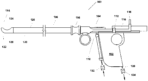
[0048] FIG. 1 is a simplified side view of one example variant of a
resectoscope 100
incorporating the present invention. As shown in FIG. 1, this example
resectoscope 100 is, in
summary overview, made up of a partially hollowed out handle 102 and a control
mechanism
104, both of which will be described in greater detail below, a shaft 106
connected at its
proximal end to the handle 102, a port through which a telescope or other
viewing apparatus,
which may or may not involve use of fiber optic technology can be inserted
(not shown), and a
finger grip 108 on the shaft 106. The resectoscope 100 further includes a
trolley mechanism 110
that facilitates movement of the telescope or other viewing apparatus that is
contained within the
shaft, a stop 112 that acts as a handle to allow manipulation of the trolley
110 and also limits
movement of the trolley 110 mechanism towards the distal end 114 of the shaft,
a power
connector 116, a fluid inlet 118 and a vacuum port/fluid outlet 120. As can be
seen in FIG. 1, the
tip 122 of the shaft 106 at the distal end 114 is formed so as to have a
physically closed blunt
shape to dramatically reduce, if not eliminate, puncture or laceration risk.
In addition, the shaft
includes an opening or resection port 124 located on a side surface 126 of the
shaft 106 near the
distal tip 122.
[0049] Depending upon the particular implementation and intended use, the
length of the
shaft 106 can be anywhere from relatively short, for example (i.e. a few
centimeters or less)
where a shallow body cavity is involved, relatively long (i.e. in excess of 40
centimeters) where
long cavities like the bowel or intestines are the intended application, or
lengths in between, for
applications such as intrauterine resection. Similarly, depending upon the
particular
implementation and intended use, the shaft can be rigid along its entire
length, flexible along a
portion of its length, or configured for flexure at only certain specified
locations.
[0050] Still further, in some implementation variants, the shaft can be made
up of two or
more detachably interlocking segments 128, 130 for purposes of modularization.
[0051] The fluid inlet 118 is configured for connection to an adjustable
pressure fluid
infusion line via a stopcock 132 or other appropriate valve and, in most
cases, also having a
parallel free flow one way fluid reservoir to accommodate vacuum boosting.
9
CA 02649095 2008-10-10
WO 2007/121109 PCT/US2007/066055
[0052] The vacuum port/fluid outlet 120 is configured for attachment to, for
example, a
foot-pedal actuated boosted vacuum source via a stopcock 134 or other
appropriate valve.
[0053] FIG. 2 is a simplified view of an example shaft 106 component suitable
for use as
part of the resectoscope of FIG. I and further includes cross sectional slices
2A through 2D taken
at the points indicated to illustrate various features of this example
implementation. Moreover,
and advantageously, in some variants, the shaft 106 itself is separable from
the non-handle
components that make up the body of the resectoscope 100, for example as in
FIG. 2, and in
some cases, made up of two or more discrete modules. Some shaft variants are
also disposable,
whereas others can be sterilized for reuse. In general, the shaft 106 is
formed as a hollowed
multi-channel shaft or cannula, the details of which are explained with
reference to the cross
sectional slices of the shaft shown in FIGS. 2A through 2D taken at 2A-2A, 2B-
2B, 2C-2C
and 2D-2D. However, it should be understood that the cross sectional shapes
are simply for
illustrative purposes, the particular cross sectional shape being more
relevant to the particular
application for which the resectoscope will be used than to the invention.
[0054] Referring now to the cross sectional slice of FIG. 2A taken at 2A-2A,
this
variant of resectoscope shaft 106 incorporates a channel or portal for a
telescope or other
viewing apparatus 202, a fluid infusion channe1204 through which fluid can be
infused from the
proximal end towards the distal end, a capture or return channel 206 through
which fluid and
resected tissue morsels are conveyed from the distal to the proximal end, one
or more optional
auxiliary channels 208 or other arrangement of appropriate size extending to
at least the working
area and, if desired, to the distal tip itself to allow for, for example:
further connections to be
made; objects, for example, catheters, drains, ureteral stents or tubal
occlusion devices to be
inserted; to provide a brief flow of liquid nitrogen or other cryo-cautery
fluid to accomplish
hemostasis; allow for passage of an auxiliary cauterization element to perform
conventional
cautery; or allow for a stylet to be passed to the vicinity of the working
area or beyond the distal
tip. In addition, in this variant, the shaft 106 is of a different modular
configuration from that of
FIG. 1, with the grip 108 of this variant being used as a coupler to couple a
main portion 200A of
the shaft from a proximal portion 200B. The shaft 106 also optionally includes
a pair of guides
210 that limit the cutting member in this variant to longitudinal movement. In
this illustrated
CA 02649095 2008-10-10
WO 2007/121109 PCT/US2007/066055
variant, the guides are configured for when an electrosurgical wire loop is
illustratively used (not
shown in this FIG.).
10055] The cross sectional slice of FIG. 2B taken at 2B-2B is similar to that
of cross
section 2A-2A except that the guides 210 are not present because, in this
variant, they are not
needed along the entire length of the shaft 106.
[0056] The cross sectional slice of FIG. 2C taken at 2C-2C is also similar to
that of
cross section 2A-2A except, because this section is beyond the entry point for
the infusion fluid
the fluid infusion channel 204 is no longer present. In addition, this portion
of the shaft will be
situated above the handle 102 so, as will be described later, the return
channel 206 is open 212 to
the handle 102 for reasons that will become evident below. It also contains a
pair of handle
guides 214 that allow for attachment/detachment of the handle or shaft
relative to the other, and
guides 210 (similar to that shown in FIG. 2A) for a proximal portion of the
wire loop apparatus.
[00571 The cross section al slice of FIG. 2D taken at 2D-2D is similar to the
lower
portion of FIG. 2C with respect to the handle guides 214 and also includes a
handle cap portion
216 and a trolley guide 218 to receive the trolley 110 mechanism.
[0058] FIG. 3 illustrates, in simplified form, the trolley 110 mechanism for
the variant of
FIG. 1. As shown, the trolley mechanism may be of solid (FIG. 3A) or hollow
(FIG. 3B) cross
sectional configuration (or some combination thereof) and includes a companion
insert port 302
for the port 202 referred to in FIG. 1 through which a telescope or other
viewing apparatus can
be inserted, an exit port 304 that will guide and align the telescope or
viewing apparatus for
proper engagement with the channel or port 202 of the shaft 106, a pair of
rails 306 on each side
that conforms in shape to the trolley guide 218 of the shaft 106, for example,
the sliding "v-
groove" arrangement shown, a pair of stops/handles 310 that can be used to
move the trolley I 10
through its range of motion along the longitudinal axis of the shaft 106 and
act as a forward-
movement limiting element, and a constraining arrangement 312a, 312b that will
clamp, affix or
otherwise constrain the telescope or viewing apparatus (once fully inserted)
in a particular
orientation.
11
CA 02649095 2008-10-10
WO 2007/121109 PCT/US2007/066055
100591 As shown, the telescope or viewing apparatus can be anchored to the
trolley by a
grooved nipple-pin pit 312 at the proximal end. Two grooved pits 312a, 312b
are seen on the
proximal end, one above, and one below the channel 314 between the two ports
302, 304. These
grooved pits 312a, 312b accept the anchoring pin found on conventional scopes,
and through
provision of two such pits 312a, 312b, allow for rotation of the scope through
180 to allow for
viewing in either a downward or upward inclination when, for example, angled
scopes of, for
example, common angles such as 12 , 30 , or 45 are used. This feature aids in
oblique optical
targeting through the opening or resection port 124. With and angled scope and
the scope
attachment pin in the inferior pit 312a, the scope is thus directed to an
upward viewing angle
providing, with those variants, a direct line-of-sight through resection port
124 to the target area
for direct optical targeting.
[0060] Advantageously, by affixing the telescope or viewing apparatus to the
trolley 110
movement of the trolley 110 along the guides 218 will effect equal movement of
the portion of
the telescope or viewing apparatus in the shaft 106 towards or away from the
distal end 114. In
this manner, the trolley 110 provides an external visual indication of the
location of the end of
the telescope or viewing apparatus.
[0061] At this point it should be noted that the telescope or viewing
apparatus per se is
conventional in the sense that numerous types are already well known and
regularly used in
performing various types of surgery. The particular type of apparatus, be it a
telescope, fiber
optic or other device, is conceptually unimportant for an understanding of the
invention so long
as an appropriate one is selected in terms of size, bevel angle if applicable
(i.e. 0 , 12 , 30 , etc.),
field of view, type, etc. so as to be compatible with the concepts described
herein. Moreover, as
will be discussed below, in some cases, two or more different telescopes or
viewing apparatus
may be used, for example, to change among different conventional bevel angles.
Thus, except as
is specifically pertinent to an understanding of the invention, particular
details regarding the
telescope or viewing apparatus are omitted for both brevity and simplicity.
[0062] FIG. 4 illustrates, in simplified form, the example control mechanism
104 for the
instrument of FIG. 1. As illustrated, the control mechanism 104 includes a
movement ring 402
that is used to maneuver a cutting member (described below) through its range
of motion via a
12
CA 02649095 2008-10-10
WO 2007/121109 PCT/US2007/066055
connection 404 thereto. In addition, the control mechanism 104 can be arranged
to cooperate
with or constrain the trolley mechansim as necessary to effect the desired
operation. In addition,
in this particular variant, the control mechanism optionally includes a power
connection 116
through which power can be supplied to a cauterization element which may or
may not be the
cutting member.
[0063] FIG. 5 illustrates, in simplified form, the handle 102 for the
resectoscope variant
of FIG. 1. As shown, the handle 102 includes an optional finger hole 502 that
facilitates
manipulation of the resectoscope 100 during insertion or while in use. In
addition, the handle
102 has an internal cavity 504 of sufficient size to enable capture of the
resected tissue entering
via the return channel 206 through an opening 506 in the top of the handle 102
while allowing
for the unobstructed, filtered exit of the return fluid via the vacuum
portlfluid outlet 120. In
addition, as noted above, the handle 102 also has a pair of rails 508 on each
side of the upper
portion that conforms in shape to the handle guides 214 shown in the cross
sections of FIG. 2C
and FIG. 2D.
[0064] As shown, the handle 102 for this variant also optionally includes a
fluid inlet
hose guide 510 that keeps the fluid inlet hose out of the way.
[0065] FIG. 6 illustrates, in simplified form, a top view of a portion 600 of
the distal end
114 of the shaft 106 near the tip 602. Note that, for clarity of presentation,
internal details have
been omitted from this view. As can again be seen from this view, the distal
end 114 is formed
so as to have a physically closed blunt shape 122. In addition, as shown in
more detail in FIG. 6,
the shaft 106 includes an opening or resection port 124 of a geometrically
closed shape that is
located on a surface 126 of the shaft 106 near the distal tip 602. In
particular, as illustrated in
this variant, the opening or resection port 124 is located immediately above,
and defines a
working area for the underlying cutting member and has a longitudinal length
~that is typically
equal to or slightly less than the range of movement of the cutting member, in
this case, between
its proximal and distal limits.
100661 It should be understood that the size, shape and exact location of the
opening or
resection port 124 may vary depending upon the particular implementation or
intended use.
Similarly, a sliding shim or cover plate can be incorporated, for example, to
provide size or
13
CA 02649095 2008-10-10
WO 2007/121109 PCT/US2007/066055
shape adjustability, and even, in some cases, to close off the opening or
resection port entirely,
for example, to facilitate insertion into body cavities where the opening or
resection port in and
of itself could cause trauma during insertion or withdrawal. Depending upon
the particular
implementation, in some variants, the movement of the shim or cover plate can
be tied to that of
the telescope so that when the scope is fully extended the shim or cover plate
will close off the
opening or resection port 124 entirely or at least cover the cutting member
itself. Optionally, for
some applications, it may be desirable to ensure that a seal is formed between
the periphery of
the opening or resection port and the tissue about the tissue that would be
resected, for example,
when used in an application such as removal of tissue from a sinus or the
trachea which are both
fairly rigid. In such cases, this desire can be accommodated in any of
multiple ways. One
example approach can involve making some portion of the shaft about the
periphery of the
opening or resection port slightly flexible so that it can conform to the
abutting tissue. Another
example approach can involve use of a deformable "gasket" material 606, such
as a closed cell
foam, putty, gel or other appropriate non-toxic deformable material. Depending
upon the
particular use, such deformable material can be part of the shaft itself or
provided separately, that
latter being advantageous for those cases where a surgeon may wish to have the
option to do so
up until about the time that insertion of the shaft begins.
[0067] In addition, and advantageously, some variants may be implemented in a
kit that
includes only certain components, for example, a shaft by itself, a shaft and
handle, a shaft and
associated cutting member, different length shafts, or multiple shafts of
different lengths, cross
sectional shapes and sizes, flexibility, curvature, or that each have openings
or resection ports of
a different size and/or shape so as to better match or accommodate the size
and shape of the
tissue to be resected and assist in confining the resected tissue within the
shaft so that, it can be
conveyed along the shaft 106 for capture in the handle 102.
100681 Still further, in some cases it may be desirable to have a more
modularized shaft,
in that, the shaft itself would be made up two or more separable pieces, an
extension section 608
representing the bulk of the shaft length, and a shaft module 610 containing
some or all of the
shaft components described herein as being located between the distal end and
a location to the
proximal side of the working area. In this manner, a particular shaft module
610 could be used,
14
CA 02649095 2008-10-10
WO 2007/121109 PCT/US2007/066055
for example, with different length or flexibility shaft extensions 608 or
different configuration
modules 610 could be used with a common shaft extension 608 in a mix-and-match
manner as
needed or desired. In addition, this approach provides further advantages in
terms of the ability
to be produced, production cost and configuration flexibility.
[0069] As illustrated by way of example, the opening or resection port 124 is
of ovoid
shape and the shaft is of a length and cross section appropriate for resection
of tissue within the
uterus. Advantageously, and irrespective of the dimensions of the shaft 106 or
particular shape
of the opening or resection port 124, the opening or resection port 124
defines the only zone for
interaction between the cutting member and the tissue to be resected while
acting as a passive
port for removal of fluid from the tissue area or body cavity.
[00701 Moreover, depending upon the phase of resectoscope use, the opening or
resection port 124 will create the path for regulation of the "inflation", if
any, of the cavity where
the resection will occur by acting as the outlet (from the perspective of the
body cavity) for
excess inflation fluid and/or will serve as a passive functional portal for
fluid and/or tissue.
Optionally, one or more small pore(s) 604 can be provided, that couple to the
return channel 206,
to provide an additional or alternative route for fluid external to the shaft
to pass into the return
channel 206, for example, during an inflation phase where it may be difficult
or undesirable to do
so through the opening or resection port. Depending upon the particular
implementation, such
pores can be sized small enough so that they do not de-inflate the cavity
during working or,
alternatively, can be selectably blocked for example, by the slidable shim or
some other means,
so as to only be open at a particular time, for example, only when, as will be
described below,
the telescope or viewing apparatus is in the extreme distal position or when a
switch is in a
position where infusion fluid is routed out of the shaft for purposes of
inflation or irrigation.
[00711 FIG. 7 illustrates, in simplified form, an external end view of a blunt
distal end
122 portion 700 of the shaft 106. As can be seen from this view, at least a
portion 702 of the tip
602 is optically transparent so as to act like a window and is aligned with
the channel or portal
202 for the telescope or viewing apparatus so as to provide for forward
viewing through the
distal end portion 702 via the telescope or viewing apparatus under the
appropriate conditions.
CA 02649095 2008-10-10
WO 2007/121109 PCT/US2007/066055
[0072] Depending upon the particular implementation, the optically transparent
portion
702 can simply be a hole or it can be a physical element. In the case of a
physical element, it can
be an integral part of the shaft, for example, if at least that portion of the
shaft is, or is made,
transparent, or it can be a separately formed and inserted element, like a
membrane, a piece of
plastic or glass (whether flat or lens shaped) or other optically transparent
material. Moreover, in
some cases, this portion 702 or window area can be, in whole or part, a lens
that can work in
conjunction with the telescope or viewing apparatus to provide a different
field of view than
would be provided by the telescope or viewing apparatus alone. For example,
the window area
702 could be an element that is flat or convex on the external side of the
distal end 122, but flat
and beveled at a specified angle on the internal side (i.e. inside the shaft)
so that a comparably
opposite beveled end of the telescope or viewing apparatus can be butted
against it to allow for
straight-ahead, angled or wide-field viewing (as determined by the shape of
the external side)
when the trolley 110 is in the extreme forward position.
[0073] Alternatively, by appropriate sizing, the window area 702 can be a hole
that, for
purposes of insertion, can be filled or blocked by the end of the telescope or
viewing apparatus
itself through maintaining it in a suitably spaced extreme forward position.
[0074] Optionally, the auxiliary channel 208 can be carried forward to the
distal end 122,
such as is shown. Additionally or alternatively, as shown in this variant, the
portion 702 is
ringed with an electrical conductor 704 that can be selectively connected to a
power source to
directly effect cauterization while viewing the tissue to be cauterized
through the distal end 122.
[0075] FIG. 8 illustrates, in simplified form, a longitudinal cross section of
the portion
800 of a shaft 106 of one example variant. In FIG. 8, the longitudinal fluid
infusion channel 204
and return channel 206 can be readily seen as can the blunt nature of the
distal end 122.
Although a single fluid infusion channel 204 is illustrated for simplicity, in
some variants, two or
more separate inflow channels with combined or associated individual
respective controls could
alternatively be used. In addition, a telescope 802 having a 30 bevel resides
in the telescope
channel 202, at a retracted location, and is aligned with the telescope end
portal 804. In this
variant, the telescope end portal 804 is capped with a lens 806 that is
slightly convex on its outer
16
CA 02649095 2008-10-10
WO 2007/121109 PCT/US2007/066055
surface 808 and beveled 810 at a mirror image 30 bevel so that, at the
extreme extended
position, the end of the beveled telescope 802 and the internal surface if the
lens 806 will mate.
[0076] The opening or resection port 124 described in connection with FIG. 5
is also
clearly visible.
[0077] A cutting member 812 also resides within the shaft 106. Depending upon
the
particular intended use and implementation, the cutting member 812 can be a
wire loop (such as
shown), a sharpened blade, a rotary cutting implement, a micro-vibrational or
harmonic or
shutter-type cutting device, or other cutting implement (each with or without
cauterization
capability). Alternatively or additionally, the particular cutting member 812
can be configured
for movement in an arcuate, axial, rotational, diagonal, transverse,
reciprocating or other manner
to effect cutting in a direction other than through pure longitudinal
movement.
j00781 In yet other variants, the cutting member 812 can be configured so that
its
orientation within the shaft 106 is changeable to provide for cutting at two
or more different
angles. In such variants, an auxiliary or reconfigurable telescope or viewing
apparatus may be
necessary or desirable to allow for angulation.
[0079] Depending upon the particular implementation, the cutting member can be
supplied with, and integral to, the instrument or shaft as packaged or it can
be of a separately
provided snap-in and/or snap-out design.
[0080] Irrespective of the particular cutting member 812 used, its mode of
integration
with the shaft, and its direction of movement or orientation, the cutting
portion of the cutting
member 812 is wholly constrained within the shaft. Moreover, ideally the
cutting member
conforms, through at least a part of its range of motion, to either an inner
or outer surface 126 of
the shaft 106 and/or an imaginary surface of the opening or resection port
that would be formed
if the shaft contour was continuous across the region of the opening or
resection port. Thus, if
the shaft near the opening or resection port is arched, because the shaft is
circular or oval in cross
section, the cutting member will typically have a similar or lesser arch. If
the shaft near the
opening or resection port is flat or near flat, the cutting member can be
similarly contoured in
shape.
17
CA 02649095 2008-10-10
WO 2007/121109 PCT/US2007/066055
[0081] However, if a type of cutting member 812 other than a wire or blade is
used, for
example a micro-vibrational or harmonic cutter (i.e. a harmonically vibrated
blade), scissor or
shutter-type mechanism, the cutting member may not follow the contour. This is
not a problem,
as following the contour is not critical to implementation of the invention
but highly desirable for
some implementations or intended uses. Rather, the important aspect is that
the cutting member
812 remains within the working area, whether or not the cutting member 812 is
a blade, loop,
scissor, shutter, harmonic or other type of cutting mechanism.
[0082] For purposes of illustration, as shown in the variant of FIG. 8, the
cutting member
812 is a wire loop that is moved by longitudinal movement of the forefinger
loop 402 on the
control mechanism 104 near the handle 102 and is constrained against non-
longitudinal
movement by the guides 210 of FIG. 2. As should be understood from FIG. 8, the
cutting
member has a height "h" that keeps it wholly within the shaft 106 through its
range of motion
from a fully distal position 814 to a fully proximal position 816 and, in this
variant, is curved in
an arc of approximately a 4 mm radius so that the telescope or viewing
apparatus 802 can pass
through and underneath the cutting loop with minimal to no contact therewith.
In this regard, it
should be noted that, at either or both extremes 814, 816, the cutting member
812 may or may
not be visible through the opening or resection port 124. As a result, the
cutting member 812 can
"guarded" by the outer surface 126 of the shaft 106, thereby preventing it
from causing
undesirable laceration or puncture of tissue during insertion or at any point
in the procedure
where cutting is not warranted or desired. Still further, through this
configuration, the outer
surface 126 of the shaft 1061imits the depth of cut, again greatly reducing
the risk of undesirable
laceration or perforation.
[0083] In addition, in this variant, the shaft 106 includes one or more fluid
export pores
818 and a fluid routing switch 820 with the fluid export pore(s) 818 being
under the fluid routing
switch 820 and beyond the termination point 822 of the fluid infusion
channe1204. The fluid
export pore(s) 818 can be of any geometric shape(s) or number.
[0084] As shown in FIG. 8, the fluid routing switch 820 is a binary position
pivoting
switch that is sized and shaped so that, in one position (the infusion
position), the switch will
direct a substantial portion, if not all, of the fluid passing through the
fluid infusion channel 204,
18
CA 02649095 2008-10-10
WO 2007/121109 PCT/US2007/066055
from the proximal end towards the distal end, and out through the fluid export
pore(s) 818, for
example, in the case of a device for intrauterine resection, to inflate the
uterus. In the other
position (the circulation position), the switch 820 will substantially, if not
completely, inhibit
fluid flow out of the pore(s) 818 and instead, direct fluid into the return
channel 206 in the
vicinity of the opening or resection port 124. As illustrated in this variant,
the switch 820 is
normally biased into the circulation position. Advantageously, this allows the
end 824 of the
telescope or viewing member 802 to be used to actuate the switch 820 and
divert the infusion
fluid out the pore(s) 818.
[0085] Optionally, in some variants, the internal surface 826 of the switch
820, about the
switch 820, and/or surfaces 828 facing the return channe1206 (whether or not
there is a switch)
can be specifically inclined and polished or otherwise made reflective so as
to act like a mirror
and enable a further or additional range of view through the opening or
resection port than could
potentially be available using only the telescope or viewing apparatus (i.e.
provide accessory
optical capability for, for example, tissue targeting or cauterization).
[0086] Alternatively, in other variants, the fluid routing switch within the
shaft 106 can
be dispensed with entirely.
[00871 FIG. 9 illustrates, in simplified form, a"switchless" (with respect to
the shaft)
variant 900 by employing at least two separate fluid inflow channels 902, 904
routed to
effectively create the two flow patterns obtained by the switch. In other
words, at least one of
the fluid inflow channels 902 is connected to the export pore(s) (analogous to
one of the binary
switch positions) and another of the fluid inflow channels 904 is configured
to cause fluid to
remain within the shaft and flow into and through the working area (analogous
to the other
binary switch positions). Such a switchless variant has the advantage that,
with respect to the
shaft itself, fluid routing becomes a passive function, formation of the shaft
becomes simpler and
a moving part is eliminated. As a result, it is easier to create an inclined
polished or mirror area
within the shaft as described above.
[0088] Of course, such a "switchless" approach would still require some form
of
selection element which could be located, for example, on or adjacent to the
handle, the control
mechanism, or wholly external to the resectoscope itself. In addition, this
alternative approach
19
CA 02649095 2008-10-10
WO 2007/121109 PCT/US2007/066055
enables specific control of the flows so that a dual or combination flow can
optionally be
achieved (i.e. an intermediate point between full output through the infusion
port and full
circulation flow at a desired flow rate).
[0089] FIG. 10 illustrates, in simplified form, an aiternative shaft portion
1000 variant
that is similar to that of FIG. 8 except a portion of the distal end 1002 is
itself transparent, so no
separate membrane, lens or other cap is required, there is specifically one
circular infusion pore
1004, and the opening or resection port 1006 is rectangular. For completeness,
FIG. 1 0A is a
cross sectional slice of the shaft taken at 10A-10A (through the pivot point
of the switch), FIG.
l OB is a cross sectional slice of the shaft taken at 10B-1 OB (through the
infusion pore), and
FIG. l OC is a cross sectional slice of the shaft taken at I OC-10C (through a
portion of the
opening near the proximal working area limit for the cutting member). In
addition FIG. l OD
illustrates a view of the upper surface of the shaft 1000 taken from above the
opening or
resection port 1006.
[0090] FIG. I 1 illustrates, in simplified form, a longitudinal, cross
sectional side-view of
a further alternative shaft portion 1100 variant that uses a sliding reed as
the fluid routing switch.
In addition, in this particular variant the reed also optionally defines and
segregates the fluid
infusion channel 1102 from the return channel 1104. For ease of understanding
this variant,
example cross sectional slices, taken at I 1A-11A through 11G-11G, are also
provided in FIG.
11 A through I 1 G. For further simplicity, details such as the cutting
member, its constraint and
movement control, as well as any optional additional auxiliary channel(s) have
been omitted but
it should be understood that any or all of them could also be present.
[0091] In this variant, two fluid export pores 1106, 1108 are provided and are
best
illustrated in FIG. 11 E. As illustrated in the various cross sectional
slices, the fluid infusion
channel 1102 splits into a pair of smaller channels I 110, 1112 as it
approaches the distal end
1114 in order to reach the two fluid export pores 1106, 1108. Fluid infusion
into the body cavity
occurs via fluid flow from the fluid infusion channel 1102, through the two
channels I 110, 1112
to the fluid export pores 1106, 1108. Again, it should be understood that each
individual fluid
export pore 1106, 1108 could be readily implemented as two or more individual
pores.
Alternatively, fluid circulation occurs via a fluid circulation channel 1116
that is located between
CA 02649095 2008-10-10
WO 2007/121109 PCT/US2007/066055
the two smaller infusion channels 1110, 1112 and is shaped to direct the fluid
into the vicinity of
the working area 1120 for return down the return channel 1104.
[0092] Of course, it should be understood that, in other implementation
variants, the fluid
circulation channel could be split up into the two channels and the fluid
infusion channels could
be the central channel, the only differences being a potential change in
relative sizing of the
channels, the export pore(s) would be centrally located and there would be two
portions used to
direct the flow into the working area to account for the split channels.
[0093] The above two variants reflect a desire for longitudinal symmetry about
the
vertical. However, it should be understood that symmetry is not required and,
in some variants,
asymmetry may provide advantages for particular uses or applications, for
example, to cause a
turbulent or specific pattern of flow near the distal tip or working area. In
such cases, some form
of side-by-side arrangement or other arrangement would likely be used.
[0094] As noted above, one example variant mechanism for switching of fluid
flow is
comprised of a blade-like control reed which spans the hollowed shaft 106
transversely. The
control reed also spans from proximal to distal within the shaft along the
greater length of the
instrument, and is held in place by small lateral grooves 1118 along the inner
wall of the shaft.
The control reed is capable of sliding longitudinally distally and proximally
along the shaft.
FIG. 12 illustrates, in simplified form, an example sliding control reed 1200
configured for use in
conjunction with the shaft portion of FIG. 11. As illustrated, when the
control reed 1200 is
within the shaft 106, it will create an eccentric partition axially along said
shaft such that the
inflow channel 11021ies beneath, and the larger outflow/return channel 1104
lies above the
control reed 1200 along the length of the shaft 106. Depending upon the
particular
implementation, the control reed 1200 may be flat, such as shown, or may be
curved in some
manner to, for example, increase the diameter of the overlying return channel
1104, or contribute
to the overall stiffness of the instrument 100. Similarly, depending upon the
particular design,
control reed material or intended use for the instrument, the control reed
1200 itself can be
reinforced, for example, by fins, ribs, or a differing thickness across its
width or along its length
to increase stiffness or create a specific flexure pattern.
21
CA 02649095 2008-10-10
WO 2007/121109 PCT/US2007/066055
[0095] The control reed 1200 accomplishes its switching task through use of
holes 1202,
1204, 1206 located near its distal end 1208. The holes 1202, 1204, 1206 are
placed, sized and
shaped to effect the desired fluid flow control based upon the position of the
control reed 1200 at
a particular point in time. As illustrated, a center hole 1202 is located near
the distal end 1208 of
the control reed 1200 and provides a flow path up and into the working area
1120 when the
center hole 1202 is aligned, in whole or part, with the fluid circulation
channel 1116. In
addition, the center hole 1202 is located relative to the end of the reed 1200
so that the control
reed 1200 can be placed in a position where an element 1122 blocks all flow
through that hole
1202. As illustrated, that position is an extreme forward (i.e. distal)
position, but could
alternatively be a rearward position or some position in between.
10096] In addition, the control reed 1200 contains a pair of lateral holes
1204, 1206 to
either side that can be aligned with the infusion channels 1110, 1112 to
direct fluid flow from the
main fluid infusion channel 1102 to the export pore(s) 1106, 1108. As with the
center hole 1202,
the lateral holes 1204, 1206 are located on the control reed 1200 so that they
can, based upon the
position of the control reed 1200, provide a fluid flow path, in this case
between the infusion
channel 1102 and export pore(s) 1106, 1108 (in this variant, in the vicinity
of the shaft 106 at the
cross section taken at C-C), or to cut off all flow to the export pore(s)
1106, 1108. As
illustrated, in cross section B-B, in this variant, solid protrusions 1124,
1126 above the control
reed groove 1118 are positioned act so as to block flow through the lateral
holes 1204, 1206
when the control reed 1200 is at its most proximal operational setting. Of
course, as with the
central hole 1202, this lateral hole 1204, 1206 blockage could also occur at a
forward position or
some position in between.
[0097] For purposes of understanding, in the variant of FIG. 11 and FIG 12,
the distal
end 1208 of the control reed 1200 and distal end slot 1128 are arranged so
that the control reed
1200 can move a distance essentially equal to, or slightly more than, the
diameter of the central
hole 1202. With the control reed 1200 in its proximal position, the lateral
holes 1204, 1206 are
blocked and the central hole 1202 is open to accomplish an internalized fluid
circuit. Thus, with
the control reed 1200 in the proximal position, fluid will be passively
directed from the inflow
channel 1102 into the center channel 1116, creating a flow circuit within the
instrument's shaft
22
CA 02649095 2008-10-10
WO 2007/121109 PCT/US2007/066055
106. In the variant illustrated in FIG. 11, when assembled and in use, fluid
flows in the distal
direction from the inflow channel 1202 under the control reed 1200, and then
upward via a
curved surface 1130 within the center channel 1116 proceeding out through the
center hole 1202
in the overlying control reed 1200. The fluid then flows or is drawn
proximally into the
outflow/return channel 1104 with or without vacuum assist. This is the typical
control reed 1200
position for resection.
[0098] In its distal position, the lateral holes 1204, 1206 of the control
reed 1200 are
open while the central hole 1202 is blocked by the distal end slot 1128 so
that fluid will be
routed out the export pore(s) 1106, 1108. Thus, as the control reed 1200 is
moved to block the
center hole 1202, the nearest 1206 of two lateral pores 1204, 1206, one on
each side, will move
from under the blocking surface 1124, 1126 and thus allow diversion of flow up
and into the
lateral channels 1110, 1112 whose ultimate path leads to the export pore(s)
1106, 1108.
[0099] Depending upon the particular implementation, movement of the control
reed
1200 can be integral with movement of the telescope or viewing apparatus 802
or not. As
shown, the sliding control reed 1200, when within the shaft 106, is activated
from the proximal
end of the instrument by an open linkage mechanism (not shown) to the trolley
110, and is
automatically activated with full advancement of the scope/trolley in unison.
The control reed
1200 is pushed forward by the trolley I 10, or independently by a finger
leaving the trolley 110 in
its home/resection position. This allows, for example, uninterrupted re-
inflation of the tissue
cavity while keeping the telescope or viewing apparatus 802 in a diagnostic or
targeting mode or
during active resection, as desired. Alternatively, movement of the control
reed 1200 can be
made independent of the other components, for example, the telescope or
viewing apparatus 802.
This can be accomplished in a straightforward manner by providing an element
at or near the
proximal end of the shaft 106 that is connected to the control reed 1200 and
thus, its movement
will move the control reed 1200.
[01001 Advantageously, some variants using the control reed 1200 arrangement
for fluid
switching will thus, have the ability to provide variable flow, not readily
obtainable via internal
switching alone with the preceding mechanical switch by: a) design, through
placement, sizing
and shape of the holes themselves so that, for example, there is an inverse
linear ratio of
23
CA 02649095 2008-10-10
WO 2007/121109 PCT/US2007/066055
diversion between completely internalized and externalized flow as the control
reed is moved, b)
movement of the control reed into any of an infinite number of intermediate
positions
irrespective of the particular flow relationship provided by the hole
placement, shape and sizing,
or c) both. As a result, finely controlled flow splitting between internalized
and externalized
routes can be achieved, for example, in order to maintain slow balanced cavity
infusion while
concurrently performing resection with vacuum assisted evacuation and/or
tissue export.
[0101] In general, the approach to controlling the fluid flow that is used to
convey the
resected tissue from the working area towards the handle 102 will likely vary
depending upon
the particular implementation and intended use. For example, in some cases,
the control can be
fully manual. In other cases it can be a result of movement of another
element, for example the
telecsope or viewing apparatus of FIG. 8 or the movement of the cutting member
itself. In yet
other cases, the control can result from a combination of manual adjustment
based upon
mechanical, electric or electronic feedback. In yet other cases, fully
automated control is
possible through use of, for example, electrically activated fluid gates,
electromagnetic,
mechanical, hydraulic, or other switches. In some variants that use a distally
placed switch that
is not directly manipulable via an external control, the switch can be
designed to be externally
controlled through fluid flow itself in conjunction with vacuum, or through
only positive
pressure fluid flow (i.e. without the use of vacuum at all). In addition, and
advantageously, when
in the fluid circulation mode or configuration (i.e. fluid will not flow
generally out the export
pore(s)) flow rates of 100 cc/min or more can be used and, with vacuum boost,
instantaneous
flow rates within the shaft can exceed 4000 cc/min.
[0102] FIG. 13 illustrates, in simplified form, a cutaway view of a shaft
portion 1300 for
a resectoscope, that is similar to the shaft portion 800 of the resectoscope
of FIG. 8 except that
the distal end has a window area 702 that is made up of a transparent membrane
1302 instead of
a lens 806. As illustrated the shaft portion 1300 is configured as it would
look during insertion.
In this configuration, the telescope or viewing apparatus 802 is fully
extended (i.e. the trolley
110 has been moved to its forward limit position so that viewing out the
window area 702 of the
shaft 106 is possible using the telescope or viewing apparatus 802. The
cutting member 812 is in
its "home" position which, although illustrated as being at the distal limit
1306 (due to the
24
CA 02649095 2008-10-10
WO 2007/121109 PCT/US2007/066055
surgical convention of preferably only cutting in the distal to proximal
direction due to the
puncture risk inherent with conventional devices) it could alternatively be at
a proximal limit or
somewhere in between. In the fully extended position, the telescope or viewing
apparatus 802
impinges against the upper portion 1304 of the switch 820 thereby opening the
fluid export
pore(s) 818 to the fluid input channel 204 to allow fluid to pass out of the
shaft 106 while
preventing infusion fluid from directly entering the return channel 206 from
inside the shaft 106.
[0103] Advantageously, it should be recognized that variants configured in
this manner
can be used in circumstances where organ "inflation" may or may not be
necessary. For uses
where inflation is not necessary, this is accomplished by limiting trolley 110
movement or
clamping the telescope or viewing apparatus 802 such that, when the trolley
110 is in the fully
extended position, the telescope or viewing apparatus 802 will fall just short
of the upper portion
1304 and thus avoid actuating the switch 820. Although, by doing so, this
could result in some
minor reduction or distortion in the forward field of view due to the gap
between the end of the
telescope or viewing apparatus 802 and the window area 702, any such reduction
or distortion
will likely occur, if at all, at the periphery of the field of view so the
reduction will have minimal
to no impact in most cases.
[0104] FIG. 14 illustrates, in simplified form, the portion of the
resectoscope of FIG. 13
as it would look during the "working" or resection process. As shown, in this
configuration, the
telescope or viewing apparatus 802 is at or near its fully retracted position
and, as a result, the
switch 820 will block passage of fluid to the fluid export pore(s) 818 and
cause the infusion fluid
to circulate up into the return channel 206 where the applied vacuum will
cause it to traverse
towards the proximal end of the shaft 106. Moreover, the placement of the
telescope or viewing
apparatus 802 allows for unobstructed view of the opening or resection port ]
24 as the cutting
member 812 is moved throughout its range to perform unidirectional or bi-
directional resection.
In addition, since they are independently maneuverable, the end of the
telescope or viewing
apparatus 802 can be used to "clear" or dislodge any resected tissue pieces
that may get caught
on the cutting member 812 by simply moving the two with respect to each other
so that the
telescope or viewing apparatus 802 passes by the cutting member 812. Still
further, should the
end of the telescope or viewing apparatus 802 become partially or totally
obstructed by tissue or
CA 02649095 2008-10-10
WO 2007/121109 PCT/US2007/066055
clouded by turbid fluid from the resection (if any), the telescope or viewing
apparatus 802 can be
moved forward of the cutting member 812 and into the clean flow of infusion
fluid, thereby
cleaning the end without the need to withdraw the shaft 106 of the
resectoscope 100 or the
telescope or viewing apparatus 802 from the body cavity.
[0105] Alternatively or additionally, in instances where there are one or more
optional
auxiliary channels 208 present and a piece of tissue or debris becomes stuck
on the cutting
member 812 or telescope/viewing apparatus 802, a stylet can be passed through
an auxiliary
channel 208, in order to bump the cutting member 812 or piece of stuck tissue
or debris and
dislodge it from the cutting member 812 or telescope/viewing apparatus 802.
Alternatively, a
home position "groove" or recessed area, configured to closely conform to and
accept the cutting
member 812, can be used to aid clearing of stuck tissue or debris from the
cutting member 812
through return to this home position.
[0106] FIG. 15 illustrates, in simplified form, the portion 1300 of the
resectoscope of
FIG. 13 wherein the telescope or viewing apparatus 802 has been moved ahead of
the cutting
member 812 as described above.
[0107] FIG. 16 illustrates, in simplified form, the portion 1300 of the
resectoscope of
FIG. 13 in an optional third configuration. In this configuration, the
resectoscope is optionally
designed to lock the cutting member 812 at a position within the working area -
illustratively
shown in this example for simplicity at the midpoint of the range of motion.
In this position, the
cutting member 812 can be connected to a power source to effect cauterization
or, for example in
the case of a cutting loop as shown in FIGS. 8 and 16, drag cutting of tissue
(i.e. along a plane
formed by the cutting member 812 or angled from that plane within an angle 0
as necessary.
Again, it is worth noting that the shaft 106 and/or periphery 1604 of the
opening or resection port
124 will act to limit the depth of cut and reduce the risk of unwanted
extraneous lacerations.
[0108] Of course, in some variants, the cutting member 812 can optionally be
configured
to cauterize throughout all, or in other variants a limited portion, of the
range of movement.
26
CA 02649095 2008-10-10
WO 2007/121109 PCT/US2007/066055
[0109] With respect to the use of the resectoscope, operationally, there are
generally two
home positions for the hand to accomplish the basic movements used to employ
many variants of
a resectoscope 100 such as described herein.
[0110] The first hand-home position is used to advance/retract the telescope
or viewing
apparatus 802 to/from the diagnostic position. In the diagnostic position, an
unobstructed
panoramic view beyond the blunt distal tip 122 is provided. To do so, the
index and middle
fingers grasp the shaft 106 via the grip/stop 108 and the thumb rests on the
handle 112 portion of
the trolley 110. Movement of the thumb distally is used to advance the
telescope or viewing
apparatus 802 and movement in the opposite direction is used to retract the
telescope or viewing
apparatus 802 and, in some variants, that same movement thereby also controls
the switch in the
distal end of the shaft. In the retracted position, a view of the working area
as well as a view
external to the shaft 106 via the opening or resection port 124 is provided.
[0111] The second hand-home position is used to configure the resectoscope 100
for
surgical working (i.e. resection, drag cutting and/or cauterization as well as
targeting). In this
position, the thumb is typically placed in the handle ring 502 and the
forefinger is placed in the
ring 402 of the control element 104. Since the cutting member 812 is connected
to the control
element 104, the cutting member 812 is actuated by movement of the control
element 104 via its
ring 402 while the instrument is stabilized by the thumb being in the thumb
hole 502.
Alternatively, in some variants, the working position can involve placement of
the index finger
in the handle ring 502 (with the remaining fingers wrapped around the back of
the handle) and
the thumb in the ring 402 of the control element 104. When used in this
manner, movement of
the thumb will move the cutting member.
[0112] Alternatively, the resectoscope 100 is further configured so that the
index and
middle fingers can pinch the finger grip 108 while the thumb works the control
element 104 or
the trolley 110 from a side position.
[0113] Having described aspects of representative example devices
incorporating aspects
of the invention, the operation of one example variant will now be described
with reference to
FIGS. 17, 18, 19 and 20 to illustrate the operation of a resectoscope 100
using such variant with
FIGS. 17-20 specifically illustrating, in simplified form, different stages of
tissue resection. For
27
CA 02649095 2008-10-10
WO 2007/121109 PCT/US2007/066055
simplicity and purposes of contrast with conventional approaches, the
operation of one such
device will now be described for the same procedure as described above in the
"Background"
section. As illustrated, the variant of this example employs a hybrid of
adjustable positive
pressure infusion into the infusion channel from a free flow reservoir, and
fluid return through
the return channel is vacuum driven with optional boosting.
[0114] Just as in any prior method, the patient is positioned with adequate
analgesia, the
cervix sterilized and dilated, except that here the dilation proceeds directly
to the diameter of
resection instrument, in this example, around 10 millimeters.
[0115] Presuming that a fully disposable version or partially disposable kit
version is
used, the pre-assembled instrument or the pertinent kit component(s) is/are
removed from one or
more sterile packages, and if in kit form assembled, and if not simply removed
ready for hookup
to the telescope 802, power source, and fluid/vacuum lines. After a quick
vacuum driven flush,
the telescope 802 is positioned to provide a view out the distal tip through
the window area (FIG.
13) and the instrument is inserted into the cervix and directly into the
uterine cavity with the aid
of its blunt, enclosed distal tip without significant concern of laceration or
puncture.
Advantageously, due to its configuration, should the surgeon encounter
cervical polyps during
insertion, they can be removed as part of the entry process. Upon insertion to
the uterine cavity
the usual visual assessments are made with fluid infusion hydrometra.
[0116] After the surgeon has done the usual photo documentation and optically
identified
the areas intended for resection, the instrument is reconfigured into the
resection mode by a
single hand motion to withdraw the telescope 802 back to the resection
position (FIG. 17) and
bring the cutting member 812 into position. This also causes the infusion
fluid to begin
circulating from the infusion channe1204 into the return channel 206. Through
minor external
adjustment of the infusion fluid flow and return vacuum rates, fluid flow
patterns are actively
internalized within the shaft 106 and re-made to serve the purposes of
resection with concurrent
tissue exportation.
[0117] Next, the tissue to be resected is brought into proximity of the
resection port (FIG.
18), and with optical guidance and low pressure vacuum, contact to the
intended area is made.
28
CA 02649095 2008-10-10
WO 2007/121109 PCT/US2007/066055
[01181 Now, the protected cutting member is brought into motion (FIG. 19), in
this case
removing a slice 1902 of tissue. Depending upon the particular patient needs
and size of the
tissue to be resected, the cutting member 812 may moved through the zone
defined by the
resection port multiple times taking multiple slices, advantageously, without
need for re-
positioning or reassessment due to inherent depth control provided by the
surface 126 of the
shaft 106. The resected tissue slice 1902 passes down the shaft 106 proximally
toward the
handle assembly via the return channel 206 and, in this implementation, into
the handle 102 body
through an opening in the channel floor just past the beginning of the fluid
infusion port 204, for
capture and retention either in the handle 102 itself or an auxiliary
container inserted in or
associated with the handle 102. In the event a slice of tissue 1902 becomes
lodged on the cutting
member 812, the telescope 802 can be independently moved forward to dislodge
it. Similarly, if
smaller fragments or turbid fluid cloud the end of the telescope 802, it can
readily be moved into
the clean fluid flow for clearing without withdrawal from the patient or
concern that pathological
tissue will be lost. In addition, a foot pedal actuated vacuum booster can be
used in a pulsed
fashion to further augment clearance and export of tissue. If necessary, with
some
implementations, the cutting member 812 can be further cleaned by return to a
rest position, for
example, if the rest position is at a location of maximum fluid flow or if a
mechanical element is
provided that is designed to clear the cutting member 812 through relative
movement.
[0119] After tissue resection to the flush level, the instrument 100 can again
be
reconfigured to the diagnostic conformation with a single hand motion and
without withdrawal
of the device from the patient, re-attaining the initial diagnostic
conformation, panoramic view,
and fluid flow patterns to support post resection reassessment or
documentation.
Advantageously, with some implementations, if a bleeding vessel is encountered
during the
process, it can be treated by cauterization using, for example, the cutting
member 812 itself with
optical targeting, or in other variant implementations where the cutting
member 812 can not be
used for cauterization, by an electrode that is passed through the auxiliary
channel 208 without,
as would be required with conventional instruments, disassembly/reassembly to,
for example,
substitute a roller-ball electrode.
29
CA 02649095 2008-10-10
WO 2007/121109 PCT/US2007/066055
[0120] Since, in this example, the resected tissue piece 1902 has been
conveyed to and
collected in the handle 102, the handle 102 can simply be removed, closed or
packaged and the
tissue sent for pathological examination without removal from the handle 102.
Alternatively, if
the handle 102 holds or is connected to some other removable tissue connection
receptacle, that
receptacle can be removed from the handle 102 or its connection or, in some
other variants, the
tissue 1802 can be removed from the handle 102 or other collection container
and placed in the
appropriate receptacle for transfer to pathology for analysis.
[0121] Note that, throughout the procedure, no components need be fully
withdrawn
from the patient and the external shape of the portion of the device contained
within the patient
does not change.
[0122] If all or part of the instrument is disposable, the disposable elements
are now
discarded. If the device is not disposable, it is disassembled, cleaned,
sterilized and repackaged
in the conventional manner.
[0123] Based upon the above, it should be understood that different variants
can be used
in many different medical disciplines for different surgical applications. For
purposes of
understanding, the following identifies some representative examples of some
surgical
applications that can benefit through use of one or more variants, it being
understood that those
enumerated are not intended to be exhaustive with respect to the particular
discipline or to the
specific application(s) within any particular medical discipline.
[0124] In the area of cystoscopic and urologic surgery the applications are
evident from
the foregoing description with the shaft being appropriately sized (length and
cross section) for
entry into the particular body cavity.
[0125] In the area of neurosurgery the invention may be used to enter
ventricles, spaces,
crevices or between cranial tissue lobes with visual assistance through the
blunt enclosed distal
tip and, with the telescope or viewing apparatus withdrawn to the working
position, to provide
for substantially concurrent tissue excision and export from within the
particular area. In
practice, intracranial spaces would be entered and viewed directly, for
example, following a burr-
hole craniotomy. Depending upon the particular circumstances a sealing grommet
can be placed
CA 02649095 2008-10-10
WO 2007/121109 PCT/US2007/066055
to allow articulation of the instrument shaft around a soft fulcrum like
pivot. For such an
application, the shaft would likely be curved or have at least some flex
capability to allow it to be
maneuvered into spaces as needed.
[0126] In order to avoid increased intracranial pressures, where necessary, a
brief fluid
infusion via the export port(s) can be accomplished concurrent with or prior
to incremental
advancement of the instrument by using rest phases and fluid pressure
decompression to allow
for venous cerebral circulation to resume. Utilizing the inherent depth of
resection control aspect
provided by the resection port, the instrument can biopsy or remove tissues
without the need for
concurrent cavity inflation once the targeting and positioning movements are
finished. Brief
fluid re-infusion can then allow for overall assessment of the progress if
necessary.
[0127] In a similar fashion, variants can be used in microdiscectomy of the
spine. Here
an appropriately shaped and dimensioned shaft would be passed through a small
paraspinal
incision through a ligamentum flavum while viewing through the blunt, enclosed
distal tip. The
shaft end would be inserted between the disc and the nerve accompanied by
slight fluid infusion
would then provide additional space for movement. The distal section of the
shaft would then be
positioned so that the solid surface would be positioned away from the disc
and used as a
retractor. Through peripheral viewing through the working area upon withdrawal
of the
telescope or viewing apparatus to the proximal side of the working area
specific resectable tissue
targeting will occur, followed by switching to the fluid circulation mode and
shaving or chipping
away of disc or spinal tissue with an appropriate cutting member and
concurrent tissue export
towards the proximal end of the shaft.
[0128] In the area of chest and pulmonologic surgery variants could be used in
bronchoscopy to enable targeted removal of lesions ranging from suspected
cancer to warty
tracheal growths or laryngeal or vocal chord polyps or nodules. In such a
case, the instrument
would be used initially as a bronchoscope viewing through the blunt, enclosed
distal end.
Thereafter, targeting through the working area in conjunction with
internalized fluid circulation
would be used to remove multiple lesions quickly with concurrent cauterization
of the base of
the cut surface. In such a variant, an auxiliary port or additional soft and
flexible tube would be
included and protrude from the distal tip to vacuum any fluid that might exit
or leak from the
31
CA 02649095 2008-10-10
WO 2007/121109 PCT/US2007/066055
resection port during the actual cutting to avoid any flooding of the lung(s).
Optionally or
alternatively, a non-flammable, oxygen carrying fluid can be utilized as the
infusion fluid if
necessary or desired. Advantageously, use of the instant approach provides a
speed advantage
over laser ablation.
[0129] In the area of gastrointestinal surgery variants would be sized and
dimensioned to
allow for insertion, tissue biopsy and export with visual targeting. Here the
shaft would
primarily be a long and flexible fiberoptic shaft, as in the case of
colonoscopic instruments
generally, with only a small, rigid section near the distal end housing the
working area and an
intermediate reservoir piece intervening between the cutting member and the
remainder of the
flexible shaft.
[01301 In the area of cardiothoracic surgery variants could be used for biopsy
of
mediastinal cavity structures such as lymph nodes, or pericardial surfaces.
Here the variant
could incorporate, or be used in conjunction with, a specialized chest tube to
allow for evaluation
and diagnosis within the pleural space. In such applications, the shaft would
be configured to be
articulable or of a pre-specified shape and would be passed through a
grommeted chest tube into
the pleural space. Upon doing so, the shaft would be moved or articulated
within the space to
view and biopsy pleural lesion such as mesotheliomas, lymphomas or other
lesions. Optionally,
the tip can be configured to rotate axially to allow for initial drainage of a
pyoma or malignant
effusion via the chest tube with immediate rinsing, viewing and possible
biopsy without recourse
to standard thoracotomy incisions or multiple instrument insertion/removals.
[01311 In the area of orthopedic surgery variants can be sized and dimensioned
to allow
for passage into compartmental spaces or articular/joint spaces to allow for
single incision joint
space treatments.
[01321 In the area of maxillofacial surgery, variants would likely be
configured to
incorporate features applicable to the pulmonologic variant such as an
optional integral inferior
aspirator tube to clear mucous and rinsing fluid. Again, the shaft would be of
appropriate
dimensions and likely be at least slightly flexible along at least a portion
of its length. Insertion
into the nasal or sinus cavities, as with the above approaches, would occur
while viewing
32
CA 02649095 2008-10-10
WO 2007/121109 PCT/US2007/066055
through the blunt, enclosed distal end followed by specific targeting of, for
example, polyps from
the proximal side of the working area.
[0133] In all of the examples noted above, as well as any other non-enumerated
surgical
applications, owing to the tremendous variances among patients themselves, the
numerous types
and kinds of instances (as well as tissues) for which such variants might be
used, it is
contemplated that a "one-size fits all" approach may not be suitable.
Advantageously, to
accommodate such cases, variants incorporating the invention can be created in
kit form so that,
immediately prior to or during the surgical operation, the surgeon can have,
for example,
multiple shafts or other components available to them each of different
configuration in terms of
dimensions, resection ports, cutting members, etc. so that specialized,
multiple different, or
initial inspection-discovered atypical circumstances can be accommodated by,
for example, last
minute attachment of a particular shaft or changeover to a different shaft.
While such cases
could lead to more than one insertion, it is to be understood that such a
disadvantage relative to
single-insertion cases would still represent a marked improvement over
conventional approaches.
[0134] Moreover, there may be certain instances where a surgeon may be unable
to
determine prior to beginning a surgical operation whether a variant
incorporating the invention
or a conventional approach can be used. Advantageously, in such cases, one or
more variants
can be kept "on-hand", the procedure can be initiated using a conventional
approach and, should
the need arise, the surgeon can quickly switch over to the variant if
necessary or advantageous.
[01351 As a final note with respect to potential applications, although all of
the above
variants have been described with respect to typical human surgical
applications, it is to be
understood that the invention is applicable to use in animals generally (i.e.
is by no means
limited solely to use in humans) although human surgical applications are
expected to be the
primary use. Thus, it should be understood that implementations of the
invention will have
application in veterinary surgical operations as well.
[0136] Thus, it will be appreciated that, in many of the above variants,
export of resected
tissue occurs simply through fluid flow from the area of the distal
tip/working region back
towards the handle at the proximal end. Moreover, due to the unique
configurations of many
variants, tissue export can be augmented by using flow rates well in excess of
what can be used
33
CA 02649095 2008-10-10
WO 2007/121109 PCT/US2007/066055
with conventional instruments. This can be illustrated with reference to a
intrauterine
hysteroscopic procedure, bearing in mind that, increased flow rates may not be
appropriate for all
surgical procedures (i.e. where the tissue to be resected or the particular
organ involved could be
unacceptably damaged by such a flow).
[01371 When performing hysteroscopic surgery, surgeons typically use a 1.5%
glycine
solution. This glycine solution is hypotonic because it only has a 200-210
osmolarity, as
compared with human serum which has better than roughly a 280 osmolarity. As a
result, it is
dangerous to absorb because it can cause hyponatremia, a low sodium condition
which can result
in coma, brain edema or, if such a condition occurs and is corrected too
rapidly, central pontine
dysmyelinosis. Thus, when performing conventional hysteroscopic operations,
infusion
pressures must be maintained during the entire operation and infusion pressure
is intentionally
limited in order to reduce and limit intravasation of the glycine solution.
This limiting function
is typically accomplished through use of an infusion pump operating at a
setting derived from the
average mean arterial pressure and abdominal weight of the patient in
conjunction with the
general experience of the surgeon. Generally, the setting is typically on the
order of 75-80
mmHg (although the specific setting will vary depending on the mean arterial
pressure and
abdominal weight for a given patient).
[0138] With a conventional 8 mm Olympus resectoscope of 30 cm length similar
to the
resectoscope illustrated in U.S. Pat. No. 3,835,842 to Iglesias, a full open
(i.e. maximum)
infusion flow is about 1.2 liters per minute.
[0139] Based upon those parameters, it is expected that the same viscosity
1.5% glycine
fluid used in a shaft according one variant of the invention configured with:
a 12 mm external
diameter, a 10 mm internal diameter, a length of 30 cm, a 2.7 mm diameter
scope in its channel,
a 5 mm diameter inflow tube, and one or more export pores with a total area
equal to that of a
single, circular export pore of 5 mm diameter, would be configured for a fluid
infusion flow rate
about equal to that of conventional instruments (i.e. about 1.2 liters per
minute).
[0140] However, in the internal circulation mode (e.g. where resection and
tissue export
would occur), an increased fluid pressure or vacuum assist can readily be used
to further
advantage. Specifically, the internalized flow rate could readily be increased
to double, triple or
34
CA 02649095 2008-10-10
WO 2007/121109 PCT/US2007/066055
more of the maximum infusion flow rate, in this example, a rate that would be
in excess of 4
liters per minute or more - a rate dramatically higher than would ever be used
with conventional
devices used in present surgical protocols. This is because, with conventional
instruments, to the
extent fluid is also drained by the instrument, the source for fluid is the
body cavity itself. Thus,
any removal rate that is greater than the inflow rate will tend to collapse
the cavity and the
addition of vacuum would only accelerate that collapse. In contrast,
implementation variants
such as disclosed herein would generally not deflate the cavity at all in the
internal circulation
mode because it is a substantially closed loop system (the possible exceptions
being particular
configurations where the window area is a hole that can not be blocked or
where the resection
port does not completely seal against the body cavity surface near an edge).
However, even
there, since bursts can be very short, cavity deflation risk is minimized
notwithstanding the high
fluid flow rate. In other words, with an internalized fluid circuit sourced by
the inflow channel, a
high flow rate can be used to export resected tissue without appreciably
altering inflation of the
cavity.
[01411 Of course, it is to be understood that, irrespective of the size of the
inflow
channel, the internal circulation rate will be limited by the smallest
constriction through which
the fluid will have to flow between the source and the infusion channel. Thus,
the limitation will
typically be based upon the size of the inflow tube and source line. In other
words, larger flow
rates would generally require a larger diameter inflow tube or source line or
both. However, as
will be appreciated, a larger diameter inflow tube can readily be provided up
to, including, and
through, one having of a cross sectional area that is equal to the cross
sectional area of the
infusion channel in the shaft. In addition, since it is expected that vacuum
boosted flow rates
will occur in bursts - not continuously - the use of a conventional 3 mm
source line is not a
problem if a sufficiently sized reservoir and inflow tube can be placed
between the source line
and the infusion channel.
[01421 Thus, it is expected that comparably sized variants can generally use
internal
circulation flow rates for tissue export (with or without vacuum assist) in
excess of four times the
rate of fluid infusion, thereby also providing reduced turbidity in the
working area, more efficient
CA 02649095 2008-10-10
WO 2007/121109 PCT/US2007/066055
cleaning of the scope or viewing apparatus end and/or reduced risk of a large
piece of tissue
becoming lodged within the shaft.
[0143] Although certain materials, features and configurations have been
identified in
connection with the above, they should not be considered literally the only
materials, features or
configurations that can be used. Particular materials, features and
configurations will, to some
extent, be determined by factors such as availability, cost, compatibility
with the other
components being used, compliance with regulations particular to surgical
devices or
manufacturing-related processes not pertinent to the inventive subject matter
herein, only its
particular implementation. Accordingly, it should be understood that those
factors may result in
a particular implementation having a configuration, features or materials that
are not specifically
described herein but should be considered as being suitable and within the
contemplated scope,
without specific itemization of all possible alternatives thereof.
[0144] For example, it may be desirable to use different cross sectional
shapes for the
shaft or its constituent channels for different applications. In other words,
different
configurations of ovoid, round or other geometric, non-geometric, symmetrical
or non-
symmetrical cross-sectional shapes may be desirable. Still further, the cross
sectional shape may
vary in different areas of the instrument. Similarly, different applications
may make it desirable
to use different orientations or groupings of channels due to specific fluid
flow, tissue export, or
other operational needs and/or to change parameters of the instrument itself
to make it more
suitable for a particular intended use, for example, to accommodate particular
configurations or
types of cutting members, deal with different organ particulars, increase or
decrease shaft
stiffness or maintain a given shaft stiffness or flexibility for different
shaft diameters.
[0145] In some variants, tissue export can be augmented through use of a
mechanical
conveyor of some form or multiple elements acting in concert, for example,
flexible millipede-
like "graspers" or "pushers" between the working area and the location where
the tissue exits the
shaft or some form of "conveyor=belt"-Iike or large pitch helical screw
arrangement which can
be driven, for example, by the fluid flow in the shaft. Note however, that
such an approach
increases the mechanical complexity and consequently the likelihood of
mechanical problems or
failure and renders the instrument more difficult to clean and re-use.
Nevertheless, such
36
CA 02649095 2008-10-10
WO 2007/121109 PCT/US2007/066055
alternative approaches can be used from a pathology standpoint due to the
increased size of the
resected tissue relative to that obtained through current macerators or other
resection devices
used for similar purposes.
[0146] Still further, with respect to materials, any material that meets
satisfies the
intended use can be used in construction of the various elements, e.g. the
shaft, cutting member,
distal tip, handle, etc. For example, if the instrument will be reusable, in
whole or part, in some
applications, one or more of those components can be made from a metal, like
stainless steel, or
a polymer or polymer composite of suitable chemical or temperature resistance
to enable it to
withstand one or more re-sterilization cycles. One suitable example polymer is
polyamideimide,
also known as "PAI" or under the trade name Torlon (a trademark of BP Amoco),
which is
commercially available form various suppliers including Quadrant Engineering
Plastics Products
of Reading, PA (www.quadrantepp.com). Another suitable example polymer is a
polyethylene
terephthalate thermoplastic polyester resin that is commercially available
under the name
Rynite from E. I. du Pont de Nemours & Co. or one of its distributors.
[0147] In cases where the instrument or any of its components will be
disposable after a
single use, or may be re-usable a very limited number of times, less expensive
materials that still
meet the requirements for the particular component or action can be used, for
example, low
temperature plastics or materials that may only be suitable for a single or
limited use because, for
example, they can not stand up to re-sterilization or can only stand up to
limited re-sterilization.
[0148] It should thus be understood that this description (including the
figures) is only
representative of some illustrative embodiments. For the convenience of the
reader, the above
description has focused on a representative sample of all possible
embodiments, a sample that
teaches the principles of the invention. The description has not attempted to
exhaustively
enumerate all possible variations. That alternate embodiments may not have
been presented for
a specific portion of the invention, or that further undescribed alternate
embodiments may be
available for a portion, is not to be considered a disclaimer of those
alternate embodiments. One
of ordinary skill will appreciate that many of those undescribed embodiments
incorporate the
same principles of the invention as claimed and others are equivalent.
37