Note: Descriptions are shown in the official language in which they were submitted.
CA 02652010 2008-11-12
WO 2008/001037 PCT/GB2007/002067
METHODS FOR CHARACTERIZING TISSUES
FIELD OF THE INVENTION
The invention relates to methods for characterizing tissues and to automated
and semi-
automated diagnostic methods for in vivo screening, and for clinical
diagnosis,
respectively. The methods rely on the quantitative assessment of dynamic
optical
phenomena occurring in tissues after the application of specific biomarkers
and on the
determination of the predictive values of dynamic optical parameters.
BACKGROUND OF THE INVENTION
The existing diagnostic and screening procedures for detecting and grading
epithelial cancers and pre-cancers are qualitative, subjective, multi-step,
labour intensive
and overall they are characterized by low cost effectiveness.
In the case of the cervix of the uterus the development of screening programs
for
cancer prevention targets the early detection and identification of its
curable precursors
such as Cervical Intraepithelial Neoplasia (CIN).
The Pap-test is the primary screening method for cervical neoplasia. During
this
test, a large number of cells are obtained from the cervical epithelium, and
are
cytologically examined after appropriate fixation and staining. The accuracy
of this
method is limited by both sampling and reading errors, leading to a
significant false
negative rate. A great number of studies have been performed aiming to
determine the
performance of the Pap-test over the past years. Researchers agree that the
mean
sensitivity is 0.59 and the mean specificity is 0.69-0.75 [Nanda K et al.
(2000) Annals of
Internal Medicine, 16;132(10): 810-819; Sankaranarayanana R, etal. (2005)
International Journal of Gynecology and Obstetrics, 89:S4-S12; and Fahey MT et
al.
(1995) American Journal of Epidemiology, 141: 680-6891. It is also widely
accepted that
the Pap-test is unable to achieve concurrently high specificity and
sensitivity. For
example, a possible increase of specificity in the 0.90-0.95 range will result
in a
decrease of sensitivity in the 0.20-0.35 range [Fahey MT et al. (1995)
American Journal
of Epidemiology, 141: 680-689].
Typically, sensitivity (SS) and specificity (SP) are used as quantitative
statistical
CA 02652010 2008-11-12
WO 2008/001037 PCT/GB2007/002067
parameters to describe the performance of diagnostic tests. The sensitivity
expresses the
percentage of the True Positives (TP), while specificity expresses the
percentage of the
True Negatives (TN). For example, a sensitivity of 80% (or 0.80) means that
the test
diagnoses correctly 80 of the 100 cases diagnosed as positive for the disease,
with the
aid of the gold standard test.
In a routine clinical setting, an abnormal Pap stained smear is followed by
colposcopy, which involves examination of the cervix using a low power
microscope.
The cervical tissue is evaluated according to the following criteria: a) the
morphology of
the lesion's margins; b) the vascular pattern of abnormal epithelium; and c)
the degree
of staining after topical application of a marker, such as an acetic acid
solution.
Colposcopic grading is based solidly on visual examination, and the detected
lesions are
classified according to empirically qualitative scales. Clinical diagnosis
based on the
visual assessment (colposcopy) features a sensitivity of 0.77 and a
specificity of 0.64
[Mitchell MF, et al. (1998) Obstetrics & Gynecology, 91:626-631]. Conventional
colposcopy fails to diagnose 56% of microinvasive and 30% of invasive cervical
cancer,
leading to an inability to treat the lesion at its curable state. In addition,
there is a high
disagreement (77%) between two different physicians in identifying the most
atypical
site for biopsy. Researchers have reported a considerable inter-observer
variability in
identifying cervical lesions through colposcopy [Schiffman M, et al. (2003)
Arch.
Pathol. Lab. Med., 127: 946-949; NHS Report. Cervical Screening Programme,
England: 2003-04 Statistical Bulletin 2004/20. October 2004. U.K; and Cantor
SB, et al.
(1998) Obstetrics & Gynecology, 91;(2): 270-277]. This diminishes the
reproducibility
of colposcopy and it is mainly attributed to the fact that the colposcopic
assessment is
qualitative and subjective.
In order to obtain more accurate CIN diagnosis and grading, biopsy samples are
obtained from suspicious areas, which are then submitted for histological
examination.
Biopsy sampling poses several problems though, such as: a) subjectivity and
high inter-
observer disagreement (>30%), as revealed by the studies of Ismail et al.
[Ismail SM, et
al. (1989) British Medical Journal, 298;(6675): 707-710] Bellina et al.
[Bellina JH, et
al. (1982) South Med. I, 75;(1): 6-8. 56] and Robertson etal. [Robertson AJ,
et al.
(1989) J. Clin. Pathol., 42;(3): 231-238], and b) risks of sampling errors in
selecting an
abnormal site for biopsy.
-2-
CA 02652010 2008-11-12
WO 2008/001037 PCT/GB2007/002067
The existing diagnostic chain for cervical neoplasia has reduced the incidence
and mortality to historically low levels but further substantial reduction
seems unlikely
with the existing diagnostic procedures. This fact highlights the need for
alternative,
more efficient technologies, implementing the stand alone, and single step
"see and
treat" concept.
Over the last decade there has been a considerable effort towards the
development of novel optical technologies capable of providing improved and
objective
information for the tissue pathology. These approaches are usually based on
the fact that
a tissue change from a normal to pathologic condition alters the tissue's
structure and
functionality, and also these alterations can be detected in vivo, by
exploiting the light-
tissue interaction phenomena. The measurement and analysis of the
characteristics of the
remitted light from the tissue can also provide information about the presence
of
different molecules, or about the various structural and functional changes
occurring
during the progress of the disease, thus providing a means for the in vivo
identification
and grading of the lesion.
Previous attempts towards this direction include a variety of spectroscopic
and
spectral imaging techniques targeting the detection of biochemical and/or
structural
alterations in vivo. Indicatively, U.S. Pat. No 4,930,516 discloses a method
for detecting
cancerous tissue, where a tissue sample is illuminated with excitation light
at a first
wavelength, producing a fluorescent radiation in response to the excitation
light
detected. The discrimination between a cancerous vs normal tissue is based on
the
wavelength and amplitude of the emitted fluorescent radiation. Alternatively,
the
spectral amplitude of normal tissue will differ from that of a cancerous
tissue at the same
wavelength.
It is known that time resolved spectroscopy, which is based on monitoring the
fluorescent decay time, has also a potential in discriminating the type, or
condition, of an
illuminated tissue. For example, U.S. Pat. No 5,562,100 discloses a method for
determining tissue characteristics based on illuminating a target tissue with
a short pulse
of excitation radiation at a particular wavelength, and detecting fluorescent
radiation
emitted by the target tissue in response to the excitation. Tissue
characteristics are
determined from the recorded amplitude of the emitted radiation. In a similar
manner,
U.S. Pat. No 5,467,767 discloses a method for determining the malignant
condition of a
tissue, using time-resolved fluorescence spectroscopy.
-3-
CA 02652010 2008-11-12
WO 2008/001037 PCT/GB2007/002067
Other inventions focus on combining two or more measurement techniques to
determine tissue characteristics. For instance, U.S. Pat. No 6,975,899
discloses an
apparatus and method utilizing fluorescence in combination with reflectance in
order to
de-couple the biochemical changes from the morphological changes occurring in
a
cancerous tissue. This combined approach is based on the fact that as tissue
undergoes
changes from a normal to a cancerous condition, fluorescence spectroscopy
becomes
less effective in determining tissue characteristics, as compared to
absorption
spectroscopy.
Other patents, such as U.S. Pat. No 5,369,496 to Utzinger et al., disclose a
method and apparatus for diagnostic multispectral digital imaging using
fluorescence,
reflectance, and polarized reflectance spectroscopy. In U.S. Pat. No 6,427,082
a method
and a system is provided for discriminating healthy from pathologic cervical
tissue
based on the fluorescence response of the tissue to laser excitation (LIF),
and the back-
scattered response to illumination by white light.
In general, prior art spectroscopic methods focus on tissue characteristics at
a
limited number of points on the tissue, whereas optical imaging methods focus
on time-
independent measurements of optical parameters over the entire tissue area.
Moreover,
these methods provide information only for the altered biochemical or cellular
tissue
structure, and not for the altered functionality of the epithelium.
Another approach developed by C. Balas is substantially different to the
previous
inventions since it involves measuring quantitatively the dynamic phenomena
occurring
in tissues after the application of biomarkers (PCT Publication No. WO
01/72214 Al
[Balas C. (2001) IEEE Trans. on Biomedical Engineering, 48:96-104], and [Balas
CJ, et
al. (1999) SPIE 3568: 31-37]). The measurement of the dynamic phenomena could
potentially provide information for both structural and functional features of
the tissue,
facilitating an in vivo diagnosis.
The method and device disclosed therein relies on the administration of a
pathology differentiating agent (biomarker), which has the property of
enhancing the
visualization of the altered structure and functionality of the abnormal cells
selectively,
and then it measures at any spatial point and in various wavelength bands, the
remitted
light as a function of time. The recorded intensity of the remitted light (for
example
intensity of back-scattered light (IBSL), defuse reflectance (DR) and
fluorescence
intensity), as a function of time is defined as the 'Dynamic Optical Curve'
(DOC),
-4...
CA 02652010 2008-11-12
WO 2008/001037 PCT/GB2007/002067
which expresses the temporal characteristics of the optical phenomena
generated during
the tissue-biomarker interaction. Modeling and analysis of the acquired DOC
enables
calculation of a variety of Dynamic Optical Parameters (DOPs) characterizing
the
biomarker-tissue interaction kinetics at every image location (pixel or group
of pixels).
The spatial distribution of these parameters comprises the kinetic map, which
can be
overlaid onto the colour image of the tissue. These data could potentially
provide a
means for the in vivo detection, mapping and grading of the lesion for
diagnosis,
screening, and follow up, while simultaneously enabling guidance for biopsy
sampling,
and surgical treatment.
Typically, the clinical value of such a diagnostic technique is determined by
its
performance both in terms of its sensitivity (SS) and specificity (SP)
positive and
negative predictive value . If the SS and SP are greater than those of the
existing
diagnostic methods, then this new technology could be deemed suitable for
screening
or/and clinical diagnosis purposes.
SUMMARY OF THE INVENTION
The invention described herein provides improved methods as compared to the
methods disclosed in PCT Publication No. WO 01/72214 Al; Balas C. (2001) IEEE
Trans. on Biomedical Engineering, 48:96-104; and Balas CJ, et al. (1999) SPIE
3568:
31-37. Specifically, the present invention provides methods for automated
diagnosis for
screening purposes, or for semi-automated clinical diagnosis in colposcopy,
based on
selecting appropriate DOPs, along with their corresponding cut-off values,
that best
discriminate various pathologic conditions. This is achieved via correlation
of the DOPs,
extracted from the DOC, with both qualitative and quantitative pathology. The
invention
disclosed herein also provides methods for assessing both structural and
functional
features in a living tissue via modelling of epithelial transport phenomena,
and their
correlation with in vivo measured dynamic optical characteristics.
The present invention provides methods e.g., automated or semi-automated
methods,
for characterizing (e.g., grading) a tissue, such as, for example, a cancerous
or pre-
cancerous tissue (e.g., of a cervical, uterine, oral, skin, respiratory, and
gastrointestinal
cancerous and/or pre-cancerous tissue). Thus, in a first aspect the invention
provides a
method for determining structural and functional characteristics and/or the
pathological
status of a tissue, comprising:
-5-
CA 02652010 2008-11-12
WO 2008/001037 PCT/GB2007/002067
generating data for a dynamic optical curve over a period of time based on an
optical property of a tissue, or portion thereof, that has been exposed to a
biomarker;
based on said data, determining a value of a dynamic optical parameter;
comparing the value of the dynamic optical parameter with reference values of
the dynamic optical parameter known to be linked to a structural or functional
characteristic and/or the pathological status of the tissue; and
based on the comparison, determining a structural or functional characteristic
and/or the pathological status of the tissue, or portion thereof. The methods
of the
present invention are useful in, for example, facilitating the screening,
clinical diagnosis,
guided biopsy sampling or treatment of a tissue. The tissue may be an
epithelial pre-
cancer tissue or a cervical, uterine, oral, skin, respiratory or
gastrointestinal pre-
cancerous or cancerous tissue. The methods include plotting a dynamic optical
curve
based on the intensity of backscattered light from a tissue, or portion
thereof, that has
been exposed to a biomarker over time; based on the dynamic optical curve,
determining
a dynamic optical parameter, e.g., 'Integral', 'Max', 'Time to Max', 'Area to
Max',
`SlopeA', and `SlopeB; based on the value of one or more of the dynamic
optical
parameters or sub-combinations thereof, characterizing the tissue. The dynamic
optical
curve represents the temporal variation of the intensity of the back-scattered
light
obtained from a tissue site after application of a biomarker and the dynamic
optical
parameter may be derived via a mathematical analysis of one or more of the
dynamic
optical curves or via empirical, manual, or visual analysis of one or more of
the dynamic
optical curves.
In a particularly preferred embodiment, the tissue under test is a cervical
tissue.
In a further embodiment, the methods are preferably used to diagnose or
characterize a
neoplasia and/or to detect an HPV infection. In a still further embodiment,
the methods
are used to determine the nuclear to cytoplasmic ratio of the cells of the
tissue. The
tissue under test preferably comprises epithelial cells.
The methods of the invention preferably give at least 60% sensitivity and at
least
60% specificity, even more preferably at least 65% or 70% sensitivity and at
least 65%
or 70% specificity and most preferably at least 75%, 76%, 77%, 78%, 79% or 80%
sensitivity and at least 75%, 76%, 77%, 78%, 79% or 80% specificity.
-6-
CA 02652010 2008-11-12
WO 2008/001037 PCT/GB2007/002067
In one embodiment, the biomarker is selected from a solution of acetic acid
(e.g.,
a 3-5% acetic acid solution), formic acid, propionic acid, butyric acid,
Lugol's iodine,
Shiller's iodine, methylene blue, toluidine blue, osmotic agents, ionic
agents, and indigo
carmine.
In another embodiment, the dynamic optical parameter is the Integral and a
value
of at least about 480-650 normalized, (dimensionless) (e.g., at least about
480, 490, 500,
510, 520, 530, 540, 550, 560, 570, 580, 590, 600, 610, 620, 630, 640 or 650)
indicates
that the cervical tissue being tested is a high grade cervical neoplasia
(e.g., distinguishes
a high grade cervical neoplasia from a non high grade cervical neoplasia).
In a further embodiment, the dynamic optical parameter is the integral and a
value of at least about 420-490 normalized, (dimensionless) (e.g., at least
about 420,
430, 440, 450, 460, 470, 480, 485 or 490) indicates that an HPV infection is
the cause of
said cervical cancer tissue (e.g., distinguishes an HPV infection from a high
grade
cervical neoplasia).
In yet another embodiment, the dynamic optical parameter is the Max and a
value of at least about 70-90 calibrated units (e.g., at least about 70, 75,
80, 85, 86, 87,
88, 89 or 90) indicates that said cervical tissue is a high grade cervical
neoplasia (e.g.,
distinguishes a high grade cervical neoplasia from a non high grade cervical
neoplasia).
In a further embodiment, the dynamic optical parameter is the Max and a value
of at least about 65-90 calibrated units (e.g., at least about 60, 65, 70, 75,
80, 85, 86, 87,
88, 89 or 90) indicates that an HPV infection is the cause of said cervical
tissue (e.g.,
distinguishes an HPV infection from a high grade cervical neoplasia).
In yet another embodiment, the dynamic optical parameter is the Area to Max
and a value of at least about 120-170 normalized, (dimensionless) (e.g., at
least about
120, 130, 140, 150, 160 or 170) indicates that said cervical tissue is a high
grade cervical
neoplasia (e.g., distinguishes a high grade cervical neoplasia from a non high
grade
cervical neoplasia).
In yet another embodiment, the dynamic optical parameter is the Time to Max
and a value of at least about 80-100 sec, (e.g., at least about 80, 85, 90,
95, 100)
indicates that the cervical tissue being tested is a high grade cervical
neoplasia (e.g.,
distinguishes a high grade cervical neoplasia from a non high grade cervical
neoplasia)
-7-
CA 02652010 2008-11-12
WO 2008/001037 PCT/GB2007/002067
In yet another embodiment, the dynamic optical parameter is the 'Area to Max'
and a value greater than or about equal to an 'Area to Max' cut-off value for
a high
grade cervical neoplasia, indicates that said cervical tissue is a high grade
cervical
neoplasia (e.g., distinguishes a high grade cervical neoplasia from a non high
grade
cervical neoplasia), where the 'Area to Max' cut-off value is between about
120 and
about 170 normalized, (dimensionless) (e.g.,about 120, 130, 140, 150, 160 or
170)
In yet another embodiment, the dynamic optical parameter is the SlopeA and a
value of at least about 1.1 to 1.3 (rad) (e.g., at least about 1.1, 1.2 or 1.3
rad) indicates
that said cervical tissue is a high grade cervical neoplasia (e.g.,
distinguishes a high
grade cervical neoplasia from a non high grade cervical neoplasia).
In yet another embodiment, the dynamic optical parameter is the SlopeB and a
value of at least about -0.012 to -0.090 (rad) (at most about e.g.,-0.012, -
0.020, -0.025, -
0.030, -0.040, -0.050, -0.060, -0.070, -0.080, or -0.090) indicates that said
cervical tissue
is a high grade cervical neoplasia (e.g., distinguishes a high grade cervical
neoplasia
from a non high grade cervical neoplasia).
In another aspect, the present invention provides methods for characterizing a
cervical tissue, such as a cervical cancer, or a pre-cancer tissue by plotting
a dynamic
optical curve based on an optical property observed from an imaged cervical
tissue (for
example the intensity of backscattered light from a cervical cancer or pre-
cancer tissue)
or portion thereof, that has been exposed to a biomarker over time; based on
the
dynamic optical curve, determining a dynamic optical parameter selected from
the group
consisting of 'Integral', 'Max', 'Time to Max', 'Area to Max', `SlopeA', and
`SlopeB';
based on the value of one or more of the dynamic optical parameters or sub-
combinations thereof characterizing the cervical cancer or pre-cancer tissue.
In yet another aspect, the present invention provides methods for
characterizing a
tissue comprising the steps of: administering a biomarker to a tissue, e.g.,
by means of
an applicator; capturing and aligning a series of spectral and color images in
time
succession and for a predetermined time period, before and after the biomarker
administration and with proper synchronization between biomarker
administration and
initiation of image capturing; calculating from the series of spectral and
color images a
dynamic optical curve at every image point, expressing the remitted light as a
function
of time, at a predetermined spectral band; calculating one or more dynamic
optical
-8-
CA 02652010 2008-11-12
WO 2008/001037 PCT/GB2007/002067
parameters from the dynamic optical curves, and displaying the one or more
dynamic
optical parameters in the form of a pseudocolor map, thereby characterizing a
tissue.
In a further aspect, the present invention provides methods for determining in
vivo functional and structural characteristics of a tissue. The methods
include
administering a biomarker to a tissue, e.g., by means of an applicator;
capturing, and
preferably aligning, a series of spectral and color images in time succession
and for a
predetermined time period, before and after the biomarker administration and
with
proper synchronization between biomarker administration and initiation of
image
capturing;
calculating from the series of spectral and color images a dynamic optical
curve at
selected image points or at every image point, expressing optical
characteristics of the
tissue such as the remitted light as a function of time, at predetermined
spectral band;
calculating one or more dynamic optical parameters (e.g., 'Integral', 'Max',
'Time to
Max', 'Area to Max', SlopeA', and `SlopeB') from the data (i.e. the dynamic
optical
curves), and displaying the one or more dynamic optical parameters in the form
of a
pseudocolor map, thereby determining in vivo functional and structural
characteristics of
a tissue.
It is confirmed that all embodiments listed in respect of the various aspects
of the
invention apply mutatis mutandis to the other related aspects of the invention
and are not
repeated for reasons of conciseness.
In one embodiment, the dynamic optical parameter 'Integral' is used to obtain
information for the functional and structural characteristics of the tissue.
In another
embodiment, the dynamic optical parameter is 'Max' and the functional and
structural
characteristics of the tissue are selected from the group consisting of
extracellular
acidity, passive diffusion constant, number of cell layers of the stratified
epithelium, and
nuclear-to-cytoplasmic-ratio.
In a further embodiment, the mathematical formulas correlating the nuclear-to-
cytoplasmic-ratio (NCR) with the 'Integral' and 'Max' parameters are:
NCR = ______ 1 x Integral ¨ 0.278 and NCR = Max ¨ 0.309 .
1349 181
In yet another embodiment, the dynamic optical parameter is SlopeA' and the
functional and structural characteristics of the tissue are selected from the
group
-9-
CA 02652010 2008-11-12
WO 2008/001037 PCT/GB2007/002067
consisting of cell malfunction in regulating the intracellular pH, existence
of
disorganized vasculature, and poor lymphatic drainage.
In related aspects, the invention also provides a computer readable medium
holding computer program instructions for characterizing a cancer tissue,
which when
executed by a computing device causes the computing device to perform the
steps of:
calculating from a series of spectral and color images a dynamic optical curve
at
selected image points expressing remitted light as a function of time at a
predetermined
spectral band, after application of a biomarker;
determining one or more dynamic optical parameters from the dynamic optical
curves, and
storing said one or more dynamic optical parameters for use in characterizing
a
cancer tissue.
In a preferred embodiment, the dynamic optical parameters are used for
discriminating pathologic conditions via combination of dynamic optical
parameters
with the aid of an Artificial Neural Network, statistical pattern recognition
algorithm,
Bayesian classification, or classification trees.
In a further aspect there is provided a computer readable medium holding
computer executable instructions for performing a method for characterizing a
tissue,
comprising
determining data for a dynamic optical curve from a captured optical property
of
a tissue, or portion thereof, that has been exposed to a biomarker over time;
based on said data, determining a dynamic optical parameter; and
based on the value of one or more of said dynamic optical parameters or sub-
combinations thereof, characterizing said tissue.
Similarly, the invention provides a computer readable medium holding computer
executable instructions for characterizing a cervical tissue, comprising
instructions for
plotting a dynamic optical curve based on one or more optical properties of a
cervical tissue, or portion thereof, that has been exposed to a biomarker over
time;
based on said dynamic optical curve, determining a dynamic optical parameter
selected from the group consisting of 'Integral', 'Max', 'Time to Max', 'Area
to Max',
`SlopeA', and SlopeB'; and
-10-
CA 02652010 2008-11-12
WO 2008/001037 PCT/GB2007/002067
based on the value of one or more of said dynamic optical parameters or sub-
combinations thereof characterizing said cervical tissue.
There is also provided a computer readable medium holding computer
executable instructions for performing a method for characterizing a tissue
comprising
the steps of:
administering a biomarker to a tissue;
capturing a series of spectral and color images in time succession and for a
predetermined time period, before and after the biomarker administration and
with
proper synchronization between biomarker administration and initiation of
image
capturing;
calculating from the series of spectral and color images a dynamic optical
curve
at selected image points, expressing an optical property as a function of
time, at a
predetermined spectral band;
calculating one or more dynamic optical parameters from the dynamic optical
curves, and
displaying said one or more dynamic optical parameters in the form of a
pseudocolor map, thereby characterizing a cancer tissue.
Further provided herein is a computer readable medium holding computer
executable
instructions for performing a method for determining in vivo functional and
structural
characteristics of a tissue, comprising the steps of:
administering a biomarker to a tissue;
capturing and aligning a series of spectral and color images in time
succession
and for a predetermined time period, before and after the biomarker
administration and
with proper synchronization between biomarker administration and initiation of
image
capturing;
calculating from the series of spectral and color images a dynamic optical
curve
at every image point, expressing remitted light as a function of time, at
predetermined
spectral band;
calculating one or more dynamic optical parameters from the dynamic optical
curves, and
-11-
CA 02652010 2008-11-12
WO 2008/001037 PCT/GB2007/002067
displaying said one or more dynamic optical parameters in the form of a
pseudocolor map, thereby determining in vivo functional and structural
characteristics of
a tissue.
All appropriate embodiments relating to the methods of the invention apply
mutatis mutandis to the computer readable medium aspects of the invention and
vice
versa.
Other features and advantages of the invention will be apparent from the
following detailed description and claims.
BRIEF DESCRIPTION OF THE DRAWINGS
Preferred embodiments of the invention will now be described with reference to
the following drawings, and the claims. In the drawings, like reference
numerals are
used to refer to like elements throughout the various views.
FIG. 1 is an illustration of the flowchart of the diagnostic method disclosed
herein.
FIG. 2 shows typical DOCs obtained from cervical tissue sites interacting with
acetic acid, corresponding to Human Papiloma Virus ( HPV) infections, as
classified by
histology.
FIG. 3 shows typical DOCs obtained from cervical tissue sites interacting with
acetic acid, corresponding to inflammation, as classified by histology.
FIG. 4 shows typical DOCs obtained from cervical tissue sites interacting with
acetic acid, corresponding to Cervical Intraepithelial Neoplasia I (CIN I), as
classified
by histology.
FIG. 5 shows typical DOCs obtained from cervical tissue sites interacting with
acetic acid, corresponding to high-grade (HG) lesions (CIN II, III, micro
invasive
cancer), as classified by histology.
FIG. 6 illustrates the DOPs corresponding to a typical DOC, which may be used
for diagnosing various pathological conditions of the tissue.
FIG. 7 illustrates the Receiver Operator Characteristics (ROC) curve
corresponding to an indicative DOP (Integral) and the 'area under the ROC
curve',
expressing the performance of this particular DOP in discriminating low-from
high-
-12-
CA 02652010 2008-11-12
WO 2008/001037 PCT/GB2007/002067
grade CIN. The results have been obtained from cervical epithelia in vivo,
interacting
with acetic acid solution, in a clinical setting where 310 women have been
enrolled.
FIG. 8 shows the sensitivity (grey) and specificity (black) plots derived from
ROC analysis corresponding to an indicative DOP (Integral), expressing the
performance of this particular DOP in discriminating low-grade from high-grade
lesions.
Integral values selected from the range 480 to 650 comprise a cut-off value
for
discriminating Low from High Grade cervical neoplasias with both SS and SP
being
greater than 60%. The results have been obtained from cervical epithelia in
vivo,
interacting with acetic acid solution 3%, in a clinical setting where 310
women have
been enrolled.
FIGS. 9A-E show the mean values, with corresponding error-bars, for five
different DOPs extracted from the DOC. The results have been obtained from
cervical
epithelia in vivo, interacting with acetic acid solution, in a clinical
setting where 310
women have been enrolled.
FIGS. 10A and 10B show scatter plots and linear regression curves of nuclear-
to-cytoplasmic-ratio (NCR), assessed quantitatively in tissue samples against
two
different DOPs (Integral and Max) obtained from the same samples before
biopsy. The
results have been obtained from cervical epithelia in vivo, interacting with
acetic acid
solution, in a clinical setting where 310 women have been enrolled.
FIG. 11 shows typical DOCs obtained from cervical tissue sites interacting
with
acetic acid, corresponding to healthy (normal) tissue, as classified by
histology.
FIG. 12 shows the steps followed by a software implementation of the invention
disclosed herein in connection with an exemplary embodiment of the hardware
setup
utilized to acquire the image tissue data.
DETAILED DESCRIPTION OF PREFERRED EMBODIMENTS
Optical biomarkers are chemical substances that induce impermanent alterations
of the optical response of the abnormal tissue. In the case of efficient
biomarkers, the
structural, morphological and functional alterations of the abnormal tissue
are
manifested in the optical signal generated during the biomarker tissue
interaction
facilitating lesion identification and localization.
A typical diagnostic procedure involving biomarker application includes:
= Administrating topically or systematically one or more biomarkers.
-13-
CA 02652010 2008-11-12
WO 2008/001037 PCT/GB2007/002067
= Inspection of the biomarker induced alterations in the optical properties
of the
tissue.
= Locating abnormal areas for diagnosis and treatment.
Traditional diagnostic methods involving biomarkers suffer from several
drawbacks
mainly related to the fact that the visual assessment of dynamic optical
phenomena
cannot be effective, due to physiological limitations of the human optical
system in
detecting and recording fast changing phenomena with different kinetics in
different
tissue sites.
A solution to this problem is provided by a method and device disclosed by
Balas C. (2001) IEEE Trans. on Biomedical Engineering, 48:96-104; Balas CJ,
etal.
(1999) SHE 3568: 31-37; and PCT Publication No. WO 01/72214 Al, wherein
quantitative assessment and mapping of the dynamic optical phenomena generated
from
the biomarker-tissue interaction is provided.
As indicated above, the present invention provides improved methods as
compared to the foregoing methods. For example, the present invention provides
a
systematic parametric analysis of DOC and comparative evaluation of the
derived DOPs
in terms of both predictive value and efficiency in discriminating various
normal and
pathologic conditions.
The invention described herein pertains to methods for automated diagnosis for
screening purposes, or for semi-automated clinical diagnosis in colposcopy,
based on
selecting appropriate DOPs, along with their corresponding cut-off values,
that best
discriminate various pathologic conditions. This is achieved via correlation
of the DOPs,
extracted from the DOC, with both qualitative and quantitative pathology.
Another
objective of the invention disclosed herein is to present a method for
assessing both
structural and functional features in a living tissue via modelling of
epithelial transport
phenomena, and their correlation with in vivo measured dynamic optical
characteristics.
As used interchangeably herein, the terms "dynamic optical curve" or "DOC"
are intended to include a curve representing an optical characteristic of a
tissue under
observation, such as intensity of backscattered light from a tissue or portion
thereof,
reflectance of light, diffusive reflectance of light from a tissue or a
portion thereof, or
fluorescence from a tissue or a portion thereof that has been exposed to a
biomarker over
time.
-14-
CA 02652010 2008-11-12
WO 2008/001037 PCT/GB2007/002067
As used herein, the term "biomarker" is intended to include any chemical agent
capable of altering an optical signal from the tissue sample being tested. Non-
limiting
examples of such agents include, but are not limited to acetic acid, formic
acid,
propionic acid, butyric acid, Lugol's iodine, Shiner's iodine, methylene blue,
toluidine
blue, osmotic agents, ionic agents, and indigo carmine. Any solutions of the
foregoing
agents may be used. In a preferred embodiment, the biomarker is an acetic acid
solution, e.g., a 3-5% acetic acid solution.
As used herein, the term "dynamic optical parameter" is intended to include
the
one or more parameters based on which one of skill in the art may
characterize, e.g.,
grade, a tissue. As described herein such parameters may be derived via a
mathematical
analysis of one or more of the dynamic optical curves plotted based on the
intensity of
backscattered light from a cancer tissue, or portion thereof, that has been
exposed to a
biomarker over time. Such parameters may also be derived by an empirical,
manual, or
visual analysis of one or more of said dynamic optical curves. Non-limiting
examples of
the dynamic optical parameters contemplated by the present invention are
'Integral',
'Max', 'Time to Max', 'Area to Max', `SlopeA', and `SlopeB'.
Numerical values of these dynamic optical parameters are based upon those
obtained with a digital imaging system (DySIS technology, Forth Photonics)
calibrated
against an 18% reflecting calibration specimen to produce for the latter a
gray value of
105 in a 0-255 gray scale in the green channel of the system. Based on this
calibration
protocol Max is given as green gray value max difference in calibrated units
(scale 0-
255) or as reflectance max difference (scale 0-100%)
The integral cut-off values referred to herein have been calculated from a DOC
corresponding to a T=240 sec integration time:
240
Itg=c f(1,¨.1õ0)dt
Where c is a scaling factor with value c= 8[TIt=0]-1 or by substituting c=1/30
(intensity units)-Isec-I , It is the remitted intensity at a given time point
after the
application of the biomarker , and I t=o is the remitted intensity before the
application of
the biomarker.
-15-
CA 02652010 2008-11-12
WO 2008/001037 PCT/GB2007/002067
Area to max is calculated from the same Itg formula and the only difference is
that T=Tmax. Accordingly, both Itg and Area-to-Max values are presented herein
as
dimensionless quantities.
Different acquisition, integration time periods and calibration protocols and
samples may result in different cut-off values. The 240 sec integration time
period is
selected as an optimum time period and it is presented here as an example and
not as a
restriction. The "calibrated units" and "dimensionless quantities" disclosed
herein may
also be referred to as "arbitrary units".
Thus, the values referred to herein indicate those obtained via the specific
protocol above. This provides the skilled person with a readily identifiable
method for
comparison of quantitative values obtained through use of other imaging
systems.
The articles "a" and "an" are used herein to refer to one or to more than one
(i.e.
to at least one) of the grammatical object of the article. By way of example,
"a dynamic
optical parameter" means one or more dynamic optical parameters.
As used herein, the term "tissue" is intended to include any tissue, or
portions
thereof, including cancerous and pre-cancerous tissues. For example, the
tissue may be
an epithelial tissue, a connective tissue, a muscular tissue or a nervous
tissue. In a
preferred embodiment of the invention, the tissue is an epithelial tissue, or
a portion
thereof, e.g., covering and lining epithelium or glandular epithelium. For
example, the
tissue may be cervical tissue; skin tissue; gastrointestinal tract tissue,
e.g., oral cavity
tissue, stomach tissue, esophageal tissue, duodenal tissue, small intestine
tissue, large
intestine tissue, pancreatic tissue, liver tissue, gallbladder tissue or colon
tissue; or nasal
cavity tissue. In a preferred embodiment, the tissue is a pre-cancer or cancer
tissue, such
as, for example, a dysplasia, a neoplasia or a cancerous lesion.
As used herein, the phrase "characterizing" a cancer tissue is intended to
include
the characterization of a cancer tissue using the methods described herein
such that the
screening, clinical diagnosis, guided biopsy sampling and/or treatment of a
cancer tissue
is facilitated. For example, a cancer tissue may be graded, e.g.,
characterized as a low
grade (LG) lesion (i.e., an HPV infection, an inflammation or a CIN Grade I
lesion, or a
sub-combination thereof) or a high grade (HG) lesion (i.e., a CIN Grade II
lesion, a CIN
Grade III lesion, or Invasive Carcinoma (CA) or a sub-combination thereof).
-16-
CA 02652010 2008-11-12
WO 2008/001037 PCT/GB2007/002067
As used herein, tissue characteristics include, but are not limited to,
structural
characteristics, functional characteristics and a pathological status of a
tissue, as well as
any combination of the aforementioned.
There are various degrees of cervical intraepithelial neoplasia (CIN),
formerly
called dysplasia. Histologically evaluated lesions are typically characterized
using the
CIN nomenclature; cytologic smears are typically classified according to the
Bethesda
system; and cervical cancer is typically staged based on the International
Federation of
Gynecology and Obstetrics (FIGO) system. CIN Grade I (mild dysplasia) is
defined as
the disordered growth of the lower third of the epithelial lining; CIN Grade
II (moderate
dysplasia) is defined as the abnormal maturation of two-thirds of the lining;
CIN Grade
III (severe dysplasia): encompasses more than two thirds of the epithelial
thickness with
carcinoma in situ (CIS) representing full-thickness dysmaturity. There are
well known
classification systems for the characterization of cervical dysplasia, i.e.,
the disordered
growth and development of the epithelial lining of the cervix (see, for
example,
DeCherney, A. etal., Current Obstetric & Gynecologic Diagnosis & Treatment,
9th ed.,
The McGraw-Hill Companies, New York, NY (2003), the contents of which are
incorporated herein by reference).
"Reference values" relate to predictive and cut-off values of the various
dynamic
optical parameters (DOPs) which correlate with and can be used to discriminate
specific
tissue pathological conditions and/or structural and functional
characteristics of a tissue.
FIG. 1 illustrates the basic steps of the method of the invention.
= Acquisition of a reference image of the tissue before biomarker
application, 102.
This step is required in order to record the original optical properties of
the
examined tissue.
= Application of a biomarker, e.g., by means of an applicator, 104. The
biomarker
applicator may also provide a triggering signal to initiate image acquisition,
right
after (i.e., less than 1 second) the biomarker application, thus ensuring the
synchronization and the standardization of the acquisition process.
= Acquisition of a series of images in time succession at a sampling or
acquisition rate
of between about five and seven seconds, at predetermined spectral bands, and
for a
predetermined time period of about four minutes, 106. The time period is
determined
taking into account the duration of the optical phenomena induced by the
biomarker.
-17-
CA 02652010 2008-11-12
WO 2008/001037 PCT/GB2007/002067
Those skilled in the art will recognize that the time period can extend beyond
four
minutes to one or two hours or any time interval therebetween, but factors
such as
patient comfort, patient convenience, effectiveness of optical phenomena
induced by
the biomarker beyond a certain period, system capabilities such as storage
capacity
and processing capacity, and other like factors can be used to determine a
desired
time period. Alternatively, the time period can be measured in terms of the
number
of images acquired, for example, thirty images, thirty-five images, forty
images and
the like. Spectral bands are selected such that maximum contrast between
biomarker
responsive and non responsive areas is achieved.
= Align captured images, 108. This step is essential for obtaining the
temporal
variation of light intensity emitted by every tissue point. Image pixels
corresponding
to a specific image location need to correspond to the same tissue point. In
several
cases of in vivo measurements, the optical sensor-tissue relative movements
are
present due to breathing, etc, during successive acquisition of tissue images.
Constant relative position between the optical sensor and the examined tissue
area
may be ensured, for example, through either mechanical stabilization means,
and/or
image registration algorithms. Proper alignment of the captured images with
the
reference image (102) ensures also valid extraction of the DOC from every
image
pixel or group of pixels corresponding to a specific location of the examined
tissue.
= Calculation from some or all of said acquired series of images of the DOC at
every
image location (i.e., every pixel location or a location defined by a group of
pixels)
for selected images, expressing the diffuse reflectance [DR], or fluorescence
intensity (Fl), as a function of time at predetermined spectral bands, 110.
The
selection of the optical property (DR, Fl) is determined by the property of
the
employed biomarker to alter either the diffuse reflectance, or fluorescence
characteristics, respectively. As indicated above, proper spectral bands are
selected
providing the maximum contrast between biomarker responsive and non-responsive
tissues and tissue areas. In an illustrative embodiment, FIG. 2-5 to be
described
below, show DOC curves obtained from cervical tissue sites interacting with
acetic
acid solution (biomarker) corresponding to various pathologies, as classified
by
histology.
= Calculation of DOPs from DOC obtained from each image location (i.e.,
every pixel
location or a location defined by a group of pixels) for selected images, 112.
A
-18-
CA 02652010 2008-11-12
WO 2008/001037 PCT/GB2007/002067
number of parameters expressing the dynamic characteristics of the phenomenon
are
derived. Depending on the efficiency of the biomarker in selectively staining
tissue
abnormalities, DOPs could potentially provide a quantitative means for
assessing in
vivo various tissue pathologies. These parameters can then be displayed in the
form
of a pseudocolor map, with different colors representing different parameter
values.
Such a pseudocolor map can be used for determining the lesion's grade and
margins,
thus facilitating biopsy sampling, treatment, and in general lesion
management. In
one embodiment of the current invention, a variety of DOPs are calculated from
DOC (e.g., DOC integral over selected time ranges, maxima, slopes as indicated
in,
for example, Table 1 below) expressing the dynamic characteristics of the
optical
phenomena generated by biomarker-tissue interaction. Detailed analysis of
indicative DOPs is provided below for the case where the tissue is cervical
epithelium and the biomarker is an acetic acid solution with reference to FIG.
6.
= In another embodiment the predictive value of the DOPs and DOC is
determined
experimentally in a statistically sufficient tissue population by comparing
DOP and
DOC vales with standard methods providing definite diagnosis, such as
histology
(gold standards). For those DOPs displaying adequate predictive values, cut-
off
values that best discriminate various pathological conditions are determined,
116.
For a specific biomarker and epithelial tissue this step could be performed
separately
and not as a part of the routine implementation of the method. This step is
essential
for correlating DOPs and DOC with specific pathological conditions. After
establishing this correlation discrimination of pathological conditions based
on
predetermined cut-off values of DOPs is enabled 120. Detailed analysis of the
assessment of the predictive values of various DOPs in the case where the
tissue is
cervical epithelium and the biomarker is acetic acid solution is provided
below with
reference to FIGS. 7-9.
= DOP and DOC values representing different pathological conditions and
grades can
be displayed in a form of a pseudocolor map, wherein different colors
represent
different grades, 124. The pseudocolor map expresses a pathology map which can
be
used for the in vivo grading of the lesion, and the determination of the
lesion
margins, facilitating biopsy sampling, treatment and in general the management
of
the lesion.
-19-
CA 02652010 2008-11-12
WO 2008/001037 PCT/GB2007/002067
= In another embodiment of the current invention, biophysical models of
both
transport phenomena and structural features of an epithelial tissue are
developed
based on the understanding and the analysis of biomarker-tissue interaction
through
in vivo and in vitro experiments, 114. In cases where epithelial transport
phenomena
are determined by the functional characteristics of the tissue, and in cases
where the
functional characteristics are expressed in DOPs and DOC, the model parameters
are
correlated with the later, thus providing a means for the in vivo assessment
of
functional and structural characteristics of the tissue. In particular, DOP
values may
be converted to express functional and/or structural features of the tissue in
various
normal and pathological conditions, 118. It is worth noticing that functional
properties can be determined only in living tissues, whereas structural
features can
be determined in vitro by analyzing tissue samples (biopsies). The methods of
the
present invention provide a means for assessing both features in vivo, thus,
enabling
more complete epithelial system characterization or identification. Complete
epithelial system characterization/identification is expected to improve
diagnostic
performance since various pathological conditions affect both functional and
structural properties of an epithelial tissue. As an example, and referring to
structural
phenomena for the case of cervical cancer where acetic-acid solution is used
as a
biomarker, DOP values are correlated with quantitative data expressing nuclear
density obtained through quantitative pathology methods. The correlation is
illustrated in FIG. 10-11, which enables the conversion of DOP to nuclear-to-
cytoplasmic-ratio. In both cases of either functional or structural features,
a
pseudocolor map may be generated with different colors representing different
functional and structural features, 122. The pseudocolor map expresses either
a
tissue functionality and/or structural map, which can be used for the in vivo
grading
of the lesion, and the determination of the lesion margins, facilitating
biopsy
sampling, treatment and in general management of the lesion. The pseudocolor
map
may be also used for in vivo monitoring of the effects of the biomarker in
both
structural and functional features of the tissue and, consequently, for
assessing the
efficiency of the biomarker in highlighting abnormal tissue areas.
As an illustrative embodiment of the present invention in the case of cervical
tissue, the appropriate DOPs, and corresponding cut-off values were determined
that
best discriminate among conditions including normal, HPV (Human
Papillomavirus)
-20-
CA 02652010 2008-11-12
WO 2008/001037 PCT/GB2007/002067
infection, Inflammation, and Cervical Intraepithelial Neoplasia (CIN) of
different
grades. Acetic acid solution 3-5% was used as the biomarker and the above
mentioned
measuring procedure for obtaining the DOC was followed. In order to determine
the
predictive value of DOC and DOPs, experimental data were obtained from a multi-
site
clinical trial, where 308 women with abnormal Pap-test were enrolled and
examined.
DOCs were obtained though image capturing in time sequence of the cervical
tissue in
the blue-green spectral range. The acetic acid responsive tissue areas, as
depicted by a
DOC and DOPs pseudocolor map, were biopsied and submitted for histological
evaluation and grading. The histology classification was then compared with a
set of
DOPs in order to determine those that best correlate with histology grading
through
ROC analysis. From the ROC curve, the optimum cut-off values for each
parameter, or
for a set of parameters, were derived providing the desirable SS and SP
values.
In an illustrative embodiment, FIG. 2 to FIG. 5 show typical DOC obtained
from cervical tissue sites classified by the histologists as: HPV infection,
Inflammation,
CIN1, and high-grade (HG) lesions, respectively. As a further categorisation
used
commonly in clinical practice, HPV, Inflammation, CIN1, or combination
thereof, are
referred to as low-grade (LG) lesions. HG lesions correspond to either, or
combination
of, CIN2, CIN3, or Invasive Carcinoma (CA). Histological grades CIN1, CIN2,
and
CIN3 are precursors of CA (CIN1-lowest, CIN3-highest). The vertical axis
corresponds
to the IBSL (expressed in arbitrary units), and the horizontal axis represents
the elapsed
time (in seconds) after the application of acetic acid to the tissue. It is
clearly seen that
the DOC corresponding to the various pathologic conditions differ in various
ways in
terms of intensity-temporal alterations.
In particular, it can be seen that the HPV-classified curves increase almost
exponentially and then reach a saturation level, whereas the curves
corresponding to
inflammation reach a higher peak value earlier, and then decay abruptly. CIN1-
classified curves reach their maximum later than the curves corresponding to
HPV or
inflammation, and then decay with a slow rate, that is notably slower than
that observed
in the inflammation cases. For the HG lesions, the maximum of the curves is
reached
later and with a higher value than that observed in the HPV and CIN1 cases,
whereas the
decay rate is very small; much smaller than that seen in the inflammation-
classified
curves. In contrast to these findings, the DOC obtained from a normal tissue
site are
almost constant across the entire measurement period (see FIG 11).
-21-
CA 02652010 2008-11-12
WO 2008/001037 PCT/GB2007/002067
Although helpful, the previous description of the DOC in relation to a
specific
pathological condition is rather qualitative. Hence, the following sections
describe the
quantitative parameters extracted from the dynamic curves which are able to
discriminate robustly LG from HG lesions, and HPV infections from HG lesions.
In a preferred embodiment of the invention, the DOC obtained from the tissue
can be further processed using mathematical formulations, including, but not
limited to,
polynomial, single-, bi-, and multi-exponential fitting, linear and non-linear
decomposition, or combinations thereof, in order to derive a single, or
combination of,
DOPs depicting various characteristics of the recorded DOC in relation to a
pathological
condition.
In another embodiment, the derived DOPs can be also weighted based on
features particular to the examined tissue sample, such as, for example,
patient age,
menopausal period (for women), or on features characterizing the regional,
global,
population of the subject whose tissue is examined, or both.
In another preferred embodiment of the method, the DOPs with a high diagnostic
value in discriminating LG from HG lesions are the following:
1. Max
This parameter is defined as the difference between maximum value of the
recorded
DOC, after the application of a biomarker and DOC value at
2. Integral
This parameter is defined as the area surrounded by the recorded DOC, and the
parallel
to the time axis line intersecting the first DOC experimental point. The
integral is
calculated for a predetermined time period, which depends on the time duration
of
optical effects generated by the biomarker-tissue interaction. In the case of
cervical
tissue and acetic acid solution (biomarker) the integral is taken for t=0 to
t=4 mm. This
parameter can be also calculated analytically through the integral of a
mathematical
formula, after approximation of the measured curve with a closed mathematical
form.
3. Tmax
This parameter is defined as the time required for reaching the maximum of the
DOC,
where said maximum is the Max parameter.
4. Area to Max
This parameter is defined as the area of the curve corresponding to the DOC
from t = 0
sec (i.e., initialization time of the acetowhitening phenomenon), until t =
Tmax. Again,
-22-
CA 02652010 2008-11-12
WO 2008/001037 PCT/GB2007/002067
this parameter can also be calculated analytically through the integral of a
mathematical
formula, after approximation of the measured curve with a closed mathematical
form.
5. SlopeA
This is a parameter expressing the rate of intensity increase until the 'Max'
value.
Indicatively, it can be calculated as the first derivative of the curve, or as
the average of
the intermediate slopes until the 'Max' value is reached.
6. SlopeB
This is a parameter expressing the rate of intensity decrease starting from
the 'Max'
value of the curve. Indicatively, it can be calculated as the last derivative
of the curve, or
as the average of the intermediate slopes, starting from the 'Max' value.
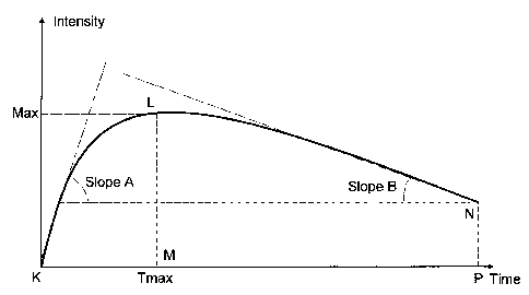
FIG. 6 illustrates four of the previously defined parameters on the curve of a
DOC: 'Max', `Tmax', SlopeA', and `SlopeB'. The other two parameters
('integral',
and 'Area to Max'), represent essentially the area enclosed by the indicated
points:
KLNP, and KLM, respectively.
FIG. 7 illustrates the LG/HG ROC analysis of the cumulative results for the
'Integral' parameter described previously. The area under the ROC curve is
0.83,
implying high discrimination (sensitivity).
FIG. 8 illustrates the sensitivity (grey) and specificity (black) plots
derived from
the ROC analysis for various values of the 'Integral' parameter used for the
quantification of the acetowhitening characteristics. It is clearly seen that
for a certain
value both sensitivity and specificity are maximized reaching 78%.
FIG. 9 illustrates the mean values, with corresponding error-bars representing
95% confidence intervals, for some of the parameters described previously, for
the LG
and HG diagnostic conditions, as concluded through biopsy examination
performed by
the histologists.
The optimum value ranges in discriminating LG from HG lesions were
calculated with ROC analysis, as shown previously for the 'Integral'
parameter. In
particular, for each parameter type the percentage of true positives (TP) and
false
positives (FP) was calculated for various threshold values spanning the entire
range:
[Pmin, Pmax], where P denotes the value of a specific parameter. The threshold
value
where the sensitivity (SS = TP), and specificity (SP = 100-FP), approximately
coincide
-23-
CA 02652010 2008-11-12
WO 2008/001037 PCT/GB2007/002067
with one another was used as an optimum (cut-off) value for discriminating LG
from
HG.
TABLE 1 illustrates the optimum value ranges for discriminating LG from HG
lesions for some of the previously defined parameters, leading to a
performance dictated
by specificity and sensitivity greater than 60%.
TABLE 1
Parameter Optimum parameter cut-off values for
LG/HG discrimination
Max* 70 to 90 (calibrated units) or 15-25%
(reflectance)
Integral** 480 to 650 (dimensionless quantity)
Tmax 80-100 sec
Area to Max** 120 to 170 (dimensionless quantity)
SlopeA 1.1 to 1.3 (rad)
SlopeB -0.012 to -0.090 (rad)
*The parameters listed above have been obtained with a digital imaging system
(DySIS technology, Forth Photonics) calibrated against an 18% reflecting
calibration
specimen to produce for the later a gray value of 105 in a 0-255 gray scale in
the green
channel of the system. Based on this calibration protocol Max is given as
green gray
value max difference in calibrated units (scale 0-255) or as reflectance max
difference
(scale 0-100%)
**The presented integral cut-off values have been calculated from a DOC
corresponding to a T=240 sec integration time:
240
Itg = c j(I - Ifr.,o)dt
Where c is a scaling factor with value c= 8[TIt-0]-1 or by substituting c=1/30
(intensity units)-Isec-1, It the remitted intensity at a given time point
after the application
of the biomarker , and It=0 the remitted intensity before the application of
the biomarker.
-24-
CA 02652010 2008-11-12
WO 2008/001037 PCT/GB2007/002067
Accordingly, Area to max is calculated from the same Itg formula and the only
difference is that T=Tmax. Both Itg and Area-to-Max are presented here as
dimensionless quantities.
Different acquisition, integration time periods and calibration protocols and
samples may result in different cut-off values. The 240 sec integration time
period is
selected as an optimum time period and it is presented here as an example and
not as a
restriction.
While the invention has been shown and described having reference to specific
embodiments, those skilled in the art will understand that variations in the
form and
detail may be made without departing from the spirit and the scope of the
invention.
Based on the previous analysis, in one preferred embodiment the 'Integral'
parameter of the DOC with the about 480-650 cut-off value range is used for
discriminating LG from HG lesions.
In another preferred embodiment the 'Max' parameter of the DOC with the
about 70-90 or 35%-45% cut-off value range is used for discriminating LG from
HG
lesions.
In yet another embodiment, the 'Area to Max' parameter with the about 120-170
cut-off value range is used for discriminating LG from HG lesions.
In yet another embodiment, the `Tmax' parameter with the about 80-100 sec cut-
off value range is used for discriminating LG from HG lesions.
In another preferred embodiment, the `SlopeA' parameter with the about 1.1-1.3
value range is used for discriminating LG from HG lesions.
In a still further embodiment, the `SlopeB' parameter with the about -0.012 to
-0.090 cut-off value range is used for discriminating LG from HG lesions.
A similar analysis was also performed for deriving the appropriate cut-off
values
of the previous parameters for discriminating HPV infections from HG lesions.
TABLE 2 illustrates the optimum value ranges generating specificity and
sensitivity greater than 60% for HPV/HG discrimination, for the 'Max' and
'Integral'
parameters.
-25-
CA 02652010 2008-11-12
WO 2008/001037 PCT/GB2007/002067
TABLE 2
Parameter Optimum parameter cut-off values for
HPV/HG discrimination
Max 65 to 90 (calibrated units)
Integral 380 to 490 (dimensionless quantity)
In a preferred embodiment, the 'Integral' parameter of the DOC with the about
380-490 cut-off value range is used for discriminating HPV infections from HG
lesions.
In another embodiment the 'Max' parameter of the DOC with the about 65-90
cut-off value range is used for discriminating HPV infections from HG lesions.
In yet another embodiment combinations of parameters including but not limited
to the above mentioned may provide a means for determining the pathology of
tissue.
For example, such a parameter may be the product of the average slope DOC
until about
40 sec sampling time after the application of said biomarker, by the Max
value. Product
values greater than about 2.05 0.2 (calibrated intensity units/time) may
indicate the
presence of high grade neoplasia, whereas lower values may indicate low grade
neoplasia or healthy tissue.
Beyond the 'hard-clustering' approach using a cut-off parameter value for
discriminating LG from HG lesions, or HPV from HG lesions, more advanced
statistical
and pattern recognition analysis techniques (such as Bayesian classification,
Artificial
Neural Networks (ANNs), classification trees), may be employed to extract
other linear,
or non-linear, of single or combinations of multiple, parameters for achieving
high
discrimination. In yet another embodiment, a parametric approach, using
Bayesian
modelling (as described in, for example, Fukunaga K. (1990) New York:
Academic, 2nd
Ed.), and a non-parametric approach, using ANNs (Learning Vector Quantization-
LVQ,
see as described in, for example, Kohonen T., (1986) Int. J. Quant. Chem.,
Suppl. 13,
209-21), were employed for differentiating the DOPs obtained from
corresponding DOC
of tissue sites with LG and HG neoplasia. For both Bayes and NN
classification, the
overall discrimination performance of LG and HG lesions was greater than 75%,
for
various combinations of the optical parameters described previously, and for a
variable
number of training sets selected from the overall sample.
-26-
CA 02652010 2008-11-12
WO 2008/001037 PCT/GB2007/002067
In another embodiment, the invention comprises a means for automated cervical
screening through the mapping of the dynamic parameter values, and the
corresponding
cut-off values, showing presence of the disease.
In yet another embodiment, the invention comprises a means for semi-automated
colposcopy through the mapping of the dynamic parameter values and
corresponding
cut-off values showing presence of the disease. Such a methodology ensures a
base-line
colposcopy performance independently of the practitioner's skills,
facilitating the
overall diagnostic procedure, follow-up, and guidance during biopsy sampling
and
treatment.
Another aspect of the present invention comprises the interpretation of the
acetowhitening phenomenon dictated by the dynamic parameters in relation to
the
functional and structural alterations in the epithelium. In one embodiment,
distinctive
parameters related to the cervical tissue structural properties are computed
and
correlated with a number of functional features derived from the DOC recorded
from the
same tissue sites. Specifically, there is a common agreement in terms of the
direct
correlation between the nuclear volume and grading of neoplasia (HPV, CIN 1,
CIN 2
and CIN3), or cervical cancer [Walker DC, et al. (2003) Physiological
Measurement,
24:1-15]. The nuclear-to-cytoplasmic-ratio (NCR), which expresses the nuclear
density
in the epithelial tissue, is the most common parameter used to describe this
correlation
with certain diagnostic conditions. In a preferred embodiment, the cellular
structure of
the tissue is assessed by finding the correlation formula between either, or
combination,
of the aforementioned dynamic parameters with the NCR computed from the biopsy
material extracted from corresponding cervical locations. To this end, the NCR
was
correlated with the DOC parameters reflecting the abnormal functioning of the
epithelium, after acetic acid induction into the tissue area.
In yet another embodiment, this correlation could lead to the extraction of a
pseudocolor map representing the structural properties of the examined
cervical tissue at
every location, in addition to the map representing the acetowhitening kinetic
characteristics, along with highlighted sites of high nuclear density. Such an
implementation has an exceptional value if one thinks that by quantifying the
in vivo
optical curve obtained from the tissue, which represents an in vivo assessment
of the
macro-structural tissue state; one can also derive direct conclusions about
the cellular
-27-
CA 02652010 2008-11-12
WO 2008/001037 PCT/GB2007/002067
properties of the tissue, which constitutes a representative view of its
structure at a
microscopic level.
In order to calculate the NCR for a corresponding number of epithelial tissue
sites from which the dynamic parameters were obtained by the method disclosed
herein,
an equal number of cervical biopsy samples were obtained during colposcopy.
The
biopsied tissue was processed through standard procedures,
immunohistochemically
stained, and placed on slides for further evaluation through microscopic image
analysis.
After acquiring an equivalent number of microscopic histological images, a
multistage
image-analysis algorithm was employed for segmenting the cell-nuclei displayed
in the
images [Loukas CG, et al. (2003) Cytometry, 55A(1): 30-42]. The NCR quantity
was
calculated as the sum of the area occupied by the nuclei enclosed in the
epithelium,
divided by the overall area of the epithelial tissue. NCR is also known as the
'cell-
packing' property of the epithelial tissue, expressing essentially the cross-
sectional
structure of the tissue's cellular population.
In an illustrative embodiment, FIG. 10A and FIG. 10B show scatter plots of two
different DOPs exhibiting the strongest correlation coefficient (R), against
NCR. These
parameters are the 'Integral', and the maximum value (Max), of the dynamic
optical
curve, as defined previously. The lines in the graphs represent linear
regression curves,
whereas the DOP to NCR conversion equation and correlation results obtained
from
least-squares fitting on the experimental data are shown in TABLE 3.
TABLE 3
NCR vs DOP Correlation Coefficient Conversion Equation
1
NCR vs 'Integral' 0.71 NCR = 1349 x Integral ¨ 0.278
NCR vs 'Max' 0.64 NCR = ¨1 x Max ¨ 0.309
181
From this table it can be seen that both parameters present a significant
correlation with
the cell-packing property of the tissue. In one embodiment of the method, the
linear
equations allow conversion of a DOP corresponding to a DOC obtained from a
specific
tissue site, to the underlying NCR property of the tissue site.
-28-
CA 02652010 2008-11-12
WO 2008/001037 PCT/GB2007/002067
In another embodiment of the method, either of the quantitative pseudocolor
maps of 'Integral', or 'Max', can be converted to the NCR map of the
epithelial tissue,
using the previously shown conversion formulas.
In addition to the structural alterations of the epithelial tissue in relation
to
neoplasia progression, there are also several functional changes in the
extracellular and
intracellular space of the epithelium after applying the acetic acid solution.
In particular,
solid tumours are known to live in an acidic microenviroment [Webb SD, at al.
(1999)
.1 Theor. Biol., 196: 237-250; Lee AH, et al. (1998) Cancer Research, 58: 1901-
1908;
Yamagata M et al. (1996) Br. J. Cancer, 73: 1328-1334; and Marion S, et al.
(2000)
Molecular Medicine Today, 6: 15-19]. Besides, experimental measurements have
shown
that extracellular pH in tumors is on average 0.5 units lower than that of
normal tissues,
with tumor extracellular pH lying typically in the range [6.6-7.0] (see
[Yamagata M et
al. (1996) Br. J. Cancer, 73: 1328-1334]). Tumor cells also have a neutral or
slightly
alkaline intracellular pH [Marion S, etal. (2000) Molecular Medicine Today, 6:
15-19].
Similar to the normal cells, tumor cells regulate their cytoplasmic pH within
a narrow
range to provide a favorable environment for various intracellular activities.
Although the issue regarding the presence of acidic extracellular pH in tumors
is
still controversial, there is a common belief that the acidic environment of
tumors arises
from the high rate of metabolic acid production, such as lactic acid, and from
its
inefficient removal from the extracellular space [Webb SD, at al. (1999)J.
Theor. Biol.,
196: 237-250; Lee AH, etal. (1998) Cancer Research, 58: 1901-1908; Marion S,
etal.
(2000) Molecular Medicine Today, 6: 15-19; and Prescott DM, et al. (2000)
Clinical
Cancer Research, 6;(6): 2501-2505]. Besides, tumor cells have a high rate of
glycolysis,
regardless of their oxygen supply level. As a consequence, large quantities of
lactic acid
(and subsequently H+) are produced outwards from the cellular environment. Due
to a
number of factors such as a disorganized vasculature, or poor lymphatic
drainage, and
elevated interstitial pressure, the acid clearance (H+ clearance) to the blood
is very slow,
and thus a reversed pH gradient between the extracellular and the
intracellular space of
tumors cells is observed, [Webb SD, at al. (1999)1 Theor. Biol., 196: 237-250;
Lee
AH, etal. (1998) Cancer Research, 58: 1901-1908; Yamagata M et al. (1996) Br.
1
Cancer, 73: 1328-1334; and Marion S, et al. (2000) Molecular Medicine Today,
6: 15-
19]. It is also reasonable to assume that the CIN extracellular environment is
also acidic
(perhaps less acidic), provided that cancer is a transitional process and CIN
is a
-29-
CA 02652010 2008-11-12
WO 2008/001037 PCT/GB2007/002067
precursor of cancer. Moreover, tumor as well as dysplastic cells are known to
employ
the same short-term, [Marion S, etal. (2000) Molecular Medicine Today, 6: 15-
19], and
long-term [Lee AH, etal. (1998) Cancer Research, 58: 1901-1908; Yamagata M et
al.
(1996) Br. J. Cancer, 73: 1328-1334 and Prescott DM, et al. (2000) Clinical
Cancer
Research, 6;(6): 2501-2505], pH regulation mechanisms as those of normal
cells. The
excess of protons produced by tumor cell metabolism is excreted from the cell
via
specific hydrogen pumps [Prescott DM, et al. (2000) Clinical Cancer Research,
6;(6):
2501-2505].
The observation of the acetowhitening effect in the cervix is used in
colposcopy
to characterize abnormal tissue (i.e. HPV, CIN, or cancer). The acetowhitening
effect
refers to the phenomenon induced by the application of acetic acid solution to
the
cervical transformation zone. The acetic acid application selectively induces
a transient
whitening of abnormal cervical areas. Although it has been used for more than
70 years
in clinical practice to locate abnormal areas, the exact physicochemical
mechanisms
involved in tissue whitening remain still unknown. Similar phenomena are
observed
when Formic, Propionic, and Butyric, acids are employed as biomarkers.
Two major explanations for the interpretation of the acetowhitening effect
prevail in the relative literature. In vitro studies have shown that the
acetic acid effect is
related to the amount of certain cytokeratines (proteins present in epithelial
cells)
[Maddox P, et al. (1999) Journal of Clinical Pathology, 52: 41-46 and Carrilho
C, et al.
(2004) Human Pathology, 35: 546¨ 551]. Since in cervical neoplasias the extra-
cellular
environment is acidic, the topically administered acidic acid molecule is not
disassociated to its composing ions and as such can penetrate passively the
cell
membrane. Entering into the neutral pH cytoplasm the acetic acid molecules are
disassociated giving hydrogen and carboxylic ions which interact with nuclear
proteins
resulting in the alteration of the scattering properties of the abnormal cells
selectively.
Cytosolic pH value is crucial for the conformational stability of these
proteins.
At neutral pH values, proteins are stable in solution. As pH drops, they
become unstable
and insoluble depending on their pI (isoelectrical point). The process of
protein
destabilization is called denaturation and this partial denaturation is a
reversible process
which lasts only for some milliseconds. Denatured or unfolded proteins have a
different
refractive index, and this may be the reason for the whitening effect. The
decrease of pH
in normal cells may not be enough to cause the proteins to unfold and perhaps
this is the
-30-
CA 02652010 2008-11-12
WO 2008/001037 PCT/GB2007/002067
reason that in normal tissue no variation in the IBSL is detected. Thus, the
back-
scattered light is strongly related to the pH dynamics influenced by the
acetic acid
penetration in the cervical epithelium. Nevertheless, the proteins that
contribute to the
effect are not well established. Moreover, each of these proteins may denature
at a
different pH value.
According to the other interpretation, the action of acetic acid on the
epithelium
of the transformation zone is related to its concentration [MacLean AB. (2004)
Gynecologic Oncology, 95: 691-694]. Acetic acid enters in the cellular
environment of
the dysplastic layers altering the structure of different nucleoproteins and
hence causing
the cells to appear opaque. Thus, the dynamics of the back-scattered light
follows the
dynamics of the acetic acid concentration. In normal tissue, no whitening
occurs because
the quantity of nucleoprotein is very small.
Based on the above mentioned analysis of the functional and structural
features
of the epithelium undergoing changes during neoplasia development it is
possible to
correlate dynamic optical data with epithelial features of diagnostic
importance. In
particular, the measured dynamic characteristics can be used to decouple
various
epithelial structural and transport phenomena occurring in time sequence after
the
application of the biomarker, and to correlate them with in vivo measurable
optical
parameters thus providing a solution to the inverse problem. In other words,
it is
possible to obtain information for various epithelial features by measuring in
vivo
dynamic characteristics and parameters.
In one embodiment of the method, `SlopeA' is used to obtain information for
the
extracellular acidity, and in turn for the passive diffusion constant, and for
the number of
cell layers of the stratified epithelium. In another embodiment of the method,
'Max' is
used to determine the NCR of the epithelium since the intensity of the back-
scattered
light is proportional to the density of signal sources (cell nuclei). In
another embodiment
of the method, `Slopeir is used to obtain information in regard to the cell
malfunction in
regulating the intracellular pH, and to the existence of disorganized
vasculature, or to the
poor lymphatic drainage associated with neoplasia development. In another
embodiment, the 'Integral' parameter is used to obtain combined information
for both
functional and structural features as described above.
Clinical validation of this biophysical model has been performed by
correlating
NCR with the 'Max' and 'Integral' parameters described previously. However,
clinical
-31-
CA 02652010 2008-11-12
WO 2008/001037 PCT/GB2007/002067
validation of the functional features is clinically impracticable due to the
lack of
reference methods capable of measuring these features in vivo. In contrast,
the method
disclosed herein is capable of modelling and predicting in vivo functional
features of the
tissue, based on its inherent capability of recording, analysing, and
displaying dynamic
optical characteristics obtained in vivo from a tissue interacting with a
biomarker.
FIG. 12 depicts another illustrative embodiment of the present invention.
Computing device 1070 executes instructions embodied on a computer readable
medium
defining at least the steps illustrated in image processing engine 1085 and in
conjunction
with a hardware set-up utilized to obtain the tissue image data. In
particular, the tissue
1020, is constantly illuminated with a light source 1010. After application of
a suitable
biomarker by means of an applicator 1030, a trigger signal is provided to
initiate image
acquisition using an image acquisition device 1040 such as a video CCD or
other
suitable image acquisition device. Between the tissue 1020 and the image
acquisition
device 1040 are optical filter 1050 and lenses 1060, for example, one or more
zoomable
lenses can be interposed. The optical filter 1050 can be tuned to a preferred
spectral
band, at which maximum contrast is obtained between areas that are subjected
to
different grade of alterations in their optical reflectance or fluorescence
characteristics
after administering an appropriate agent.
Before agent administration a tissue image is obtained as a reference. After
agent
administration, a series of images 1080, in time succession, at predetermined
spectral
bands, and for a predetermined time period, is obtained and stored in memory
or a
storage device internal to or external to the computing device 1070, for
further
processing by the image processing engine 1085. After proper alignment of some
or all
of the acquired images, a DOC 1090 is generated for a specific image location
corresponding to the same tissue point. In step 1100, a number of dynamic
optical
parameters expressing the dynamic characteristics of the phenomenon are
derived from
the DOCs, 1100.
After extracting the DOPs, in step 1110 their values can be compared with
predetermined cut-off values to, in turn, in step 1120, classify various
pathological
conditions of the tissue. As one result, a pseudolor map 1130, can then be
displayed on a
display device 1140, with different colors, or grey-shades, representing
different
pathologies. Alternatively, the classification of the various pathological
conditions of
the tissue can be stored for display at another time or sent to another
computing device
-32-
CA 02652010 2014-06-04
by, for example, a packet or other unit suitable for use in transporting data
in a network
environment.
Alternatively, in step 1150, the DOP values can be converted using
predetermined mathematical formulas, to express functional and structural
features of
the tissue. In this case, a pseudolor map 1130, can be displayed on the
display device
1140 with different colors, or grey-shades, representing different functional
and
structural features.
The contents of all references, figures, patents and published patent
applications
cited throughout this application are hereby incorporated by reference.
Those skilled in the art will recognize, or be able to ascertain using no more
than
routine experimentation, many equivalents to the specific embodiments of the
invention
described herein and the
scope of the claims should not be limited by any preferred embodiments or
examples set tbrth, but should be given the broadest interpretation,
consistent with the description as a whole.
-33-