Note: Descriptions are shown in the official language in which they were submitted.
CA 02653942 2014-12-04
ULTRASONIC SURGICAL INSTRUMENT
Field of the Invention
[0001] The present invention relates, in general, to ultrasonic surgical
instruments and,
more particularly, to an ultrasonic surgical clamp coagulator apparatus
particularly configured to provide increased tissue transection forces.
Background of the Invention
[0002] This application contains subject matter that relates to the
following non-
provisional applications:
[0003] ULTRASONIC SURGICAL BLADE AND INSTRUMENT HAVING A GAIN STEP,
U.S. Patent No. 7,163,548.
[0004] TISSUE PAD FOR USE WITH AN ULTRASONIC SURGICAL INSTRUMENT,
Ser. No. 11/245,819, filed October 7,2005, published as US 2006-0079874;
[0005] COMBINATION TISSUE PAD FOR USE WITH AN ULTRASONIC SURGICAL
INSTRUMENT, Ser. No. 11/246,794, filed October 7, 2005, granted as US
7,544,200;
[0006] ACTUATION MECHANISM FOR USE WITH AN ULTRASONIC SURGICAL
INSTRUMENT, Ser. No. 11/246,826, filed October 7, 2005;
[0007] CLAMP MECHANISM FOR USE WITH AN ULTRASONIC SURGICAL
INSTRUMENT, Ser. No. 11/246,264, filed October 7, 2005, published as US
2006- 0079879;
[0008] FEEDBACK MECHANISM FOR USE WITH AN ULTRASONIC SURGICAL
INSTRUMENT, Ser. No. 11/246,384, filed October 7, 2005, published as US
CA 02653942 2014-04-16
- 2 -
2006;
[0009] HANDLE ASSEMBLY HAVING HAND ACTIVATION FOR USE WITH AN
ULTRASONIC SURGICAL INSTRUMENT, Ser. No. 11/246,330, filed October 7,
2005 and granted as US 7,846,155;
[0010] ULTRASONIC SURGICAL SHEARS AND TISSUE PAD FOR SAME, serial no.
11/065,378, filed February 24, 2005, published as US 2005 - 0192610; and
[0011] HAND ACTIVATED ULTRASONIC INSTRUMENT, serial no. 10/869,351, filed
June 16, 2004, published as US 2005 - 0033337.
[0012] Ultrasonic surgical instruments are finding increasingly widespread
applications
in surgical procedures by virtue of the unique performance characteristics of
such
instruments. Depending upon specific instrument configurations and operational
parameters, ultrasonic surgical instruments can provide substantially
simultaneous cutting of tissue and hemostasis by coagulation, desirably
minimizing patient trauma. The cutting action is typically effected by an end-
effector or blade tip at the distal end of the instrument, which transmits
ultrasonic
energy to tissue brought into contact with the end-effector. Ultrasonic
instruments of this nature can be configured for open surgical use,
laparoscopic
or endoscopic surgical procedures including robotic-assisted procedures.
[0013] Ultrasonic surgical instruments have been developed that include a
clamp
mechanism to press tissue against the blade of the end-effector in order to
couple ultrasonic energy to the tissue of a patient. Such an arrangement
(sometimes referred to as a clamp coagulator shears or an ultrasonic
transector)
is disclosed in U.S. Pat. Nos. 5,322,055; 5,873,873 and 6,325,811. The surgeon
activates the clamp arm to press the clamp pad against the blade by squeezing
on the handgrip or handle.
END5881W0PCT
CA 02653942 2014-04-16
- 3 -
[0014] Some current ultrasonic shears devices, however, have the tendency
to create
tissue tags. Tissue tags are the tissue that remains clamped in the jaw that
is not
transected after the majority of the tissue in the jaw has been transected and
falls
away. Tissue tags may result from insufficient end-effector or blade tip
proximal
loading and/or lower proximal blade activity. Surgeons may mitigate tissue
tags
either through the addition of vertical tension (i.e. putting tension on the
tissue
using the blade) or rearward traction on the device in order to move the
untransected tissue to a more active portion of the blade to complete the cut.
[0015] Some current ultrasonic shears devices utilize tissue pads that
close in parallel
with the surface of the blade. This presents certain problems in terms of the
pressure profile exerted on the tissue. As tissue is compressed between the
jaw
and the blade, the distal portion of the blade deflects under load more than
the
proximal portion of the blade. This deflection is created in part by the
portion of
the blade distal to the most distal node of the device. It is also partly
created by
the deflection of the waveguide or transmission rod proximal to the most
distal
node. Additionally, the fact that blade amplitude decreases moving proximal of
the tip of the blade makes the situation worse since the amount of energy
transferred to the tissue, even if the pressure was constant, is reduced.
[0016] Current tissue pad designs utilize PTFE material to contact the
tissue and blade.
Although these designs have been adequate, they tend to suffer from longevity
issues since the pads tend to deteriorate over long surgical procedures.
Additionally, newer designs of clamp coagulator shears increase blade
amplitude
and/or the loading of the pad against the tissue and blade and overwhelm the
pad material, resulting in less than required tissue pad life. The pad
material
limits the amount of force that may be applied against the tissue and blade,
which in turn limits the tissue thickness or vessel size that some current
clamp
END5881W0PCT
CA 02653942 2014-04-16
- 4 -
coagulator shears may effectively cut and coagulate. Current composite pads
may be difficult or expensive to manufacture.
[0017] Some current designs of clamp coagulator ultrasonic shears are
limited in the
length of the active blade available for use by surgeons due to inherent
limitations in the effective transfer of mechanical motion along the
longitudinal
path of the blade from the transducer assembly. Although new blade geometry
has mitigated some of these problems, longer active blade lengths, or blades
that
have more mass (created by larger diameter or larger lengths) have a tendency
to shrink the frequency window between resonant and anti-resonant frequencies
making it difficult or impossible for ultrasonic generators to lock on to the
proper
frequency to drive the waveguide, blade and transducer assembly.
[0018] Some current designs of clamp coagulator shears utilize elastonner
material such
as silicone for node supports along the length of the blade. The most distal
node
support is typically silicone to provide for a seal around the blade. Where
higher
clamp forces are desired, as is the case with longer active blade lengths, it
is
desirable to have a rigid distal node support. Many problems, however, are
inherent with rigid node supports. Materials such as thermoset polymers that
are
capable of withstanding the pressure and temperature requirements of an
ultrasonic blade node support are often too expensive to be utilized in
production.
The use of thermoplastics would improve manufacturability from a cost
perspective but may not hold up to the pressure and temperature requirements
of
an ultrasonic blade node support.
[0019] Some current designs of clamp coagulator shears utilize a constant
force spring
mechanism that prevents the application of too much force to the clamp arm and
blade. Although the mechanism provides relatively constant force to the
system,
the spring imparts some slope to the force curve. In applications where the
END5881W0PCT
CA 02653942 2014-04-16
- 5 -
clamp force is low, the slope is not significant. In applications with high
clamp
forces, however, the difference in force attributable to the slope over the
possible
range of spring compressions becomes very significant and may exceed the
maximum force allowable in the blade, in the tube assemblies or in other
components of the system. The high slope could allow the maximum force to be
exceeded under abuse modes or through normal manufacturing tolerance
variations. If this occurs, the blade may bend, the actuation mechanism may
fail
or undesirable tissue effects may occur (i.e. fast cutting, but minimal tissue
coagulation). This situation is aggravated by the fact that a portion of the
jaw
(the clamp arm and pad) of the device can meet sufficient resistance to engage
the force limiting mechanism when the clamp pad almost contacts the blade
(when transecting thin tissue or at the end of the transection or clamping
solid
objects such as other devices) or when the clamp arm is still open with
respect to
the blade (when transecting thick tissue).
[0020] Some current designs of clamp coagulator shears utilize force-
limiting springs to
ensure that clamp forces are within a specified range. It is also necessary
for the
force-limiting spring design to allow the surgeon to "feather" (apply less
than the
maximum force and slowly increase to the maximum force). In these
mechanisms, therefore, the jaw closes until a predetermined force is met and
then the additional stroke drives the mechanism into the force limiting range.
In
some cases, though, the surgeon may, unknowingly, fail to apply the full force
of
the jaw against the tissue resulting in incomplete tissue cuts or insufficient
coagulation. Alternatively, the surgeon may unknowingly open the clamp arm
during a transection that results in incomplete tissue cuts or insufficient
coagulation.
[0021] Some current designs of clamp coagulator shears utilize a foot pedal
to energize
END5881W0PCT
CA 02653942 2014-12-04
,
- 6 -
the surgical instrument. The surgeon operates the foot pedal while
simultaneously applying pressure to the handle to press tissue between the jaw
and blade to activate a generator that provides energy that is transmitted to
the
cutting blade for cutting and coagulating tissue. Key drawbacks with this type
of
instrument activation include the loss of surgeon focus on the surgical field
while
the surgeon searches for the foot pedal, the foot pedal gets in the way of the
surgeon's movement during a procedure and surgeon leg fatigue during long
cases.
[0022] Some current designs of torque wrenches for ultrasonic surgical
instruments
utilize a multi-piece torque wrench for use in properly torqueing an
instrument to
an ultrasonic handpiece. A multi-piece assembly is more costly in that
separate
pieces have to be molded and then assembled. In addition, the pieces have a
tendency to wear rapidly leading to failure of the wrench.
[0023] It would be desirable to provide an ultrasonic surgical instrument
that overcomes
some of the deficiencies of current instruments. The ultrasonic surgical
instrument described herein overcomes those deficiencies.
Summary of the Invention
[0023a] The present invention relates to a tissue pad for use in an ultrasonic
clamp
coagulator, comprising a first tissue pad portion having a distal end and a
proximal end, defining a length L, a first tissue engaging surface having a
length
corresponding to the length L and a cavity having an opening circumscribed by
the first tissue engaging surface.
[0024] The present invention meets the above stated needs for an improved
tissue pad
for an ultrasonic surgical instrument. The tissue pad for use in an ultrasonic
clamp coagulator comprises i) a first tissue pad portion, the first tissue pad
portion having a distal end a proximal end, a tissue engaging surface and a
CA 02653942 2014-12-04
- 6a -
cavity having an opening coinciding with the tissue engaging surface, and ii)
a
second tissue pad portion, the second tissue pad portion made from a
composition having a greater resistance to heat than the first tissue pad
portion,
the second tissue pad portion having a tissue engaging surface and sized for
placement within the cavity.
CA 02653942 2014-12-04
- 7 -
[0025] A method of mounting the first tissue pad portion onto a clamp arm
of an
ultrasonic clamp coagulator, comprises the steps of: i) inserting the first
tissue
pad portion into the clamp arm, the first tissue pad portion being oriented
during
the insertion so that the cavity is positioned at the proximal portion of the
arm,
and ii). inserting into the cavity a second tissue pad portion made from a
composition having a greater resistance to heat than the first tissue pad
portion,
the second tissue pad portion having a tissue engaging surface and sized for
placement within the cavity.
[0026] The present invention meets the above stated needs for an waveguide and
blade
that enable larger wave amplitude and a longer active blade length and still
provide sufficient frequency margin or window. The waveguide is provided with
a
series of gain steps located at the distal portion of the waveguide preferably
at
the two most distal nodes in relation to the handpiece, or the two most
proximal
nodes in relation to the blade tip.
[0027] The present invention meets the above stated needs for a waveguide
support
that enables high clamp forces along with longer active blade lengths. The
waveguide is supported by a rigid support positioned between the waveguide
and an outer sheath. The support preferably is annular that has an inner
diameter that coincides with the waveguide outer diameter and an outer
diameter
that coincides with the inner diameter of the outer sheath. In a second
expression of the embodiment, the support has support legs for attaching to
the
outer sheath and which further channel heat away from the waveguide and to the
outer sheath.
[0028] A method for providing a nodal support to an ultrasonic surgical
instrument
CA 02653942 2014-04-16
- 8 -
comprising an outer sheath and waveguide positioned within the outer sheath
and having a distal blade and a most-distal vibration node, the steps
comprising:
a) providing a rigid nodal support comprising an annular collar; b)
positioning the
rigid annular collar between the waveguide and outer sheath; c) providing at
least
one support member attached to the annular collar; d) positioning the at least
one support member in contact with the outer sheath; and e) positioning the
annular collar at the most-distal vibration node.
[0029] The present invention meets the above stated needs for a one-piece
torque
wrench for an ultrasonic surgical instrument. The torque wrench includes
cantilever arms aligned in an annular fashion about the centerline of the
torque
wrench. The cantilever arms include teeth in an inward perpendicular fashion
in
relation to cantilever arms. The surgical instrument includes an outer tube
retainer that includes spline gears projecting in a perpendicular fashion
along the
outer circumference of retainer. Torque is transmitted through the cantilever
arms to the spline gears for attaching a handpiece to the clamp coagulator.
Brief Description of the Figures
[0030] The novel features of the invention are set forth with particularity
in the appended
claims. The invention itself, however, both as to organization and methods of
operation, may best be understood by reference to the following description,
taken in conjunction with the accompanying drawings in which:
[0031] FIG. 1 is a plan view illustrating an embodiment of an ultrasonic
surgical
instrument in accordance with the present;
[0032] FIG. 2 is a perspective assembly view of an embodiment of an
ultrasonic surgical
instrument in accordance with the present invention;
END5881W0PCT
CA 02653942 2014-04-16
- 9 -
[0033] FIG. 3 is a plan view of one embodiment of the waveguide and blade
assembly in
accordance with the present invention;
[0034] FIG. 3a is a sectional view of one embodiment of the distal end of
the blade
assembly in accordance with the present invention;
[0035] FIG. 3b is a plan view of one embodiment of the waveguide and blade
assembly
and silicone support rings in accordance with the present invention;
[0036] FIG. 3c is a graph depicting waveform along the length of the
waveguide and
blade of one embodiment of the present invention;
[0037] FIG. 3d is a perspective, side view and cross sectional view of one
embodiment
of a distal blade node support;
[0038] FIG. 4a depicts plan, top and cross sectional views of outer sheath
and clamp
arm assembly of one embodiment of the present invention;
[0039] FIG. 4b is a perspective assembly view of one embodiment of a clamp
arm and
clamp pad assembly of the present invention;
[0040] FIG. 4c is a plan and cross sectional view of one embodiment of a
clamp arm of
the present invention;
[0041] FIG. 4d is a perspective elevation view of one embodiment of a
tissue pad insert
of the present invention;
[0042] FIG. 5 is a plan view and side view of one embodiment of the outer
tube of the
present invention;
[0043] FIG. 6 is a side view and plan view of one embodiment of the inner
tube of the
present invention;
END5881W0PCT
CA 02653942 2014-04-16
- 10 -
[0044] FIG. 7 is a perspective assembly view of the distal end of a
handpiece assembly
and electrical ring contactors;
[0045] FIG. 8a is a perspective view of the front and rear sides of a
connector and
flexboard assembly of one embodiment of the present invention;
[0046] FIG. 8b is a plan view of the rocker switch of one embodiment of the
present
invention;
[0047] FIG. 8c is an electrical schematic of the switch circuit;
[0048] FIG. 9 is a plan view of an ultrasonic surgical instrument in
accordance with the
present invention with the a first finger accessing a first activation button
gripped
by a left-handed surgeon;
[0049] FIG. 10 is a plan view of an ultrasonic surgical instrument in
accordance with the
present invention with the a first finger accessing a first activation button
gripped
by a right-handed surgeon;
[0050] FIG. 11 is a perspective, side view and cross sectional end view of
one
embodiment of a torque wrench;
[0051] FIG. 12 is a perspective and cross sectional end view of one
embodiment of an
outer tube retainer of the present invention; and
[0052] FIG. 13 is a force curve illustrating various forces as a function
of the trigger
position.
Detailed Description of the Invention
[0053] Before explaining the present invention in detail, it should be
noted that the
invention is not limited in its application or use to the details of
construction and
END5881W0PCT
CA 02653942 2014-04-16
-11 -
arrangement of parts illustrated in the accompanying drawings and description.
The illustrative embodiments of the invention may be implemented or
incorporated in other embodiments, variations and modifications, and may be
practiced or carried out in various ways. Further, unless otherwise indicated,
the
terms and expressions employed herein have been chosen for the purpose of
describing the illustrative embodiments of the present invention for the
convenience of the reader and are not for the purpose of limiting the
invention.
[0054] Further, it is understood that any one or more of the following-
described
embodiments, expressions of embodiments, examples, etc. can be combined
with any one or more of the other following-described embodiments, expressions
of embodiments, examples, etc.
[0055] The present invention is particularly directed to an improved
ultrasonic surgical
clamp coagulator apparatus which is configured for effecting tissue cutting,
coagulation, and/or clamping during surgical procedures. The present apparatus
can be readily configured for use in open surgical procedures, as well as
laparoscopic or endoscopic procedures and robot-assisted surgical procedures.
Versatile use is facilitated by selective use of ultrasonic energy. When
ultrasonic
components of the apparatus are inactive, tissue can be readily gripped and
manipulated, as desired, without tissue cutting or damage. When the ultrasonic
components are activated, the apparatus permits tissue to be gripped for
coupling with the ultrasonic energy to effect tissue coagulation, with
application of
increased pressure efficiently effecting tissue cutting and coagulation. If
desired,
ultrasonic energy can be applied to tissue without use of the clamping
mechanism of the apparatus by appropriate manipulation of the ultrasonic
blade.
[0056] As will become apparent from the following description, the present
clamp
coagulator apparatus is particularly configured for disposable use by virtue
of its
END5881W0PCT
CA 02653942 2014-04-16
- 12 -
straightforward construction. As such, it is contemplated that the apparatus
be
used in association with an ultrasonic generator unit and transducer of a
surgical
system, whereby ultrasonic energy from the generator unit provides the desired
ultrasonic actuation through the transducer for the present clamp coagulator
apparatus. It will be appreciated that a clamp coagulator apparatus embodying
the principles of the present invention can be configured for non-disposable
or
multiple uses, and non-detachably integrated with an associated hand piece (or
transducer) unit. However, detachable connection of the present clamp
coagulator apparatus with an associated ultrasonic hand piece is presently
preferred for single-patient use of the apparatus.
[0057] The present invention will be described in combination with an
ultrasonic
instrument as described herein. Such description is exemplary only, and is not
intended to limit the scope and applications of the invention. For example,
the
invention is useful in combination with a multitude of ultrasonic instruments
including those described in, for example, U.S. Pat. Nos. 5,938,633;
5,935,144;
5,944,737; 5,322,055, 5,630,420; and 5,449,370.
[0058] With reference to FIGS. 1-3, an embodiment of a surgical system 19,
including
an ultrasonic surgical instrument 100 in accordance with the present invention
is
illustrated. The surgical system 19 includes an ultrasonic generator 30
connected
to an ultrasonic transducer 50 via cable 22, and an ultrasonic surgical
instrument
100. It will be noted that, in some applications, the ultrasonic transducer 50
is
referred to as a "hand piece assembly," or simply "hand piece," because the
surgical instrument of the surgical system 19 is configured such that a
surgeon
may grasp and manipulate the ultrasonic transducer 50 during various
procedures and operations. A suitable generator is the GEN 300TM sold by
Ethicon Endo-Surgery, Inc. of Cincinnati, Ohio.
END5881W0PCT
CA 02653942 2014-04-16
- 13 -
[0059] The ultrasonic surgical instrument 100 includes a multi-piece handle
70 adapted
to isolate the operator from the vibrations of the acoustic assembly contained
within transducer 50. The handle 70 can be shaped to be held by a user in a
conventional manner, but it is contemplated that the present ultrasonic
surgical
instrument 100 principally be grasped and manipulated by a scissor-like
arrangement provided by a handle assembly of the instrument, as will be
described. While single-piece handle 70 is illustrated, the handle 70 may
comprise a single or unitary component. The proximal end of the ultrasonic
surgical instrument 100 receives and is fitted to the distal end of the
ultrasonic
transducer 50 by insertion of the transducer into the handle 70. The
ultrasonic
surgical instrument 100 may be attached to and removed from the ultrasonic
transducer 50 as a unit.
[0060] Referring specifically now to FIG. 2, the ultrasonic surgical
instrument 100 may
include a handle assembly70, comprising mating housing portions 68 and 69,
together forming handle 70 and a transmission assembly 71. The ultrasonic
surgical instrument 100 has application in both open and endoscopic surgical
procedures. The construction can be dimensioned such that transmission
assembly 71 has an outside diameter of approximately 8.5 mm. The elongated
transmission assembly 71 of the ultrasonic surgical instrument 100 extends
orthogonally from the instrument handle 70. The handle 70 may be constructed
from a durable plastic, such as polycarbonate or a liquid crystal polymer. It
is also
contemplated that the handle 70 may alternatively be made from a variety of
materials including other plastics, ceramics or metals.
[0061] The transmission assembly 71 may include an outer tubular member or
outer
sheath 72, an inner tubular actuating member 76, a waveguide 80 and end-
effector 81 (blade 79, clamp arm 56, pin 56b and one or more clamp pads 58).
END5881W0PCT
CA 02653942 2014-04-16
. .
- 14 -
As will be described, the outer sheath 72, the actuating member 76, and the
waveguide or transmission rod 80 may be joined together for rotation as a unit
(together with ultrasonic transducer 50) relative to handle 70. The waveguide
80,
which is adapted to transmit ultrasonic energy from transducer 50 to blade 79
may be flexible, semi-flexible or rigid.
[0062] The ultrasonic waveguide 80 may further include at least one
radial hole or
aperture 66 extending there through, substantially perpendicular to the
longitudinal axis of the waveguide 80. The aperture 66, which may be
positioned
at a node, is configured to receive an insulated connector pin 27, which
connects
the waveguide 80, to the tubular actuating member 76, and the tubular outer
sheath 72, as well the outer tube retainer 29. A rotation knob 28 (not shown)
may be added to or may replace retainer 29 to facilitate rotation of the blade
assembly 80, including the end effector 81 relative to instrument handle 70,
as is
known and understood in the art.
[0063] The blade 79 may be integral with the waveguide 80 and formed as a
single unit.
In an alternate expression of the current embodiment, a threaded connection, a
welded joint, or other coupling mechanisms may connect blade 79 to waveguide
80. The distal end of the blade 79 is disposed near an anti-node in order to
tune
the acoustic assembly to a preferred resonant frequency fo when the acoustic
assembly is not loaded by tissue. When ultrasonic transducer 50 is energized,
the distal end of blade 79 is configured to move longitudinally in the range
of, for
example, approximately 10 to 500 microns peak-to-peak, and preferably in the
range of about 20 to about 200 microns at a predetermined vibrational
frequency
fo of, for example, 55,500 Hz.
[0064] Referring now to FIG. 3, the waveguide 80 may also be configured
to amplify the
mechanical vibrations transmitted through the waveguide 80 to the blade 79 as
is
END5881W0PCT
CA 02653942 2014-04-16
,
- 15 -
well known in the art and more fully described in ULTRASONIC SURGICAL
BLADE AND INSTRUMENT HAVING A GAIN STEP, Ser. No. 10/701,558, filed
November 5, 2003, now U.S. Patent, No. 7,163,548 B2. In one embodiment of
the present invention, the waveguide 80 may further have features to control
the
gain of the longitudinal vibration along the waveguide 80 and features to tune
the
waveguide 80 to the resonant frequency of the system. In particular, waveguide
80 may have any suitable cross-sectional dimension. For example, the
waveguide 80 may have a substantially uniform cross-section or the waveguide
80 may be tapered at various sections or may be tapered along its entire
length
as is described in more detail herein.
[0065] In one embodiment of the present invention, the waveguide 80
includes a hollow
bore 101 located between the most distal vibration node and the distal tip of
the
blade 79a. This hollow bore 101 in the instant embodiment, facilitates longer
active blade length by stretching or expanding wavelength as is known and
understood in the art. This longer active blade length may require larger
diameter blades 79 to facilitate the bore. To ensure proper performance of the
blade 79 and to achieve desired cutting and coagulation action of the blade, a
larger wave amplitude may be used. Increasing active blade length and wave
amplitude may create difficulties for the system to achieve resonance. For
instance, a system tuned to resonate at 55,500 Hz, with the hollow bore blade,
may achieve anti-resonance at 55,550 Hz. This narrow frequency window may
make it difficult or impossible for the generator 30 (see FIG. 1) to
continuously
drive the waveguide 80 and blade 79 system at its resonant frequency.
[0066] To enable larger wave amplitude and longer active blade lengths and
still provide
sufficient frequency margin or window, a waveguide 80 is provided with a
series
of gain steps in the waveguide 80. The gain of a gain step less than unity
END5881 WOPCT
CA 02653942 2014-04-16
- 16 -
results from an increase in mass of the ultrasonic waveguide at a node, and
the
gain of a gain step greater than unity results from a decrease in mass of the
waveguide at a node. A gain feature is any one of geometric constructions of
the
waveguide or blade that either increases or decreases the mass of the
waveguide or blade at a node and include: a discrete change in outer diameter
or
perimeter, a taper, a longitudinal hole, a transverse hole, a void, a surface
flat, a
surface slot, and a change in material. The term hole includes a through hole
and a non-through hole. Other gain features are left to the artisan.
[0067] In one embodiment of the present invention, a gain step 102, located
at the
second most distal vibration node (see FIG. 3), is provided in the waveguide
80.
Gain step 102 decreases the cross sectional area of the blade facilitating
greater
wave amplitude in the decreased diameter (see FIG. 3c), as is known and
understood in the art. To facilitate the longer active blade length and to
maintain
a desired blade diameter, a step up 103 is provided at or near an antinode,
which
increases the cross sectional area of waveguide 80 without affecting the gain.
In
one embodiment, the step up 103 is located at the second most distal vibration
antinode in relation to the distal blade tip 79a. A second step down or gain
step
104 is provided adjacent to the blade 79. The second gain step 104 results in
a
second amplitude increase. In one embodiment, the second gain step 104 is
located at the first most distal vibration node in relation to the blade tip
79a.
[0068] As is known and understood in the art, in an ultrasonic blade
system, a generator
produces a current to drive a transducer located within handpiece 50. This
transducer imparts mechanical energy at a specific frequency to a waveguide
and to a blade attached thereto. The generator continues to impart electrical
energy to convert to mechanical energy as it varies the frequency in an effort
to
find and drive the system at its resonant frequency. Equating the transducer
and
END5881W0PCT
CA 02653942 2014-04-16
, .
- 17 -
waveguide as an equivalent electrical model, as the frequency of cycling is
increased, starting at a non-resonant condition below the desired resonant
frequency, the system's oscillations first approach a frequency at which
impedance is minimum (maximum admittance). This minimum impedance
frequency approximates the series resonance frequency, the frequency at which
impedance in an electrical circuit describing the element is zero (assuming
resistance caused by mechanical losses is ignored). The minimum impedance
frequency also is the resonant frequency of the waveguide and blade assembly,
which by design is nominally the same resonant frequency of the transducer.
The composition of the transducer material and the shape and volume of the
waveguide and blade assembly determine the resonance frequency. As the
cycling frequency is further increased, impedance increases to a maximum
(minimum admittance). The maximum impedance frequency, approximates the
parallel resonance frequency, the frequency at which parallel resistance in
the
equivalent electrical circuit is infinite (assuming resistance caused by
mechanical
losses is ignored). The maximum impedance frequency also is the anti-
resonance frequency. The larger the difference between resonant and anti-
resonant frequencies (that is, the frequency window or phase margin), the
easier
it is for a generator to establish and maintain resonance in the waveguide and
blade assembly as frequency tolerances are relaxed.
[0069] In the present invention, the gain step described above may cause
a significant
acoustic impedance mismatch, causing some of the mechanical energy
transmitted along the waveguide to be reflected. As is seen in FIG. 3 and 3c,
a
gain in wave amplitude is caused by a thinner cross section in the waveguide
80
adjacent gain step 102. At gain step 102, the change in thickness results in a
lowering of the anti-resonant frequency after the step 102. This results in a
narrower frequency window or steeper trough between resonance and anti-
END5881W0PCT
CA 02653942 2014-04-16
, =
- 18 -
resonance. A step up 103 results in an increase in waveguide cross-section or
thickness that in part addresses manufacturing requirements.
[0070] Applicants have determined that locating the gain steps in the
distal portion of the
waveguide results in a greater phase margin or wider trough between resonant
and anti-resonant frequencies. What is meant as the "distal portion" is the
distal
half of the waveguide. By delaying waveguide narrowing to the distal end of
the
waveguide, more mechanical energy is stored along the waveguide and any
negative effects due to reflection at the gain steps are mitigated. It is
appreciated
that the gain step or combination step up/down/up may be located anywhere
along the waveguide. For ideal system performance, however, the gain step(s)
should be located in the distal half of the blade, preferably at the two most
distal
nodes in relation to the handpiece, or the two most proximal nodes in relation
to
the blade tip. Surprisingly, the Applicants found that the phase margin
increased
by almost 100 % by relocating the gain steps to the distal portion of the
waveguide. In early experiments of a waveguide having two gain steps, one at
the proximal end and one at the distal end, the phase margin measured 30 to 40
Hz. In experiments of a waveguide having two gain steps, both located at the
distal portion, the phase margin measured between 50 and 80 Hz. In
experiments of a waveguide having two gain steps, one at each of the two most
distal nodes, the phase margin measured between 75 and 80 Hz.
[0071] In another embodiment (not shown), a single gain step is located
at either the
first or second most distal node in relation to the tip of the blade 79a. A
single
gain step may obviate the need for a step up and step down on the blade. To
accommodate the hollow tip blade, the waveguide 80 must be of sufficient cross
section to transmit a wave from the handpiece to the first gain step 102 and
the
difference in diameters between the waveguide and the blade must be sufficient
END5881W0PCT
CA 02653942 2014-04-16
. .
- 19 -
to result in the wave amplitude gain from a step down, step up and step down
combination. The diameter difference must be large enough to achieve correct
blade longitudinal excursion while providing a sufficient frequency window for
the
system to lock on to resonance.
[0072] Referring again to FIG. 3, a waveguide 80 and blade 79 combination
is shown.
In one embodiment, the overall length of the combination is 8.854 inches. The
first gain step 102 is located 6.139 inches from proximal end 90. The step up
103 is located 6.922 inches from end 90. The second gain step 104 is located
7.912 inches from end 90. The bore 101 is 0.384 inches measured from blade
tip 79a.
[0073] Referring now to FIG. 3c, graph 91 displays wave amplitude vs.
blade distance
and geometry. The y-axis represents wave amplitude given as a percentage of
maximum displacement. The x-axis represents blade length. The wave 92
represents the response or gain along the length due to the varying cross
sections in waveguide 80 and blade 79 discussed in reference to FIG. 3. The
points at which the wave 92 crosses the x-axis are referred to as nodes or
vibration nodes. The points at which wave 92 reaches maximum amplitude are
referred to as antinodes. It can be seen that the first gain in wave amplitude
92
corresponds with gain step 102 and the second gain in wave amplitude 92
occurs at second gain step 104.
[0074] Referring to FIG. 3b, waveguide 80 may have a plurality of
stabilizing silicone
rings 80a or compliant supports to prevent the waveguide 80 from making
contact with the inner tube 76 during activation. The silicone rings 80a are
ideally located at nodes on the waveguide 80 as is known and understood in the
art. Rings 80a are preferably over molded on waveguide 80 to ensure accurate
location. A seal may be provided at the distal-most node, nearest the end-
END5881W0PCT
CA 02653942 2014-04-16
- 20 -
effector 81, to abate passage of tissue, blood, and other material in the
region
between the waveguide 80 and actuating member 76. A silicone ring may not be
sufficient at the most distal node of the waveguide 80 in the instant
embodiment
of the present invention. As discussed above, the waveguide 80 is provided
with
amplitude gain steps 102 and 104 that amplify the wave transmitted to the
blade
79. The greatest deflection of the blade occurs at the blade tip 79a. To
facilitate
tissue cutting in the proximal portion of the blade 79, a node support more
rigid
than silicon (a "rigid" support) is preferable to promote wave transmission to
the
blade to prevent wave absorption or losses and further provide more accurate
dimensional stability of the blade deflection relative to the clamp arm 56. A
rigid
support may be useful in promoting higher clamp forces in the proximal portion
of
blade 79 since a rigid support will not compress or yield at higher clamp
forces as
would silicone or like materials.
[0075] Referring back to FIG. 2 and now FIG. 3d, a blade support 450 is
shown. In a
first expression of a first embodiment, support 450 is comprised of two
pieces. A
sleeve 451 preferably composed of a thermoset polymer is disposed around
annular opening 452. The sleeve may be manufactured from bar stock resin
such as VESPEL 211. The sleeve 451 is then insert molded or pressed into a
less expensive thermoplastic resin collar 453. The collar 453 is preferably
composed of a filled thermoplastic resin with high thermal-conductivity, such
as a
filled PEEK. In this two-piece configuration, the support 450 is able to
withstand
the temperature and pressures associated with operation of the ultrasonic
blade
79 and waveguide 80. In operation, when the blade is activated, heat built up
in
the blade is transferred through the thermoset polymer insert 451 to the
collar
453. The collar 453 transfers the heat to fins 454. Fins or supports 454
support
the collar 453 in the outer tube or sheath 72 and also transfer heat away from
the
collar 453 and insert 451 into the void between the blade 79 and the actuating
END5881 WOPCT
. . CA 02653942 2014-04-16
- 21 -
member 76. In a second expression of a first embodiment, blade support 450 is
comprised of a thermoplastic resin, such as PEEK, of unitary construction. In
a
third expression of a first embodiment, blade support 450 is comprised of a
thermoset polymer of unitary construction or a high temperature injection
molded
polyamide or compression molded materials of unitary construction. Other
materials, such as ceramics and metals are also contemplated for the blade
support 450 as is well known to the artisan.
[0076] Ultrasonic transducer 50, and an ultrasonic waveguide 80 together
provide an
acoustic assembly of the present surgical system 19, with the acoustic
assembly
providing ultrasonic energy for surgical procedures when powered by generator
30. The acoustic assembly of surgical instrument 100 generally includes a
first
acoustic portion and a second acoustic portion. In the present embodiment, the
first acoustic portion comprises the ultrasonically active portions of
ultrasonic
transducer 50, and the second acoustic portion comprises the ultrasonically
active portions of transmission assembly 71. Further, in the present
embodiment,
the distal end of the first acoustic portion is operatively coupled to the
proximal
end of the second acoustic portion by, for example, a threaded connection.
[0077] With particular reference to FIGS. 2, 9 and 10, reciprocal
movement of actuating
member 76 drives the clamp member 60 open and closed. A force-limiting
mechanism 95 is operatively connected to actuating member 76 and comprises a
tube collar 98 that secures distal washer 97, wave spring 94, onto threaded
collar
cap 93. Outer tube retainer 29 engages with openings 75 in the proximal
portion
of tubular actuating member 76 via insulated pin 27. A shoulder 74 on the tube
collar 78 receives drive yoke 33 for engagement with the inside surface of
outer
sheath 72.
[0078] The force limiting mechanism 95 provides a portion of the clamp
drive
END5881 WOPCT
CA 02653942 2014-04-16
- 22 -
mechanism of the instrument 100, which affects pivotal movement of the clamp
member 60 by reciprocation of actuating member 76. The clamp drive
mechanism further includes a drive yoke 33 which is operatively connected with
an operating trigger handle 34 of the instrument, with the operating trigger
handle
34 thus interconnected with the reciprocable actuating member 76 via drive
yoke
33 and force limiting mechanism 91. Trigger handle 34 is connected to drive
yoke 33 and link 37 via pins 35 and 36. Spring 12 is located between drive
yoke
33 and handle assembly 68 and 69 biasing reciprocable actuating member 76 to
the open position.
[0079] Movement of trigger handle 34 toward handgrip 70 translates
actuating member
76 proximally, thereby pivoting clamp member 60 toward blade 79. The scissor-
like action provided by trigger handle 34 and cooperating handgrip 70
facilitates
convenient and efficient manipulation and positioning of the instrument, and
operation of the clamping mechanism at the distal portion of the instrument
whereby tissue is efficiently urged against the blade 79. Movement of trigger
handle 34 away from handgrip 68 translates actuating member 76 distally,
thereby pivoting clamp member 60 away from blade 79.
[0080] With particular reference to FIG. 4a, therein is illustrated one
embodiment of
clamp member 60 for use with the present ultrasonic surgical instrument 100
and
which is configured for cooperative action with blade 79 (not shown). The
clamp
member 60 in combination with blade 79 is commonly referred to as the end
effector 81, and the clamp member 60 is also commonly referred to as the jaw.
The clamp member 60 includes a pivotally movable clamp arm 56, which is
connected to the distal end of outer sheath 72 and actuation member 76 (not
shown), in combination with a tissue-engaging pad or clamp pad 58. In one
expression of the embodiment, clamp pad 58 is formed from TEFLON
END5881W0PCT
CA 02653942 2014-04-16
=
- 23 -
trademark name of E. I. Du Pont de Nemours and Company, a low coefficient of
friction polymer material, or any other suitable low-friction material. Clamp
pad
58 mounts on the clamp arm 56 for cooperation with blade 79, with pivotal
movement of the clamp arm 56 positioning the clamp pad in substantially
parallel
relationship to, and in contact with, blade 79, thereby defining a tissue
treatment
region. By this construction, tissue is grasped between clamp pad 58 and blade
79. As illustrated, clamp pad 58 may be provided with non-smooth surface, such
as a saw tooth-like configuration to enhance the gripping of tissue in
cooperation
with the blade 79. The saw tooth-like configuration, or teeth, provide
traction
against the movement of the blade. The teeth also provide counter traction to
the blade and clamping movement. As would be appreciated by one skilled in
the art, the saw tooth-like configuration is just one example of many tissue-
engaging surfaces to prevent movement of the tissue relative to the movement
of
the blade 79. Other illustrative examples include bumps, criss-cross patterns,
tread patterns, a bead or sand blasted surface, etc.
[0081] Tissue pads having composite construction, while having benefits and
advantages over TEFLON pads, have cost and manufacturing disadvantages.
Composite tissue pads are typically compression molded into a flat coupon.
Such compression molding can be time consuming and expensive. Once the flat
coupon is produced, it must be machined to produce a tissue pad suitable for
use
with a clamping ultrasonic device adding further time and expense to the
manufacturing process.
[0082] With particular reference to FIGS. 4a and b, a first expression of
the current
embodiment includes a clamp pad 58 having a proximal portion 58b and a distal
portion 58a that include a saw tooth like configuration. Clamp pad 58 may be a
single component, which has an annular opening 58c. Annular opening 58c is
END5881W0PCT
CA 02653942 2014-04-16
=
- 24 -
configured to receive pad insert 58d. The advantage of two separate
components 58 and 58d is that the pad and the insert may be constructed from
different materials. For example, having a two-piece tissue pad allows the use
of a very lubricious material at the distal end that is not particularly wear
resistant
to high temperatures compared to a very high temperature material at the
proximal end that is not particularly lubricious because the proximal end is
an
area of lower amplitude. Such a configuration matches the tissue pad materials
to the amplitude of the blade 79. Applicants found, in one experiment, that a
15% graphite-filled, 30% PTFE-filled polyimide tissue pad showed substantially
the same or better wear with a 4.5 pound clamping force as a 100%
polytetrafluoroethylene (PTFE) tissue pad showed with a 1.5 pound clamping
force. The advantage of a 15% graphite-filled, 30% PTFE-filled polyimide
tissue
pad is increased heat resistance, which improves the overall wear resistance
of
the tissue pad. This polyimide-composite clamp pad has a useful heat
resistance
up about 800 F to about 1200 F, as compared to a useful heat resistance up to
about 660 F of a PTFE clamp pad. Alternatively, Other materials are also
useful
for a portion of the tissue pad (that is element 58d), such as ceramics,
metals,
glasses and graphite.
[0083] In an alternate expression of the current embodiment, clamp pad 58
includes a
proximal portion 58b that is smoother than distal portion 58a (not shown)
where
distal portion 58a includes a saw tooth like configuration. Proximal portion
58b
may be devoid of saw-tooth-like teeth or other tissue engaging surfaces
contemplated. Utilizing a smooth proximal portion 58b on clamp pad 58 allows
tissue in the proximal region to move distally, following the vibratory motion
of the
blade, to the more active region of the blade 79 to prevent tissue tagging.
This
concept takes advantage of the inherent motion profile of blade 79. Due to
sinusoidal motion, the greatest displacement or amplitude of motion is located
at
END5881 WOPCT
CA 02653942 2014-04-16
. .
- 25 -
the most distal portion of blade 79, while the proximal portion of the tissue
treatment region is on the order of 50% of the distal tip amplitude. During
operation, the tissue in the proximal region of end effector (area of portion
58b)
will desiccate and thin, and the distal portion of end effector 81 will
transect
tissue in that distal region, thereby allowing the desiccated and thin tissue
within
the proximal region to slide distally into the more active region of end
effector 81
to complete the tissue transaction.
[0084] In another expression of the current embodiment of the present
invention, clamp
pad 58a is formed from TEFLON or any other suitable low-friction material.
Pad
insert 58d is formed from a composite material, such as a polyimide.
[0085] In one expression of one embodiment of the invention, a pad insert
58d is formed
from a cylinder of composite material. Referring to FIG. 4c, a pad insert
having a
varying cross section is depicted. Pad 58d may be a cylinder that is sized to
press fit within annular opening 58c. Alternatively, pad insert 58d maybe
provided with cutout 58d'. The cutout 58d' is matched to a lip in annular
opening
in 58c in pad 58. This lip-cutout arrangement facilitates easy insertion of
the pad
insert 58d into the pad 58 and further promotes mechanical holding action of
the
pad insert 58d in the pad 58. This mechanical holding action may obviate the
need for welding, gluing and the like. In other embodiments, the pad insert
58d
may be cylindrical with varying steps in the bore to facilitate holding, may
be of
cylindrical cross section with increasing or decreasing diameter, or may be
square, or rectangular and may be provided with barbs on the surface to
facilitate
holding in the clamp pad 58. Other embodiments may use glue or welding to
hold the pad insert 58d in place. In other embodiments, the clamp pad 58 may
have multiple pad inserts located anywhere along the length of the pad 58.
[0086] Several benefits and advantages obtain from one or more of the
expressions of
END5881W0PCT
CA 02653942 2014-04-16
- 26 -
the invention. Having a tissue pad with a base material and at-least-one pad
insert material allows the base material and the at-least-one pad insert
material
to be chosen with a different hardness, stiffness, lubricity, dynamic
coefficient of
friction, heat transfer coefficient, abradability, heat deflection
temperature, glass
transition temperature and/or melt temperature to improve the wearability of
the
tissue pad, which is important when high clamping forces are employed because
tissue pads wear faster at higher clamping forces than at lower clamping
forces.
Further benefits and expressions of this embodiment are disclosed in United
States patent application, serial number 11/065,378, filed on February 24,
2005,
published as US 2005-0192610 and commonly assigned to the assignee of the
present application.
[0087] Referring to FIG. 4c, one expression of clamp arm 56 is shown. In
this
configuration, the clamp arm is stamped from a single piece of material. This
stamping process yields a cross section 56a and flanges 56a' tapered inward to
form a channel and adapted to receive a correspondingly shaped wedge or
flange 58e on clamp pad 58. This single piece stamping process has the
advantage of rapid and inexpensive clamp arm 56 manufacture. Additionally,
clamp arm 56 omits a pad stop that is found in traditional ultrasonic clamp
arms
and instead relies on weld pin 56b (see FIG. 4a) to hold the clamp pad 58 in
place further reducing manufacturing cost.
[0088] Although a single clamp arm is depicted, clamp arm 56 may comprise a
distal T-
shaped slot for accepting a T-shaped flange of distal clamp pad and a proximal
wedged-shaped or dove tailed-shaped slot for accepting a wedge-shaped flange
of a proximal clamp pad as is known and understood in the art. As would be
appreciated by those skilled in the art, flanges and corresponding slots have
alternate shapes and sizes to secure the clamp pads to the clamp arm. The
END5881W0PCT
CA 02653942 2014-04-16
- 27 -
illustrated flange configurations shown are exemplary only and accommodate the
particular clamp pad material of one embodiment, but the particular size and
shape of the flange may vary, including, but not limited to, flanges of the
same
size and shape. For unitary tissue pads, the flange may be of one
configuration.
Further, other methods of mechanically attaching the clamp pads to the clamp
arm, such as rivets, glue, press fit or any other fastening means well know to
the
artisan are contemplated.
[0089] A first expression of a method for replacing clamp pads 58 would
include one or
more of the steps of: a) removing weld pin 56b; b) removing clamp arm 56 from
outer sheath 72; c) removing clamp pad 58 from the clamp arm 56; c) removing a
pad insert 58d from the clamp pad 58; d) inserting a clamp pad into a clamp
arm
56; and e) engaging clamp arm 56 with outer sheath 72 via weld pin 56b. In
this
removal and replacement process, the new clamp pad 58 inserted in step "d"
may be of unitary TEFLON construction, may be of composite construction, may
be multiple pieces of TEFLON or composite material or may contain a pad insert
or any combination thereof. Pad insert may be a new pad insert or may be the
pad insert from the "used" clamp pad.
[0090] A second expression of a method for replacing clamp pads 58 would
include one
or more of the steps of: a) opening flanges on clamp arm 56 (see FIG. 4b); b)
removing clamp pad 58 from clamp arm 56; c) removing pad insert 58d from
clamp pad 58; d) inserting pad insert 58d into a clamp arm; and e) closing
flanges on clamp arm 56. In this removal and replacement process, the new
clamp pad 58 inserted in step "d" may be of unitary TEFLON construction, may
be of composite construction, may be multiple pieces of TEFLON or composite
material or may contain a pad insert or any combination thereof. Pad insert
may
be a new pad insert or may be a pad insert from a "used" clamp pad.
END5881W0PCT
CA 02653942 2014-04-16
- 28 -
[0091] A third expression of a method for replacing a clamp pad having a
base material
and at-least-one pad insert material would include one or more of the steps
of: a)
removing the clamp pads from clamp arm 56; b) providing a new clamp pad
having an opening at a proximal end thereof; c) inserting a pad insert sized
to fit
the opening into the opening; and d) attaching the clamp pad to the clamp arm.
[0092] Referring now to FIGS. 2, 4a-b, 5 and 6, pivotal movement of the
clamp member
60 with respect to blade 79 is affected by the provision of a pair of pivot
points on
the clamp arm 56 that interface with the outer sheath 72 and inner tube 76
respectively. The outer sheath 72 is grounded to handle 70 through outer tube
retainer 29. Clamp arm 56 is pivotally connected to outer sheath 72 via
corresponding through holes 52a and 52b on clamp arm 56 and 52c and 52d on
outer sheath 72. Pin 56b slides through holes 52a-d to secure clamp arm 56 to
outer sheath 72. In one embodiment pin 56b is laser welded to clamp arm 56 so
that pin 56b is fixed to clamp arm 56 and rotates relative to outer sheath 72.
Any
linear fastener will suffice and the invention is not limited to weld pin
fastening.
[0093] Inner tube 76 translates along the longitudinal axis of outer sheath
72 and is
grounded to the handle 70 through outer tube retainer 29. Legs 54a,b on clamp
arm 56 engage slots 54c at the distal end of inner tube 76. The pivotal
connection of clamp arm 56 to the inner and outer tubes 76, 72 provide more
robustness to the end effector 81 and minimize failure modes due to excessive
axial or torsional abuse loads. Further, the embodiment increases the
effectiveness of the end effector 81 to provide clamp forces in excess of 5
lbs.
Reciprocal movement of the actuating member 76, relative to the outer sheath
72
and the waveguide 80, thereby affects pivotal movement of the clamp arm 56
relative to the end-blade 79.
[0094] In one embodiment of the present invention, the inner tube 76 and
outer sheath
END5881W0PCT
CA 02653942 2014-04-16
- 29 -
72 are manufactured through rolled construction as is known and understood in
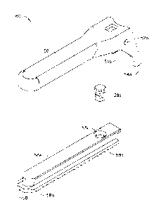
the art. This rolled construction may result in significant cost savings over
extrusion or other like manufacturing processes. Other manufacturing
techniques, such as a drawn tube, are also contemplated herein.
[0095] Referring now to FIGS. 1, 2 and 7, housing 70 includes a proximal
end, a distal
end, and a cavity 59 extending longitudinally therein. Cavity 59 is configured
to
accept a switch assembly 300 and the transducer assembly 50, which interfaces
with housing 68 via switch assembly 300.
[0096] Transducer 50 includes a first conductive ring 400 and a second
conductive ring
410 which are securely disposed within the handpiece transducer body 50. In
one expression of the current embodiment, first conductive ring 400 comprises
a
ring member, which is disposed between the transducer 50 and the horn 130.
Preferably the first conductive ring 400 is formed adjacent to or as part of
the
flange member 160 within the cavity 162 and is electrically isolated from
other
electrical components. The first conductive ring 400 is anchored to and
extends
upwardly from a non-conductive platform or the like (not shown) which is
formed
within the transducer body 50. The first conductive ring 400 is electrically
connected to the cable 22 (FIG. 1) by means of one or more electrical wires
(not
shown), which extend along the length of the transducer body 50 to the first
conductive ring 400.
[0097] The second conductive ring 410 of the transducer 50 similarly
comprises a ring
member that is disposed between the transducer body 150 and the horn 130.
The second conductive ring 410 is disposed between the first conductive ring
400 and the horn 130 and therefore the first and second conductive rings 400,
410 are concentric members. The second conductive ring 410 is likewise
electrically isolated from the first conductive ring 400 and other electrical
END5881W0PCT
CA 02653942 2014-04-16
- 30 -
components contained within the transducer 50. Similar to the first conductive
ring 400, the second conductive ring 410 preferably is anchored to and extends
upwardly from the non-conductive platform. It will be understood that the
first
and second conductive rings 400, 410 are sufficiently spaced from one another
so that they are electrically isolated from each other. This may be
accomplished
by using one or more spacers 413 disposed between the first and second
conductive rings 400, 410 or between the rings 400, 410 and other members
within the transducer 50. The second conductive ring 410 is also electrically
connected to the cable 22 (FIG. 1) by means of one or more electrical wires
(not
shown), which extend along the length of the transducer 50 to the second
conductive ring 410. The second conductive ring 410 is thus provided to
partially
define a second electrical pathway from the cable 22 to the switch mechanism
300. A suitable ultrasonic transducer 50 is Model No. HP054, sold by Ethicon
Endo-Surgery, Inc. of Cincinnati, Ohio.
[0098] In one expression of the current embodiment, the distal end of
transducer 50
threadedly attaches to the proximal end of waveguide 80. The distal end of
transducer 50 also interfaces with switch assembly 300 to provide the surgeon
with finger-activated controls on surgical instrument 100.
[0099] With reference now to FIGS. 8a-c, switch assembly 300 comprises a
pushbutton
assembly 310, a flex circuit assembly 330, a connector assembly 350, a first
spring slip ring conductor 360 and a second spring slip ring conductor 370.
Connector assembly 350 is generally cylindrical and is supported within handle
70 by way of corresponding supporting mounts on switch assembly 350 and
housing portions 68 and 69. Connector assembly 350 defines a first cavity 353,
a mounting boss 352 and a second cavity 351. Cavity 353 is sized to accept the
proximal end of transducer 50, whereby horn 130 passes through cavity 351 to
END5881W0PCT
= CA 02653942 2014-04-16
- 31 -
interface with waveguide 80. Mounting boss 352 accepts slip ring conductors
360 and 370, which in turn electrically engage ring contacts 400 and 410,
respectively.
[00100] With particular reference now to FIG. 8a, slip ring conductors 360 and
370 are
generally open-ended 0-shaped springs that slip onto connector assembly 350.
Each spring slip ring comprises two pressure point contacts (361a-b and 371a-
b)
that contact the respective ring conductor 400 and 410 of transducer 50. The
spring tension of the slip rings 360 and 370 cause positive contact between
contacts 361a-b, 371a-b and conductors 400 and 410. It is evident that the
slip
ring construction may allow electrical contact to be made even as the surgeon
may rotate transducer 50 during use of the instrument. Posts 364 and 374 (not
shown) of the respective slip rings electrically connect to the respective
conductor within flex circuit 330 to complete the electrical circuit as is
known and
understood in the art.
[00101] A flex circuit 330 provides for the electro-mechanical interface
between
pushbuttons 311, 312 and the generator 30 via transducer 50. Flex circuit
comprises two dome switches 332 and 334 that are mechanically actuated by
depressing pushbuttons 311 or 312 respectively of corresponding pushbutton
assembly 310. Dome switches 332 and 334 are electrical contact switches, that
when depressed provide an electrical signal to generator 30 as shown by the
electrical wiring schematic of Fig. 8d. Flex circuit 330 also comprises two
diodes
within a diode package 336, also illustrated in Fig. 8d. Flex circuit 330
provides
conductors, 335 and 337 as is known to those in the art, that connect to slip
ring
conductors 360 and 370 via electrical tabs 364 and 374, respectively, which in
turn provide electrical contact to ring conductors 400 and 410, which in turn
are
connected to conductors in cable 22 that connect to generator 30. Tabs 364 and
END5881W0PCT
CA 02653942 2014-04-16
,
- 32 -
374 (not shown) are soldered to conductors 335 and 337.
[00102] Flex circuit 330 is partially folded and is generally fixedly attached
in handle
assembly 68 so that dome switches 334 and 332 interface with backing surfaces
on handle assembly 69 (not shown). Backing surfaces provide a firm support for
the dome switches during operation, discussed below. Dome switches 334 and
332 may be fixedly attached to backing surfaces by any convenient method, such
as, an adhesive. Flex circuit is secured to connector assembly 350 via
alignment
pins 354 on switch assembly 350 and corresponding alignment holes 338 on flex
circuit 330. As is well appreciated by one skilled in the art various
electrical
constructions are available to provide electrical interface between the
pushbuttons and the generator, which may include molded circuits or standard
wire connections.
[00103] Layered on top of flex circuit is pushbutton assembly 310, which has a
corresponding saddle-shape as flex circuit 330. Pushbutton assembly 310
comprises two pushbuttons, distal pushbutton 312 and proximal pushbutton 311
which have corresponding pressure studs 315 and 314 arranged in a rocker
fashion. In one embodiment, push button assembly 310 comprises a rocker style
pushbutton. Other types of switches, known to the skilled artisan, are equally
applicable. Rocker pushbutton assembly 310 is rotationally attached to handle
70 to provide centering action to the pushbutton assembly 310. As is readily
apparent, by depressing pushbuttons 311 and 312 the corresponding pressure
studs 314 and 315 depress against corresponding dome switches 334 and 332
to activate the circuit illustrated in FIG. 8c. Switches 312 and 311 are
located on
the ultrasonic instruments centerline so that a surgeon may operate the
pushbuttons using either a left hand or a right hand. When the surgeon
depresses switch 312, the generator will respond with a certain energy level,
END5881W0PCT
CA 02653942 2014-04-16
- 33 -
such as a maximum ("max") power setting; when the surgeon depresses switch
311, the generator will respond with a certain energy level, such as a minimum
("min") power setting, which conforms to accepted industry practice for
pushbutton location and the corresponding power setting.
[00104] Alternatively, the pushbuttons may be molded into the connector
assembly 350
or into the handle assembly 68 to reduce the number of components and
increase the reliability of the overall device. The pushbuttons may be
attached
through small cantilever sections, which allow for sturdy attachment of the
pushbutton to the other components, while at the same time allowing for a low
force to activate the pushbuttons.
[00105] In the foregoing embodiment of the present invention, switches 311 and
312
configured in such a way to provide an ergonomically pleasing grip and
operation
for the surgeon. Switches may be placed in the range of the natural swing of
the
surgeon's index or middle fingers, whether gripping surgical instrument 100
right-
handed or left handed. Referring again to FIG. 8b, in a current embodiment a
series of partitions, such as ridges 312a and/or depressions or "peaks and
valleys" are integrated onto the pushbutton 312. The ridges provide tactile
feedback to the surgeon as to the location of the pushbuttons and whether the
button represents min or max power activation. Such tactile feedback is
essential to the surgeon, so the surgeon may continuously assess the surgical
site, but confidently understand which pushbuttons are being activated,
without
the need to view the instrument 100.
[00106] Referring to FIG. 9, a surgeon's left hand is accessing instrument
100. The
thumb is poised to activate trigger handle 34, and the index and middle
fingers
easily engage pushbutton assembly 310. The surgeon's ring finger and pinkie
END5881W0PCT
CA 02653942 2014-04-16
, .
- 34 -
grasp handle 70.
[00107] In FIG. 10, a right-handed the surgeon has depressed trigger handle 34
to close
clamp arm 56 against blade 79. The right forefinger can easily access
pushbutton 312 to activate max power, and the left middle finger can easily
access pushbutton 311b to activate min power. It can be observed that the
surgeon may use the index finger to activate max power and the middle finger
to
activate min power. The rocker type switch allows the surgeon to rest both
fingers on the min and max buttons while ensuring that both buttons are not
activated simultaneously. The rocker type switch facilitates rapid change of
cutting speed from max to min, back to max, etc. In previous devices, the
surgeon would have to move from a foot from one pedal to another, or move his
or her finger from one button to another. In some instances, the surgeon would
have to look away from the operative field to locate either the desired foot
pedal
or desired button. The rocker switch permits the surgeon to rest two fingers
on
the switches during all phases of surgery obviating the need to look at or
search
for the desired button.
[00108] Referring to FIGS. 9 and 10, an expression of surgical instrument 100
is shown
graphically illustrating a surgeon's finger placement on instrument 100.
Closing
of the instrument 100 is achieved by the placement of the thumb through the
opening 34a in trigger handle 34 and depressing trigger handle 34. (Inserting
the
thumb through the opening 34a to activate trigger handle 34 is exemplary only.
Surgeons with larger hands may opt to activate trigger handle 34 with the
thumb
on the outside of trigger handle 34 and trigger handle 34 is provided with
ridges
34b to enable use of the thumb or any other finger or part of the hand on the
outside of trigger handle 34 during surgery). Opening 34a is generally sized
to
accept different sized fingers and thumbs, a common variable as is evident
END5881W0PCT
CA 02653942 2014-04-16
- 35 -
depending upon the sex and size of the surgeon.
[00109] In an alternate expression of the invention, trigger handle 34 and
grip handle 70
have a soft-touch molded thermo plastic elastomer liner (not shown) on their
inner surfaces defining openings 34a and 68a. Plastic liner provides comfort
to
the surgeon and prevents finger and hand fatigue. The plastic liner also
provides
an enhanced gripping surface between the handles and the surgeon's thumb and
fingers. This is particularly advantageous for accepting multiple digit sizes
of
male and female surgeons and still providing a comfortable and positive
gripping
surface. Plastic liner be smooth or have contours molded onto the surface of
liner, such as ribs. Other contours may be bumps, and peaks and valleys.
Various other shapes and interfaces are within the scope of this invention as
would be obvious to one skilled in the art.
[00110] Referring now to FIGS. 2, 11 and 12, a one-piece torque wrench 500 is
shown.
The torque wrench 500, in one embodiment, is provided with cantilever arms 501
disposed in an annular fashion about the centerline of torque wrench 500.
Cantilever arms 501 include teeth 501a disposed, in one embodiment, in an
inward perpendicular fashion in relation to cantilever arms 501. Teeth 501a,
in
one embodiment of the current invention, are disposed with a cam ramp 501b at
a 25 angle with respect to the perpendicular angle between arm 501 and teeth
501a.
[00111] Referring now to FIG. 12, an outer tube retainer 29 is shown. Outer
tube retainer
29 includes spline gears 29a projecting in a perpendicular fashion along the
outer
circumference of retainer 29. Spline gears 29a include cam ramps 29b disposed
at a 25.6 angle with respect to the perpendicular angle between the outer
circumference of retainer 29 and spline gears 29a. Other angles of the teeth
and
END5881W0PCT
CA 02653942 2014-04-16
. .
- 36 -
cam ramps are contemplated and left up to the designer.
[00112] In operation, torque wrench opening 502 is aligned with outer sheath
72 and
guided along substantially the entire length of sheath 72. Torque wrench lip
503
engages the distal end of handgrip 70. Cantilever teeth 501a slidably engage
spline gears 29a on outer tube retainer 29. Cam ramp 501b slidably engages
retainer cam ramps 29b. Clockwise annular motion or torque is imparted to
torque wrench 500 through paddles 504. The torque is transmitted through arms
501 and teeth 501a to gears 29a, which in turn transmit the torque to the
waveguide 80 via insulated pin 27. When a user imparts 5-12 in-lbs. of torque
and holds the handpiece 50 stationary, the ramps 501b and 29b cause the arms
501 to move or flex away from the centerline of wrench 500 ensuring that the
user does not over-tighten the waveguide 80 onto horn 130 (Fig. 7). When a
counter-clockwise torque is applied to wrench 500 via paddles 504 (and holding
the handpiece 50 stationary), the perpendicular flat sides of teeth 501a and
29a
abut allowing a user to impart a torque to the interface between the waveguide
80 and horn 130 in proportion to the force applied to the paddles facilitating
removal of the instrument 100 from the handpiece 50. The torque wrench 500
may be constructed from a durable plastic, such as polycarbonate or a liquid
crystal polymer. It is also contemplated that the wrench 500 may alternatively
be
made from a variety of materials including other plastics, ceramics or metals.
[00113] In another embodiment (not shown), the paddles and cantilever arm
assembly
may be separate components attached by mechanical means or chemical means
such as adhesives or glue.
[00114] Referring now to FIGS. 2, 5, 6 and 13, force limiting mechanism 95
provides a
wave spring 94. Wave spring 94 is operationally coupled to yoke 33, which in
turn is driven by trigger handle 34. Wave spring 94 generates the end effector
END5881W0PCT
CA 02653942 2014-04-16
. .
- 37 -
load and maintains the consistency of the end effector load. As a result, the
end
effector load is more tightly controlled and component abuse load conditions
are
reduced. Mechanical interference or contact between trigger handle 34 and
handle 70 are a safe guard against wave spring 94 being fully compressed,
thereby preventing the spring material to yield and render wave spring 94
useless in subsequent clamp arm closures. As would be appreciated by one
skilled in the art, the application of a mechanical stop spring force limiting
system
has applicability in other energy-based surgical devices (such as RE,
microwave
and laser) that encounter clamping forces, as well as mechanical devices, such
as, clip appliers, graspers and staplers.
[00115] In one expression of the current embodiment, wave spring 94 has a
spring
constant about 43 pounds per inch. Wave spring 94 is preloaded to a force
necessary to achieve the desired transection force, and is a function of the
mechanical advantage of the clamp arm 56 coupling means and frictional losses
in the device. In a second expression of the current embodiment, wave spring
94 is preloaded at about 13 pounds.
[00116] Referring now to FIG. 13, curve 82 illustrates trigger handle 34 force
as a
function of the angular rotation of trigger handle 34 for clamp arm closure
and
curve 83 represents trigger handle force as a function of angular rotation of
trigger handle 34 for clamp arm opening. Point 82a represents the point at
which
yoke 33 begins to deflect or compress wave spring 94 and the force increases
as
trigger handle 34 is depressed further until the force reaches the preload
value of
wave spring 94 at inflection point 82b, and the slope of the force curve
decreases. Point 82c represents trigger handle force where trigger handle 34
contacts handle 70 resulting in a mechanical stop.
[00117] In FIG. 13, curve 83 illustrates the force exerted on the trigger
handle 34 by
END5881W0PCT
CA 02653942 2014-04-16
. .
- 38 -
spring 12. Point 83a represents the point at which trigger handle 34
disengages
from handle 70 and wave spring 94 and spring 12 both exert force against yoke
33. Point 83b represents the maximum force that trigger handle 34 exerts
against a user during the opening stroke of trigger handle 34 and clamp arm
56.
As can be appreciated by curves 82 and 83 in the "closed" region in FIG. 13,
less
force is required to maintain the trigger handle 34 in a closed position than
required to achieve the closed position. This difference in forces proves
advantageous during long surgical procedures where hand fatigue may be a
factor. For example, a surgeon may unknowingly relax pressure on the trigger
handle 34 during a long transection; with lower forces required to keep
trigger
handle 34 fully closed, consistent cutting and hemostasis is achieved.
[00118] Referring now to FIG. 2, surgical instrument 100 further provides for
audible and
tactile means for indicating to the surgeon that the trigger has reached full
travel
and the clamp arm 56 is applying the correct coaptation force to the tissue.
Clicker 339 located in handle 70 contacts trigger handle tab 34a upon closing
or
opening in a momentary fashion such that trigger handle tab 34a causes clicker
339 to yield and spring back to its original position resulting in an audible
and
tactile "click." This is useful during protracted surgical operations or
tissue
transection activities when the surgeon's grip may relax, without the
surgeon's
knowledge, and the pressure delivered to the tissue from the clamp arm 56 may
be unknowingly decreased.
[00119] Clicker 339 is generally planar and made of a flexible plastic that
adequately
deflects when it engages trigger handle tab 34a thereby providing an audible
and/or tactile signal to the surgeon that there is full end effector 81
closure.
Advantageously, tab 34a strikes and deflects clicker 339 when trigger handle
34
is rotated from the full closure position and in the opposite direction
thereby
END5881W0PCT
CA 02653942 2014-04-16
. .
- 39 -
providing an audible and/or tactile signal to the surgeon that full closure of
end
effector 81 no longer exists. As would be appreciated by the skilled artisan,
the
indicating means may be either tactile, audible or visual or a combination.
Various types of indicators may be used including dome switches, solid stops,
cantilever springs or any number of mechanical or electrical switches known to
those skilled in the art. Further various means may be used to provide
feedback
to the surgeon, including, but not limited to, lights, buzzers, and vibratory
elements.
[00120] Preferably, the ultrasonic clamp coagulator apparatus described above
will be
processed before surgery. First, a new or used ultrasonic clamp coagulator
apparatus is obtained and if necessary cleaned. The ultrasonic clamp
coagulator
apparatus can then be sterilized. In one sterilization technique the
ultrasonic
clamp coagulator apparatus is placed in a closed and sealed container, such as
a plastic or TYVEK bag. Optionally, the ultrasonic clamp coagulator apparatus
can be bundled in the container as a kit with other components, including a
torque wrench. The container and ultrasonic clamp coagulator apparatus, as
well as any other components, are then sterilized in any conventional medical
sterilization technique, such as gamma radiation, x-rays, high-energy
electrons
or ETO (ethylene oxide). The sterilization kills bacteria on the ultrasonic
clamp
coagulator apparatus and in the container. The sterilized ultrasonic clamp
coagulator apparatus can then be stored in the sterile container. The sealed
container keeps the ultrasonic clamp coagulator apparatus sterile until it is
opened in the medical facility.
[00121] While the present invention has been illustrated by description of
several
embodiments, it is not the intention of the applicant to restrict or limit the
invention to such detail. Numerous variations, changes, and substitutions will
END5881W0PCT
CA 02653942 2014-04-16
- 40 -
occur to those skilled in the art without departing from the scope of the
invention.
Moreover, the structure of each element associated with the present invention
can be alternatively described as a means for providing the function performed
by the element.
END5881W0PCT