Note: Descriptions are shown in the official language in which they were submitted.
CA 02655024 2011-10-27
= 28569-95
1
MEASUREMENT APPARATUS FOR ENUMERATION OF
WHITE BLOOD CELLS AND RELATED METHOD
Technical Field
The present invention relates to a measurement apparatus and a
method for enumeration of particles, such as white blood cells in a sample,
such as a blood sample. The present invention further relates to a computer
program for analysing a sample.
Background of the Invention
Determining a white blood cell count is often important in connection to
treating a patient_ This analysis may be needed for diagnosing e.g.
leukaemia, or infectious or inflammatory diseases or for monitoring treat-
ments. It is desirable to enable analysis results to be obtained as quickly as
possible in order to minimize waiting times for patients and enabling a
physician to make a decision of treatment and diagnosis directly when making
a first examination of the patient. It would therefore be preferable to
provide
an analysis method which may be quickly performed by the physician or a
nurse without the need of sending a test away to a laboratory. Determining
the white blood cell count is one of the most common tests being performed
on patients in establishing a diagnosis. Therefore, it would be very
advantageous to have a quick and simple method of performing the analysis.
Today, a white blood cell count is normally obtained through a manual
procedure by staining a blood sample and microscopically viewing the sample
in a special counting chamber, e.g. a Butter chamber. The counting chamber
is provided with a grid dividing the chamber in well-defined small volumes.
The white blood cells are allowed to settle at the bottom of the counting
chamber in order to enable the microscope to focus on all cells in the
chamber and, thus, facilitate counting. Thus, the sample needs to settle for
several minutes before the counting may be performed. The white blood cell
count can then be determined by counting the number of blood cells per box
in the grid. The white blood cell count is obtained manually by an analyst,
who
needs to be experienced in performing the analysis in order to be able to
perform a reliable analysis.
This analysis is time-consuming. Further, since it is performed
manually, the results of the analysis may vary depending on the person
performing the analysis.
CA 02655024 2008-12-10
PCT/SE2007/000656
WO 2008/010761
2
There are a few number of existing automated analysis methods for
determining a white blood cell count. The white blood cell count may be
determined by means of the Coulter principle, which is based on determining
cell size and thereby the cell type by sensing an impedance. A method for
counting white blood cells by the Coulter principle is described in US
5,262,302. Measurement apparatus according to the Coulter principle is
expensive and it is therefore a considerable investment. Thus, a hospital or
laboratory will be reluctant to invest in more than one apparatus. This
implies
that the analysis will need to be performed in a centralised location and a
patient will need to wait for analysis results.
The Coulter principle is the dominating, automated analysis method
that is presently being used. However, there are a few other methods that
have been described. One such method for determining a white blood cell
count is disclosed in US 5,585,246. Here, a blood sample has to be prepared
by being mixed with a fluorescent dye and ligand complex which tags the
white blood cells. The sample is introduced into a capillary and is irradiated
by
a laser source which scans over the sample in the capillary. The fluorescence
is measured in order to determine the number of white blood cells. A similar
method is disclosed in WO 97/02482, using a fluorescent dye and a laser
source scanning over a capillary. This method is adapted for enumeration of
white blood cells in apheresis products containing a low number of white
blood cells. Here, the capillary is quite thick and it is necessary to wait
until
the white blood cells have settled at the bottom of the capillary before the
capillary may be scanned.
In WO 99/45384, a sample-containing chamber having varying
thickness is shown. The varying thickness separates different compounds of
blood. The blood sample is stained with a colorant to differentially highlight
at
least three different white blood cell types in the blood sample. The white
blood cells may be enumerated by using an optical scanning instrument to
view a portion of the chamber.
In WO 98/50777, a method for assessment of the number of somatic
cells in milk is disclosed. The method comprises applying a volume of a
sample in a sample compartment and transmitting electromagnetic signals,
having passed from the sample compartment, onto an array of detection
elements. The intensities of detected electromagnetic signals are processed
and the results are correlated to the number of cells present in the sample.
= CA 02655024 2011-10-27
28569-95
3
There is still a need to speed up and simplify existing automated
methods for determining a white blood cell count such that the analysis may
be performed by any user, not requiring special training, and such that
measurement apparatuses may be relatively inexpensive. This would imply
that the analysis may be provided at a point of care. Further, since the white
blood cell count is such a commonly performed analysis, any improvement to
the analysis method would have a positive impact on patient care. An
analysis method providing a possibility to obtain results at a point of care
would be particularly advantageous.
Also, it may be advantageous to obtain a differential white blood cell
count, that is to examine the distribution of different types of white blood
cells
in a blood sample. This differential white blood cell count may reveal if the
cells are present in a normal distribution, or if any cell type is increased
or
decreased. The information may be useful in diagnosing specific types of
illness. For example, an increase in neutrophils indicates a bacterial
infection,
whereas an increase in lymphocytes is common in acute viral infections.
The differential white blood cell count may also be obtained by
microscopically viewing and manually counting stained blood cells in a Barker
chamber. There also exist some automated methods. For example, a
differential count may be obtained with the Coulter principle by analysing the
form and size of the electrical pulse generated by a cell passing through an
electrical field. The form and size of the pulse may be related to the type of
white blood cell being detected. One such method is described in US
4,528,274.
In US 5,123,055, another method for identifying different types of white
blood cells is described. This method requires several size and colour
parameters to be sequentially analysed in order to differentiate the types of
white blood cells.
It is still desired to speed up and simplify existing automated methods
for determining a differential white blood cell count. It would be
particularly
advantageous to provide a quick, simple and relatively inexpensive analysis
method such, that the analysis may be provided at a point of care.
Summary of the Invention
It is an object of some embodiments to provide a simple analysis for
determining a volumetric enumeration of particles, such as white blood cells
=
CA 02655024 2011-10-27
28569-95
4
in a sample, such as a blood sample and determining a differential particle
count, such as a differential white blood cell count.
Thus, according to one aspect of the invention, there is provided a
measurement apparatus for enumeration of particles, such as white blood
cells in a sample, such as a blood sample.
The apparatus comprises a holder, which is arranged to receive a
sample acquiring device comprising a measurement cavity that holds a
sample, an imaging system adapted to acquire at least one magnified digital
image of the sample. The apparatus further comprises an image analyser,
which is arranged to analyse the at least one acquired digital image for
identifying the particles and determining the number of particles in the
sample, wherein the image analyser is arranged to analyse the at least one
acquired digital image for identifying particles that are imaged in focus and
determining types and number of these particles, the types being
distinguished by physical features of the particles, whereby the ratio of.
different types of particles in the sample is determined.
The imaging system may comprise a magnifying means and at least
one digital image acquiring means.
In accordance with one embodiment the apparatus is adapted for
enumeration of white blood cells in a blood sample, the measurement cavity
is adapted to hold a stained and hemolysed blood sample, and wherein the
image analyser is arranged to analyse the at least one acquired digital image
for identifying stained white blood cells and determining the number of white
blood cells in the sample, wherein the image analyser is arranged to analyse
the at least one acquired digital image for identifying white blood cells that
are
imaged in focus and determining types and number of these white blood cells,
the types being distinguished by geometric features of the white blood cells,
whereby the ratio of different types of white blood cells in the sample is
determined.
CA 02655024 2011-10-27
28569-95
According to another aspect, there is provided a measurement
apparatus for enumeration of white blood cells in a sample, the apparatus
comprising: a holder, which is arranged to receive a sample acquiring device
comprising a measurement cavity that holds a sample, wherein the measurement
5 cavity comprises a reagent comprising a hemolysing agent for lysing red
blood cells
in the sample and a staining agent for staining white blood cells in the
sample, an
imaging system adapted to acquire a plurality of digital images of the sample
at
different levels in the direction along an optical axis using different
optical settings,
and an image analyser, which is arranged to analyse each acquired digital
image for
identifying stained white blood cells and determining a number of white blood
cells in
the sample, wherein the image analyser is arranged to analyse each acquired
digital
image for identifying white blood cells that are imaged in focus and
determining types
and number of these white blood cells, the types being distinguished by
physical
features of the white blood cells, whereby a ratio of different types of white
blood cells
in the sample can be determined.
According to another aspect of the invention, there is provided a
method for enumeration of particles in a sample, said method comprising:
acquiring a
sample into a measurement cavity of a sample acquiring device, acquiring at
least
one digital image of a magnification of the irradiated sample in the
measurement
cavity, digitally analysing the at least one digital image for identifying the
particles and
determining the number of particles in the sample, and digitally analysing the
at least
one digital image for identifying particles that are imaged in focus and
determining
types and number of these particles, the types being distinguished by
geometric
features of the particles, whereby the ratio of different types of particles
in the sample
is determined.
In accordance with one embodiment the method is adapted for
enumeration of white blood cells in a blood sample. The method comprises
acquiring
a blood sample into a measurement cavity of a sample acquiring device. The
blood
sample is mixed with a reagent, comprising a hemolysing agent for lysing the
red
CA 02655024 2011-10-27
= 28569-95
5a
blood cells in the blood sample and a staining agent for staining the white
blood cells
in the blood sample. The staining agent preferably selectively stains white
blood cells
and does not stain other cells in the blood sample. The method further
comprises
acquiring at least one digital image of a magnification of the sample in the
measurement cavity. The method further comprises digitally analysing the at
least
one digital image for identifying stained white blood cells and determining
the number
of white blood cells in the sample, and digitally analysing the at least one
digital
image for identifying white blood cells that are imaged in focus and
determining types
and number of these white blood cells, the types being distinguished by
geometric
features of the stained cells, whereby the ratio of different types of white
blood cells in
the blood sample is determined.
According to another aspect, there is provided a method for
enumeration of white blood cells in a sample, said method comprising:
acquiring a
sample into a measurement cavity of a sample acquiring device, the sample
being
mixed with a reagent, comprising a hemolysing agent for lysing red blood cells
in the
sample and a staining agent for staining white blood cells in the sample,
acquiring a
plurality of digital images at different levels in the direction along the
optical axis of
the sample of a magnification of an irradiated sample in the measurement
cavity
using different optical settings, digitally analysing each acquired digital
image for
identifying the stained white blood cells and determining a number of white
blood
cells in the sample, and digitally analysing each acquired digital image for
identifying
stained white blood cells that are imaged in focus and determining types and
number
of these white blood cells, the types being distinguished by physical features
of the
stained white blood cells, whereby a ratio of different types of white blood
cells in the
sample can be determined.
CA 02655024 2011-10-27
28569-95
5b
The measurement apparatus and the method of the invention both
enable simple analysis of a sample of whole blood. To this end, the
measurement apparatus is arranged to acquire at least one digital image of a
blood sample, which sample has been mixed with a staining agent for staining
the white blood cells. The staining of the white blood cells implies that the
white blood cells may be distinguished in a digital image and different types
of
white blood cells may be distinguished by geometric features of the cells in
the same or another digital image.
The measurement apparatus and the method are thus arranged to
both determine a volumetric enumeration of all white blood cells within the
blood sample and determine a differential white blood cell count.
Whereas many existing methods are able to count different blood cells
and even subgroups of blood cells, the measurement apparatus according to
the invention is specifically adapted to analysis of white blood cells. The
reagent comprises a hemolysing agent which will lyse the red blood cells in
the blood sample. This destroys the possibilities to enumerate the red blood
cells in the sample. On the other hand, the lysing of the red blood cells
simplifies the distinguishing and identification of the white blood cells
within
the blood sample.
CA 02655024 2008-12-10
PCT/SE2007/000656
WO 2008/010761
6
Further, the measurement apparatus is specifically adapted to analyse
the at least one digital image such that cells that are imaged in focus are
identified. This allows an image to be acquired of a relatively thick sample,
while only the cells that are in focus are counted. This feature is
particularly
useful considering that the enumeration of the total number of white blood
cells is more easily made than the identification of the type of white blood
cells, since typing requires more details of the cell to be analysed. Thus, by
ensuring that only cells that are in focus are counted, the identification of
the
type of white blood cells may be performed in a sample that may simultane-
ously be used for determining a statistically reliable volumetric enumeration
of
the white blood cells in the sample.
The measurement apparatus and the method of the invention provide a
very simple analysis of a sample of whole blood. The analysis does not
require complicated measurement apparatus or advanced steps to be
performed by an operator. Therefore, it may be performed in direct
connection to examination of a patient, without the need for a qualified
technician. It is merely required that a blood sample is acquired and mixed
with a staining agent. Then, the blood sample may be placed in the holder of
the measurement apparatus and, in direct response thereto, the
measurement apparatus may present analysis results.
In fact, the blood sample may be allowed to be mixed with the reagent
in the measurement cavity. Thus, there will be no need to perform a sample
preparation manually. Within a few minutes or less, the reaction of the blood
sample with the reagent will have hemolysed the red blood cells and stained
the white blood cells such that the sample is ready for optical measurement to
acquire the at least one digital image. The blood sample may be mixed with
the reagent by e.g. dispersion or diffusion of the reagent into the blood
sample or by actively vibrating or moving the sample acquiring device so that
an agitation is caused in the measurement cavity.
The measurement apparatus may further comprise an electromagnetic
radiation source, which is arranged to irradiate the sample held in the meas-
urement cavity of the sample acquiring device.
The imaging system may be arranged to acquire a plurality of digital
images of the sample using different optical settings, wherein the image
analyser is arranged to analyse each acquired digital image for identifying
particles or stained white blood cells and determining the number of particles
or white blood cells in the sample, wherein the image analyser is arranged to
_
CA 02655024 2008-12-10
PCT/SE2007/000656
WO 2008/010761
7
analyse each acquired digital image for identifying particles or white blood
cells that are imaged in focus and determining types and number of these
particles or white blood cells, the types being distinguished by geometric
features of the particles or stained white blood cells, whereby the ratio of
different types of particles or white blood cells in the sample is determined.
By acquiring a plurality of digital images at different levels in the
direction of depth of field in the sample, it is possible to analyse a
relatively
large sample volume even when using a high magnification. A high
magnification makes it, due to the resulting small depth of field, difficult
to
view the complete volume in one image. Since the magnification level affects
the depth of field, the step of acquiring a plurality of digital images allows
the
use of a greater magnification, which in turn makes it possible to, in each
image, differentiate between different kinds of white blood cells depending,
amongst others, upon the shape, number or size of the nuclei.
According to another embodiment, the imaging system is arranged to
provide information about the direction of light in the acquired image to
facilitate focusing, whereby shifting focus in the acquired image is enabled.
This implies that a single image may be used both for enumeration of the total
number of white blood cells in the sample analysing the entire depth of the
sample at once, and for determining the ratio of different types of white
blood
cells in the blood sample by analysing cells in the image when the image is
shown with a portion of the thickness of the blood sample being in focus. An
image comprising information of direction of light into the image may be
obtained using an array of small lenses (e.g., a compound lens)providing
ability to trace rays in the acquired image such that different parts of the
image may be placed in focus.
The imaging system may be arranged to acquire a first and a second
digital image of the sample using different optical settings, and wherein the
image analyser is arranged to analyse the first acquired digital image for
determining the number of particles or white blood cells in the sample and the
image analyser is arranged to analyse the second acquired digital image for
determining the ratio of different types of particles or white blood cells in
the
sample.
Thus, the measurement apparatus is specifically adapted to acquire
two digital images using different optical settings. This implies that the
optical
settings may be optimised and adapted to, firstly, determine the number of
CA 02655024 2008-12-10
WO 2008/010761 PCT/SE2007/000656
8
white blood cells within a volume and, secondly, determine a ratio of
different
types of white blood cells.
The imaging system may comprise two at least partly separate parts,
which direct light from an irradiated sample to a first and a second part of
the
imaging system.
The action to determine if a white blood cell is in focus or not may be
performed by making use of the fact that the cytoplasm of the cell may act as
a lens refracting the light. For a white blood cell imaged in focus the nuclei
appear as dark shadows whereas the surrounding cytoplasm is almost
invisible. The nuclei appear as regions with significantly lower light
intensity
whereas the cytoplasm leaves the light intensity unaffected.
For a white blood cell imaged too close to the imaging system (too
close to be in focus) the nuclei appear as dark shadows whereas the
surrounding cytoplasm acts as a lens and refracts the light which results in a
dark circle around the nuclei. The nuclei appear as a region with
significantly
lower light intensity relative to a focused image of the nuclei and the
cytoplasm appears with low light intensity.
For a white blood cell imaged too far away from the imaging system
(too far to be in focus) the nuclei appear as dark shadows whereas the
surrounding cytoplasm acts as a lens and refracts the light resulting in a
bright circle around the nuclei. The nuclei appear as a region with
significantly
lower light intensity relative to a focused image of the nuclei whereas the
cytoplasm appears with high light intensity.
Alternatively, the identifying of the cells that are imaged in focus may
be performed by analysing the edges of imaged cells in order to assess
whether the cell is imaged in focus based on a slope of intensity at the edge.
Cells that are not in focus will show a slow decrease in intensity at the
edges,
whereas cells in focus will be imaged with a sharp edge represented as a
large decrease in intensity at the edge of the cell. Thus, by analysing how
the
intensity varies at an edge of an imaged cell, it may be determined whether
the cell is imaged in focus or not.
An alternative way to determine the cell type is by, in the image
analyser, for a specific particle or cell, determining the number of said
images
in which said particle or cell is imaged counting from an image in which the
particle or cell is determined to be out of focus in a first direction to an
image
in which the particle or cell is determined to be out of focus in a second
direction.
CA 02655024 2008-12-10
PCT/SE2007/000656
WO 2008/010761
9
The image analyser may be arranged to determine, based on the
counted number of images, a geometrical feature related to the size of said
particle or cell.
The imaging system with the optical settings used for acquiring said at
least one digital image may have a magnification power of 1-50x, more
preferably 1-20x, more preferably 3-20x, more preferably 5-20x and more
preferably about 10x.
The imaging system may be arranged to obtain said at least one digital
image with a depth of field in the range of 2-60 micrometers, more preferably
in the range of 2-30 micrometers, more preferably about 8-10 micrometers.
As used in this context, "depth of field" implies a length in a direction
along the optical axis that is imaged in a sufficient focus to allow image
analysis to identify cells positioned within this length. This "depth of
field" may
be larger than a conventional depth of field defined by the optical settings.
With an increased magnification power, the depth of field is decreased.
The electromagnetic radiation source may be arranged to irradiate a
wavelength corresponding to a peak in absorbance of the staining agent.
Consequently, the stained white blood cells which contain an accumulation of
staining agent will be detected by an indication of a low transmittance of
light
in the digital images.
The electromagnetic radiation source may comprise a laser source.
The laser source may provide light of a well-defined wavelength fitting the
absorbance of the staining agent. Further, the laser source may provide colli-
mated light, minimizing disturbances of stray light, such that a point of low
transmittance of light will be sharply distinguished.
The electromagnetic radiation source may alternatively comprise a light
emitting diode. This radiation source may still provide sufficient irradiating
conditions for properly distinguishing white blood cells from other matter in
the
sample.
The image analyser may be arranged to identify areas of high light
absorbance in order to determine the number of particles or white blood cells
in the sample. The image analyser may be further arranged to identify black
or dark dots in the image. Since the staining agents may be accumulated in
the nuclei of the white blood cells, the absorbance of the light may have
peaks at separate points. These points will form black dots in the digital
image and may be classified as white blood cells.
CA 02655024 2008-12-10
PCT/SE2007/000656
WO 2008/010761
The image analyser may be arranged to distinguish different types of
particles or white blood cells by analysing shape and size of identified areas
of high light absorbance in the at least one digital image. Since different
types
of white blood cells have different sizes, the type of a white blood cell may
be
5 identified by determining the size of the blood cell. Further, the
different types
may be differently stained giving different shapes of the identified areas in
the
digital image. This may also be used in order to identify the type of white
blood cells. A differential white blood cell count specifying the ratio of
three
different types of white blood cells may be obtained by analysing the size of
10 the blood cells. A differential white blood cell count distinguishing
five different
types of white blood cells may require further features of the blood cells to
be
investigated. For example, a number of nuclei of each cell, an intensity of
radiation transmitted through the blood cell, or the shape of the blood cell
may
be examined.
The staining agent may be arranged to selectively stain the nuclei of
the white blood cells. This implies that the white blood cells may be
identified
as coloured dots and therefore easily be distinguished and counted in a
digital
image. Further, the size of the stain spots may be used to identify the type
of
the white blood cells, as different types of white blood cells have different
sizes.
The staining agent may be any one in the group of Hematoxylin,
Methylene blue, Methylene green, Methylene azure, cresyl violet acetate,
Toluidine blue, Gentian violet, Sudan analogues, Gallocyanine, and Fuchsin
analogues, or any combination thereof. However, it should be appreciated
that the staining agent is not limited to this group, but many other
substances
may be contemplated.
The hemolysing agent may be a quaternary ammonium salt, a saponin,
a bile acid, such as deoxycholic acid, a digitoxin, a snake venom, a
glucopyranoside or a non-ionic detergent of type Triton. However, it should be
appreciated that the hemolysing agent is not limited to this group, but many
other substances may be contemplated.
The measurement apparatus may further comprise an objective lens
which is shared for the different optical settings. This implies that the
digital
images may be obtained by imaging along the same optical path such that
the images are centred at the same point in the measurement cavity. This
makes the measurement apparatus compact.
CA 02655024 2008-12-10
WO 2008/010761 PCT/SE2007/000656
11
According to one embodiment, the imaging system may comprise two
at least partly separate parts, which direct light from an irradiated sample
to a
first and a second part of the imaging system. This implies that the path of
light from the sample to the imaging system may be defined within a fixed
optical set-up. Thus, the measurement apparatus may be robust and
insensitive to impact.
The imaging system may further comprise a beam splitter for directing
light from the objective lens towards the first or the second part of the
imaging
system. This implies that the first and second digital images may be obtained
simultaneously, whereby the analysis may be very quickly performed.
The first part of the imaging system may be arranged to receive light
directly from the beam splitter, that is no optical element is arranged
between
the first part of the imaging system and the beam splitter. Alternatively, the
light may be arranged to pass directly from the objective lens to the first
part
of the imaging system. Then, in order to obtain the second digital image, a
mirror may be inserted into the light path for deflecting light to the second
part
of the imaging system instead.
The imaging system may further comprise an ocular lens between the
beam splitter and the part of the imaging system adapted to acquire digital
images. The ocular lens may thus provide a further magnification of the
sample in order to distinguish between different types of white blood cells.
Preferably, lens packages are used and the ocular lens package will then
move a virtual principal plane within the objective lens package to change the
relation between the image plane and the objective lens package to allow
further magnification.
The imaging system may further comprise an optical element between
the beam splitter and the part of the imaging system adapted to acquire
digital
images for affecting cells not positioned in focus of the imaging system,
whereby identifying white blood cells that are imaged in focus is facilitated.
The optical element allows an image to be acquired of a sample
thickness much larger than the depth of field of the imaging system. The
optical element ensures that the cells that are out of focus may be withdrawn
from consideration in order to increase the certainty of the measurement.
Since the optical element affects the imaging of cells out of focus, the cells
in
focus will be easily identified. The optical element may be implemented as a
spatial filter that affects the imaging of a cell such that the edge of the
cell will
. . . = - ¨
CA 02655024 2008-12-10
WO 2008/010761 PCT/SE2007/000656
12
comprise an overshoot intensity larger than the background intensity, where
the cell is imaged by absorbing light.
According to an alternative embodiment, the imaging system may
further comprise a wavefront coding element between the beam splitter and
the second part of the imaging system. A wavefront coding element
deliberately distorts the light rays by passing them through a waveplate with
a
saddle-like shape, that is relatively flat in the middle, but with scalloped
edges. This causes a specific optical aberration, the image looks blurry, but
the de-focus is the same over a large range of distances. This wavefront
coding element thus increases a depth along the optical axis that may be
analysed. The distortions in the image are mainly determined by the shape of
the de-focusing wavefront coding element, which is accurately known.
Therefore, a computer is able to remove the blur point by point. A computer
may decode the image using what is essentially a digital filter, and thus cre-
ates an image which is sharp over a large depth of field. In this way, the
depth of field of the imaging system may be increased, enabling a larger
depth of a sample to be imaged in focus.
According to another embodiment, one part of the imaging system is
arranged to acquire both the first and second images and at least part of the
magnification system of the imaging system is switchable in order to acquire
the first and second digital images using different optical settings. This
implies
that the measurement apparatus need only comprise one single part of the
imaging system. Further, it allows several different optical settings to be
used
by e.g. providing a main lens that is movable between well-defined positions
along the optical axis.
The imaging system may be arranged having a larger magnification
power in the optical settings used for acquiring the second digital image than
in the optical settings used for acquiring the first digital image. This
implies
that details may be better viewed in the second digital image, whereby
different types of white blood cells may more easily be distinguished from
each other.
The imaging system with the optical settings used for acquiring the first
digital image may have a magnification power of 1-50x, more preferably 1-
20x, more preferably 3-20x, more preferably 3-10x and more preferably about
4x. Within these ranges of magnification power, the white blood cells are
sufficiently magnified in order to be detected, while the imaging system may
be arranged to image the sample thickness within sufficient focus in order to
CA 02655024 2008-12-10
PCT/SE2007/000656
WO 2008/010761
13
assess the number of blood cells within the image. Thus, the imaging system
may have a depth of field covering the sample thickness. However, the entire
sample thickness need not be imaged within a depth of field of the imaging
system, using a conventional definition of depth of field. Cells that are
imaged
slightly out of focus may still be correctly counted using clever image
analysis.
A low magnification power implies that a large "depth of field" may be
obtained. Thus a large sample thickness may be allowed and a large volume
may be analysed. However, if a low magnification power is used, the white
blood cells may be hard to detect because each blood cell is imaged onto
very few pixels, such as 3-4 pixels. A lower magnification power may be used
by increasing the number of pixels in the acquired image, that is by improving
the resolution of the digital image. In this way, it is possible to use an
optical
magnification power of 1-4x, while still enabling the white blood cells to be
detected.
The imaging system with the optical settings used for acquiring the
second digital image may have a magnification power of 1-50x, more
preferably 1-20x, more preferably 3-20x, more preferably 5-20x and more
preferably about 10x. Within these ranges of magnification power, the white
blood cells are sufficiently magnified in order to distinguish between
different
types of white blood cells. A lower magnification power may be used by using
an optical element for emphasizing cells that are imaged in focus and
facilitating identification of these cells.
The imaging system may be arranged to obtain the first image with a
depth of field of at least the thickness of the measurement cavity of the
sample acquiring device. This implies that a sufficient focus is obtained of
the
entire sample thickness such that the entire thickness of the measurement
cavity may be simultaneously analysed in the digital image of the sample.
Thus, there is no need to await that the white blood cells settle in the meas-
urement cavity, whereby the time for making an analysis is reduced.
However, there may be a need to await a reaction causing the red blood cells
to be hemolysed and await movements caused by introduction of the sample
into the measurement cavity to settle. These waiting times would be very
short, in the order of 30 seconds or less. By choosing not to focus very
sharply on a specific part of the sample, a sufficient focus is obtained of
the
entire sample thickness to allow identifying the number of white blood cells
in
the sample. This implies that a white blood cell may be somewhat blurred and
still be considered to be in focus of the depth of field. The analysed volume
of
- - - -
CA 02655024 2008-12-10
PCT/SE2007/000656
WO 2008/010761
14
the sample may thus be well-defined by the thickness of the measurement
cavity and the size of the digital image specifying the cross-sectional area
of
the measurement cavity being imaged.
The imaging system may be arranged to obtain the first image with a
depth of field in the range of 50-200 micrometers. This depth of field may be
adapted to correspond to the depth or thickness of the measurement cavity. A
depth of at least 50 micrometers allows a larger volume of blood to be ana-
lysed over a small cross-sectional area, thus avoiding compression of the
blood cells of the sample into a monolayer. Thus, a sufficiently large volume
of the blood sample in order to give reliable values of the white blood cell
count may be analysed using a relatively small image of the blood sample.
Further, it is difficult to achieve a depth of field exceeding 200 micrometers
while obtaining a digital image with a sufficient magnification. It is even
difficult to achieve a depth of field exceeding 170 micrometers.
The imaging system may be arranged to obtain the second image with
a depth of field in the range of 2-60 micrometers. This may be achieved by
imaging a portion of the thickness of the measurement cavity. In such case,
only this portion of the thickness of the measurement cavity is imaged in
focus. The second digital image is then analysed by only taking into account
white blood cells that are imaged in sufficient focus in order to determine
their
type. Since the second digital image is used to determine the ratio of
different
types of white blood cells, it is not important to image a well-defined
volume.
Thus, it is possible to obtain appropriate first and second images by imaging
the same portion of the measurement cavity. However, the second image
may alternatively be acquired imaging a different portion of the measurement
cavity, whereby this portion may have a thickness corresponding to the depth
of field of the imaging system for obtaining the second image.
The image analyser may be arranged to electronically magnify the at
least one acquired image. While the sample is being magnified for acquiring a
magnified digital image of the sample, the acquired digital image itself may
be
electronically magnified for simplifying distinguishing between objects that
are
imaged very closely to each other in the acquired digital image.
According to another aspect of the invention, there is provided a
sample acquiring device for enumeration of white blood cells in a blood
sample. The sample acquiring device comprises a measurement cavity for
receiving a blood sample. The measurement cavity has a first and a second
predetermined fixed thickness defined between inner walls of the
CA 02655024 2008-12-10
WO 2008/010761 PCT/SE2007/000656
measurement cavity, wherein the first thickness is adapted for determining
total volumetric enumeration of white blood cells in the blood sample and the
second thickness is adapted for determining a ratio of different types of
white
blood cells within the blood sample. The sample acquiring device further
5 comprises a reagent, which is arranged in a dried form on a surface
defining
the measurement cavity. The reagent comprises a hemolysing agent for
lysing red blood cells in the blood sample, and a staining agent for
selectively
staining white blood cells in the blood sample.
The sample acquiring device provides a possibility to directly obtain a
10 sample of whole blood into the measurement cavity and provide it for
analysis. There is no need for sample preparation. In fact, the blood sample
may be sucked into the measurement cavity directly from a pricked finger of a
patient. Providing the sample acquiring device with a reagent enables a
reaction within the sample acquiring device which makes the sample ready
15 for analysis. The reaction is initiated when the blood sample comes into
contact with the reagent. Thus, there is no need for manually preparing the
sample, which makes the analysis especially suitable to be performed directly
in an examination room while the patient is waiting.
Since the reagent is provided in a dried form, the sample acquiring
device may be transported and stored for a long time without affecting the
usability of the sample acquiring device. Thus, the sample acquiring device
with the reagent may be manufactured and prepared long before making the
analysis of a blood sample.
Whereas many existing methods are able to count different blood cells
and even subgroups of blood cells, the sample acquiring device according to
the invention is specifically adapted to performing enumeration of white blood
cells. The reagent comprises a hemolysing agent which will lyse the red blood
cells in the blood sample. This destroys the possibilities to enumerate the
red
blood cells in the sample. On the other hand, the lysing of the red blood
cells
simplifies the distinguishing and identification of the white blood cells
within
the blood sample.
The staining agent provides a marking of the individual white blood
cells. This enables the white blood cells to be individually viewed or
detected.
The white blood cells may e.g. be detected by scanning the measurement
cavity or obtaining an image of the measurement cavity.
The sample acquiring device further provides a first thickness of the
measurement cavity specifically adapted to facilitate determining a volumetric
CA 02655024 2008-12-10
WO 2008/010761 PCT/SE2007/000656
16
white blood cell count. The measurement cavity may have a sufficient
thickness to allow a quite large volume of the blood sample to be analysed
and therefore allow a good statistic for determining the volumetric white
blood
cell count. The white blood cell count may thus be obtained by summing the
number of individually detected white blood cells in a defined volume.
The sample acquiring device also provides a second thickness of the
measurement cavity specifically adapted to facilitate distinguishing between
different types of white blood cells. In this regard, the second thickness may
be thinner than the first thickness allowing the entire second thickness to be
imaged within a depth of field of a larger magnification. Such a larger
magnification may be needed when distinguishing between different types of
white blood cells in comparison to imaging in order to merely determine the
total number of white blood cells, regardless of type, within the blood
sample.
The sample acquiring device may comprise a body member having two
planar surfaces forming inner walls to define said measurement cavity. The
planar surfaces may be arranged at a predetermined distance from one
another to determine a sample depth for an optical measurement. This
implies that the sample acquiring device provides a well-defined depth to the
optical measurement, which may be used for accurately determining the white
blood cell count per volumetric unit of the blood sample. A volume of an ana-
lysed sample will be well-defined by the depth of the measurement cavity and
an area of the sample being imaged. Thus, the well-defined volume could be
used for associating the number of white blood cells to the volume of the
blood sample such that the volumetric white blood cell count is determined.
The measurement cavity preferably has a first uniform depth of 50-200
micrometers. A depth of at least 50 micrometers implies that the
measurement cavity does not force the blood sample to be smeared into a
monolayer thereby allowing a larger volume of blood to be analysed over a
small cross-sectional area. Thus, a sufficiently large volume of the blood
sample in order to give reliable values of the white blood cell count may be
analysed using a relatively small image of the blood sample. The first depth
is
more preferably at least 100 micrometers, which allows an even smaller
cross-sectional area to be analysed or a larger sample volume to be ana-
lysed. Further, the first depth of at least 50 micrometers and more preferably
100 micrometers also simplifies manufacture of the measurement cavity
having a well-defined depth between two planar surfaces.
CA 02655024 2008-12-10
WO 2008/010761 PCT/SE2007/000656
17
For most samples arranged in a cavity having a thickness of no more
than 200 micrometers, the white blood cell count is so low that there will be
only minor deviations due to white blood cells being arranged overlapping
each other. However, the effect of such deviations will be related to the
white
blood cell count and may thus, at least to some extent, be handled by means
of statistically correcting results at least for large values of the white
blood cell
count. This statistical correction may be based on calibrations of the
measurement apparatus. The deviations will be even less for a measurement
cavity having a first thickness of no more than 170 micrometers, and even
less for a measurement cavity having a first thickness of no more than 150
micrometers, whereby a simpler calibration may be used. This thickness may
even not require any calibration for overlapping blood cells.
Further, the first thickness of the measurement cavity is sufficiently
small to enable the measurement apparatus to obtain a digital image such
that the entire depth of the measurement cavity may be analysed simulta-
neously. Since a magnifying system is to be used in the measurement
apparatus, it is not easy to obtain a large depth of field. Therefore, the
first
thickness of the measurement cavity would preferably not exceed 150
micrometers in order for the entire thickness to be simultaneously analysed in
a digital image. The depth of field may be arranged to handle a first
thickness
of the measurement cavity of 170 micrometers or even 200 micrometers.
The measurement cavity preferably has a second uniform thickness of
20-60 micrometers. This second thickness of the measurement cavity would
allow the entire second thickness to be imaged within a depth of field of a
magnification needed for distinguishing between different types of white blood
cells. Further, the second thickness may still allow a sufficient volume to be
imaged enabling a substantial number of white blood cells to be analysed.
This would allow the ratio of different types of white blood cells to be
determined with a good statistical certainty. Typically, it is desired to
analyse
the type of 200 white blood cells.
The sample acquiring device may be provided with a reagent that has
been applied to the surface solved in a volatile liquid which has evaporated
to
leave the reagent in a dried form.
It has been realised that the reagent is advantageously solved in a
volatile liquid before being inserted into the measurement cavity. This
implies
that the liquid may in an effective manner be evaporated from the narrow
space of the measurement cavity during manufacture and preparation of the
CA 02655024 2011-10-27
. 28569-95
18
sample acquiring device. The reagent may preferably be arranged in a dried
form in the part of the measurement cavity of the first thickness.
The reagent may preferably be solved in an organic solvent and more
preferably be solved in methanol. Such solvents are volatile and may
appropriately be used for drying the reagent onto a surface of the
measurement cavity.
The sample acquiring device may further comprise a sample inlet
communicating the measurement cavity with the exterior of the sample
acquiring device, wherein the inlet is arranged to acquire a blood sample. The
sample inlet may be arranged to draw up a blood sample by a capillary force
and the measurement cavity may further draw blood from the inlet into the
cavity. Also, the sample acquiring device may be arranged to first draw the
sample into the portion of the measurement cavity of the first thickness. Part
of the sample may then be further transported by capillary force into the
portion of the measurement cavity of the second thickness. As a result, the
blood sample may easily be acquired into the measurement cavity by simply
moving the sample inlet into contact with blood. Then, the capillary forces of
the sample inlet and the measurement cavity will draw up a well-defined
amount of blood into the measurement cavity. Alternatively, the blood sample
may be sucked or drawn into the measurement cavity by means of applying
an external pumping force to the sample acquiring device. According to
another alternative, the blood sample may be acquired into a pipette and then
be introduced into the measurement cavity by means of the pipette.
The sample acquiring device may be disposable, i.e. it is arranged to
be used once only. The sample acquiring device provides a kit for performing
a white blood cell count, since the sample acquiring device is able to receive
a blood sample and holds all reagents needed in order to present the sample
to cell counting. This is particularly enabled since the sample acquiring
device
is adapted for use once only and may be formed without consideration of
possibilities to clean the sample acquiring device and re-apply a reagent.
Also, the sample acquiring device may be moulded in plastic material and
thereby be manufactured at a low price rate. Thus, it may still be cost-
effective to use a disposable sample acquiring device.
Some embodiments also relate to a computer program product,
embodied in a computer-readable medium, for analysis of a sample,
comprising: computer code for digitally analyzing at least one image of a
sample for determining a number of particles in the sample; computer code
for digitally analyzing the at
=
CA 02655024 2013-12-17
28569-95
19
least one image of the sample for identifying one or more types of particles
in a
focused region of the sample, each type of particle being associated with one
or more
distinguishing physical features; and computer code for outputting information
corresponding to the number and types of particles in the sample.
According to another aspect, there is provided a computer readable
memory having recorded thereon statements and instructions for execution by a
computer for implementing a method of analysing a sample, the statements and
instructions comprising: code means for digitally analyzing a plurality of
images of the
sample for determining a number of white blood cells in the sample, the
plurality of
digital images of the sample acquired at different levels in the direction
along an
optical axis using different optical settings; code means for digitally
analyzing the
plurality of images of the sample acquired at different levels in the
direction along an
optical axis using different optical settings, for identifying one or more
types of white
blood cells in a focused region of the sample, each type of white blood cell
being
associated with one or more distinguishing physical features; code means for
outputting information corresponding to the number and types of white blood
cells in
the sample.
Some embodiments also relate to a computer program for analysing a
sample, the computer program comprising computer program code for: analysing
at
least one digital image for identifying the particles and determining the
number of
particles in the sample, and analysing the at least one digital image for
identifying
particles that are imaged in focus and determining types and number of these
particles, the types being distinguished by physical features of the
particles, whereby
the ratio of different types of particles in the sample is determined.
According to another aspect, there is provided a computer readable
memory having recorded thereon statements and instructions for execution by a
computer for implementing a method of analysing a sample, the statements and
instructions comprising: code means for analysing a plurality of digital
images for
identifying white blood cells, the plurality of digital images of the sample
acquired at
CA 02655024 2013-12-17
28569-95
19a
different levels in the direction along an optical axis using different
optical settings,
and determining a number of white blood cells in the sample, and code means
for
analysing the plurality of digital images acquired at different levels in the
direction
along an optical axis using different optical settings for identifying white
blood cells
that are imaged in focus and determining types and number of these white blood
cells, the types being distinguished by physical features of the white blood
cells,
whereby a ratio of different types of white blood cells in the sample can be
determined.
Brief Description of the Drawings
The invention will now be described in further detail by way of example
under reference to the accompanying drawings.
Fig. 1 is a schematic view of a sample acquiring device.
Fig. 2 is a schematic block diagram of a measurement apparatus
according to a first embodiment.
Fig. 3 is a schematic block diagram of a measurement apparatus
according to a second embodiment.
Fig. 4 is a flow chart of a method according to a first embodiment of the
invention.
Fig. 5 is a schematic view of an arrangement for a movable lens
according to an embodiment of the invention.
Fig. 6 is a schematic view of an arrangement according to an
embodiment of the invention.
Fig. 7 illustrates a sample imaged at three different layers.
Fig. 8a illustrates a white blood cell in camera view when the cell is
positioned out of focus of a measurement apparatus according to Fig. 10.
CA 02655024 2013-12-17
,
28569-95
19b
Fig. 8b illustrates a white blood cell in camera view when the cell is
positioned in focus of a measurement apparatus according to Fig. 10.
Fig. 8c illustrates a white blood cell in camera view when the cell is
positioned out of focus of a measurement apparatus according to Fig. 10.
Fig. 9a illustrates recorded intensities of a cross-section of a cell to be
analysed, when the cell is positioned out of focus of a measurement apparatus
according to Fig. 10.
CA 02655024 2008-12-10
WO 2008/010761 PCT/SE2007/000656
Fig. 9b illustrates recorded intensities of a cross-section of a cell to be
analysed, when the cell is positioned in focus of a measurement apparatus
according to Fig. 10.
Fig. 9c illustrates recorded intensities of a cross-section of a cell to be
5 analysed, when the cell is positioned out of focus of a measurement
apparatus according to Fig. 10.
Fig. 10 is a schematic view of a measurement apparatus according to
a third embodiment.
Fig. 11 is a flow chart of a method according to a second embodiment
10 of the invention.
Fig. 12 is a schematic view of a measurement apparatus according to
a fourth embodiment.
Detailed Description of Preferred Embodiments
15 Referring now to Fig. 1, a sample acquiring device 10 according to a
first embodiment will be described. The sample acquiring device 10 is
preferably disposable and is to be thrown away after having been used for
analysis. This implies that the sample acquiring device 10 does not require
complicated handling. The sample acquiring device 10 is preferably formed in
20 a plastic material and may be manufactured by injection-moulding. This
makes manufacture of the sample acquiring device 10 simple and cheap,
whereby the costs of the sample acquiring device 10 may be kept down.
The sample acquiring device 10 comprises a body member 12, which
has a base 14, which may be touched by an operator without causing any
interference in analysis results. The base 14 may also have projections 16
that may fit a holder in an analysis apparatus. The projections 16 may be
arranged such that the sample acquiring device 10 will be correctly positioned
in the analysis apparatus.
The sample acquiring device 10 further comprises a sample inlet 18.
The sample inlet 18 is defined between opposite walls within the sample
acquiring device 10, the walls being arranged so close to each other that a
capillary force may be created in the sample inlet 18. The sample inlet 18
communicates with the exterior of the sample acquiring device 10 for allowing
blood to be drawn into the sample acquiring device 10. The sample acquiring
device 10 further comprises a chamber for counting white blood cells in the
form of a measurement cavity 20 arranged between opposite walls inside the
sample acquiring device 10. The measurement cavity 20 is arranged in
CA 02655024 2008-12-10
WO 2008/010761 PCT/SE2007/000656
21
communication with the sample inlet 18. The walls defining the measurement
cavity 20 are arranged closer together than the walls of the sample inlet 18,
such that a capillary force may draw blood from the sample inlet 18 into the
measurement cavity 20.
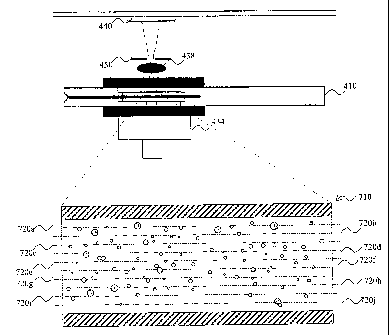
The measurement cavity 20 has a first portion 20a having a first
thickness and a second portion 20b having a second, smaller thickness. The
first portion 20a is in communication with the sample inlet 18, whereas the
second portion 20b is in communication with the first portion 20a. Thus, a
capillary force may draw blood from the first portion 20a of the measurement
cavity 20 into the second portion 20b.
The walls of the first portion 20a of the measurement cavity 20 are
arranged at a distance from each other of 50-200 micrometers. The first
portion 20a is more preferably at least 100 micrometers thick. Further, the
first
portion 20a is more preferably no more than 150 micrometers thick. The
distance is generally uniform over the entire first portion 20a. The thickness
of
the first portion 20a defines the volume of blood being examined. Since the
analysis result is to be compared to the volume of the blood sample being
examined, the generally uniform thickness of the first portion 20a needs to be
very precise, i.e. only very small variations in the thickness are allowed
between first portions 20a of different sample acquiring devices 10. The
thickness is chosen to allow a relatively large sample volume to be analysed
in a small area of the cavity so that a sufficient number of particles or
cells are
available for counting. The The first portion 20a of the measurement cavity
20 is specifically adapted for determining a volumetric total white blood cell
count in a blood sample. The entire thickness of the first portion 20a may be
chosen to allow it to be imaged within a depth of field of an imaging system.
Then, an image may be analysed and the number of white blood cells present
in the image may be counted in order to determine the volumetric white blood
cell count.
The sample acquiring device 10 is typically adapted for measuring
white blood cell counts above 0.5 x 10⁹ cells/litre blood. At much lower white
blood cell counts, the sample volume will be too small to allow statistically
significant amounts of white blood cells to be counted. Further, when the
white blood cell count exceeds 12 x 109 cells/litre blood, the effect of blood
cells being arranged overlapping each other will start to be significant in
the
measured white blood cell count. At this white blood cell count, the white
blood cells will cover approximately 8% of the cross-section of the sample
CA 02655024 2008-12-10
WO 2008/010761
PCT/SE2007/000656
22
being analyzed, if the thickness of the first portion 20a is 140 micrometers.
Thus, in order to obtain correct white blood cell counts, this effect will
need to
be accounted for. Therefore, a statistical correction of values of the white
blood cell count above 12 x 109 cells/litre blood may be used. This
statistical
correction will increase with increasing white blood cell counts, since the
effect of overlapping blood cells increases with increased white blood cell
counts. The statistical correction may be determined by means of calibration
of a measurement apparatus. As an alternative, the statistical correction may
be determined at a general level for setting up measurement apparatuses to
be used in connection to the sample acquiring device 10. This statistical cor-
rection is of similar magnitude as statistical corrections that are presently
performed in analysis apparatus that use the Coulter principle. It is
contemplated that the sample acquiring device 10 could be used to analyse
white blood cell counts as large as 50 x 109 cells/litre blood.
The second portion 20b of the measurement cavity 20 is specifically
adapted for determining a ratio of different types of white blood cells in a
blood sample. The entire thickness of the second portion 20a is to be imaged
within a depth of field of an imaging system. Then, an image may be analysed
and the number of white blood cells of each type present in the image may be
counted in order to determine the ratio of different types of white blood
cells.
The walls of the second portion 20b of the measurement cavity 20 are
arranged at a distance from each other of 20-60 micrometers. The distance is
generally uniform over the entire second portion 20b. Since the analysis is
mainly intended to compare the number of different types of white blood cells
to each other, it is not critical to know the exact volume being analysed.
Therefore, the thickness of the second portion 20b need not be as precise as
the thickness of the first portion 20a. The thickness of the second portion
20b
needs to allow a sufficient amount of white blood cells to be analysed in
order
to obtain statistically significant results. Further, as stated above the
thickness
of the second portion 20b should be adapted to be imaged in its entirety
within a depth of field of an imaging system. Thus, all white blood cells
within
the sample are imaged in focus and the analysis of the sample is not
hampered by noise in the image from parts of the sample imaged out of
focus. The second portion 20b is thinner than the first portion 20a in order
to
enable a larger magnification to be used while allowing the entire second
portion to be imaged within a depth of field of the imaging system. The larger
CA 02655024 2008-12-10
WO 2008/010761 PCT/SE2007/000656
23
magnification may be needed in order to allow not only counting the total
number of white blood cells but also determining the type of white blood
cells.
A surface of a wall of the measurement cavity 20 is at least partly
coated with a reagent 22. The reagent 22 may be freeze-dried, heat-dried or
vacuum-dried and applied to the surface of the measurement cavity 20. When
a blood sample is acquired into the measurement cavity 20, the blood will
make contact with the dried reagent 22 and initiate a reaction between the
reagent 22 and the blood.
The reagent 22 is applied by inserting the reagent 22 into the
measurement cavity 20 using a pipette or dispenser. The reagent 22 is solved
in a volatile liquid, e.g. an organic solvent such as methanol, when inserted
into the measurement cavity 20. The solvent with the reagent 22 may fill the
measurement cavity 20. Then, drying is performed such that the solvent will
be evaporated and the reagent 22 will be attached to the surfaces of the
measurement cavity 20.
Since the reagent is to be dried onto a surface of a narrow space, the
liquid will have a very small surface in contact with ambient atmosphere,
whereby evaporation of the liquid is rendered more difficult. Thus, it is
advantageous to use a volatile liquid, such as methanol, which enables the
liquid to be evaporated in an effective manner from the narrow space of the
measurement cavity.
According to an alternative manufacturing method, the sample
acquiring device 10 may be formed by attaching two pieces to each other,
whereby one piece forms the bottom wall of the measurement cavity 20 and
the other piece forms the top wall of the measurement cavity 20. This allows a
reagent 22 to be dried onto an open surface before the two pieces are
attached to each other. Thus, the reagent 22 may be solved in water, since
the solvent need not be volatile.
The reagent 22 comprises a red blood cell hemolysing agent and a
white blood cell staining agent. The hemolysing agent may be a quaternary
ammonium salt, a saponin, a bile acid, such as deoxycholic acid, a digitoxin,
a snake venom, a glucopyranoside or a non-ionic detergent of type Triton.
The staining agent may be Hematoxylin, Methylene blue, Methylene green,
Methylene azure, cresyl violet acetate, Toluidine blue, Gentian violet, a
Sudan
analogue, Gallocyanine, or a Fuchsin analogue, or any combination thereof.
When a blood sample makes contact with the reagent 22, the hemolysing
agent will act to lyse the red blood cells such that the lysed red blood cells
are
CA 02655024 2008-12-10
WO 2008/010761 PCT/SE2007/000656
24
mixed with the blood plasma. Further, the staining agent will accumulate in
the nuclei of the white blood cells. The reagent 22 should contain sufficient
amounts of staining agent to distinctly stain all the nuclei of the white
blood
cells. Thus, there will often be a surplus of staining agent, which will be
intermixed in the blood plasma. The surplus of staining agent will give a
homogenous, low background level of staining agent in the blood plasma.
The accumulated staining agent in the white blood cells will be
distinguishable
over the background level of staining agent.
The reagent 22 may also comprise other constituents, which may be
active, i.e. taking part in the chemical reaction with the blood sample, or
non-
active, i.e. not taking part in the chemical reaction with the blood sample.
The
active constituents may e.g. be arranged to catalyse the hemolysing or
staining action. The non-active constituents may e.g. be arranged to improve
attachment of the reagent 22 to the surface of a wall of the measurement
cavity 20.
Within a few minutes or even less than a minute, the blood sample will
have reacted with the reagent 22, such that the red blood cells have been
lysed and the staining agent has accumulated in the nuclei of the white blood
cells.
Referring now to Fig. 2, a first embodiment of a measurement
apparatus 30 for analysis of white blood cells in a blood sample will be de-
scribed. The apparatus 30 comprises a sample holder 32 for receiving a
sample acquiring device 10 with a blood sample. The sample holder 32 is
arranged to receive the sample acquiring device 10 such that the
measurement cavity 20 of the sample acquiring device 10 is correctly
positioned within the apparatus 30. The apparatus 30 comprises a light
source 34 for illuminating the blood sample within the sample acquiring
device 10. The light source 34 may be an incandescent lamp, which irradiates
light in the entire visible spectrum. The staining agent which is accumulated
in
the nuclei of the white blood cells will absorb light of specific wavelengths,
such that the nuclei of the white blood cells will emerge in a digital image
of
the sample. If a colour image is acquired, the white blood cells will emerge
as
specifically coloured dots. If a black and white image is acquired, the white
blood cells will emerge as dark dots against a lighter background.
The light source 34 may alternatively be a laser or a light emitting
diode. This may be used for increasing contrast in the image such that the
white blood cells may be more easily detected. In this case, the light source
CA 02655024 2008-12-10
WO 2008/010761 PCT/SE2007/000656
34 is arranged to radiate electromagnetic radiation of a wavelength that
corresponds to an absorption peak of the staining agent. The wavelength
should further be chosen such that the absorption of the non-white blood cells
components in the blood is relatively low. Further, the walls of the sample
5 acquiring device 10 should be essentially transparent to the wavelength.
For
example, when Methylene blue is used as the staining agent, the light source
34 may be arranged to irradiate with light having a wavelength of 667 nm.
The apparatus 30 further comprises an imaging system 36, which is
arranged on an opposite side of the sample holder 32 relative to the light
10 source 34. Thus, the imaging system 36 is arranged to receive radiation
which has been transmitted through the blood sample. The imaging system
36 in this embodiment comprises a magnifying means 38 that is divided into
two separate parts. A first part 38a of the magnifying means 38 is arranged to
receive radiation that has been transmitted through the blood sample in the
15 first portion 20a of the measurement cavity 20. The imaging system
further
comprises a first image acquiring means 40, which is arranged to image the
first portion 20a of the measurement cavity 20 as magnified by the first part
38a of the magnifying means 38. The first part 38a of the magnifying means
38 is arranged to provide a magnifying power of 1-50x, more preferably 1-
20 20x, and most preferably 1-4x. Within these ranges of magnifying power,
it is
possible to distinguish the white blood cells. The image may be acquired with
an improved resolution in order to allow lower magnifying power to be used.
Further, the depth of field of the first part 38a of the magnifying means 38
may be arranged to include the thickness of the measurement cavity 20.
25 The first part 38a of the magnifying means 38 comprises an objective
lens or lens system 42, which is arranged close to the sample holder 32, and
an ocular lens or lens system 44, which is arranged at a distance from the
objective lens 42. Each of the objective lens or lens system 42 and the ocular
lens or lens system 44 may include one or a plurality of individual lenses or
other optical components. The objective lens 42 provides a first magnification
of the sample, which is further magnified by the ocular lens 44. The
magnifying means 38 may comprise further lenses for accomplishing an
appropriate magnification and imaging of the sample. The first part 38a of the
magnifying means 38 is arranged such that the sample in the first portion 20a
of the measurement cavity 20 when placed in the sample holder 32 will be
focussed onto an image plane of the first image acquiring means 40.
CA 02655024 2008-12-10
WO 2008/010761 PCT/SE2007/000656
26
The first image acquiring means 40 is arranged to acquire a first digital
image of the sample. The first image acquiring means 40 may be any kind of
digital camera, such as a CCD- or CMOS-camera. Reference to a digital
camera as described herein should be considered as only one embodiment of
an image analysis portion. The pixel size of the digital camera sets a
restriction on the imaging system 36 such that the circle of confusion in the
image plane may not exceed the pixel size within the depth of field. However,
the white blood cells may still be detected even if they are somewhat blurred
and, therefore, the circle of confusion may be allowed to exceed the pixel
size
while being considered within the depth of field, as defined in this context.
As
used herein, "depth of field" will thus imply a length in a direction along
the
optical axis that is imaged in a sufficient focus to allow image analysis to
identify cells positioned within this length. This "depth of field" may be
different from a conventional depth of field defined by the optical settings
and
may depend on the specific image analysis to be performed.
The digital camera 40 will acquire a first digital image of the sample in
the first portion 20a of the measurement cavity 20, wherein the entire sample
thickness is sufficiently focussed in the first digital image for counting the
white blood cells. The imaging system 36 will define an area of the first
portion 20a of the measurement cavity 20, which will be imaged in the first
digital image. The area being imaged together with the thickness of the first
portion 20a of the measurement cavity 20 defines the volume of the sample
being imaged.
A second part 38b of the magnifying means 38 is arranged to receive
radiation that has been transmitted through the blood sample in the second
portion 20b of the measurement cavity 20. The imaging system further com-
prises a second image acquiring means 41, which is arranged to image the
second portion 20b of the measurement cavity 20 as magnified by the second
part 38b of the magnifying means 38. The second part 38b of the magnifying
means 38 is arranged to provide a magnifying power of 5-200x, more
preferably 5-100x, and most preferably 5-20x. Within these ranges of
magnifying power, it is possible to distinguish between white blood cells. The
image may be acquired with an improved resolution in order to allow lower
magnifying power to be used. Further, the depth of field of the second part
38b of the magnifying means 38 may still be arranged to include the thickness
of the measurement cavity 20.
CA 02655024 2008-12-10
WO 2008/010761 PCT/SE2007/000656
27
Like the first part 38a, the second part 38b of the magnifying means 38
also comprises an objective lens or lens system 43, which is arranged close
to the sample holder 32, and an ocular lens or lens system 45, which is
arranged at a distance from the objective lens 43. Again, each of the
objective
lens or lens system 43 and the ocular lens or lens system 45 may include one
or a plurality of lenses or other optical components. The objective lens 43
provides a first magnification of the sample, which is further magnified by
the
ocular lens 45. The magnifying means 38 may comprise further lenses or
other optical components for accomplishing an appropriate magnification and
imaging of the sample. The second part 38b of the magnifying means 38 is
arranged such that the sample in the second portion 20b of the measurement
cavity 20 when placed in the sample holder 32 will be focussed onto an image
plane of the second image acquiring means 41.
The second image acquiring means 41 is arranged to acquire a second
digital image of the sample. The second image acquiring means 41 may be
any kind of digital camera, such as a CCD- or CMOS-camera. Since the
second image is to be used for determining different types of white blood
cells, the circle of confusion in the image plane may not exceed the pixel
size
within the depth of field. The digital camera 41 will acquire a second digital
image of the sample in the second portion 20a of the measurement cavity 20,
wherein the entire sample thickness is sufficiently focussed in the second
digital image for identifying the type of the white blood cells present.
The imaging system 36 can be arranged for imaging blood samples in
sample acquiring devices 10 without the need to adjust the imaging system
36. Preferably, the imaging system 36 is arranged within a housing which
maintains the imaging system in a fixed relationship to the sample holder.
The apparatus 30 further comprises an image analyser 46. The image
analyser 46 is connected to the first and second digital cameras 40, 41 for
receiving first and second digital images acquired by the digital cameras 40,
41. The image analyser 46 is arranged to identify patterns in the first
digital
image that correspond to a white blood cell for counting the number of white
blood cells being present in the digital image. Thus, the image analyser 46
may be arranged to identify dark dots in a lighter background. The image
analyser 46 may be arranged to first electronically magnify the digital image
before analysing the digital image. This implies that the image analyser 46
may be able to more easily distinguish white blood cells that are imaged
CA 02655024 2008-12-10
WO 2008/010761 PCT/SE2007/000656
28
closely to each other, even though the electronic magnifying of the digital
image will make the digital image somewhat blurred.
The image analyzer 46 may include a processor adapted to receive
image information from the first and second digital cameras 40, 41. The
processor may be configured with image analysis software or algorithms, for
example, the precise nature of which may be adapted to perform analyses as
described herein.
The image analyser 46 may calculate the number of white blood cells
per volume of blood by dividing the number of white blood cells being
identified in the first digital image with the volume of the blood sample,
which
is well-defined as described above. The volumetric white blood cell count may
be presented on a display of the apparatus 30.
The image analyser 46 is further arranged to identify patterns in the
second digital image that correspond to a white blood cell for counting the
number of white blood cells being present in the digital image. The image
analyser 46 will further analyse the shape and size of each detected white
blood cell in order to determine the type of the white blood cell. Thus, the
image analyser 46 may be arranged to identify dark dots in a lighter back-
ground as white blood cells. The image analyser 46 may be arranged to first
electronically magnify the digital image before analysing the digital image.
This implies that the image analyser 46 may be able to more easily
distinguish white blood cells that are imaged closely to each other, even
though the electronic magnifying of the digital image will make the digital
image somewhat blurred. The image analyser 46 will then determine the type
of the white blood cell by various physical criteria, an important one being
the
size of the imaged white blood cell. In accordance with literature lymphocytes
have a diameter of about 5-11 micrometers, granulocytes have a diameter of
about 8-15 micrometers, and monocytes have a diameter of about 16-25
micrometers. Since the expected sizes in some cases overlap further
information is preferably used to discriminate different white blood cell
types
from each other. Such information may e.g. be the shape and/or size of the
nucleus. Granulocytes may e.g. be identified by presence of two or more dots
within a cell corresponding to a segmented nucleus. This may be used to
improve the assessment made by the size classification.
A white blood cell count differentiated in five parts, wherein the
granulocytes are further differentiated as eosinophils, neutrophils and
basophils, may be obtained by using further physical characteristics. Also,
the
CA 02655024 2008-12-10
WO 2008/010761 PCT/SE2007/000656
29
three part white blood cell count may be improved using these additional
physical characteristics. Thus, the analysis may further examine the shape of
the detected blood cells. Also, the analysis may further examine an intensity
of radiation transmitted through the detected blood cells.
The image analyser 46 may calculate the number of white blood cells
of each type. Typically, the image analyser 46 may count and classify a
certain number, e.g., 1000 white blood cells. The percentage or ratio of each
type of white blood cells may then be determined as the number of white
blood cells classified to belonging to the type divided with the total number
of
analysed white blood cells. A statistically significant measure may be
determined by analysing about 200 white blood cells for type. However, it is
desired that a larger number of white blood cells are analysed for type in
order to improve statistics. Further, the image analyser 46 may be arranged
to analyse white blood cells that are imaged in sufficient focus to be
properly
classified. Also, where two or more white blood cells are very close to each
other, they may be difficult to separate correctly, and thus such white blood
cells may be disregarded completely by the image analyser 46. On the other
hand, since the imaging system 36 is arranged to image the entire thickness
of the second portion 20b of the measurement cavity 20 in focus, the image
analyser 46 may determine a volumetric enumeration of each type of white
blood cells from the second digital image only.
The image analyser 46 may be realised as a processing unit, which
comprises codes for performing the image analysis.
Referring now to Fig. 3, a second embodiment of a measurement
apparatus 130 for analysis of white blood cells in a blood sample will be
described. The apparatus 130 comprises a sample holder 132 for receiving a
sample acquiring device 110 with a blood sample. The apparatus 130 is
arranged to receive sample acquiring devices 110, wherein the measurement
cavity 120 has one uniform thickness over the entire area being imaged.
Thus, the measurement cavity 120 has a thickness corresponding to the first
portion 20a of the measurement cavity 20 of the sample acquiring device 10
according to the embodiment described above with reference to Fig. 1. The
sample holder 132 is arranged to receive the sample acquiring device 110
such that the measurement cavity 120 of the sample acquiring device 110 is
correctly positioned within the apparatus 130. The apparatus 130 comprises a
light source 134 for illuminating the blood sample within the sample acquiring
CA 02655024 2008-12-10
WO 2008/010761 PCT/SE2007/000656
device 110 in a corresponding way as the light source 34 of the first
embodiment.
The apparatus 130 further comprises an imaging system 136, which is
arranged on an opposite side of the sample holder 132 relative to the light
5 source 134. Thus, the imaging system 136 is arranged to receive radiation
which has been transmitted through the blood sample. The imaging system
136 in this embodiment is arranged to acquire a first and a second digital
image along the same optical path such that the images are centred at the
same point in the measurement cavity 120. Still, the first and second digital
10 images of the sample are acquired using different optical settings. This
may
be achieved in a number of different ways, as will be described below.
As shown in Fig. 3, the imaging system comprises a magnifying means
138 that comprises a common part and two separate parts. The magnifying
means 138 may thus comprise an objective lens or lens system 142, which is
15 arranged close to the sample holder 132 and which is shared for the two
optical settings for acquiring both the first and the second digital images.
The
objective lens 142 provides a first magnification of the sample. The imaging
system 136 may further comprise a beam splitter 139 for directing light in two
different directions towards a first and a second image acquiring means 140,
20 141, which may be any kind of digital camera, such as a CCD-camera. The
magnifying means 138 comprises a first ocular lens or lens system 144,
which is arranged between the beam splitter 139 and the first digital camera
140. The objective lens 142 provides a first magnification of the sample,
which is further magnified by the ocular lens 144. The magnifying means 138
25 may comprise further lenses or optical elements for accomplishing an
appropriate magnification and imaging of the sample in the first digital
image.
The magnifying means 138 further comprises a second ocular lens or
lens system 145 may be, which is arranged between the beam splitter 139
and the second digital camera 141. The objective lens 142 provides a first
30 magnification of the sample, which is further magnified by the ocular
lens 145.
The magnifying means 138 may comprise further lenses or optical elements
for accomplishing an appropriate magnification and imaging of the sample in
the second digital image. The objective lens 142 and the ocular lens 145 may
be implemented as lens packages and the ocular lens package 145 will then
move a virtual principal plane within the objective lens package 142 to change
the relation between the image plane and the objective lens package 142 to
allow the further magnification, while the sample acquiring device 110 is not
CA 02655024 2008-12-10
WO 2008/010761
PCT/SE2007/000656
31
moved in relation to the objective lens package 142. In this way, different
magnifications may be obtained in the first and second digital images.
In particular, the magnifying means 138, as shown in this embodiment,
comprises an optical element 147 that emphasizes imaging of white blood
cells that are placed in focus. This enhances possibilities to identify which
blood cells types are being imaged in focus and thereby are to be considered
when distinguishing between different types of white blood cells.
The optical element 147 allows an image to be acquired of a sample
thickness much larger than the depth of field of the part of the imaging
system
136 that captures the second digital image. The optical element 147 ensures
that the cells that are out of focus may be withdrawn from consideration in
order to increase the certainty of the measurement. Since the optical element
147 affects the imaging of cells out of focus, the cells in focus will be
easily
identified. The optical element 147 may be implemented as a spatial filter
that
affects the imaging of a cell such that the edge of the cell will comprise an
overshoot intensity larger than the background intensity, where the cell is
imaged by absorbing light. This may easily be detected in image analysis
and, therefore, these cells may be quickly discarded from consideration.
According to an alternative embodiment, the magnifying means may
comprise a wavefront coding element between the beam splitter 139 and the
second digital camera 141. The wavefront coding element may thus replace
the optical element 147. A wavefront coding element deliberately distorts the
light rays by passing them through a waveplate with a saddle-like shape, that
is relatively flat in the middle, but with scalloped edges. This causes a
specific
optical aberration, the image looks blurry, but the de-focus is the same over
a
large range of distances. This wavefront coding element thus increases a
depth along the optical axis that may be analysed. The distortions in the
image are mainly determined by the shape of the de-focusing wavefront
coding element, which is accurately known. Therefore, a computer is able to
remove the blur point by point. A computer may decode the image using what
is essentially a digital filter, and thus create an image which is sharp over
a
large depth of field. In this way, the magnifying means may increase the
depth of field of the imaging system, enabling a larger depth of a sample to
be
imaged in focus.
In this embodiment, the beam splitter may be replaced with, the image
system 136 may comprise a mirror or other element (not shown) for directing
essentially all light from the sample towards a selected one of the two
digital
CA 02655024 2008-12-10
WO 2008/010761 PCT/SE2007/000656
32
cameras 140, 141. The mirror may then be turned or moved for shifting the
camera 140, 141 that is viewing the sample. This allows more light to pass to
the digital cameras 140, 141 and thus gives better light conditions for
acquiring the images. However, the two images may not be recorded
simultaneously and the image system 136 will need moving parts. According
to one alternative, one of the cameras may be arranged to view the sample
when the mirror is completely removed from the optical path.
According to another alternative, the objective lens 142 may provide all
magnification needed for obtaining the first image. Thus, light may be passed
directly from the beam splitter or mirror to the first digital camera 140.
According to yet another alternative, no objective lens is shared by the
first and second optical settings. Thus, a beam splitter or mirror may be
arranged close to the sample holder 132 and the magnifying means 138 may
comprise both an objective lens and an ocular lens in both optical paths
between the beam splitter and the first digital camera 140 and between the
beam splitter and the second digital camera 141.
According to a further alternative, the first and second digital images
are obtained by means of one digital camera. In this case, the magnifying
means 138 needs to be switched or changed in order to change the optical
settings for obtaining the two digital images. Thus, an objective lens 242 may
be movable between two different well-defined positions, as shown in Fig. 5.
The objective lens 242 may thus be arranged to be moved along the optical
axis and will make contact with a stop, e.g. an edge of the optical axis
making
contact with a protrusion 250, 252. The distance between the objective lens
and the sample acquiring device may thus be accurately controlled for
controlling the magnification of an image to be acquired.
In any of the above described alternatives, the first digital camera 140
is arranged to image the measurement cavity 120 with a first optical setting
provided by the magnifying means 138. The magnifying means 138 is thus
arranged to provide a magnifying power of 1-50x, more preferably 1-20x, and
most preferably 1-4x. Within these ranges of magnifying power, it is possible
to distinguish the white blood cells. The image may be acquired with an
improved resolution in order to allow lower magnifying power to be used.
Further, the depth of field of the magnifying means 138 may still be arranged
to include the thickness of the measurement cavity 120.
As described with reference to the first embodiment shown in Fig. 2,
the first digital camera 140 in the second embodiment is arranged to acquire
CA 02655024 2008-12-10
WO 2008/010761 PCT/SE2007/000656
33
a first digital image of the sample. The first digital camera 140 views the
sample such that the entire thickness of the measurement cavity 120 is within
the depth of field as defined for the first embodiment. The imaging system
136 will define an area of the measurement cavity 120, which will be imaged
in the first digital image. The area being imaged together with the thickness
of
the measurement cavity 120 defines the volume of the sample being imaged.
Further, in any of the above described embodiments, the second digital
camera 141 is arranged to image the measurement cavity 120 with a second
optical setting provided by the magnifying means 138. The magnifying means
138 is arranged to provide a magnifying power of 5-200x, more preferably 5-
100x, and most preferably 5-20x. Within these ranges of magnifying power, it
is possible to distinguish the white blood cells. The image may be acquired
with an improved resolution in order to allow lower magnifying power to be
used. However, since the second digital image views the same part of the
measurement cavity 120 as the first digital image, the greater magnification
used in the second optical setting may prevent the entire thickness of the
measurement cavity 120 from being imaged within a depth of field. The
second digital image will thus image white blood cells in focus, but will also
image white blood cells and other parts of the blood sample that are out of
focus causing a blurring disturbance in the image. In these conditions, the
optical element 147, as described above, will improve the chances to identify
cells that are imaged in focus making easier to classify as to type.
The magnifying means 138 may advantageously be arranged to place
a top portion of the thickness of the measurement cavity 120 in focus in the
image plane of the second digital camera 141. This implies that the distur-
bances of the parts of the blood sample that are out of focus are near the
bottom and remain relatively low. However, it is conceivable that any portion
of the measurement cavity 120 is imaged in focus in the second digital image.
Further, the magnifying means 138 may be arranged to typically image a
thickness of 20-60 micrometers of the measurement cavity 120 in focus.
According to yet another alternative, as illustrated in Fig. 6, only one
digital image is acquired. However, this digital image needs to provide
information of the direction of light being detected. This implies that the
digital
image contains information of not only the detected radiation, but also a
point
in space from which the detected radiation was emitted. This digital image
may then be presented in such a way that the focus of the digital image may
be shifted as desired. The digital image may thus be used firstly to count a
CA 02655024 2008-12-10
WO 2008/010761 PCT/SE2007/000656
34
total number of white blood cells within the entire depth of the measurement
cavity and secondly, by shifting focus to a portion of the thickness, to
determine the ratio of different types of white blood cells in the sample.
This
alternative may be implemented as shown in Figure 6 comprising a light
source 334, an objective lens 342 and one digital camera 340. These parts
may be implemented in a similar way as described above. The apparatus
further comprises an array of small lenses 360 being provided in the optical
path between the sample acquiring device 110 and the digital camera 340.
The array of small lenses 360 provides a possibility to trace rays in the
acquired image such that different parts of the image may be placed in focus.
Returning now to Fig. 3, the apparatus 130 further comprises an image
analyser 146. The image analyser 146 is connected to the first and second
digital cameras 140, 141 for receiving first and second digital images
acquired
by the digital cameras 140, 141. Alternatively, the image analyser 146
receives only one digital image containing information of direction of light
as
described in the above paragraph. The image analyser 146 is arranged to
analyse the first and second digital images in a similar way as described for
the image analyser 46 of the first embodiment above. However, since the
second digital image may be obtained by imaging only part of the thickness of
the sample within a depth of field, the image analyser 146 may need to
handle the second digital image more carefully. First of all, the image
analyser 146 will only analyse white blood cells that are identified to be
imaged in focus. This is possible since the image analyser 146 may only
determine the ratio of different types of white blood cells and will therefore
not
need to exactly know the volume of the sample being analysed. Cells that are
imaged out of focus may be blurred in such a way that the image analyser
146 could determine incorrect sizes of the cells and therefore incorrectly
classify the cells. Thus, by ensuring that only cells that are imaged in focus
are analysed, the certainty of the analysis is improved.
Fig. 7 illustrates a sample 710 imaged at three different layers 720a-c
of the sample 710. Layer 720b indicates a focus plane to be discussed in
detail below. An optic system has a depth of field in which objects may be
considered to be in focus even if they are not positioned exactly in the focus
plane. In Fig. 7 the depth of field of focus plane 720b is indicated by the
dashed area 720b'.
Figs. 8a-c illustrate three different white blood cells in camera view and
Fig. 9a-c illustrate their respective light distributions.
CA 02655024 2008-12-10
WO 2008/010761 PCT/SE2007/000656
Fig. 8b illustrates a white blood cell imaged in focus. The nuclei appear
as dark shadows whereas the surrounding cytoplasm is almost invisible. In
Fig. 9b the distribution of light intensity is shown. The nuclei appear as
portions with significantly lower light intensity whereas the cytoplasm leaves
5 the light intensity unaffected.
Fig. 8a illustrates a white blood cell imaged too close to the image
acquiring means 441 to be in focus. The nuclei appear as dark shadows
whereas the surrounding cytoplasm acts as a lens and refracts and diffuses
the light which results in a dark circle around the nuclei. In Fig. 9a the
10 distribution of light intensity is shown. The nuclei appear as a portion
with
significantly lower light intensity and the cytoplasm appears with low light
intensity.
Fig. 8c illustrates a white blood cell imaged too far away from the
image acquiring means 441 to be in focus. The nuclei appear as dark
15 shadows whereas the surrounding cytoplasm acts as a lens and refracts
the
light resulting in a bright circle around the nuclei. In Fig. 9c the
distribution of
light intensity is shown. The nuclei appear as a portion with significantly
lower
light intensity whereas the cytoplasm appears with high light intensity.
The image analyser 146 is further arranged to determine the size of the
20 white blood cells imaged in focus. This determined size may then be used
to
classify the white blood cells in a manner corresponding to the manner
described above with reference to the first embodiment. Since the second
digital image may be a bit blurry and difficult to analyse, the image analyser
146 may be arranged to count and classify only a relatively small number,
25 200 say, of white blood cells. This may still be sufficient for forming
a
statistically significant result of the ratio of different types of white
blood cells
in the sample. As an alternative, the image analyser 146 may be arranged to
perform measurement of size and verification whether a cell is imaged in
focus within the same image processing step. Thus, the size of every imaged
30 cell is determined, but only the cells that are imaged in focus are
considered
when determining the ratio of different types of white blood cells in the
sample.
The image analyser 146 may be realised as a processing unit, which
comprises codes for performing the image analysis.
35 When using the principal of Fig. 7-9a-c and the setup of Fig. 10 the
apparatus 30 may be arranged to acquire several digital images of the
sample using different optical settings. For example, the several digital
CA 02655024 2008-12-10
WO 2008/010761 PCT/SE2007/000656
36
images may image ten different layers 720a-j of the sample 710 as shown in
Figure 10. The image analyser is arranged to, for a specific particle or cell,
determine the number of said images in which said particle or cell is imaged.
The counting of images starts from an image in which the particle or cell is
determined to be out of focus in a first direction continues via the image(s)
in
which the particle or cell is determined to be in focus and ends in an image
in
which the particle or cell is determined to be out of focus in a second
direction. The first and second directions are basically opposite normals to
the
focus plane. In fig 8a and fig 9a the cell is determined to be out of focus in
a
first direction. The limit for being out of focus in this direction is
determined to
be the image in which the greatest contrast is measured for a specific cell
for
the different areas (central and ring area). For cells being located even
closer
to the imaging system the same basic shape with a dark ring around a dark
nuclei will be detected, but the they will be more blurred and the contrast
will
be lower than in the image determined as the limit for being out of focus in
the
first direction. Similarly will the other limit be determined by identifying
in
which image the greatest contrast between the dark nuclei and the light
encircling the nuclei is detected. In the images with a focus plane even
further
from the imaging system, the cells will still be detected as a dark nuclei and
a
light circle, but the they will be more blurred and the contrast will be lower
than in the image that is considered as the limit for being out of focus in
the
second direction.
This will give information concerning the radius of curvature of the
respective white blood cell. A comparably small white blood cell will give a
comparably short focus length and will, when counting images between the
limiting images, result in a comparably low number of images. This may also
be said as that they move quickly in and out of focus. A comparably large
white blood cell will give a longer focus length and the distance between the
image in which they are out of focus in one direction and the image in which
they are out of focus in the other direction will be comparably greater. This
may also be said as that they will move slowly in and out of focus when
comparing the different acquired images with images from neighbouring
layers. It may be noted that the focus length and the limiting images relate
to
a distance, but with a specified distance in focus plane for respective image
a
distance may instead be denoted as a number of images.
The embodiment of Fig. 10 comprises a light source 434, a sample
acquiring device 410, an optical system 438 (with a magnification factor of
CA 02655024 2008-12-10
WO 2008/010761 PCT/SE2007/000656
37
1 Ox), a diaphragm 450 directing the light to an image acquiring means 440.
Referring to Fig. 4, a method for volumetric enumeration of white blood cells
will be described. The method comprises acquiring a blood sample in a
sample acquiring device, step 102. An undiluted sample of whole blood is
acquired in the sample acquiring device. The sample may be acquired from
capillary blood or venous blood. A sample of capillary blood may be drawn
into the measurement cavity directly from a pricked finger of a patient. The
blood sample makes contact with a reagent in the sample acquiring device
initiating a reaction. The red blood cells will be lysed and a staining agent
is
accumulated in the nuclei of the white blood cells. Within a few minutes from
acquiring the blood sample, the sample is ready to be analysed. Alternatively,
a blood sample is acquired and mixed with a hemolysing agent and a staining
agent before being introduced into the sample acquiring device. The sample
acquiring device is then placed in an analysis apparatus, step 104. An
analysis may be initiated by pushing a button of the analysis apparatus.
Alternatively, the analysis is automatically initiated by the apparatus
detecting
the presence of the sample acquiring device.
The sample is irradiated, step 106, and a first and a second digital
image of the sample is acquired, step 108, using different optical settings.
The sample is being irradiated with electromagnetic radiation of a wavelength
corresponding to an absorption peak of the staining agent. This implies that
the digital images will contain black or darker dots in the positions of the
white
blood cell nuclei.
The acquired digital images are transferred to an image analyser,
which performs image analysis of the first and second digital images, step
110. The image analyser counts the number of black dots in the first digital
image in order to determine a volumetric enumeration of all white blood cells
in the blood sample. The image analyser also analyses the size and shape of
a certain number of black dots in the second digital image in order to
classify
the white blood cells and obtain a ratio of different types of white blood
cells in
the blood sample.
According to another embodiment illustrated in Fig. 11, the image
acquiring step 108b involves acquiring a plurality of digital images at
different
layers.
In the image analyser the respective digital image (from each layer) is
analysed in order to determine which white blood cells are in focus and for
CA 02655024 2008-12-10
WO 2008/010761 PCT/SE2007/000656
38
these white blood cells the image is analysed to classify the white blood
cells
and obtain a ratio of different types of white blood cells in the blood
sample.
It should be emphasized that the preferred embodiments described
herein are in no way limiting and that many alternative embodiments are
possible within the scope of protection defined by the appended claims.
The apparatus of Fig. 10 may be a separate unit since the total number
of white blood cells may be determined while determining the classification of
respective white blood cell. Alternatively the apparatus of Fig. 10 may be
used as image acquiring means 41 in the embodiment of Fig. 2 or as image
acquiring means 141 in the embodiment of Fig. 3. Such a design is in
principle shown in Fig. 12. This embodiment comprises a light source 534, a
diaphragm 550, a sample acquiring device 510, a beam splitter 539, a first
optical system 538a (with a first magnification factor of about 3x) directing
the
light to a first image acquiring means 540 and a second optical system 538b
(with a second magnification factor of about 10x) directing the light to a
second image acquiring means 541 via a mirror 534. The second optical
system also comprises means for changing the focus 542 or may be
movable. Thereby the second acquiring means 541 is capable of acquiring a
plurality of digital images. In one embodiment the first image acquiring means
540 is omitted and the total number of particles or white blood cells is
determined from the images acquired by the second image acquiring means
541.