Note: Descriptions are shown in the official language in which they were submitted.
CA 02656004 2008-12-17
WO 2006/138698 PCT/US2006/023738
METAL-ENHANCED FLUORESCENCE-BASED SENSING METHODS
BACKGROUND OF THE INVENTION
[01l Field of the Invention
[02] The present invention relates to detection methods, and more
particularly, to the use of
metallic surfaces to enhance intensity of fluorescence species or reactions in
capture assays
thereby increasing the sensitivity and rapidity of these assays. The present
invention is applicable
for determining free unbound bilirubin in serum and for capturing nucleotide
sequences.
[031 Background of the Related Art
[04] Assays are used widely for the detection and determination of a variety
of proteins,
peptides and small molecules. Currently, there exists a large diverse family
of assays today and
the basic principles are generally the same. These assays typically use
receptor-ligand binding for
target molecule recognition and fluorescence based readouts for signal
transduction. Fluorescent
based assay systems are available in many forms, such as time-resolved assays,
energy transfer
assays and fluorescence polarization assays.
[05] Fluorescence detection is the basis of most assays used in drug discovery
and high
throughput screening (HTS) today. In all of these assays, assay rapidity and
sensitivity is a
primary concern. The sensitivity is determined by both the quantum yield of
the fluorophores and
efficiency of the detection system, while rapidity is determined by the
physical and biophysical
parameters of temperature, concentration, assay bioaffinity, etc.
[06] Heretofore, assay methods and/or systems have been lacking in sensitivity
for determining
and quantifying the amount of free unbound bilirubin in neonatal serum or
isolating target
nucleotide sequence.
[07] Technology has been developed that recognizes that close-proximity to
noble metallic
surfaces can alter the radioactive decay rate and/or excitation rate of
fluorophores. Further, it has
CA 02656004 2008-12-17
WO 2006/138698 PCT/US2006/023738
been shown that quantum yield of low quantum yield fluorophores can be
increased by proximity
to such metallic surfaces. However, the use of such technology, termed Metal
Enhance
Fluorescence (1NIEF), has been limited and heretofore has not been envisioned
for the use of
determining the level of free unbound bilirubin in neonatal serum or for
isolating a desired
nucleotide sequence.
[081 The most commonly used method for serum free-bilirubin measurement is the
peroxidase
method. The concentration of unbound bilirubin is determined from the
peroxidase-catalyzed
oxidation of bilirubin by a peroxide [47]. The protocol for measurement of
free bilirubin
according to the peroxidase method requires a blood sample to be drawn from
the baby. The
serum, the portion of the sample to be tested, is then separated by
centrifugation. The serum is
taken on ice and shielded from the light, and is used to measure free
bilirubin using the unbound
bilirubin UB Analyzer, a direct free bilirubin measurement. The UB Analyzer
(FDA approved) in
essence utilizes the peroxidase method, but in a standardized instrument.
First, a measurement is
performed using the full concentration of the peroxidase enzyme, and a readout
is obtained which
indicates both total and free bilirubin levels. A second measurement is
performed using half the
initial concentration of peroxidase. To improve the accuracy of the free
bilirubin measurement,
both the readouts are used to derive the final estimated value of free
bilirubin using a known
algorithm table.
[09] However, the UB Analyzer has some technical pitfalls including the need
for reagent
manipulation and sample dilution before analysis. A 40-fold dilution must be
made to the serum
sample, which can alter intrinsic bilirubin binding properties and mask the
presence of binding
competitors to albumin. Moreover, there is a possibility of interference with
free bilirubin
measurement by direct or conjugated bilirubin [48]. The test also requires the
use of at least two
peroxidase concentrations in order to improve the accuracy of the free
bilirubin measurement, as
an estimate of the equilibrium free bilirubin in the sample being measured.
This necessary and
repeated measurement with two different peroxidase concentrations increases
both the amount of
blood and time required for each sample. Furthermore, the light absorption of
bilirubin varies
with the type of albumin present and the number of bilirubin molecules bound
per albumin. There
are also factors that can cause the overestimation or underestimation of the
free bilirubin
measurement, depending on the rate of the peroxidase reaction [49].
2
CA 02656004 2008-12-17
WO 2006/138698 PCT/US2006/023738
[010] There are also several other cuinbersome techniques that indirectly
measure unbound
bilirubin. For example, the HBABA method, utilizes 2-(4'-hydroxybenzeneazo)
benzoic acid to
measure the available albumin binding sites of a sample, by a shift in the
absorbance spectrum of
the dye when bound to albumin [50]. This gives an estimate of how much
bilirubin is unbound.
The fluorescence-quenching method allows the determination of the binding
capacity and affinity
of albumin, whereby the concentration of unbound bilirubin may be indirectly
calculated, based on
the quenching of the ultraviolet fluorescence of albumin upon binding to
bilirubin [51].
[011] Providing a sensitive and reliable assay for determining serum free
bilirubin would be of
great value because jaundice (unconjugated hyperbilirubinemia) is one of the
most common
probleins of prematurity. Almost all premature babies have some degree of
jaundice during their
first week. Jaundice can lead to neurotoxicity including deafness, auditory
neuropathy, athetoid
cerebral palsy, supranuclear gaze palsy, neonatal seizures, and apnea [31-33].
Premature infants
are at a higher`. risk of bilirubin-induced neuronal injury than term infants
[34]. To prevent
bilirubin-induced neurotoxcity, neonates are often treated with intensive
phototherapy. In rare
cases with severe hyperbilirubinemia and unresponsiveness to phototherapy,
exchange transfusion
is used. Uniform guidelines, however, do not exist for the management of
unconjugated
hyperbilirubinemia in premature infants. Currently, serum total bilirubin
levels are used to
evaluate and manage premature infants with unconjugated hyperbilirubinemia.
However, there is
substantial evidence that serum total bilirubin levels correlate poorly with
bilirubin-induced
neurotoxicity in premature infants [35-37]. Moreover, institutional variations
in the levels of
bilirubin at which phototherapy and exchange transfusions are initiated in
jaundiced premature
newborns indicate that the current management of hyperbilirubinemia in these
babies is not
evidence based [38].
[012] Various biochemical factors are involved in the pathogenesis of
bilirubin encephalopathy.
Bilirubin binding is a complex function of the concentrations of total
bilirubin, free unbound
bilirubin and serum albumin. According to current theory, unbound bilirubin
(UB; also referred to
as non-albumin-bound or free bilirubin) is capable of crossing the intact
blood brain barrier and
causing subsequent neuronal damage [39]. Current literature supports the
notion that the risk of
bilirubin neurotoxicity increases with increasing free bilirubin (or UB)
concentration. According
to "free bilirubin thinking," the free bilirubin concentration deternines the
distribution of bilirubin
between the tissues and vascular space [40]. There exists overwhelming
clinical evidence to
3
CA 02656004 2008-12-17
WO 2006/138698 PCT/US2006/023738
support this free bilirubin theory [41-46]. Studies in neonates supporting
free bilirubin theory
have involved autopsy findings of kernicterus, and auditory brainstem response
(ABR) findings of
transient bilirubin encephalopathy. The fmdings of these studies have
suggested that the
neurological outcome of hyperbilirubinemia correlate better with free
bilirubin than total serum
bilirubin levels. In premature infants, overt kernicterus becomes likely with
unbound bilirubin
levels _15 nmol/L (0.87 g/dl) [42-43], and ABR changes are seen at unbound
bilirubin levels >
0.5 g/dl [41]. In term neonates, ABR changes are seen at unbound bilirubin
levels > 1.0 g/dl
[45]. In summary, as far as the available biochemical measures are concerned,
most of the
published studies indicate that free bilirubin is the most sensitive
biochemical measure to evaluate
premature infants with jaundice.
[013] Due to the shortcomings of the techniques discussed above, it would be
advantageous to
have a system for measuring unbound bilirubin that not only directly measures
the metal-amplified
fluorescence of the unbound bilirubin itself but also provides a direct
correlation between the
fluorescence emission and the concentration of the free bilirubin, even in
whole unseparated
blood.
[014] Notably, the present invention also addresses the problems relating to
isolation and
quantitation of specific nucleotide sequences, such as RNA molecules, from
biological samples.
Isolating and determining a specific nucleotide sequence is an essential tool
for the study of
regulated gene expression [119] and is routinely employed in studies of gene
transcription, [120]
RNA stability, [121] RNA transport and a host of other biological processes
[122]. In addition,
RNA detection and quantitation also present an appealing strategy for rapidly
identifying
unknown biological agents (bacterial, viral, etc.) [123, 124]. Furthermore,
nucleotide sequence
detection is of great utility for gene expression profiling in clinical
settings, where the expression
of a subset of genes within tissue (i.e. biopsy) or blood samples may be
rapidly measured,
revealing diagnostic information to direct patient-specific therapeutic
strategies [120, 125].
[015] All current techniques for quantifying specific RNAs exploit base-pair
complimentarity
between a target RNA and one or more nucleic acid probes, either in the form
of extended DNA or
RNA sequences including Northern blots,[119]; RNase protection assays, [126,
127]; [RPAs]) or
short oligonucleotides (reverse transcription-PCR [RT-PCR], [128]; or RNA
capture assays [129].
This principle allows for extremely precise target recognition, yet current
methods of probe;target
4
CA 02656004 2008-12-17
WO 2006/138698 PCT/US2006/023738
hybrid detection face a number of technological restrictions. In particular,
the utility of RNA
sensing in microbial detection and/or clinical gene expression profiling may
be hindered by two
principal constraints, namely: sensitivity and rapidity [130].
[016] RNA capture assays offer a simple and rapid approach to RNA
quantitation. Target
RNAs are selected based on complimentarity to an oligonucleotide probe which
is attached to a
solid surface or matrix, then detected by annealing a radio- or chemically-
labeled probe at a
distinct site on the target RNA [129]. At present, however, these assays are
subject to the same
sensitivity limitations as those described for Northern blots and RPAs,
namely, that detection
relies on the activity of radiolabels, the sensitivity of conjugated
fluorophores, or the use of bright
secondary chemiluminescent assays. These conditions make RNA capture assays
currently useful
only for abundant RNA species, thus limiting their general utility as a
biosensor platform [128].
[017] Thus, there is a need for biosensor systems and methods of using same
that overcome the
shortcomings of the prior art and provide for increased sensitivity and signal
production for use in
determining free bilirubin in blood or serum, and isolating target nucleotide
sequences.
SUMMARY OF INVENTION
[018] In one aspect, the present invention relates to a metallized surface
micro-assay based
detection system for determining unbound bilirubin in neonatal serum in the
presence of a
predominantly high background of bilirubin bound Human Serum Albumin (I-iSA).
The system
comprises a polymeric material which is coated and/or at least surface
impregnated with HSA that
is applied over the metallized surface for capture of unbound bilirubin.
[019] In another aspect, the present invention relates to a metallized surface
assay based
detection system for determining unbound bilirubin in neonatal serum, the
detection system
comprising:
a) metallic particles or film deposited on a substrate surface; and
b) a polymeric film positioned on the metallic particles or metallic film,
wherein
at least the surface of the polymeric film is impregnated with HSA in an
amount sufficient to capture of unbound bilirubin.
CA 02656004 2008-12-17
WO 2006/138698 PCT/US2006/023738
[020] In yet another aspect, the present invention relates to a detection
system for determining
free unbound bilirubin, the system comprising:
a) a metallic material applied to at least a portion of a substrate surface;
b) a polymeric layer applied to the metallic material and any exposed
substrate
surface to form a detection substrate, wherein the polymeric layer is coated
with and/or at least surface impregnated with human serum albumin (HSA) in
an amount sufficient to bind with free bilirubin;
c) a source of electromagnetic energy for applying energy to the detection
system; and
d) a detector for measuring fluorescence emission of the bound bilirubin in
the
polymeric material, wherein the polymeric layer is of sufficient thickness to
position the bound bilirubin a distance from the metallic surface to enhance
fluorescence.
[021] Preferably, the thickness of the polyineric layer is from about 20 nm to
about 300 nm, and
more preferably from about 40 nm to about 120 nm.
[022] The metallic material may take the form of metallic islands, colloids,
nanostructures of
any geometric shape, porous matrix or a continuous metallic surface. The
metallic element may
include any form of noble metals such, as silver, gold, platinum and copper,
and more preferably,
the metallic material is gold or a low density silver. The substrate
positioned beneath the metallic
material may include glass and/or a polymeric material.
[0231 The HSA impregnated and/or coated polymeric material may further include
a tag that
emits a radiative signal when excited by electromagnetic energy. Still
further, the system may
include a fluorophore having binding affinity for the bound bilirubin that
provides a fluorescence
signal and an enhanced signal when positioned a sufficient distance from the
metallic material.
[024] In a still further aspect, the present invention relates to a method of
detecting unbound
bilirubin in neonatal serum, the method comprising:
a) contacting a detection substrate with neonatal serum, wherein the detection
substrate comprises:
i. metallic material applied to at least a portion of a substrate surface; and
6
CA 02656004 2008-12-17
WO 2006/138698 PCT/US2006/023738
ii. a polymeric layer applied to the metallic material, wherein the polymeric
layer is coated with and/or at least surface impregnated with human serum
albumin in an amount sufficient to bind with free bilirubin;
b) applying a source of electromagnetic energy to the detection substrate; and
c) detecting fluorescence emission of the bilirubin bound on the human serum
albumin and/or in the polymeric material, wherein the free bilirubin diffuses
into the polymeric material and its intrinsic fluorescence is enhanced by
positioning near the metallic material.
[0251 Another aspect of the present invention relates to a target nucleotide
sequence sensing
platform comprising:
a) a glass or polymeric substrate at least partially coated with metallized
material, wherein the metallized material comprises an anchor probe;
b) a first probe having binding affinity for the target nucleotide sequence
and
comprising a fluorophore;
c) a second probe having binding affinity for the target nucleotide sequence
nucleotide sequence, wherein the second probe binds to a different region of
the target nucleotide sequence and at a predetermined distance from the first
probe and wlierein the second probe comprises a linking molecule having
binding affinity for the anchor probe;
d) a first annealing solution for binding the first and second probes to any
target
nucleotide sequence in the sample;
e) a second annealing solution for binding the linking molecule to the anchor
probe; and
f) a single or multiple photon excitation system for exciting the fluorophore
label.
[0261 In yet another aspect, the present invention relates to a method for
capturing a target RNA
in a sample, the method comprising:
a) providing a metallized surface at least partially coating a substrate,
wherein
the metallized surface further comprises an anchor probe;
7
CA 02656004 2008-12-17
WO 2006/138698 PCT/US2006/023738
b) preparing a first nucleotide sequence probe essentially complementary to
the
target RNA for binding to one area of the target RNA, wherein the first probe
comprises a fluorescence label;
c) preparing a second nucleotide sequence probe essentially complementary to
the target RNA, wherein the second nucleotide probe binds to a region of the
target RNA sequence different from and at a predetermined distance from the
binding of the first probe and wherein the second probe comprises a linlcing
molecule having binding affmity for the anchor probe;
d) providing annealing conditions for binding the first and second nucleotide
sequence probes to any target RNA in the sample; and
e) providing annealing conditions for binding the linking molecule to the
anchor
probe, wherein the linlcing molecule is positioned a sufficient distance from
the fluorescence label to position the fluorescence label a distance from the
metallized surface for enhanced fluorescence upon single or multiple photon
excitation.
[027] The excitation energy may be generated by any electromagnetic energy
source having the
ability to generate single or multiple photons, and preferably, generated by a
laser diode, light
emitting diode source or pulsing systems thereof.
[028] The metallized surface may take the form of metallic islands,
nanostructures, colloids,
porous matrix or a continuous metallic surface. The metallic element may
include any form of
noble metals such as silver, gold, platinum and copper, and more preferably,
the inetallic material
is a low density silver. The substrate that comprises the metallized surface
may include glass or
polymeric material, or combinations thereof.
[029] In a still further aspect, the present invention relates to a target RNA
sensing platform
comprising:
a) a glass or polymeric substrate at least partially coated with metallized
material, wherein the metallized material comprises an anclior probe;
b) a first DNA probe having binding affinity for the target RNA and comprising
a fluorescence label.
8
CA 02656004 2008-12-17
WO 2006/138698 PCT/US2006/023738
c) a second DNA probe having binding affinity for the target RNA, wherein the
second DNA probe binds to a different region of the target RNA sequence and
at a predetermined distance from the first DNA probe and wherein the second
probe comprises a linking molecule having binding affinity for the anchor
probe;
d) a first annealing solution for binding the first and second DNA probes to
any
target RNA in the sample;
e) a second annealing solution for binding the linking molecule to the anchor
probe; and
f) a single or multiple photon excitation system for exciting the fluorescence
label.
[0301 Another aspect relates to a kit for use in determining free unbound
bilirubin in a test
sample of neonatal serum, the kit comprising
a) a metallic material applied to at least a portion of a substrate surface,
wherein
the substrate surface is positioned within a container; and
b) a polymeric layer applied to the metallic material surface to form a
detection
substrate, wherein the polymeric layer is coated and/or at least surface
impregnated with human serum albumin (HAS) in an amount sufficient to
bind with free bilirubin, wherein the polymeric layer is of sufficient
thickness
to position any bound bilirubin a sufficient distance from the metallic
material
to enhance fluorescence.
1031] The metallic material may take the form of metallic islands, colloids,
nanostructures of
any geometric shape, porous matrix or a continuous metallic surface. The
metallic material may
include any form of a noble metal such as silver, gold, platinum, copper and
combinations thereof,
and more preferably, the metallic material is gold or a low density silver.
The substrate positioned
beneath the metallic material may include glass and/or a polymeric material.
[032] Other features and advantages of the invention will be apparent from the
following
detailed description, drawings and claims.
BRIEF DESCRIPTION OF THE FIGURES
9
CA 02656004 2008-12-17
WO 2006/138698 PCT/US2006/023738
[033] Figure 1 shows the MEF free unbound bilirubin assay of the present
invention.
[034] Figure 2 shows the effects of local metallic structures on a nearby
fluorophore.
[035] Figure 3 shows a classical Jablonski diagram for the free space
condition and the modified
from in the presence of metallic particles, islands or colloids. E-
Excitation. r,,, - radiative rate in
the presence of metal.
[036] Figure 4 shows standard front face excitation and off-axis collection of
the enhanced
intrinsic bilirubin fluorescence, (TOP) and Total-Internal Reflection
Fluorescence excitation
geometry.
[037] Figure 5 shows cleaned glass slides with surface-immobilized PEG-DA
(Polyethylene
glycol diacrylate) polymer coated over the entire surface. Both slides have
been exposed to 50 ml
0.2 mg/dl free bilirubin (Sigma) in 2 spotted areas. The left hand slide
contained embedded HSA,
while the right hand slide contained no HSA. Both slides were washed after the
10 minute
incubation period for 2 mins with PBS buffer.
[038] Figure 6 shows the synthetic scheme for the fabrication of the HSA
embedded PEG-DA
polymer coating.
[039] Figure 7 illustrates representative cover-well micro chambers that
readily stick to the
surface of many polymers and even glass (wet or dry), can be readily sealed,
preventing potential
evaporation, trapping a known volume of fluid on the surface of the film.
Multiple spot chambers
are also available allowing many more measurements per assay.
[040] Figure 8 shows one embodiment of the MEF-based RNA sensing platform
technology of
the present invention.
[041] Figure 9 shows the fluorescence emission spectra (intensity: arbitrary
units) of TAMRA-
linked oligo annealed to the RNA substrate that was hybridized with the
thiolated Oligo anchor
probe on the surface of the SiFs.
CA 02656004 2008-12-17
WO 2006/138698 PCT/US2006/023738
[042] Figure 10 shows the fluorescence emission intensity measured at 585 nm
versus the
amount of RNA used in the RNA capture assay (Signal to Noise, S/N > 20) for
three separate
measurements.
[043] Figure 11 shows another embodiment of the RNA biosensing assay of the
present
invention.
[044] Figure 12 shows the 0-globin mRNA substrate with the positions of
translational initiation
(AUG) and termination (UGA) codons indicated. The 3'-coding sequences targeted
by the anchor
and fluorescent primers are indicated below. Base numbering is relative to the
translation
initiation codon Accession number for the rabbit b-globin mRNA sequence is
V00879.
[045] Figure 13 shows fluorescence emission spectrum measured from a 40 uL
solution of 500
finoles of TAMRA-linked oligo anchor probe on glass slide (TAMR.A-linked oligo
is not linked to
the surface).
[0461 Figure 14 shows fluorescence emission spectra (intensity: arbitrary
units) of TAMRA-
linked oligo annealed to the 500 finoles of RNA substrate that was hybridized
with the thiolated
oligo anchor probe on the surface of the SiFs and control experiments: 1)
Control RNA (tRNA,
random sequence, Sigma) is used instead of Target RNA, 2) thiolated-oligo
anchor probe is
omitted, 3) TAMRA-linked oligo is omitted from the RNA capture assay.
[047] Figure 15 shows the experimental scheme used for the detection of RNA in
the absence of
SiFs (on glass, Top-Left) and in the presence of SiFs using avidin-biotin
interactions.
[048] Figure 16 shows fluorescence emission spectra (intensity: arbitrary
units) of TAMRA-
linked Oligo annealed to the RNA substrate (500 finoles) that was hybridized
with the biotinylated
Oligo anchor probe that was brought to the glass surface via avidin-biotin
interactions.
[049] Figure 17 shows fluorescence eniission spectra (intensity: arbitrary
units) of TANIRA-
linked Oligo annealed to the RNA substrate (500 finoles) that was hybridized
with the biotinylated
Oligo anchor probe that was brought to the SiFs-coated surface via avidin-
biotin interactions.
11
CA 02656004 2008-12-17
WO 2006/138698 PCT/US2006/023738
DETAILED DESCRIPTION OF THE INVENTION
[050] The present invention provides assays utilizing Metal-Enhanced
Fluorescence (MEF) for
detection, isolation and/or amplification of free unbound bilirubin or target
nucleotide sequences.
[051] Most knowledge relating to fluorescence is based on measurements of the
spectroscopic
properties of fluorophores that upon excitation, radiate into a homogeneous
and non-conducting
medium, typically referred to as free space. These spectral properties are
well described by
Maxwell's equations for a radiating oscillating dipole. However, the
interactions of an emitting
dipole with physical objects can be considerably more complex, as known from
antenna and
receiver design. The size and shape of an antenna are designed with the goal
of directing the
radiation and accounting for its interactions with the earth's surface. A
fluorophore is also like an
antenna, but one, which oscillates at high frequency and radiates short
wavelengths. Local effects
are not usually seen because of the small size of fluorophores relative to the
experimental
apparatus.
[052] However, literature is rapidly starting to emerge whereby nearby
conducting metallic
surfaces can respond to a fluorophores oscillating dipole and modify the rate
of emission, that is
the intrinsic radiative decay rate, and the spatial distribution of the
emitted radiation.
Theoreticians describe this effect as due to changes in the photonic mode
density near the
fluorophore [30]. In most spectroscopic measurements, the solution or mediuin
is transparent to
both the emitted and sampling radiation. However, there are several important
exceptions to the
free space condition. One well-known example is Surface Enhanced Raman
Scattering (SERS)
[53-57]. It is known that the presence of a metallic surface can enhance the
Raman signals by
factors of 103 to 108, and reports of even larger 1014_1016 fold enhancements
have appeared [58-
60]. The presence of a nearby metal film, island or particle can also alter
the emission properties
of fluorophores. The most well known effect is the quenching of fluorescence
by a near-by metal.
The emission of fluorophores within 50 A of a metal surface is almost
completely quenched. This
effect is used in fluorescence microscopy with evanescent wave excitation. The
emission from
membranes cellular regions near the quartz-water interface is quenched,
allowing selective
observation of the emission from the cytoplasmic region more distance from the
solid-liquid
interface [61]. In addition to quenching, it is known that metal surfaces or
particles can cause
significant increases in fluorescence. Remarkably, depending on the distance
and geometry, metal
12
CA 02656004 2008-12-17
WO 2006/138698 PCT/US2006/023738
surfaces or particles can result in enhancement factors of many 1000 fold for
the fluorescence
emission [62-64].
[053] Fluorophores near a metal film are not expected to emit isotropically,
but rather the
emission is directed into selected directions that depends on the sample
configuration and the
nature of the metallic surface [65-70]. In addition to directionality, the
decay times of
fluorophores are altered by the metal and under certain conditions can lead to
an enhanced
photostability of fluorophores [71].
[054] The effects of metallic particles and surfaces on fluorophores are due
to at least three
known mechanisms as shown in Figure 2. One is energy transfer quenching, km,
to the metal with
a d'3 dependence [68]. This quenching can be understood by damping of the
dipole oscillations by
the nearby metal and as mentioned above, typically occurs within about 30 to
50 A of the surface.
A second mechanism is an increase in the emission intensity due to the metal
increasing the local
incident field on the fluorophore, E,,,, with a maximum theoretical
enhancement effect of 140.
This effect has been observed for metal colloids and is appropriately called
the "Lightening Rod
effect" [69, 70, 72]. This enhancement can be understood as due to the metal
particles on
concentrating the local field and subsequently increasing the rate of
excitation. The third
mechanism is that a nearby metal can increase the intrinsic decay rate of the
fluorophore, I',,,, that
is, to modify the rate at which a fluorophore emits photons [1-30]. The last
two fluorophore-metal
interactions offer remarkable opportunities for advanced fluorescence assay
technology, and is the
major focus of the present invention and heretofore have not been utilized in
assays for clinical
sensing.
[055] "Fluorophore," and "fluorescence label," used interchangeably herein,
means any
substance that emits electromagnetic energy such as light at a certain
wavelength (emission
wavelength) when the substance is illuminated by radiation of a different
wavelength (excitation
wavelength) and is intended to encompass a chemical or biochemical molecule or
fragments
thereof that is capable of interacting or reacting specifically with an
analyte of interest in a sample
to provide one or more optical signals. Additionally fluorophore includes both
extrinsic and
intrinsic fluorophores. Extrinsic fluorophore refer to fluorophores bound to
another substance.
Intrinsic fluorophores refer to substances that are fluorophores themselves.
Exemplary
fluorophores include but are not limited to those listed in the Molecular
Probes Catalogue which is
13
CA 02656004 2008-12-17
WO 2006/138698 PCT/US2006/023738
incorporated by reference herein.
[056] Representative fluorophores include but are not limited to Alexa Fluor
350, Dansyl
Chloride (DNS-Cl), 5-(iodoacetamida)fluoroscein (5-IAF); fluoroscein 5-
isothiocyanate (FITC),
tetramethylrhodamine 5-(and 6-)isothiocyanate (TRITC), 6-acryloyl-2-
dimethylaminonaphthalene
(acrylodan), 7-nitrobenzo-2-oxa-1,3,-diazol-4-yl chloride (NBD-Cl), ethidium
bromide, Lucifer
Yellow, 5-carboxyrhodamine 6G hydrochloride, Lissamine rhodamine B sulfonyl
chloride, Texas
RedTM. sulfonyl chloride, BODIPYTM., naphthalamine sulfonic acids including
but not limited to
1-anilinonaphthalene-8-sulfonic acid (ANS) and 6-(p-toluidinyl)naphthalen- e-2-
sulfonic acid
(TNS), Anthroyl fatty acid, DPH, Parinaric acid, TMA-DPH, Fluorenyl fatty
acid, Fluorescein-
phosphatidylethanolamine, Texas red-phosphatidylethanolamine, Pyrenyl-
phophatidylcholine,
Fluorenyl-phosphotidylcholine, Merocyanine 540, 1-(3-sulfonatopropyl)-4-[-
.beta.-[2 [(di-n-
butylamino)-6 naphthyl]vinyl]pyridinium betaine (Naphtyl Styryl),
3,3'dipropylthiadicarbocyanine
(diS-C3-(5)), 4-(p-dipentyl aminostyryl)-1-methylpyridinium (di-5-ASP), Cy-3
lodo Acetamide,
Cy-5 N-Hydroxysuccinimide, Cy-7-Isothiocyanate, rhodamine 800, IR-125,
Thiazole Orange,
Azure B, Nile Blue, Al Phthalocyanine, Oxaxine 1, 4', 6-diamidino-2-
phenylindole (DAPl),
Hoechst 33342, TOTO, Acridine Orange, Ethidium Homodimer,
N(ethoxycarbonylmethyl)-6-
methoxyquinolinium (MQAE), Fura-2, Calcium Green, Carboxy SNARF-6, BAPTA,
coumarin,
phytofluors, Coronene, and metal-ligand complexes.
[057] Representative intrinsic fluorophores include but are not limited to
organic compounds
having aromatic ring structures includ'uig but not limited to NADH, FAD,
tyrosine, tryptophan,
purines, pyrirmidines, lipids, fatty acids, nucleic acids, nucleotides,
nucleosides, amino acids,
proteins, peptides, DNA, RNA, sugars, and vitamins. Additional suitable
fluorophores include
enzyme-cofacto'rs; lanthanide, green fluorescent protein, yellow fluorescent
protein, red
fluorescent protein, or mutants and derivates thereof.
[058] Also included are novel quaternary nitrogen heterocyclic boronic acid-
containing
compounds including:
(A)
14
CA 02656004 2008-12-17
WO 2006/138698 PCT/US2006/023738
x~
B(aH)2
a)
(B)
(HO)2B n//\)
_ - o
R \ / \ N O
X
(C)
(HO)2B , I
0
N
(D)
(HO)2B
H Me
N +
e
x
(E)
(HO)2B p
0
N
~.v v v
v v v
N
d x
B(OH)2
CA 02656004 2008-12-17
WO 2006/138698 PCT/US2006/023738
and
(F)
(HO)2B /
O
X p
N
X
~ B(OH)2
[059] wherein X is chloride, bromide or iodide and R is selected from the
group consisting of H,
straight chain or branched Ci-C4 alkyl group, Cl-C4 alkoxy group, aryl group,
hydroxyl, cyano,
sulfonyl, and NR'R2, wherein R' and RZ may be the same as or different from
one another and is
independently selected from the group consisting of H and C, - C4 alkyl
groups.
[060] In one embodiment, the present invention provides enhanced emissions
using metallized
islands of elliptical, spherical, triangular or rod-like forms. In exemplary
cases, the elliptical
islands have aspect ratios of 3/2, and the spherical colloids have diameters
of 20-60 nm. However,
the invention is not limited to any particular geometry. Using known coating
techniques, the
placement of metallic islands could be controlled precisely, as close as 50 nm
apart. In the
continuous metallic film case, the fluorophore emissions could be detected in
the analyte solution
up to 500 nm away from the surface of the metal. In the case where the
metallic coating is formed
by islands, the enhanced fluorophore emissions could be detected in the
solution up to 200 nm
away from the surface of the metal.
[061] In another embodiment, the present invention provides for metallic
material and a
fluorophore label capable of fluorescing, wherein the metallic material and
the fluorophore are
separated by at least one film spacer layer. The thickness of said film may be
chosen so as to
enhance the fluorescence of the fluorophore due to the distance of the
fluorophore from the
metallic material. The fihn spacer layer may be one or multiple layers of a
polymer film, a layer
formed from a fatty acid or a layer formed from an oxide. In a preferable
embodiment, the film
spacer layers and the metallic material are chemically inert and do not bind
to the fluorophore to
16
CA 02656004 2008-12-17
WO 2006/138698 PCT/US2006/023738
be detected or to intermediates that are bound to the compounds to be
detected, for example
covalently bound. The layer formed from a fatty acid may be formed by a
Langmuir-Blodgett
technique. The film spacer layer may be a spin coated polymer film. The oxide
layer may be
formed from a deposition technique, such as vapor deposition.
(062] Further, the metallic material may be in the form of a porous three
dimensional matrix.
The three dimensional matrix may be a nano-porous three dimensional matrix.
The metallic
material may include metal colloid particles and/or metal-silica composite
particles. The metallic
material may comprise agglomerated metal particles and/or binary linked
particles or metal
particles in a polymer matrix. The three dimensional matrix may be formed from
controlled pore
glasses or using matrices assembled from the aggregation of silver-silica
composites themselves.
The matrices may be metallic nanoporous matrix, through which species will
flow and be both
detected and counted more efficiently.
[063] It is known that a nearby metal can increase the intrinsic decay rate of
a fluorophore, that
is, to modify the rate at which the fluorophore emits photons. In
fluorescence, the spectral
observables are governed by the magnitude of X, the radiative rate, relative
to the sum of the non-
radiative decay rates, ka, such as internal conversion and quenching.
[064] Fluorophores with high radiative rates have high quantum yields and
short lifetimes.
Increasing the quantum yield requires decreasing the non-radiative rates lcnr,
which is often only.
accomplished when using-a low solution temperature or a fluorophore bound in a
more rigid
environment. The natural lifetime of a fluorophore, T,,, is the inverse of the
radiative decay rate or
the lifetime which would be observed if their quantum yields were unity. This
value is deterrnined
by the oscillator strength (extinction coefficient) of the electronic
transition. Hence, for almost all
examples currently employed in fluorescence spectroscopy, the radiative decay
rate is essentially
constant. The modification and control of the radiative rate have also been
referred as Radiative
Decay Engineering (RDE), or "lightening rod" fluorescence enhancement effect.
For example,
enhanced intrinsic DNA fluorescence above metallic particles has recently been
observed, which
is typically not readily observable because of DNA's very low quantum yield of
less than 10'.
The second favorable "lightening rod" effect also increases the fluorescence
intensity by locally
enhanced excitation. In this case, emission of fluorophores can be
substantially enhanced
irrespective of their quantum yields.
17
CA 02656004 2008-12-17
WO 2006/138698 PCT/US2006/023738
[065] The reduction in lifetime of a fluorophore near a metal is due to an
interaction between the
fluorophore and metal particle, which enhances the radiative decay rate
(quantum yield increase)
or depending on distance, d"3, causes quenching. It should be noted that
lifetimes of fluorophores
with high quantum yields (0.5) would decrease substantially more than the
lifetimes of those with
low quantum yields (0.1 and 0.01). A shorter excited-state lifetime also
allows less photochemical
reactions, which subsequently results in an increased fluorophore
photostability. Notably, the use
of low quantum yield fluorophores would lead to much larger fluorescence
enhancements (i.e. 1/
Qo) and could significantly reduce unwanted background emission from
fluorophores distal from
the silvered assay.
[066] Fluorophore photostability is a primary concern in many applications of
fluorescence.
This is particularly true in single molecule spectroscopy. A shorter lifetime
also allows for a
larger photon flux. The maximum iiumber of photons that are emitted each
second by a
fluorophore is roughly limited by the lifetime of its excited state. For
example, a 10 ns lifetime
can yield about 10$ photons per second per molecule, but in practice, only 103
photons can be
readily observed. The small number of observed photons is typically due to
both photo-
destruction and isotropic emission. If a metal surface decreases the lifetime,
one can obtain more
photons per second per molecule by appropriately increasing the incident
intensity.
[067] On the other hand, the metal-enhanced fluorescence provides enhanced
intensity, while
simultaneously shortening the lifetime. That is, it may be possible to
decrease the excitation
intensity, yet still see a significant increase in the emission intensity and
photostability.
[068] The emission enhancement may be observed at distances according to the
type of
fluorophore to be detected and the type, shape of the metal material, noting a
difference between a
film and a metallic island or colloid. For example, emission enhancement may
be observed when
a fluorophore distances about 4 nm to about 200 nm to metal surfaces.
Preferable distances are
about 4 nm to about 30 nm, and more preferably, 4 nm to about 20 nm to metal
surfaces. At this
scale, there are few phenomena that provide opportunities for new levels of
sensing, manipulation,
and control. In addition, devices at this scale may lead to dramatically
enhanced performance,
sensitivity, and reliability with dramatically decreased size, weight, and
therefore cost.
18
CA 02656004 2008-12-17
WO 2006/138698 PCT/US2006/023738
[069] Different surface enhanced fluorescence effects are expected for
mirrors, sub-wavelength
or semi-transparent metal surfaces, silver island films or metal colloids.
More dramatic effects are
typically observed for islands and colloids as compared to continuous metallic
surfaces. The
silver islands had the remarkable effect of increasing the intensity 5-fold
while decreasing the
lifetime 100-fold. Such an effect can only be explained by an increase in the
radiative decay rate.
[070] Fluorescence can be detected using devices including, but not limited
to, a
spectrofluorometer having a light source and detector. Additional detectors
may include GaAs-
cathode PMT. Further detectors may include photomultiplier tubes.
Additionally, it is
advantageous for the device to have a monochromator so that specific
wavelengths of light may be
used to excite a molecule or to detect emissions at a specific wavelength.
[071] Excitation light sources can include arc lamps and lasers, laser diodes
and light emitting
diode source, and both single and multiple photon excitation sources. In
another embodiment, use
of a Ti-sapphire laser, Laser Diode (LD) or Light Emitting Diode Sources
(LEDs) may be used
with the RNA assay of the present invention. For example, using 2-photon
excitation at 700-1000
nm and also using short pulse width (< 50 pi), high repetition rate (1-80
MHz), laser diode and
LED (1 ns, 1- 10 MHz) sources. The enlianced sensitivity of the assay using 2-
photon excitation,
as compared to 1-photon, can be shown by using series dilution with RNA,
initially with the Ti-
Sapphire system, and later with LEDs and LDs. If a fluorophore absorbs two
photons
simultaneously, it will absorb enough energy to be raised to an excited state.
The fluorophore will
then emit a single photon with a wavelength that depends on the fluorophore
used and typically in
the visible spectra. The use of the Ti-sapphire laser with infrared light has
an added benefit, that
being, longer wavelengths are scattered less, which is a benefit to high-
resolution imaging.
Importantly, there is reduced background signal level gained by using 2-photon
excitation as
compared to 1-photon excitation by utilizing localized excitation near by a
metallic particles.
[072] When a sample containing a fluorophore is placed in the
spectrofluorometer and exposed
to an amount of exciting radiation, the fluorophore emits radiation that is
detected by a
photomultiplier tube. The fluorescence intensity of a fluorophore can be
increased in response to
an amount of exciting radiation when the distance between the metal particle
and the fluorophore
is from about 4 nm to about 2000 nm, preferably from about 40 nm to about 200
nm. The
enhancement of fluorescence is, in part due to the localized excitation of the
fluorophores when in
19
CA 02656004 2008-12-17
WO 2006/138698 PCT/US2006/023738
close proximity to the silver nanoparticles and results in improved
photostability of the
fluorophores [131, 132]. When the metal (silver, aluminum or gold) is a
continuous 45 nm-thick
film, the spatially isotropic fluorescence emission can be converted into
directional emission
towards a detector further improving the detectability [134].
[073] In applications of MEF, it was found that the enhanced fluorescence
signals (Quantum
yields - Qm) of fluorophores in close proximity (< 10 nm) to metallic
nanostructures could be
well described by the following equations:
[0741 Q.= (r + rm) i (r+rm+kor) (1)
[075] where r is the unmodified radiative decay rate, rm is the metal-modified
radiative decay
rate and knr are the non-radiative rates. Similarly, the metal-modified
lifetime, 7'rn, of a
fluorophore is decreased by an increased radiative decay rate:
[0761 Tm = 1 / (r+rm+kn,) (2)
[077] These equations have resulted in most unusual predictions for
fluorophore-metal
combinations, and it is these predictions and observations that are currently
finding profound
implications and applications in fluorescence based nanotechnology. From
equations 1 and 2, it
can be seen that as the value of rm increases, the quantum yield Qm increases,
while the lifetime,
nn, decreases. This is contrary to most observations in fluorescence where the
free-space
quantum yield, Qo, and lifetime, To, usually change in unison as described by
the well known
equations:
[078] Qo = I' / (r + knr) (3)
[079] To =1 / (r + kor) (4)
[0801 In addition, one major criterion for choosing fluorophores in current
immunoassays has
been a high quantum yield. This can lead to a high background from either
unlabelled
fluorophores or a high fluorescence background from non-specific assay
absorption. However,
metal-enhanced fluorescence is ideally suited in this regard, in that low
quantum yield
CA 02656004 2008-12-17
WO 2006/138698 PCT/US2006/023738
fluorophores are more favorable, the fluorescence enhancement factor in the
presence of silver
nanostructures given by 1/Qo where Q is the free-space quantum yield in the
absence of metal.
Subsequently MEF when applied to immunoassays, yields ultra bright assays,
with a much higher
Signal:Noise as compared to identical assays not employing the MEF phenomenon.
[081] Preparation of Metal Islands
[082] Metallic island particles are prepared in clean beakers by reduction of
metal ions using
various reducing agents. For example, sodium hydroxide is added to a rapidly
stirred silver nitrate
solution forming a brown precipitate. Arnmonium hydroxide is added to re-
dissolve the
precipitate. The solution is cooled and dried quartz slides are added to the
beaker, followed by
glucose. After stirring for 2 minutes, the mixture is warmed to 30 C. Affter
10-15 minutes, the
mixture turns yellow-green and becomes cloudy. A thin fihn of silver particles
has formed on the
slides as can be seen from their brown green color. The slides are rinsed with
pure water prior to
use.
[083] Preparation of Silver Colloids
[084] Colloids can be prepared as suspensions by citrate reduction metals.
Preferred metals are
silver and gold. Again, gold may be used because of the absorption of gold at
shorter
wavelengths. However, gold colloids may also be used with longer wavelength
red and NIR
fluorophores. The size of the colloids and their homogeneity can be determined
by the extensive
publications on the optical properties of metal particles available and the
effects of interface
chemistry on the optical property of colloids.
[085] Silver island films can be formed by a chemical reduction of a silver
salt on the quartz
surface, which are relatively simple to fabricate. However, this approach does
not provide a
control of particle size, or distance of the fluorophores from the surface.
Enhancements of 1000
fold have been with the realization that sample geometries have been
heterogeneous and the
enhancement factors spatially averaged.
[086] Metal particles can be bound to a surface by placing functional chemical
groups such as
cyanide (CN), amine (NH2) or thiol (SH), on a glass or polymer substrate.
Metal colloids are
21
CA 02656004 2008-12-17
WO 2006/138698 PCT/US2006/023738
known to spontaneously bind to such surfaces with high affinity.
[087] Positioning of the biomolecule or metal particle at a desired distance
can be achieved by
using a film. The film may be a polymer film, a Langmuir-Blodgett fihn or an
oxide film.
[088] Langmuir-Blodgett Films
[089] Metal-fluorophore distances may be achieved by using Langmuir-Blodgett
films with fatty
acid spacers. The fatty acids may be from natural sources, including
concentrated cuts or
fractionations, or synthetic alkyl carboxylic acids. Examples of the fatty
acids include, but not
limited to, caprylic (C8), capric (C,o), lauric (C12), myristic (C14),
palmitic (C16), stearic (C18), oleic
(C18), linoleic (C18), linolenic (Cl8), ricinoleic (C18) arachidic (C20),
gadolic (C20), behenic (C22)
and erucic (C22). The fatty acids with even numbered carbon chain lengths are
given as illustrative
though the odd numbered fatty acids can also be used.
[090] Metal-fluorophore distances may be achieved by using polymer films.
Examples of the
polymer include, but not limited to, polyvinyl alcohol (PVA). Absorbance
measurements and
ellipsometry may be used to determine polymer film thickness. One type of
polymer films is spin
coated polymer film. The technology of spin coated polymer spacer films
readily allows films to
be coated onto a variety of surfaces, with varied thickness from >0.1 um. The
coating can be
performed on a spin coater, which allows uniform surface thickness by varying
polymer
concentration (viscosity) and spin speed. For example, Model P6700 spin coater
(Specialty
Coating Systems Inc.), allows uniform surface thickness by varying polymer
concentration
~
(viscosity) and spin speed.
[091] Metallic colloids (or various other non-spherical shapes/particles) may
also be
incorporated into organic polymers, covalently or non-covalently, to form
polymeric matrices,
wherein the distance from diffusing species affords an increase in radiative
decay rate and thus, an
imcrease in quantum yield. Such polymeric matrices are ideal for
sensing/flowing sensing
applications of low concentration species.
[092] Polymers containing metal particles may have other applications,
including but not limited
to, size inclusion/exclusion sensing of a fluorescent or a non-fluorescent
species, increased
22
CA 02656004 2008-12-17
WO 2006/138698 PCT/US2006/023738
photostability of embedded fluorophores, single pore single molecule
detection, and porous
polymers which allow diffusing analytes or antibodies, resulting in a
detectable and quantifiable
signal change in the analyte or antibody or respective transduction element.
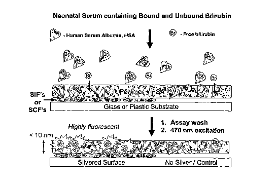
[093] Figure 1 illustrates the new assay for the detection of unbound
bilirubin in neonatal serum.
Briefly, the new assay, as shown in Figure 1, provides for immobilizing noble
metallic
nanostructures on either glass or plastic supports. A thin polymeric layer is
then coated and
immobilized on both the metallized and nonmetallized portions of the glass /
plastic supports. The
polymeric film contains an optimized amount of HSA (Human Serum Albumin) to
bind any
unbound bilirubin. The molecular weight of the polymer has been chosen such
that small
molecules, like bilirubin, can readily diffuse into the polymer film and bind
with HSA, but once
bound can't diffuse out from the film due to the crosslinking density and
therefore pore size of the
polymer. The polymer film also prevents bilirubin bound HSA from diffusing
into the polymer
film. In essence, the polymer films acts as a membrane through which only free
bilirubin diffuses.
Free bilirubin is typically weakly fluorescent and for the most part
considered to be non-
fluorescent [74]. However, upon complexation with HSA becomes fluorescent, and
due to the
close proximity of the silver, is further fluorescently enhanced.
[094] The albumin bound bilirubin on the surface of the polymer is washed away
before
measurements, providing for enhanced fluorescence intensities from the polymer
immobilized free
bilirubin fraction of the sample.
[095] The silver surfaces required for MEF and the present assay can be
obtained using silver
metal island films (SiFs), sandwiched films or even spin coated silver islands
or colloids. A
quartz surface or plastic may be used as substrates for forming the metal
islands thereon.. If
quartz is used, the quartz slides are soaked in 10 parts 98% H2SO4 and 1 part
30% H202 for at least
24 hrs. The SiFs are prepared in clean beakers by reduction of silver ions
using various reducing
agents [75]. Sodium hydroxide is added to a rapidly stirred silver nitrate
solution forming a brown
precipitate. Ammonium hydroxide is added to redissolve the precipitate. The
solution is cooled
and dried quartz slides are added to the beaker, followed by glucose. After
stirring for 2 mins the
mixture is warmed to 30 C. After 10-15 min the mixture turns yellow-green and
becomes cloudy.
A thin film of silver particles has formed on the slides as can be seen from
their brown green
color. The slides are rinsed in pure water prior to the experiment. Additional
procedures for
23
CA 02656004 2008-12-17
WO 2006/138698 PCT/US2006/023738
preparing silver and gold particles are also available [76-80], but primarily
silver is used because
of the longer surface plasmon absorption of gold, which accounts for its
familiar color. It is also
possible to silanize (and uniformly amino coat) the slides by placing them in
a 2 % solution (v/v)
of 3-aminopropyltrimethoxysilane (APS) in dry methanol for 2 hrs, rinsed and
then air-dried. The
silanized substrates should be used within one hour or stored under a dry
nitrogen atmosphere.
Silver nanostructures readily bind to surface amino groups with high affinity
[81,82], and
therefore this process can be used to produce films, where the silver is
tightly surface bound.
[096] While SiFs have been successfully used for MEF studies [2,6,7,9,25],
other metallic
particles and surfaces may be employed, if required, e.g. colloids can be
prepared as suspensions
by citrate reduction of silver or gold, where the size of the colloids and
their homogeneity can be
judged quite simply by the extensive publications on the optical properties of
metal particles
available [83,84], and the effects of interface chemistry on the optical
property of colloids [85]. It
is also possible to prepare bimetallic metal nanoparticles [86] or hollow
sphere colloids [87]. In
addition, the present inventor has recently published two new procedures for
the seed-mediated
growth and deposition of silver nanorods [17] and nanotriangles [16] on
substrates, and these may
be employed, if required. Pre-formed metal particles or colloids can also be
bound to glass
surfaces by placing functional groups such as cyanide (CN), amine (NH2), or
thiol (SH) on a glass
or polymer substrate. In this regard, the present inventor has recently shown
that MEF can occur
from plastic substrates, when inert polymers are firstly functionalized with
amino groups [29].
Silver and gold colloids spontaneously bind to such surfaces with high
affinity [81,82].
Procedures for coating particles with silica have also been developed and will
be used if required
[89,90].
[097] In a typical preparation, glass microscope slides, as shown in Figure 6,
were cleaned with
"piranha solution" (3:7 30% hydrogen peroxide/concentrated sulfuric acid) for
at least 2 hours.
Then, the glass substrates were rinsed extensively with deionized water and
dried in a stream of
dry nitrogen prior to use. The cleaned slides were silanized by immersing them
in a solution of 3-
((trichlorosiyl)propyl) metacrylate (TPM) in heptane and carbon tetrachloride
(4:1, v/v) Then, the
TPM-coated glass slides were rinsed in ethanol and then water. Finally, the
TPM-coated slides
were dried in a stream of nitrogen gas. The polymer precursor solution was
prepared by
combining 50 mg of PEG-DA (Polyethylene Glycol diacrylate), 200 pL of
deionized water, and 6
L photoinitiator Darocur 1173 (From Ciba Special Chemicals, NY) and vortexing
for 5 mins. A
24
CA 02656004 2008-12-17
WO 2006/138698 PCT/US2006/023738
few drops of the HSA / Polymer precursor solution was placed on TPM-coated
glass slides. Free
radical polymerization of the acrylate end groups was initiated by exposure to
a 100 W long wave
UV spot lamp (CTVP Inc.; Upland, CA) for 30 min. The thickness of the polymer
can be
controlled by spin coating before curing [91], and also monitored by a variety
of other techniques
[91]. In this regard, the CFS is equipped with a Speciality Coating Systems
Inc., Model P6700
spin coater, which allows uniform surface thickness by varying polymer
concentration (viscosity)
and spin speed. This allows polymer film thicknesses down to several nm to be
achieved [91,92].
In this regard, film thicknesses are preferably less than 100 nm to optimize
MEF, noting that the
surface is non continuous and features "valleys and mountains" in its surface
topography. The
film thickness and HSA ratio is optimize to allow the polymer films to freely
diffusing bilirubin,
where the film thickness and HSA extent of loading is simply optin:uzed by
considering the
maximum observable fluorescence intensity at ;zt~ 520 nm, the emission maxima
for bilirubin. The
optimum concentration of HSA is loaded into the polymer precursor solution
before spin coating
and UV curing. This concentration is optimized with regard to the maximum
fluorescence
observed by exposure to free solution bilirubin after the polymer is cured.
Films are optimized
with regard to sensor response times and maximum fluorescence signal. After
polymerization, the
PEG layer is washed in PBS for at least 2 hours. This step serves to both
hydrate the matrix and to
remove any unbound surface HSA.
[098] It has been found that the PEG-DA polymer is suitable for the MEF assay.
However,
other polymers may be used, for example, polymers of HEMA (hydroxy ethyl
methacrylate) [93-
97], used in the development of aqueous anion sensors [96] and ethyl cellulose
[98], used in the
construction of dissolved CO2 sensors [98], would be considered. In addition,
plasticized PVC is
simple to prepare, can be made moderately hydrophilic [99] and can be coated
on a variety of
surfaces [100-103].
[099] Free bilirubin calibration plots can be determined for the optimum
polymer formulation,
which includes the optimized polymer thickness, extent of HSA loading and w/v
PEG-DA in the
final formulation. These parameters directly affect the free bilirubin
diffusion rates into the
polymer film (sensor response time) as well as both the enhanced and total
fluorescence signal
observed. For example, a polymer film 10 M thick would not be appropriate for
a MEF assay, as
the MEF phenomenon has been found to occur in a range from 50 to 300 nm from
the glass
substrate and < 10 nm from the peak (top) of the SiFs. Hence, polymer films
ranging from about
CA 02656004 2008-12-17
WO 2006/138698 PCT/US2006/023738
50 nm to about 300 nm, are deemed appropriate depending on the level of
inclusion of HSA in the
firm, and preferably, the film is approximately 100 nm thick. 50 l of
buffered free bilirubin
solution is pipetted into small micro sample chambers as shown in Figure 7,
which are used to trap
small volumes on the assay surface. The emission intensity maxima for
bilirubin upon 470 nm
laser line excitation, and observed through a Semrock 488 razor edge filter,
is recorded. The
calibration plots is constructed, using identical assay formulations, starting
at the clinically
significant concentration 2 g/dl and decreased through series dilutions (from
the master stock
solution) until the S/N ratio drops below 3. This value is deemed the highest
sensitivity, lowest
free bilirubin concentration, the assay can measure. Each concentration is
measured four (4) times
and the mean value determined and plotted. Preferably, the calibration plot
contains no fewer than
25 concentration data points, each the mean of four (4) 4 separate
measurements.
[0100] Fluorophore or analyte photostability is a primary concern in many
applications of
fluorescence, particularly platform type assays and single molecule studies
[61,107]. The
maximum number of photons that are emitted by a fluorophore each second is
roughly limited by
the lifetime of its excited state. If the silver assay surface decreases the
lifetime of bilirubin due its
close proximity as suggested by equations 3 and 4, then one can obtain more
photons per second
per molecule, by appropriately increasing the incident intensity. On the other
hand, the MEF
effect enhances the intensity while simultaneously shortening the lifetime, so
it may in fact be
possible to decrease the excitation intensity yet still see a significant
increase in the emission
intensity and therefore photostability of bound bilirubin. Thus, laser
irradiances can be lowered,
significantly reducing the likelihood of any bilirubin photochemistries
[108,109]. Radiation
excitation frequencies are used that do not cause bilirubin photochemical
reactions and frequencies
such as 516 or 532 nm may be used, by using notch or razor edge filters for
emission.
[0101] Bilirubin samples were prepared by using a solid, powdered form of
bilirubin that can be
purchased with high purity from Sigma. Both solid and solutions of bilirubin
preferably are kept
cold and away from direct light when not in use, due to bilirubin's well-known
photochemistries
[111]. A stock solution was first prepared by dissolving 1 mg of bilirubin
into 10 1 of 1N sodium
hydroxide and then 25 1 of 0.1M EDTA to dissolve the bilirubin into a slurry.
3 ml of buffer was
then added to equilibrate the pH to ;:z~ 7. The concentration of the stock
solution was
approximately about 500pM, and from this, dilutions can'be made in order to
test a range of free
26
CA 02656004 2008-12-17
WO 2006/138698 PCT/US2006/023738
bilirubin concentrations. Low concentrations are especially important, because
free bilirubin
concentration in infants is between 0.05 to 2.511g/dl. Both the stock solution
and samples to be
measured should be kept at 5 C and wrapped in aluminum foil until use. Samples
to be tested,
should be prepared on the day of use. The stock bilirubin solution lasts for
about a week, one
readily observing color change as a function of bilirubin instability [111].
[0102] 50 l of buffered free bilirubin was pipetted into a small plastic
cover which covers one
area of the polymer-coated silvered surface. The small micro-sample chambers,
readily available
from Invitrogen, as shown in Figure 7, come in a range of volume sizes from
10's of 1's up to
several ml. The sample chambers simply stick to surfaces, retaining and
trapping a known surface
volume. Typically, a 500 l blood sample provides s:t; 250 1 of serum, 50 l
to be used for the
new MEF assay.
[0103] In addition to using standard 470 nm front face excitation and off-axis
collection of the
enhanced intrinsic bilirubin fluorescence, Figure 4, the present invention
contemplates using a
TIRF (Total-Internal Reflection Fluorescence) excitation geometry, but with
the same collection
angle/geometry for fluorescence. The fluorescence will be collected through a
488 nm Semrock
Razor edge filter, the emission spectra collected on a Ocean Optics HR4000
fiber-optic
spectrometer. Using a TIRF geometry, as shown in Figure 4, one produces a
metal-amplified
evanescent wave above the assay, far greater than is observed than without the
silver [113,114],
which penetrates several hundred nanometers away from the silver particles
[113]. Given the fact
that the free bilirubin is in close proximity to the silver particles in the
film, then this mode of
excitation provides for a good way of suppressing unwanted background
fluorescence, as distal
material from the silver is not excited and therefore does not fluoresce.
[0104] While the surface of the polymer film has shown very little fouling by
HSA, (tested using
fluorescein labeled HSA from Invitrogen), this approach is still likely to
increase the S / N ratio of
our system. It is for this reason that TIRF geometries are widely used in many
assays today
[115,116].
[0105] Figure 5 shows the presence of diffused bilirubin into photocured PEG-
DA (Polyethylene
Diacrylate) polyiner after incubation, evident by the yellow color. In this
Figure 5, 50 l of 0.2
27
CA 02656004 2008-12-17
WO 2006/138698 PCT/US2006/023738
g/dl laboratory free bilirubin (Sigma) in PBS buffer was incubated onto the
surface of a
metallized slide according to the present invention. After a 10 minute
incubation period, the assay
was washed with buffer for 2 mins to remove any unbound material. From the
photograph shown
in Figure 5, the presence of the bilirubin can be clearly seen, confirming the
plausibility of the
proposed assay. In addition, this free bilirubin concentration is towards the
lower end of the
clinically important concentration range scale to be assayed. While no silver
is present on these
substrates, silver Island films only occupy a;t; 40 % mass surface coverage
and therefore the
polymer adheres to the 60 % non silvered glass using the same chemistries.
[0106] In an anotlier embodiment, the present invention, relates to a new
sensing platform
technology based on Metal-Enhanced Fluorescence (MEF), where the detected
fluorescence
emission is significantly amplified for detection of a nucleotide sequence.
The nucleotide
sequence communicatively connect to the metallic material can be quantified
compared to the
undetectable emission on non metallized surface. In this regard, the detection
of RNA is
accomplished by annealing a target RNA, tagged with a fluorophore, to an
oligonucleotide anchor
probe in a single step on a solid surface, where the, fluorescence signal is
intrinsically enhanced by
silver nanoparticles as shown in MEF based RNA sensing platform systems of
Figures 8 and 11.
[0107] "Nucleotide," as used herein refers to deoxyribonucleic acid (DNA) or
ribonucleic (RNA),
RNA can be unspliced or spliced mRNA, rRNA, tRNA, or antisense RNAi. DNA can
be
complementary DNA (cDNA), genomic DNA, or an antisense.
[0108] The nucleotides used as hybridization probes in the present inventor
are typically designed
to be specific for the desired sequeiice in order to decrease the probability
of hybridizing to
unrelated sequences. Such probes can be modified so as to be detectable using
radionuclides,
luminescent moieties, and so forth. Hybridization conditions also can be
modified in order to
achieve the desired specificity. For example, a moderately stringent
hybridization condition may
include: 2X SSC/0.1% SDS at about 37 C or 42 C (hybridization conditions);
0.5X SSC/0.1% SDS
at about room temperature (low stringency wash); 0.5X SSC/0. 1% SDS at about
42 C (moderate
stringency wash). An example of moderately-high stringency hybridization
conditions is as
follows: 0.1 X SSC/0.1% SDS at about 52 C (moderately-high stringency wash).
An example of
high stringency hybridization conditions is as follows: 0.1 X SSC/0.1% SDS at
about 65 C (high
stringency wash).
28
CA 02656004 2008-12-17
WO 2006/138698 PCT/US2006/023738
[0109] The nucleotides sequences of the present invention can be obtained
using standard
techniques known in the art (e.g., molecular cloning, chemical synthesis) and
the purity can be
determined by polyacrylaniide or agarose gel electrophoresis, sequencing
analysis, and the like.
Polynucleotides also can be isolated using hybridization or computer-based
techniques that are
well known in the art. Such techniques include, but are not limited to: (1)
hybridization of
genomic DNA or cDNA libraries with probes to detect homologous nucleotide
sequences; (2)
antibody screening of polypeptides expressed by DNA sequences (e.g., using an
expression
library); (3) polymerase chain reaction (PCR) of genomic DNA or cDNA using
primers capable of
annealing to a nucleic acid sequence of interest; (4) computer searches of
sequence databases for
related sequences; and (5) differential screening of a subtracted nucleic acid
library.
[0110] Formation of Silver Island Films (SiFs) on APS-coated Glass Substrates
[0111] Silver nitrate (99.9%), sodium hydroxide (99.996%), ammonium hydroxide
(30%),
trisodium citrate, D-glucose and premium quality APS-coated glass slides
(75x25 mm) were
obtained from Sigma-Aldrich. The sources for enzymes, RNA and DNA are
described below. In
a typical SiFs preparation a solution of silver nitrate (0.5 g in 60 ml of
deionized water) in a clean
100-m1 glass beaker, equipped with a Teflon-coated stir bar, is prepared and
placed on a Coming
stirring/hot plate. While stirring at the quickest speed, 200 L of freshly
prepared 5% (w/v)
sodium hydroxide solution is added. This results in the formation of dark
brown precipitates of
silver particles. Approximately 2 ml of anunonium hydroxide is then added,
drop by drop, to re-
dissolve the precipitates. The clear solution is cooled to 5 C by placing the
beaker in an ice bath,
followed by soalcing the APS-coated glass slides in the solution. While
keeping the slides at 5 C,
a fresh solution of D-glucose (0.72 g in 15 ml of water) is added.
Subsequently, the temperature
of the mixture is then warmed to 30 C. As the color of the mixture turns from
yellow-green to
yellow-brown, and the color of the slides become green, the slides are removed
from the mixture,
~
washed with water, and sonicated for a few seconds at room temperature. SiFs-
deposited slides
were then rinsed with deionized water several times and dried under a stream
of nitrogen gas.
[0112] Preparation of the a-globin mRNA substrate
[0113] The complete protein coding sequence of rabbit ,6-globin mRNA was
amplified from
29
CA 02656004 2008-12-17
WO 2006/138698 PCT/US2006/023738
plasmid pC7(.iG23 by polymerase chain reaction using Pfu DNA polymerase
(Stratagene, La Jolla,
CA) from primers 5'-GCAGTCTAGAATGGTGCATCTGTCCAG-3' and 5'- GCACAAG
CTTCAGTGGTATTTGTGAGCCAGG-3' (Integrated DNA Technologies, Coralville, IA).
Underlined bases indicate the XbaI and HinrIIII restriction sites incorporated
into the 5'- and 3'-
termini of the PCR product. This DNA fragment was then inserted into the XbaI
+ HindlQ
restriction sites of pGEM7Zf(+) (Promega, Madison, WI) using standard
subcloning techniques
[142] to generate plasmid pG7(+)RG-CDS. The fidelity of the 0-globin cDNA
insert was verified
by restriction digests and automated DNA sequencing.
[0114] A 484-nt RNA substrate containing the fl-gtobin coding sequence (See
Figure 12) was
prepared by in vitro transcription using T7 RNA polymerase (Ambion, Austin,
TX) from a
HinellIl-linearized pG7(+),QG-CDS DNA template. Following digestion of
template DNA with
RQ1-DNase (Promega), templates were purified by duplicate extractions with
phenol:chloroform:isoamyl alcohol (25:24:1). Unincorporated nucleotides were
removed from the
preparation by spin column chromatography tlirough RNase-free G-50 Quick Spin
columns
(Roche, Indianapolis, IN). The integrity of the fl-globin RNA substrate was
evaluated by
electrophoresis through formaldehyde-agarose gels stained with ethidium
bromide. Fluorescence
intensity of ethidium bromide-stained RNA was measured using the EDAS 290 gel
documentation
system (Kodak, Rochester, NY), with synthesis yield calculated by comparison
to co-fractionated
RNA size markers (InVitrogen, Carlsbad, CA).
[0115] MEF-based RNA sensing assays
f01161 The following RNA capture assay [143] was used to detect specific RNA
substrates on
SiFs-coated glass slides, as shown in Figure 11. First, fl-globin mRNA or
yeast tRNA substrates
(10 ng) were incubated with an antisense primer 5'-GTGAGCCAGGGCATT-TAMRA-3'
(fluorescent probe; 10 pmol) in a total volume of 100 l hybridization buffer
[10 mM
HEPES-KOH [pH 7.4] containing 100 mM KCI, 2 mM dithiolthreitol, and 1 mM
MgC12] at 70 C
for 5 minutes, then the RNA/DNA construct was slowly cooled to 37 C over 20-
30 minutes. The
anchor probe (5'-thiol-CACCTTCTGATAGGC-3', 10 pmol) was attached to the SiFs
by an
overnight incubation at 4 C in a humidified chamber. Excess thiol-conjugated
oligo was removed
by washing the surface with the hybridization buffer several times. The TAMRA-
linked oligo
annealed to RNA substrates were annealed to the thiol-linked anchor oligo on
the surface of the
CA 02656004 2008-12-17
WO 2006/138698 PCT/US2006/023738
SiFs at 37 C for 30 minutes in a humidified chamber. Non-binding RNA
substrates were removed
as described above prior to fluorescence measurements. This procedure brings
the fluorophore to
a distance, approximately 10 nm, from the surface of the SiFs where the
fluorescence emission is
expected to increase by MEF as described previously [131, 132].
[0117] Fluorescence measurements on SiFs were performed byplacing the SiFs on
a stationary
stage equipped with the fiber-optics mount on a 15-cm-long arm (normal to
sample). The output
of the fiber was connected to an Ocean Optics HD2000 spectrofluorometer for
the emission
spectra. The excitation light was provided by a 532 nm laser at an angle of 45
degrees. The
emission spectra were observed through a 532-nm-notch filter (Samrock).
j01181 The deposition of Silver Island films onto glass slides was performed
as described
previously [136]. In a typical SiF preparation, a solution of sodium hydroxide
and ammonium
hydroxide are added to a continuously stirred solution of silver nitrate at
room temperature.
Subsequently, the mixture is cooled down in an ice bath, Silane-prepTM glass
slides (Sigma) are
inserted and a solution of D-glucose is added. As the temperature is
increased, the color of the
mixture turns yellow-brown and the SiFs-deposited slides are removed from the
mixture, washed
with water, and sonicated for a few seconds at room temperature. SiFs-
deposited glass slides were
stored in deionized water until they were used. Fluorescence emission spectra
of TAMRA-
labeled oligo with RNA substrate hybridized to the thiolated-oligo anchor
probe on SiFs is shown
in Figure 9. The emission intensity peak of TAMRA-labeled oligo that was
annealed to RNA
substrates ranging from 25 finoles to 500 finoles is clearly observed at 585
nm, and increased
linearly as the amount of RNA substrate is increased, as shown in Figure 9.
The fluorescence
emission spectra of TAMRA shown in Figure 9 (especially for the RNA substrates
of 250 finoles
or higher) appear broader than the spectrum of TAMRA-labeled oligo anchor
probe measured
from a solution on plain glass, as shown in Figure 13, due to the background
scattering from the
SiFs-coated glass slide.
[0119] The control experiments revealed that when the RNA sequence was changed
(that is,
control tRNA with random sequence is used in the RNA capture assay) the
fluorescence emission
from TAMRA-labeled oligo was not observed, as shown in Figure 14, since the
control tRNA
lacked the specific sequence that is required for the annealing of TAMRA-
labeled RNA. In
addition, when either of the other components of the RNA capture assay,
thiolated-oligo or
31
CA 02656004 2008-12-17
WO 2006/138698 PCT/US2006/023738
TAMRA-labeled oligo is omitted, almost no fluorescence emission was observed.
Thus, the RNA
capture assay is highly specific and the contribution of the non-specific
interactions to the detected
signal is minimal.
[0120] The lower detection limit (LDL) of the RNA capture assay described here
was 25 finoles
of RNA (S/N > 20) and made possible by the amplification of fluorescence
emission intensity
based on our previously described phenomenon metal-enhanced fluorescence [131,
132]. The
amplification of fluorescence emission intensity is a property of the silver
nanoparticles deposited
on the glass slides and thought to occur due to partial non-radiative energy
transfer between the
excited state of the fluorophore and the surface plasmons of the silver
nanoparticles, as well as due
to the spatially localized excitation of fluorophores created by the
nanoparticles within close
proximity [137].
[01211 Although the LDL of the MEF-based RNA capture assay is 100-200-fold
less sensitive
than the current RNA capture assays [129, 140], the MEF-based RNA sensing
method offers a
considerably simpler, cheaper and quicker alternative to RT-PCR, since it does
not require the
amplification of the RNA target and can be performed relatively quickly. Given
that the S/N > 3-
4 for fluorescence-based assays is considered acceptable, [133] the actual
lower detection limit of
the MEF-based RNA capture assay is approximately 5 finoles.
[0122] In a coniparison experiment, RNA was detected in the absence of SiFs on
glass (Figure 15
on glass, Top-Left) and in the presence of SiFs using avidin-biotin
interactions. In this regard,
firstly, the RNA is annealed to a TAMItA-labeled oligo and then the RNAITAMRA-
labeled oligo
is annealed to a biotinylated Oligo. Finally, the resultant RNA/Oligo
construct is brought the
surface due to the interaction of avidin and biotin. Figure 16 shows
fluorescence emission spectra
(intensity: arbitrary units) of TAMRA-linked Oligo annealed to the RNA
substrate (500 fmoles)
that was hybridized with the biotinylated Oligo anchor probe that was brought
to the glass surface
via avidin-biotin interactions. The emission intensity peak of TAMRA-labeled
oligo that was
annealed to RNA substrates (Target and non-specific RNA: 500 finoles) is
observed at 585 nm but
are similar (20 AU). That is, the RNA assay on glass substrate in the absence
of SiFs is not
sensitive enough to distinguish between the actual assay and the non-specific
interactions. In
contrast, Figure 17 shows fluorescence emission spectra (intensity: arbitrary
units) of TAMRA-
linked Oligo annealed to the RNA substrate (500 finoles) that was hybridized
with the biotinylated
32
CA 02656004 2008-12-17
WO 2006/138698 PCT/US2006/023738
Oligo anchor probe that was brought to the SiFs-coated surface via avidin-
biotin interactions. The
emission intensity peak of TAMRA-labeled oligo that was annealed to RNA
substrates (Target
and non-specific RNA: 500 finoles) is observed at 585 nm and significantly
larger than the
background. Although, the sensitivity of the RNA assay using SiFs (and avidin-
biotin
interactions) is improved compared to the assay on the glass surface, the
sensitivity is much less
when compared to the RNA assay on SiFs with thiolated-oligo is used. This is
due to the fact that
the fluorophore is located approximately 10 nm away from the surface of the
silver when avidin-
biotin system is used (the thicknesses of avidin and biotinylated BSA are 4
nm) and approximately
4 mu when thiolated oligo is used. In all the previously published MEF papers
[131, 132, 136,
137] the maximum enhancement of fluorescence by silver was observed when the
fluorophore was
located within 8 nm of the surface and the enhancement is decreased for the
distances larger than 8
nm. Thus, the RNA assay using the thiolated-oligo on SiFs is more sensitive
than the assays using
avidin-biotin interactions on glass and SiFs.
[0123] The rapidity of the MEF-based RNA capture assays could be increased
further with the
help of low-power microwaves, as shown previously for the MEF based protein
and antibody
assays that were completed within 20 seconds, i.e., microwave-accelerated
metal-enhanced
fluorescence (MAMEF) [136, 138]. Similar to RT-PCR, the MEF-based RNA capture
assays
could potentially be multiplexed by simply using SiFs-coated high throughput
screening (HTS)
wells [139]. Ultimately, ultra-rapid MEF-based multiplexed RNA capture assays
comparable to
RT-PCR could be achieved by combining MAMEF technology with the use of SiFs-
coated HTS
wells once the sensitivity of the MEF-based method is improved. In this
regard, MEF-based
enhancements in excess 3000-fold using fractal silver surfaces was recently
reported[132].
33
CA 02656004 2008-12-17
WO 2006/138698 PCT/US2006/023738
References
The contents of the following references are hereby incorporated by reference
herein for all
purposes.
[1] Geddes, C. D. and Lakowicz, J. R. (2002). Metal-Enhanced Fluorescence, J.
Fluorescence, 12 (2), 121-129.
[2] Lakowicz, J. R. (2001). Radiative Decay Engineering: Biophysical and
Biomedical
Applications, Anal. BioClzein., 298, 1-24.
[3] Lakowicz, J. R., Shen, Y., D'Auria, S., Malicka, J., Fang, J., Gryczynski,
Z. and
Gryczynslei, I. (2002). Radiative Decay Engineering 2. Effects of silver
island films on
fluorescence intensity Lifetimes and Resonance energy transfer, Anal.
Biochem., 301,
261-277.
[4] Lakowicz, J. R., Gryczynski, I, Shen, Y. B., Malicka, J., and Gryczynski,
Z, (2001).
Intensified fluorescence, Pliotonics Spectra, 35(10), 96-104.
[5] Gryczynski, I., Malicka, J., Gryczynski, Z., Geddes, C. D. and Lakowicz,
J. R. (2002) The
CFS engineers the intrinsic radiative decay rate of low quantum yield
fluorophores, J.
Fluorescence,12(1), 11-13.
[6] Malika, J., Gryczynski, I., Maliwal, B. P., Fang, J. F. and Lakowicz, J.
R. (2003).
Fluorescence spectral properties of cyanine dye labeled DNA near metallic
silver
particles, Biopolymers, 72(2), 96-104.
[7] Malicka, J., Gryczynski, I., Kusba, J., Shen, Y. B., and Lakowicz, J. R.
(2002). Effects of
metallic silver particles on resonance energy transfer in labeled bovine serum
albumin,
Biochem. Biophys. Res. Comm., 294(4), 886-892.
[8] Gryczynski, I., Malika, J., Shen, Y. B., Gryczynski, Z., and Lakowicz J.
R. (2002).
Multiphoton excitation of fluorescence near metallic particles: Enhanced and
localized
excitation, J. Phys. Chena. B., 106(9), 2191-2195.
[9] Malika, J., Gryczynski, I., Fang, J. Y., Kusba, J. and Lakowicz, J. R.
(2002).
Photostability of cy3 and cy5-labeled DNA in the presence of metallic silver
particles, J.
Fluorescence, 12(3-4), 439-447.
[10] Geddes, C. D., Cao, H., Gryczynski, I., Gryczynski, Z., Fang, J. and
Lakowicz, J. R.
(2003). Metal-enhanced Fluorescence (MEF) Due to silver colloids on a planar
surface:
Potential applications of Indocyanine green to in vivo imaging, J. Phys
Cliern. A., 107,
3443-3449.
[11] Geddes,= C. D., Parfenov, A., Gryczynski, I., Malicka, J., Roll, D., and
Joseph R.
Lakowicz, (2003). Fractal silver structures for metal-enhanced fluorescence:
Applications
for ultra-bright surface assays and lab-on-a-chip based nanotechnologies, J.
Fluorescence,
34
CA 02656004 2008-12-17
WO 2006/138698 PCT/US2006/023738
13(2), 123-128.
[12] Lakowicz, J. R., Malicka, J., Gryczynski, Z., Huang, J., Geddes, C. D.
and Gryczynski, I.,
(2003), Increased sensitivity of Fluorescence detection, PharmaGenomics, 3(3),
38-46.
[13] Geddes, C. D., Parfenov, A., and Lakowicz, J. R., (2003). Photodeposition
of silver can
result in Metal-enhanced fluorescence, Applied Spectroscopy, 57(5), 526-531.
[14] Geddes, C. D., Gryczynski, I., Malicka, J., Gryczynslci, Z., and
Lakowicz, J. R. (2003).
Metal-Enhanced Fluorescence: Potential applications in HTS, Combinatorial
Chemistry
and HTS, 6(2), 109-117.
[15] Lakowicz, J. R., Gryczynski, I., Malicka, J., Gryczynski, Z., and Geddes,
C. D. (2002).
Enhanced and localized multi-photon excited fluorescence near metallic silver
islands:
Metallic islands can increase probe photostabilityJ. Fluorescence, 12(3/4),
299-302.
[16] Aslan, K., Lakowicz, J. R., and Geddes, C. D., (2005). Rapid Deposition
of Triangular
Silver Nanoplates on Planar Surfaces: Application to Metal-enhanced
Fluorescence,
Journal ofPhysical CliemistryB.,109(13), 6247- 6251,
[17] Aslan, K., Lakowicz, J. R., and Geddes, C. D., (2005). Fast and slow
deposition
of Silver Nanorods on Planar-Surfaces: Application to Metal-enhanced
fluorescence.
Journal ofPhysical Chemistry B., 109(8), 3157-3162,
[18] Aslan, K., Lakowicz, J. R., Szmacinski, H., and Geddes, C. D., (2005).
Enhanced
ratiometric pH sensing using SNAFL-2 on silver island films: Metal-enhanced
fluorescence sensing. Jn. Fluorescence, 15(1), 37-40,
[19] Wu, M., Lakowicz, J. R, and Geddes, C. D., (2005). Enhanced lanthanide
luminescence
using silver nanostructures: Opportunities for a new class of probes with
exceptional
spectral characteristics. Jn. Fluorescence,15(1), 53-59, 2005.
[20] Aslan, K., Lakowicz, J. R., Szmacinsld, H., and Geddes, C. D., (2004).
Metal-enhanced
fluorescence solution based sensing Platform. Jn. Fluorescence, 14 (6), 677-
679, 2004.
[21] Geddes, C. D., Parfenov, A., Roll, D., Gryczynski, I., Malicka, J., and
Lakowicz, J. R.,
(2004). Roughened silver electrodes for use in Metal-enhanced fluorescence.
Spectrochemica Acta A., 60 (8-9), 1977-1983,
[22] Parfenov, A., Gryczynski, I., Malicka, J., Geddes, C. D., and Lakowicz,
J. R., (2003).
Enhanced fluorescence from fluorophores on fractal silver surfaces. Jn. Plays.
Citem. B.,
107(34), 8829-8833,
[23] Pugh, V. J., Szmacinski, H., Moore, W. E., Geddes, C. D., and Lakowicz,
J. R.,
(2003).Submicrometer spatial resolution of Metal-enhanced fluorescence.
Applied
Spectroscopy, 57(12), 1592-1598,
[24] Geddes, C. D., Parfenov, A., Roll, D., Fang, J., and Lakowicz, J. R.,
(2003).
Electrochemical and laser deposition of silver for use in metal-enhanced
fluorescence.
Langmuir, 19, 6236-6241,
CA 02656004 2008-12-17
WO 2006/138698 PCT/US2006/023738
[25] Lakowicz, J. R., Geddes, C. D., Malicka, J., Gryczynski, K., Lukomska, J.
Huang, C.,
Aslan, K., and Gryczynski. I., (2004).Advances in Surface-enhanced
Fluorescence. Jn.
Fluorescence, 14(4),425-441,
[26] Aslan, K., et al and Geddes, C. D., (2005) Metal-enhanced fluorescence:
An emerging
tool in biotechnology. Current opinions in Biotechnology, 16(1), 55-62,
[27] Geddes, C. D., Aslan, K., Gryczynski. I., Malicka, J., and Lakowicz, J.
R, Radiative
Decay Engineering (RDE). Topics in Fluorescence Spectroscopy Volume 8, Edited
by
Chris D. Geddes and Joseph R. Lakowicz, Springer, New York, Pgs 405-448.
[28] Geddes, C. D., Aslan, K., Gryczynski. I., Malicka, J., and Lakowicz, J.
R., (2004)
Noble-metal surfaces for Metal-enhanced fluorescence, In Reviews in
Fluorescence 2004,
Ed by Chris D. Geddes and Joseph R. Lakowicz, Kluwer Academic Plenum
Publishers,
New York, pgs 365-401. ISBN: 0-306-48460-9.
[29] Aslan, K., Bagugu, R., Lakowicz, J. R., and Geddes, C. D., (2005). Metal-
Enhanced
Fluorescence from Plastic substrates. Jn. Fluorescence, 15(2), 99-104,
[30] Lakowicz, J. R., Malicka, J., Gryczynski. I., Gryczynski. Z., Geddes, C.
D., (2003)
Radiative Decay Engineering: The role of photonic mode density in
biotechnology. Jn.
Physics D. Appl. Phys. 38, R240-249,
[31] Volpe, J.J., (2001). Bilirubin and Brain Injury, in Neurology of the
Newborn [ed 4].
[32] Amin, S.B., (2004). Clinical assessment of bilirubin-induced
neurotoxicity in premature
infants. Seminars in Perinatology 28:340-347.
[33] Amin, S.B., Charafeddine, L., Guillet, R., (2005). Transient Bilirubin
Encephalopathy and
Apnea of Prematurity in 28 to 32 Weeks Gestational Age Infants. JPerinatol.
2005.
[34] Pledger, D.R., Scott, J., Belfield, A., (1982). Kernicterus at low levels
of serum bilirubin:
The impact of bilirubin albumin -binding capacity. Biol Neonate. 1982; 41:38-
44
[35] Garziani, L.J., Mitchell, D.G., Kornhauser, M., et al., (1992).
Neurodevelopment of
preterm infants. Neonatal neurosonographic and serum bilirubin studies.
Pediatrics.
89(2):229-234.
[36] O'Shea, T.M., Dillard, R.G., Klinepeter, K.L., Goldstein, D.J., (1992).
Serum bilirubin
levels, intracranial hemorrhage, and the risk of developmental problems in
very low birth
weight neonates. Pediatrics. 90(6):888-892.
[37] Van de Bor, M., Dokkum, M.E., Schreuder, A.M., Veen, S., Brand, R.,
Verloove -
Vanhorick, S.P., (1992). Hyperbilirubinemia in low birth weight infants and
outcome at 5
years of age. Pediatrics. 89(3):359-364.
[38] Hansen,. T.W., (1996). Therapeutic approaches to neonatal jaundice: an
international
survey. Clin Pediatr. 35(6):309-316
[39] Bratlid, D. (1990). How bilirubin gets into the brain. Clin
Perinatol.17:449-465.
[40] Ahlfors, C.E. (2001). Bilirubin-albumin binding and free bilirubin. J.
Perinatol. 21:
S40-42
36
CA 02656004 2008-12-17
WO 2006/138698 PCT/US2006/023738
[41] Amin, S.B., AhlforsõC., Orlando, M.S., Dalzell, L.E., Merle, K.S.,
Guillet, R., (2001).
Bilirubin-albumin binding variables in premature infants. Pediatrics
107(4):664-668.
[42] Nakamura, H., Yonetani, M., Uetani, Y., Funato, M., Lee, Y., (1992).
Determination of
serum unbound bilirubin for prediction of kernicterus in low birth weight
infants. Acta.
Paediatr. Jpn. 34:642-647.
[43] Cashore, W.J., Oh, W., (1982). Unbound bilirubin and kernicterus in low
birth weight
infants. Pediatrics 69(4):481-485.
[44] Ritter, D.A., Kenney, J.D., Norton, H.J., Rudolph, A.J., (1982). A
prospective study of
free bilirubin and other risk factors in the development of kernicterus in
premature infants.
Pediatrics 69(3):260-266.
[45] Nakamura, H., Takada, S. Shimabuku, R., et al. (1985). Auditory nerve and
brainstem
responses in newborn infants with hyperbilirubinemia. Pediatrics. 75:703-708.
[46] Funato, M., Tamai, H., Shimada, S., et al. (1994) Vigintiphobia, unbound
bilirubin, and
auditory brainstem responses. Pediatrics. 93:50-53.
[47] Jacobsen, J. and Wennburg, P., (1974). Determination of Unbound Bilirubin
in the Serum
of Newborns. Clin. Chem. 20(7): 783-789.
[48] Ahlfors, C., (1981). Effect of Serum Dilution on Apparent Unbound
Bilirubin
Concentration as Measured by the Peroxidase Method. Clin. Chem. 27(5): 692-
696.
[49] Ahlfors, C., (2000).Measurement of Plasma Unbound Unconjugated Bilirubin.
Anal.
Biochem. 279: 130-135.
[50] Porter, E, and Waters, W., (1966). A rapid micromethod for measuring the
reserve
albumin binding capacity in serum from newborn infants with
hyperbilirubinemia. J. Lab
& Clin. Med. 67(4):660-668.
[51] Berde, C., Benitz, W., Rasmussen F., et al., Bilirubin Binding in the
Plasma of Newborns:
Critical Evalutation of a Fluorescence Quenching Method and Comparison to the
Peroxidase Method., (1984) Pediatric Research.l8(4): 349-354.
[52] Blackmon, L.R., Fanarof,f A.A., and Raju, T.N.K., (2004). Research on
prevention of
bilirubin-induced brain injury and kernicterus: National Institute of Child
Health and
Human Development Conference Executive Summary. Pediatrics 114:229-233.
[53] Flelschmann, M., Hendra, P. J., and McQuillan, A. J. (1974). Raman
spectra of pyridine
absorbed at a silver electrode, Cliena. Plzys. Lett., 26(2), 163-166.
[54] Jeanmaire, D. L. and Van Duyne, R. P. (1997). Surface Raman
spectroelectrochemistry.
Part 1. Heterocyclic, aromatic and aliphatic amines adsorbed on the anodised
silver
electrode,J. Electroanal. Cliem., 84, 1-20.
37
CA 02656004 2008-12-17
WO 2006/138698 PCT/US2006/023738
[55] Aroca, R., Jennings, C., Kovacs, G. J., Loutfy, R. G. and Vincett, P. S.
(1985). Surface-
enhanced Raman scattering of Langmuir-Blodgett monolayers of phthalocyanine by
indium and silver island films, J. Phys. Chein., 89, 4051-4054.
[56] Pettinger, B., and Gerolymatou, A. (1984). Dyes adsorbed at Ag-colloids:
Substitution of
fluorescence by similarly efficient surface fluorescence and surface Raman
scattering,
Ber. Bungens. Plzys. Chem., 88, 359-363.
[57] DeSaja-Gonzalez, J., Aroca, R., Nago, Y. and DeSaja, J. A. (1997).
Surface enhanced
fluorescence and SERS spectra of N-octadecyl-3,4:9,10-perylenetetracarboxylic
monohydride on silver island films, Spectrochim. Acta. Part A, 53, 173-181.
[58] Hildebrandt, P. and Stockburger, M., (1984). J. Phys Chem. B., 88, 5935.
[59] Kneipp, K., Wang, Y., Kneipp, H., Itzkan, L., Dasari, R. R. and Feld, M.
S., (1996). Phys.
Rev. Lett., 76, 2444.
[60] Kneipp, K., Wang, Y., Kneipp, H., Perelman, L. T., Itzkan, L., Dasari, R.
R., and Feld, M.
S., (1997). Phys. Rev. Lett., 78, 1667.
[61] Axelrod, D., Hellen, E. H. and Fulbright, R. M. (1992). Total internal
reflection
fluorescence, in Topics in Fluorescence Spectroscopy, Vol. 3: Biochemical
applications,
(Lakowicz J. R., Ed.), Plenum Press, New York, pp. 289-343.
[62] Wokaun, A., Lutz, H.-P., King, A. P., Wild, U. P. and Ernst, R. R.
(1983). Energy transfer
in surface enhanced fluorescence, J Chem. Plzys., 79(1), 509-514.
[63] Holland, W. R. and Hall, D. G. (1985). Waveguide mode enhancement of
molecular
fluorescence. Optics Letts., 10(8), 414-416.
[64] Glass, A. M., Liao, P. F., Bergman, J. G. and Olson, D. H. (1980).
Interaction of metal
particles with adsorbed dye molecules: absorption and luminescence. Optics
Letts., 5(9),
368-370.
[65] Axelrod, D., Burghardt, T. P. and Thompson, N. L. (1984). Total internal
reflection
fluorescence, Ann. Rev. Biophys. Bioeng:, 13, 247-268.
[66] Benner, R. E., Dornhaus, R. and Chang, R. K. (1979). Angular emission
profiles of dye
nlolecules excited by surface lasmon waves at a metal surface, Optics Commun.,
30(2),
145-149.
[67] Barnes, W. L. (1998). Fluorescence near interfaces: The role of photonic
mode density, J.
Modern Optics, 45(4), 661-699.
[68] Camplon, A., Gallo, A. R., Harris, C. B., Robota, H. J. and Whitmore, P.
M. (1980).
Electronic energy transfer to metal surfaces: A test of classical image dipole
theory at
short distances, Chem. Pliys. Letts., 73(3), 447-450.
[69] Sokolov, K., Chumanov, G. and Cotton, T. M. (1998). Enhancement of
molecular
38
CA 02656004 2008-12-17
WO 2006/138698 PCT/US2006/023738
fluorescence near the surface of colloidal metal films, Anal. Chem., 70, 3898-
3905.
[70] Hayakawa, T., Selvan, S. T. and Nogami, M. (1999). Field enhancement
effect of small
Ag particles on the fluorescence from Eu3+-doped Si02 glass, Appl. Plays.
Lett., 74(11),
1513-1515.
[71] Geddes, C. D., Cao, H., and Lakowicz, J. R. (2003). Enhanced
photostability of ICG in
close proximity to Gold colloids, Spectrochenaica Acta A. 59, 2611-2617.
[72] Selvan, S. T., Hayakawa, T. and Nogami, M. (1999). Remarkable influence
of silver
islands on the enhancement of fluorescence from Eu3+ ion-doped silica gels, J.
Phys.
Chena. B., 103, 7064-7067.
[73] Strickler, S. J. and Berg, R. A. (1962). Relationship between adsorption
intensity and
fluorescence lifetime of molecules, J. Cl2em. Phys., 37, 814-822.
[74] Knudsen, A., Pedersen, A.O., Brodersen, R., (1986). Spectroscopic
Properties of
Bilirubin-Human Serum Albumin Complexes: A Stoichiometric Analysis. Arch.
Biochem
and Biophys. 244(1): 273-284.
[75] Rivas, L., Sanchez-Cortes, S., Garcia-Ramos, J. V. and Morcillo, G.
(2001), Growth of
silver colloidal particles obtained by citrate reduction to increase the Ramen
enhancement
factor, Langmuir, 17(3), 574-577.
[76] Shirtcliffe, N., Nickel, U. and Schneider, S. (1999). Reproducible
preparation of silver
sols with small particle size using borohydride reduction:For use as nuclei
for preparation
of larger particles, J Colloid Interface Sci., 211(1), 122-129.
[77] Pastoriza-Santos, I., and Liz-Marzan, L. M. (2000). Reduction of silver
nanoparticles in
DMF. Formation of monolayers and stable colloids, Pure Appl. Chem., 72(1-2),
83-90.
[78] Pastoriza-Santos, I., Serra-Rodriguez, C. and Liz-Marzan, L. M. (2000).
Self-assembly of
silver particle monolayers on glass from Ag} solutions in DMF, J. Colloid
Interface Sci.,
221(2), 236-241.
[79] Bright, R. M., Musick, M. D. and Natan, M. J. (1998). Preparation and
characterization of
Ag colloid monolayers, Langmuir,14(20), 5695-5701.
[80] Ni, F. and Cotton, T. M. (1986). Cheniical procedure for preparing
surface-enhanced
Raman scattering active silver films, Anal. Cltem., 58(14), 3159-3163.
[81] Freeman, R. G., Grabar, K. C., Allison, K. J., Bright, R. M., Davis, J.
A., Guthrie, A. P.,
Hommer, M. B., Jackson, M. A. , Smith, P. C., Walter, D. G. and Natan, M. J.
(1995).
Self-assembled metal colloid monolayers: An approach to SERS substrates,
Scierice, 267,
1629-1632.
[82] Grabar, K. C., Freeman, R. G., Hommer, M. B. and Natan, M. J. (1995).
Preparation and
characterisation of Au colloid monolayers, Anal. Chem., 67, 735-743.
39
CA 02656004 2008-12-17
WO 2006/138698 PCT/US2006/023738
[83] Link, S. and El-Sayed, M. A. (1999). Spectral properties and relaxation
dynamics of
surface plasmon electronic oscillations in gold and silver nanodots and
nanorods, J. Plzys.
Chenz. B., 103, 8410-8426.
[84] Kreibig, U. and Genzel, L. (1985). Optical absorption of small metallic
particles, Surface
Science, 156, 678-700.
[85] Krelbig, U., Gartz, M. and Hilger, A. (1997). Mie resonances: Sensors for
physical and
chemical cluster interface properties, Ber. Bunsenges, Plzys. Chenz., 101(11),
1593-1604.
[86] Toshima, N. and Yonezawa, T. (1998). Bimetallic nanoparticles-novel
materials for
chemical and physical applications, New J. Chezn., 1179-1201.
[87] Caruso, F., Caruso, R. A. and Mohwald, H. (1998). Nanoengineering of
inorganic and
hybrid hollow spheres by colloidal templating, Science, 282, 1111-1114.
[88] Yee, J. K., Parry, D. B., Caldwell, K. D. and Harris, J. M. (1991).
Modification of quartz
surfaces via thiol-disulphide interchange, Langrnuir, 7, 307-313.
[89] Farmer, S. C. and Patten, T. E. (2000). Synthesis of luminescent
organic/inorganic
polymer nanocomposites, Polym. Mater. Sci. Eng., 82, 237-238.
[90] Comor, M. I. And Nedeljkovic, J. M. (1999). Enhanced photocorrosion
stability of
colloidal cadmium sulphide-silica nanocomposites, J Mater. Sci. Lett., 18,
1583-1585.
[91] Gryczynski, I., Malicka, J., Nowaczyk, K., Gryczynski, Z., and Lakowicz,
J. R. (2004).
Effects of Sample thickness on the Optical Properties of Surface Plasmon-
Coupled
Emission. J Plzys Chenz. B., 108, 12073-12083.
[92] Gryczynski, I., Malicka, J., Gryczynski, Z., Nowaczyk, K., and Lakowicz,
J. R. (2004).
Ultraviolet Surface Plasmon-Coupled Emission Using Thin Aluminum Films. Anal.
Chem., 76(21), 4076-4081.
[93] Geddes, C. D., Douglas, P., Moore, C.P., Wear, T.J., Egerton, P.L.,
(1999) Optical thin
film serisors for the determination of aqueous halide Ions. Jrz. Fluorescence,
9(3), 163-
171, 1999.
[94] Geddes, C. D., and Douglas, P., (2005). Fluorescent dyes bound to
hydrophilic
copolymers - Applications for aqueous halide sensing. App. Poly. Sci., 76(5),
603-615,
2000.
[95] Geddes, C. D., Optical thin film polymeric sensors for the determination
of aqueous
chloride, bromide and iodide ions at high pH, based on the quenching of
fluorescence of
two acridinium dyes. Dyes and Pignzents, 45(3), 243-251, 2000.
[96] Geddes, C. D., A Halide sensor based on the quenching of fluorescence of
an immobilised
indolium salt. P/totochemistry and Photobiology A: Citemistry, 137(2-3), 145-
153, 2000.
[97] Geddes,.C. D., Halide sensing using the SPQ molecule. Sensors and
Actuators Cizeznical,
CA 02656004 2008-12-17
WO 2006/138698 PCT/US2006/023738
72(2), 188-195, 2001.
[98] Mills, A., and Chang, Q., (1994). Colorimetric polymer film sensors for
dissolved carbon
dioxide. Sensors and Actuators B: Chemical, 21, 83-89.
[99] Herrero, M., Tiemblo, P., Reyes-Labarta, J., Mijangos, C., and Reinecke,
H., (2002). PVC
modification with new functional groups. Influence of hydrogen bonds on
reactivity,
stiffness and specific volume. Polymer, 43, 2631- 2636.
[100] Yu, Z.J., Kang, E.T., Neoh, K.G., and Tan, K.L., (2001). Surface
Passivation of epoxy
resin with a covalently adhered poly(tetrafluoroethylene) layer. Surface &
Coatings
Technology, 138, 48-55.
[101] Endo, K., (2002) Synthesis and Structure of poly(vinyl chloride).
Progess in Polymer
Science, 27, 2021-2054.
[102] James, N.R., and Jayakrishnan, A., (2003). Surface thiocyanation of
plasticized poly(vinyl
chloride) and its effect on bacterial adhesion. Biomaterials, 24, 2205- 2212.
[103] Liu, B., Yang, Y., Zhao-Yang, W., Wang, H., Shen, G., and Yu, R., (2005)
A
potentiometric acetylcholinesterase biosensor based on plasma-polymerized
film. Sensors
and Actuators B: Chemical, 104, 186-190.
[104] Geddes, C. D., Parfenov, A., and Lakowicz, J.R. (2003). Luminescent
Blinking from
Noble-metal Nanostructures: New Probes for localization and imaging. Jn.
Fluorescence,
13(4), 297-299,
[105] Geddes,C.D.,Parfenov, A., Gryczynski, I., and Lakowicz,J.R.(2003).
Luminescent
blinking from silver nanostructures.Jn. Phys. Chem. B. 107(37), 9989-9993,
[106] Geddes,C.D.,Parfenov,A.,Gryczynski, I., and Lakowicz, J.R.,
(2003).Luminescent
blinking of gold nanoparticles. Chem. Phys. Letts, 380(3-4), 269-272, 2003.
[107] Lakowicz, J. R. (1999). Principles of Fluorescence Spectroscopy, 2 d
Edition, Kluwer
Academic Plenum Publishers, New York.
[108] Turro, N.J., (1991). Modern Molecular Photochemistry, University Science
Books,
California. ISBN 0-935702-71-7
[109] Wayne, C.E., and Wayne, R.P. (1999) Photochemistry, Oxford University
Primers, Bath,
ISBN 0-19-855886-4
[110] DeGraff, A., and Demas, J.N., (2005) Luminescence-Based Oxygen Sensors.
Reviews In
Fluorescence 2005.Springer, New York, 125-151.
[111] Lamola, A., Eisinger, J., Blumberg, W., Patel S., Flores J., (1979).
Fluorometric Study of
the Partition of Bilirubin among Blood Components: Basis for Rapid Microassays
of
Bilirubin Binding Capacity in Whole Blood. Anal. Biocliem.100:25-42.
41
CA 02656004 2008-12-17
WO 2006/138698 PCT/US2006/023738
[112] Geddes,C.D., (2001). Optical halide sensing using fluorescence
quenching: Theory,
simulations and applications - A review. An invited review article
commissioned by the
Institute ofPhysics. Meas. Sci. Technol., 12(9), R53-R88.
[113] Matveeva, E., Gryczynski, Z., Malicka, J., Gryczynski, I., and Lakowicz,
J. R. (2004).
Metal-enhances fluorescence immunoassays using total internal reflection and
silver
island-coated surfaces. Anal. BioChem., 334, 303- 311.
[114] Malicka, J., Gryczynski, I., Gryczynski, Z., and Lakowicz, J. R. (2004).
Use of Surface
Plasmon-Coulped Emission to measure DNA Hybridization. Journal of Biomolecular
screening. 9(3), 208 - 214.
[115] Matveeva, E., Gryczynski, Z., Gryczynski, I., Malicka, J., and Lakowicz,
J. R. (2004).
Myoglobin Immunoassay Utilizing Directional Surface Plasmon-Coupled Emission.
Anal.
Chem., 76(21), 6287-6292.
[116] Matveeva, E., Malicka, J., Gryczynski, I., Gryczynski, Z., and Lakowicz,
J. R. (2004).
Multi-wavelength immunoassays using surface plasmon coupled emission.
Biocltemical
and Biophysical Research Conamunications., 313, 721- 726.
[117] Ludbrook, J., (2002). Statistical Techniques for Comparing Measurers and
Methods of
Measurement: A Critical Review. Clin. and Expt Pharm. and Physiol. 29: 527-
536.
[118] Ludbrook, J., (1997). Comparing methods of measurement. Clin. and Expt
Pharin. and
Physiol. 24: 193-203.
[119] Krumlauf,"R. Mol. Biotechnol., 1994, 2, 227-242.
[120] Elkahloun, A.G.; Gaudet, J.; Robinson, G.S.; Sgroi. D.C.Cancer Biol.
Ther. 2002, 1,
354-358.
[121] Wilson, G.M.; Deeley, R.G. Plasmid 1995, 33,198-207.
[122] Kindler, S.; Wang, H.; Richter, D.; Tiedge, H. Annu. Rev. Cell. Dev.
Biol. 2005, 21,
223-45.
[123] van Doom, L.J.; Kleter, B.; Voermans, J.; Maertens, G.; Brouwer, H.;
Heijtink, R.;
Quint, W., J. Med. Virol. 1994. 42, 22-28.
[124] Call, D.R.; Borucki, M.K.; Loge, F.J. J.Microbiol. Methods 2003, 53, 235-
243.
[125] Ramaswamy, S.; Golub, T.R. J. Clin. Oncol., 2003, 20, 1932-1941.
[126] Haines, D.S.; Gillespie, D.H. Biotechniques 1992, 12, 736-741.
[127] Rosenau, C.; Kaboord, B.; Qoronfleh. M.W. Biotechniques 2002, 33, 1354-
1358.
[128] Bustin, S.A. J. Mol. Endocrinol. 2002, 29, 23-39.
42
CA 02656004 2008-12-17
WO 2006/138698 PCT/US2006/023738
[129] Tsai, S.P.; Wong, A.; Mai, E.; Chan, P.; Mausisa, G.; Vasser, M.;
Jhurani, P.; Jakobsen,
M.H.; Wong, W.L.T.; Stephan. J.-P. Nucleic Acids Res., 2003, 31, e25.
[130] The sensitivity of RNA detection becomes limiting when only minute
quantities of
biological material are available (e.g. few bacterial cells or spores from an
air or soil
sample). This is also reflected in poor signal-to-noise when the RNA of
interest is
expressed at very low levels relative to the bulk RNA population (e.g.
expression of
oncogene mRNAs as a function of total cell RNA mass, low levels of viral mRNAs
in a
blood sample from a patient with a latent infection). The Rapidity is highly
desirable
in an RNA sensing system, particularly in microbial screening, since early
identification
of pathogens provides better opportunities for containment, decontamination,
and
treatment.
[131] Aslan, K.; Gryczynski I.; Malicka J.; Matveeva E.; Lakowicz, J.R;
Geddes, C.D. Current
Opinion in Biotechnology, 2005,16(1), 55-62.
[132] Parfenov, A.; Gryczynski, I.; Malicka, J.; Geddes, C.D.; Lakowicz, J.R.;
J. Phys. Chem.
B., 2003, 107, 8829-8833.
[133] Lakowicz, J.R. Principles of Fluorescence Spectroscopy,1999,Kluwer, New
York.
[134] Malicka, J.; Gryczynsld, I.; Gryczynski, Z.; Lakowicz J.R. J. Biomol
Screen. 2004, 9(3),
208-215.
[135] Sastry, M.; Mayya, K.S.; Bandyopadhyay, K. Coll. Surf. A, 1997, 127 (1-
3), 221-228.
[136] Aslan, K.; Geddes, C D. Anal. Chern., 2005, 77(24), 8057-8067.
[137] Aslan, K.; Leonenko, Z.; Lakowicz, J.R.; Geddes, C.D. J. Fluores.
2005,15(5), 643-654.
[138] Aslan,K.; Geddes, C.D. J. Fluores. DOI.=10.1007/s] 0895-005-0026-z
[139] Aslan, K.; Holley, P.; Geddes, C.D. J. Immun. Methods (Submitted).
[140] Xie, H.; Yu, Y.H.; Xie, F.; Lao, Y.Z.; Gao, Z. Anal. Claern. 2004, 76,
4023-4029.
[141] Wilson, G.M., and Deeley, R.G. (1995) An episomal expression vector
system for
monitoring sequence-specific effects on mRNA stability in human cell lines.
Plasmid
33:198-207.
[142] Sambrook, J., Fritsch, E.F., and Maniatis, T (1989) Molecular Cloning: A
Laboratory
Manual, 2"d ed. Cold Spring Harbor: Cold Spring Harbor Laboratory, NY.
[143] Tsai, S.P., Wong, A., Mai, E., Chan, P., Mausisa, G., Vasser, M.,
Jhurani, P., Jakobsen,
M.H., Wong, W.L.T., and Stephan, J.-P. (2003) Nucleic acid capture assay, a
new method
for direct quantitation of nucleic acids. Nucleic Acids Res. 31:e25.
43