Note: Descriptions are shown in the official language in which they were submitted.
CA 02658243 2009-01-15
WO 2008/014496 PCT/US2007/074664
POLYSPHERIC ACCOMMODATING INTRAOCULAR LENS
BACKGROUND
[00011 Intraocular lenses have for many years had a design of a single optic
with loops
attached to the optic to center the lens and fixate it in the empty capsular
bag of the human
lens. In the mid '80s plate lenses were introduced, which comprised a silicone
lens, 10.5 mm
in length, with a 6 mm optic. These lenses could be folded but did not fixate
well in the
capsular bag, but resided in pockets between the anterior and posterior
capsules. The first
foldable lenses were all made of silicone. In the mid 1990s an acrylic
material was introduced
as the optic of lenses. The acrylic lens comprised a biconvex optic with a
straight edge into
which were inserted loops to center the lens in the eye and fixate it within
the capsular bag.
[0002) Recently accommodative or accommodating intraocular lenses have been
introduced to the market, which generally are modified plate haptic lenses. A
plate haptic lens
may be referred to as an intraocular lens having two or more plate haptics
joined to the optic.
[00031 Flexible acrylic material has gained significant popularity among
ophthalmic
surgeons. h12003 more than 50% of the intraocular lenses implanted had acrylic
optics.
Hydrogel lenses have also been introduced. Both the acrylic and hydrogel
materials are
incapable of multiple flexions without fracturing.
[00041 The advent of an accommodating lens which functions by moving along the
axis
of the eye by repeated flexions somewhat limited the materials from which the
lens could be
made. Silicone is the ideal material, since it is flexible and can be bent
probably several
million times without showing any damage. Additionally a groove or hinge can
be placed
across the plate adjacent to the optic as part of the lens design to
facilitate movement of the
optic relative to the outer ends of the haptics. On the other hand, acrylic
material fractures if it
is repeatedly flexed.
SUMMARY OF THE INVENTION
[00051 According to a preferred embodiment of this invention, an accommodating
lens
comprises a lens with a flexible solid optic attached to which are two or more
extended
portions which may be plate haptics capable of multiple flexions without
breaking, preferably
along with fixation and centration features at their distal ends. There may be
a hinge or groove
across the extended portions adjacent to the optic to facilitate the anterior
and posterior
movement of the optic relative to the outer ends of the extended portions.
-1-
CA 02658243 2009-01-15
WO 2008/014496 PCT/US2007/074664
[00061 Importantly, the center of the optic of the lens of the present
invention has a
central area of less than 1.0 diopter to aid in near vision. Preferably, the
accommodating lens is
to be implanted in the patient's non-dominant eye to provide improved instant
near vision.
[00071 Thus, the present invention is directed to an accommodating lens with a
polyspheric optic, and a method wherein a conventional accommodating lens,
such as the type
disclosed in U.S. Patent 6,387,126 and others in the name of J. Stuart
Cumming, is implanted
in the dominant eye of the patient, and the lens of the present invention
having an increased
depth of focus is implanted in the non-dominant eye.
[0008] Accordingly, features of the present invention are to provide an
improved form
of accommodating lens including a polyspheric optic, and a method of
implanting that type of
lens in a patient's non-dominant eye and implanting a conventional
accommodating lens in the
dominant eye.
BRIEF DESCRIPTION OF THE DRAWINGS
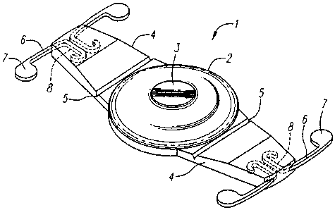
[0009] Figure 1 is a perspective view of a preferred embodiment of the present
invention.
[0010] Figure 2 is a front elevational view.
[0011] Figure 3 is a side elevational view.
[0012] Figure 4 is an end view.
[0013] Figure 5 illustrates the lens, showing T-shaped haptics engaged in the
capsular
bag having been depressed by the bag wall toward the optic.
[0014] Figures 6a and 6b provide details of the blended polyspheric design
transition of
the anterior optic surface from the outside to the center of the lens.
[0015] According to the present invention the optic is of a foldable, flexible
silicone,
acrylic or hydrogel material and the haptic plates are of a foldable material
that will withstand
multiple foldings without damage, e.g., silicone. Preferably, the end of the
plate haptics have
T-shaped fixation devices and are hinged to the optic.
-2-
CA 02658243 2009-01-15
WO 2008/014496 PCT/US2007/074664
DESCRIPTION OF THE PREFERRED EMBODIMENT
[00161 Turning now to the Figures, a preferred embodiment is illustrated in
detail
comprising an intraocular lens 1 formed as a flexible solid optic 2 preferably
made of silicon,
and flexible extending portions 4 of any suitable form which may be plate
haptics or fingers
which are capable of multiple flexations without damage and formed, for
example, of silicone.
The optic 2 and haptics 4 preferably are uniplanar, and one or more haptics 4
extend distally
from opposite sides of the optic 2.
[00171 According to the present invention, the optic 2 has a central blended
area 3. The
lens 1 preferably comprises an accommodating intraocular lens currently
available from
eyeonics, inc., Aliso Viejo, California, such as shown in U.S. Patent number
6387126,
typically with a 4.5 mm diameter optic, but with a polyspheric optic 3 and
which has an added
of less than 1 dioptor of power in the center of the lens 1 producing a single
focal point. The
area 3 is on the anterior side of the lens, and the posterior side can be any
conventional form or
can be toric if desired, or just the posterior surface behind the bulls eye
could be toric. The
added power area 3 is to aid in near vision by producing a single focal point
with increased
depth of focus. The optic diameter can range from approximately 3.5-8.0 mm but
a typical one
is 4.5-5.0 mm.
[00181 Non-accommodating intraocular lenses have been disclosed with a central
area
with a power of 2.0 diopters or more. Examples are in Nielson, U.S. Patent No.
4,636,211, and
Keats, U.S. Patent No. 5,366,500. Such lenses result in the patient having two
separate images,
although the brain tends to ignore an unwanted image.
[00191 Importantly, with the present acconimodating lens having a central area
of less
than 1.0 diopter the distant vision of the patient will slightly blur with no
separate images, but
also improve the near vision principally through an increased depth of field.
Thus, there will
not be two separate images, but a blurred primary image which when seen in one
eye only,
preferably with the other eye having a standard intraocular lens, is believed
to essentially be
not noticeable by the patient.
[00201 The haptics preferably are plate haptics having arcuate outer edges
including
loops 6. The loops 6 when unrestrained are somewhat less curved in
configuration as shown in
Figures 1-2, but compare an example of an inserted lens 1 as seen in Figure 5.
The lens 1,
including the optic 2, haptics 4, and loops 6 is preferably formed of a semi-
rigid material such
as silicone, acrylic, or hydrogel, and particularly a material that does not
fracture with time.
The loops 6 can be of a material different from the haptics 4 and retained in
the haptics by
-3-
CA 02658243 2009-01-15
WO 2008/014496 PCT/US2007/074664
loops 8 molded into the ends of the haptics. Grooves or thin areas 5 forming
hinges preferably
extend across the haptics 4 adjacent to the optic 2.
[0021] The flexible haptics 4 and loops 6 can be connected to an acrylic optic
2 by
means of an encircling elastic band (not shown) which fits into a groove in
the acrylic optic 2
as shown and described in co-pending Application Serial No. 10/888536 filed
July 8, 2004 and
assigned to the assignee of the present application.
[0022] There can be a sharp edge 12 around the posterior surface 14 of the
optic 2. The
junction of the posterior surface 14 of the optic 2 to the edge of the lens 1
is a sharp edge or
junction 12 designed to reduce the migration of cells across the posterior
capsule of the lens
post-operatively and thereby reduce the incidence of posterior capsular
opacification and the
necessity of YAG posterior capsulotomy. The anterior surface 16 of the optic 2
is closer to the
groove 2 than is the posterior surface 14.
[0023] Figure 1 illustrates the haptics 4, loops 6, hinge 5 across the haptics
adjacent to
the optic 2. Hard knobs 7 can be provided on the ends of the loops 6 and are
designed to fixate
the loops 6 in the capsular bag of the eye and at the same time allow the
loops 6 to stretch
along their length as the optic 2 of the lens 1 moves backward and forward and
the haptics 4
move or slide within pockets formed between the fusion of the anterior and
posterior capsules
of the capsular bag.
[0024] The present polyspheric concepts are applicable to several forms of
lenses, such
as lenses shown in Cumming U.S. Patent Nos. 5,476,514, 6,051,024, 6,193,750,
and 6,387,126.
[0025] Figures 6a and 6b illustrate more detail of the blended polyspheric
design of the
anterior optic surface 16 and thus show the transition of the anterior optic
surface from the
outside surface of spherical radius SRl to the center surface of the spherical
radius of SR2
which comprises the central area 3 illustrated in the other Figures. Figures
6a and 6b
demonstrate the transition area as a varying radius that ranges from SRI to
SR2, and it should
be noted that the difference between SR1 and SR2 has been enhanced to better
show the
transition. In particular, SRI is > SR3 > SR4 > SR5 > SR2.
[0026J As is well known in the art, the intraocular lens 1 such as that in the
drawings is
implanted in the capsular bag of the eye after removal of the natural lens.
The lens is inserted
into the capsular bag by a generally circular opening torn in the anterior
capsular bag of the
human lens and through a small opening in the cornea or sclera. The outer ends
of the haptics
4, or loops 6, are positioned in the cul-de-sac of the capsular bag. The outer
ends of the
haptics, or the loops, are in close proximity with the bag cul-de-sac, and in
the case of any form
of loops, such as 6, the loops are deflected from the configuration as shown
for example in
-4-
CA 02658243 2009-01-15
WO 2008/014496 PCT/US2007/074664
Figure 2 to the position shown in Figure 5. The knobs 7 can be provided on the
outer end
portions of the loops 6 for improved securement in the capsular bag or cul-de-
sac by
engagement with fibrosis, which develops in the capsular bag following the
surgical removal of
the central portion of the anterior capsular bag. Additionally, according to
the present
invention, the lens with the central area 3 is intended to be implanted in the
non-dominant eye
of the patient, and a conventional intraocular lens like that seen in the
drawings but without the
central area 3 is intended to be implanted in the dominant eye of the patient.
The present lens
implanted in the non-dominant eye is intended to give superior instant near
vision than if the
non-dominant eye has implanted therein a lens without the central area 3. The
lenses are
implanted in the same manner as described above and as known in the art.
[0027] There are two descriptions of central diopter and range that should be
considered.
= The first looks at the distribution of the lens over the dioptric power
range of 4.0 to
33.0, the mode - or the most commonly used dioptric power of the lens is 22.0
diopter.
= A histogram of the lens is basically a bell curve with a peak at 22.0
diopter. Often
analysis is done with a 22 diopter lens for this very reason.
The second can be relative to the lens design with the central diopter being
the
dioptric power of the center portion 3 of the lens of typically 1.5 mm
diameter. The
dioptric power of this area will be <1.01arger than that of the surrounding
area -
thus the <1.0 diopter add region.
[0028] The lens design is sewed on the existing eyeonics Crystalens to the
extent of the
following:
= Lens and plate haptics are manufactured from the same mold; however, one of
the
pins for molding the anterior optical surface of the present lens is
different.
= Lens and plate material is Biosil (Silicone).
= Haptic is the same design.
= Haptic material is the same Kapton HN (polyimide).
= The posterior surface SRO may be the same as or different than SR1 (e.g. a
23
diopter pin on the anterior side and a 21 diopter pin on the posterior side
will give a
22 diopter lens).
[0029] Below are calculated dimensions of the optical section of the IOL for
the
minimum, average and maximum diopter lens. Diopter 1 is the dioptric power
through the
-5-
CA 02658243 2009-01-15
WO 2008/014496 PCT/US2007/074664
outer perimeter of the lens, and Diopter 2 is through the center section. Note
that the radii are
approximate as SRO (posterior surface spherical radius) and SR1 (anterior
surface spherical
radius - outer area) aren't necessarily the same. The center thickness on the
center area 3 is
approximately 3 microns (0.003 mm) thicker over the 4 to 33 diopter range.
Diopter Diopter T SRO & SR2 Center
1 2 SR1 (mm) Thickness
(mm) (mm)
4 5 45.47 30.30 0.46
~ - ~
22 23 8.24 7.55 0.97
1- - - -
33 34 5.47 5.16 1.32
After the lens is manufactured, it is tumbled with a slurry of glass beads to
remove any
flashing, smooth the edges and integrate the radii, and it shrinks, resulting
in an absence of
discrete radii SR1 - SR5, and thus ends up not a multiple power lens but a
lens with a
polyspheric front surface. The resulting blended design after completion does
not cause
separate images as does a multifocal lens, but actually provides a central
curve which provides
additional focusing power and actually results in an extended region of depth
of field about the
far point of the patient's vision. Thus, a desired depth of field increase
about the focal point
occurs, and the retinal image has been determined to be superior over a wider
range than a
standard accommodating intraocular lens. The through focus wavefront
aberrations peak to
valley and RMS graphs and Waveforms 1 and 2 below show quantitatively how the
present
ED-AIOL provides superior overall optical performance in the range of object
vergence from
infinity to 2 D. Thus, the lens functions simply by extending the range of
accommodation
about the far point by increasing the static depth of field. A patient's
vision is improved by
virtue of an increased depth of field, and this depth of field also will be
present if the patient
wears spectacles for near vision.
-6-
CA 02658243 2009-01-15
WO 2008/014496 PCT/US2007/074664
The Waveforms 2 are RMS wavefront aberrations for AIOL and ED-AIOL for object
vergence
distance from 0 D (object at infinity) to 2 D (500 mm).
In the Waveforms 1 and 2 it can be seen that the AIOL provides lower wavefront
aberration
errors in terms of peak to valley and RMS values over the rage of object
distance from infinity
to about 4 M(0.25 D). For closer object distances (4 M to 500 mm), the ED-AIOL
provides
better optical performance. In the majority of the object vergence range, the
ED-AIOL
provides about 33% better P-V performance and about 50% better RMS performance
compared to the AIOL. As can be seen from the lateral shift in the graphs,
this corresponds to
about a 0.3D improvement for the ED-AIOL. This again demonstrates the fact
that the ED-
AIOL should provide better overall performance over the depth of field range
about the
AIOL's focal point.
[0030] The end of the loops 6 containing the knobs 7 may be either integrally
formed
from the same material as the haptics 4 or the loops may be of a separate
material such as
polyimide, prolene, or PMMA as discussed below. The loops if formed of a
separate material
are molded into the terminal portions of the haptics 4 such that the flexible
material of the loop
6 can extend by elasticity along the internal fixation member of the loop.
[0031] As noted above, the haptics 4 may have a groove or thin area 5 forming
a hinge
across their surface adjacent to the optic. This facilitates movement of the
optic anteriorly and
posteriorly relative to the outer ends of the haptics.
[0032] Accordingly, there has been shown and described a lens that ideally
comprises a
silicon optic and silicone haptic plates, loops that can be of a different
material than the plate,
and a fixation device at the end of each loop allowing for movement of the
loops along the
tunnel formed in the fusion of the anterior and posterior capsules of the
human capsular bag,
and wherein the anterior surface of the optic has a central area of increased
power of less than 1
diopter as well as a method of implanting the lens in the non-dominant eye.
[0033] Various changes, modifications, variations, and other uses and
applications of
the subject invention will become apparent to those skilled in the art after
considering this
specification together with the accompanying drawings and claims. All such
changes,
modifications, variations, and other uses of the applications which do not
depart from the spirit
and scope of the invention are intended to be covered by the claims which
follow.
-7-
SUBSTITUTE SHEET (RULE 26)
CA 02658243 2009-01-15
WO 2008/014496 PCT/US2007/074664
APPENDIX 1
Through Focus Wavefront Peak to Valley
1.8
1.6
...
:
:::::::::::::::::::::::::::::::::::::::::::::::::::::::::::::::::.::.::.::.::.:
:.::.::.::.::: ...
....
f<2
.. .,
1.4 f:? ;><;
:
~;i?:: <::::: >:::::: >;;;;;;;;;;;;;
1
2
0
.?:::>:::::: ::::::>::::::>::::
<iY?~::i:::i:::::i:::iz:::
. >>>>>:>;:
; A AIOL
..............................
ED-AIOL
4' ~ - -
0.8
::.::.::.:::::::.::.::.::.::.::.::.:::::::::::::::::::::
~if <i<i ii <: :<i:<i:>::<: >:<
0.6
s::::>::::::::>::::::::>::::::::>::::::::>::::::::>::::::::>::::::::>::::::::>:
:::::
~ie:>::::>;::::>::::>:::;:::>:>:::s:~ >3
...::::: .: ...........................................
d z; zzzzzzzzizzz:z:> ' ::z: :i::: v .>;c,.>, ,,,,:v>:::=;::::v>a::::v,:
0.4
>::::>:::<i::;:>::::>::
::>::::>::::::::>:<`:::::::::::::::::::::::::::: ::
..:.;:.;:.::.:.;:.;:.;:.;:.;:.;:.;:..... <;: <::.<u
1IiiIIiJ1II.jT
\\ ~
~..
. :. .
,:~:<:<:::::< .:::::::::::::>:<::::;::::.;,.;:,,
.. .. ii:,....<i;i;<..
:.......
...
0 . ................ .
0 0.5 1 1.5 2
Object vergence (D)
Waveforms 1
The Waveforms 1 are peak to valley wavefront aberrations for AIOL and ED-AIOL
for object
vergence distance from 0 D (object at infinity) to 2D (500 mm).
Through Focus Wavefront RMS
1.4 \~k~
1.2 -
c xxxxxx: >:::>:::
;;;;;a
0.8 \;;;;<z
<..AIOL
:
\...
>.::::::<:::::
ED-AIOL
06 ; ;;:<:"'` >
0.4 MM
\\\\
0.2
I MEM"I",
p , < ~ ~_ ME>:z
0 0.5 1 1.5 2
Object vergence (D)
Waveforms 2
-8-
SUBSTITUTE SHEET (RULE 26)