Note: Descriptions are shown in the official language in which they were submitted.
CA 02658735 2009-03-17
ANCHORING DEVICE
Technical Field
[0002] The present disclosure relates generally to the field of surgical
devices, and more
particularly to anchoring devices, such as sutures, which include a loop
having anchors
disposed along a surface.
Background of Related Art
[0003] Surgical sutures have been successfully used for various types of
medical
procedures, including tissue and wound closure. Surgical sutures typically
have a needle
attached at one end. As the needle penetrates tissue, the suture enters,
passes through, and
exits tissue, at which point knots may be used to secure the tissue or wound.
[0004] Additionally, sutures typically employ a knot at the distal end to
secure the suture
end in tissue, permitting movement of the free end through tissue. Knot tying
adds time to a
procedure and may result in additional bulk material being left at the wound
site. Improvements
in the field are desired.
[0005] Furthermore, specific patient populations such as patients with
diabetes T1, T2,
or other immuno-compromised patients (such as chemotherapy patients) have less
elastic
tissue. These patient populations have longer healing profiles and less
compliant tissue and
these factors may lead to lower suture holding forces in tissue. Needles tend
to be oversized for
given suture diameter and a larger needle may leave behind a larger hole at
the needle
1
CA 02658735 2009-03-17
penetration point in the tissue. The suture generally needs to fill this hole.
Also, improvements
in suture holding forces are desired.
SUMMARY
[0006] The present disclosure relates to methods for securing, and more
specifically
methods for securing tissue using anchoring devices. The method includes the
steps of
providing a suture having a proximal portion and a distal portion wherein the
proximal portion
includes an elongate body and the distal portion of the suture terminates in a
loop, and at least
a portion of the loop includes a plurality of barbs and the suture further
includes a bifurcation of
the elongate body into two branches. The method includes inserting the
proximal portion of the
suture into a tissue penetration point; advancing the two branches of the
suture through the
tissue such that the two branches are simultaneously pulled through a
penetration point; and
securing the suture in the tissue. The method may further include the step of
inserting the
proximal portion of the suture through a segment of the loop remaining outside
the body tissue.
The method may further include the step of pulling the suture through tissue
until movement of
the loop through tissue is limited by an end effector.
[0007] Additionally, the passage of the two branches of the loop through the
penetration
points may lead to an increase the holding force of the suture in tissue. The
two branches also
exert pressure on the tissue at the penetration point. The two branches of the
anchoring device
further includes barbs on a surface thereof and these barbs may secure the two
branches in the
body tissue.
[0008] In some embodiments, the anchoring device includes and end effector
which
limits movement of at least a portion of the loop through tissue such that a
segment of the loop
remains outside the body tissue. In certain embodiments, the end effector
engages the tissue to
prevent movement of the loop in a proximal direction.
2
CA 02658735 2009-03-17
[0009] The present disclosure also contemplates an anchoring device having an
elongate body including a proximal portion and a distal portion, the proximal
portion of the
elongate body terminating in a free end and the distal portion of the elongate
body forming a
loop; the loop includes a first plurality of anchors disposed along a surface
of the loop and the
loop further includes at least one end effector.
[00010] In certain embodiments the end effector is selected from the group
consisting of
barb, knot, pledget, and buttress. In other embodiments the end effector is a
bulk of a material
configured and dimensioned to limit movement of a distal segment of the loop
in a proximal
direction through tissue. In some embodiments, the end effector may be
integral with the loop,
alternatively, the end effector may be a separate device which is secured to
the loop. In
alternate embodiments, the end effector limits movement of at least a portion
of the loop
through tissue such that a segment of the loop remains outside a body tissue.
[00011] The anchoring device of the present disclosure may further include a
needle
secured to a proximal portion of the elongate body.
[00012] Another method for securing tissue is also provided, the method
comprises the
steps of providing an anchoring device, inserting a proximal portion of the
medical device into
tissue, pulling the two branches of the loop through tissue such that the two
branches are
simultaneously pulled through a penetration point; and, advancing the proximal
portion of the
loop through tissue such that the end effector limits movement of the distal
portion of the loop
through the tissue.
BRIEF DESCRIPTION OF DRAWINGS
[00013] Various preferred embodiments of the sutures are described herein with
reference to the drawings, in which:
[00014] FIGS. 1A-1 B are side views illustrating one embodiment of a looped
suture;
[00015] FIGS. 2A-2B are side views illustrating another embodiment of a looped
suture;
3
CA 02658735 2009-03-17
[00016] FIG. 3 is a side view illustrating one embodiment of an anchoring
suture
including barbs;
[00017] FIG. 3A is an enlarged view of the area of detail designated in FIG.
3;
[00018] FIG. 4 is a side view illustrating an alternate embodiment of an
anchoring suture
including barbs;
[00019] FIG. 5 is an enlarged side view showing a barb of an alternate
embodiment of an
anchoring suture with a compound barb; and,
[00020] FIG. 6 is a side view illustrating another embodiment of an anchoring
suture
including barbs;
[00021] FIG. 7 is a side view illustrating an alternate embodiment of an
anchoring suture
with an end effector;
[00022] FIG. 8A is a side view of another embodiment of an anchoring suture
with an end
effector;
[00023] FIG. 8B is a side view of a different embodiment of an anchoring
suture with an
end effector;
[00024] FIG. 8C is a side view of an alternate embodiment of an anchoring
suture with an
end effector;
[00025] FIG. 9A is a plan view of the anchoring suture of FIG. 4 in tissue
with portions of
tissue removed;
[00026] FIG. 9B is a side view of the anchoring suture of FIG. 4 in tissue
with portions of
tissue removed;
[00027] FIG. 10A is a plan view of the anchoring suture of FIG. 8C in a first
position in
tissue, with portions of tissue removed;
[00028] FIG. 10B is a plan view of the anchoring suture of FIG. 8C in a second
position in
tissue, with portions of tissue removed;
4
CA 02658735 2009-03-17
[00029] FIG. 1 1A shows a perspective view, partially in cross-section, of a
suture filling a
needle penetration point; and,
[00030] FIG. 11 B shows a perspective view, partially in cross-section, of an
anchoring
suture of the present disclosure filling a needle penetration point.
DETAILED DESCRIPTION OF EMBODIMENTS
[00031] The present disclosure is directed to an anchoring device and in
certain preferred
embodiments, a suture, herein referred to as an anchoring suture. The
anchoring sutures of
certain embodiments of the present disclosure have an elongate body, which
connects to a
needle at a proximal end thereof, and a distal end of the elongate body forms
an anchoring
loop. The anchoring loop further includes a plurality of anchors (tissue
engaging members).
Medical devices of the present disclosure include sutures formed from fibers,
filaments, and
yarns.
[00032] Anchoring devices, including anchoring sutures of the present
disclosure may be
absorbable or non-absorbable. It should be understood that combinations of
filaments made
from different materials (e.g. natural and synthetic, or bioabsorbable and non-
bioabsorbable
materials) may be used to make the present anchoring suture.
[00033] Suitable synthetic absorbable materials include polymers such as those
made
from lactide, glycolide, caprolactone, valerolactone, carbonates (e.g.,
trimethylene carbonate,
tetramethylene carbonate), dioxanones (e.g., 1,4-dioxanone), 1,dioxepanones
(e.g., 1,4-
dioxepan-2-one and 1,5-dioxepan-2-one), ethylene glycol, ethylene oxide,
esteramides, y-
hyd roxyval e rate, R-hydroxypropionate, alpha-hydroxy acid, hydroxybuterates,
orthoesters,
hydroxy alkanoates, tyrosine carbonates, polyimide carbonates, polyimino
carbonates such as
poly (bisphenol A-iminocarbonate) and poly (hydroquinone-iminocarbonate), and
polymer drugs
(e.g., polydiflunisol, polyaspirin, and protein therapeutics) and copolymers
and combinations
thereof. Suitable natural absorbable polymers include collagen, cellulose and
gut. In
CA 02658735 2009-03-17
embodiments, glycolide and lactide based polyesters, including copolymers of
lactide and
glycolide may be used.
[00034] Suitable non-absorbable materials which may be used to form the
anchoring
sutures disclosed herein include non-absorbable natural materials such as
cotton, silk, and
rubber. Suitable non-absorbable synthetic materials include monomers and
polymers derived
from materials such as nylons, polyolefins such as polypropylene and
polyethylene, ultra high
molecular weight polyethylene (UHMWPE), polyamides, polyesters such as poly
ethylene
terephthalate (PET), polyaryletherketone, polyvinylidene difluoride (PVDF),
acrylic, polyamides,
aramids, fluropolymers, polybutesters, silicones, and polymer blends,
copolymers thereof and
combinations with degradable polymers. Polypropylene can also be utilized to
form the suture.
The polypropylene can be isotactic polypropylene or a mixture of isotactic and
syndiotactic or
atactic polypropylene. Additionally, non-absorbable synthetic and natural
polymers and
monomers may be combined with each other and may also be combined with various
absorbable polymers and monomers to create fibers and filaments for the
present anchored
device.
[00035] In certain embodiments, anchoring devices, including anchoring
sutures, may, in
whole or in part (e.g. anchors) may be constructed using shape memory
polymers. Suitable
polymers used to prepare hard and soft segments of shape memory polymers
include
polycaprolactone, dioxanone, lactide, glycolide, polyacrylates, polyamides,
polysiloxanes,
polyurethanes, polyether amides, polyurethane/ureas, polyether esters, and
urethane/butadiene
copolymers and combinations thereof.
[00036] In some embodiments, the sutures may include metals (e.g. steel and
degradable magnesium), metal alloys or the like.
[00037] As used herein, the terms "fibers", "filaments" and "yarns" each may
be used to
construct in whole or in part anchoring devices. The term "fibers," in this
context, are generally
used to designate natural or synthetic structures that have a length
approximately 3 orders of
6
CA 02658735 2009-03-17
magnitude greater than their diameter or width. The term "filaments" are
typically used to
describe "fibers" of indefinite or extreme length, and "yarns" as a generic
term for a continuous
strand of twisted or untwisted "fibers" or "filaments" in a form suitable for
knitting, weaving,
braiding or otherwise intertwining.
[00038] Sutures of the present disclosure may be monofilament or multifilament
(e.g.
braided). Methods for making sutures from these suitable materials are within
the purview of
those skilled in the art (e.g. extrusion and molding). The filaments may be
combined to create a
multifilament suture using any technique within the purview of one skilled in
the art such as
commingling, twisting, braiding, weaving, entangling, and knitting. For
example, filaments may
be combined to form a yarn or they may be braided. In another example,
filaments may be
combined to form a yarn and then those multifilament yarns may be braided.
Those skilled in
the art reading this disclosure will envision other ways in which filaments
may be combined.
Fibers may also be combined to produce a non-woven multifilament large
diameter suture. In
certain embodiments, a multifilament structure useful in forming an anchoring
suture according
to the present disclosure may be produced by braiding. The braiding can be
done by any
method within the purview of those skilled in the art. For example, braid
constructions for
sutures and other medical devices are described in U.S. Patent Nos. 5,019,093;
5,059,213;
5,133,738; 5,181,923; 5,226,912; 5,261,886; 5,306,289; 5,318,575; 5,370,031;
5,383,387;
5,662,682; 5,667,528; and 6,203,564.
Furthermore, the anchoring device may include portions which are
monofilament and portions which are multifilament. In some embodiments, the
proximal end of
the elongate body may be a multifilament and the looped portion (loop portion
described below)
may be a monofilament.
[00039] Additionally, the anchoring device may include biologically acceptable
additives
such as plasticizers, antioxidants, dyes, dilutants, bioactive agents and
combinations thereof,
7
CA 02658735 2009-03-17
which can be coated on the filaments or fibers, or impregnated into the fibers
or filaments (e.g.
during compounding or extrusion) used to form the anchoring suture of the
present disclosure.
[00040] Various compositions and materials may also be applied to the
anchoring sutures
or included in the filaments or fibers to improve mechanical properties such
as handling and
knot strength or to deliver medicinal agents. Suitable coating materials
include any materials
conventionally applied to sutures. For example, suitable materials include
fatty acid esters
which may be combined with the metal salt of a fatty acid in the coating
composition. Such
esters include, for example, calcium stearate, stearoyl lactylate esters,
paimityl lactylate esters,
oleyl lactylate esters such as calcium, magnesium, aluminum, barium, or zinc
stearoyl lactylate,
calcium, magnesium, aluminum, barium, or zinc palmityl lactylate; calcium,
magnesium,
aluminum, barium, or zinc oleyl lactylate; with calcium stearate and calcium
stearoyl-2-lactylate
(such as the calcium stearoyl-2-lactylate commercially available under the
trade name VERV
from American Ingredients Co., Kansas City, Mo.) being preferred. When
desirable, the fatty
acid ester may be combined with a solvent. Suitable solvents include polar and
non-polar
solvents including but not limited to alcohols (e.g., methanol, ethanol,
propanol), chlorinated
hydrocarbons (such as methylene chloride, chloroform, 1, 2-dichloro-ethane),
and aliphatic
hydrocarbons such as hexane, heptene, ethyl acetate.
[00041] In embodiments, the anchoring device may be combined with and/or
coated with
suitable materials including polyalkylene oxides such as polyethylene oxide,
polypropylene
oxide, polyethylene glycol (PEG), polypropylene glycol, copolymers thereof,
and the like,
including those having acrylate groups such as acrylate PEGs, and acrylate
PEG/PPG
copolymers. Such combinations may include blends or copolymers with
polyalkylene oxide
oligomers or polymers or other non-toxic surfactants. The resulting
composition may possess
antimicrobial properties due to the presence of the copolymers described
above. In other
embodiments, the sutures may be combined with silicone acrylates. Coatings may
be applied
to the individual filaments or the anchoring suture at any time prior to
sterilization techniques.
8
CA 02658735 2009-03-17
Coatings can be applied to the filaments using any technique within the
purview of those skilled
in the art.
[00042] Additionally, the anchoring device may incorporate various
pharmaceuticals and
medicinal agents. Medicinal agents and drugs may be applied to the sutures
and/or construct
materials by methods within the purview of those skilled in the art, including
but not limited to
dipping, spraying, brushing, vapor deposition, coextrusion, capillary wicking,
film casting,
molding and the like. Additionally, solvents may be used to incorporate
various agents into the
anchoring suture. Suitable solvent include those listed above.
[00043] Medicinal agents which may be incorporated into the device include
antimicrobial
agents, anti-virals, anti-fungals, and the like. Antimicrobial agents as used
herein is defined by
an agent which by itself or through assisting the body (immune system) helps
the body destroy
or resist microorganisms which may be pathogenic (disease causing). The term
"antimicrobial
agent" includes antibiotics, quorum sensing blockers, surfactants, metal ions,
antimicrobial
proteins and peptides, antimicrobial polysaccharides, antiseptics,
disinfectants, anti-virals, anti-
fungals, and combinations thereof.
[00044] Agents may be incorporated into a coating using solvents or mixed with
various
monomers or polymers and applied to the anchoring device. Additional suitable
medicinal
agents which may be used include colorants, dyes, preservatives, protein and
peptide
preparations, protein therapeutics, polysaccharides such as hyaluronic acid,
lectins, lipids,
probiotics, antibiotics, angiogenic agents, anti-thrombotics, anti-clotting
agents, clotting agents,
analgesics, anesthetics, wound repair agents, chemotherapeutics, biologics,
anti-inflammatory
agents, anti-proliferatives, diagnostic agents, antipyretic, antiphlogistic
and analgesic agents,
vasodilators, antihypertensive and antiarrhythmic agents, hypotensive agents,
antitussive
agents, antineop-astics, local anesthetics, hormone preparations,
antiasthmatic and antiallergic
agents, antihistaminics, anticoagulants, antispasmodics, cerebral circulation
and metabolism
improvers, antidepressant and antianxiety agents, vitamin D preparations,
hypoglycemic agents,
9
CA 02658735 2009-03-17
antiulcer agents, hypnotics, antibiotics, antifungal agents, sedative agents,
bronchodilator
agents, antiviral agents, dysuric agents, brominated or halogenated furanones,
and the like. In
embodiments, polymer drugs, i.e., polymeric forms of such compounds for
example, polymeric
antibiotics, polymeric antiseptics, polymeric chemotherapeutics, polymeric
anti-proliferatives,
polymeric antiseptics, polymeric non-steroidal anti-inflammatory drugs
(NSAIDS), and the like
may be utilized and combinations thereof.
[00045] The anchoring device of the present disclosure can additionally
contain suitable
medicinal agents such as viruses and cells, peptides, polypeptides and
proteins, analogs,
muteins, and active fragments thereof, such as immunoglobulins, antibodies
(monoclonal and
polyclonal), cytokines (e.g. lymphokines, monokines, chemokines), blood
clotting factors,
hemopoietic factors, interleukins (IL-2, IL-3, IL-4, IL-6), interferons ([3-
IFN, a-IFN and y-IFN),
erythropoietin, nucleases, tumor necrosis factor, colony stimulating factors
(e.g., GCSF, GM-
CSF, MCSF), insulin, anti-tumor agents and tumor suppressors, blood proteins,
gonadotropins
(e.g., FSH, LH, CG, etc.) hormones and hormone analogs (e.g., growth hormone),
vaccines
(e.g., tumoral, bacterial and viral antigens), somatostatin, antigens, blood
coagulation factors,
growth factors, protein inhibitors, protein antagonists, and protein agonists,
nucleic acids, such
as antisense molecules, DNA, RNA, oligonucleotides, polynucleotides and
ribozymes and
combinations thereof.
[00046] Methods for combining these medicinal agents with compositions of the
present
disclosure are within the purview of those skilled in the art and include, but
are not limited to
mixing, blending, dipping, spraying, wicking, solvent evaporating and the
like.
[00047] In the description that follows, the term "proximal" as used herein,
means the
portion of the device which is nearer to the user, while the term "distal"
refers to the portion of
the device which is further away from the user.
[00048] Sutures of the present disclosure include an elongate body, having
both distal
and proximal portions, the distal portion of which transitions from the
elongate body to an
CA 02658735 2009-03-17
anchoring loop. Methods for creating anchoring loops are within the purview of
those skilled in
the art and include but are not limited to welding, ultrasonic energy,
cutting, molding and gluing.
In preferred embodiments to be described later, the anchoring loop includes
barbs along a
surface.
[00049] Adjuncts to making loops, such as adhesives and glues, may also be
employed
in the anchoring suture. In some embodiments (FIGS. 1A, 1B), the distal
portion of suture may
be folded and fixed to elongate body using adhesives and glues. In alternate
embodiments, as
shown in FIGS. 2A and 2B, loop portion may initially be a separate component
which connects
to an elongate body and optionally glued in place. It should be understood
that embodiments
and methods described in FIGS. 1 and 2 can be used to create any of the
anchoring suture
embodiments described herein (FIGS 3-6). Suitable materials such as absorbable
and non-
absorbable materials include, but not limited to cyanoacrylates, isocyanates,
polyurethanes,
polyamines, polyamides, polyacrylates, polymethacrylates, silicones,
carbonates, and other
synthetic monomers and polymers and combinations thereof.
[00050] Adhesives such as cyanoacrylates can be employed in creating sutures
of the
present disclosure. Suitable cyanoacryiates include materials derived from
methyl
cyanoacrylate, ethyl cyanoacrylate, butyl cyanoacrylate, octyl cyanoacrylate,
isobutyl
cyanoacrylate, and methoxypropyl cyanoacrylate and combinations thereof and
the like.
[00051] The anchoring loop further includes anchors disposed along a surface.
Anchors
can be created on the anchoring suture using any technique, including but not
limited to lasers,
molding, knives, biades, stamping, and other cutting means within the purview
of those skilled in
the art. Ultrasonic energy can also be used to create barbs or anchors as
described in U.S.
Patent Application No. 60/994,173 filed on September 17, 2007 entitled "Method
of Forming
Barbs on a Suture".
[00052] In some embodiments, anchoring sutures of the present disclosure
include loops
which are integral to an elongate body, as shown in FIGS. 1 and 3. Sutures
with integral loops
11
CA 02658735 2009-03-17
may be defined as having one structure or component in which the elongate body
is continuous
with the loop. For example, FIG. 1 shows an elongate body 10 in which the
distal end is folded
or "looped" to create a loop 14 (FIG. 1 B) at the distal end of the medical
device. The suture as
shown in FIGS. 1 and 2 further includes transition area 16 and anchors which
will be described
in further detail below. An anchoring suture may also contain an integral loop
as shown in FIG.
3, wherein the loop portion may be molded. In alternate embodiments, such as
FIG. 2,
anchoring sutures may comprise two components which are fixed or fitted
together in a fashion
as to create the anchoring suture. For example, the elongate body 10 may
include a female
component while the loop 14 may include a male component and the two
components may be
fitted together to create a final product. One skilled in the art can envision
other manufacturing
processes in which to create integral loops and medical devices with integral
and non-integral
loops.
[00053] Another embodiment of the anchoring suture of the present disclosure
is shown
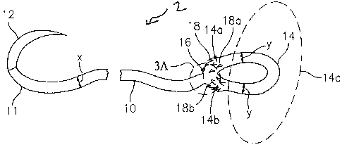
in FIG. 3 and is designated generally by reference numeral 2. Suture 2 has an
elongate body
10, a proximal portion of elongate body 10 terminating in a free end 11, and a
distal portion of
the elongate body 10 which forms, transitions into, or terminates in a loop
14. As shown in FIG.
3, the free end 11 further comprises a needle 12. The elongate body 10 has a
diameter "x" and,
in preferred embodiments, the elongate body 10 is generally elliptical in
transverse cross-
section. The distal end of elongate body 10 extends into a loop 14,
bifurcating at transition area
16 (FIGS. 3 and 3a). Loop 14 includes two branches 14a and 14b, which may be
identical in
shape and cross-sectional area, to both each other and elongate body 10. In
preferred
embodiments, sections 14a and 14b are generally elliptical in shape and cross-
sectional area,
although other shapes are envisioned such as circular, oval, square, and
rectangular. In the
embodiment shown in FIG. 3, the loop 14 may be integral with the elongate body
10 of the
suture 2. In alternate embodiments, the loop 14 may be a separate component
prior to
assembly (FIGS.1 and 2), and during assembly the loop 14 may be attached to
the elongate
12
CA 02658735 2009-03-17
body 10. The loop 14 has a generally arcuate surface, and each branch (14a and
14b) has an
independent diameter "y", of which 14a and 14b may be of similar or different
diameters. The
loop may be of any shape including circular, oval, polygonal.
[00054] Furthermore, anchoring suture of FIG. 3 includes a first plurality of
anchors 18
disposed along a surface of the loop 14. Anchors 18a are disposed along
surface of branch
14a and anchors 18b are disposed along branch 14b. Additionally, segment 14c
is used to
designate a loop segment in which barbs are absent. In the illustrated
embodiment, anchors 18
are located adjacent transition area 16 of elongate body 10 and anchoring loop
14.
Furthermore, the first plurality of anchors 18 is oriented such that movement
of the anchoring
loop 14 towards the proximal end is limited. As shown in FIG. 3, anchors 18
are oriented
towards transition area 16 to prevent movement of anchoring loop 14 through
tissue. In
embodiments shown, anchors 18 are integral to the anchoring loop 14.
[00055] It will be understood that FIG. 4 is a generally similar to FIG. 3 and
therefore all
numerals and descriptions which are the same in FIG. 3 are designated with the
prime mark
and have some differences. FIG. 4 shows an alternate embodiment of an
anchoring suture 2' in
which a second plurality of anchors 22 is disposed along the elongate body
10'. The second
plurality of anchors 22 extends in the second direction which is different
from a first direction of
the first plurality of anchors. In the embodiment shown, the first plurality
of anchors 18' are
disposed along a loop surface and extend in the first direction, generally
towards transition area
16' of the anchoring suture 2'. The second plurality of anchors 22 extend in a
second direction,
towards the loop 14', with respect to longitudinal axis A of the elongate body
10. As shown in
FIG 4. the first plurality of anchors 18' and the second plurality of anchors
22 extend in
directions substantially opposite to one another. The second plurality of
anchors 22 permits
movement of the elongate body 10' in the direction of the leading or proximal
end while
preventing movement of the elongate body 10' towards the loop end.
13
CA 02658735 2009-03-17
[00056] In the alternate embodiment shown in FIG. 5, anchoring suture 30
includes a
compound barb 26 having an inner surface 30 including a first angle a,
disposed at a first
orientation relative to a longitudinal axis "A"' of the elongate body and a
second angle P having
a second inner surface 32, disposed at a second orientation relative to a
longitudinal axis b of
the elongate body. The anchoring suture may optionally include a third
orientation (not shown).
In the embodiment shown, the first, second and third orientations are each
disposed at different
angles with respect to the longitudinal axis. In some embodiments, the
anchoring suture may
include a staggered arrangement of large or small barbs. In other embodiments,
an anchoring
suture may have a random configuration of both large and small barbs. It will
be understood
that the embodiment shown in FIG. 5 is generally similar to FIGS. 3 and 4, but
has a different
geometry for the barbs. In alternate embodiments, the above-mentioned compound
barb
geometry may also be present on the anchoring loop (not shown).
[00057] The surface area of the plurality of anchors can also vary. For
example, fuller-
tipped anchors can be made of varying sizes designed for specific surgical
applications. When
joining fat and relatively soft tissues, larger anchors may be desired,
whereas smaller anchors
may be more suitable for collagen-dense tissues. In some embodiments (FIG. 4),
a
combination of large and small anchors within the same structure may be
beneficial, for
example when a fiber is used in tissue repair with differing layer structures.
Use of the
combination of large and small anchors with the same fiber wherein anchor
sizes are
customized for each tissue layer will ensure maximum holding properties.
[00058] Another embodiment of an anchoring device is shown in FIG. 6. The
anchoring
device 40 includes a needle 42 at a proximal end 41 of the device. The device
bifurcates at a
transition area 45, and a distal portion of the device terminates in an
anchoring loop 49. The
anchoring loop 49 includes two branches 46a and 46b at a proximal end 47 of
the anchoring
loop 49. The anchoring loop 49 has a generally arcuate surface, branches 46a
and 46b may
have similar or different diameters. In the illustrated embodiment, a first
plurality of anchors 48
14
CA 02658735 2009-03-17
are located adjacent the transition area 45. Furthermore, the first plurality
of anchors 48 is
oriented such that movement of the anchoring loop 49 in tissue, in a direction
towards the distal
end 43 of the device, is limited. As illustrated in Figure 6, the device may
have an elongate
body 44 that is shorter in longitudinal length as compared to the anchoring
loop 49. The two
branches of the loop may be advanced through a single needle penetration point
and pulled
through tissue; the method of which will be described in detail later.
[00059] Figure 7 illustrates an alternate embodiment of an anchoring device 50
which
may be used in combination with a mechanical suturing device such as an Endo
Stitchr""
suturing device commercially available from Tyco Healthcare Group LP.
Anchoring device 50
includes a needle 52 which is compatible with a mechanical suturing device
such as an Endo
StitchTM suturing device. The proximal portion 51 of the anchoring suture
includes an elongate
body 54, and the distal portion 55 of the suture terminates in a loop 59. The
loop 59 includes
two branches 56a and 56b and each branch includes a plurality of barbs 58 on a
surface
thereof. In other embodiments, a plurality of barbs may only be on a surface
of at least one
branch or a portion of the anchoring device. The loop 59 also includes an
unbarbed distal
portion 56c. The loop further includes an end effector 57 which limits
movement of the
anchoring device through tissue. In some embodiments, the end effector is
located on the
unbarbed distal portion 56c of the loop (Figure 7). As illustrated, the end
effector 57 is a bulk
(large mass) of suture material, which is generally "T"-shaped and in the
current embodiment,
the end effector is welded to the loop 59.
[00060] This disclosure contemplates different end effectors and non-limiting
alternate
embodiments are illustrated in FIGS. 8A, 8B and 8C. In Figure 8A an end
effector is illustrated
as a second plurality of barbs 60 which are oriented such that movement of an
end portion of
the loop 63 through tissue towards a proximal end of the anchoring suture is
limited. In this
embodiment, a first plurality of barbs 62 located at a proximal portion of the
loop 63 are shown
oriented in a generally opposite direction to a second plurality of barbs 60,
which are located at
CA 02658735 2009-03-17
a distal portion of loop 63. In another embodiment, Figure 8B, the end
effector is a bead 65 of a
polymeric material. In some embodiments, the bead may of a similar material to
the anchoring
loop and in alternate embodiments; the bead may be comprised of a different
material than the
anchoring loop. In some embodiments, such as Figures 8A and 8B, the end
effector is integral
with the loop. In yet other embodiments, the end effector may be a separate
device such as a
pledget or buttress. As illustrated in Figure 8C, the end effector is a
pledget 67 formed on or
otherwise attached to the loop. In this embodiment, prior to creating a loop,
a suture may
penetrate the pledget 67 and a length of the suture may be pulled through the
pledget. It should
be noted that once the pledget has been moved across a portion of barbs 68
projecting from the
suture surface, the barbs will prevent the pledget from disengaging the suture
and the barbs will
retain the pledget in place on the suture. Next, a loop may be created via
various means
including those described above, and the pledget 67 may be positioned at a
distal most point of
the anchoring loop. It should be understood that end effectors are not limited
to those structures
described herein and one skilled in the art may contemplate other shapes and
devices which
may be used for a similar purpose. End effectors may be constructed using
methods within the
purview of those skilled in the art, including but not limited to glues,
adhesives, lasers, ultrasonic
or heat welding, molding, overmolding and the like. Any of the suture
materials and structures
discussed above may be used to form the anchoring devices discussed herein.
[00061] As used herein, the term "tissue" includes, but is not limited to,
tissues such as
skin, fat, fascia, bones, muscles, tendons, ligaments, organs, nerves, and
blood vessels. Also
used herein, the term "wound" includes, but is not limited to, a surgical
incision, cut, laceration
or severed tissue in human or animal skin or other human or animal bodily
tissue.
[00062] Tissue may be sutured by inserting proximal portion of an anchoring
suture into
tissue at a first section and advancing the proximal portion of the suture
through a second
section of the tissue, and exiting tissue at an exit point. The suture is
pulled through the exit
point until the first plurality of barbs on the anchoring loop engages tissue
and resists movement
16
CA 02658735 2009-03-17
in direction of needle advancement, thus preventing further advancement of
anchoring loop
through tissue. The proximal portion of the suture may optionally be inserted
through the
segment of the loop remaining outside the body tissue for enhanced fixation.
FIGS. 9A and 9B
show the embodiment of FIG. 4, where an unbarbed loop segment 14c' remains
exterior to the
wound site (or external to skin in dermal closure) due to the anchors 18a' and
18b' and lack of
anchors on segment 14c'. It should be understood that all embodiments
described herein can
be used in a similar fashion. Upon exit of tissue, needle and proximal end of
suture may be
passed through segment of loop which remains exterior to wound site to secure
suture in place.
User may then continue suturing wound, entering and exiting tissue until wound
site is closed
(or implant attached).
[00063] Figures 10A and 10B illustrate the embodiment of Figure 8C in tissue.
Tissue
may be secured in a similar manner as described above, by inserting a proximal
portion of the
anchoring device into tissue at a first section and advancing the proximal
portion of the
anchoring device, including a proximal portion of the loop, through a second
section of the
tissue, and exiting tissue at an exit point. In the embodiments described in
FIGS 6, 7, 8A, 8B,
and 8C once the needle is advanced through tissue, the remainder of the suture
follows
including the two branches of the loop. More specifically, the two branches of
the loop are
advanced through a needle penetration point (or points through which the
needle and elongate
body have passed). Figure 10A illustrates a first position of the embodiment
of an anchoring
device as described in Figure 8C. As illustrated, proximal portion of suture
70 and proximal
portion 71 of loop 72, including two branches 72a and 72b, are advanced
through tissue. Both
branches 72a and 72b are advanced through needle penetration points (74a,
74b,74c, and
74d), the barbs engage tissue and suture holding force is increased. Figure
10B shows the
embodiment of Figure 8C in a second position. Once the anchoring suture has
been further
advanced through tissue, the pledget 67 prevents any further movement of the
distal loop
portion through tissue. It should be understood that other embodiments of end
effectors and
17
CA 02658735 2009-03-17
shown and described would function in a manner similar to the embodiment
described with
respect to Figures 10A and 10B. It should also be understood that anchoring
sutures without
end effectors may also be inserted and advanced through tissue in a similar
manner.
[00064] Figure 11A shows the prior art in which an oversized needle 80
penetrates
tissue, and leaves a tissue penetration point (82a and 82b) that a single
suture strand 84 may
not fill. Figure 11 B shows one embodiment of the current disclosure in which
an oversized
needle 90 penetrates tissue and the two branches (94a and 94b) of the
anchoring device 94 can
better fill the needle penetration point 92. The two branches of the loop in
combination with the
barbs allow an increase in tissue holding strength which may be desirable in
certain
applications.
[00065] In order to facilitate needle attachment to an anchoring suture or
device of the
present disclosure, conventional tipping agents can be applied to the braid.
Two tipped ends of
the fiber may be desirable for attaching a needle to each end of the fiber to
provide a so-called
double armed suture. The needle attachment can be made by any conventional
method such
as crimping, swaging, etc, as is known within the purview of those skilled in
the art.
Alternatively, a reduced diameter may be provided at the end of the suture to
be inserted into
the drilled end of a needle. To provide a reduced diameter, the suture may by
machined using
any technique within the purview of those skilled in the art, such as cutting,
grinding, laser
machining or the like.
[00066] Anchoring devices, including anchoring sutures of the present
disclosure may be
employed in medical devices, drug delivery devices and cell growth substrates.
Examples of
suitable medical devices and/or surgical devices employing the anchoring
sutures may include,
but are not limited to meshes, wound dressings, bandages, drug delivery
devices, anastomosis
rings, stents, grafts, catheter systems, soft tissue repair and augmentation
devices, scaffolds,
buttresses, lap bands, tapes, anchors, ribbons, orthopedic devices, tissue
engineering scaffolds,
various cell growth substrates, and other implantable devices. In some
embodiments, devices
18
CA 02658735 2009-03-17
of the present disclosure may be knitted or woven with other fibers, either
absorbable or non-
absorbable, to form surgical devices. The anchoring devices and/or sutures
also can be made
into meshes or non-woven materials to form fabrics, such as matted fabrics and
felts.
[00067] Additionally, anchoring devices of the present disclosure may be
packaged using
materials known to those within the purview of those skilled in the art,
including foil and various
plastics ( e.g. polyethylene), which may provide a moisture barrier.
[00068] Once the anchoring device is constructed, it can be sterilized by any
means
within the purview of those skilled in the art including but not limited to
ethylene oxide, electron
beam (e-beam), gamma irradiation, autoclaving, and the like.
[00069] Example 1
[00070] Distal end of MaxonTM suture is folded towards elongate body to create
a loop,
and suture (loop) is then placed in an ultrasonic welding apparatus, where
loop is welded
closed. Suture is then affixed to an ultrasonic cutting apparatus to create
barbs. Elongate body
and anchoring loop of anchoring suture is cut via ultrasonic blades at various
angles.
[00071] Example 2
[00072] Distal end of SurgiproTM suture is folded towards elongate body to
create loop
and glue is placed on elongate body and distal suture end is folded over and
attached to
elongate body, creating a fixed loop. Suture is then affixed to a cutting
apparatus and anchoring
suture is cut at various angles using a knife. Anchoring suture is then coated
with a
chemotherapeutic agent using solvent casting.
[00073] Example 3
19
CA 02658735 2009-03-17
[00074] Distal end of MaxonTM suture is folded towards elongate body to create
a loop,
and suture (loop) is then placed in an ultrasonic welding apparatus, where
loop is welded
closed. The ultrasonic welding apparatus is then used to weld a distal end of
the loop into a
generally "T"-shape, creating an end effector. Suture is next affixed to an
ultrasonic cutting
apparatus to create barbs. Elongate body and a proximal portion of the
anchoring loop of
anchoring suture is cut via ultrasonic blades at various angles.
[00075] It should be noted that the present disclosure is not limited to wound
closure and
contemplates other procedures such as cosmetic and orthopedic procedures.
Additionally, the
above description contains many specifics; these specifics should not be
construed as
limitations on the scope of the disclosure herein but merely as
exemplifications of particularly
useful embodiments thereof. Those skilled in the art will envision many other
possibilities within
the scope and spirit of the disclosure as defined by the claims appended
hereto.