Note: Descriptions are shown in the official language in which they were submitted.
CA 02660117 2009-02-05
WO 2008/021771 PCT/US2007/075237
INSERTION SYSTEM FOR IMPLANTING A MEDICAL
DEVICE AND SURGICAL METHODS
BACKGROUND OF THE INVENTION
1. Field of the Invention.
[0001] The present invention relates to an insertion system for implanting a
medical
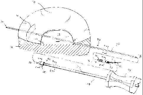
device and to surgical methods for utilizing the same.
2. Description of the Related Art.
[0002] Orthopedic surgeries are commonly performed to repair and replace
damaged
bone and tissue in the human body. To perform orthopedic surgery, a surgeon
may create an
incision and, if necessary, may retract the surrounding tissue to provide the
necessary visual
and physical access to the damaged bone and tissue. Once the incision is made
and the tissue
retracted, the surgeon will perform the necessary repair or replacement
procedures. For
example, if the meniscus of the knee is damaged the surgeon may perform a
meniscectomy,
i.e., remove a portion of a meniscus in the knee.
[0003] While surgical procedures requiring exposure of the damaged bone and
tissue
are effective, healing time for the patient may be greatly reduced if the
surgery is performed
using minimally invasive or arthroscopic techniques. These techniques allow
the surgeon to
make a small incision and perform the entire surgery therethrough. To perform
surgery in
this manner, the surgeon may utilize cannulated devices and other specially
designed tools.
While these procedures are effective, the insertion and manipulation of
medical devices
through the small incision is difficult. For example, certain medical devices
for repairing
tissue are extremely small and may require accurate placement to achieve
optimum results.
SUMMARY OF THE INVENTION
[0004] The present invention relates to an insertion system for medical
devices and to
surgical methods for the implantation of the same. In one embodiment, the
implantation
system includes a guide wire and an insertion tool. The insertion tool
includes a longitudinal
bore extending therethrough for receipt of the guide wire. A portion of the
longitudinal bore
of the insertion tool mates with the outer surface of the guide wire to
rotationally lock the
insertion tool and the guide wire. Additionally, the medical device to be
inserted includes a
CA 02660117 2009-02-05
WO 2008/021771 PCT/US2007/075237
longitudinal bore for receipt of the guide wire. Similarly, at least a portion
of the longitudinal
bore of the medical device may also mate with the outer surface of the guide
wire to
rotationally lock the medical device and the guide wire. The rotational locks
allow for
rotation of the insertion tool to result in corresponding rotation of the
medical device via
rotation of the guide wire. Additionally, in another embodiment, the insertion
tool includes
features which allow for filament management, i.e., reduce or prevent filament
entanglement
during medical device insertion. In a further embodiment, the insertion system
further
includes a needle having an eyelet for receiving and positioning filaments
attached to the
medical device.
[0005] Advantageously, the present insertion system allows for the insertion
of the
medical device without any direct mechanical connection between the medical
device and the
insertion tool. This allows the surgeon to easily maneuver the insertion tool
without
correspondingly altering the position of the medical device, i.e., the
insertion tool and the
medical device are independently axially movable along the guide wire.
Additionally, by
utilizing the guide wire to rotate the medical device, rotational force may be
provided along a
greater length of the medical device instead of at a single point connected to
the insertion
tool.
[0006] In one form thereof, the present invention provides an insertion system
for a
medical device comprising a guide wire having an outer surface; a medical
device having a
first inner surface, the first inner surface defining a first bore adapted for
receiving the guide
wire therethrough; whereby the guide wire and the medical device are
rotationally locked
when the guide wire is received through the first bore; and an insertion tool
having a second
inner surface, the second inner surface defining a second bore adapted for
receiving the guide
wire therethrough, whereby the guide wire and the insertion tool are
rotationally locked when
the guide wire is received in the second bore.
[0007] In another form thereof, the present invention provides An insertion
system for
a medical device comprising: a guide wire having an outer surface; a medical
device having
an inner surface defining a bore extending therethrough, the bore adapted to
receive the guide
wire therein, wherein at least a portion of the outer surface of the guide
wire and at least
portion of the inner surface of the medical device are in mating engagement
when the guide
wire is received in the bore; and an insertion tool having an inner surface
defining a
longitudinal bore extending therethrough, the longitudinal bore adapted to
receive the guide
wire therein, wherein at least a portion of the inner surface of the
longitudinal bore and at
2
CA 02660117 2011-07-29
WO 2008/021771 PCT/US2007/075237
least of portion of the outer surface of the guide wire are in mating
engagement when the
guide wire is received in the longitudinal bore.
BRIEF DESCRIPTION OF THE DRAWINGS
[0008] The above-mentioned and other features and advantages of this
invention, and
the manner of attaining them, will become more apparent and the invention
itself will be
better understood by reference to the following description of an embodiment
of the
invention taken in conjunction with the accompanying drawings, wherein:
[0009] FIG. 1 is a fragmentary prospective view of a meniscus and other knee
anatomy and an exploded view of the insertion system of the present
application;
[0010] FIG. 2 is a fragmentary prospective view of a meniscus and other knee
anatomy including the insertion system of FIG. 1;
[0011] FIG. 3 is a cross-sectional view along line A-A of FIG. 1;
[0012] FIG. 4 is a cross-sectional view along line B-B of FIG. 1;
[0013] FIG. 5 is a cross-sectional view along line C-C of FIG. 1;
[0014] FIG. 6 is a cross-sectional view along line D-D of FIG. 1;
[0015] FIG. 7 is a fragmentary prospective view of a meniscus and other knee
anatomy including a needle; and
[0016] FIG. 8 is a perspective view of the medical device implanted in a
meniscus.
[0017] The exemplification set out herein illustrates a preferred embodiment
of the
invention and such exemplification is not to be construed as limiting the
scope of the
invention in any manner.
DETAILED DESCRIPTION
[0018] As shown in FIG. 1, meniscus 10 is located on tibial plateau 12 of
tibia 14.
Meniscus 10 includes tear 16 extending partially therethrough. To transfer
blood, biological
factors, and cells from a vascular region of tissue to tear 16 and thereby
effect healing,
medical device 18 may be used. Medical device 18 is the subject of U.S. Patent
Application
No. 11/462,728 entitled MEDICAL DEVICE FOR REPAIR OF TISSUE AND METHOD
FOR IMPLANTATION AND FIXATION filed on even date herewith.
While medical device 18 and the
insertion system of the present application are described and depicted herein
with specific
reference to a knee and fixation of a tear in a meniscus, medical device 18
and the insertion
3
CA 02660117 2009-02-05
WO 2008/021771 PCT/US2007/075237
system of the present application may be used in any situation where diseased
or damaged
tissue or bone exists. Medical device 18 , shown in FIG. 1, includes threads
20 for retaining
medical device 18 in the proper position within meniscus 10 and filaments 21,
22 for securing
tissue, e.g., the opposing planes of tear 16, in the desired position.
Additionally, medical
device 18 includes inner surface 23 defining longitudinal bore 24, shown in
FIG. 4, extending
therethrough.
[0019] The insertion system of the present application may be used to insert
medical
device 18 into tissue, such as meniscus 10. The insertion system includes
guide wire 26 and
insertion tool 28. Guide wire 26 may include sharpened tip 30 to facilitate
insertion of guide
wire 26 into tissue. As shown in FIG. 3, guide wire 26 has a square cross-
section formed by
outer surface 32, shown in FIG. 3. While the cross-section of guide wire 26
formed by outer
surface 32 is depicted and described herein as square, guide wire 26 may have
any non-
circular cross-sectional geometry. Further, guide wire 26 and insertion tool
28 may be made
of a flexible material, such as Nitinol.
[0020] Insertion tool 28 includes inner surface 33 defining longitudinal bore
34,
shown in FIG. 6, extending through shaft portion 36, shown in FIGS. 1 and 2,
having a round
cross-section. The cross-section of longitudinal bore 34 may have any
geometric
configuration. In one embodiment, the cross-section of longitudinal bore 34 is
square and
made slightly larger than the cross-section of guide wire 26 to facilitate
mating engagement
of inner surface 33 of bore 34 and outer surface 32 of guide wire 26. Further,
insertion tool
28 includes tip portion 38 at distal end 40, shown in FIG. 1, thereof. Tip
portion 38 of
insertion tool 28 includes interior bore 42, shown in FIG. 5, defined by inner
surface 33
extending longitudinally therethrough and having a square cross-section
slightly larger than
the cross-section of outer surface 32 of guide wire 26. This size and shape of
interior bore 42
allows for mating engagement of inner surface 33 of tip portion 38 of
insertion tool 28 and
outer surface 32 of guide wire 26. Handle 44 is located adjacent proximal end
46 of shaft
portion 36 and facilitates the rotation and gripping of insertion tool 28 by a
surgeon.
[0021] To insert medical device 18, guide wire 26 is first inserted through
meniscus
10. Guide wire 26 should be positioned coaxially with the desired implantation
line of
medical device 18, which, in the case of FIG. 1, follows a path through the
plane of tear 16.
Longitudinal bore 24 of medical device 18, shown in FIG. 4, has a square cross-
section
slightly larger than the cross-section of outer surface 32 of guide wire 26.
The size and shape
of longitudinal bore 24 allow for mating engagement of inner surface 23 of
medical device 18
4
CA 02660117 2009-02-05
WO 2008/021771 PCT/US2007/075237
and outer surface 32 of guide wire 26. Once guide wire 26 is properly
positioned at least
partially in meniscus 10, longitudinal bore 24 of medical device 18 is aligned
with end 48 of
guide wire 26. Once aligned, medical device 18 may be slid onto guide wire 26
with inner
surface 23 of medical device 18 and outer surface 32 of guide wire 26 in
mating sliding
engagement and rotationally locking medical device 18 and guide wire 26. In
the same
manner as medical device 18, interior bore 42 of tip portion 38 of insertion
tool 28 is aligned
with end 48 of guide wire 26. Once aligned, insertion tool 28 is slid onto
guide wire 26 and,
as insertion tool 28 is advanced toward the patient's body, guide wire 26 may
be allowed to
extend from proximal end 46 of insertion tool 28, as shown in FIG. 2. In this
position, at
least a portion of inner surface 33 of insertion tool 28 and outer surface 32
of guide wire 26
are in mating engagement, rotationally locking insertion tool 28 and guide
wire 26.
[0022] Once the insertion system is assembled as shown FIG. 2, with guide wire
26
properly positioned at least partially within meniscus 10 and medical device
18 and insertion
tool 28, respectively, positioned on guide wire 26, insertion tool 28 is
advanced toward the
patient until distal end 50 of tip portion 38 of insertion tool 28 contacts
proximal end 52 of
medical device 18. In this position, additional axial movement of insertion
tool 28 by the
surgeon along guide wire 26 in the direction of Arrow A will push medical
device 18 forward
toward tear 16. As threads 20 of medical device 18 encounter meniscus 10,
insertion tool 28
may be rotated by the surgeon to facilitate insertion of medical device 18
into meniscus 10.
Due to the cross-section of outer surface 32 of guide wire 26 corresponding to
the cross-
sections of bores 24, 42 of medical device 18 and tip portion 38,
respectively, as described in
detail above, rotation of insertion tool 28 in the direction of Arrow C will
result in
corresponding rotation of guide wire 26 and medical device 18. The surgeon can
utilize the
appropriate combination of axial movement and rotation to properly position
medical device
18 within meniscus 10. In one embodiment, medical device 18 lacks threads 20.
To insert
this embodiment of medial device 18, only axial movement of insertion tool 28
is required.
[0023] Once medical device 18 is positioned in its desired location in
meniscus 10, as
shown in FIG. 2, insertion tool 28 may be removed from guide wire 26 by
sliding insertion
tool 28 axially along guide wire 26 in the direction indicated by Arrow B, as
shown in FIG. 2.
Once insertion tool 28 is removed, guide wire 26 may be removed from meniscus
10 by
pulling guide wire 26 in the direction of Arrow A or, alternatively, by
pulling guide wire 26
in the direction indicated by Arrow B (FIG. 2).
CA 02660117 2009-02-05
WO 2008/021771 PCT/US2007/075237
[0024] In one exemplary embodiment, shown in FIG. 7, the insertion system
further
includes needle 60. Needle 60 is utilized to position filaments 21, 22 of
medical device 18
within meniscus 10 to close tear 16 by fixating the planes of tear 16 into
mating engagement.
In another embodiment, insertion tool 28 contains features to maintain
filaments 21, 22 and
needle 60, i.e., prevent rotation or entanglement of filaments 21, 22 and
needle 60, during
medical device insertion. Needle 60 includes eyelet 62 at proximal end 64. In
an exemplary
embodiment, needle 60 further includes sharpened tip 66 at distal end 68 to
facilitate
insertion. To position filaments 21, 22, needle 60 is inserted into the
patient's body using
known minimally invasive or arthroscopic techniques. Either before or after
needle 60 is
inserted into the patient's body, filament 21 is inserted through eyelet 62
and distal end 68 of
needle 60 is inserted into meniscus 10 at insertion point 70. Insertion point
70 may be any
point in meniscus 10 through which a surgeon desires to place filament 21.
Needle 60 may
then be pushed through outer wall 72 of meniscus 10 and is then removed from
meniscus 10.
Once needle 60 is removed from meniscus 10, filament 21 may be removed from
eyelet 62 of
needle 60 and needle 60 may be completely removed from the patient's body.
Depending on
the surgical technique utilized, removal of needle 60 from the patient's body
may require an
additional incision adjacent outer wall 72 of meniscus 10.
[0025] To position filament 22, needle 60 is reinserted using known minimally
invasive or arthroscopic techniques and filament 22 is then inserted through
eyelet 62 of
needle 60. Distal end 68 of needle 60 is then inserted into meniscus 10 at
insertion point 71.
Similarly, insertion point 71 may be any point in meniscus 10 through which a
surgeon
desires to place filament 22. Needle 60 may then be pushed through outer wall
72 of
meniscus 10 and removed from meniscus 10. Once needle 60 is removed from
meniscus 10,
needle 60 may be completely removed from the patient's body and filament 22
removed from
eyelet 62 of needle 60. In another exemplary embodiment, a pair of needles 60
are used, one
for filament 21 and one for filament 22, allowing the surgeon to position
filaments 21, 22
substantially simultaneously. Filaments 21, 22 may then be secured to one
another by any
known method, such as by knot 74, shown in FIG. 8. Additionally, other methods
of
securing filaments 21, 22 together may be used, such as those methods
disclosed in the
above-incorporated patent application entitled MEDICAL DEVICE FOR REPAIR OF
TISSUE AND METHOD FOR IMPLANTATION AND FIXATION. Once secured, the
ends of filaments 21, 22 may be trimmed and the excess removed.
6
