Note: Descriptions are shown in the official language in which they were submitted.
CA 02660149 2009-02-05
WO 2008/017363 PCT/EP2007/006217
Specification
MOG antibodies
The present invention concerns in general the field of antigen-antibody-
interaction-
based analysis-methods and kits therefore. In particular, the present
invention
concerns a method for a quantitative in vitro analysis to diagnose, to
categorise, to
predict and/or to monitor the progression of a condition in accordance with
claim 1
and a kit for carrying out such a method in accordance with claim 26.
Antigens are large molecules, usually proteins, viruses, fungi, bacteria, and
also
substances such as toxins, chemicals, drugs, and other particles that are
foreign to
an organism. The immune system recognizes antigens and produces antibodies as
a
part of the humoral immune response.
An antibody is a protein used by the immune system to identify and neutralise
antigens. During an immune response against specific antigens antibodies
evolve
that specifically binds to these antigens.
Antibodies can be anchored to the cell membrane of immune cells or they can
exist
freely in the blood and in tissue fluids, as well as in many secretions. Free
antibodies
have two primary functions:
- combining with specific immunoglobulin receptors and exerting effector
functions,
and
- binding to antigens and crosslinking them.
In binding to antigens, they can cause agglutination and precipitation of
antibody-
antigen products primed for phagocytosis by macrophages and other cells, block
viral
receptors, and stimulate other immune responses, such as the complement
pathway.
Because antibodies are generated by the humoral immune system of the body
almost immediately after detection of the presence of antigens, they usually
appear
at a very early stage of development of a condition.
This early appearance makes the detection of antibodies in theory an
attractive tool
to diagnose a condition early.
SUBSTITUTE SHEET (RULE 26)
CA 02660149 2009-02-05
WO 2008/017363 PCT/EP2007/006217
Because of the antigen specificity of antibodies, the detection of specific
antibodies is
used in medical diagnostics.
Serology depends on these methods. Autoimmune disorders sometimes can be
traced to antibodies that bind the body's own proteins; a few can even be
detected
through blood tests. Antibodies directed against RBC surface antigens in
immune
mediated hemolytic anemia can be detected with the Coombs test. The Coombs
test
is also used for antibody screening in blood transfusion preparation and also
for
antibody screening in antenatal women.
However, problematic with all these approaches is that in general the kinds
and the
amounts of antibodies present in the immune systems of two individuals are
hardly
comparable.
One field where such an early diagnostics tool would be highly desirable is
the
diagnosis of cancer and autoimmune disorders.
Cancer results when cells lose their response to growth regulatory pathways
and
multiply abnormally. This uncontrolled outgrow is connected to evolution and
abnormal expression patterns of gene products, which often results in immune
recognition and antibody production of the body against certain tumor specific
(tumor
marker) structures. Clearly, measurement of the antibody appearance against
tumor
markers could lead to early diagnosis of cancer or determination of the
progression
and prognosis of cancer.
Autoimmune disorders are conditions caused by an immune response against the
body's own tissues. This is caused by a hypersensitivity reaction similar to
allergies,
where the immune system reacts to a substance that it normally would ignore.
In
allergies, the immune system reacts to an external substance that would
normally be
harmless. With autoimmune disorders, the immune system reacts to normal "self'
body components.
Normally, the immune system is capable of differentiating "self' from "non-
self'
tissue. Some immune system cells (lymphocytes) become sensitized against
"self'
tissue cells, but these faulty lymphocytes are usually removed or controlled
2
CA 02660149 2009-02-05
WO 2008/017363 PCT/EP2007/006217
(suppressed) by other lymphocytes. Autoimmune disorders occur when the normal
control process is disrupted. They may also occur if normal body tissue is
altered so
that it is no longer recognised as "self."
An autoimmune disorder may affect only one organ or tissue type or may affect
multiple organs and tissues. Organs and tissues commonly affected by
autoimmune
disorders include blood components such as red blood cells, blood vessels,
connective tissues, endocrine glands such as the thyroid or pancreas, muscles,
joints, and skin.
One example of an autoimmune disorder is multiple sclerosis (MS).
MS is a central nervous system disorder marked by decreased nerve function
with
initial inflammation of the protective myelin nerve covering and eventual
scarring.
Symptoms and severity of symptoms vary widely and often progress into episodes
of
crisis alternating with episodes of remission.
It was discovered that myelin oligodendrocyte protein (MOG), that is expressed
exclusively in the central nervous system (CNS), is the immunodominant target
of
demyelinating auto antibodies in the guinea pig model of experimental
autoimmune
encephalomyelitis (EAE), the animal model of MS (Lebar, R., et al.,1986,
Clinical and
Experimental Immunology, 66:423-34; Linnington, C., et al., 1984, Journal of
Neuroimmunology, 6:387-96).
The pathogenic role of antibodies targeting MOG in EAE and the exposed
location of
MOG at the outermost lamella of CNS myelin indicate that MOG may also act as
important auto antigen in MS, as evidenced by the detection of MOG-specific
antibodies in the CNS tissue of MS patients (O'Connor, et al., 2001, Journal
of
Clinical Immunology, 21:81-92).
However, no clear evidence exists about the presence of MOG-specific
antibodies in
serum or in the cerebrospinal fluid (CSF) of MS patients. Several laboratories
have
attempted to detect these anti-MOG antibodies with quite differing results.
While some laboratories detect significantly elevated anti-MOG antibody levels
(De
March, A. K. et al., 2003, Journal of Neuroimmunology, 135:117-125; Gaertner,
S. et
al., 2004. Neurology, 63:2381-2383; Iglesias et al., 2001, Glia, 36:220-234;
Berger,
3
CA 02660149 2009-02-05
WO 2008/017363 PCT/EP2007/006217
et al., 2003, New England Journal of Medicine, 349:139-145) others measure
similar
concentrations in patients with other inflammatory neurological diseases or
even in
healthy controls (Haase, et al., 2001, Journal of Neuroimmunology, 114:220-
225;
Lampasona et al., 2004, Neurology, 62:2092-2094; Lim, et al., 1986, Journal of
Biological Chemistry, 261:5140-5146). These results were in general obtained
by
either using ELISA or RIA techniques.
This discrepancy was attributed to differences in the selection of patients
and assay
performance.
The amounts and kinds of antibodies present in the immune system of a subject
to
be analysed varies considerably based on a number of factors such as its race,
sex,
area of living, lifestyle, age, previous antigens encountered, inheritance,
other
present diseases or nutrition. These individual variations may render the
detection of
specific antibodies impossible when the level of these antibodies is low
and/or the
unspecific background is high.
Nevertheless, auto antibodies often appear a long time before the first
symptoms of a
condition become evident and an early diagnosis of autoimmune diseases is
highly
desirable to guarantee an optimal therapy.
Yet, today an early diagnosis of conditions such as, e.g., autoimmune
disorders, in
particular MS is extremely difficult, a prediction with respect to the
progression of
such a condition is next to impossible.
Using this early appearance as an analytical tool could help to drastically
increase
the success rate for the treatment of these conditions and in some instances
could
even help to prevent that symptoms ever appear.
In addition, early detection of autoantibodies could help to determine
subtypes of a
disease. MS patients are categorized into four groups depending on the type of
the
immune reaction that dominates. In Type II MS the progression of the disease
is
dependent on auto-antibodies against constituents of the Myelin sheath. Since
these
patients usually benefit from specific therapies like IVIG, Rituxan or
Plasmapheresis,
4
CA 02660149 2009-02-05
WO 2008/017363 PCT/EP2007/006217
it would be highly desirable to diagnose these subgroup of patients early and
convey
them to their effective therapy.
In an attempt to use the appearance and specificity of antibodies as an
analytical tool
and to overcome the above mentioned and other disadvantages and problems of
the
present state of the art the present inventors have completed the following
invention.
It was the object of the present invention to provide a fast, simple and easy-
to-use
method for a quantitative in vitro analysis to diagnose, to categorise, to
predict and/or
to monitor the progression of a condition based on antibody-antigen
interactions, that
overcomes or at least reduces the problems associated with the methods of the
prior
art, in particular that overcomes or at least reduces the problems associated
with the
individual properties of each subject to be analysed, in particular the
amounts and
kinds of antibodies present in its immune system, and its incomparability with
other
subjects.
This object is solved by a method in accordance with claim 1-25.
It was a further object of the present invention to provide the state of the
art with a kit
that contains all necessary parts to carry out the method of the present
invention.
This object is solved by a kit in accordance with claim 26-33.
Those skilled in the art will understand that it is possible to freely combine
any
features of the present invention disclosed herein. This will result in
further
embodiments of the present invention, that are considered to be comprised by
its
scope.
It is furthermore referred to all references cited herein. Their relevant
content is to be
considered a part of the disclosure of the present invention.
The method of the present invention is a method for a quantitative in vitro
analysis to
diagnose, to categorise, to predict and/or to monitor the progression of a
condition.
The diagnosis, preferably an early diagnosis, of a wide variety of different
conditions
is one field of application of the method of the present invention. Most
disorders of an
5
CA 02660149 2009-02-05
WO 2008/017363 PCT/EP2007/006217
organism are reflected at a very early state in the humoral immune system of
the
corresponding subject. Detecting the presence of specific antibodies for
antigens that
cause a condition reliably is therefore a powerful tool to diagnose a
condition,
preferentially even before symptoms of the condition appear. As it is commonly
known, it is of a significant value in medicine, to be able to diagnose a
condition
early. The method of the present invention can be applied after symptoms of
the
condition have appeared to provide further evidence to safely diagnose the
condition,
but equally well also before the appearance of any symptoms at all with
apparently
healthy individuals in the framework of, e.g., regular and/or irregular
medical check-
ups. The method of the present invention is also applicable after the death of
a
subject, e.g., to determine its cause of death or to determine any other
disorders the
dead subject might have suffered from.
The categorisation of a condition, in particular of disorders, is another
important field
of application for the method of the present invention. Oftentimes a single
disorder
with its symptoms can be the result of differing underlying biochemical or
physiological causes. In order to be able to advise a correct therapy it is
therefore
crucial, to determine the cause of the disorder correctly. The method of the
present
invention allows it to discriminate between different types of a disorder even
though
the symptoms might be identical for all types of that disorder.
The method of the present invention can also be applied for the correct
prediction of
the progression of a condition, in particular of a disorder. Such a correct
prediction
allows to choose the appropriate therapy. It furthermore adds to the
atmosphere of
trust between medical practitioner and the patient and avoids, that the
patient does
not know what to expect in the future. Appropriate preparations can be made in
time.
Finally, the monitoring of a condition is another application example of the
subject
matter of the present invention. This application allows it for example, that
the
effectiveness of a medication is checked after a relatively short time after
application
a medication, a long time before symptoms of healing can be expected to show.
This
allows to abort ineffective medication early, while avoiding a time loss and
inadvertent and unnecessary side effects, and also allows to detect the
effectiveness
of a medication early, which will add to the comfort of a patient.
6
PCT/EP 2007/0^r ~,' ^^ ~ ~ ~^'18
'rinted: 15=0.~-2009 DESCPAMD: PCT/EP2007/006 217
WO 2008/017363 , PC'II'/EP2007/006217
to diagnose, to categorise, to predict and/or to monitor the progression of a
con ' ion
comprising the following steps:
a) , Obtaining a sample suspected of containing anti-A-antibodies fro a
subject to
be analysed,
b) Providing native and mutant antigen A,
c) Contacting the sample suspected of containing an '-antibodies with mutant
antigen A and with native antigen A,
d) Detecting the amount of anti-A-antibodies b nd to native antigen A after
step
c)
wherein the presence of anti-A-antibo ' s bound to native antigen A allows the
diagnosis, the categorisation, the pr iction and/or the monitoring of the
progression
of a condition.
Optionally, the sample spected of containing anti-A-antibodies from a subject
to be
analysed can be f' t brought into contact with mutant antigen A. Further
optionally,
the complexe ormed with mutant antigen A can be removed from the sample by
technique nown to those skilled in the art prior to bringing the sample into
contact
with tive antigen A. This will help to eliminate any unspecific binding from
this
In one embodiment of the present invention the method comprises the fotlowing,
steps:
a) Obtaining a first sample suspected of containing anti-A-antibodies from a
subject to be analysed,
b) Providing the native antigen A,
c) Contacting the first sample suspected of containing anti-A-antibodies with
the
native antigen A,
d) Detecting the amount of bound anti-A-antibodies after step c),
e) Providing mutant antigen A,
t) Obtaining a second sample suspected of containing anti-A-antibodies from
the
same subject to be analysed as in step a),
7
1/3; CA 02660149 2009-02-05 AMENDED SHEET 09_1 ~^200$
CA 02660149 2009-02-05
WO 2008/017363 PCT/EP2007/006217
g) Contacting the second sample suspected of containing anti-A-antibodies from
the same subject as in step a) with mutant antigen A,
h) Detecting the amount of bound anti-A-antibodies after step g),
i) Determining the ratio and/or the difference of anti-A-antibodies bound to
antigen A of step d) to anti-A-antibodies bound to mutant antigen A of step
h),
wherein the ratio and/or the difference of anti-A-antibodies bound to antigen
A
compared to anti-A-antibodies bound to mutant antigen A allows the diagnosis,
the
categorisation, the prediction and/or the monitoring of the progression of a
condition.
For the purpose of the present invention is a sample suspected of containing
anti-A-
antibodies any sample that is obtained in order to check it for anti-A-
antibodies. Thus,
for a sample to be suspected of containing anti-A-antibodies it is not
necessary, that
there is reason to believe that the sample might contain anti-A-antibodies, in
particular it is not necessary that symptoms for the condition associated with
anti-A-
antibodies already show.
The sample suspected of containing anti-A-antibodies can be in principle any
sample
obtained from an organism that contains antibodies. It is preferred, that the
first and
the second sample are derived from the same origin in the subject to be
analysed,
e.g., both are blood samples.
It is even more preferred that only one sample is obtained from the subject to
be
analysed that after removal is split into two portions, one of which then
serves as first
sample and the other one serves as second sample, in order to ensure sample
homogeneity.
As sample size for carrying out the method of the present invention, 1-5 l,
preferably
1-25 l, even more preferred 1-1000 l is sufficient, although larger samples
are
usable, too. Using equal sample volumes for the first and the second sample is
preferred, because equal amounts of both samples will simplify the comparison
of
anti-A-antibodies bound to antigen A and anti-A-antibodies bound to mutant
antigen
A.
8
CA 02660149 2009-02-05
WO 2008/017363 PCT/EP2007/006217
Preferably, the samples employed as first and second sample should have an
anti-A
antibody concentration of about 1 pg/mI - 0.001 Ng/mI, in particular preferred
of 0.5
pg/ml - 0.01 Ng/mI.
Undiluted samples, as they are obtained from a subject to be analysed, e.g.,
from a
human, should have a total antibody A concentration of at least 1 pg/mI, more
preferred 10 to 100 Ng/mI, even more preferred 10 Ng/mI to 1 mg/mI or even
higher if
available.
Prior to the analysis with the method of the present invention the samples are
preferably diluted to a desired total antibody A concentration of , e.g., 1
Ng/mI to 0.1
Ng/mI.
Antigen A and mutant antigen A are preferably provided in equal molar amounts.
The
total amount of antigen A and mutant antigen A used in each experiment is 0.1-
100
g, preferably 0.2-50 g, even more preferred 0.3-25 g, most preferred 0.5-10
g.
More antigen can be provided, however this will require rather large amounts
of
protein.
It is one advantage of the method of the present invention, that it is
possible to
surprisingly improve the accuracy of the analysis methods of the state of the
art,
while still requiring extremely small sample volumes.
To bring the first sample suspected of containing anti-A-antibodies in contact
with the
native antigen A, any method is suitable that allows an antigen-antibody-
interaction
to take place.
Similarly, to bring the second sample suspected of containing a anti-A-
antibodies
from the same subject as in step a) in contact with mutant antigen A, any
method is
suitable that allows an antigen-antibody-interaction to take place.
It is preferred, even though not required, that the sample suspected of
containing
anti-A-antibodies is brought into contact with antigen A by the same method as
the
second sample is brought into contact with mutant antigen A.
9
CA 02660149 2009-02-05
WO 2008/017363 PCT/EP2007/006217
After formation of antigen-antibody interaction any method can be used to
detect and
quantify the formed antigen-antibody-complexes that can discriminate antigen-
antibody-complexes from the remaining components of the samples. Quantitative
chromatography such as gel chromatography, column chromatography, in
particular
size exclusion chromatography, chromatography based on ionic interactions or
affinity chromatography, density centrifugation or simple filtering are only
some
examples of applicable methods. Other alternatives are optical methods, such
as
electron microscopy or light scattering. Those skilled in the art will know,
how these
methods are carried out and how they can be used to quantify the components of
a
sample.
Those skilled in the art will also be able to use alternative methods that are
known in
the art to quantify antigen-antibody-complexes.
In one embodiment of the present invention the determination of the ratio
and/or the
difference of anti-A-antibodies bound to antigen A of step d) compared to anti-
A-
antibodies bound to mutant antigen A of step h) is simply carried out by
calculation
by hand.
In a preferred embodiment of the present invention, the amounts of anti-A-
antibodies
bound to antigen A of step d) and of anti-A-antibodies bound to mutant antigen
A of
step h) are measured by a detection means, which then transfers corresponding
signals to a computational unit. The computational unit will then calculate
the ratio
and/or the difference of anti-A-antibodies bound to antigen A of step d)
compared to
anti-A-antibodies bound to mutant antigen A of step h) and transmit a
corresponding
signal to a display unit which displays the obtained ratio and/or the
difference.
In one embodiment of the present invention the method of the present invention
further comprises the step of providing the antigen A and/or the mutant
antigen A
with at least one detectable moiety.
A detectable moiety is any atom or group of atoms that alone or after
activation,
possibly after combination with another reagent, emits a signal. This signal
can be
emitted permanently or only after binding to the antibody or until the antigen
provided
CA 02660149 2009-02-05
_
Printed PCT/EP 2007/P G.,,,
:..1 5-01-2009 DESCPAMD ; CT/EP 2007/006 217
WO 2008/017363 ]PCT/EP2007/006217
with the detectable moiety is bound to a corresponding antibody. In case the
detectable moiety emits a signal only after activation, it is possible to
first remove all
unbound antigens with a detectable moiety from the sample and then to activate
the
detectable moiety.
If antigen A and mutant antigen A are provided with a detectable moiety it is
possible
to provide both antigens with the same detectable moiety or with different
detectable
moieties.
Providing both antigens with the same detectable moiety has the advantage,
that in
the quantification step the obtained signals are easy to compare and errors
from
different detection systems for different signals are avoided.
thi ase it is possible to carry out the invention in a one-pot assay. Antigen
A
provide with a first detectable moiety ' and mutant antigen A provided with a
detectable iety that is different from the first detectable moiety are in this
case
brought into con ct simultaneously with the sample suspected of containing
anti-A-
antibodies. The ratio d/or the difference of anti-A-antibodies bound to
antigen A of
step d) compared to anti- -antibodies bound to mutant antigen.A of step h) is
then
obtained as ratio and/or the ifference of the signal of the detectable moiety
of
antigen A compared to the signal the detectable moiety of mutant antigen A.
If only one of antigen A or mutant antige is provided with a detectable
moiety,
then it is again possible to carry the method o 4he present invention out as a
one-
pot-reaction. In case antigen A is provided with detectable moiety and mutant
antigen A is not provided with a detectable moiety, equ amounts of antigen A
and
mutant antigen A are brought into contact simultaneously wi, the sample
suspected
of containing anti-A-antibodies. As reference sample, an equal a unt of the
sample
suspected of containing anti-A-antibodies is simultaneously brought i o
contact with
antigen A labelled with a detectable moiety in similar amounts as it is pr ent
in the
mixture of antigen A and mutant antigen A. The ratio and/or the difference o
ti-A-
antibodies bound to antigen A of step d) to anti-A-antibodies bound to mutant
antig
2/3', AMENDED SHEET 09 1_2 200~;;
CA 02660149 2009-02-05
nn
`18
Printed: 15-0t-20Q9; PCT/EP 200710DESCPAMD.; PCT/EP 2007/006 2:17.
WO 2008/017363 .PCT1EP20071006217
The detectable moiety is preferably selected from the group consisting of
radioactive
markers or enzymes, such as, e.g., alkaline phosphatase or horseradish
peroxidase,
colloidal gold, urease, fluorescein, rhodamine, and biotin-streptavidin.
According to one embodiment of the present invention the individual steps of
the
described method are carried out in the framework of. an immuno-absorbance
essay,
in particular in an enzyme-linked immunosorbent assay (ELISA),
radloimmunoassay
(RIA), BIACORE or an enzyme immuno assay (EIA), preferably in an automated
form.
These assays are state of the art and those 'skilled in the art will know, how
to use
the method and the kit of the present invention in these analysis methods.
RIA is a method used to test antigens without the need to use a bioassay. It
involves
mixing known quantities of a radioactively labelled antigen, frequently
labelled with
radioactive isotopes of iodine attached to tyrosine, with antibodies specific
to that
antigen, then adding unlabeled or "cold" antigen and measuring the amount of
labelled antigen displaced.
The Biacore technology is based on the natural phenomenon of surface plasmon
resonance. A protein, e.g. an antigen, is attached to the sensor surface,
while the
ligand, e.g. a specific antibody, is part of the mobile phase which is running
along the
surface. On the backside of the sensor surface light is reflected with an
intensity that
changes when the ligand from the mobile phase binds to the fixed protein.
EIA is an assay that uses enzyme-bound antibodies to detect antigens or enzyme
bound antigens to detect antibodies. The enzyme catalyses a reaction with a
detectable product when exposed to a substrate.
The method of the present invention is ideally suited to be carried out in an
automated form. For example, antigen A and the mutant antigen A, both labelled
with
a detectable moiety can be added into a multiwell-plate. A multitude of
samples
Il2
313 AfVIENDED SHEET 09-12 2008`:
CA 02660149 2009-02-05
WO 2008/017363 PCT/EP2007/006217
suspected of containing anti-A-antibodies can then be added thereto, the
formed
antibody-antigen complexes can be automatically detected thereafter and the
desired
ratios and differences can be calculated by a computer. This would allow to
screen a
large number of patients simultaneously for a particular condition and/or
disorder.
Similarly, a single sample of a subject to be tested can be brought into
contact with a
multitude of antigens and corresponding mutant antigens. This way, an
individual can
be tested simultaneously for a multitude of conditions, for example for
research
purposes or as part of a medical check-up.
Variations of these automated methods according to the present invention can
be
made by and are within the skill of those skilled in the art and are part of
the present
invention.
In the present invention it is preferred that the condition to be diagnosed,
to be
categorised and/or its progression to be monitored is a physiological or a
clinical
condition.
In particular, the subject matter of the present invention can be used to
diagnose
and/or categorise cancers, in particular carcinoma, lymphoma, leukaemia,
sarcoma,
mesothelioma, gliome, germ cell tumors and choriocarcinoma and/or to predict
and/or monitor their progression.
The subject matter of the present invention can also be used to diagnose
and/or to
categorise an infectious disease. An infectious disease in this respect is a
disease
caused by a biological agent such as, e.g., a virus, a bacterium, a fungi and
protozoa, or a parasite. Examples of infectious diseases that can be
diagnosed,
categorised, predicted and/or their progression monitored are lower
respiratory
infections, HIV/AIDS, diarrheal diseases, tuberculosis (TB), malaria, measles,
pertussis, tetanus, meningitis, syphilis, hepatitis B, poliomyelitis,
diphtheria and
tropical diseases, such as, e.g., chagas disease, dengue fever, lymphatic
filariasis,
leishmaniasis, onchocerciasis, schistosomiasis and trypanosomiasis.
13
CA 02660149 2009-02-05
WO 2008/017363 PCT/EP2007/006217
One important application of the subject matter of the present invention is to
check
the success of a vaccination and to monitor the status of a vaccination. In
particular
in disease control programs the subject matter of the present invention can be
applied to check the status of vaccination of whole populations. In particular
the
applicability of the subject matter of the present invention to automated
analysis
methods, in particular to high throughput screening is very useful in this
respect.
In one further embodiment the subject matter of the present invention is used
to
diagnose, to categorise, to predict and/or to monitor the progression of an
auto
immune disorder such as, e.g., Hashimoto's thyroiditis, pernicious anemia,
Addison's
disease, diabetes, in particular type I, rheumatoid arthritis, systemic lupus
erythematosus, dermatomyositis, Sjogren's syndrome, lupus erythematosus,
myasthenia gravis, Reiter's syndrome and Grave's disease.
In particular the subject matter of the present invention can be used to
diagnose, to
categorise, to predict and/or to monitor the progression of EAE and/or MS.
The ratio or the difference of anti-A-antibodies bound to antigen A compared
to anti-
A-antibodies bound to mutant antigen A, that allows the diagnosis, the
categorisation,
the prediction and/or the monitoring of the progression of a condition can be
obtained
from reference examples obtained from individuals that exhibit the particular
condition.
Contrary to the diagnosis methods of the prior art, the subject matter of the
present
invention surprisingly overcomes the problems that arise from the general
incomparability of samples of different individuals because of factors such as
race,
sex, area of living, lifestyle, age, previous antigens encountered,
inheritance, other
present diseases or nutrition.
Hence, the measured difference and/or ratio of one individual that suffers
from a
condition can serve as a reference example and provide indicative figures that
allow
the diagnosis of the same condition in other individuals.
14
CA 02660149 2009-02-05
WO 2008/017363 PCT/EP2007/006217
Based thereon it is possible to establish a meaningful databank with reference
figures that allow the diagnosis, the categorisation, the prediction and/or
the
monitoring of the progression of different conditions.
In general, the medical practitioner will know, what ratio and/or what
difference is
indicative for a certain condition.
Usually, a ratio of anti-A-antibodies bound to antigen A to anti-A-antibodies
bound to
mutant antigen A of >1, preferably of >1.5, in particular preferred of >2
allows the
diagnosis of a particular condition.
In one embodiment of the present invention the sample suspected of containing
anti-
A-antibodies from a subject is immobilised on a matrix prior to the contact
with the
antigen A and/or mutant antigen A. This has the advantage that after contact
with the
antigen A and the mutant antigen A the formed antigen-antibody complexes will
remain bound on the matrix, whereas any unbound antigen A or mutant antigen A
can be washed off from the matrix. Thereafter a readout of a detectable signal
can be
obtained directly from the matrix with the bound antigen-antibody complexes
thereon.
It is furthermore possible to immobilise the native antigen A and/or the
mutant
antigen A on a matrix prior to the contact with the sample suspected of
containing
anti-A-antibodies from a subject. This has the advantage that after contact
with anti-
A-antibodies only antigen-anti A antibody-complexes will remain bound on the
matrix,
whereas the remaining components of the sample can be washed off from the
matrix,
so that the possibility that they might interfere with the measured signal is
eliminated.
Finally, it is also possible to immobilise both, the antigens and the
antibodies on a
matrix, as long as it is still possible for the antigens and antibodies to
interact. Also
this approach has the advantage, that other components of the sample can be
easily
removed before a signal is measured.
Washing is an optional step after contacting antigen A or mutant antigen A
with the
anti-A-antibody in the subject matter of the present invention. Washing can
help to
CA 02660149 2009-02-05
WO 2008/017363 PCT/EP2007/006217
remove any sample components from the sample that might interfere with the
generation or detection of a detectable signal.
Washing in this respect can be carried out with polar solvents, in particular
aprotic
solvents such as, e.g., 1,4-Dioxane , tetrahydrofuran (THF), acetone,
acetonitrile
(MeCN), dimethylformamide (DMF), dimethyl sulfoxide (DMSO) or protic solvents
such as, e.g., acetic acid, n-butanol, isopropanol, n-propanol, ethanol,
methanol,
formic acid , water or mixtures thereof.
It is preferably, that the solvents are buffered at a pH, that can be
tolerated by the
antibody-antigen-complexes, such as, e.g., pH 2-11, 3-10, 4-9, 5-8,
particularly
preferred pH 6,5-7,5, and mostly preferred pH 7,3
Suitable buffers are any buffers that buffer at these pH-ranges. Preferred
are, e.g.,
TAPS (tris(hydroxymethyl)methyl]amino}propanesulfonic acid), bicine (N,N-bis(2-
hydroxyethyl)glycine), tris (tris(hydroxymethyl)methylamine), tricine (N-
tris(hydroxymethyl)methylglycine), HEPES (4-2-hydroxyethyl-l-
piperazineethanesulfonic acid), TES (2-
{[tris(hydroxymethyl)methyl]amino}ethanesulfonic acid), MOPS (3-(N-
morpholino)propanesulfonic acid), PIPES ( piperazine-N,N'-bis(2-ethanesulfonic
acid)), Cacodylate ( dimethyl arsenate), MES (2-(N-morpholino)ethanesulfonic
acid)
and/or acetate, PBS (phosphate buffered saline).
In one embodiment of the present invention the method further comprises the
step of
contacting the anti-A-antibody - antigen A complexes after step c) and/or the
step of
contacting the anti-A-antibody - mutant antigen A complexes after step g) with
a
secondary antibody binding antibody.
In another embodiment the method of the present invention further comprises
the
step of contacting the anti-A-antibody - antigen A complexes after step c)
and/or the
step of contacting the anti-A-antibody - mutant antigen A complexes after step
g)
with a secondary antigen A-binding antibody.
16
CA 02660149 2009-02-05
WO 2008/017363 PCT/EP2007/006217
According to further embodiments of the present invention the secondary
antibody
binding antibody and/or the secondary antigen A-binding antibody contains a
detectable moiety. As with respect to the detectable moiety that the antigens
can be
provided with, the detectable moiety for the secondary antibodies can also be
any
atom or group of atoms that alone or after activation, possibly after
combination with
another reagent emits a signal and is preferably selected from the group
consisting of
radioactive markers, enzymes, such as, e.g., alkaline phosphatase or
horseradish
peroxidase, colloidal gold, urease, fluorescein, rhodamine, and biotin-
streptavidin.
In this respect and as mentioned above, the subject matter of the present
invention is
ideally suited to be used in the framework of an ELISA assay.
ELISA uses at least one antibody that is specific to the antigen and another
so-called
secondary antibody that can be provided with a detectable moiety, such as an
enzyme, e.g., alkaline phosphatase or horseradish peroxidase.
This secondary antibody, e. g. provided with alkaline phosphatase or
horseradish
peroxidase as detectable moiety can cause, e.g., a chromogenic and/or
fluorogenic
substrate to produce a signal.
ELISA can be performed to evaluate the presence of anti-A-antibodies in a
sample, it
is thus a useful tool for determining serum antibody concentrations for one or
more
conditions to be investigated.
The steps of ELISA for determining the presence of anti-A-antibodies and or
their
concentrations can be for example:
- Applying a sample of antigen A to a surface, often the well of a microtiter
plate.
1 1he antigen can be fixed to the surface to render it immobile.
- Washing the plate to remove unbound antigen.
- Applying a large amount of an unreactive agent (blocking agent) to the
surface
that does not or does hardly bind antibodies (e.g. bovine serum albumin) to
bind to empty spaces that are not occupied by the antigen A.
- Washing the plate to remove unbound blocking agent.
17
CA 02660149 2009-02-05
WO 2008/017363 PCT/EP2007/006217
- Applying samples suspected of containing anti-A-antibodies of unknown
antibody concentration, usually in a diluted form, to the plate. Additional
reagents like bovine serum albumin can be added to the solution to stabilize
the antibodies and to reduce unspecific binding.
- Washing the plate, so that any unbound antibodies are removed. After this
wash, only the anti-A-antibody-antigen A complexes remain attached to the
well.
- Adding the secondary antibodies to the wells, which will bind to any antigen-
antibody complexes. These secondary antibodies are, e.g., provided with an
enzyme, that is capable of producing a signal, once it can interact with a
substrate.
- Washing the plate, so that excess unbound secondary antibodies are
removed.
- Applying a substrate which is converted by the enzyme to elicit a detectable
signal.
- Detecting the signal.
- Repeating the procedure with mutant antigen A instead of antigen A.
- Determining the ratio and/or the difference of anti-A-antibodies bound to
antigen A compared to anti-A-antibodies bound to mutant antigen A from the
detected signals.
In this method the enzyme can act as an amplifier: even if only few enzyme-
linked
antibodies remain bound, the enzyme molecules will produce many signal
molecules.
To evaluate the obtained optical density or fluorescent units of the sample
advantageously a standard curve can be used for interpolation, that can be
obtained
from a set of experiments using a serial dilution of the secondary antibody
provided
with the enzyme and/or of the substrate.
An alternative for an applicable ELISA-method is a "double antibody sandwich
ELISA" technique.
The steps are, e.g., as follows:
- Binding an antibody to the wells of the plate that specifically binds
antibodies
of the species from which the anti-A antibodies are obtained.
- Washing the plate, so that any unbound antibody is removed.
18
CA 02660149 2009-02-05
WO 2008/017363 PCT/EP2007/006217
- Applying a large amount of an unreactive agent (blocking agent) to the
surface
that does not or hardly bind antibodies (e.g. bovine serum albumin) to bind to
empty spaces that are not occupied by the antibody.
- Washing the plate to remove unbound blocking agent.
- Applying samples suspected of containing anti-A-antibodies of unknown
antibody concentration, usually in a diluted form, to the plate.
- Washing the plate, so that any unbound components are removed.
- Applying antigen A to the plate that is specifically bound by anti-A
antibodies.
- Washing the plate to remove unbound antigen A.
- Applying secondary enzyme-linked antibodies to the plate which are also
specific to the antigen A, however that bind at a position that differs from
the
position that the anti-A-antibodies bind to.
- Washing the plate, so that unbound enzyme-linked antibodies are removed.
- Applying a substrate which is converted by the enzyme into a detectable
signal.
- Detecting the signal and quantifying it.
- Repeating the procedure with mutant antigen A
- Determining the ratio and/or the difference of anti-A-antibodies bound to
antigen A compared to anti-A-antibodies bound to mutant antigen A from the
detected signals.)
A third possible alternative for an applicable ELISA-method is a variant of
the
"competitive ELISA" technique.
The steps for this ELISA method can be, e.g., as follows:
- The sample suspected of containing anti-A-antibodies is incubated in the
presence antigen A to form antibody-antigen complexes.
- This sample comprising the bound antibody/antigen complexes is then added
to an antigen A coated well.
- The plate is washed, so that any unbound antibody is removed. The more anti-
A-antibodies were present in the sample, the more anti-A-antibodies will still
available for binding to the immobilised antigen A in the well, hence
"competition".
19
CA 02660149 2009-02-05
WO 2008/017363 PCT/EP2007/006217
- The secondary antibody, specific to the primary anti-A-antibody is added.
This
secondary antibody is coupled to an enzyme.
- A washing step is employed to remove all unbound secondary antibodies.
- A substrate of the enzyme is applied, which is converted by the enzyme into
a
detectable signal, preferably a chromogenic or fluorescent signal.
- The signal is detected and quantified.
- The procedure is repeated with mutant antigen A
- The ratio and/or the difference of anti-A-antibodies bound to antigen A
compared to anti-A-antibodies bound to mutant antigen A is determined from
the detected signals.
Possible matrices used in the present invention to immobilise antigen A and/or
mutant antigen A and/or anti-A-antibodies can be any material that antigen A
and/or
mutant antigen A and/or anti-A-antibodies can be attached to without disabling
the
antigen-binding capacity of the antibodies or the antibody-binding capacity of
the
antigens. Preferably is the matrix a membrane, a cell membrane, a chip, a
dish, an
ELISA well, a tube, in particular a plastic or a glass tube, a cuvette, a
polymer
particle, a bead, a pellet or a resin for a chromatographic column.
The sample used in the framework of the present invention can be any sample
that
potentially contains antigens, in particular antigen A. It is, however,
preferred that the
sample is a blood sample, a cerebrospinal fluid sample, a CNS sample or a
serum
sample of a patient.
The amount of bound antibodies can be detected depending on the kind of
detectable moiety used, if any. If a detectable moiety is used, it is within
the skill of
those skilled in the art to select a suitable method of detection. Preferably
the
generated signals are detected by visual or automated detection, e.g., by
spectrometry, preferably of a precipitate or a colour change, by light or
electron
microscopy, by radiometric measurements or by fluorescence microscopy.
It is wherever appropriate preferred, to calibrate the method of detection
and/or the
employed detection means, e.g., by using a dilution series of antibodies
provided
CA 02660149 2009-02-05
WO 2008/017363 PCT/EP2007/006217
with enzymes as detectable moieties and corresponding substrates. The
calibration
of such a detection method is within the skill of a person skilled in the art.
The subject matter of the present invention is applicable independently of the
nature
of the antigen. Any antigen, such as, e.g., foreign proteins, viruses, fungi,
bacteria,
and also substances such as toxins, chemicals, drugs, and other particles that
are
foreign to an organism, can be used as native antigen A. Preferably, the
native
antigen A is selected from the group consisting of Ro, La, Jo-1, SM, Sc170, SS-
A,
SS-B, Pr3, MPO, thyroglobulin, TPO, thyrotropin receptor, insulin, insulin
receptor,
GAD, DNA topoisomerase II , IA-2, IA-2beta, TSH receptor, PM/ScI100, acetyl
choline receptor, BP180, NC1, Histone, U1 RNP, tissue transglutaminase, type
IV
collagen, MOG and MBP. All these antigens are known in the art
( Mahler, M., Bluthner, M. & Pollard, K. M. (2003) Clinical Immunology 107, 65-
79;
Scofield, R. H. (2004) Lancet 363, 1544-1546; D'Cruz, D. (2002) Toxicology
Letters
127, 93-100; and references therein ) . Additionally, the employed antigen A
can also
comprise only antigenic domains of antigens or can comprise antigenic parts of
these
antigens that share an amino acid sequence homology with the complete native
antigen sequence of at least 10 % identical amino acids, preferably at least
25 %
identical amino acids, more preferred at least 50 % identical amino acids and
in
particular preferred at least 75 % identical amino acids.
In general the native antigen A can be obtained by any method known in the
art. It is
preferred, however, that the antigen A and/or the mutant antigen A is provided
from a
recombinant expression system. If the sequence of an antigen is known, it is
within
the skill of those skilled in the art to select a suited expression system, in
particular
an appropriate vector and an appropriate organism along with appropriate
growth
rnnrlitinnc fnr nrntcin cvnrcccinn
r= = =r= =
Using recombinant protein expression has the advantage that it is possible to
generate large amounts of protein in a short period of time with relatively
inexpensive
equipment and at low costs.
Oftentimes, expression systems work so well that quantities of protein are
generated
that are no longer foided correctly but that are expressed in inclusion bodies
instead.
Inclusion bodies contain denatured protein.
21
CA 02660149 2009-02-05
WO 2008/017363 PCT/EP2007/006217
Denatured protein is in general much easier to handle and to store than
protein in its
native fold. Denatured antigen A can be transformed into its native state by a
procedure called "refolding". It is within the skill of those of skill in the
art to select
proper refolding conditions for a particular denatured antigen.
According to one embodiment of the present invention the native antigen A
and/or
the mutant antigen A is used in a refolded form.
The mutant antigen A used in the subject matter of the present invention
comprises
at least one altered amino acid with respect to the native antigen A sequence
that is
located within an epitope of the native antigen A.
In particular, the mutant antigen A used in the subject matter of the present
invention
comprises 1, 2, 3, 4, 5, 6, 7, 8, 9 or 10 altered amino acids with respect to
the native
antigen A sequence that are located within an epitope of the native antigen A.
An epitope is the part of a molecule that is recognised by the immune system,
specifically B-cell epitopes are recognized by antibodies or B cells and T-
cell
epitopes by T-cells, or T cells. In the following "epitope" stands for B-cell
epitope.It is
within the skill of those skilled in the art to determine such epitopes; in
particular they
can be mapped by techniques such as using protein microarrays, ELISPOT or
ELISA.
Most epitopes that are recognised by antibodies and B-cells can be thought of
as
three-dimensional surface features of an antigen molecule; that fit precisely
and thus
bind to the anti-A-antibody, in particular to its paratope. Exceptions are
linear
epitopes, which are determined by the amino acid sequence, the primary
structure,
rather than by the tertiary structure of a protein.
In one embodiment of the present invention the native antigen A is Myelin
Oligodendrocyte Glycoprotein (MOG) or comprises antigenic parts of MOG that
share an amino acid sequence homology with the native MOG sequence of at least
10 identical amino acids, preferably at least 25 identical amino acids, more
preferred
22
CA 02660149 2009-02-05
Printed: 13.-11-2008 DESCPAMD; PCT/EP 2007/CP6T/EP 20071006 217)8
WO 2008/017363 PCT/EP2007/006217
at least identical 50 amino acids and in particular preferred at least 75
identical amino
acids.
The present inventors were able to solve the three dimensional protein
structure of
MOG (Breithaupt et al., 2003, Proceedings of the National Academy of Sciences
of
the United States of America, 100: 9446-51). Using this structure, it was
possible to
define amino acids that are located on the surface of MOG and that, hence can
contribute to the formation of epitopes.
.10 Consequently, the mutant antigen A is MOG or an antigenic part of MOG that
shares
an amino acid sequence homology with the native MOG sequence of at least 10
identical amino acids, preferably of at least 25 identical amino acids, more
preferred
of at least identical 50 amino acids and in particular preferred of at least
75 identical
amino acids where at least one amino acid is altered with respect to the
native MOG
sequence, preferably is the at least one altered amino acid located within the
MOG-
sequence that is part of an epitope, more preferred of the immuno dominant
epitope,
even more preferred is the at least one altered amino acid located within
amino acids
28-35, 42-55, 72-80, 86-93 and/or 101-108 of the native MOG sequence, still
more
preferred within the FG-loop of native MOG, namely the amino acids 101-108,
preferably contains the mutant MOG-sequence 1, 2, 3, 4, 5, 6, 7 or 8
mutations, in
particular preferred is the mutant antigen A selected from the group
consisting of the
single mutant Ser104Glu, the double mutant His103G1y, Ser104Glu and the double
mutant His103A1a, Ser104G1u.
mutan OG) is used to bind (absorb) all molecules (e.g. unspecifi nding
antibodies) tha e present in the sample of interest and th ntribute to the
background of the ass hen it is used to determ' he amount of specific
antibodies against the particular a" en A (e. G). Two variations of the method
can be applied:
1. The mutant antigen A.. MOG-mutant) is a directly to the sample to be
measured. The ad age would be for example in an ELI ssay that contains
pre-boun igen (e.g. MOG) that substances (e.g. unspecific antibo t hat would
o the antigen unspecifically and that would contribute to the background o e
23
1f2 = AMENDED SHEET 29-10-2008'
CA 02660149 2009-02-05
-. ., .. .. õ . -. ..
Printed.`13-11-2008' DESCPAMD PCT/EP 2007/CPCT/EP 2007/006 217~
WO 2008/017363 iPCT/EP2007/006217
ay will also bind to the added but soluble mutant antigen (or artificial
polymer
the mutan tigen). In the following washing steps these unspecific bi s can be
washed away prior e detection step.
2. Alternatively the sam can be depleted m substances that react
unspecifically with antigen A (e.g. M ' cubating the sample with a material
(e.g. chromatography raisin) to w the mutan i en A (e.g. mutated MOG) is
attached_ In this case t nspecific binders remain bou o the raisin and are
removed from t ample of interest.
The r of both procedures is an increase of the signal to noise ratio whe e
The subject matter of the present invention is in general applicable to any
organism
that exhibits an immune system. The present inventors, however, intend to use
the
subject matter of the present invention primarily for mammalian subjects, in
particular
humans.
Also comprised by the subject matter of the present invention is a kit for
carrying (jut
the method of the present invention comprising a native antigen A and a mutant
antigen A.
Preferably, the kit of the present invention is a kit to diagnose, to
categorise, to
predict and/or to monitor the progression of EAE and/or MS comprising
a) native MOG or antigenic parts of MOG that share an amino acid sequence
homology with the native MOG sequence of at least 10 identical amino acids,
preferably of at least 25 identical amino acids, more preferred of at feast
identical 50
amino acids and in particular preferred of at least 75 identical amino acids;
b) mutant MOG or an antigenic part of MOG that shares an amino acid sequence
homology with the native MOG sequence of at least, 10 identical amino acids,
preferably of at least 25 identical amino acids, more preferred of at least
identical 50
amino acids and in particular preferred of at least 75 identical amino acids ;
where at least one amino acid is altered with respect to the native MOG
sequence,
preferably is the at least one altered amino acid located within the MOG-
sequence
thatis part of an epitope, more preferred of the immuno dominant epitope, even
more
24
2/~ AMENDED SHEET 29-10 20:08.
CA 02660149 2009-02-05
WO 2008/017363 PCT/EP2007/006217
preferred is the at least one altered amino acid located within amino acids 28-
35, 42-
55, 72-80, 86-93 and/or 101-108 of the native MOG sequence, still more
preferred
within the FG-loop of native MOG, namely the amino acids 101-108, preferably
contains the mutant MOG-sequence 1, 2, 3, 4, 5, 6, 7 or 8 mutations, in
particular
preferred is the mutant antigen A selected from the group consisting of the
single
mutant Ser104GIu, the double mutant His103GIy, Ser104GIu and the double mutant
His103AIa, Ser104GIu.
In one embodiment, the kit of the present invention can also comprise a
secondary
antibody-binding antibody and/or a secondary MOG binding antibody.
Furthermore, the kit of the present invention can comprise a detectable unit
linked or
to be linked to the native MOG and/or mutant MOG and/or secondary antibody-
binding antibody and/or secondary MOG binding antibody, preferably a
radioactive
marker, an enzyme such as, e.g., alkaline phosphatase or horseradish
peroxidase,
colloidal gold, urease, fluorescein, rhodamine, biotin-streptavidin .
According to one embodiment of the present invention the kit also comprises a
matrix
to immobilise the antigens and/or the antibodies wherein the matrix is
preferably a
membrane, a cell membrane, a polymer particle, a chip, a dish, an ELISA well,
a
tube, in particular a plastic or a glass tube, a cuvette, a bead, a pellet or
a resin for a
chromatographic column.
On embodiment of the present invention comprises a chip or an ELISA well
provided
with an array of different antigens and mutant antigens immobilised thereon.
Such a
chip or ELISA well can be used to screen for multiple conditions
simultaneously and
would be ideally suited for automated applications.
In the kit of the present invention at least one of the antigens or antibodies
can be
provided in a lyophilised or denatured form. This would allow an easier
handling, a
prolonged storage time and a longer lifetime of the kit. In this case it is
preferred that
the kit further comprises a corresponding refolding solution that allows to
refold the
antigens or antibodies prior to their use.
CA 02660149 2009-02-05
WO 2008/017363 PCT/EP2007/006217
Finally, the kit of the present invention can furthermore comprise a washing
solution,
preferably a polar washing solution, in particular preferred buffered water.
Washing solutions can be any polar solvents, in particular aprotic solvents
such as,
e.g., 1,4-Dioxane, tetrahydrofuran (THF), acetone, acetonitrile (MeCN),
dimethylformamide (DMF), dimethyl sulfoxide (DMSO) or protic solvents such as,
e.g., acetic acid, n-butanol, isopropanol, n-propanol, ethanol, methanol,
formic acid ,
water or mixtures thereof. Preferred is buffered water. It is preferred, that
the solvents
are buffered at a pH, that can be tolerated by the antibody-antigen-complexes,
such
as, e.g., pH 2-11, 3-10, 4-9, 5-8, and particularly preferred pH 6.5-7.5.
Suitable
buffers are any buffers that buffer at these pH-ranges. Preferred are, e.g.,
TAPS
(tris(hydroxymethyl)methyl]amino}propanesulfonic acid), bicine (N,N-bis(2-
hydroxyethyl)glycine), tris (tris(hydroxymethyl)methylamine), tricine (N-
tris(hydroxymethyl)methylglycine), HEPES (4-2-hydroxyethyl-l-
piperazineethanesulfonic acid), TES (2-
{[tris(hydroxymethyl)methyl]amino}ethanesulfonic acid), MOPS (3-(N-
morpholino)propanesulfonic acid), PIPES ( piperazine-N,N'-bis(2-ethanesulfonic
acid)), Cacodylate ( dimethyl arsenate), MES (2-(N-morpholino)ethanesulfonic
acid)
and/or acetate, PBS (phosphate buffered saline).
Further features and advantages of the subject matter of the present invention
will be
apparent from the following examples and drawings:
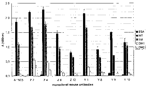
Fig. 1 shows the extend of monoclonal mouse anti-MOG antibody - binding to rat
MOG (WT), rat MOG mutants (SM, S42P, DM1, DM2), human MOG (hMOG) and
BSA as control. The data were obtained according to the procedure described in
example 1.It is evident from figure 1 that antibody binding to the MOG mutants
SM,
DM1 and DM2 that contain mutations in the antigenic FG loop is strongly
reduced for
all monoclonal antibodies. The double mutants DM1 and DM2 yield an ELISA
signal
of 0 to 25 % compared to the not mutated MOG (WT). The single mutant SM yields
ELISA signals in the range of 47 % to 79 %.
Fig. 2 shows the result of an experiment described in detail in example 2.
Sera were
obtained from healthy individuals and from MS patients. These serum samples
were
26
CA 02660149 2009-02-05
WO 2008/017363 PCT/EP2007/006217
brought into contact with human MOG (WT) and two mutant rat MOGs and with BSA
as control. Displayed is the amount of antibody binding to the presented
antigens.
Example 1:
Binding of several mouse monoclonal antibodies to MOG and its mutants
Design of mutant MOG and site-directed mutagenesis
The protein crystal structure of the extracellular domain of MOG (MOGex) was
recently solved (Breithaupt et al., 2003, Proceedings of the National Academy
of
Sciences of the United States of America, 100: 9446-51). Based on this
structure
possible intermolecular contacts between MOG as antigen and corresponding
antibodies were analyed using programs of the program package CCP4
(Collaborative Computational Project, 1994, Acta Crystallographica Section D-
Biological Crystallography, 50:760-763.) and the model building program
O(Jones
etal., 1991, Acta Crystallographica Section a, 47:110-119.). Electrostatic
potentials
were calculated in GRASP (Nicholls et al., 1991, Proteins-Structure Function
and
Genetics, 11:281-296) by employing atomic charges according to Weiner and
colleagues (Weiner et al., 1984, Journal of the American Chemical Society,
106(3),
765-784). The solvent accessible surface of MOGex was calculated with the
utility
SURFACE of the CCP4 program package.
Mutagenesis was carried out using the extracellular domain of rat MOG (MOGe,)
subcloned into the His-tag expression vector pQE-12 by following the method of
"QuikChange Site-Directed Mutagenesis" by Stratagene (LaJolia, USA). The
oligonucleotides used were: 5'-CTTCAGAGA CCACGAATA CCAAGAAGA
AGCCGCCG-3' (SM1, Ser104GIu), 5'-CACATGCTT CTTCAGAGA CGGCGAATA
CCAAG-3' (DM1, His103GIy, Ser104GIu), 5'-CACATGCTT CTTCAGAGA
CGCTGAATA CCAAG-3' (DM2, His103AIa, Ser104GIu) and the corresponding
reverse complementary oligonucleotides. The identity of the mutations was
verified
by DNA sequencing of the purified plasmids.
Protein expression and refolding of recombinant MOG
27
CA 02660149 2009-02-05
WO 2008/017363 PCT/EP2007/006217
Plasmids containing the extracellular domain of human MOG and the "humanized"
rat MOG mutant Ser42Pro were a kind gift of Nancy Ruddle (Oliver et al., 2003,
Journal of Immunology 171(1), 462-468).The extracellular domain of rat and
human
MOG and the mutant proteins were overexpressed in inclusion bodies in
Escherichia
coli. After disruption of the cells by sonification the inclusion bodies were
purified by
repetitive steps of centrifugation and resuspension in 50 mM Tris / HCI (pH
8.0), 0.3
M NaCI, 0,5 % LDAO. The inclusion bodies were solubilized in solubilisation
buffer
(100 mM NaH2PO4, 10 mM Tris, 6 M guanidinium chloride, 40 mM mercaptoethanol,
pH 8.0). After dilution in mercaptoethanol-free solubilisation buffer the
denatured
MOG was bound to Ni-NTA Superflow (Qiagen, Hilden, Germany) material and
refolded on the column in two steps. At first, a linear gradient from
solubilisation
buffer (1 mM mercaptoethanol) to 100 mM NaH2PO4, 10 mM Tris, 3 mM glutathione,
pH 8.0 over 10 hours and 80 column volumes was applied, followed by a short
linear
gradient (2 hours, 2 column volumes) to remove the glutathione for complete
oxidation of the refolded MOG. After elution, unfolded and aggregated MOG was
removed by a final gel filtration chromatography step. Identity and integrity
of the
proteins were checked by mass spectrometry and one-dimensional'H-NMR. Protein
concentrations were determined by UVNis spectroscopy, relative concentrations
by
the Bradford protein assay (BioRad, Hercules, USA).
ELISA
Antibody binding to MOG and to the mutant proteins was measured by ELISA. The
mouse monoclonal antibodies (mAb) 8-18C5 (Linnington et al., 1984, Journal of
Neuroimmunology, 6:387-96.), Yl, Y8, Y9, Y10, Z2, Z4, Z8 and Z12 (Piddlesden
et
al., 1993, American Journal of Pathology, 143:555-564) were purified from
hybridoma supernatants by affinity chromatrography on Protein G. Their
concentration was estimated by UVNis spectroscopy and colorimetrically by the
Bradford method. 96-Well plates (Maxisorb, Nunc, Rosklide, Denmark) were
coated
with 100 l 10 g/ml antigen in PBS (1 h, 30 C), washed three times with PBS
containing 0.2 % Tween20 and blocked with PBS containing 1 % w/v BSA (2 h,
30 C). After washing, the plates were incubated with the monoclonal antibodies
( -
0.5 g/ml in PBS) or the plasma samples of the MOG-vaccinated mice diluted
1:250
for lh at 30 C. The washing procedure was repeated and anti-mouse IgG (Fab')2,
28
CA 02660149 2009-02-05
WO 2008/017363 PCT/EP2007/006217
conjugated with horseradish peroxidase (Amersham Biosciences, Uppsala,
Sweden),
that was diluted 1:10000 in PBS was added and the plates were incubated for lh
at
30 C. Antibody binding was detected by oxidation of o-phenylene diamine and
quantified by measuring the absorbance at 490 nm after stopping the reaction
with
H2SO4. The in figure 1 displayed values correspond to the means of triplicate
(plasma samples) and quadruplicate (hybridoma supernatants) measurements of a
representative experiment.
Example 2:
Binding of human antibodies obtained from patients suffering from MS and from
healthy controls to native MOG and to two mutant MOGs
Samples of human sera obtained from two patients suffering from MS, I. M. and
N. K.
and one serum sample obtained from T. K. as healthy control were brought into
contact with wildtype human MOG and with double mutant rat MOG (double mutant
1, His103GIy, Ser104GIu; and double mutant 2, His103AIa, Ser104GIu) by using
the
following protocol:
(1) Coat 96 well ELISA plates with 100 NI 10 Ng/mI MOG, MOG mutants and BSA
(control).
(2) Remove unbound antigen by washing 3x with 240 pi PBS / 0.2% Tween 20.
(3) Block the plates with 240 pi 2% BSA dissolved in PBS / 0.02 % sodium
azide.
(4) Remove unbound BSA by washing 3x with 240 NI PBS / 0.2% Tween 20.
(5) Incubate plates with 100 pi sera of patients and healthy controls serially
diluted
(1:250 - 1:2000) in PBS complemented with BSA.
(6) Remove unbound antibodies by washing 3x with 240 pi PBS / 0.2% Tween 20.
(7) Bind 100 pl diluted secondary human-IgG-specific antibody fused to
horseradish
peroxidase (HRP).
(8) Remove unbound antibody by washing 3x with 240 pi PBS / 0.2% Tween 20.
(9) Add 100 NI ortho-phenylene diamine (1 mg / ml) in PBS and stop the
enzymatic
reaction by adding 50 NI 4 molar sulphuric acid.
(10) Measure absorption at 490 nm.
The results are displayed in figure 2.
29
CA 02660149 2009-02-05
WO 2008/017363 PCT/EP2007/006217
It is obvious from these data that an analysis of the binding of serum
antibodies to
native MOG alone allows no meaningful diagnosis whatsoever.
The antibodies from the serum of I. M. show only little binding to MOG. This
would
suggest that I. M. is healthy. However, this diagnosis would be incorrect,
since I. M.
suffers from MS.
The antibodies from the serum of N.K. show a similar behaviour as the
antibodies
from I.M. Note, that they were used in a concentration that was twice as high
as the
antibodies from I.M.. This again would wrongly suggest that N.K. is healthy.
The antibodies from the serum of T. K. show a mediocre binding to MOG, about
50%
more binding than the serum of I. M. Knowing that I. M. suffers from MS, one
would
assume that T. K. suffers from MS, too. Again, this diagnosis would be wrong
because T. K. is healthy.
In contrast, however, if one considers the ratio of anti-MOG-antibodies bound
to
native MOG compared to anti-MOG-antibodies bound to mutant MOG, one can
clearly see, that this ratio is >1 for both patients suffering from MS and <1
for the
healthy control. This result is obtained independently from the type of mutant
MOG
used and also independently from the life circumstances of the tested
individuals.
Consequently, contrary to the methods of the prior art, the method of the
present
invention allows a safe and precise diagnosis.
Moreover, the ratio of anti-MOG-antibodies bound to native MOG compared to
anti-
MOG-antibodies bound to double mutant 1 is for both MS patients about 1.2,
independently from the individual influences on the immune system of both
patients.
Similarly, the ratio of anti-MOG-antibodies bound to native MOG compared to
anti-
MOG-antibodies bound to double mutant 1 is for both MS patients about 1.4,
despite
the very different absolute amount of binding to MOG.
Consequently, for double mutant 1 a ratio of anti-MOG-antibodies bound to
native
MOG compared to anti-MOG-antibodies bound to double mutant 1 of about 1.2
allows to diagnose MS.
CA 02660149 2009-02-05
WO 2008/017363 PCT/EP2007/006217
Similarly, for double mutant 2 a ratio of anti-MOG-antibodies bound to native
MOG
compared to anti-MOG-antibodies bound to double mutant 2 of about 1.4 allows
to
diagnose MS.
It is important to notice that these figures only depend on the specific
mutant
antibody used and that they are independent from the particular life
circumstances of
the tested individuals.
These examples demonstrate that the present inventors were able to provide a
fast,
simple and easy-to-use method for a quantitative in vitro analysis to
diagnose, to
categorise, to predict and/or to monitor the progression of a condition based
on
antibody-antigen interactions, that overcomes or at least reduces the problems
associated with the methods of the prior art, in particular that overcomes or
at least
reduces the problems associated with the individual properties of each subject
to be
analysed, in particular the amounts and kinds of antibodies present in its
immune
system and its incomparability with other subjects and have, hence solved the
object
of the present invention.
31