Note: Descriptions are shown in the official language in which they were submitted.
CA 02660802 2009-02-13
WO 2008/020062
PCT/EP2007/058539
Method, apparatus and computer program for displaying marks in an image
data set
TECHNICAL FIELD
This disclosure generally relates to a method, an apparatus and a computer
program for displaying marks in an image data set.
BACKGROUND INFORMATION
Image data sets are in general generated by image generation devices. These
image generation devices are, for example, mammography scanners, computed
tomography scanners, magnetic resonance imaging scanners and ultra sound
scanners, which are, in particular, used for diagnostic purposes. The
generated
image data sets are often transferred to marks generation devices, like a
io computer-aided-detection device (CAD device), for determining marks
indicating
certain locations within the image data sets. For example, in the case of a
medical image data set, the CAD device can determine marks indicating
CA 02660802 2009-02-13
PCT/EP2007/058539
- 2 -
locations within the image data set, which are suspicious of showing cancer.
In
this case, a user, like a radiologist, could examine the locations within the
image
data set indicated by the marks, in order to determine, whether cancer is
present
or not.
Such an image data set comprises often a large amount of marks, whereby a
viewer, for example, a radiologist, can be confused and might overlook
important
or suspicious marks.
It is known to visualize the marks within the image data set with variable
size. It
is, for example, known to visualize marks of higher importance, e.g. of higher
io suspiciousness of marking cancer, with a larger size than marks having a
smaller
importance in order to draw the attention of a user to the most important
marks.
But there are still a lot of marks present in the image data set having
different
sizes, wherein a user, like a radiologist, can still be confused. Furthermore,
if the
computer program, which is used to determine the importance of a mark, like a
CAD computer program of a CAD device, does not correctly determine the
importance of a mark, this mark is visualized with a smaller size and the
probability of overlooking an important mark is even increased.
BRIEF SUMMARY OF THE INVENTION
Embodiments of the present invention provide a method, an apparatus and a
computer program for displaying marks in an image data set, which display the
marks in an image data set such that the marks within the image data set are
clearly shown, without confusing a user, like a radiologist, decreasing the
probability of overlooking important marks.
In a first aspect of the present invention a method for displaying marks in an
image data set is presented, wherein an image data set comprising marks is
provided and wherein during a review phase not all marks within the image data
set are displayed at the same time, wherein the method comprises:
generating a list of the marks by sorting the marks depending on a
predetermined sorting criterion,
CA 02660802 2009-02-13
._
- 2a -
grouping the marks into groups wherein each group contains marks being
successive in the generated list,
displaying the groups temporally one after another within the image data set
during the review phase, wherein marks of a group are shown at the same time,
wherein the image data set is a medical image data set, wherein the marks
are CAD marks and wherein the sorting criterion is the probability of marking
illness, in particular the suspiciousness.
CA 02660802 2009-02-13
- 3 -
One embodiment of the invention is based on the idea, that, if less marks are
shown within the image data set at the same time, a user is less confused and
the probability of overlooking an important mark is decreased.
A list of marks is generated by sorting the marks depending on a predetermined
sorting criterion and the marks are displayed temporally one after another
during
the review phase within the image data set in accordance with the generated
list.
Furthermore, since the marks are shown in a sorted way, it is possible to show
the marks in a sequence, which suits to a certain application. For example,
the
sorting criterion could be the importance of a mark with respect to a certain
io application, e.g. the suspiciousness of marking cancer in the case of
medical
image data sets, and the list can be generated such that the most important
marks are shown first, since the attention of the user might be higher at the
beginning of a review of the marks than at the end. The sorting criterion can,
for
example, be predetermined by inputting a sorting criterion by a user, like a
radiologist, into an apparatus for displaying marks in an image data set,
which is
adapted to perform the method in accordance with one embodiment of the
invention.
It is further preferred in one embodiment that the image data set is a medical
image data set, for example, a set of digital mammograms, that the marks are
zo CAD marks and that the sorting criterion is the probability of marking
illness, in
particular the suspiciousness. The medical image data set can, for example,
also
be a tomosyntheses image data set, a computed tomography image data set, a
breast magnetic resonance imaging data set or any other medical image data
set. In this case, the importance of a mark is correlated with the probability
of
marking illness, e.g., a mark having a higher probability of marking illness
has a
higher importance than a mark having a lower probability of marking illness.
The
illness is, for example, cancer. The suspiciousness is a well defined CAD term
and is, for example, provided by a CAD mark generation device and a CAD mark
CA 02660802 2009-02-13
WO 2008/020062
PCT/EP2007/058539
- 4 -
generation computer program. A CAD mark generation device using a CAD mark
generation computer program is, for example, the Image Checker System
produced by R2 Technology Inc. or the Second Look Digital System produced by
iCAD Inc. The suspiciousness is a part of the DICOM standard, in particular,
the
suspiciousness is a part of the DICOM CAD Structure Report (SR), which is, for
example, disclosed in Digital Imaging and Communications in Medicine (DICOM)
2006, published by the ACR (the American College of Radiology) and the NEMA
(the National Electrical Manufacturers Association), in particular, in DICOM
Base
Standard 2006 Part PS 3.16; Supplement 50: Mammography Computer-Aided
io Detection SR SOP. The SR is, for example, generated by the above
mentioned
CAD mark generation device and CAD mark generation computer program.
Since in the above mentioned embodiment the marks are sorted in accordance
with their probability of marking illness and since they are shown in this
sequence
temporally one after another, the marks can be presented to the user, like a
radiologist, in accordance with the actual level of attention of the user. For
example, if the user has the highest attention at the beginning of a review of
the
marks, the marks will be sorted such that the first shown marks have the
highest
probability of marking illness. lf, for example, the attention of the user
reaches the
maximum in the middle of the review of the marks, the marks can be sorted such
that the mark with the highest probability of marking illness are arranged in
the
middle of the generated list. Thus, the probability of marking illness of the
CAD
marks can be adapted to the attention of the user, further decreasing the
probability of overlooking important marks.
It is preferred in one embodiment that the next mark in the generated list is
displayed after fulfilling a predetermined switching criterion. The
predetermined
switching criterion is, for example, an input to an apparatus for displaying
marks
in an image data set, which performs the method in accordance with one
embodiment of the invention. The apparatus for displaying marks in an image
data set can comprise an input device, for example, a keyboard or a mouse, for
inputting a signal indicating that the next mark can now be shown. Thus, in
this
example, the switching criterion is an input criterion, e.g., whether a signal
has
CA 02660802 2009-02-13
- 5 -
been inputted indicating that the next mark can now be shown or whether such a
signal has not been inputted. Thus, a user can review a mark within the image
data set as long as needed and, after the user has reviewed the respective
mark,
the user can switch to the next mark. This further decreases the probability
of
overlooking an important mark and of a misassessment of a mark, in particular,
of
generating a false negative or a false positive.
It is further preferred in one embodiment that an indication is displayed
indicating
the position of the displayed mark within the list and/or the overall number
of
marks to be displayed. This allows the user to see directly how many marks
have
io already been displayed and how many marks will be displayed.
Furthermore,
since the marks are sorted in dependence on a predetermined sorting criterion,
this allows to give the user an impression of the importance of the displayed
mark. For example, if the sorting criterion is the probability of marking
illness, the
position of the displayed marks within the list gives the user an impression
of the
probability of marking illness of the displayed mark.
The method comprises following steps:
- generating a list of the marks by sorting the marks depending on a
predetermined sorting criterion,
- grouping the marks into groups wherein each group contains marks being
successive in the generated list,
- displaying the groups temporally one after another within the image data
set during the review phase.
Groups of marks being successive in the generated list are shown at the same
time. This allows review of several marks, which might be similar with respect
to
the sorting criterion, at the same time, wherein the speed of reviewing is
CA 02660802 2009-02-13
=
- 6 -
increased, while, since only groups and not all markers are shown, the user is
still
not confused by the amount of marks displayed at the same time.
Also, if groups of marks are displayed within the image data set temporally
one
after another, the image data set is a medical image data set, the marks are
CAD
marks and the sorting criterion is the probability of marking illness, in
particular,
the suspiciousness. Furthermore, also in this case, it is preferred in one
embodiment that the next group is displayed after fulfilling a predetermined
switching criterion.
It is further preferred in one embodiment that only marks are displayed
fulfilling at
1 o least one given displaying criterion. This at least one given
displaying criterion
can, for example, be the size of a mark, e.g., only marks are displayed having
a
size larger than a predetermined threshold value. This reduces the amount of
marks, which are displayed at the same time within the image data set, whereby
the probability of overlooking important marks is decreased. It is further
preferred
in one embodiment that the image data set is a medical image data set, that
the
marks are CAD marks and that the at least one given displaying criterion is at
least one of a microcalcification criterion, a mass criterion and an operating
point
criterion. This at least one given displaying criterion can be predefined
and/or
inputted by a user into an apparatus for displaying marks in an image data
set,
which is adapted to perform the method in accordance with one embodiment of
the invention.
As mentioned above, the CAD marks are provided by CAD mark generation
devices and CAD mark generation computer programs, which are known in the
state of the art. These devices and computer programs provide a SR, which is
defined in the DICOM standard and which contains information about each CAD
mark. This information describes, in particular, whether a CAD mark is a
microcalcification mark or a mass mark. If a CAD mark is a microcalcification
mark, this CAD mark fulfils the microcalcification criterion. If a CAD mark is
a
mass mark, this CAD mark fulfils the mass criterion. Two displaying criterions
can
be present, e.g., for example, the number of microcalcification marks and the
CA 02660802 2009-02-13
WO 2008/020062
PCT/EP2007/058539
- 7 -
number of mass marks can be adjusted independently. If a microcalcification
criterion and a mass criterion are given, for example, inputted by a user in
an
apparatus for displaying marks in an image data set, which is adapted to
perform
the method in accordance with one embodiment of the invention, marks are
displayed fulfilling both criterions, e.g., microcalcification marks and mass
marks
are displayed. If only one of these criterions is given, only
microcalcification
marks or mass marks are displayed, respectively.
The operating point is the operating point of the CAD algorithm used by the
CAD
mark generation device and CAD mark generation computer program. The
io operating point of the CAD algorithm, which has been used for
determining the
respective CAD mark, is also contained in the information of the SR provided
from the CAD mark generation device and CAD mark generation computer
program. The operating point is a measure for the probability of the
determination
of false positives and false negatives by the CAD algorithm. The operating
point
criterion is fulfilled for a respective CAD mark, if the operating point of
this
respective CAD mark is equal to a given operating point. The given operating
point can be given by a user, which inputs the operating point via an input
device,
like a keyboard or a mouse, to an apparatus for displaying marks in an image
data set for performing the method in accordance with one embodiment of the
invention. Furthermore, the operating point can also be predetermined. The
operating point is, for example, described in the section "Correction Items ¨
By
Number ", CP 624 of the DICOM Standard.
The user can select via the input device, that only CAD marks are displayed
which fulfil the microcalcification criterion, e.g., that only
microcalcification marks
are shown. Since microcalcification marks are known to be determined by the
CAD algorithm with a high performance (e.g., high specificity and high
sensitivity), the use of microcalcification marks for reviewing marks of an
image
data set can further improve a diagnosis based on this review.
The user can select via the input device, that only CAD marks are displayed
which fulfil the mass criterion, e.g., that only mass marks are shown. Since
CAD
CA 02660802 2009-02-13
WO 2008/020062
PCT/EP2007/058539
- 8 -
algorithms determine in general mass marks with a large number of false
positives, it is particularly important to review these mass marks carefully.
Therefore, particularly during reviewing mass marks the present invention,
which
assists a user, like a radiologist, in focussing on, in this embodiment, mass
marks, is very useful. The user can select the mass criterion as the display
criterion by inputting a corresponding signal into the apparatus for
displaying
marks in an image data set via an input device. If the apparatus receives such
a
signal, only mass marks are displayed. And, if the user has chosen a certain
operating point by inputting a signal into the apparatus via the input device
io indicating this certain operating point, only marks are displayed
belonging to this
certain operating point. Since the operating point is a measure for the
probability
of a mark of being a false negative or false positive, by choosing a certain
operating point the user can show marks having a certain probability of being
a
false positive or false negative.
It is preferred in one embodiment that an input device is provided for
entering at
least one displaying criterion as the at least one given displaying criterion
in an
apparatus for displaying marks in an image data set. This allows to change the
number of displayed marks based on user interaction.
It is preferred in one embodiment that the marks are displayed at least in a
first
display area and in a second display area, wherein in both display areas one
or
several marks are displayed within the image data set and wherein in the
second
display area a region around the one or several marks is displayed with a
larger
magnification than in the first display area. Since in the second display area
a
region around the one or several marks is displayed with a larger
magnification
than in the first display area, the marks, e.g., the region within the image
data set,
which is marked by the respective mark, can be reviewed in more detail,
whereby
the probability of a missassessment is reduced. Furthermore, since the
magnification is shown, in particular, automatically and at the same time, at
which
the corresponding mark is shown in the first display area, both, an overall
view
and a magnification, are shown at the same time, wherein a further interaction
of
CA 02660802 2009-02-13
WO 2008/020062
PCT/EP2007/058539
- 9 -
the user for obtaining such a magnification is not needed, decreasing the time
needed for reviewing the marks.
As already mentioned above, the image data set is preferentially a medical
image
data set in one embodiment, and the marks are preferentially CAD marks in one
embodiment. The image data set is preferentially a mammogram data set in one
embodiment, for example, for each breast of a patient a craniocaudal (CC) view
image and a mediolateral oblique (MLO) view image, e.g., a mammogram data
set comprises preferentially four images in one embodiment, a left and a right
CC
image, and a left and a right MLO image. It is preferred in one embodiment
that a
io displayed mark is selected, in particular, to be discarded. For example,
if, as
described above, the marks are shown temporally one after another or groups of
marks are shown temporally one after another, a displayed mark can be
selected,
for example, by using an input device, like a keyboard or a mouse, by the
user.
The user can, for example, choose, whether a selected mark has to be discarded
or whether a selected mark has to be accepted. The method of one embodiment
is preferentially adapted such that selected marks are discarded and that non-
selected marks are accepted. It is further preferred in one embodiment that,
after
selecting a displayed mark, only non-selected marks are displayed. This allows
a
user to reduce the number of marks during the review of the marks, in order to
decrease the overall number of marks within the image data set, further
decreasing the probability of overlooking important marks.
It is also preferred in one embodiment that a displayed mark is provided with
a
further mark. This can, for example, be performed by inputting the further
mark
via an input device into an apparatus for displaying marks in an image data
set,
which performs the method in accordance with one embodiment of the invention.
In this case, the image data set comprises the initially provided marks and
the
later provided added marks. This allows a user, for example, a radiologist, to
add
further marks to the image data set.
It is further preferred in one embodiment that, if the image data set
comprises
several images and if a mark is displayed in one of these images, being a
first
CA 02660802 2009-02-13
WO 2008/020062
PCT/EP2007/058539
- 10 -
image, in at least one of the other images of the image data set a corridor is
displayed, which includes a location, which corresponds to a first location in
the
first image, which is marked by the mark. This allows a user, like a
radiologist, to
review corresponding regions in different images, wherein the user is assisted
in
reviewing a mark displayed in a first image, in particular, in diagnosing,
whether a
mark displayed in the first image indicates an illness, like cancer or not.
The corridor is preferentially determined by using following steps in one
embodiment:
- providing a starting image, being the first image, of an object, and at
least
io one target image of the object, being at least one of the other images,
- providing a second location within the starting image and the at least
one
target image, being the location of a reference feature detectable in the
starting
image and in the at least one target image,
- determining a corridor in the at least one target image, wherein the
corridor
contains a circular line, wherein the circular line defines a circle, whose
radius
corresponds to a distance between the first location and the second location
in
the starting image and whose centre position corresponds to the second
location,
- presenting the corridor and the at least one target image.
In the starting image and/or in the at least one target image, the object can
be
deformed, and in each or in some of these images the deformation of the object
can be different. Furthermore, the starting image and/or the at least one
target
image can be a two-dimensional image, like a absorption or transmission
projection, for example, a CC image or a MLO image of a breast, or a three-
dimensional image, like a three-dimensional computed tomography image or a
three-dimensional magnetic resonance image. For example, the starting image
and the at least one target image can be two-dimensional images, or one of
these images, the starting image or the at least one target image, can be a
two-
dimensional image, and another image of these images can be a three-
dimensional image.
CA 02660802 2009-02-13
WO 2008/020062
PCT/EP2007/058539
- 11 -
The first location and the second location can be an image point or an
extended
region within the respective image. Furthermore, since in different images the
object can comprise different deformations and can be imaged from different
directions, the shape of the first location and/or the second location can
differ
from image to image. For example, a location, being in the starting image an
image point, can be a line in the at least one target image.
The distance between the first location and the second location is
preferentially in
one embodiment the distance between the centres of these locations. But, the
distance can also be any distance between a point at or within the first
location
io and a point at or within the second location.
The circular line can be a complete circular line, e.g., a line forming a
circle, or a
circular segment line, e.g., a line being a segment of a circle. The corridor
is
therefore preferentially in one embodiment formed substantially like a ring or
like
a segment of a ring containing a complete circular line or a circular segment
line,
respectively. The circular line is preferentially a virtual line in one
embodiment,
e.g., this circular line is preferentially not displayed in one embodiment.
The centre position of the circle defined by the circular line, e.g., the
complete
circular line or the circular segment line, is preferentially in one
embodiment the
centre of the second location in the at least one target image. Alternatively,
the
centre position of this circle can be any point at or within the second
location
within the at least one target image.
The circular line defines a circle, whose radius corresponds to the distance
between the first location and the second location in the starting image. This
means that this radius can be substantially equal to this distance, e.g., it
is
preferred in one embodiment that a difference length between this radius and
this
distance is smaller than 10 percent of this distance. It is further preferred
in one
embodiment that this difference length is smaller than 5 percent of this
distance,
and it is further preferred in one embodiment that this distance is equal to
the
radius. That the radius corresponds to the distance also means that, if the
object
CA 02660802 2009-02-13
WO 2008/020062
PCT/EP2007/058539
- 12 -
in the at least one target image is magnifiedly shown with respect to the
starting
image of this object, this radius is substantially equal to an accordingly
magnified
distance. For example, if the object is magnified by a factor of two in the at
least
one target image relative to the starting image, the radius is substantially
equal to
twice a distance between the first location and the second location in the
starting
image. If the starting image and the at least one target image show the object
with different magnifications, the difference between the radius and the
accordingly magnified distance is preferentially in one embodiment smaller
than
percent of the accordingly magnified distance, further preferred in one
io embodiment smaller than 5 percent of the accordingly magnified distance
and
further preferred in one embodiment equal to zero.
This embodiment is based on the idea that the first location in the starting
image
corresponds with a high probability to a region within the at least one target
image, which is located within the corridor within the at least one target
image.
Therefore, the user, like a radiologist, can focus on the corridor, if the
user wants
to find a region within the at least one target image, which corresponds to
the first
location within the starting image. The determination of this corridor can be
carried out with low computational effort.
This low computational effort is particularly important, if the user is a
radiologist
and if the images are medical images. A radiologist has to review a large
number
of medical images in a short time, for example, 200 images in two hours. It is
therefore important to determine the corridors very fast, in order to not let
a
radiologist wait and waist his time.
It is preferred in one embodiment that the object is a breast, wherein the
starting
image and the at least one target image are medical diagnostic images of the
breast, in particular, mammograms, wherein the reference feature is a nipple
of a
breast. The mammograms are preferentially in one embodiment a MLO or ML
image and a CC image of the same breast. The deformation of the breast in a
MLO or ML image is different to the deformation of the breast in a CC image.
Furthermore, the breast in a MLO or ML image has been viewed from another
CA 02660802 2009-02-13
WO 2008/020062
PCT/EP2007/058539
- 13 -
direction than in a CC image. But, although the breast is viewed from
different
directions and deformed differently, the distance between the first location
within
the breast and the nipple of the breast being the second location is
substantially
invariant in the different mammographic views, e.g., this distance does not
change very much. It exists therefore a high probability, that a region, which
corresponds to a first location in a starting mammogram, is located within the
corridor in at least one target mammogram. The presentation method of
presenting an image of an object is therefore particularly applicable, if
corresponding locations or regions have to be found in different mammograms of
io the same breast.
The first location is in one embodiment preferentially marked by a CAD mark
determined by using a CAD algorithm.
CAD marks are used, in order to help a radiologist to find suspicious regions
within a medical image, like a mammogram. A CAD mark marks a region within a
mammogram, which is, with respect to the CAD algorithm used by a CAD
computer program and/or a CAD device, suspicious of marking an illness, in
particular, of marking cancer. If different CAD marks are shown one after
another
within the image data set in accordance with a generated list, the first
location is
preferentially in one embodiment the location within an image, being the first
image, which is marked by the currently displayed CAD mark. This allows a user
to easily review regions, which correspond to the first location, in the at
least one
target image. Thus, the same region can be viewed in different images of the
same object. This improves the review results of a user, like a radiologist.
It is further preferred in one embodiment that the step of providing the
second
location within the starting image and the at least one target image comprises
a
step of automatically detecting the second location in at least one of the
starting
image and the at least one target image. The automatic detection of the second
location allows to automatically determine and display the corridor in the at
least
one target image, if a mark marking a first location in the starting image is
displayed.
CA 02660802 2009-02-13
WO 2008/020062
PCT/EP2007/058539
- 14 -
It is further preferred in one embodiment that the step of providing the
second
location within the starting image and the at least one target image comprises
a
step of providing an input unit for inputting the second location within the
starting
image and the at least one target image. The input unit, for example, a
graphical
user interface, allows a user to input the second location in the starting
image
and/or in at least one of the at least one target image. The second location
can
preferentially be provided in one embodiment within an image by clicking with
a
mouse pointer on the respective location within the respective image. This
allow
a user to easily provide the second location in at least one of the starting
image
io and the at least one target image.
It is further preferred in one embodiment that the step of providing the
second
location within the starting image and the at least one target image comprises
a
step of providing an input unit for correcting the position of the second
location at
least within one of the starting image and the at least one target image. The
input
unit is, for example, a graphical user interface, which allows a user, for
example,
by using a drag-and-drop operation, to move the second location to another
position within the respective image. This allows a user to easily modify the
position of a second location which has been provided, for example, by an
automatic second location detection system. Such a system is, for example, a
nipple detection system using a nipple detection computer program for
detecting
the nipple of a breast automatically. Such a system is, for example, disclosed
in
Zhou C, Chan HP, Paramagul C, Roubidoux MA, Sahiner B, Hadjiiski LM, Petrick
N, "Computerized nipple identification for multiple image analysis in computer-
aided diagnosis", Med Phys. 2004 Oct, 31(10):2871-82.
It is further preferred in one embodiment that the presentation method for
presenting an image of an object further comprises the step of providing an
input
unit for inputting the distance for providing the distance or the step of
determining
the distance between the first location and the second location in the
starting
image. Thus, preferentially in one embodiment a user can enter the distance
between the first location and the second location in a presentation device
for
presenting an image of an object in accordance with one embodiment of the
CA 02660802 2009-02-13
WO 2008/020062
PCT/EP2007/058539
- 15 -
invention. For example, if the object is a breast and ultrasound and/or breast
magnetic resonance imaging images are present, a user, like a radiologist, can
determine the distance between a first location and a second location being
the
location of the nipple of the breast from the present ultrasound and/or breast
magnetic resonance imaging images, and the user can enter this determined
distance into the presentation device for presenting an image of an object in
accordance with one embodiment of the invention. Thus, the distance between
the first location and the second location has not to be determined within the
starting image, but this distance can be provided by a user, who knows this
io distance, for example, from present images of the object, for example,
of the
breast. This present images can differ from the starting image and the at
least
one target image. For example, the starting image and the at least one target
image can be digital mammograms acquired by a digital mammography device,
and the present images, from which the user has determined the distance, can
be an image from other modalities, like images from an ultrasound device, a
magnetic resonance imaging device or a computed tomography device.
However, it is also possible that a user does not enter this distance in the
presentation device for presenting an image of an object, but that this
distance is
determined by the presentation device for presenting an image of an object
itself.
It is further preferred in one embodiment that the corridor is determined such
that
the circular line is located in the middle of the corridor. This leads to a
corridor
having a high probability of containing a region, which corresponds to the
first
position, in the at least one target image.
It is further preferred in one embodiment that the first location is extended
and
that the step of determining the corridor comprises following steps:
- determining the smallest distance and the largest distance between the
second location and the extended first location within the starting image,
- determining the corridor such that it contains all image points of the at
least
one target image having a distance to the second location which is smaller
than
the largest distance and larger than the smallest distance. This further
increases
CA 02660802 2009-02-13
WO 2008/020062
PCT/EP2007/058539
- 16 -
the probability of the corridor of containing a region, which corresponds to a
first
location within the starting image, within the at least one target image.
It is also preferred in one embodiment that an input unit is provided for
inputting a
width of the corridor being sufficient to contain all image points of the at
least one
target image having a distance to the second location which is smaller than
the
largest distance and larger than the smallest distance. The input unit is
preferentially a graphical user interface in one embodiment, which allows a
user
to input a width of the corridor, which is sufficient to contain all image
points of
the at least one target image having a distance to the second location, which
is
io smaller than the largest distance and larger than the smallest distance.
This
allows a user to modify the width of the corridor, thus, to further increase
the
probability of the corridor of containing a region, which corresponds to a
first
location within the starting image, within the at least one target image by
increasing the width of the corridor.
It is further preferred in one embodiment that an input unit is provided for
inputting a width value or that a width value is predefined, wherein the
corridor is
determined such that it contains all image points of the at least one target
image
having a distance to the second location which is smaller than the largest
distance multiplied by the sum of one and the width value and larger than the
smallest distance multiplied by the difference between one and the width
value.
The input unit for inputting a width value is preferentially a graphical user
interface in one embodiment, which allows a user to input a width value, for
example, via a keyboard or a mouse. Since, in this embodiment, the width
depends on a fraction of the largest distance and a fraction of the smallest
distance, the width of the corridor depends on the distance between the first
location and the second location and, thus, a useable width of the corridor
can
easily be inputted by a user. The width value can also be predefined. If the
width
value is predefined, the input unit for inputting a width value does not have
to be
provided. A preferred width value is 25 percent in one embodiment. It is
further
preferred in one embodiment that the part of the at least one target image,
which
is located inside of the corridor, is presented more prominent than the part
of the
CA 02660802 2009-02-13
- 17 -
at least one target image which is located outside of the corridor. Since the
part
of the at least one target image, which is located inside of the corridor, is
presented more prominent than the part of the at least one target image, which
is
located outside of the corridor, the user, like radiologist, is focused on the
corridor
and not distracted by the part of the at least one target image located
outside of
the corridor. This further increases the assistance of finding corresponding
regions in different images.
The part of the at least one target image, which is located outside of the
corridor
is preferentially in one embodiment shown less prominent by dimming this part.
o The dimming is preferentially in one embodiment carried out such that the
user
can still see the outline of the object, in particular, of the breast, in the
dimmed
part, and that the user can easily focus on the corridor. The dimming is
preferentially performed by decreasing the brightness in one embodiment.
Alternatively, only the part of the at least one target image can be shown,
which
is located inside of the corridor, e.g., the part of the at least one target
image,
which is located outside of the corridor, is, in this embodiment, not shown.
It is further preferred in one embodiment that the image data set comprises a
first
kind of marks and at least one second kind of marks, wherein during the review
phase not all marks of the first kind of marks are displayed at the same time
and
wherein during the review phase all marks of at least one kind of the at least
one
second kind of marks are displayed at the same time. Preferentially in one
embodiment, during the review phase the marks of the first kind of marks are
displayed in at least one of the manners described in further detail below.
Since
during the review phase not all marks of the first kind of marks are shown, a
user
is still less confused by the marks, while, since all marks of at least one
kind of
the at least one second kind of marks are displayed at the same time, the
information from these at least one second kind of marks can also be used
during
the review phase, which can improve the assistance of a user, like a
radiologist,
in reviewing marks. For example, if the marks are CAD marks, the first kind of
marks are preferentially mass marks in one embodiment, and the at least one
second kind of marks are preferentially microcalcification marks in one
embodiment.
CA 02660802 2009-02-13
- 18 -
In a further aspect of the present invention a method for displaying marks in
an
image data set is presented, wherein an image data set comprising marks is
provided, wherein in an overview phase all marks are displayed, wherein in a
review phase the marks are displayed in one of the manners described in
further
detail below and wherein the overview phase is arranged before and/or after
the
review phase. This allows a user to have an overview over all marks within the
image data set before and/or after the method described in further detail
below is
performed.
It is further preferred in one embodiment that a selection is performed,
wherein
an overview phase is arranged after the review phase and wherein in the review
phase after the overview phase only non-selected marks are displayed. Since
finally only non-selected marks are displayed, marks, which have been selected
by the user, in particular, as being of no importance, are not shown, further
decreasing the probability of overlooking important marks.
In a further aspect of the invention an apparatus for displaying marks in an
image
data set is presented, comprising a displaying unit, wherein the apparatus is
adapted for displaying not all marks within the image data set at the same
time
on the displaying unit during a review phase, wherein the apparatus comprises
a
list generation unit for generating a list of the marks by sorting the marks
zo depending on a predetermined sorting criterion, wherein the apparatus
comprises
a grouping unit for grouping the marks into groups wherein each group contains
marks being successive in the generated list, wherein the displaying unit is
adapted for displaying the groups temporally one after another within the
image
data set during the review phase, wherein marks of a group are shown at the
same time, wherein the image data set is a medical image data set, wherein the
marks are CAD marks and wherein the sorting criterion is the probability of
marking illness, in particular the suspiciousness.
It is preferred in one embodiment that the apparatus for displaying the next
mark
in the generated list after fulfilling a predetermined switching criterion.
CA 02660802 2009-02-13
- 19 -
It is further preferred in one embodiment that the apparatus is adapted for
displaying an indication indicating the position of the displayed mark within
the list
and/or the overall number of marks to be displayed.
The apparatus comprises a list generation unit for generating a list of the
marks
by sorting the marks depending on a predetermined sorting criterion, wherein
the
apparatus comprises a grouping unit for grouping the marks into groups wherein
each group contains marks being successive in the generated list and wherein
the displaying unit is adapted for displaying the groups temporally one after
io another within the image data set during the review phase. The image
data set is
a medical image data set, wherein the marks are CAD marks and wherein the
sorting criterion is the probability of marking illness. It is further
preferred in one
embodiment that the sorting criterion is the suspiciousness. It is further
preferred
in one embodiment that the apparatus is adapted for displaying the next group
after fulfilling a predetermined switching criterion. It is further preferred
in one
embodiment that the apparatus is adapted for displaying only marks fulfilling
at
least one given displaying criterion. It is further preferred in one
embodiment that
the image data set is a medical image data set, wherein the marks are CAD
marks and wherein the at least one given displaying criterion is at least one
of a
microcalcification criterion, a mass criterion and an operating point
criterion.
It is further preferred in one embodiment that the displaying unit comprises
at
least a first display area and a second display area, wherein the apparatus is
adapted for displaying in both display areas one or several marks within the
image data set and for displaying in the second display area a region around
the
one or several marks with a larger magnification than in the first display
area. It is
further preferred in one embodiment that the image data set is a medical image
data set and that the marks are CAD marks. It is further preferred in one
embodiment that the image data set is a mammogram data set. It is further
preferred in one embodiment that the apparatus is adapted for selecting a
displayed mark, in particular, to be discarded. It is further preferred in one
CA 02660802 2009-02-13
- 20 -
embodiment that the apparatus is adapted for displaying only non-selected
marks, after selecting a displayed mark. It is further preferred in one
embodiment
that the apparatus is adapted for providing a displayed mark with a further
mark.
In a further aspect of the invention an apparatus for displaying marks in an
image
data set is presented, comprising a displaying unit for displaying the image
data
set and the marks, wherein the apparatus is adapted for displaying in an
overview phase all marks, for displaying in a review phase the marks and for
arranging the overview phase before and/or after the review phase. It is
preferred
in one embodiment that the apparatus is adapted for arranging an overview
io phase after the review phase and for displaying in the overview phase
after the
review phase only non-selected marks.
In a further aspect of the present invention an imaging system is presented,
comprising:
- an image data set generating unit for generating an image data set,
- a marks generating unit for generating marks depending on the generated
image data set,
- an apparatus for displaying marks within an image data set.
In a further aspect of the present invention computer programs for displaying
marks in an image data set are provided.
It shall be understood that the methods, apparatuses, imaging system and
computer programs described above have corresponding embodiments as
further described below.
BRIEF DESCRIPTION OF THE SEVERAL VIEWS OF THE DRAWINGS
CA 02660802 2009-02-13
WO 2008/020062
PCT/EP2007/058539
-21 -
These and other aspects of the invention will be apparent from and elucidated
with reference to the non-exhaustive and non-limiting embodiments described
hereinafter. In the following drawings:
Fig. 1 shows a schematic view of an imaging system in accordance with
the invention,
Fig. 2 shows a schematic view of an apparatus for imaging marks
within
an image data set in accordance with the invention,
Fig. 3 shows a flow chart of an embodiment of a method for displaying
marks within an image data set in accordance with the invention,
io Fig. 4 shows exemplarily an image data set and CAD marks,
Fig. 5 shows exemplarily an image data set and a CAD mark in a first
display area and in a second display area, wherein the CAD mark
and the image data set are magnified in the second display area,
Fig. 6 shows a flowchart of a further embodiment of a method for
displaying marks within an image data set in accordance with the
invention,
Fig. 7 shows a further embodiment of a method for displaying marks
within an image data set in accordance with the invention, and
Fig. 8 shows a further embodiment of a method for displaying marks
within an image data set in accordance with the invention.
DETAILED DESCRIPTION OF EMBODIMENTS
In the following description, numerous specific details are given to provide a
thorough understanding of embodiments. One skilled in the relevant art will
CA 02660802 2009-02-13
WO 2008/020062
PCT/EP2007/058539
- 22 -
recognize, however, that the invention can be practiced without one or more of
the specific details, or with other methods, components, materials, etc. In
other
instances, well-known structures, materials, or operations are not shown or
described in detail to avoid obscuring aspects of the invention.
Reference throughout this specification to "one embodiment" or "an embodiment"
means that a particular feature, structure, or characteristic described in
connection with the embodiment is included in at least one embodiment. Thus,
the appearances of the phrases "in one embodiment" or "in an embodiment" in
various places throughout this specification are not necessarily all referring
to the
io same embodiment.
Furthermore, the particular features, structures, or
characteristics may be combined in any suitable manner in one or more
embodiments.
The headings provided herein are for convenience only and do not interpret the
scope or meaning of the claimed invention.
Fig. 1 shows a schematic view of an imaging system 1 comprising an image data
set generating unit 2, a CAD mark generation unit 3 and an apparatus for
displaying marks in an image data set 4. The image data set generation unit 2
is,
for example, a device for acquiring digital mammograms, a computed
tomography device, a magnetic resonance imaging device, a tomosyntheses
device or any other medical imaging device. The image data set generating unit
2
generates image data sets and transfers them to the CAD mark generating unit 3
and to the apparatus 4 for displaying marks in an image data set. The CAD mark
generation unit 3 generates from the image data sets received from the image
data set generation unit 4 CAD marks using known CAD algorithms. Such CAD
mark generation units 3 and CAD algorithms are, for example, the Image
Checker System produced by R2 Technology Inc. and the Second Look Digital
System produced by iCAD Inc.
The generated CAD marks are transferred from the CAD mark generation unit 3
to the apparatus 4 for displaying marks in an image data set.
CA 02660802 2009-02-13
WO 2008/020062
PCT/EP2007/058539
- 23 -
The apparatus 4 for displaying marks in an image data set is, in more detail,
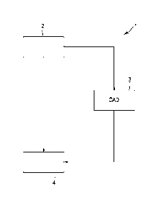
schematically shown in Fig. 2.
The apparatus 4 for displaying marks in an image data set comprises an image
data set receiving unit 5 for receiving image data sets from the image data
set
generation unit 2. The apparatus 4 comprises further a CAD mark receiving unit
6
for receiving CAD marks from the CAD mark generation unit 3. The CAD marks
are preferentially in one embodiment transferred and received in the DICOM
format, in particular, as a DICOM CAD Structure Report (SR). The CAD marks,
e.g., in particular, the SR, is transferred from the CAD mark receiving unit 6
to a
io list generation unit 7. The list generation unit 7 can forward the CAD
marks to a
control unit 8 without modifications, or the list generation unit 7 can
generate a list
of the marks by sorting the marks depending on a predetermined sorting
criterion
and the sorted CAD marks can be transferred from the list generation unit 7 to
the control unit 8. The control unit 8 receives image data sets from the image
data set receiving unit 5 and inputs from an input unit 9. The input unit 9
is, for
example, a keyboard or a mouse. The control unit 8 is connected to a display
unit
10 for displaying CAD marks and image data sets.
The apparatus 4 for displaying marks in an image data set further comprises a
grouping unit 11, which receives sorted CAD marks from the list generation
unit 7
for grouping these CAD marks into groups of CAD marks, which are transferred
to the control unit 8. The control unit 8 controls the image data set
receiving unit
5, the CAD mark receiving unit 6, the list generation unit 7, the input unit
9, the
displaying unit 10 and the grouping unit 11 in accordance with a method for
displaying marks in an image data set, which will be described further below.
The apparatus for displaying marks in an image data set is preferentially a
computer system comprising the different units 5 to 11 in one embodiment.
Theses units can be realised by computer programs and/or dedicated hardware.
In accordance with one embodiment of the invention, the apparatus for
displaying
marks in an image data set can also comprise less, additional and/or other
units,
as long as the apparatus still allows to display not all marks within the
image data
CA 02660802 2009-02-13
WO 2008/020062
PCT/EP2007/058539
- 24 -
set at the same time. For example, instead of separate list generation unit 7
and
grouping unit 11, a single unit can be used having the same functional
features
as the list generation unit 7 and the grouping unit 11 together. Furthermore,
the
image data set receiving unit 5 and the CAD mark receiving unit 6 could be
integrated into one receiving unit, which receives both, the image data set
and
the CAD marks. In addition, the invention is not limited to the data paths
within
the apparatus 4 for displaying an image data set, which have been described
above. For example, the image data sets and CAD marks could be directly
transferred to the display unit 10, and not via the control unit 8, while the
control
io unit 8 still controls the data transfer within the apparatus 4 for
displaying an
image data set.
An embodiment of a method for displaying marks in an image data set in
accordance with one embodiment of the invention will now be described in more
detail with respect to a flowchart shown in Fig. 3.
In step 101 the apparatus 4 for displaying marks in an image data set receives
an
image data set from the image data set generation unit 2 and CAD marks from
the CAD mark generation unit 3. In another embodiment, the image data set and
the corresponding CAD marks can already be present on the apparatus 4 for
displaying marks in an image data set such that an acquisition and/or
determination and/or receiving of these data is not necessary. Step 101 would
therefore be omitted.
In step 102 the image data set and the CAD marks have been transferred to the
displaying unit 10, which displays the image data set and the CAD marks. Such
a
visualization of the image data set containing the CAD marks can, for example,
be performed after a user has requested such a visualization by using the
input
unit 9. If a user inputs such a request, in this embodiment, the control unit
8
receives this request and controls the displaying unit 10 such that it shows
the
image data set and the CAD marks, in particular, in an overview phase, all CAD
marks, which haven been determined by the CAD mark generation unit 3. In
other embodiments, step 102 can be omitted.
CA 02660802 2009-02-13
WO 2008/020062
PCT/EP2007/058539
- 25 -
A visualization of the image data set and the CAD marks is exemplary shown in
Fig. 4. In Fig. 4 digital mammograms 20a, ..., 20d, 21a, ...., 21d are shown.
The
digital mammograms 20a, ...., 20d are current mammograms, and the digital
mammograms 21a, ..., 21d are prior mammograms of an earlier examination of
the breasts. The digital mammograms 20a, ..., 20d (for each breast a CC-view
image and a MLO-view image) constitute one image data set. Within this image
data set four CAD marks C1, C2, C3, M1 are displayed.
In step 103 the list generation unit 7 generates a list of the CAD marks by
sorting
the marks depending on a predetermined sorting criterion. This sorting
criterion is
io in this embodiment the suspiciousness. The suspiciousness is also known
as the
certainty of finding. A mark having a large suspiciousness has a large
probability
of marking illness, in particular, of marking cancer. The suspiciousness is
contained in the SR, which has in step 101 been transferred from the CAD mark
generation unit 3 to the apparatus 4 for displaying marks in an image data
set.
Thus, in step 101 a SR has been transferred containing the CAD marks and
further information, which is defined in the corresponding DICOM standard. In
this exemplary embodiment, the CAD marks' degree of suspiciousness
decreases in the following order: C1, M1, C2, C3.
In step 104 the CAD marks are displayed temporally one after another within
the
image data set on the displaying unit 10 in accordance with the generated
list,
e.g., in this embodiment, at first the CAD mark C1 is displayed. The
displaying
unit 10 displays in this embodiment the respective CAD mark on a first display
area and on a second display area. In the first display area the respective
CAD
mark is displayed within a view of the respective image of the image data set
showing the whole image containing the respective CAD mark. In the second
display area the respective CAD mark is shown in a magnified view. Such a
visualization on the displaying unit 10 is exemplarily shown in Fig. 5.
Fig. 5 shows two images 20a, 20b of the current image data set, and images
21a,
21b of the prior image data set. The, in the generated list, first CAD mark C1
is
displayed in a first display area 22 such that substantially the whole
CA 02660802 2009-02-13
WO 2008/020062
PCT/EP2007/058539
- 26 -
corresponding image 20a is shown, while in a second display area 23 the CAD
mark and the surrounding breast tissue are shown magnifiedly. The first
display
area 22 and the second display 23 show an indication 24 indicating the
position
of the displayed CAD mark C1 within the list, which has been generated in step
103.
In step 105 the user has the possibility to select the present CAD mark. In
this
embodiment, a selected CAD mark is discarded. The selection of a CAD mark
can be performed by using the input unit 9.
Fig. 5 shows in the second display area 23 the letters "X" and "A". If the
user
io pushes with a mouse pointer the letter "X" the CAD mark C1 will be
selected,
e.g., will be discarded, while, if the user pushes with the mouse pointer the
letter
"A", the CAD mark C1 will be accepted, and preferentially the next mark will
be
shown in one embodiment. The apparatus 4 for displaying marks in an image
data set can also be configured that each non-selected CAD mark is an accepted
mark.
In other embodiments in accordance with the invention, step 105 can be
omitted.
After the apparatus for displaying marks in an image data set has received a
signal from the input unit 9 indicating that the next CAD mark can be
displayed, it
is determined whether a next CAD mark exists in the list, which has been
generated in step 103. If such a next CAD mark exists, the method continues
with
the next CAD mark with step 104. If a next CAD mark does not exist, all
selected
CAD marks are displayed on the displaying unit 10 in an overview phase in step
107. Step 107 can be omitted.
The next CAD mark can, for example, be displayed on the display unit 10, if a
user pushes with a mouse pointer the letter õA", which is shown in the second
display area 23 in Fig. 5.
CA 02660802 2009-02-13
WO 2008/020062
PCT/EP2007/058539
- 27 -
The steps 103 to 106 define the review phase, during which not all marks
within
the image data set are displayed at the same time, wherein the attention of
the
user is focused on the respective CAD mark and wherein, thus, the probability
of
overlooking an important CAD mark is decreased.
During the method for displaying marks in an image data set a user can add
additional marks, which can be displayed within the image data set.
The method for displaying marks within an image data set can be modified such
that during performing this method a user can input a signal to the apparatus
4
indicating that now all marks, which have been reviewed and which have not
io been discarded, shall be shown on the displaying unit 10. The method for
displaying marks within an image data set can further be modified such that,
after
all of these marks have been shown, the method continues, if a further signal
is
inputted to the apparatus 4 by using the input unit 9 indicating that the
review of
the marks should continue.
A further embodiment of the method for displaying marks in an image data set
in
accordance with the invention will in the following be described with
reference to
a flowchart shown in Fig. 6.
Steps 201 and 202 are identical to steps 101 and 102.
In step 203 only CAD marks are displayed at the same time during the review
phase, which fulfil a predetermined displaying criterion. This displaying
criterion
can, for example, be a microcalcification criterion, a mass criterion or an
operating point criterion. The displaying criterion can also be a combination
of
these criterions. It can be configured, for example, by the user, which of
these
criterions or which combination of these criterion form the displaying
criterion.
Step 204 corresponds to step 105 and can be omitted. Furthermore, as already
described in more detail with respect to step 107, in step 205 all marks are
CA 02660802 2009-02-13
WO 2008/020062
PCT/EP2007/058539
- 28 -
displayed within the image data set, which have not been selected, e.g., in
this
embodiment, which have not been discarded.
A further embodiment of the method for imaging marks within an image data set
in accordance with the invention will in the following be described with
respect to
a flowchart shown in Fig. 7.
Steps 301 and 302 correspond to steps 101 and 102. In step 303 the CAD marks
are restricted as described with respect to step 203. The following steps 304
to
308 are performed only with the CAD marks, which fulfil the displaying
criterion of
step 303. Except for the restriction to CAD marks, which fulfil the displaying
io criterion, the steps 304 to 308 correspond to the above described steps
103 to
107. Also step 306 can be omitted.
A further embodiment of the method for displaying marks within an image data
set will now be described with respect to a flowchart shown in Fig. 8.
Steps 401 and 402 correspond to steps 101 and 102. The restriction in step 403
corresponds to the restriction of step 203. This restriction step 403 can be
omitted in this embodiment. The list generation step 404 corresponds to step
103. In step 405 the grouping unit 11 groups CAD marks into groups, wherein
each group contains CAD marks being successive in the list, generated in step
404. In the above mentioned example, in which four CAD marks C1, C2, C3, M1
are present within the image data set, a first group could contain the marks
C1,
M1 and a second group could contain the marks C2, C3. The steps 406 to 408
correspond to steps 104 to 106, except for the difference that in step 406 all
marks of the group are displayed at the same time and that, in step 408, it is
checked, whether a next group exists. Step 409 corresponds to step 107.
The order of the above described steps is not strict. For example, the viewing
step 102, 202, 303, the restriction step 203, 303 and the list generation step
103,
304 can be mixed in the respective flowcharts shown in Figures 3, 6 and 7, if
present.
CA 02660802 2009-02-13
WO 2008/020062
PCT/EP2007/058539
- 29 -
If the image data set comprises several images and if a mark is displayed in
one
of these image, being a first image, in at least one of the other images,
being at
least one target image, of the image data set a corridor is in one embodiment
preferentially displayed, which includes a location, which corresponds to the
location in the starting image, which is marked by the respective mark.
If the display criterion is an operating point criterion, preferentially only
CAD
marks are displayed in one embodiment, which correspond to a given operating
point. This operating point can be entered into an apparatus for displaying
marks
in an image data set, for example, by using a graphical user interface
comprising
a sliding scale, wherein, preferentially in one embodiment, by using the
sliding
scale one of three operating points can be selected.
The sorting criterion and/or the displaying criterion can also be a value,
which is
or depends on the architectural noise. The architectural noise is related to
the
probability of marking illness, in particular, the probability of marking
cancer. The
architectural noise can be formed by a seemingly random pattern, which is
formed by various tissues in the breast (ducts, lobules and connective
tissue). In
the science of image analysis there are various measures for noise (entropy,
Fourier power spectrum, fractal dimension, etc.). In combination with some
image
processing, such as edge enhancement, and enhancement of linear structures
etc., this mathematical concepts can be used to quantify the architectural
noise in
mammograms.
Although some embodiments of the invention, which have been described above,
use CAD marks, these embodiments are not limited to a certain CAD mark
generation unit. These embodiments can be performed independent of the
respective CAD mark algorithm used by the CAD mark generation unit. These
embodiments only require CAD marks, but it is not important how these CAD
marks have been determined.
While one or more embodiments of the invention has been illustrated and
described in detail in the drawings and foregoing description, such
illustration and
CA 02660802 2012-08-22
WO 2008/020062
PCT/EP2007/058539
- 30 -
description are to be considered illustrative or exemplary and not
restrictive. The
invention is not limited to the disclosed embodiments.
Other variations to the disclosed embodiments can be understood and effected
by those skilled in the art in practicing the claimed invention, from a study
of the
drawings, the disclosure, and the appended claims.
In the claims, the word "comprising" does not exclude other elements or steps,
and the indefinite article "a" or "an" does not exclude a plurality.
A computer program may be stored/distributed on a suitable medium, such as an
optical storage medium or a solid-state medium supplied together with or as
part
io of other hardware, but may also be distributed in other forms, such as
via the
Internet or other wired or wireless telecommunication systems.
Any reference signs in the claims should not be construed as limiting the
scope.
It is apparent for a skilled person that the features of the dependent claims
can
be combined and added to the corresponding independent claims.
U.S. Application Serial No. 11/465.078, Attorney Docket No. 650069.401,
entitled
"METHOD, APPARATUS AND COMPUTER PROGRAM FOR PRESENTING
CASES COMPRISING IMAGES," filed August 16, 2006, with inventors Dr. Carl
J.G. Evertsz and Dr. Anke Bodicker: and U.S. Application Serial No.
11/465,074,
Attorney Docket No. 650069.402, entitled "PRESENTATION METHOD.
PRESENTATION DEVICE AND COMPUTER PROGRAM FOR PRESENTING
AN IMAGE OF AN OBJECT," filed August 16, 2006, with inventors Dr. Carl J.G.
Evertsz and Dr. Anke Bodicker, both provide additional disclosure.