Note: Descriptions are shown in the official language in which they were submitted.
CA 02661128 2009-02-18
WO 2007/021185 PCT/NL2006/000429
Title: A method and apparatus for generating hardness and/or strain
information of a tissue
The invention relates to a method and apparatus, which can be used
for generating deformation and/or hardness information of tissue, for example
a circular (or cylindrical) tissue subject. In particular, the method relates
to a
method for generating hardness information of the wall of a blood vessel or
body cavity.
Such a method is known from European application EP-A 0 908 137.
In this application, the strain (deformation) of the vessel walls is derived
with
ultrasound from the relative displacement of a more inward layer and a more
outward layer of the vessel wall as a result of the varying intra vessel
pressure
through the heartbeat. These relative displacements are (at an assumed equal
speed of sound in the medium) equal to the difference of relative time delays
of
the ultrasound beam, measured at two or more times.
The relative time delay can be measured by correlating with each
other sound signals obtained consecutively over time from one specific
direction and deriving the relevant time delay from a correlation optimum.
This optimum occurs when the time difference between the respective signals
is equal to the relevant time delay. By taking the difference of time delays
measured at two different echo depths along the measuring beam and relating
this to the time difference between these echo depths, it is possible to
derive
the degree of strain of the vessel wall in the direction of the sound beam as
a
result of pressure changes induced by the heartbeat. By measuring several
relative displacements along the measuring beam, it is possible to derive the
degree of strain in different areas of the vessel wall in the direction of the
sound beam as a result of pressure changes induced by the heartbeat. By
measuring the local relative displacements with a measuring beam in a
specific direction and performing this measurement in a measuring plane
CA 02661128 2012-12-28
2
oriented transversely to the vessel wall, it is possible to display elasticity
information about respective measuring positions in the measuring plane, a so-
called elastogram. By measuring only one strain value per measuring beam
corresponding to the layer at the lumen-tissue boundary and superimposing
this information on the conventional echo-image as a color coded contour at
the
lumen-tissue boundary a so-called palpogram is generated. The information
derivable form such an elastogram/palpogram is important to identify and
characterize plaques on the vessel walls. The composition of plaques can be
important to the assessment of the injuriousness to health.
Such information is often not derivable from a conventional
echogram, since the image of high-risk plaques cannot be distinguished from
less high-risk plaques. Moreover, practical and theoretical studies show that
the degree of strain of the vessel wall is indicative of the stresses that can
occur in such plaques. If stresses become too high, a plaque can tear open, so
that a life threatening thrombosis can arise.
A satisfactory image of the strain can be generated by inserting a
transducer (a combination of a transmitter and receiver) in the lumen of the
vessel, see for example WO 03/017845A1. In a vessel wall, the interesting
strain of tissue is primarily from the lumen vessel wall boundary to outside
(radial strain) and perpendicular to the radial strain (circumferential
strain).
If the transducer is positioned in the lumen, the direction of the measuring
beam and the radial strain are aligned. Consequently, a proper estimate of
this
component of the strain can be obtained. Using lateral strain estimation
methods as disclosed in an article in Ultrasound in Medicine and Biology, vol.
24(8), pp 1183-1199, 1998, the circumferential strain in the vessel wall can
be
determined.
CA 02661128 2012-12-28
3
However, it is not always desirable to insert a transducer in the
lumen of a vessel. Preferably, vessels are visualized from outside the body.
However, it has been found that it is difficult to obtain a complete strain
image
of the tissue using non-invasive techniques since the radial strain and the
measuring beam are not aligned, see for example theoretical studies as
disclosed in an article in IEEE Transactions on Ultrasonics, Ferroelectrics
and
Frequency Control, vol. 46(3), pp. 616-625, 1999.
The present invention aims to provide an improved method and an
improved apparatus for strain estimation and/or hardness imaging of tissue or
tissue structures, for example tissue structures having substantially circular
cross-sections such as blood vessels or similar tissue.
According to an embodiment of the invention, a method for
generating hardness or strain information of a tissue which is subject to a
varying pressure, to determine deformation of the tissue, wherein the tissue
(10) is the wall of a blood vessel, the tissue having a substantially circular
cross-section, wherein the varying pressure is induced by the heartbeat, the
method comprising:
positioning at least one signal transmitter (1) outside the tissue (10);
positioning at least one signal receiver (1) outside the tissue (10);
using the signal transmitter (1) in a first and a second step, to send
signals (S) at different angles towards the tissue, when viewed in said cross-
section;
using the signal receiver (1) to receive signals from the tissue (10),
wherein the received signals result from the signals that have been sent by
said signal transmitter,
CA 02661128 2012-12-28
3a
wherein said transmitter or said receiver (1) is substantially not moved with
respect to the tissue during the sending and receiving of said signals,
wherein the signal transmitter (1) is positioned such and dimensioned such,
that the signals emitted in the first step encompass the contour of the tissue
(10) which is to be examined, and such that the signals emitted in the
second step encompass the contour of the tissue (10) which is to be examined,
wherein the direction of the signals (S) and the direction of radial strain in
the
blood vessel wall are generally not aligned, wherein each step of emitting of
signals (S) involves emission of a plurality of signals to perform tissue
deformation imaging.
For example, in an embodiment of the invention, this invention uses
steering of measuring beams (sent by the signal receiver) over multiple
angles,
with respect to the tissue, and detecting resulting reflected signal parts.
CA 02661128 2009-02-18
WO 2007/021185 PCT/NL2006/000429
4
Also, in an embodiment, the mentioned transmitter and receiver can
be integrated with each other, or be provided by a single component or device,
for example in the case a suitable signal transducer is applied.
For non-invasive assessment of the geometry of carotid arteries with
ultrasound, linear array transducers can be used. Using a linear array
transducer, ultrasound (measuring) beams can not only be emitted
perpendicular to the transducer surface (an angle of 90 degrees), from that
surface, but also under smaller angles (down to 45 degrees). Therefore, images
of the carotid artery can be made under different angles. In this way, regions
where no strain can be determined are at other positions for the images
acquired at different angles. Compounding of these images can result in a full
reconstruction of the radial and circumferential strain for all regions of the
vessel wall.
According to an embodiment of the invention, there is provided an
apparatus for generating hardness and/or strain information of a tissue, for
example specifically adapted to carry out a method according to the invention,
wherein the apparatus comprises:
-at least one signal transmitter which is configured to be located
outside a tissue during use to transmit at least one signal towards the
tissue;
and
- at least one signal receiver which is configured to be located
outside the tissue during use, to receive signals from the tissue;
-wherein the transmitter is arranged to generate signals having
different directions of propagation.
For example, according to an embodiment, the signal transmitter
can be arranged to send signals in a first direction which encloses at least a
first angle with a surface of the signal transmitter, wherein the signal
transmitter is also arranged to send signals in a second direction which
encloses at least a second angle (differing from the first angle) with the
surface
of the signal transmitter. Said first angle and said second angle can differ,
for
CA 02661128 2009-02-18
WO 2007/021185 PCT/NL2006/000429
example, about 25 to 65 degrees with each other, for example about 35 to 55
degrees, particularly for example about 45 degrees. Also, said first angle can
be, for example, in the range of about 80-100 degrees, for example about 90
degrees. Also, said second angle can be, for example, in the range of about 25-
5 65 degrees, for example about 35 to 55 degrees, particularly for example
about
45 degrees.
Further advantageous embodiments of the invention are described
in the dependent claims.
The invention will be further explained by non-limiting examples on
the basis of the description of the drawings in which:
Figures 1A, 1B schematically depict a method according to a first
embodiment of the invention;
Figures 2A, 2B schematically depict a method according to a second
embodiment of the invention;
Figure 3 schematically depicts an apparatus according to an
embodiment of the invention; and
Figure 4 schematically depicts an embodiment of the invention.
In the present application, similar or corresponding features are
denoted by similar or corresponding reference signs.
Figure 1A depicts the application of a transducer 1 to emit a
measuring beam (or a beam of a plurality of measuring signals) S towards a
circular (or cylindrical) tissue 10. For example, the measuring beams/signals
S
can be ultrasound beams. The measuring beams S can be emitted, for example,
by a transmitter of a suitable transducer 1, for example a linear array
transducer 1. The transducer 1 can also be configured to detect signals that
are
reflected back towards the transducer 1 by the tissue 10. As an example, the
tissue can be a vessel wall of a blood vessel 10.
As is depicted in figure 1A, if the transducer 1 is not positioned in
the lumen of the vessel wall of the blood vessel 10, the direction of the (for
example ultrasound) beam S and the direction of the radial strain in the
vessel
CA 02661128 2009-02-18
WO 2007/021185 PCT/NL2006/000429
6
wall are generally not aligned. The radial and circumferential components of
the strain make an angle between -180 and +180 degrees with the measuring
beam, depending on the location in the arterial wall.
In Figure 1A, in region I and III (depicted by respective dotted
circles in Fig. 1A), the angle between the measuring beam S and the radial
strain in the vessel wall is small. In these regions, the radial strain in the
vessel wall can be determined by calculating the strain in the direction of
the
measuring beam. In these regions, the circumferential strain in the vessel
wall
can be determined using lateral strain estimation techniques. In regions II
and
IV (also depicted by respective dotted circles in Fig. 1A), the angle between
the
measuring beam S and the circumferential strain in the vessel wall is small.
In
these regions, the circumferential strain in the vessel wall can be determined
by calculating the strain in the direction of the measuring beam. In these
regions, the radial strain in the vessel wall can be calculated by using
lateral
strain estimation techniques.
For example, in Fig. 1A, in the locations between regions I, II, III
and IV, the angle between the radial strain and the measuring beam can be
around 45 and 75 degrees, or for example circa 50 and 70 degrees (see Fig.
1A).
Particularly, figures 1A-1B are a schematic representation of a
method according to the invention, to acquire data of a circular tissue
structure, i.e. a structure 10 having a circular cross-section, using
subsequent
different imaging planes. In this case, the different imaging planes are
composed of or provided by measuring beams S, or a plurality of signals S that
are or run parallel in a particular image plane (see Fig. 1A and 1B). For
example, the angle between the measuring beams and the transducer surface
is different for the different imaging planes (compare Fig 1A and 1B).
Therefore, a plurality of signals is first being sent in a first direction
which
encloses at least a first angle with a surface of the signal transmitter
(which is
shown in Fig. 1A), and after that in a second direction which encloses at
least a
second angle (different from the first angle) with the signal transmitter (see
CA 02661128 2009-02-18
WO 2007/021185 PCT/NL2006/000429
7
Fig. 1B). Thus, various outer surface parts of the tissue 10 (which surface
parts can be reached by the beam emanating from the transducer 1) receive
the measuring beam subsequently with different angles of incidence.
As will be appreciated by the skilled person, each plurality of signals
S can be configured to determine strain in the tissue, which can be achieved
by
transmitting more than one signal in the same direction while the tissue under
interrogation is at various levels of deformation. This is known as such from
prior art ultrasound tissue elastography.
For example, the transmitting of the signals in the first direction can
include: transmitting at least one signal towards the tissue while the tissue
is
at a certain (first) level of deformation (in the first direction) and shortly
thereafter transmitting at least one signal towards the tissue while the
tissue
is at a different level (i.e. different from the first level) of deformation
(also in
the first direction). In the same way, the transmitting of the signals in the
second direction can include: transmitting at least one signal towards the
tissue while the tissue is at a certain level of deformation (in the second
direction) and shortly thereafter transmitting at least one signal towards the
tissue while the tissue is at a different level of deformation (in the second
direction). Thus, each step of sending of signals in a certain direction can
involve a short time period, in which at least two signals are being sent
after
each other while the tissue is deforming.
In Fig. 1A, the transducer 1 is positioned outside the tissue 10, and
sends a plurality of signals S in a first direction which encloses at least a
first
angle with a surface of the transducer (or signal transmitter). After the
transmission of the signals in the first direction, the transducer 1 sends
signals in a second direction (see fig. lb) which encloses at least a second
angle
with the transducer 1. Herein, the positioning of the transducer 1 with
respect
of the tissue 10 remains unaltered. Thus, in Fig. 1, the transducer 1 is
configured to subsequently generate signals having different directions of
propagation, i.e., different with respect to the transducer 1. From Figures
1A,
CA 02661128 2009-02-18
WO 2007/021185 PCT/NL2006/000429
8
1B it clearly follows, that the transducer can be configured and dimensioned
such, that at least part of the transmitted signals can reach the tissue 10,
in
case of sending signals at the first angle as well as the second angle.
Herein,
for example, the width of the transducer can depend on the width of the tissue
as well as on the distance between transducer and tissue. Clearly, a wider
transducer can be provided in case the transducer is located further away from
the tissue. For example, the width of the transducer (measured in Fig. 1A
perpendicularly to the signals S and to the imaging plane) can be
(significantly) larger than the width of the tissue 10 (measured in the same
direction). Also, for example, as follows from Fig. 1A-1B, the transducer 1
can
be positioned approximately centrally with respect to the tissue 10,
opposite/outside the tissue 10, during operation.
The mentioned second angle differs from the first angle. For
example, said first angle and said second angle can differ about 35 to 55
degrees with each other, for example about 45 degrees (see Fig. 1B). For
example, said first angle can be in the range of about 80-100 degrees, for
example about 90 degrees (as in Fig. 1A). Also, for example, said second angle
can be in the range of about 25-65 degrees, for example about 35-55 degrees,
particularly for example about 45 degrees. Signals, which result from said
signals being sent to the tissue, for example reflections or echo's of those
signals, are received by a receiver, which is also part of the transducer. For
example, from echo's resulting from signals (that have been sent in a certain
direction), tissue deformation can be determined, as will be appreciated by
the
skilled person. These echo's can be compared with each other to determine
tissue deformation.
In an embodiment of the invention, by (for example electronically)
steering the image plane of a linear array transducer, several images of the
tissue with different angles between the transducer and the ultrasound signals
can be acquired without moving the transducer with respect to the tissue or a
body which contains the tissue.
CA 02661128 2009-02-18
WO 2007/021185 PCT/NL2006/000429
9
In a first step (see fig. 1A), signals from the tissue with a transducer
1 (particularly an acoustic transducer) positioned outside the tissue or
tissue
structure for measuring the deformation of the tissue 10 in a first measuring
plane defined by the sensor in which the measuring beams as generated by the
transmitter are all parallel and with a certain angle towards the transducer
surface (see figure la). It has been found that in this first step, in regions
where the angle between ultrasound beam and radial strain is around plus or
minus 60 degrees, the strain cannot be determined.
Next, without moving the transducer with respect to the tissue, in a
second step, signals S are generated by the transmitter 1 and received by the
receiver 1 from the tissue for measuring deformation of the tissue 10 in a
second measuring plane (or one or more other measuring planes) defined by
the transducer 1, in which the measuring beams are parallel but with a
different angle to the transducer surface than in the previous step (figure
lb).
The difference in angle between the measuring beams of the first and the
second step is preferably in the order of plus or minus 45 degrees. In this
way,
in regions were the angle between ultrasound beam and radial strain were
around plus or minus 60 degrees in the above-mentioned first step (figure 1A),
the strain can be determined using said second step. Consequently, a complete
strain image will be generated. As mentioned above, each step of emitting
signals S can involve emission of a plurality of signals to perform tissue
deformation imaging.
For example, during use, the transmitter 1 can transmit subsequent
signals S with time differences of about 100 ms or less.
In this way, the radial and circumferential strain can be assessed for
all regions by combining the strain images acquired under the different angles
used in said first and second step. Since this images can be acquired very
fast
after each other (time between images lower than 100 ms), this technique can
be used for vascular applications (see figure 1). Also, the method can
comprise
CA 02661128 2009-02-18
WO 2007/021185 PCT/NL2006/000429
the step of displaying elasticity and/or hardness parameters of the tissue.
Also,
the signals can be used to provide echographic data.
Figure 2 is a schematic representation of an alternative method to
acquire data of tissue structure 10 in which measuring beams S with different
5 angles with respect to the transducer surface are being transmitted at
the
same time. In Fig. 2A, 2B, only a first (1st), second (2nd) and third (3rd)
beam S
are shown, however, a different number of measuring beams can also be
emitted at the same time, at different angles. In this case, the beams S can
originate from the same spot or location of the transducer or transducer
10 surface (see Fig. 2). An image plane is formed by repeating this
procedure (i.e.
the emission of different beams ¨at different angles- at the same time) for
measuring beams originating from different locations of the transducer. A
shift
of the location of emission of a group of measuring beams (towards the
location
shown in Fig. 2B) is indicated by an arrow X in Fig. 2A. As follows from the
drawing, this shift leads to the beams S intersecting the tissue 10 at
subsequently different angles.
In the embodiment of Figure 2, for example, diverging measuring
beams, or a group of diverging signals, can be sent to the tissue by the
transducer 1, from subsequently different locations of the transducer or
transducer surface. The signals, or parts thereof, that return from the tissue
towards the transducer can be detected by the transducer. The diverging
measuring beams can be arranged in several ways. For example, the diverging
measuring beams can also include signals that enclose said first angles with
the transducer surface, and signals that enclose said second angles with the
transducer surface. Also, in this case, emission of each group of diverging
signals can include at least two sub-emissions, shortly after each other, for
tissue deformation imaging.
Thus, as in Fig. 2, it is also possible steer separate ultrasound beams
under different angles. In this way, two or more signals from the tissue are
received with a transducer 1 positioned outside the tissue or tissue structure
CA 02661128 2009-02-18
WO 2007/021185 PCT/NL2006/000429
11
for measuring the deformation of the tissue in a measuring plane defined by
the transducer in which the measuring beams have different angles towards
the transducer surface but originate from the same location of the transducer.
The difference in angle between the first signal and the other signals is
preferably in the order of plus or minus 45 degrees (figure 2a). Next, without
moving the transducer, two or more signals from the tissue for measuring the
deformation of the tissue are received in a measuring plane defined by the
transducer in which the measuring beams have different angles towards the
transducer surface but all these beams originate from the same location of the
transducer but this location is another location than the previous location.
The
difference in angle between the first signal and the other signals is
preferably
in the order of plus or minus 45 degrees.
Figure 3 is a diagrammatic representation of an embodiment of an
apparatus according to the invention, which can be used in the embodiments of
Figures 1 and/or 2. This apparatus can comprise a linear array transducer 1,
or another transducer, which can serve as a signal transmitter as well as a
signal receiver. Ultrasound beams, that can be generated by this transducer,
can be steered with different angles with respect to the transducer surface by
the beam steering device 2 (see above). A processor 3 is present to collect
and
process the echographic data. Also, the apparatus can comprise a display
device 4 for displaying elasticity and/or hardness parameters of the tissue.
The
processor 3 can be connected to the display device 4, which can be achieved in
various ways as will be clear to the skilled person, for example by suitable
communication means. Also, the apparatus can comprise a position recording
device 5, which is preferably coupled with the processor device 3, to record
positions of said transmitter and/or receiver with respect to said tissue 10.
The
transducer can be moved with respect to the blood vessel 10 by a motion
actuator 7 that is controlled by an activation device 6. Particularly, the
movement is parallel to the blood vessel 10, along the blood vessel, so that
subsequent (axially neighbouring) blood vessel parts can be examined by the
CA 02661128 2009-02-18
WO 2007/021185 PCT/NL2006/000429
12
apparatus. Also, for example, the activator 6 can be configured to activate
said
signal transmitter to transmit said signal, and/or to activate said signal
receiver to receive said signal.
Besides, in an embodiment, the apparatus can be arranged to be
connected to an ECG recording device and/or pressure monitoring device,
preferably such that during use the apparatus becomes active during a
predetermined part of the heartcycle.
During use of the apparatus, the acquisition of data can be limited to
certain times in the pressure cycle. Using an ECG signal 8 or a signal
generated by a device that continuously measures the blood pressure 9, data
acquisition can be limited to certain parts of the pressure cycle.
Furthermore,
it is possible to acquire three dimensional information of the tissue by
moving
the transducer in a direction perpendicular to the initial imaging plane. If a
motorized device 7 is used to move the transducer, movement of the transducer
can be controlled using the ECG or blood pressure signal (6).
In the present application, a multi-angle approach can be used to
assess strain in regions of the vascular wall that are difficult to be
assessed
using one acquisition at a fixed angle only.
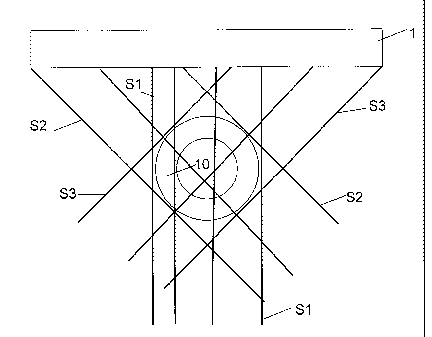
Figure 4 shows an embodiment, similar to that of Figures 1A-1B,
2A-2B. In Figure 4, a plurality of ultrasound signals Si (four depicted, as an
example) is emitted at an angle of about 90 degrees from a transducer 1
towards the tissue 10. Ultrasound signals 52 (three depicted) are emitted at
an
angle of about 450 from the transducer 1 towards the tissue 10. Ultrasound
signals S3 (three depicted) are emitted at an angle of about -45 degrees from
the transducer 1 towards the tissue 10. This embodiment can provide the
above-mentioned advantages. As follows from Fig. 4 (and also from Figures 1-
2), the transducer 1 can be positioned such and be dimensioned such, that each
group of emitted signals Si, S2, S3 can encompass the contour (in cross-
section) of the tissue 10 which is to be examined.
CA 02661128 2009-02-18
WO 2007/021185 PCT/NL2006/000429
13
In the above, methods are described that are based on acoustic
measuring beams generated and received by an ultrasound transducer. The
method can also be performed using optical measuring signals or a
combination of acoustical and optical signals. Furthermore, a method has been
described using the pulsatile blood pressure as a source for deformation. This
method can also be applied on tissues that are deformed by other sources than
the blood pressures originating from inside the body (like breathing or active
muscle contraction) or a by means of artificially exerting a pressure
variation
of the tissue.
Also, for example, according to another embodiment (see Figures 1),
there can be provided a method for generating strain and/or hardness
information of a tissue or tissue structure which is or can be subject to a
varying pressure, comprising the steps of:
1) receiving signals from the tissue with a sensor 1 positioned outside
the tissue for measuring the deformation of the tissue in a measuring plane
defined by the sensor 1 in which the measuring beams are all parallel and with
a certain angle towards the sensor surface;
2) without moving the sensor, receiving the signals from the tissue for
measuring deformation of the tissue in a measuring plane defined by the
sensor 1 in which the measuring beams are parallel but with a different angle
to the sensor surface than in the first step; herein, the angle with respect
to
the sensor surface between the first and the second step is preferably in the
order of about 45 degrees;
Identifying the strain of the tissue by compounding the information
acquired in step 1) and 2); and
Relating the strain to elasticity and/or hardness parameters of the
tissue.
Also, for example, a method for generating hardness information of a
tissue subject to a varying pressure can comprise the steps of (see Figures
2):
CA 02661128 2009-02-18
WO 2007/021185 PCT/NL2006/000429
14
1) receiving two or more signals from the tissue with a sensor 1
positioned outside the tissue for measuring the deformation of the tissue in a
measuring plane defined by the sensor in which the measuring beams have
different angles towards the sensor surface but originate from the same
location of the sensor; herein, the angle with respect to the sensor surface
between the first signal and the other signals is preferably in the order of
about 45 degrees;
2) Without moving the sensor 1, receiving two or more signals from the
tissue for measuring the deformation of the tissue in a measuring plane
defined by the sensor in which the measuring beams have different angles
towards the sensor surface but originate from the same location of the sensor
in which this location is another location; the angle with respect to the
sensor
surface between the first signal and the other signals is preferably in the
order
of about 45 degrees;
- 3) Repeating step 2 until an image is acquired; and
Identifying the strain of the tissue by compounding the information
acquired in step 1, 2 and 3.
Relating the strain to elasticity and/or hardness parameters of the
tissue.
Furthermore, as follows from the above, the method can comprise
the step of displaying elasticity and/or hardness parameters of the tissue.
Also,
the signals can be echographic data detected with an acoustic sensor. Besides,
the signals can be optic data detected with an optical sensor. After
acquisition
of data of one single plane, the sensor can be moved to acquire data in
another
plane (which, for example, is parallel to the previous plane) to assess
deformation of tissue in more than one image plane. The signals, at an
assumed cyclic pressure change, can also be received at predetermined time
intervals in the period of motion. The signals may come from a blood vessel
wall and that the data are received only during a specific time interval of
the
period of the heartbeat. Data can be acquired in certain parts of the heart
cycle
CA 02661128 2009-02-18
WO 2007/021185 PCT/NL2006/000429
using the ECG or using a device to measure the pulsatile blood pressure in the
tissue under investigation or another tissue or tissue structure than the
tissue
or tissue structure under investigation.
Besides, as follows from the above, an apparatus for generating
5 hardness and/or strain information of tissue, which tissue can be subject
to a
varying pressure, can comprise:
- a sensor capable of generating and receiving measuring beams or
signals with different angles towards the sensor surface, wherein the sensor
preferably includes at least one transducer, preferably a linear array
10 transducer.
- a beam steering device to control the way in which the measuring
beams are generated and received by the sensor.
- a processor device for collecting and processing signals received by
the sensor to identify strain of the tissue and to relate the strain to
elasticity
15 and/or hardness parameters of the tissue.
- a display device for displaying said elasticity and/or hardness
parameters of the tissue.
- a position recording device coupled with the processor device to
record sensor positions
The apparatus can further comprise an actuator for moving the
sensor. Besides, the apparatus can further comprise an activating means or
activator for activating the actuator. Also, in this case, the activating
means
can be connected to an ECG recording device and/or pressure monitoring
device to become active during a predetermined part of the heartcycle. As an
example, the sensor can be an acoustic sensor, or an optical sensor
It is to be understood that in the present application, the term
"comprising" does not exclude other elements or steps. Also, each of the terms
"a" and "an" does not exclude a plurality. Any reference sign(s) in the claims
shall not be construed as limiting the scope of the claims. Also, a single
CA 02661128 2009-02-18
WO 2007/021185 PCT/NL2006/000429
16
processor, controller or other unit may fulfil functions of several means
recited
in the claims.
Although the invention has been discussed on the basis of the above
mentioned exemplary embodiments, it is clear that the invention can also be
used when detecting and analyzing other tissues, such as (for cancer research
of) the prostate, the esophagus, the cervix etc. Such and other variations are
deemed to be within reach and the scope of protection of the appended claims.
For example, in the embodiments of Figures 1 and 2, said transducer
surface is a substantially flat surface, and the transducer surface is faced
toward the location of the tissue to be investigated. However, the transducer
surface can also be shaped.
In the present application, the term "tissue" should be interpreted
broadly, as will be clear to the skilled person. Particularly, the tissue can
be
part of a human or animal body.
Besides, said transducer, signal transmitter signal receiver or
transducer can be arranged and constructed in various ways, as will be clear
to
the skilled person. Preferably, the signal transmitter and signal receiver are
located at the same side of a tissue to be investigated. This can be the case,
for
example, when the signal transmitter and signal receiver are provided by an
acoustic transducer. Besides, a plurality of sensors, transducers, signal
transmitters and/or signal receivers can be utilized.