Note: Descriptions are shown in the official language in which they were submitted.
CA 02661221 2012-08-30
WO 2008/024434 PCT/US2007/018636
- 1 -
DEVICE FOR REMOVING FLUID FROM BLOOD IN A PATIENT
FIELD OF THE INVENTION
The present invention relates to filtration devices and methods for
continuously treating patients suffering from a condition of fluid overload,
retention
of excess fluids, or hypervolemia, as may be a result of renal or cardiac
disease. The
present disclosure also relates to hemCidialysis devices for treating people
with renal
failure. The devices can be worn extracorporeally or surgically implanted into
patients.
BACKGROUND OF THE INVENTION
Excessive fluid can accumulate in patients suffering from end stage renal
disease (ESRD) or congestive heart failure (CHF). The excess fluid first
accumulates in the blood and expands the volume of blood leading to
hypertension
and places increased stress on the heart. This added stress often leads to
heart
failure and death. The fluid also can accumulate in the pleural cavities of
the lungs
leading to shortness of breath. Oxygen uptake in the lungs is reduced as air
becomes
displaced by water. Again, if this condition is not reversed, death can
result.
According to the National Kidney Foundation, 20 million people have
Chronic Kidney Disease (C1CD) in the, US, which is one in every nine
Americans.
The most severe stage of C1CD, when kidneys cease to function, is End Stage
Renal
Disease (ESRD). According to the USRDS 2005 Annual Data Report, 452,957
people had ESRD in the US in 2003 and, of these, there were 324,826 prevalent
dialysis patients. The mortality rate of ESRD patients who receive traditional
hemodialysis therapy is 24% per year. The leading causes of death in patients
with
ESRD are cardiac related which accounts for 43% of all deaths in this
population.
CA 02661221 2009-03-24
WO 2008/024434 PCT/US2007/018636
- 2 -
In ESRD patients, fluid accumulates because their kidneys no longer can
effectively
remove the water and other fluids, which are consumed daily. The fluid
accumulates first in the blood where the blood volume can expand by as much as
= .
20%. The fluid then accumulates throughout the body ending up in the
extremities
such as the anldes, hands, and other tissues as edema (swelling). Volumes as
large
as 7-10 liters or about 15-20 pounds can commonly accumulate. This causes
increased stress on the heart as evidenced by significant increases in blood
pressure
or hypertension and subsequent heart failure. About 60% of hemodialysis
patients
have chronic hypertension as defined by the World Health Organization
guidelines.
This fluid overload volume can only be removed from ESRD patients by
direct ultrafiltration or by the ultrafiltration action of a dialysis
procedure, generally
carried out weekly in three 4 hour sessions. Removal of the large amounts of
water
in severe cases of fluid overload often causes fatigue and nausea and, in some
cases,
arrhythrnias, "crashing," and heart failure.
The fluid begins to re-accumulate again once the dialysis session is over. To
minimize the fluid accumulation,isevere fluid intake guidelines have been
established for these patients. Frequently because of continual thirst,
however, these
fluid restrictions are not complied with because of the hardship they impose
on the
quality of life of these patients.
= After the excess fluid has been removed and the proper blood volume has
been obtained, blood pressure will drop and the cardiac stress will be
reduced.
However, repeated increases and decreases in blood volume may also eventually
lead to damage to the heart and vascular system, thus further increasing the
risk of
cardiac disease. As re-accumulation of water occurs when the patient is not on
the
machine in a relatively short period of time, hypertension is nearly always
present in
hemodialysis patients to some degree. For those patients with residual kidney
function, this chronic hypertension may cause rapid decay of this residual
kidney
function leading to the high mortality rates of the general ESRD population
rather
than the lower mortality rates of those,ESRD patients with some residual
kidney
function.
The incidence of advanced CHF continues to grow and has become a disease
of epidemic proportions throughout the world. According to the National Health
CA 02661221 2009-02-18
WO 2008/024434 PCT/US2007/018636
- 3 -
and Nutrition Examination Surveys, an estimated 4.8 million Americans have
CHF.
In CHF patients, there is a progressive deterioration of the heart muscle that
leads to
an inability to pump enough blood to support the vital organs. As a result,
fluid
retention occurs because the blood perfusion pressure in the kidneys is
reduced and
the kidneys become inefficient in removing fluid.
While fluid overload in CHF patients can often be treated with numerous
pharmacological agents, these drugs become gradually ineffective over time and
may also cause undesirable effects such as kidney failure. There continues to
be a
growing body of literature that supports the concept of physic4lly removing
the fluid
by blood ultrafiltration, which has been shown to improve patient outcomes and
shorten hospital stays and intensive care unit utilization. In fact, fluid
removal may
be superior to the administration of very large doses of diuretic drugs.
There are several advantages to treating CHF fluid overload patients with
ultrafiltration over diuretic drugs. LT1trafiltration offers an efficient
fluid removal
without those side effects seen with drugs such as kidney failure and blood
pressure
drops. Furthermore, ultrafiltration quickly relieves the symptoms of shortness
of
breath and joint swelling.
Ultrafiltration is a process by which blood is exposed (under pressure) to a
semi-permeable membrane. The membrane properties dictate that water, salts,
and
other small molecular weight moleculbs pass through the membrane, but blood
cells,
proteins, and other large molecular weight molecules are not separated. The
ultrafiltration cartridge is generally made up of a very large number of small
diameter hollow fiber membranes. Typically, blood is removed from the patient
via
a catheter placed in an artery or large vein and is pumped into the
ultrafiltration
cartridge to generate the pressure necessary to carry out the ultrafiltration
process.
The hollow fibers are arranged so that the blood is perfused through these
hollow
fiber membranes and the filtered fluid is then removed and discarded, while
the
treated blood is then returned via another catheter back to the patient.
Conventional ultrafiltration devices have several drawbacks. The procedures
are carried out on machines that must be plugged into an electrical circuit
and
therefore the patients have limited mobility during the typically thrice
weekly, 4-
hour procedures. Because ultrafiltration is generally carried out during a
standard
CA 02661221 2009-03-24
WO 2008/024434 PCT/US2007/018636
- 4 -
dialysis session, the excessive water volume must be removed in this 4-hour
period,
which places additional physiological burdens on the patients.
Because of the close relationship between blood volume and blood pressure,
there is an additional complication using conventional ultrafiltration
procedures
related to total amount of fluid removed during a typical session. The fluid
amount
to be removed is generally determined by the amount of weight the dialysis
patient
has gained since the last dialysis and/or ultrafiltration session. Excessive
fluid
;
removal often leads to a significant drop in the patient's blood pressure
(hypotension), which can lead to hemodynamic instability and fainting, cardiac
arrest, or death.
There is an increasing body of evidence that continuous removal of
accumulated water through daily home dialysis or continuous ambulatory
peritoneal
dialysis (CAPD) results in significantly improved patient outcomes and far
fewer
physiological burdens being placed on the patients. However, the complexity
and
immobility of home dialysis procedures as well as the medical complications,
such
as infection and scarring, associated with long-term peritoneal dialysis,
severely
restricts the use of these ultrafiltration procedures to effectively treat
hypervolemia.
Another drawback of conventional ultrafiltration is the need to use
anticoagulants, such as heparin or citrate, to prevent the blood from clotting
in
conventional ultrafiltration devices. In order to adapt conventional
ultrafiltration
devices for continuous use, continuous anticoagulation must be utilized at
anticoagulant levels sufficient to prevent clots from forming in the device.
Prolonged use of anticoagulants presents a significant risk to patients in
general
because of the possibility of uncontrolled bleeding occurring and particularly
to the
majority of ESRD patients who are undergoing thrice weekly hemodialysis
procedures during which they also receive anticoagulation.
An additional drawback of the adaptation of conventional ultrafiltration to
the continuous treatment of hypervolemia resides in the complications of blood
access and the use of pumps. Most blood access for conventional
ultrafiltration
devices is carried out via indwelling venous catheters or arterio-venous
fistulas in
the case of certain ESRD patients. Notwithstanding the complications
associated
with the long term use of these blood access devices, they require the use of
special
CA 02661221 2009-02-18
WO 2008/024434 PCT/US2007/018636
- 5 -
blood pumps in the extracorporeal circuit in order to generate the flow rates
and
perfusion pressures required to achieve fluid removal in the ultrafiltration
device.
Blood access catheters that are placed in high pressure arteries have been
utilized to
obviate the need for additional pumping mechanisms to achieve the blood flow
rates
and pressures required, but safety concerns for their use outside an intensive
care
environment render them impracticable.
The use of membrane-based ultrafiltration systems for the treatment of blood
has been well documented in extracorporeal systems for over 30 years. However,
the use of these systems for continuous applications has been hampered by a
number.
of technical hurdles relating primarily to blood clotting and
biocompatibility.
Firstly, the cartridges contain a large number of small diameter hollow fiber
: = 1
membranes, which presents a large contact surface for filtration and toxin
clearance.
While this large surface area, approximately 1-2 m2 (10,000 ¨ 20,000 cm2) is
required to achieve the performance characteristics required for a short term
(2-6 hr)
extracorporeal ultrafiltration session, it exposes the blood to an equally
large surface
area of foreign material. The small diameter membranes are used to minimize
the
extracorporeal volume of blood that is required to be used during typical
hemodialysis or ultrafiltration. This combination of large numbers of fibers
coupled
with their small diameters results in an overwhelming surface-to-volume ratio
with
which the natural coagulation system of the patient must deal. As a result, a
high
level of anticoagulation is required to prevent the blood from clotting in the
cartridge. While this anticoagulation is medically acceptable over the
relatively
short period of the hemodialysis or hemofiltration sessions, long-term chronic
use of
high doses of anticoagulants is Medically unacceptable. Even with the use of
anticoagulation, continuous use in an extracorporeal circuit of existing
dialyzers is
generally not possible for more than approximately 48-72 hours.
This inherent thrombogenicity of the existing hollow fiber ultrafiltration
devices is further complicated by the design of the inlet and outlet elements
of the
cartridges which are used in existing devices to (i) distribute blood from a
single
inlet conduit to the large number of hollow fiber membranes and (ii) to
collect the
blood from the large number of hollow fiber membranes and channel the blood to
a
single outlet conduit. These designs allow for a number of stagnation points
within
CA 02661221 2009-02-18
WO 2008/024434 PCT/US2007/018636
- 6 -
these elements of the cartridge increasing the thrombogenicity of existing
devices.
Furthermore, these elements do not distribute the blood uniformly to all the
hollow
fiber membranes resulting in significant differences in blood velocity and
performance within different areas of hollow fiber membranes.
Secondly, long-term blood access continues to be problematic. Percutaneous
catheter use in hemodialysis patients is plagued with issues related to
bleeding,
infection, and clotting that require a high level of attention to maintain
these blood
conduits patent for use. There have been some recent developments in catheter
design that may improve these catheters, but currently they are unsatisfactory
for
The use of large bore, approximately 6 mm diameter, vascular grafts have
been largely successful as a long-term blood access conduit in vascular
reconstruction surgery. Graft survivals of over 5 years continuous use have
been
The devices and techniques disclosed herein are designed to address these
and other deficiencies of prior art devices and techniques for addressing
SUMMARY OF THE INVENTION
The present invention provides methods and apparatuses for continuous
CA 02661221 2009-02-18
WO 2008/024434 PCT/US2007/018636
- 7 - .
continuously and automatically remove excess fluid and/or blood toxins,
without the
use of perfusion pumps or percutaneous access devices.
Accordingly, one embodiment of the present invention is an apparatus for
removing fluid from the body of a patient. The apparatus includes a first
header
defining a first flow path with a single inlet and multiple outlets and a
second header
defining a second flow path having multiple inlets and a single outlet. A
filter is in
fluid communication with the first header and the second header. A first graft
is
included for connecting the vascular system of the patient to the single
inlet. A
second graft is included for connecting the single outlet to the vascular
system of the
patient. A housing is adapted to colldbt fluid that passes through the filter.
A drain
conduit is connected to the housing.
Another aspect of an embodiment of the invention includes the first flow
path being adapted to uniformly distribute fluid flow in the first flow path,
and the
second flow path being adapted to uniformly distribute fluid flow in the
second flow
path. Uniform fluid flow may be achieved by including one or more flow
restricting
neck regions or necks in the first flow path, the second flow path, or both.
The flow
restricting neck regions may be located near one or more of the multiple
outlets of
the first header, one or more of the multiple inlets of the second header, or
both.
The flow restricting neck regions near the multiple outlets of the first
header may be
more flow restrictive the closer they are to the single inlet of the first
header.
Similarly, the flow restricting neck regions near the multiple inlets of the
second
header may be more flow restrictive the closer they are to the single outlet
of the
second header. Uniform fluid flow niny also be achieved by having the first
flow
path progressively bifurcate divergently from the single inlet to the multiple
outlets,
having the second flow path progressively converging from the multiple inlets
to the
single outlet, or both.
In a further aspect of an embodiment of the present invention, the first
header
and the second header are elongated. The first header, the second header, the
filter
and the housing are substantially coplanar, and their thickness is about 10 mm
or
less.
In a further aspect of an embodiment of the present invention, the drain
conduit may be connected to the bladder of the patient. The patient may then
CA 02661221 2009-02-18
WO 2008/024434 PCT/US2007/018636
- 8 -
remove the fluid by natural urination. Also; a valve may be adapted to
restrict fluid
flow through the drain conduit. The valve may be controlled by a sensor and a
microprocessor based on physiological parameters of the patient.
Alternatively, the
valve may be controlled manually.
Another embodiment of the present invention includes a first header having a
first inlet and multiple outlets and a second header having multiple inlets. A
filter is
in fluid communication with the first header and the second header. The first
header, the second header and the filter define a flow path. The flow path may
include one or more neck regions near one or more of the multiple outlets. The
flow
path may also include one or more neck regions near one or more of the
multiple
inlets. The filter may include multiple hollow fiber membranes. The filter may
be
substantially permeable to water and substantially impermeable to blood cells
and
proteins.
A further embodiment of the present invention is an implantable
hemoconcentrator for removing fluid from the blood of a patient. The
implantable
hemoconcentrator includes a first header, a second header, and a filter. The
filter is
in fluid communication with the first header and the second header. The filter
includes a plurality of hollow fiber membranes. The first header, the second
header
and the filter are adapted to define a flow path that provides substantially
uniform
flow of blood through each of the hollow fiber membranes with minimal
stagnation
in the flow of blood.
A further embodiment of the present invention is a method for removing
fluid from the body of a patient. A fluid removing device is surgically
implanted in
the patient. The fluid removing device includes a first header defining a
first flow
path having a first inlet and multiple outlets and one or more necks located
near one
or more of the multiple outlets. The device also includes a second header with
multiple inlets and a second outlet, a filter in fluid communication with the
first
header and the second header, a first graft for connecting to the vascular
system of
the patient to the first inlet, a second graft for connecting the second
outlet to the
vascular system of the patient, a housing adapted to collect fluid that passes
through
the filter, and a drain conduit to the housing. The first graft is connected
to a first
blood vessel of the patient, which may be the femoral artery. The second graft
is
CA 02661221 2009-03-24
WO 2008/024434
PCTfUS2007/018636
- 9 -
connected to a second blood vessel of,' the. patient, which may be the femoral
vein.
The drain conduit is connected to the bladder of the patient. The device may
be
implanted in a subcutaneous location, such as the retropubic space. The method
may also include controlling the volume of fluid removed.
Another embodiment of the present invention involves an ultrafiltration
device containing a small number of large bore hollow fiber membranes and
inlet
and outlet distribution elements to evenly distribute and consolidate the
fluid flow so
as to maximize the efficiency of the device and minimize the disturbance of
the
blood flow to enable operation of the ultrafiltration device with a minimum of
or no
anticoagulant.
Another aspect of an embodiment of the present invention is an ultrafiltration
device adapted for direct implantation into the Patient's blood circulatory
system
incorporating a material suitable to attach (1) the blood inlet of the
ultrafiltration
device directly to an artery, (2) the blqo4 outlet of the ultrafiltration
device directly
to a vein, and (3) the filtered fluid outlet of the ultrafiltration device to.
the bladder of
the patient.
A further aspect of an embodiment of the present invention is an exemplary
ultrafiltration device incorporating a system that controls the removal of
excess fluid
from the circulatory system based upon a change in a relevant physiological
parameter, e.g., blood pressure, blood oncotic pressure, blood osmolality,
blood
constituent level, blood gas levels (e.g., p02, pCO2) and combinations
thereof.
An additional aspect of an embodiment of the present invention is an
ultrafiltration device that includes a system to transmit real time diagnostic
data.
Devices and procedures according the present invention may eliminate or
reduce excess fluid and eliminate or reduce the complications associated with
hypervolemia. This will help to reduce the incidence of hypertension and
associated
cardiac disease. In embodiments of thedisclosure the device operates to lower
blood pressure and reduce the incidence of pulmonary edema, and allows
patients to
ingest fluids as needed without the constant concern of controlling and
monitoring
fluid intake. Such a system is expected to lead to improvements in patient
health,
quality of life, and patient morbidity and mortality. These improvements may
be
CA 02661221 2009-03-24
WO 2008/024434 PCT/US2007/018636
- 10 -
achieved by slowly and continuously removing excess fluid from patients
suffering
from hypervolemia.
BRIEF DESCRIPTION OF THE DRAWINGS
FIG. 1 is a simplified view of a device according to the present disclosure
showing a relatively small number of relatively large bore hollow fibers and
the
inlet and outlet bifurcated distribution elements;
FIG. 2 is a detailed view of a bifurcated distribution element shown in FIG. 1
according to the present invention;
FIG. 3 is a simplified view of the filtration device according to the present
disclosure showing the bifurcated distribution elements;
FIG. 4 is an exploded detail view of the bifurcated distribution element
according to the present disclosure;
FIG. 5 is a simplified view of the device according to the present disclosure
showing implanting a device in a body by attaching the blood path of the
device to an
artery or vein and attaching the filtrate path of the device to the bladder
and showing the
device that controls the amount of fluid removed based on a change in a
physiological parameter;
FIG. 6 is a simplified view of the device according to the present disclosure
showing a device attached to the vascular system and a collection bag (in the
case of a
wearable, extracorporeal embodiment);
FIG. 7 is a schematic view of a filter device with the connections desirable
to
implant it in a body;
FIG. 8 is a schematic view an embodiment of the filter device that is
implantable in a body;
FIGS. 9-12 are a series of views of an embodiment of the header and flow
paths through the header;
FIGS. 13 and 14 are views of the flow path of the entire device illustrating
necking of the flow path; .
FIG. 15 is a schematic view of a filter device configured to provide
hemodialysis;
FIG. 16 is a schematic view of a filter device with exemplary dimensions;
CA 02661221 2009-03-24
WO 2008/024434 PCT/US2007/018636
- 11 -
FIG. 17 is a detailed view of the filter header with exemplary dimensions;
FIGs. 18, 19 and 20 are views of the flow velocity of an embodiment
illustrating the effect of neck regions on uniformity of flow;
FIG. 21 is a graph showing pressure drop versus blood flow rate through an
embodiment of the disclosure; and
FIG. 22 is a graph showing shear forces versus exposure time for
embodiments of the disclosure.
DETAILED DESCRIPTION OF THE INVENTION
The invention provides methods and apparatuses for continuous blood
ultrafiltration and/or hemodialysis which minimize thrombosis in and caused by
the
apparatuses. The apparatuses so limit apparatus-related thrombosis via the use
of
variable diameter and/or bifurcating blood channel designs which assure that
the
blood constituents are not exposed to undue shear forces, while at the same
time
minimizing the number of blood flow stagnation points. The apparatuses also
use
large-bore filter fibers that minimize the processed blood's exposure to any
thrombogenic filter surfaces within the apparatuses. The present invention
provides
devices and methods for the ultrafiltration of water, salts and other small
molecular
weight molecules from the bIoodi Blood cells and other large molecular weight
I,
molecules like proteins are typically not removed from the blood during this
ultrafiltration process. The process takes place by exposing blood, contained
in one
chamber, under pressure to one side of a semipermeable membrane whereby the
small molecules contained in the blood are filtered across the membrane, which
is
then collected in a second chamber. Once treated, the blood is then returned
to the
body and the filtrate is then discarded. The present invention also relates to
devices
that can provide hemofiltration and hemodialysis for both volume control
and/or
toxin removal within the blood.
Referring to FIG. 1, an ultrafiltration device 10 is comprised of a bundle of
hollow fiber membranes 5 contained within a housing 1. The device contains a
conduit 2 to form a single blood flow path into the device and a conduit 3 to
form a
single blood flow path exiting the device and a conduit 4 for the filtered
fluid to exit
the device. The conduits 2 and 3,may,be referred to as flow headers. The
hollow
CA 02661221 2009-03-24
WO 2008/024434
PCT/US2007/018636
- 12 -
fiber membranes (tubes) 5 can be made of any biocompatible material used for
hemodialysis or hemofiltration membranes to remove toxins and/or fluid. These
materials include, but are not limited to, polysulfone, cellulose acetate,
polyacrylonitrile, or polymethylmethacrylate. The fabrication of these hollow
fiber
membranes can be accomplished by any number of known methods used in the
manufacturing of medical grade hollow fibers for hemodialysis or
hemofiltration
devices. The housing and conduits of the device can be made of any
biocompatible
material including, but not limited to,.polymers like styrene acrylonitrile
(SAN),
polycarbonate (PC), polymethylmethacrylate (PMMA), polytetraflouroethane
(PTFE), polyethylethylketone (PEEK), polydirnethylsiloxane (PDMS),
polyurethane
(PU), or polysulfone (PS), or metals like stainless steel or titanium, or
ceramics.
The hollow fiber membranes can be sealed into the housing using a variety of
biocompatible potting compounds including, for example, polyurethane or epoxy,
An embodiment of an ultrafiltration device according to the present
invention preferably produces between 0-4 liters of fluid per day (0
ml/min)
which is readily achievable in a device containing high flux hollow fiber
membranes
having a total membrane surface area of less than 600 cm2 when operated at an
average transmembrane pressure gradient of about 50 mmHg and a blood velocity
of
approximately 30 cm/sec. The term high flux refers to the (increased) pore
size of
the filter element. Dialysers can have increased pore size of the filter
element to
increase the efficiency of the dialysis treatment. Preferably, the flux is
about 1
ml/min/m2/mmhg. In this embodiment,' the filter surface area is preferably
about 3
to about 6% of the membrane filter surface area of hemodialyzers and
hemofilters.
Accordingly, devices built according to this embodiment may be considerably
smaller than that of existing hemodialyzers and hemofilters. With the reduced
size
improvement it is possible to design a system that is sized for implantation
within
the body of hypervolemic patients.
FIG. 2 illustrates the conduit 2 distribution element of the ultrafiltration
device according to an embodiment of the present invention that splits and
channels
the single incoming flow path of blood into discrete flow paths 6 which flow
into the
core of the hollow fiber membranes. Similarly the conduit 3 (illustrated in
FIG. 1)
collects and channels the discrete flow paths of blood exiting the hollow
fiber
CA 02661221 2009-03-24
WO 2008/024434
PCT/US2007/018636
- 13 -
membranes into a single exiting flow path. Referring to FIG. 3 and FIG. 4, the
design of the flow headers of this embodiment of the disclosure is based on
the
formation of a bifurcated channel network which optimizes the hydrodynamic
forces
acting on the blood as it passes through the conduit in a manner so as to
minimize
the disturbance of the blood flow path and to eliminate any stagnation points
within
the flow path. In a preferred form, the header diverges into four different
conduits at
. .1 =
a pass. See for example, FIG. 4. Alternatively, the diverging fluid paths
created at
a single stage could be more or fewer than 4. Furthermore, significant to the
design
of this bifurcated network is the angle of divergence for each successive
level of
In a preferred form, the number of hollow fibers contained within the device
housing is significantly lower than the number of hollow fibers generally
found in
many dialyzers. The present invention also provides larger inner diameters of
the
hollow fiber membranes, in order to prevent clotting in the long-term use of
the
=
20 device. According to one aspect of the present invention, hollow fiber
membranes
generally used in existing hemodialyzers or hemofilters are smaller than the
currently contemplated preferred embodiment. In the device according to the
present invention, the number of the hollow fiber membranes is about
12 to about 60 hollow fiber membranes. Further, the hollow fiber
25 membranes have an inner diameter of between about 1 to about 7 mm, about
10 to
about 15 times that of most hollow fiber membranes incorporated into
hemodialyzers and hemofilters.
The increased inner diameter of the hollow fiber membranes reduces the
surface to volume ratio of the membrane and such reduction of surface to
volume
30 ratio provides improved thrombogenicity. The lower surface to volume
ratio can
also lead to a higher device volume per unit surface area than those devices
utilizing
smaller diameter hollow fiber membranes. However, as noted earlier, the total
CA 02661221 2013-07-19
- 14 -
membrane surface area that appears to be needed to meet the performance
requirements of a device according to one embodiment is about (approximately)
3 to
about 6% of that in existing hemodialyzers and hemofilters. Specifically, the
volume per surface area with hollow fibers (even with inner diameters 10 to15
times
that of most hemodialyzers and hemofilters) is less than 20 ml or about 20%
that of
common hemodialyzers or hemofilters.
In another aspect of the disclosure, the discrete flow paths emanating from
'
the flow headers 2, 3 are also aliened :with the corresponding hollow fiber
membranes 5 so as to form a stepless junction between the conduit and the core
of
the hollow fiber membrane. In a preferred form, conduits 22 are perpendicular
to an
end face 24 of the header and the fiber membranes are also perpendicular to
end face
26 (identified in FIG. 1) of the filter body. A template may be used to
precisely
align the hollow fibers prior to their being sealed into the housing 1 during
the
fabrication of the device. Using a template, each hollow fiber membrane is
aligned
with a discrete blood flow path so that blood flow in all hollow fiber
membranes is
uniform and turbulent flow or boundary layer separation is significantly
reduced
or eliminated, maintaining the low thrombogenicity of the overall device.
With reference to FIG. 3, the housing I may have an external shell 30 that
contains
the filter elements and endplates 32 that secure the filter element in axial
alignment
with the-device. In a preferred folia, the axis of the filter elements are
disposed
perpendicular to the endplates.
Referring to FIG. 5, according to some embodiments, the ultrafiltration
device I can be modified for implantation directly into the patient by forming
a
conduit between the inlet of the device and artery 8 using a large bore
vascular graft
7 and the outlet of the device and a vein 9 also using a large bore vascular
graft 7.
By attaching the device as an arteriovenous shunt, the pressure difference
between
the artery and vein are sufficient to provide the necessary driving force to
perfuse
the blood through the device and establish a high enough transmembrane
pressure to
allow the required fluid to be removed from the blood using the small membrane
surface area incorporated into the device. The material of the vascular graft
can be
any material used today for grafts such as polytetraflouroethane (PTFE) or
woven
TM
Dacron, but the diameter of the graft should be large enough to permit
unhindered
CA 02661221 2009-03-24
=
WO 2008/024434
PCT/US2007/018636
- 15 -
blood flow to and from the device. In the preferred form, the inner diameter
of the
vascular graft connections is between 2 mm and 7 mm. The connection between
the
vascular graft and the device should be such that there is essentially a
stePless
conduit so as to avoid generating turbulence in the blood flow path and
maintain a
low level of thrombogenicity in the overall device.
The filtrate from the device in a fully implanted device can readily be
collected in the bladder ha by connecting a suitable conduit 11 between the
filtrate
outlet of the device and the bladder. The material of this conduit can be of
any
biocompatible material including, but, not limited to, silicone or
polyurethane. Many
commercially available nephrostomybatheters may be used for this purpose. By
using the bladder as the collection site for the filtered fluid, normal
urination will
periodically remove the fluid from the body and provide for additional
capacity for
future filtration volumes. Normal urination provides patients with the
psychological
benefit over use of a urinary bag. However, should the bladder not be
functioning in
the patient due to chronic atrophy, an external connection via a standard
percutaneous catheter can be made between the filtrate outlet of the device
and a
standard urinary collection bag.
= One aspect of the invention provides a system to prevent overfiltration
of the
hypervolemic patients. In a preferred form, the device is fitted with a
control valve
13 on the filtrate outlet of the device. When this valve is closed, no
ultrafiltration
takes place, but the blood still readily flows through the device maintaining
its
patency.
In one embodiment, the valveiis connected to a blood pressure sensor on the
blood inlet conduit of the device so that the inlet blood pressure determines
the
status of the control valve. At high blood pressures corresponding to the
condition
of hypervolemia, the filtrate control valve is opened and ultrafiltration of
the blood
occurs to remove excess fluid. However, as the fluid is removed from the
hypervolemic patients, blood pressure will drop correspondingly. When the
blood
pressure drops to a predetermined level, the sensor sends a signal to the
filtrate
control valve and the valve is closed and the fluid removal terminates. When
the
blood pressure increases as excessive fluids subsequently begin to accumulate
in the
blood, the filtrate valve opens and the ultrafiltration resumes to remove the
excess
, CA 02661221 2013-07-19
- 16 -
fluid. When the device is used a patient with CHF, the physiological parameter
that
could be monitored to control the device may be lung capacity or volume.
Alternatively, blood parameters such as, e.g., blood pressure, blood ODODI/C
pressure,
blood osmolality, blood constituent level, and/or blood gas levels (pa>, pCO2)
can be
so monitored. Of course, other physiological parameters, even a combination of
parameters, could be used to control the device and thus the volume of fluid
in the
patient.
In another embodiment, the ultrafiltration device is attached external to the
body in such a fashion as to permit the patient full range of ambulatory
motion.
Referring to FIG. 6, the blood inlet of the device 1 is attached to an artery
15 via a
percutaneous arterial catheter 17 and the blood outlet of the device is
connected to a
vein 14 via a percutaneous venous catheter 16. The inherent blood pressure
difference between an artery and a vein eliminates the need for an additional
blood
pump to generate the required blood flow rate and transmembrane pressure
difference to establish the ultrafiltration required to alleviate the
hypervolemic
condition. The filtrate outlet of the device is connected to a standard
urinary
collection bag 19 via a suitable catheter IS.
In another embodiment, the filtration volume control system 33 is present. The
system includes, but is not limited to, a manual on-off valve, an automatic
valve
connected to a blood pressure sensor, or a battery controlled mini-pump.
Methods to
immobilize the external elements in one embodiment include, but are not
limited to,
attaching the external elements to a vest or belt.
In another embodiment, referring to FIG. 7, the ultrafiltration device is
attached to the blood circulatory system of the patient by attaching both the
blood
inlet and blood outlet of the device to veins 14 via percutaneous venous
catheters 16.
However, because there is an insufficient blood pressure gradient between
veins, a
blood perfusion pump 20 is used to establish the blood flow rate and
transmembrane
pressure gradient to achieve the required ultrafiltration performance of the
device.
The transmembrane pressure gradient can also be achieve through the use of a
pump
21 on the filtrate outlet conduit to establish a negative pressure in-the
filtrate
chamber, thus creating a su.ificient traasmembrane pressure gradient to
establish the
required ultrafilt-ation performance of the device.
CA 02661221 2009-03-24
WO 2008/024434 PCT/US2007/018636
- 17 -
In another embodiment, the device can be used with sensors that have the
capacity for real time diagnostic data igatering. For example, blood sensors
can be
disposed in the conduits, 'e.g., 7,. so that various types of parameters may
be
measured. Some of the physiological parameters that could be measured include
blood cell counts, blood pressure, blood oncotic pressure, blood osmolality,
blood
constituent level, and blood gas levels (p02, pCO2) or other parameters that
can be
productively measured within the bloodstream. , The diagnostic data that is
collected
can be used to operate the device, e.g., open a valve to allow the filter to
remove
water from the bloodstream. Alternatively, the diagnostic data can be used for
some
other productive collateral benefit such as regulating medicines or machines
to
enhance patient comfort.
FIG. 8 illustrates a schematic view of an embodiment of an ultrafiltration
device 100 that is implantable into the body of a person. The embodiment
includes
an inlet header 110 and a hollow fiber ultrafiltration core 112 and an outlet
header
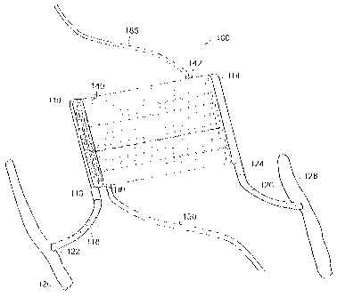
114. The ultrafiltration core 112 is di(sposed between the inlet and outlet
header in a
fluid tight manner. The inlet header 110 includes an inlet conduit 116 that
forms an
attachment point for a graft material 118 from a femoral artery 120. In a
preferred
form, the vascular graft is a 6 mm PTFE graft. A cut is made into the femoral
artery
and the graft material 118 is attached to the femoral artery 120 at location
122 in a
known manner. The headers 110 and 114 may alternately be referred to as
manifolds or grooved headers.
Similarly the outlet header 114 includes an outlet conduit 124 so that a
vascular graft 126 may be attached to the outlet header. In a preferred form,
the
vascular graft is a 6 mm PTFE graft. The other side of the graft 126 is
attached to a
femoral vein 128 at an attachment location 130.
Preferably the ultrafiltration device 100 is surgically implanted in a
subcutaneous location near and above the groin, such as the retropubic space.
This
allows for shorter vascular grafts 118 and 126 to connect the ultrafiltration
device
100 to the femoral artery 120 and femoral vein 130. In this location the valve
152
can be accessed and adjustments made without penetrating the skin, i.e.
extracorporeally (the valve 152 is discussed further below). The surgical
procedure
CA 02661221 2009-03-24
WO 2008/024434 PCT/US2007/018636
- 18 -
can be performed using local anesthesia. The ultrafiltration device 100 can be
removed or exchanged in a relatively simple surgical procedure.
The hollow fiber ultrafiltration core 112 includes a multiplicity of hollow
fibers 140 that extend from the inlet header 110 and the outlet header 114 in
a fluid
tight manner. That is, blood that leaves the femoral artery 120 at the
attachment
point 122 and travels through the graft material 118 and into the header 110
will
pass through the header into the plurality of hollow fibers 140. The housing
protects
the hollow fibers and also collects fluid that passes through the wall of the
fibers.
The hollow fibers are connected to the outlet header 114 in a manner similar
to the inlet header and fluid that passes through the fibers into the outlet
header can
be collected in the outlet header and pass through the graft material 126 and
back
into the bloodstream through the femoral vein.
The housing 142 includes a drain conduit 150 with a valve 152. The valve
operates as a safety valve with a manual control so that the device can be
properly
regulated. The outlet of the drain conduit is configured to drain into the
bladder 154.
The drain conduit, in a preferred form, may be a Filtrate Suprapubic Malecot
Bladder Catheter available through Cook Medical, Bloomington, Indiana. The
Malecot catheter includes radially expandable distal end to secure the
catheter within
a bladder. Of course alternative catheters may be used to dispose of the fluid
from
the device. Additionally, the conduit Can be directed outside the body and
connected
to an ostomy bag. Preferably the device 100 is substantially flat and the
components
of the device are substantially coplanar as shown in FIG. 8 in order to
facilitate
implantation of the device in the body of a patient.
The housing 142 and the ultrafiltration core 112 may be constructed out of
flexible materials. This flexibility will permit the device 100 to bend or
flex, further
facilitating the implantation and maintenance of the device in the body of a
patient.
Alternatively, the housing 142 may be constructed from substantially
inflexible
material.
FIGS. 9 and 12 illustrate embodiments of the inlet header 110 and outlet
header 114. Preferably the inlet header 110 and the outlet header 114 are
identical,
and they are shown as such in FIGS. 9-12. If the inlet header 110 and the
outlet
, õ.
CA 02661221 2009-02-18
WO 2008/024434 PCT/US2007/018636
- 19 -
header 114 are identical this will likely streamline design, manufacture and
assembly of embodiments disclosed herein.
As shown in FIGS. 9-12, the inlet header 110 defines a flow path 153
beginning at the inlet conduit 116 which then is split or bifurcated into
multiple
separate flow passages 156. The separate flow passages 156 connect to the
hollow
fibers 140. Similarly, the outlet header 114 defines a flow path 153 beginning
at the
separate flow passages 156 at the juncture with the hollow fibers 140 and
combines
or converges the separate flow passages 156 into a single outlet conduit 124.
The
flow paths 153 defined by the inlet hdader 110 and outlet header 114 may be
adapted
to optimize the hydrodynamic forces acting on the fluid passing through the
flow
paths 153. FIG. 12 shows a partial cutaway view illustrating the flow path
153.
FIGS. 10 and 11 illustrate the flow paths 153 defined by the headers 110
and 114. As illustrated in FIG. 10-12, the header flow paths 153 are
configured to
have smoothly diverging/converging conduits. Reference numeral 153 in FIGS. 10
and 11 illustrates the volume of the flow paths 153 themselves. The headers
110
and 114 define the flow path 153. Flow passages 156 are adapted to fit the
hollow
tubes 140 of the ultrafiltration core 112. As in other embodiments, the
connection
preferably is made to be as smooth as possible (without discontinuities) so
that the
possibility of blood clotting is minimized.
The headers 110 and 114 including the corresponding flow paths 153 may
be adapted to optimize the hydrodynamic forces acting on the blood as it
passes
through the flow paths 153 in a'manr&r s'o as to minimize the disturbance of
blood
flow and to reduce or eliminate any stagnation points within the blood flow.
In a
preferred embodiment, there are thirty-two flow passages 156 in each of the
headers
110 and 114. In another preferred embodiment there are sixteen flow passages
156
in each of the headers 110 and 114. The angle and path of divergence for each
flow
passage 156 may be adapted to minimize thrombogenicity in blood flow, which
eliminates or minimizes the amount of anticoagulant that must be used to
maintain
the system clot-free throughout its intended use.
FIGS. 13 and 14 illustrate the flow path through an embodiment of the
invention. As shown in FIGS. 13 and 14 the flow path includes neck regions or
necks, e.g. 170, 172. Neck regions are shown as constrictions or restrictions
in the
=
= . õ
CA 02661221 2009-03-24
WO 2008/024434 PCT/US2007/018636
=
- 20 -
flow passages 156. Alternatively one or more of the neck regions may be
located in
the hollow fibers 140, preferably located towards the end of the hollow fibers
140.
The neck regions closer to the header inlet conduit 116 and header outlet
conduit
124 , e.g. 170, are narrower (i.e., more flow restrictive) than the neck
regions, e.g.
172, at the regions further away from the header inlet conduit 116 and header
outlet
conduit 124. The variation in neck region size may be adapted to provide for
more
uniform volume of blood flowing through each of the hollow fibers 140,
minimize
blood flow disturbance, and reduce or eliminate any stagnation points within
the
blood flow.
FIGS. 13 and 14 show an embodiment of the invention with neck regions,
e.g. 170, 172, located in the inlet header 110 and the outlet header 114.
Alternatively, neck regions could be present only in the inlet header 110 or
the outlet
header 114, Such an arrangement may require the neck regions to be more
constricting as compared to the embodiment with neck regions located in both
the
inlet header 110 and the outlet header 114.
FIG. 15 illustrates a device configured for hemodialysis. Like reference
numerals will be used for like elements from FIG. 8 and need not be described
here.
In a hemodialysis device 180 a fluid conduit 185 is used to deliver dialysate
to the
housing 142 so that the filter elements 142 that have blood passing through
can
remove toxins through a convection gradient across the filter element so that
the
toxins are removed from the blood into the dialysate and the dialysate fluid
removed
from the hemodialysis element.
FIG. 16 illustrates exemplary dimensions of a device according to the
present disclosure. Specifically, as illustrated, the inlet header 110 has an
approximately 6mm inlet and is about (approximately) 75 mm in length.
Additionally, the width is approximately.23 mm. The hollow fiber
ultrafiltration
core 112 is about 74 mm wide and has a length of about 75 mm. About 32 tubes
are
disposed in the core and are connected to the inlet and outlet. The outlet
header 114
has similar dimensions as the inlet header. As illustrated, the headers have
an
overall length of approximately 95mm and the inlet/outlet conduit is
approximately
6mm in diameter. FIG. 17 illustrates the header in more detail in two side
views
taken at 90 degrees apart. As illustrated in the detail header view of FIG.
17, the
CA 02661221 2009-02-18
WO 2008/024434 PCT/US2007/018636
- 21 -
thickness or dimension across a header 110/114 is about 6mm. The inlet header
110, outlet header 114, and housing 142 may all be of substantially similar
thickness. As shown in FIG. 16 and 17, the thickness of the inlet header 110,
outlet
header 114, and housing 142 is about 6 mm. In another exemplary embodiment the
thickness may be 8 mm, 10 mm, 12 mm or such other thickness as may be
desirable.
Finite element analysis was performed on a device with features substantially
similar to those in the embodiments shown in FIGS. 9-12, 16 and 17. FIGS. 13
and 14 are based on the device used in the finite element analysis. The finite
element analysis was performed using finite element analysis software from
Adina
R&D, Inc. and computer aided design drawings produced using software from
Solid Works Corporation. The fluid was assumed to have a viscosity of 0.003 Pa-
s.
Results of the finite elements analysis are shown in FIGS. 18-20.
FIG. 18 shows the velocity of fluid flow through a device that does not
include neck regions. The fluid flow through the embodiment exhibits some non-
uniform flow through the holloWlibeis of the embodiment, especially the hollow
fibers closest to the inlet conduit 116 Of the inlet header 110 and the outlet
conduit
124 of the outlet header 114. Non-uniform flow can result in increased shear
forces
in the fluid and stagnation points. Approximate fluid flow velocities for this
exemplary device are indicated at various points in the device in FIG. 18.
=
FIG. 19 shows the velocity of fluid flow through a device that includes neck
regions. The fluid flow through the device shows significantly reduced non-
uniform
flow through the hollow fibers of the embodiment as compared to the fluid flow
shown in FIG. 18. This is most apparent when comparing the fluid flow through
the
hollow fibers closest to the inlet conduit 116 of the inlet header 110 and the
outlet
conduit 124 of the outlet header 114 in FIGS. 18 and 19. Approximate fluid
flow
velocities are indicated at various points in the device in FIG. 19. =
FIG. 20 shows the velocity of the fluid flow through the portion of the
embodiment that includes neck ;regions and is most susceptible to stagnation
points
and shear forces in fluid. The velocity and direction of fluid flow are
depicted using
directional arrows. The longer and larger the arrows the faster the fluid is
flowing,
and conversely the shorter and smaller the arrows the slower the fluid is
moving.
CA 02661221 2009-02-18
WO 2008/024434 PCT/US2007/018636
-22 -
The arrows in FIG. 20 show uniform fluid flow and no stagnation points in the
fluid
flow.
FIG. 21 is a graph of the results of the finite element analysis described
above showing the pressure drop in relation to blood flow rate over the entire
embodiment and over portions of the embodiment, such portions including the
hollow fibers 140, headers (inlet header 110 and outlet header 114), and the
grafts
(graft 118 connected to the inlet header 110 and graft 126 connected to the
outlet
header 114). As is shown in this embodiment, a low pressure drop over the
device is
achieved.
It is desirable to have a low pressure differential over an ultrafiltration
device. The low pressure differential enables the device to be utilized
without use of
a pump, and therefore makes the device more suitable for implantation. The
pressure differential over the embodiment as shown in FIG. 21 is less than the
typical pressure differential between a femoral artery and a femoral vein.
Typically
the femoral artery is approximately 120 mmHg and the femoral vein is at about
(approximately) 10-20 mmHg, resulting in a pressure differential of about 100-
110
mmHg. Therefore, the typical pressure differential between a femoral artery
and a
femoral vein is sufficient to cause blood to flow through the device at
acceptable
rates.
To prevent or reduce the potential of thrombosis occurring in a device it is
desirable to minimize shear forces in the blood passing through the device.
FIG. 22
is a graph showing shear forces on the y-axis and exposure time on the x-axis.
This =
graph is derived from Colton, C.K. and E.G. Lowrie, "Hemodialysis: Physical
Principles and Technical Considerations," in "The Kidney", 2nd ed., B.M.
Brenner
and F.C. Rector, Jr., eds., Vol. II, W.B. Saunders, Philadelphia, PA, pages
2425-
2489 and in particular page 2441 (1981). The lines on the graph indicate the
threshold points above which thrombosis is more likely to occur in red blood
cells
and platelets. Also included on the graph are the results of finite element
analysis as
described above. Finite element analysis results for embodiments with sixteen
and
thirty-two hollow fibers 140 are shown on the graph. The results place these
embodiments below the lines on the graph, indicating that thrombosis is not
likely to
occur in connection with the sixteen Snd thirty-two hollow fiber 140
embodiments.
CA 02661221 2013-07-19
- 23 -
To reduce or prevent thrombosis in a device it is desirable to (1) minimize
shear forces in the blood passing through the device and (2) avoid stagnation
points
that may be caused by flow irregularities. The results of the finite element
analysis
demonstrate that the embodiments including the neck regions reduce or
eliminate
non-uniform flow and therefore reduce shear forces in the fluid and reduce or
eliminate stagnation points in the fluid. The elimination or reduction of non-
uniform
flow and stagnation points reduces the likelihood of thrombosis and causes the
embodiments to be more suitable for long term implantation.
h.
Certain benefits may be adhievedby using an implantable device rather than
a non-implantable device. After implantation of the device, and after an
initial
healing period, there is a lower risk of bleeding, clotting and infection than
with a
non-implanted device. In particular, without any percutantous access the
likelihood
of infection is reduced because there is less opportunity for bacteria to gain
access to
the device or the area in which the device is implanted. In addition, there is
a lower
likelihood of thrombosis because the blood flow through the device remains
uninterrupted and there is no exposure to air. Also, patients receiving the
implantable device will be able to have greater fluid intake because of the
ability to
remove excess fluid from the body. This, combined with the ability to urinate,
can
substantially increase the quality of life for the patient suffering from
kidney disease
or heart failure.
While particular embodiments of the present disclosure have been shown and
described, it will be obvious to those skilled in the art that changes and
modifications may be made.