Note: Descriptions are shown in the official language in which they were submitted.
CA 02662845 2009-03-06
WO 2008/032249 PCT/IB2007/053611
1
Catheter and medical as sembly
FIELD OF THE INVENTION
- The present invention relates to a catheter comprising:
- a connector at a proximal side of the catheter for connecting the catheter
to an
external signal transmission/receiving unit for transmitting and/or receiving
signals,
- an electrode at a distal side of the catheter, and
- an electrical connection including an electrical wire for electrically
connecting
the electrode and the connector for the transmission of signals between the
electrode and the
connector.
Further, the present invention relates to a medical assembly comprising such a
catheter.
BACKGROUND OF THE INVENTION
So far electrophysiologic (EP) interventions cannot be performed safely under
magnetic resonance (MR) guidance due to the risk of radio frequency (RF)
heating. For EP
procedures it is necessary to monitor the intracardiac electrocardiogram
(IECG) ("mapping")
and/or to stimulate the heart ("pacing"). Both require an electrical
connection to electrodes on
the catheter tip, which is placed inside the heart. This connection can become
resonant at the
operating frequency of the MR scanner und thus act as an antenna for the
applied RF fields,
leading to excessive heating especially at the tips.
The wires, which are connected between the electrodes at the catheter tip
("distal side") and the connections at the other side ("proximal side") of the
catheter, become
resonant if they fulfill the condition
l~n~=n Xo
2 2 le rl r
with k0 denoting the wavelength in vacuum, 1 the length of the wire, Er and r
the effective relative permittivity and permeability for the common mode. The
effective Er
and r depend strongly on the properties of the tissue surrounding the wire.
As a consequence
it is hardly possible to predict if resonance will occur during an
intervention, since the length
CA 02662845 2009-03-06
WO 2008/032249 PCT/IB2007/053611
2
of wire inside the patient as well as the surrounding of the wire changes
continuously. This
means that EP catheters have to be made inherently RF-safe.
WO 2005/053555 Al discloses an electrode catheter for the defibrillation,
mapping or ablation of cardiac tissue. Said catheter comprises a terminal on
the proximal end
of the electrode catheter and one or more sensing and/or treatment electrodes
that are situated
on or in the vicinity of the distal end of the electrode catheter, in addition
to at least one
electric conductor, which is used to electrically connect a respective sensing
or treatment
electrode to the terminal. The electric conductor is composed of carbon and
the electrode
catheter is configured to be suitable for use as part of magnetic resonance
tomography and
for connection to electrophysiotherapy equipment. Said catheter comprises at
least one
defibrillation electrode, or at least one sensing electrode for the recording
and evaluation of
cardiac tissue potentials, or at least one treatment electrode for delivering
high-frequency
currents for ablation purposes. However, said carbon is not sufficiently RF-
safe for medical
applications in the field of MR.
SUMMARY OF THE INVENTION
It is an object of the present invention to provide a catheter and a medical
assembly using such a catheter which can be used during medical interventions
under MR
guidance without the risk that the electrical connection of the catheter
becomes excessively
hot due to resonance at the operating frequency of the MR scanner.
In a first aspect of the present invention a catheter as described above is
presented that is characterized in that the electrical connection has a high
electrical resistance
of at least 1 kS2, in particular of at least 5 kS2.
In a further aspect of the present invention a medical assembly is presented
comprising:
- a catheter as claimed in claim 1,
- a signal transmission/receiving unit for transmitting signals to and/or
receiving
signals from the electrode of said catheter and for processing said signals.
Preferred embodiments of the invention are defined in the dependent claims.
The present invention is based on the idea to use a highly resistive
electrical
connection for the transmission of signals between the external equipment
(e.g. the pacing
unit or the ECG unit) and the catheter tip. A high resistance limits the
induced current in the
wires and thus effectively attenuates heating. Despite the high resistance the
signal quality is
sufficient.
CA 02662845 2009-03-06
WO 2008/032249 PCT/IB2007/053611
3
According to a preferred embodiment of the present invention the usage of
highly resistive wire(s) in the catheter for connecting the electrodes at the
tip to the connector
at the other side for connection to an external equipment (generally called
signal
transmission/receiving unit) is proposed. The wire can have an electrical
resistance of at least
1 kS2/m, in particular of at least 5 kS2/m.
An alternative (or additional) solution is to insert one or more current
limiting
resistors into the conection to reduce the RF-heating. The resistor(s) can
have a resistance of
at least 1 kS2, in particular of at least 5 M.
The invention can be used for mapping as well as pacing, although in the
latter
case additional measures may need to be applied to reduce the risk for
injuries caused by the
high voltages required for the stimulation system. When used for mapping, the
wires of the
highly resistive connection are used to carry the signals (in particular ECG
signals) picked up
by the electrodes to the external equipment. When used for pacing, the wires
of the highly
resistive connection are used to carry the pacing signals applied by the
external equipment to
the connector to the electrodes at the catheter tip.
Preferably, the electrical connection comprises a pair of wires for
electrically
connecting a pair of electrodes with the connector. Often, a catheter
comprises a plurality of
electrodes from which 2 electrodes are used for a heart stimulation (bipolar).
Generally, also
one electrode of the catheter is sufficient, where an external reference
electrode is used in
addition (unipolar).
To avoid any risk that too high currents or voltages my cause any damage to a
patient's health during an intervention, a preferred embodiment of the
catheter comprises a
current sensing unit for sensing the electrical current flowing in the
electrical connection at
the distal side of the catheter and for transmitting a sensing signal
indicative of the sensed
current to an external current control unit. Thus, a current leakage between
the catheter input
and the catheter tip can be recognized, and coutermeasures can be taken by the
current
control unit, e.g. the current flowing in the wire(s) of the electrical
connection can be reduced
or stopped.
In an advantageous embodiment, that is simple to implement, the current
sensing unit comprises a sensing resistor having a small electrical resistance
of 1 kS2 for
sensing a voltage representing said sensing signal and a pair of sensing wires
for transmitting
said sensing signal to said external current control unit.
CA 02662845 2009-03-06
WO 2008/032249 PCT/IB2007/053611
4
Preferred embodiments of the materials that can be used for said wire(s) are
defined in further dependent claims enabling a practical handling, which would
not be
possible with a very thin Cu wire.
BRIEF DESCRIPTION OF THE DRAWINGS
These and other aspects of the invention will be apparent from and elucidated
with reference to the embodiment(s) described hereinafter. In the following
drawings
Fig. 1 shows a diagrammatic side elevation of an MR imaging apparatus,
Fig. 2 shows a first embodiment of the medical assembly according to the
present invention,
Fig. 3 shows a diagram illustrating the temperature change for two different
wires used in a catheter, and
Fig. 4 shows a further embodiment of the medical assembly according to the
present invention.
DETAILED DESCRIPTION OF THE INVENTION
Fig. 1 exemplarily shows components of an open MR imaging apparatus
which are of essential importance for the generation and reception of magnetic
fields in an
examination zone 1. Above and underneath the examination zone 1 there are
provided
respective magnet systems 2, 3 which generate an essentially uniform main
magnetic field
(BO field for magnetizing the object to be examined, that is, for aligning the
nuclear spins)
whose magnetic flux density (magnetic induction) may be in the order of
magnitude of
between some tenths of Tesla to some Tesla. The main magnetic field
essentially extends
through a patient P in a direction perpendicular to the longitudinal axis of
the patient (that is,
in the x direction).
Planar or at least approximately planar RF conductor structures (resonators)
in
the form of RF transmission coils 4 ("body coils") are provided for generating
RF pulses (Bl
field) of the MR frequency whereby the nuclear spins are excited in the tissue
to be
examined, said RF transmission coils 4 being arranged on the respective magnet
systems 2
and/or 3. RF receiving coils 5 are provided for receiving subsequent
relaxation events in the
tissue; these coils 5 may also be formed by RF conductor structures
(resonators) provided on
at least one of the magnet systems 2, 3. Alternatively, one common RF
resonator can also be
used for transmission and reception if it is suitably switched over, or the
two RF resonators 4,
5 can serve for the alternating transmission and reception in common.
CA 02662845 2009-03-06
WO 2008/032249 PCT/IB2007/053611
Furthermore, for the spatial discrimination and resolution of the relaxation
signals emanating from the tissue of a patient P (localization of the excited
states) there are
also provided a plurality of gradient magnetic field coils 7, 8 whereby three
gradient
magnetic fields are generated which extend in the direction of the x axis.
Accordingly, a first
5 gradient magnetic field varies essentially linearly in the direction of the
x axis, while a
second gradient magnetic field varies essentially linearly in the direction of
the y axis, and a
third gradient magnetic field varies essentially linearly in the direction of
the z axis.
In order to monitor the intracardiac electrocardiogram (IECG) ("mapping")
and/ or to stimulate the heart ("pacing") of the patient P, use is often made
of a catheter 6
which is introduced into the patient P to provide an electrical connection to
electrodes on the
catheter tip. So far electrophysio logic (EP) interventions, however, cannot
be performed
safely under MR guidance due to the risk of RF heating of the electrical
connection, which
can become resonant at the operating frequency of the MR scanner und thus act
as an antenna
for the applied RF fields, leading to excessive heating.
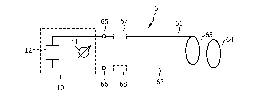
Fig. 2 shows a first embodiment of the medical assembly including a catheter
6 according to the present invention. The catheter in this embodiment
comprises two wires
61, 62 for connecting a pair of electrodes 63, 64 at the tip of the catheter 6
to an external
equipment 10 (signal transmission/receiving unit) connected to connection
terminals 65, 66
(the connector) for the transmission of signals therebetween. In the shown
embodiment the
external equipment 10 comprises an ECG monitoring unit 11 for receiving and
processing of
ECG signal received by the electrodes 63, 64 and a EP stimulating unit 12 for
generating and
transmitting of pacing signals for stimulating the heart of the patient P. It
is evident that these
are just shown as examples and that the external equipment 10 does neither
require both these
units 11, 12 nor is it limited to these units 11, 12.
According to the present invention highly resistive wires 61, 62 and/or wires
with additional lumped resistors 67, 68 are used as connections within the
catheter 6. A high
resistance limits the induced current in the leads and thus effectively
attenuates resonance
effects. In this context it has to be mentioned, that the heating of the
conductor 6 itself
generally does not pose the safety problem (as often incorrectly stated).
Instead the electric
fields E near the conductors 61, 62 lead to a power deposition in the tissue,
which is
described by the specific absorption rate
SAR=6E2
2p
CA 02662845 2009-03-06
WO 2008/032249 PCT/IB2007/053611
6
with and being the conductivity, respectively the mass density of the
tissue. Reducing the current inside the conductor 6 leads to lower electric
fields at its tip,
since both are related.
The current can principally be reduced by lumped resistors 67, 68, but they
have to be placed
at positions, where the highest currents occur, i.e. in the antinodes of the
induced standing
wave. If they are put into the nodes of the standing wave, they are not
effective. The problem
of proper a-priori placement does not occur when using highly resistive wires
61, 62, where
the resistance is distributed. Furthermore, in contrast to lumped elements
highly resistive
wires do not introduce additional joints. Nevertheless, it is principally
possible to insert
(preferably additional) lumped resistors 67, 68 into the highly resistive
wires 61, 62 to
increase the resistance further or to use low resistance cables with lumped
resistors placed at
distances much smaller than /2.
Test measurements performed on a highly conductive copper wire and a
resistive wire of 1.8 kS2/m (metallic alloy ISAOHM, Isabellenhutte) being put
into a catheter
surrounded by a phantom liquid mimicking the human body demonstrate the
advantage of the
invention. The wires were in contact with the phantom fluid at the tip of the
catheter as in a
real EP catheter. The length of the catheter was varied to achieve the
resonance condition.
The temperature increase at the tip was measured during an MR scan. The
results presented
in Fig. 3 show, that for a copper wire a pronounced resonance effect occurs
leading to
excessive heating, whereas the resistive wire does not cause clinically
significant heating.
Besides the safety aspect there is the requirement that the wire is adequate
to
transmit signals or stimulating pulses, respectively. Typical ECG recorders 11
have input
impedances of several hundred MS2 up to GS2, which means that the voltage drop
of the cable
is negligible, even if the cable resistance reaches several MS2. Also the
thermal noise of such
a line is much lower than typical ECG voltages.
The situation may need further attention in the case of pacing or any other
method, which has to bring a voltage/current to the catheter tip. Since the
resistance between
the tip electrodes 63, 64 (typically about 100 S2) is much smaller than that
of the highly
resistive wire, most of the power is lost in the cable. As a consequence high
voltages have to
be applied to achieve the required currents/voltages at the catheter tip. As
an example for a
pacing current of 2 mA between the electrodes and a 100 kS2 resistance of the
connecting
cables 200 V must be applied. This poses safety considerations, since inside
the body already
low voltages can cause dangerous shocks.
CA 02662845 2009-03-06
WO 2008/032249 PCT/IB2007/053611
7
This invention therefore provides additional, optional measures to provide
adequate safety. An embodiment of a medical assembly using such additional
safety
measures is shown in Fig. 4.
A first such measure is to use a current limited power supply 13, so that even
in the case of a short circuit no currents higher than the specified ones can
flow through the
wires 61, 62.
A second such measure is to avoid that e.g. in case of a breakdown of the
cable insulation, a voltage is applied somewhere else than at the desired
location. To this end,
it is verified that the whole current, which is fed into the catheter 6, in
fact reaches the tip.
This may be realized by sensing the voltage drop of a resistor R2 at the tip
of the catheter 6,
which directly yields the corresponding current. This value is then compared
with the current
applied by the power source 13, which can e.g. be measured in the same way.
For sensing the
current at the tip, resistive wires 69, 70 are used that have a higher
resistance than the
resistance of the wires 61, 62 connecting the electrodes to the external
equipment 10.
In this embodiment, the potential difference at the resistor R2 is monitored
by
the two wires 69, 70. The current at the tip is compared to the current fed
into the catheter
measured at the resistor Rl to detect a malfunction. Thus, the high voltage,
which is
necessary to achieve sufficiently high stimulation currents, does not pose a
danger. It is not
necessary to use lumped resistors Rl, R2, but instead it is also possible to
use small parts of
the highly resistive wires 61, 62 to measure the potential differences.
Thus, the present invention provides the following features that can be used
each on its own or in any one combination of features:
using highly resistive wires (and/or lumped resistors) inside catheters to
monitor
physiological signals (IECG) without danger of RF-heating (RF-safe);
using the same assembly to stimulate the heart (pacing);
using highly resistive wires (or cables with lumped resistors) to sense the
current at the catheter tip for providing feedback information, e.g. for
detecting malfunction
or regulation purposes;
using highly resistive wires (or cables with lumped resistors) to sense non-
physiological signals without RF-heating;
Using highly resistive wires an EP catheter can be built in the usual way,
i.e.
connecting the tip electrodes to the monitoring and pacing equipment.
Optionally, additional
filters may be inserted to remove MR-induced artefacts from the IECG signal.
CA 02662845 2009-03-06
WO 2008/032249 PCT/IB2007/053611
8
Wires with resistances starting at roughly 1 kS2/m are used to achieve RF-
safety. As shown in Fig. 3 already resistances of 1.8 kS2/m can reduce the
heating
substantially, but to avoid any risks higher values seem favourable for
practical applications.
Nevertheless, a compromise between safety (demanding high resistance) and
applied power
(demanding low resistance) has to be found. The wires can be made of metallic
alloys (as in
the example shown above: ISAOHM wire), carbon fibres, conductive polymers or
any other
non-magnetic material. A possible technique is to coat nonconductive fibres or
threads with
the conductive polymers. Also the catheter itself may be coated with such a
conductive film.
An alternative is to use nonconductive fibres or threads coated with very thin
metallic
surfaces (e.g. by sputtering). A further alternative is to use nonconductive
fibres or threads
filled (above the percolation threshold) with conductive particles. If
necessary the resistance
of the wires can be further increased adding lumped resistors.
The wires of the catheter according to the invention are of a high electrical
resistance. This resistance is high in comparison to the traditionally used
metal wires. The
electrical resistance is that high that it sufficiently attenuates the current
induced in the wire
by the MR field. On the one hand, the electrical resistance of the connecting
wires is
preferably low in order not to degrade the signal and signal quality sensed by
the electrodes.
On the other hand, the electrical resistance is preferably high in order to
attenuate the induced
current. Thus, the value of the electrical resistance of the wires according
to the invention is
to be chosen such that both criteria (low enough for signal quality and high
enough for
attenuating MR induced current) are met. These suitable values form a range of
electrical
resistance values, with a lower boundary and an upper boundary. The lower
boundary is such
that the MR induced values are still sufficiently attenuated. The upper
boundary is such that
the signal quality is still sufficient to perform reliable measurements.
The exact values of the resistance of the wire depend, for example, on the
position of such a wire inside the patient and inside the MR-scanner. Moreover
the wire
thickness, strength of excitation, number of wires inside one catheter play a
role.
For the lower boundary of the range of values of electrical resistance of the
wire, the following can be said. Experiments have shown that under bad
conditions of having
the wire very close to the wall of the MR-scanner the resistance of the wire
must be larger
that 2kS2/m (for a 30 m thick wire) to suppress induced current sufficiently.
In more
favorable conditions it appeared that a resistance as low as 1 kS2/m still
provided for
sufficient attenuation of the induced current. Preferably, values are higher
than 5 kS2/m. A
preferred value lies between 5 and 20 kS2/m.
CA 02662845 2009-03-06
WO 2008/032249 PCT/IB2007/053611
9
For the upper boundary of the range of values of the electrical resistance of
the
wire, the following can be said. When the wire is used for mapping of ECG
signals, i.e.
measuring signals at the electrode, values up to 2 MS2 still provide good
signal quality. When
the wire is used for pacing, lower values of the electrical resistance must be
used since
electrical power will have to be transferred through the wire. This makes that
the values is
preferable less then 50kS2.
The above numbers are examples of the value of the electrical resistance
according to the invention. It is to be noted, that in other circumstances
other values could be
preferable.
The invention can advantageously be used for EP interventions under MR
guidance, allowing to record IECGs and to stimulate the heart. The concept of
monitoring the
voltage of a sensor in the catheter (in this case a current sensing resistor)
may also be used for
other applications. Generally the invention can be applied to all medical
diagnosis and
therapy involving the measurement of electrical signals or the application of
electrical
stimulation. With continuously increasing field strengths of MR systems, in
future, even
comparatively short leads of e.g. implantable devices may benefit from the
proposed
invention.
In summary, the present invention provides a solution to prevent excessive
heating during EP interventions under MR guidance by using highly resistive
wires and or
lumped resistors as connections within catheters. A high resistance limits the
induced current
in the leads and thus effectively attenuates heating. The signal quality of
the recorded IECG
is preserved even if the cable resistance reaches several MS2.
Thus, according to the present invention, with highly resistive wires the IECG
can be measured safely without danger of RF-heating. The same type of cables
can be used
for cardiac pacing. The wires can be realized with a very low diameter. The
safety concept is
not limited to EP applications. It can be used to sense the output voltage of
a pressure or
temperature sensor. The optional current limitation and the current leakage
detection protect
the patient even in case of a catheter malfunction.
While the invention has been illustrated and described in detail in the
drawings
and foregoing description, such illustration and description are to be
considered illustrative or
exemplary and not restrictive; the invention is not limited to the disclosed
embodiments.
Other variations to the disclosed embodiments can be understood and effected
by those
skilled in the art in practicing the claimed invention, from a study of the
drawings, the
disclosure, and the appended claims.
CA 02662845 2009-03-06
WO 2008/032249 PCT/IB2007/053611
In the claims, the word "comprising" does not exclude other elements or steps,
and the indefinite article "a" or "an" does not exclude a plurality. A single
element or other
unit may fulfill the functions of several items recited in the claims. The
mere fact that certain
measures are recited in mutually different dependent claims does not indicate
that a
5 combination of these measured cannot be used to advantage.
Any reference signs in the claims should not be construed as limiting the
scope.