Note: Descriptions are shown in the official language in which they were submitted.
CA 02664135 2009-03-20
PCT/EP2007/059951
Method and device for automated removal
of cells and/or cell colonies
Description
The invention relates to a method and uses of the method for
automated removal of cells and/or cell colonies. The invention
further relates to a device for carrying out the method.
Within the framework of biological or medical research, work
on the care, screening and selection of cells and/or cell
colonies in cell cultures has to be carried out with reference
to most diverse criteria. At the same time, the cells and/or
cell colonies must be specifically selected and transferred
into other sample vessels or cell cultures.
The continuously growing market of biotechnology and medical
research is presently characterised by the change from largely
manual cell culture selection and care towards partially and
fully automated systems which select and separate cells with
reference to specific criteria. As a result of the very
different cell types in this area, having highly varying
properties and harvesting behaviour, special solutions adapted
to the cell type to be processed are largely produced.
Since in the area of university and also commercial research
in this field, the cell lines used are frequently changed due
to changing research tasks, the acquisition of these expensive
devices having only special uses is of no economic interest
for smaller research teams.
CA 02664135 2009-03-20
2
So far, the practical implementation has been carried out by
taking up various commercially available plastic tips for the
removal of particles from media, taking up special plastic
tips having an enlarged opening for picking particles from
viscous media, taking up metal capillaries for picking
strongly adherent particles of different size by scraping or
taking up glass capillaries inserted in adapters for
separating smaller, non-adherent or barely adherent particles.
Relatively universally usable devices for automation of this
work are not available for the separation and culture care of
strongly adherent cells and colonies. A major problem is the
strong adherence of the cells to the sample vessel. Known
methods such as, for example, the targeted cloning by means of
enzymes for releasing the binding of the cell membranes from
the sample vessel, described in DE 197 42 163 C2 fail due to
the time of action of the enzyme before the establishment of
the desired effect being too long and due to the cell death
and severe reduction in vitality initiated by the damage to
the cell membrane caused by the use of the enzyme. Thus, the
automation of the targeted selection of such cells is not
possible in the known manner. These problems arise
particularly in the area of stem cell research.
The practical implementation hitherto has been carried out by
means of scraping cannulas made of stainless steel having
different diameters, scraping cannulas of glass or metal,
which are glued into a conical adapter, or scraping cannulas
made of ceramic for smaller diameters.
Due to the mostly high requirements on sterility for
protecting the samples from contamination, the cannulas used
must usually be changed after each separation cycle. At high
AVI-22873 PCT- Fassung-adoc
17.03.2009
CA 02664135 2009-03-20
3
throughput this takes place accordingly frequently and is not
reasonable manually.
A number of mechanical devices and robot systems are already
known for the semi-automatic processing, in particular for the
separation, isolation and treatment of cell clones and
individual cells.
For example, DE 10 2004 027 661 Al discloses a drive
arrangement for a robot system for isolating and treating cell
clones and individual cells. The drive arrangement comprises
motor-driven slits which can be moved in two different axial
directions and serve to move the separating tool to the
samples.
DE 10 2004 046 740 Al describes a tool head for the automatic
isolation and treatment of cell clones. This tool head serves
to receive a pipette which in turn receives a cloning dome. A
defined force closure between the pipette tip and the cloning
dome is possible due to a staggered arrangement of leaf
springs.
DE 197 42 163 Al describes a cloning dome which encloses the
cell and/or cell colony during the removal and in which a
rinsing process takes place by which means the cell and/or
cell colony is released from the vessel. The cell and/or cell
colony released by the rinsing is then aspirated through the
cloning dome.
An automated method for picking animal cell colonies is
disclosed, for example, in EP 1 502 649 Al. The cell culture
is scanned with an optical device and image data of the cell
culture are produced. These image data are transferred to an
AVI-22873 PCT- Fassung-e.doc
17.03.2009
(Z1'
CA 02664135 2009-03-20
4
image processing unit. In the image processing unit a shape
recognition is made using the image data. At the same time,
position data of the cell and/or cell colony are detected. The
position data thus determined are stored in a position
database. The position data are then transferred from the
position database to a harvesting unit. The harvesting unit is
moved to a spatial location of the cell culture fixed on the
carrier according to the previously determined position data.
The cell and/or cell colony is then picked by the harvesting
unit at the spatial location of the cell culture. A hollow
needle in a picking head picks the cell colonies by aspiration
from the cell culture. The harvesting unit transports the
picked cell and/or cell colony to a destination and deposits
the cell and/or cell colony there.
EP 1 754 537 Al describes a method for selecting and picking
animal cell colonies in which the cell colonies are brought
into contact with a labelled protein so that the brightness of
the cell colonies corresponds to the content of a sought
protein. The cell colonies are then selected by means of the
brightness.
In the known methods, however, only the positions of cell
colonies or cell colonies having specific properties are
detected.
It is therefore the object to select cells and/or cell
colonies having special properties from the detected cells
and/or cell colonies.
The object is achieved with a method for automated removal of
cells and/or cell colonies from a cell culture having the
features of claim 1 and with a device for the removal of a
mwanpar-Faumg-eAx
17.03.2009
CA 02664135 2009-03-20
cell and/or cell colony having the features of claim 27. The
dependent claims contain expedient or advantageous embodiments
and features of the method or the device.
The method according to the invention comprises an automated
5 removal of cells and/or cell colonies from a cell culture
whilst executing a first detection step for selecting cells
and/or cell colonies with reference to corporeal and/or
physical parameters and detecting position data and storing
the detected position data of the selected cells and/or cell
colonies in a position database.
Thereafter, at least one second detection step for detecting
at least one further parameter of the cells and/or cell
colonies is executed, comparative data are created from the
data of the second detection step and the data of the first
detection step, cells and/or cell colonies are selected by
reference to the comparative data and the position data are
transferred from the position database to a harvesting unit.
For executing the first detection step during the image
processing, corporeal and/or physical parameters, in
particular surface areas, sizes and/or outlines and/or
spectral parameters, in particular brightnesses and/or
fluorescence intensities are detected. Such parameters can be
used as standard for identification of the sought cells and/or
cell colonies and can be deduced relatively simply from the
image points of the detected image information.
The second detection step is introduced for further
investigation of the cell and/or cell colony after the
scanning and the shape recognition in order to investigate the
already identified cells and/or cell colonies with regard to
AVI-22873 PCT- Fassung-e.cloc
17.03.2009
V
CD, 02664135 2012-09-20
6
further parameters. During this analysis, no longer the entire
scattering plate is scanned but only the regions in which
interesting material was found in the shape recognition. This
optimises the execution time and in the case of a fluorescence
analysis, also the time of illuminating the cell and/or cell
colony with fluorescence light, which should be as short as
possible to prevent bleaching of the sample.
The basic idea of the invention is therefore, in an automated
multi-step process, to locate cells and/or cell colonies
having a plurality of specific properties within a cell
culture, to select these according to the desired properties,
pick them and further process them.
Thus, in an alternative embodiment, the present method for
automated removal of cells and/or cell colonies from a cell
culture comprises the steps of:
executing a first detection step for identifying cells
and/or cell colonies with reference to first morphological
or qualitative parameters selected from the group consisting
of surface areas, sizes, outlines, spectral parameters and
combinations thereof to obtain first detection data, and
detecting position data and storing the detected position
data of the identified cells and/or cell colonies in a
position database,
executing at least one second detection step for detecting
at least one second fluorescence, bright-field or phase-
contrast parameter of the cells and/or cell colonies only in
the regions in which cells or cell colonies were identified
in the first detection step to obtain second detection data,
creating comparative data from the first and second
detection data and assigning the comparative data to the
position data,
CD, 02664135 2012-09-20
7
selecting cells and/or cell colonies having comparative data
specified in regard to the presence of absence of individual
fluorescence, bright-field or phase-contrast signals,
transferring the position data linked to the comparative
data from the position database to a harvesting unit, and
removing selected cells and/or cell colonies from the cell
culture by means of the harvesting unit.
In an expedient embodiment, an xy table is provided as a
support for the vessel of the cell culture. In this case, the
scanning of the cell culture and the approach of the
harvesting unit is executed by a movement of the xy table. In
this procedure, the xy table therefore guides the cell culture
gradually along under an image acquisition unit through
individual coordinate points, wherein a series of images is
recorded and processed. The identified locations are then
approached in a corresponding manner by the harvesting unit so
that the xy-table displaces the cell culture in relation to
the harvesting unit such that this is located above the
corresponding coordinate position. As a result, expensive
displacement and adjusting mechanisms for image acquisition
and harvesting unit are saved and only one adjusting device is
required for both process sections.
Preferably, partial images of the cell culture are recorded
during scanning and the first and/or second detection step are
carried out with reference to the partial images. This
constitutes a major advantage with regard to the required
image storage device and the image quality during detection.
The image recognition can additionally be made on unchanged
image data on the software side and therefore on the
physically best-possible resolution and image fidelity. It is
possible to work with full resolution at the set magnification
CD, 02664135 2012-09-20
8
whereas when analysing the combined image, which is described
further below, zooming in closer is usually used in order to
keep the quantity of image data within the frame.
Alternatively to this, an entirety of the partial images
covering the cell culture is expediently recorded during the
scanning process step. In this case, the image data of the
partial images is combined in an image processing unit to form
image data of an overview image of the cell culture. Such a
procedure is expedient because under the conditions of a
microscopic imaging, only one section of the cell culture can
be detected but on the other hand, the absolute position of a
cell or cell colony in the area of the cell culture must be
known for the subsequent removal. Under these conditions, the
overview image of the cell culture necessarily consists of a
mosaic of partial images.
During the detection of the position data, a determination of
the shape centre of gravity of identified shapes is made. In
this case, the coordinates of the shape centre of gravity are
stored as the position data for the identified shape in the
position database. For a detected shape or contour this
expedient procedure defines its position with a relatively
small data volume. In this case, the image points pertaining
to one shape are combined to form a reference image point
which specifies the position of the determined cell or cell
colony.
The shape recognition and the detection of the position data
expediently include a determination of distances between the
identified shapes. Due to this embodiment of the method, it
can be specified inter alia which detected cell or cell colony
still pertains to a detected group or should be removed
CD, 02664135 2012-09-20
8a
individually. In this case, in particular, the range with
which cells or cell colonies in the vicinity of the determined
position data can be removed with a single access can be taken
into account for a given harvesting tool. More remotely
located cells or cell colonies must then be approached
separately.
During the scanning, the shape recognition and/or the
detection of the position data, a real-time display of the
image data is expediently made on a monitor. By this means the
entire procedure can be monitored and influenced if necessary.
The second detection step preferably comprises two or more
individual steps and in each step various parameters are
detected and the parameters in the individual steps may be
recorded with different types of exposure. Thus, in the second
detection step, many different fluorescence channels and
excitation wavelengths can be scanned and evaluated. In this
case, the software merely collects particle data which are
less data-intensive than image data.
In an advantageous embodiment of the invention, the
identification of the cells and/or cell colonies in the first
detection step is made by reference to the first parameters by
means of a first interactive selection list. The selection of
cells and/or cell colonies is made by reference to comparative
data by means of a second interactive selection list or an
interactive scatter diagram. In the scatter diagram two
different particle values
CA 02664135 2009-03-20
9
are imaged with respect to one another from the result list.
An automated selection of the cells and/or cell colonies is
possible due to the interactive selection lists or the
interactive scatter diagram.
The first interactive selection list preferably contains at
least coordinates of the shape centres of gravity and image
data and/or data from the first detection step. The second
interactive selection list contains at least the data from the
first selection list and the second detection step and/or the
comparative data. With the aid of the selection lists, it is
possible to view the position database manually and check and
select the cells or cell colonies which have been found
automatically. As a result, selection errors can be corrected.
In addition, the storage of these data is less complex
compared with the storage of image data.
The selection of the cells and/or cell colonies is preferably
made by logic filters. The cells and/or cell colonies can
thereby be filtered logically by means of the presence or
absence of individual fluorescence or bright field signals.
The picking of cells and/or cell colonies in an adherent cell
culture, i.e. adhering to the bottom of a container, takes
place with the following expedient steps: firstly, a tip is
taken up and the tip is filled with an enzyme or solvent.
Then, a cloning dome is taken up and a cell and/or cell colony
is enclosed by the cloning dome. The enzyme or solvent
contained in the tip is then dispensed from the tip into an
interior of the cloning dome. The cloning dome is rinsed,
thereby releasing the cell and /or cell colony. The cell
and/or the cell colony is now aspirated.
m142873 Krr- Fasaing-exlm
17.03.2009
CA 02664135 2009-03-20
In a strongly adherent cell culture the picking of a cell
and/or the cell colony takes place with the following
expedient steps:
Firstly, the cell and/or cell colony is enclosed with a tip of
5 a cannula. Then, a relative movement of the cannula tip is
executed in the xy plane and the cell and/or cell colony is
scraped off. The scraped-off cell and/or cell colony is then
picked into the cannula. Such a procedure is recommended when
the cell or the cell colony adheres strongly to its base and
10 release by a solvent or an enzyme would severely increase the
risk of destruction or damage to the cell or cell colony.
In an alternative embodiment, the relative movement of the
cannula tip and/or the xy table is combined with an aspiration
and/or rinsing process in the cannula tip. This is
particularly advantageous when stem cells are to be picked.
If the desired determined cell and/or colony and/or partial
region of a colony is enclosed by a cannula tube and released
from the bottom of the sample vessel by relative movements of
the tool and/or the sample vessel on the cross-table and
picked into the cannula, corresponding cells and/or colonies
can be separated in a substantially shorter time and
substantially more gently compared to the conventional method.
Frequently, automation of these processes for specific types
of cells is possible for the first time by using this method.
A principal area of application of this new type of technology
for separating strongly adherent cells is research in the
field of animal and human stem cells. The targeted release
from regions of solid cell groups (cell lawns) is also
possible by this method.
AVI-22873 PCT- Fassung-e.doc
17.03.2009
CA 02664135 2009-03-20
11
The targeted separation of partial regions of a cell colony
(e.g. undifferentiated stem cells which are surrounded by
already-differentiated stem cells) is thereby possible in an
automated manner.
Compared with conventional oscillating methods, the aforesaid
scraping method furthermore has the advantage that the
undesirable uncontrolled release of individual cells from the
colony group due to the oscillating barely arises. The
structure of the colony to be harvested remains largely
preserved.
The following steps are expediently carried out for picking of
a cell and/or cell colony from a semisolid nutrient substrate,
in particular agar or methyl cellulose:
Firstly, a tip is taken up. The tip is then positioned over
the cell and/or the cell colony and the cell and/or cell
colony is enclosed by the tip. The cell and/or cell colony and
the nutrient substrate in the vicinity of the cell and/or the
cell colony is then aspirated into the tip.
Positionally fixed individual cells are expediently picked
with the following steps:
In a first step, a capillary is filled with a fluid, in
particular air or a liquid in a calibrated quantity. The
capillary opening is positioned above an individual cell
and/or individual colony. The medium in the vicinity of the
individual cell and/or individual colony is aspirated into the
capillary, wherein the individual cell and/or individual cell
colony is picked into the capillary.
AVI-22873 PCT- Fassung-e.doc
17.03.2009
CA 02664135 2009-03-20
12
In an expedient variant, the filling of the capillary with the
calibrated quantity of fluid is accompanied by an image
acquisition of the capillary in conjunction with an image data
evaluation in an image processing unit.
A cell colony whose size exceeds the usable diameter of the
tip, the cannula or the capillary, is preferably harvested
successively in parts. In this way, relatively large cell
colonies can also be harvested.
Alternatively, individual parts are separated out from a cell
colony with the aid of the tip, the cannula or the capillary.
In this way, regions of a cell colony having specified
properties can be harvested separately from other regions.
Such a method is particularly suitable for the harvesting of
undifferentiated stem cells surrounded by differentiated stem
cells.
In a further alternative embodiment, individual regions are
separated out from solid cell groups. In this way, specified
regions having desired properties can be separated from
regions having undesired properties.
The cells and/or cell colonies sorted by settling are
preferably deposited in a depositing container and the
position data of the deposited cell and/or cell colony are
detected and processed. It is thus possible for harvested
cells and/or cell colonies to be deposited in the depositing
container sorted according to classes (density, fluorescence
etc.), which can be valuable for downstream processes. In
addition, after each harvesting process, the image processing
software receives the information as to which container and at
which position in this container the robot controller has
AVI-22873 PC7- Fassung-e.doc
17.032009
0
CA 02664135 2009-03-20
13
deposited the cell and/or cell colony, thus ensuring a
complete logging of the entire process.
The method is preferably used for removing stem cells,
biological and/or chemical particles or solids, in particular
beads.
A device for removing a cell and/or a cell colony from a cell
culture is characterised by a microscope unit for microscopic
scanning of the cell culture in combination with an image
acquisition unit and an image evaluation unit for detecting
the position of the cells and/or the cell colonies in the cell
culture, a control and memory unit for storing the detected
position of the cell and/or cell colony and a harvesting
module having a removal tool for removing the cell and/or cell
colony at the detected position of the cell and/or cell
colony.
The provision of a device according to the invention should
indicate a way in which a plurality of completely different
cell types can be processed with a single device. For this
purpose, it is possible to equip the modular device with
different tools and software sequences.
These devices having a drive device according to DE 10 2004
027 661 and having the cell-typical removal heads used in the
tool head according to DE 10 2004 046 740 are capable of
implementing the various methods used for automated cell
harvesting for separating a wide range of individual cells and
cell colonies.
AVI-22873 PCT- Fassung-e.doc
17.03.2009
CA 02664135 2009-03-20
14
In these methods, capillaries having different sizes, shapes
and materials are usually used for releasing and/or picking
the clone or particle.
By implementing largely standard adapters for these different
forms of cannula, it is possible to use largely the same tools
for a wide range of applications which brings about a high
cost efficiency and a drastic reduction in development
expenditure and development time.
In one embodiment of the device, the removal tool consists of
a tip which can be filled with a dissolving or enzymatic
liquid, a cloning dome which can be coupled to the tip, which
covers selected cells and/or cell colonies and which can be
filled with the liquid contained inside the tip, as well as a
loading and aspiration device for the tip and the cloning
dome.
In a further embodiment, a removal tool is provided in the
form of a magazine having an arrangement of cannulas of
different diameters mounted inside the magazine, and a
coupling unit for automatic removal of the cannulas from the
magazine and the integration thereof in an exchangeable head.
The cannulas are preferably formed in different sizes, shapes
and/or materials. It is thereby possible to adapt to the
properties of the cells and/or cell colonies to be harvested.
These and other methods in future can be implemented by
supplementing the range of usable cannulas whereby more
extensive applications can be developed without major
technical development expenditure by slight mechanical
AVI-22873 PCT- Fassung-e.doc
17.03.2009
CD, 02664135 2012-09-20
modifications to the tool head and adaptations in the sequence
software.
In a further embodiment, a removal tool having a suction tip
with an enlarged cross-section in its tip section is provided.
5 Finally, in a removal tool in a further expedient embodiment,
a cannula, an image recording device for monitoring a quantity
of fluid contained in the cannula, an image processing device
for processing the image information of the cannula and a
suction device for aspirating cells and/or cell colonies into
10 the cannula are provided.
The method and the device will now be explained in detail
hereinafter with reference to exemplary embodiments. The
appended figures serve for illustration. The same reference
numerals are used for parts or process steps which are the
15 same or which have the same effect.
BRIEF DESCRIPTION OF THE DRAWINGS
FIG. 1 shows a device for implementing the method in one
exemplary embodiment,
FIG. 2 shows an exemplary removal tool comprising a tip and a
cloning dome,
FIG. 3 shows an exemplary cannula magazine comprising a number
of cannulas and a tool head with an adapter,
FIG. 4 shows two embodiments of an exemplary suction tip with
enlarged cross-sections in the tip region,
FIG. 5 shows an exemplary capillary with an image recording
device for calibration,
CA 02664135 2009-03-20
16
Fig. 6 shows a first process step comprising recording partial
images and combining the partial images to form an overview
image,
Fig. 7 shows an exemplary selected partial images with cells
and a cell colony,
Fig. 8 shows a schematic shape recognition of selected cells
and cell colonies,
Fig. 9 shows schematic shape centres of gravity and position
coordinates of the identified cells and cell colonies and
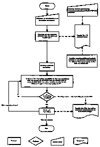
Fig. 10 shows an exemplary flow diagram of the fundamental
sequence of automatic cell harvesting using the device.
Figure 1 shows an exemplary embodiment of a device for
removing a cell and/or cell culture. The device includes a
microscope unit 1 with a number of optical components, in
particular an arrangement comprising deflecting prisms la and
a lens system lb for beam guidance and microscopic imaging.
The microscope unit 1 is coupled to an image recording unit 2,
usually a CCD camera or a CCD array. An image evaluation unit
3a is provided for processing the image information read out
from the image recording unit 2. The image processing unit 3a
consists of a personal computer 3 with image processing
software running thereon. A control and storage unit 4 is
furthermore provided, which is integrated in the personal
computer 3 and whose functions are implemented by further
software components. The control and storage unit 4 comprises
a monitor or a display 4a.
AVI-22873 PCT- Fassung-e.cloc
17.03.2009
CA 02664135 2009-03-20
17
The device furthermore contains a harvesting module 5 which is
mounted on a displacement mechanism. The displacement
mechanism consists of a lifting column 5a and a displacement
drive 5b. The lifting column 5a and the displacement drive 5b
are designed for larger displacement distances and are used
for bringing a harvesting module 5 towards a cell culture 8
located in a sample container, coarse adjustment of the
harvesting module 5 and movement of a removal tool 10a towards
the corresponding separating stations of the removed cells
and/or cell colonies.
The aforementioned microscope unit 1 is configured as a
transmission microscope. For this purpose, illumination 6 with
a series of switchable illumination filters 7 is provided. The
illumination 6 transilluminates the cell culture 8 located in
the sample container. The cell culture 8 is fixed on a support
in the form of an xy-table 9 by which means the cell culture 8
can be moved with a microscopic adjustment accuracy of a few
micrometers both in the x and in the y direction below the
optical arrangement consisting of illumination 6 and
deflecting prism la located thereunder. In this case, the
adjusting coordinates of the xy table 9 are transmitted to the
storage and control unit 4 or adjusted by the storage and
control unit 4.
The microscope unit 1 consists of a commercially available
microscope stand which is equipped with a motorised xy table
9. Optionally, this microscope unit 1 can also be equipped
with a commercially available fluorescence device. The
fluorescence device can accommodate up to 3 filter cubes
(consisting of excitation filter, dichroic mirror and emission
filter) and can be illuminated either by means of a
commercially available gas burner or by means of external
AVI-22873 PCT- Fassung-e.doc
17.03.2009
CA 02664135 2009-03-20
18
illumination 6 which is coupled in with glass fibres. It is
also possible to place the emission filter into a motorised
filter wheel before the illumination 6 in order to then
simultaneously scan fluorescences with corresponding triple or
quad-band filter cubes in the fluorescence device. In
addition, the image recording unit 2 with CCD chip is mounted
on the microscope unit 1 by which means scanning of the sample
is possible. By using commercially available phase contrast
sliders, physically optimum phase contrast illumination is
possible.
The commercially available PC is connected to the basic device
via a network connection. Running on this is commercially
available standard image processing software, which together
with a specially programmed robot controller and specially
developed modules for this image processing software, takes
over the driving of the device and the analysis of the image
data.
As will be explained in detail subsequently, in conjunction
with the movement of the xy table 9, the entire area of the
cell culture is scanned, whereby a number of microscopic
individual images of the cell culture are recorded by the
image detection unit.
The movement of the xy table also serves to position the cell
culture for the removal of the identified cells or cell
colonies. For this purpose, the harvesting module 5 is
positioned by the displacement mechanism above the cell
culture 8 whilst the xy table is adjusted to the previously
determined positions of the identified cells and cell colonies
and enables the harvesting module 5 to remove the cells or
cell colonies.
AVI-22873 POT- Fassung-e.doc
17412009
CA 02664135 2009-03-20
19
The removal of the cells or cell colonies from the cell
culture 8 located in the sample container requires a lowering
of a removal tool 10a into the cell culture 8, a picking of
the cells or cell colony and their separation. For this
purpose, the removal module 5 has a tool head 10 which is
fitted with a lowering or suction mechanism. The removal tool
10a is located at the end thereof. The picked cells or cell
colonies are deposited in a separating battery 11. This
consists of a row of test tubes or tubes which can be driven
individually by the lifting column and the displacement drive
and in which the removed cells and cell colonies can be
deposited by the tool head.
In addition, the separating battery can also be configured in
parts as a magazine for the preparation of removal tools 10a
which can be coupled onto the tool head 10 as desired, as will
be explained in detail subsequently.
In principle, the functions described here in principle are
controlled by the storage and control unit 4 and run
substantially fully automatically. However, due to the
monitoring of the functions on the monitor or display, the
user has a number of possibilities for influencing the
function by the known input means such as keypad and mouse and
a corresponding user interface at the software components
running inside the storage and control device.
Thus, in particular, an adjustment of the magnification factor
and a change in the resolving power of the image recording
device are possible by access to the control of the microscope
device. Furthermore, by controlling the illumination and the
illumination filters, spectral ranges can be modified or the
AVI-22873 PCT- Fassung-e.doc
17.03.2009
CA 02664135 2009-03-20
microscope device can be switched to fluorescence or dark
field operation.
Furthermore, it is possible to address the harvesting module 5
whereby individual cells or cell colonies determined by the
5 microscope unit can be selected in a menu-controlled manner
and allocated to a specified place in the separating battery
11. In addition, an operating mode of the harvesting module
can be selected in which, depending on the selected cells or
cell colonies, specified removal tools 10a can be taken from
10 the conical receptacle 32 of the tool head 10 in order to
remove the selected cells in a certain manner.
From this point, according to the separation method selected
in each case, different removal tools 10a now come into action
which can separate the particles and transfer them into a
15 corresponding target container with the aid of different
cannulas 15, 18 and/or capillaries and different sequences.
These methods are described in detail hereinafter.
The removal tool 10a sits at the upper end of the drive
arrangement and can be changed freely according to the
20 application. Five different removal tools 10a can be used for
the applications "adherent cell colony harvesting with
enzyme", "adherent cell colony harvesting without enzyme by
scraping", "harvesting from agar", "harvesting from methyl
cellulose" and "harvesting from positionally fixed individual
cells". All these removal tools 10a have in common that the
application-specific software part is mounted directly in the
removal tool 10a so that when the removal tool 10a is put in
place, the PC software automatically executes the correct
application and brings the correct consumables to the feed
receptacle. For the structure used in some of the removing
AVI-22873 PCT- Fassung-e.doc
17.03.2009
0
CA 02664135 2009-03-20
21
tools 10a as tool head 10 reference is made to DE 10 2004 046
740. The description of the aforesaid individual applications
is made hereinafter.
All the applications have in common that the scanning and
analysis process takes place in the same way. The process only
differs according to the application and therefore according
to the removal tool 10a used during the harvesting or picking
of the particles.
Harvesting of adherent cell colonies by means of enzyme:
This method is used for the complete or partial detachment of
cell colonies which adhere to the bottom of sample containers
(with these types of cells, the adhesion is necessary for the
survival of the cells and their multiplication).
Following the process steps of scanning and detection of the
objects of interest for the user, described further below,
these are positioned for harvesting. The tool head according
to DE 10 2004 046 740 is prepared for harvesting by first
taking up a commercially available tip 12 made of plastic and
filling this tip 12 with an enzyme or solvent (possibly
temperature-controlled) optimised for the respective cell type
in order to then take up a cloning cup 13 according to DE 197
42 163 C2. The colony to be separated is enclosed with this
cloning cup 13 and by dispensing the enzyme or the solvent
from the tip 12 into the interior of the cloning cup 13 and
therefore onto the object concerned, corresponding rinsing
cycles and times of action and finally taking up the volume
inside the cloning cup 13 into the tip 12, the desired colony
is separated from the sample container and can then be
AVI-22873 PCT- Fassung-e.doc
17.03.2009
CA 02664135 2009-03-20
22
transferred into another sample container and further
processed and investigated there.
Figure 2 shows a first removal tool 10a for this purpose. A
tip 12 which can be filled with liquid, in particular a
solvent or an enzyme, is combined with a cloning cup 13. The
tip 12 shown here comprises a tubular structure in the form of
a pipette or cannula which has an end cone 12a which is
inserted in a receiving cone 13a of the cloning cup 12 and
engages positively there. The tip 12 is expediently first
filled with liquid and receives the cloning cup 13 outside the
cell culture 8. The combination of tip 12 and cloning cup 13
thus formed is placed over the selected cells or cell colonies
in the cell culture 8. The liquid is then dispensed inside the
tip 12 into the cloning cup 13. The cells thereby detached are
then aspirated from the cloning cup 13 into the tip 12. Such a
removal tool 10a is particularly suitable for adherent cells
and cell colonies, i.e. those adhering to the bottom of a
vessel. The cloning cup 13 thereby covers a region of the cell
culture 8 stipulated by its radius. The radius of the cloning
cup 13 should be selected in this case depending on the
density of the cell population. A particular advantage of the
cloning cup 13 is that the relative positioning between
removal tool 10a and cell culture 8 which is executed by means
of the xy table 9 as mentioned, can be carried out with
comparatively limited accuracy.
A mechanical detachment of the cells can also be used for the
removal of adherent cells and cell colonies for which
experience shows that damage to the cell structures occurs
under the action of enzymes or solvents. For this purpose, the
selected cells or cell colonies are enclosed by the tip of a
cannula 15 and released from the base by scraping as a result
AVI-22873 PCT- Fassung-e.doc
17.03.2009
CA 02664135 2009-03-20
23
of a relative movement of cannula 15 and vessel. Depending on
the application or adherence strength, the cannula 15 consists
of various materials, for example, glass, plastic or metal and
has different inside diameters, wherein a plurality of
cannulas 15 in different designs can be held in readiness in
one magazine.
According to the requirements for sterility and throughput,
the cannula 15 can be changed manually or automatically. A
cannula magazine 14 is used for automatically changing the
cannula 15. Figure 3 shows a cannula magazine 14 with a number
of cannulas 15 located therein. This arrangement can be
configured as a part of the separating magazine 11 shown in
Fig. 1, reserved particularly for this purpose. An
interchangeable head 16 provided for this purpose has an
adapter 16a for grasping and withdrawing a cannula 15 from the
cannula magazine 14.
An interchangeable head 16 is moved over the cannula 15 and
lowered. This grips the cannula 15 and moves this over the
cell culture. A displacement of the xy table 9 to the position
of the selected cell or cell colony takes place there. The
cannula 15 is lowered and encloses the cell. The xy table 9
now executes slow oscillating movements whereby a negative
pressure is produced in the cannula 15 which aspirates the
cell.
Harvesting of strongly adherent cell colonies (stem cells,
cells on feeder cells, etc) by mechanical action:
This method is used for the complete or partial detachment of
cell colonies which adhere strongly to the bottom of sample
containers (with these types of cells, the adhesion is
AVI-22873 PCT- Fassung-e.doc
17.03.2009
CA 02664135 2009-03-20
24
necessary for the survival of the cells and their
multiplication).
The tool head 10 according to DE 2004 046 740 differs from the
previously explained tool head 10 by using a cannula 15 as
removal tool 10a. Depending on the application, the cannula 15
can consist of different materials (plastic, glass, metal) and
have different inside and outside diameters depending on the
size of the colonies to be harvested. Depending on the
requirements for sterility and throughput, the cannula 15 can
be changed manually (usually combined with a disinfection step
between the harvesting processes) or automatically (special
cannulas 15 are provided in racks similar to the tips 17).
Following the process steps of scanning and detection of the
objects of interest for the user, described further below,
these objects are positioned for harvesting. The colony to be
separated is enclosed with the cannula 15 (the end of the
cannula lies on the bottom of the sample vessel). The
detachment of the strongly adherent colony is effected
manually, by relative movements of the cannula 15 (scraping
and therefore displacement of the enclosed colony on the
bottom of the vessel), possibly in combination with aspiration
and rinsing processes of the syringe.
The relative movement is produced by moving the xy table 9,
the removal tool 10a or both in combination. The additional
use of cell-dissolving enzymes inside the cannula 15 is also
possible.
After detachment of the colony, this is taken up in the
cannula 15 and transferred to another container. The cannula
AVI-22873 PCT- Fassung-e.doc
17.03.2009
CA 02664135 2009-03-20
15 is now disinfected depending on the application or a new
cannula 15 is taken up. The next colony can then be harvested.
This tool head 16 with cannulas 15 was produced after problems
had arisen during the detachment and the time required for
5 this in the case of the aforesaid tool head 10 with tip 12 for
harvesting adherent cells by means of enzyme for various cell
types. The enzymes for detachment of the cells attack the cell
membrane. A too-high dosage or too-long time of action of the
enzyme, as is required for strongly adherent cells, frequently
10 leads to damage or destruction of the cells. However, the main
applications of the device lie in the separation of living
cells which are to be further cultivated and multiplied after
harvesting. Thus, a new automatable method was required for
these cell types in order to be able to separate these
15 strongly adherent cells.
By using cannulas 15 of different diameters and materials with
flat ends and a conical adapter according to DE 10 2004 046
740 which makes it possible to take up, dispense and magazine
the cannulas 15, and with the aid of corresponding devices
20 having automatic sequences as well as tool heads 10 for
picking up the cannulas 15 according to DE 10 2004 046 740,
harvesting could be carried out successfully on strongly
adherent cell types.
In addition to the use of cannulas 15, the use of relative
25 movements for the gentle detachment of individual cells and/or
colonies is a further feature of the invention. The relative
movement is either executed by the cannula 15 (movement of the
removal tool), the sample (movement of the xy table 9) or
both. Direction, travel and speed are determined according to
the respective cell type.
AVI-22873 PCT- Fassung-e.doc
17.03.2009
CA 02664135 2009-03-20
26
The selection of the diameters of the cannulas 15 is made with
reference to the size of the cells and/or colonies to be
separated. By using the conical receptacle 32 as adapter (see
Patent Application DE 10 2004 046 740), these highly varying
cannula sizes and materials can be handled with the same tool.
Special cannulas 15, primarily of smaller diameter or non-
metallic materials - can be glued into corresponding conical
adapters.
After depositing the cell in the separating magazine 11 and an
optional disinfection process, the cannula 15 can now be
deposited in the cannula magazine 14 and a new cannula 15
removed.
The method is described hereinafter with reference to feeder
cells:
The harvesting of colonies of feeder cells with picking of
feeder cells is effected by means of a scrape module. The
colony is completely enclosed by a metal capillary of the
scrape module or a part of the colony is stamped out by the
metal capillary. For this purpose, the metal capillary is
placed on the bottom of the culture dish during harvesting.
Feeder cells surrounding the colony or feeder cells located
under the colony are detached and picked by means of a scrape
movement. They are deposited in the target well. This is
usually not perturbing since the feeder cells no longer divide
and die after some time.
The harvesting of colonies of feeder cells without picking
feeder cells is effected with the glass capillary. For this
purpose, the upper region of the colony is picked by means of
aspiration at a distance of 0-50 pm above the target colony.
AVI-22873 PCT- Fassung-e.cloc
17.03.2009
CA 02664135 2009-03-20
27
Since the colony is a three-dimensional object, the aspiration
forces only act on the upper region of the colony facing away
from the bottom of the dish and not on the edge zones or
regions outside the colony. The size and depth of the piece to
be picked is thereby specified by the diameter of the
capillary, the distance from the colony, the amount of
aspiration and the aspiration speed and must be determined
empirically for each cell type. As a result, only cells or a
part of the colony are harvested without the surrounding
feeder cells or those located thereunder. It is furthermore
possible to harvest several clonal (genetically identical)
pieces of a colony by repeated picking at the same location.
For this purpose, the distance of the capillary tip from the
colony must be re-adjusted each time to always produce the
same aspiration force.
Harvesting of colonies from semi-solid nutrient substrates
(agar, methyl cellulose):
This method is used for the complete or partial removal of
cell colonies located on the base or inside the nutrient
media.
Following the process steps of scanning and detection of the
objects of interest for the user, described further below,
these are positioned for harvesting. The tool head 10
according to DE 10 2004 046 740 is prepared for harvesting by
picking up a special plastic tip 17 which is characterised by
a larger inside diameter at its tip in relation to its picking
volume compared with commercially available plastic tips (was
previously shortened). This tip 17 is positioned over the
colony to be separated or the colony is enclosed by said tip.
By aspirating the nutrient medium in the vicinity of the
AVI-22873 PCT- Fassung-e.doc
17.03.2009
CA 02664135 2009-03-20
28
colony or the included content of the special tip 17, the
colonies are taken up with the nutrient medium and can then be
transferred to another sample container and further processed
and studied.
Agar:
When harvesting from agar, insertion into the colony
surrounded by the agar is frequently sufficient so that
particles adhere to the tip 17 of the removal tool 10a and
this is then rinsed off in the target well which is filled
with nutrient medium. A special tool for this application then
requires n syringe drive for aspirating the particles.
Figure 4 shows two exemplary tips 17 with apexes having an
expanded inside diameter for removing cells from semi-solid
nutrient substrates, especially agar or methyl cellulose. The
tip 17 expediently consists of glass or plastic. It is
positioned over the previously selected cell and lowered,
whereby the cell or cell colony is enclosed in the apex of the
tip. The nutrient medium together with the cell or cell colony
contained therein is then picked up by means of the expanded
tip and can be transferred to the separating magazine.
Harvesting of positionally fixed individual cells:
This method is used for removing individual cells or small
cell colonies which are located on the bottom of the sample
container and remain largely positionally fixed there but
exhibit no or only minimal adherence.
Following the process steps of scanning and detection of the
objects of interest for the user, described further below,
AVI-22873 PCT- Fassung-e.doc
17.03.2009
CA 02664135 2009-03-20
29
these objects are positioned for harvesting. The tool head 10
according to DE 10 2004 046 740, however, does not take up a
plastic tip for this application but a capillary. This is
filled with air or a fluid via the connected syringe drive
depending on the cell type and application requirements,
calibrated by means of image processing and thus prepared for
the harvesting process.
The capillary opening (different diameters depending on cell
and colony size) is positioned over the cell or colony to be
separated. By aspirating the nutrient medium or buffer in the
vicinity of the individual cell or colony, the desired cell or
colony is picked up with the medium and can then be
transferred to another sample container and further processed
and investigated.
Figure 5 shows a cannula 18 for removing positionally fixed
individual cells. Such a cannula 18 is suitable for removing
individual cells or cell colonies which are located on the
bottom of a sample container and remain there in a
positionally fixed manner but not adherently. This is filled
with air or a fluid, the fluid level 18a of the cannula 18
being recorded by an image acquisition system 19 and
calibrated. For removal of the cell or cell colony, the
opening of the cannula 18 is positioned over the cell or cell
colony. By aspiration of the nutrient medium or buffer over
the cell, these together with the medium enters into the
interior of the cannula 18 and can then be transferred. In
this case, the diameter of the cannula opening must be adapted
to the sizes of the cells.
This and in future other methods can be implemented by
supplementing the base platform of the device and its axial
AVI-22873 PCT- Fassung-e.doc
17.03.2009
CA 02664135 2009-03-20
system according to DE 2004 027 661 and using complete
microscope optics.
The process of cell detection and image processing will be
explained in detail in the following.
5 Firstly the user loads the feed receptacle with corresponding
target plates, consumables and liquids and equips the
microscope cross table with its starting plate in which the
cell cultures to be harvested are located. These plates can be
freely defined and calibrated in the image processing
10 software. These plates can then be scanned.
For scanning the table is moved in a pattern which corresponds
to the image section of the optical camera system. The content
of the complete plate can thus be scanned image by image.
After one of these individual images has been scanned, a
15 particle detection takes place immediately based on grey
threshold values (and therefore on brightness differences).
Corresponding mathematical filters can be used before this
detection in order, for example, to optimise contrast or
prepare the image for better detection. This detection is made
20 image by image, i.e. during scanning. In this case, edge-
overlapping particles are automatically identified by the
software and combined to form one particle. This type of
detection is therefore also designated as edge-overlapping
detection. As a result, primarily only a so-called particle
25 map remains which shows in binary form where identified
particles are located and where not. Thus, image data need not
be held expensively in the memory but merely a map of the
detection result. Optionally, a reduced-size overview image
can be produced and stored. In addition to the already-
30 mentioned filters for image processing, further filters can be
AVI-22873 PCT- Fassung-e.doc
17.03.2009
CA 02664135 2009-03-20
31
used after the detection. Thus, the identified particles can
be evaluated and filtered out with regard to their
morphological (shape, size etc.) and qualitative parameters
(density, brightness differences etc.). This procedure has the
advantage that the analysis of the particles is made by
reference to the individual images recorded with 100%
resolution, not with a possibly quality-reduced overview
image.
All the remaining particles are output to a particle list and
can be individually approached, evaluated and reprocessed by
the user.
Figure 6 shows schematically the cell culture 8 in the left-
hand partial image. By means of a movement of the xy table 9,
the cell culture is scanned with a series of individual
microscope images 20. The size of the individual images
depends on the magnification factor set at the microscope unit
1. The smaller the magnification factor, the larger the
section of the cell culture 8 covered by the individual image
20, the smaller the number of individual images 20 required
for total recording of the cell culture 8 and the larger the
step movements to be executed by the xy table 9 in order to
bring the next-following image section under the microscope
unit.
It is accordingly necessary to match the step movements of the
xy table 9 with the magnification factor of the microscope
unit 1. This matching is effected by the memory and control
unit 4. In this case, each of the recorded individual images
20 is uniquely identifiable in its and y coordinates by the
position of the xy table 9. At the same time, the coordinates
given inside the individual images 20 of the image points
AVI-22873 PCT- Fassung-e.doc
17.03.2009
CA 02664135 2009-03-20
32
contained therein can simply be linked to the coordinates of
the individual image. As a result, each image point in each
individual image uniquely specifies a location in the scanned
cell culture 8.
By reference to the recorded individual images 20 of the cell
culture 8, the first and/or second detection step described
further below can be carried out to select the cells and/or
cell colonies according to specified parameters.
Alternatively to this, the individual images 20 thus recorded
are combined in the image evaluation unit 3a to form an
overview image 21 of the entire cell culture 8. This combining
is appropriate on the one hand because structures which have
been recorded at the edges of the respective individual images
are completed to form complete objects. On the other hand,
15 the overview picture 21 allows the user a stepless and
continuously executable overview of the scanned cell culture
8. The overview image 21 can be processed for this purpose by
image processing software and displayed to the user in various
resolution stages on the monitor 4a. In conjunction with the
20 generation of the overview image 21, a matching of the
coordinates of the individual images 20 and the coordinates of
the image points within adjoining individual images is carried
out in order to eliminate overlaps of the same image ranges.
The cell detection, i.e. the automatic identification of cells
or cell colonies within the individual images 20 is carried
out by means of a shape recognition explained hereinafter.
Figure 7 shows for this purpose an exemplary individual image
20, taken from the overview image 21, with cells 22 and a cell
colony 23 contained therein, in the microscope image. A
prerequisite for reliable shape recognition of the cells is a
AVI-22873 PCT- Fassung-e.doc
17.03.2009
CA 02664135 2009-03-20
33
sufficiently high contrast between the cells or cell colonies
and their background in the microscope image. This can be
achieved by various methods in microscopy. A first possibility
consists in focussing the optical system of the microscope
unit 1 onto the image plane of the cell culture 8 in which the
cells are to be expected. In the case of adherent cells, this
is the surface of the bottom of the sample vessel of the cell
culture. Cells in a semi-solid nutrient medium for example,
agar, are usually located on the surface of the agar and can
be focussed there.
In the case of cells or cell colonies which can be located
inside the cell culture in different image planes, techniques
involving the fluorescence labelling of microbiological
objects can be used. For this purpose, the cells to be
identified are marked with a fluorescence marker whilst light
having a corresponding excitation wavelength is irradiated via
the illumination system. During image acquisition in the area
of the fluorescence wavelength of the marker, the cells or
cell colonies are distinguished against a dark background as
luminous or light structures which form a sufficient colour
contrast.
Figure 8 shows a further schematic step of the image
processing. The left-hand diagram shows an individual image 20
converted into grey tones. Conversion into grey tones is
particularly advantageous when the cells stand out
sufficiently strongly from the background in the microscope
image. Naturally, emphasis of a single colour value of the
image or a reduction of the image to one colour value is also
possible. Likewise, the colour values of image points of the
cells to be detected can be predefined as reference, wherein
AVI-22873 POT- Fassung-e.doc
17.03.2009
CA 02664135 2009-03-20
34
each individual image point within the individual image 20 is
compared with this reference value.
Image points which correspond to predefined reference values,
i.e. colour values, grey levels, and similar values are
combined in the course of the image processing to form point
sets whose shape, size and outline can be analysed. The cells
22 are characterised, for example, by relatively large closed
shapes 24 which compactly enclose a specified region of the
image, the edges thereof running substantially smoothly. The
cell colony 23 on the other hand forms a set of individual
smaller closely adjacent structures 25 in an image region.
Both shapes can easily be identified within the scope of
commonly used image detection routines.
On the basis of the identified shapes, further image
processing steps are carried out in which the position of the
shapes and their mutual distances from one another are
calculated. The determination of the position is important for
the subsequent removal of the cells or cell colony, the
determination of the distance is necessary in order to specify
whether the cell or cell colony found must be removed by a
single removal process or together with other adjacent cells
of the cell culture.
Figure 9 shows an example for the determination of position
and distance. The diagram in Fig. 9 uses the result of the
image processing shown schematically in Fig. 8. In the example
shown here, a shape centre of gravity Si, S2 or S3 is
calculated for each set of image points pertaining to the
identified shape, i.e. cell or cell colony. The mode whereby
this calculation is made and the size of image section to be
selected for its calculations can be specified in advance by
AVI-22873 PCT- Fassung-e.cloc
17.03.2009
0
CA 02664135 2009-03-20
the user. As a result, it can be defined inter alia at what
point a group of identified cells should be treated as a cell
colony or a group of individual cells.
In the example shown here, its own shape centre of gravity Si
5 or S2 is calculated in each case for the individual shapes 24,
each being allocated an x coordinate xi or x2 and a y
coordinate yl or y2. For the closely adjacent shapes 25, a
shape centre of gravity S3 having coordinates x3 and y3 is
calculated, which applies to the entire set of these
10 structures and thus lies in an intermediate range of these
shapes. With the calculation of the shape centres of gravity
and the specification and storage of their coordinates, the
cells or cell colonies are uniquely identified in their
position. The coordinates are stored together with the image
15 data of the cells and cell colony in a position database and
can be uniquely located by retrieving the position database.
For determining the distances of the identified shapes and
therefore of the cells or cell colonies, use is made of the
determined coordinates, wherein the distances ISijI between
20 two arbitrary shape centres of gravity Si and Sj and their
coordinates (xi; yi) or (xj; yj) are calculated using the
Pythagoras relationship
Si.i1= 'Ajci 4)2 (Yi YA2
This calculation can be made automatically as part of the
image processing if the determined cells or cell colonies can
be found close to one another within previously specified
limits. In addition, it is naturally possible for a manual
distance calculation to be made by the user as part of editing
AVI-22873 PCT- Fassung-e.doe
17.03.2009
CA 02664135 2009-03-20
36
the position database. In this case, means for an interactive
graphical user guidance and image editing are used on the
software side in which in particular individual cells can be
marked by a mouse click and then the distance between the
marked cells is calculated by the image processing program.
The standard image processing software used has extensive
possibilities for further documentation of the detection
images and results such as graphical evaluations, report
generator etc. A continuous documentation of the process is
therefore possible.
In a second detection step, it is possible to study the
particles already identified with regard to further
parameters. In this analysis, the complete starting plate is
no longer scanned but only the regions where interesting cell
material was found in the first analysis step. This optimises
the execution time and in the case of a fluorescence analysis,
also the illumination time of the sample with fluorescence
light, which should be kept to a minimum to prevent bleaching
of the sample. The second detection step can consist of an
arbitrary number of individual steps, each of which can be
taken up with other types of exposure. Thus, many different
fluorescence channels and excitation wavelengths can be
scanned and evaluated in this second detection. The software
in this case only collects particle data, which are less data-
intensive than image data.
At the end of this second detection step, all the data
obtained are inserted in the already available particle list
for the corresponding original particles. As a result, a
corresponding overlapping effect is obtained so that the data
from the first analysis can be compared with the data of the
AVI-22873 PCT- Fassung-43.doc
17.03.2009
CA 02664135 2009-03-20
37
second analysis for the same particle. Thus, for example, the
quotient of fluorescence area (area, second analysis) divided
by the area of the bright-field particle (area, first
analysis) yields a quality feature for the antibody production
(the lighter and larger the fluorescence signal for the
smaller bright field colony, the more this colony produces
antibodies).
This particle list can furthermore be filtered logically by
means of the presence or the absence of individual
fluorescence or bright-field signals. Moreover, the user has
the possibility of filtering particles by means of a two-
dimensional scatter diagram. In this case, two different
particle values from the result table are imaged with respect
to one another.
When the table is completely filtered, this list can either be
harvested automatically or individually with line accuracy.
The AVISO robot control software is responsible for carrying
out the actual harvesting process which starts the process in
response to a signal from the image processing software. The
image processing software in this case takes over the
positioning of the cross table and ensure that in each case,
the next object to be harvested is located exactly centred in
the field of view of the camera so that the harvesting tool
which had previously been calibrated to this position,
encounters the cells and can pick them.
Communication between the two software components takes place
bidirectionally. This includes the image processing software
sending information such as particle diameter or position
index when using a multi-well plate to the robot control
before beginning the harvesting process. As a result, the
AVI-22873 PCT- Fassung-e.doc
17.03.2009
CA 02664135 2009-03-20
38
sequence control is put into a position where the harvesting
process can be carried out flexibly for the respective
harvesting candidates. A typical application here is the use
of automatically changeable cannulas and/or capillaries of
different shape, size and material to take into account the
different sizes of the particles and achieve optimum
harvesting results. It is also possible to have harvested
cells deposited in the dispensing container sorted according
to classes (density, fluorescence, ...) which can be
enormously valuable for downstream processes. Independently of
this, after each harvesting process the image processing
software obtains the information on which container and at
which position in this container the robot controller has
deposited the cell(s), thus ensuring complete logging of the
entire process.
Particles whose size exceeds the usable cannula diameter can
be successively harvested in parts. Dissolving out only parts
of a colony (e.g. undifferentiated stem cells surrounded by
differentiating ones) is also possible.
The colony, partial colony or individual cell to be harvested
in each case is positioned by the microscope cross table in
the optic axis of the microscope. An image of the particle
before harvesting is recorded and stored. The same takes place
after harvesting as evidence of the successful harvesting.
Both images are allocated to the corresponding well of the
target vessel for this particle so that precisely this
particle in this well is documented for the further
investigation.
The coordinates of the cells or cell colonies are now used for
removing the cells. For this purpose, the harvesting module is
AVI-22873 PCT- Fassung-e.doc
17.03.2009
a
CA 02664135 2009-03-20
39
moved over the cell culture 8 as described, whereby the xy
table 9 approaches each coordinate and lowers the harvesting
module 5 of the tool head 10 with the removal tool 10a at the
corresponding coordinate and activates the cell removal.
Figure 10 shows the fundamental sequence of the automated cell
harvesting using the device. At the beginning of the process,
a definition of the identification and harvesting parameter
takes place. By reference to these parameters, the scanning
process is then carried out with simultaneous identification
of the cells and/or cell colonies.
The data determined therefrom are stored in a results list
with cells and/or cell colonies found. The cells and/or cell
colonies found can be further filtered from this results list.
This is effected with by a manual reprocessing, i.e.
removing/adding/separating/combining of cells and/or cell
colonies or by applying additional analysis functions to the
cells and/or cell colonies found. For example, an examination
for different fluorescences can be made.
After the cells and/or cell colonies have been selected in
this way, the automatic harvesting can be started. For this,
it must firstly be analysed whether the cell and/or cell
colony can be harvested with the present removal tool on
account of its size and whether other cells and/or cell
colonies are located in the withdrawal region of the removal
tool. If harvesting is possible, this is then carried out. The
result of the harvesting is stored in the results list of the
harvesting including documentation of the before/after images.
Then, the analysis of the harvesting possibility described
above is carried out anew for the next cell and/or cell
colony, if possible followed by harvesting.
AVI-22873 PCT- Fassung-e.doc
17.03.2009
r"*
CA 02664135 2009-03-20
If harvesting is not possible, the reason for this is firstly
determined and the result is stored in the results list of the
harvesting including documentation of the before/after images.
This is again followed by the analysis described above for the
5 next cell and/or cell colony, if possible followed by
harvesting.
After harvesting the last cell and/or cell colony, the process
is automatically ended.
Figure 11 shows a receptacle 32 which is attached to the tool
10 head 10. The receptacle 32 comprises an outer cone 26 which is
graded in steps 28. This outer cone 26 receives a removal tool
10a which is a tip here and has an inner cone 27 which forms a
counterpart to the outer cone 26 of the receptacle 32. Due to
the meshing of the outer cone 26 of the receptacle 32 and the
15 inner cone 27 of the removal tool 10a, a non-positive
detachable connection is formed between the receptacle 32 and
the removal tool 10a.
Figure 12 shows an alternative embodiment of a receptacle 32.
The receptacle 32 here comprises an inner cone 31. This inner
20 cone 31 receives a removal tool 10a which is a capillary here
and comprises an outer cone 30 which forms a counterpart to
the inner cone 31 of the receptacle 32. Due to the meshing of
the inner cone 31 of the receptacle 32 and the outer cone 30
of the removal tool 10a, a non-positive detachable connection
25 is formed between the receptacle 32 and the removal tool 10a.
By using different conical receptacles 32, matched in their
size and design, for the very diverse, frequently newly
developed consumables used, in the form of exchangeable
cannulas, a largely uniform tool geometry can be retained.
AVI-22873 PCT- Fassung-e.doc
17.03.2009
CA 02664135 2009-03-20
41
The conical receptacle 32 results in self-centring of the
consumables (cannulas) partially to be positioned with high
precision when received by the tool. Due to the high stability
of the conical receptacle 32 and the uniform force
distribution, the application of relatively high transverse
forces to the exchangeable capillaries is possible, e.g.
during scraping without loss of positioning accuracy or
loosening of the exchangeable cannulas in the receptacle 32.
By using a thickening below the conical receptacle 32 on the
tool head, the exchangeable cannulas can be removed from the
tool again by means of a simple stripping device, not shown,
to allow receipt of the next exchangeable cannula.
At the same time, this collar also serves to allow the
magazining of even sensitive consumables such as, for example,
small-diameter glass capillaries, to feed these in large
numbers to the automated process. Usually racks of 96 are used
for this purpose.
It is possible to use various conical receptacles 32 for
adapting a wide range of automatedly exchangeable capillaries
and cannulas of very diverse size, shape and materials on a
tool of the same type for the purposes of the automated
investigation and separation of individual cells or cell
colonies.
Capillaries of glass, metal or ceramic can be used.
AVI-22873 PCT- Fassung-e.cloc
17.03.2009
CA 02664135 2009-03-20
42
Reference list
1 Microscope unit
la Deflecting prism
lb Lens system
2 Image recording unit
3 PC
3a Image evaluation unit
4 Control and memory unit
4a Monitor, display
5 Harvesting module
5a Lifting column
5b Displacement drive
6 Illumination
7 Illumination filter
8 Cell culture
9 xy table
10 Tool head
10a Removal tool
11 Separating battery
. 12 Tip
12a End cone
13 Cloning cup
13a Picking cone
14 Cannula magazine
15 Cannula
16 Exchangeable head
16a Adapter
17 Tip
18 Cannula for positionally fixed individual cell
18a Fluid level
19 Calibrating image acquisition
AVI-22873 PCT- Fassung-e.doe
17.03.2009
CA 02664135 2009-03-20
43
20 Individual image
21 Overview image
22 Cell
23 Cell colony
24 Cell shape
25 Cell colony structure
Si First shape centre of gravity
S2 Second shape centre of gravity
S3 Third shape centre of gravity
xi First x coordinate
x2 Second x coordinate
x3 Third x coordinate
yl First y coordinate
y2 Second y coordinate
y3 Third y coordinate
26 Outer cone
27 Inner cone
28 Step
30 Outer cone
31 Inner cone
32 Receptacle
AVI-22873 PCT- Fassung-e.doc
17.03.2009