Note: Descriptions are shown in the official language in which they were submitted.
CA 02666884 2009-04-17
WO 2008/051437 PCT/US2007/022237
DEVICE FOR AUTOMATED NEEDLE DEPLOYMENT
FIELD OF THE INVENTION
The present invention generally relates to medical systems and devices for
suturing
internal tissue walls, and more particularly to a device for automated needle
deployment and
to a method of using such device.
BACKGROUND OF THE INVENTION
Various medical procedures, particularly cardiology procedures, involve
accessing a
corporeal vessel through the formation of a hole or opening in the vessel wall
so that a
medical procedure can be performed. After the particular medical procedure has
been
perfonned, the access hole in the vessel wall must be closed.
A number of prior vascular closure devices and methods have been developed in
attempt to provide a solution for the problem of closing a hole in the vessel
wall. Tissue
approximation typically involves passing a length of suture into and through
adjacent vessel
and subcutaneous tissue, across the vessel opening, and back into and through
adjacent vessel
and subcutaneous tissue. Certain prior closure devices have involved
relatively complicated
methods and devices for extracting a length of suture from inside the vessel
so that the
physician can approximate tissue surrounding the hole in the vessel wall
through use of the
suture.
U.S. Pat. No. 5,643,292 and U.S. Pat. No. 6,059,800 disclose example prior
suturing
devices used for approximating tissue surrounding the opening in a vessel
wall. Most prior
closure devices enlarge the vessel opening thereby negating the benefits of
using smaller or
less invasive percutaneous products. Prior suturing devices are also
relatively complicated
and difficult to use. Furthennore, many suturing devices dilate the vessel
opening and
perform the medical procedure via the vessel opening before the suture is
extended across the
vessel opening for approximation tissue surrounding the vessel wall.
In many prior art systems, needle deployment is done manually by a physician
or
operator. Manual deployment involves estimation by the operator of how the
needle should
be deployed, how fast the trigger for the needle should be actuated, how much
force should
be applied, etc. The manual method of needles deployment require the physician
to manually
pull a lever or button proximally to deploy the needles. The speed or force
used to actuate the
lever or button will determine the force the needle will have when penetrating
the artery. The
more force the needle have in penetrating the artery the greater the
possibility of piercing an
1
CA 02666884 2009-04-17
WO 2008/051437 PCT/US2007/022237
artery. Thus, the physician must exert sufficient force to penetrate the
artery but take care not
to exert so much force as to pierce the artery. Manual deployment allows for
greater
inconsistency and user error as different physicians have differing perception
when it comes
to how much force or speed to apply when using a device It would be
advantageous to have a
device for automated needle deployment that reduces operator estimation and,
thus, operator
error, and standardizes deployment of the needle.
BRIEF SUMMARY OF THE INVENTION
A device for automated needle deployment and a method of using such device is
disclosed. Medical systems and devices for suturing internal tissue walls that
include such
automated needle deployment device are further disclosed.
In one embodiment, the automated needle deployment device comprises a pusher,
a
needle, a tube, and an actuator. The pusher has a needle engaging end. The
needle has a
sharp end and an opposite end. A suture is associated with the needle. The
pusher and
needle are slidably disposed within the tube. The actuator comprises a control
and a spring
and is operatively associated with the pusher. Actuation of the actuator moves
the pusher
towards the needle expulsion end of the tube such that the needle engaging end
of the pusher
engages the needle and expels the needle from the tube.
Other features and advantages of the invention will become apparent from the
following detailed description, taken in conjunction with the accompanying
drawings which
illustrate, by way of example, various features of embodiments of the
invention.
BRIEF DESCRIPTION OF THE DRAWINGS
Figure la illustrates an automated needle deployment device in a closed
configuration
in accordance with one embodiment.
Figure lb illustrates an automated needle deployment device in a partially
open
configuration in accordance with one embodiment.
Figure 1 c illustrates an automated needle deployment device in an open
configuration
in accordance with one embodiment.
Figure 1 d illustrates an automated needle deployment device in a needle
deploying
configuration in accordance with one embodiment.
Figure 2a illustrates a needle having a suture crimped thereto in accordance
with one
embodiment,
Figure 2b illustrates an alternative view of the needle and suture of Figure
2a.
2
CA 02666884 2009-04-17
WO 2008/051437 PCT/US2007/022237
Figure 3a illustrates a suturing system of the automated needle deployment
device, the
suturing system in a closed configuration, in accordance with one embodiment.
Figure 3b illustrates a suturing system of the automated needle deployment
device,
the suturing system in an open configuration, in accordance with one
embodiment.
Figures 4 illustrates an actuator of the automated needle deployment device,
the
actuator having a compression spring and the compression spring being in an
extended
configuration, in accordance with one embodiment.
Figure 5 illustrates the actuator of Figure 4 with the compression spring in a
relaxed
configuration.
Figure 6 illustrates an actuator of the automated needle deployment device,
the
actuator having a flywheel, in accordance with one embodiment.
Figure 7 illustrates an actuator of the automated needle deployment device,
the
actuator having a torsion spring, in accordance with one embodiment.
Figure 8 depicts a method of using the suture system of the present invention.
DETAILED DESCRIPTION OF THE INVENTION
A device for automated needle deployment and a method of using such device is
disclosed. Medical systems and devices for suturing internal tissue walls
included such
automated needle deployment device are further disclosed.. More particularly,
an automated
needle deployment device suitable for use with an internal tissue suture
delivery system for
performing medical procedures that include delivering needles and sutures to
internal tissue
for closing internal tissue walls after an opening or puncture in tissue has
been made is
provided. The automated needle deployment device may form a part of a needle
and suture
delivery unit. Tissue that may be closed in accordance with the teachings
herein may be part
of a lumen such as a blood vessel, body cavity, other organ, or any tissue
suitable for
suturing. In one example, vascular suture delivery systems such as disclosed
in copending
U.S. Patent Application No. 11/551,523, filed October 20, 2006, may be used to
deliver
needles and sutures for closing internal tissue walls after a medical
procedure is performed
through a vascular wall opening.
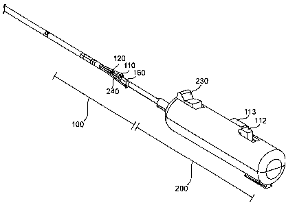
Figures la-ld illustrate one embodiment of a vascular closure delivery system
comprising a handle and needle and suture delivery unit 100. The handle
includes an actuator
201 for actuating an automated needle deployment device of the needle and
suture delivery
unit needle and suture delivery unitactuator 201 Figure la illustrates the
needle and suture
delivery unit in a closed configuration, Figures lb and 1 c illustrates the
needle and suture
3
CA 02666884 2009-04-17
WO 2008/051437 PCT/US2007/022237
delivery unit in a partially open and an open configuration, respectively, and
Figure ld
illustrates the needle and suture delivery unit in a needle deploying
configuration. The needle
and suture delivery unit 100 comprises a needle 140, a pusher 130, and a
needle carrier tube
120. The needle 140 and pusher 130 are provided within the needle carrier tube
120 when
the needle and suture delivery unit 100 is in a closed configuration, shown in
Figure 1 a. A
suture 150 is provided with the needle 140. In various embodiments, more than
one pusher
130 and needle 140 may be associated with an actuator 201. In the embodiment
of Figures
la-ld, four pushers 130 and four needles 140 are provided. Further description
of Figures
1 a-1 d is provided below in regard to the suture delivery system. Reference
to distal and
proximal positions may be made herein. Generally, proximal refers to towards
the physician
or operator and distal refers to towards the patient. Such reference is for
the purposes of
illustration only and is not intended to be limiting, and orientations of the
various components
may be altered.
The handle 200 of the vascular closure delivery system is provided at a
proximal end
thereof and may be used to control the needle and suture delivery unit 100.
First, second, and
third actuators 113, 112, and 230 may be provided on the handle 200. The first
actuator 113
may be provided on the handle 200 for actuating the legs 110 from a collapsed
position to an
operational and open position. The second actuator 112 deploys the needle by
deploying the
actuating members.. A third actuator 230 retracts the actuating members after
needle
deployment.
The needle 140 may be constructed of implant grade stainless steel, a
dissolvable
polymer, a bioresorbable material, or other material suitable for engaging
with tissue. The
needle 140 includes a sharp end and an opposite end. In one embodiment, the
face of the
opposite end is approximately perpendicular to a central axis of the needle.
In alternative
embodiments, the opposite end to the sharp end may have different
configurations. The
suture 150 may be associated with or coupled to the needle 140 in any suitable
manner and at
any suitable location. For example, the suture 150 may be threaded through the
needle 140,
adhered to the needle 140, crimped to the needle 140, injection molded into a
needle 140, or
other. In one embodiment, shown in Figures 2a and 2b, the suture 150 is
crimped to the
needle 140 generally between the sharp end of the needle 140 and the opposite
end of the
needle 140. In alternative embodiments, apparatuses other than needles may be
provided for
placing the suture 150. For example, a pronged projectile or other suitably
shaped projectile
for engaging with tissue may be provided.
4
CA 02666884 2009-04-17
WO 2008/051437 PCT/US2007/022237
In embodiments where the pusher 130 and needle 140 are provided in a needle
carrier
tube 120, the pusher 130 expels the needle 140 from the carrier tube 120.
Thus, the needle
carrier tube 120 has a needle expulsion end from which the needle 140 is
expelled to deploy
the needle 140 and suture 150. The needle expulsion end may be the distal end
of the needle
or the proximal end of the needle in various embodiments. In the embodiments
shown, the
needle expulsion end of the needle carrier tube 120 is the proximal end of the
needle carrier
tube 120. The pusher 1301ikewise has a needle engagement end. The needle
engagement
end of the pusher 130 is the end of the pusher 130 that engages the needle 140
to expel the
needle 140 from the needle carrier tube 120. The pusher 130 may be grounded
and/or the
needle engagement end of the pusher 130 may have adaptive features to enable
coupling with
the needle, described more fully below. The needle engagement end of the
pusher 130 may
be the proximal end of the pusher or the distal end of the pusher in various
embodiments. In
the embodiments shown, the needle engagement end of the pusher 130 is the
proximal end of
the pusher 130. Thus, the needle engagement end of the pusher 130 engages the
needle 140
to expel the needle 140 from the needle expulsion end of the needle carrier
tube 120. More
specifically, in the embodiments shown, the proximal end of the pusher 130
engages the
needle 140 to expel the needle 140 proximally from the proximal end of the
needle carrier
tube 120.
In one embodiment, the needle 140 is positioned in the carrier tube 120 such
that the
sharp end of the needle 140 is oriented toward the needle expulsion end of the
carrier tube
120 and the opposite end of the needle 140 is oriented toward the needle
engagement end of
the pusher 130. In this embodiment, the needle 140 is delivered from the
needle carrier tube
120 sharp end-first. Generally, the needle 140 engages with tissue after it is
fully delivered
from the tube 120. Once the needle 140 engages with tissue, such as by
embedding in tissue,
it is substantially prevented from re-entering the tube 120.
The pusher 130 may have any suitable configuration for engaging the needle
140. As
previously discussed, the needle engagement end of the pusher 130 may have
adaptive
features to enable the needle engagement end of the pusher 130 to engage the
needle 140 or
to couple with the needle 140. As shown, the pusher 130 comprises a rod-like
structure
wherein the needle engagement end of the pusher 130 is configured to be
received by the
opposite end of the needle 140 such that the needle 140 is carried by the
pusher 130. The
pusher 130 may be solid or hollow or a combination thereof. In the embodiment
shown, the
pusher 130 has a generally circular cross section. In other embodiments, the
cross section of
CA 02666884 2009-04-17
WO 2008/051437 PCT/US2007/022237
the pusher 130 may be varied. The pusher 130 is configured and positioned in
the needle
carrier tube 120 for movement towards an expulsion end of the needle carrier
tube 120 to
expel the needle 140 therefrom. Such movement is in response to triggering,
directly or
indirectly, of the actuator 201, described more fully below. The combination
of the length of
the pusher 130 and the distance the pusher 130 moves may result in the needle
engagement
end of the pusher 130 moving through and out of the tube 120. In some
embodiments, the
pusher 130 may exit the tube 120 partially or not at all. The needle
engagement end of the
pusher 130 may be the distal end of the pusher 130 or the proximal end of the
pusher 130.
As shown in Figures 1 a-1 d, the pusher 130 is located at the distal end of
the needle
and suture delivery unit 100 and pointed towards the proximal end of the
needle and suture
delivery unit 100 such that the pusher 130 pushes the needle 140 proximally
for engagement
with tissue. In this embodiment, the needle engagement end of the pusher 130
is the
proximal end of the pusher. In alternative embodiments, the pusher 130 may be
located at the
distal end of the suture assembly or between the proximal end and distal end
of the suture
assembly with the pusher 130 pointed towards the distal end of the needle and
suture delivery
unit 100 such that the pusher pushes the needle distally for engagement with
tissue. In this
embodiment, the needle engagement end of the pusher 130 is the distal end of
the pusher 130.
Similarly, the expulsion end of the needle carrier tube 120 may be the
proximal end of the
needle carrier tube 120 or the distal end of the needle carrier tube 120. In
the embodiment of
Figures la-ld, the expulsion end of the needle carrier tube 120 is the
proximal end.
When a needle 140 is provided at the needle engagement end of the pusher 130,
the
pusher 130 expels the needle 140 from the tube 120 as the needle engagement
end of the
pusher 130 moves towards an exit point or expulsion end of the tube 120. After
expulsion of
the needle 140, the pusher 130 may be retracted back into the tube 120.
The pusher 130 may be configured to push the needle 140 from the needle
carrier tube
120, for example by contacting the opposite end of the needle 140 with the
needle
engagement end of the pusher 130 and pushing it out of the needle carrier tube
120.
Alternatively, as shown in Figure 3b, the pusher 130 may be configured for
carrying the
needle 140 out of the needle carrier tube 120. In this embodiment, the cross
section of the
pusher 130 complements the cross section of the needle 140 and is smaller than
the cross
section of the needle 140. Further, the opposite end of the needle 140 is at
least partially
hollow such that it may receive the needle engagement end of the pusher 130.
Thus, the
needle 140 receives the needle engagement end of pusher 130 at the opposite
end of the
6
CA 02666884 2009-04-17
WO 2008/051437 PCT/US2007/022237
needle 140. The pusher 130 then may be moved to project from the needle
carrier tube 120
and thus carry the needle 140 from the needle carrier tube 120.
Thus, the pusher 130 and needle 140 may be slidably disposed within the needle
carrier tube 120 such that the pusher 130 moves therein to expel the needle
140 therefrom,
either by pushing the needle 140 from the needle carrier tube 120, by carrying
the needle 140
out of the needle carrier tube 120, or other. The needle carrier tube 120 may
have any
suitable cross section for slidably receiving the pusher 130 and the needle
140. For example,
the needle carrier tube 120 may have a circular cross section or a square
cross section. In the
embodiments shown, the needle carrier tube 120 has a circular cross section.
Returning to
Figures 2a and 2b, the needle carrier tube 120 may have a slot 122 to allow
loading of a
needle 140 having a suture 150 coupled thereto at between the sharp end of the
needle and
the opposite end of the needle. In alternative embodiments, for example, where
the suture
150 is coupled to the needle 140 at the opposite end thereof, no slot may be
provided in the
needle carrier tube. The needle 140 within the needle carrier tube 120 may be
provided at a
needle engagement end of the pusher 130. The needle 140 may be oriented in the
tube 120
for expulsion sharp-end first or sharp-end last.
The suture 150 may be composed of a variety of materials such as nylon, a
bioresorbable or nonresorbable suture material, metal wire, or any suitable
suture material.
The suture 150 may be braided. One or more sutures may be associated with each
needle 140
or other projectile of the needle and suture delivery unit 100. Thus, at least
one end of the
suture 150 is associated with a needle 140. Initially, the length of the
suture 150 is of a
length such that the suture 150 extends from the needle 140 as engaged with
the tissue, out of
the tissue of the patient, and toward the delivery unit handle. A portion of
the suture 150 may
be disposed in the tube 120, trailing from the needle 140, before the needle
140 is delivered
to tissue.
Movement of the pusher 130 towards the needle expulsion end of the needle
carrier
tube 120 is triggered by the actuator 201 of the handle 200. The actuator 201
uses a triggered
force to automatically deploy the needle 140 via movement of the pusher 130,
for example
with the push of a button. Such triggered force may be a spring force, a
pneumatic force, a
magnetic force, or other force. For the purposes of illustration, a spring
force is herein
described.
As shown in Figures 4-7, the actuator 201 comprise a spring 210 and a control
220
The spring is pulled to store energy. Thus, the control 220 keeps the spring
210 in tension
until actuation is desired. The control 220 is coupled directly or indirectly
to actuating
7
CA 02666884 2009-04-17
WO 2008/051437 PCT/US2007/022237
member(s) 240. The actuating member(s) 240 is coupled to the pusher(s) 130.
Such
coupling may be done in any suitable manner. In one embodiment, the actuating
member(s)
240 is crimped to the pusher(s) 130. As shown in Figure 3b, a single actuating
member 240
may be operatively associated with a plurality, for example four, pushers 130.
The control
220 is released to cause automated deployment of the needle 140. Such
automated
deployment is via the actuating members 240 acting on the pusher 130 and the
pusher 130
acting on the spring 210. In some embodiments, the control 220 may act on the
pusher 130
without an intermediate actuating member. Regardless of whether an actuating
member 240
is provided, the pusher 130 is compelled to move towards the expulsion end of
the needle
carrier tube 120 by release of the spring 210, thus deploying the needle 140
from the needle
carrier tube 120. The direction of movement of the pusher 130 may be varied to
suit the
orientation of the automated needle deployment device. Thus, when it is
desired to deploy
the needles proximally from a distal position, as shown in Figures la-ld, the
pushers 130
move proximally. This may be done, for example, by exerting a pull force on
the pushers
130. Conversely, when it is desired to deploy the needles distally from a
proximal position,
the pushers move distally. This may be done, for example, by exerting a push
force on the
pushers 130. Depending on the type of contro1220 used, described below, the
pusher 130
(and actuating member 240 if provided) may automatically be retracted or may
be manually
retracted.
In one embodiment, shown in Figures 4 and 5, the control 220 is a control
lever and
the spring 210 is a compression spring. As shown in Figure 4, the compression
spring 210 is
kept in tension by the lever 220. The control lever 220 is released, for
example by pulling of
the control lever 220, shown in Figure 5, to release the spring 210 and move
the pusher 130
or actuating member. If an actuating member 240 is provided, the actuating
member 240 in
turn acts on the pusher 130. The pusher 130 is thus moved (directly by the
spring 210 or via
the actuating member 240) towards the expulsion end of the needle carrier tube
120 to deploy
the needle 140. In one embodiment, an actuating member 240 comprising a
nitinol wire is
provided. The actuating member 240 has first and second ends. The first end is
coupled to
the pusher 130 and the second end is coupled to the compression spring 210.
When the
control lever 220 is released, the spring moves to a released position that
moves the extended
end of the spring proximally. The proximal movement of the spring causes the
actuating
member 240 to move proximally, which in turn causes the pusher 130 to move
proximally.
The proximal movement of the pusher 130 causes the needle 140 to move
proximally towards
an expulsion end of the tube 120 such that the needle 140 is expelled from the
tube 120 in the
8
CA 02666884 2009-04-17
WO 2008/051437 PCT/US2007/022237
proximal direction. The actuating member 240 may be coupled to the pusher 130
at any
suitable location such that a pull force exerted on the actuating member 240
will exert a pull
force on the pusher 130. In one embodiment, the actuating member 240 is
coupled to the
pusher 130 proximate the needle engaging end of the pusher 130. Figure 5
illustrates the
spring 210 in a released state.
The control lever 220 may be released using any suitable mechanism. For
example, a
push button or release knob may be provided to release the control lever 220.
A second
mechanism, shown in Figure 4 as a push button 230, may be provided to retract
that actuating
members. A further spring may be provided that is actuated upon pushing of the
push button
130, actuation of the spring retracting the actuating member 240.
Alternatively, a single
mechanism may be provided to release the control lever and retract the
actuating members.
Such release and retraction may be done using separate actuations of the
mechanism.
Figure 6 illustrates an alternative embodiment using a flywheel control 220
and a
compression spring 210. The flywheel control 220 comprises a flywheel 222 and
a center rod
224. The center rod 224 is operatively associated with the pusher 130 or, if
provided,
actuating member. A release button 230 is provided for releasing the
compression spring
210, causing the rotation of the flywheel 22. Rotation of the flywhee1222 in
turn moves the
center rod 224 which causes movement of the actuating member 240 and/or pusher
130. In
one embodiment, the center rod 224 is coupled to an actuating member 240 such
as a nitinol
wire. The actuating member 240 thus is coupled at one end to the pusher 130
and at the other
end to the center rod 224. When the flywheel control 220 is released, the
flywheel 222
rotates, causing movement of the center rod 224 in the proximal direction. The
proximal
movement of the center rod 224 causes the actuating member 240 to move
proximally, which
in turn causes the pusher 130 to move proximally. The proximal movement of the
pusher
130 causes the needle 140 to move proximally towards an expulsion end of the
tube 120 such
that the needle 140 is expelled from the tube 120 in the proximal direction.
The actuating
member 240 may be coupled to the pusher 130 at any suitable location such that
a pull force
exerted on the actuating member 240 will exert a pull force on the pusher 130.
In one
embodiment, the actuating member 240 is coupled to the pusher 130 proximate
the needle
engaging end of the pusher 130.
With use of a flywheel 222, after movement towards the expulsion end of the
needle
carrier tube 120, the actuating member 240 and/or pusher 130 automatically
retracts into the
tube 120 as rotation of the flywheel 222 continues. Thus, half of the
revolution of the
flywheel drives the or actuating member 240 and/or pusher 130 towards the
expulsion end of
9
CA 02666884 2009-04-17
WO 2008/051437 PCT/US2007/022237
the needle carrier tube 120 and the other half of the revolution of the
flywhee1222 retracts the
actuating member 240 and/or pusher 130. Thus, a single mechanism, release
button 230,
controls release of the spring and retracting of the actuating member 240
and/or pusher 130.
An alternative flywheel embodiment is illustrated in Figure 7 showing a
torsion
spring. Thus, the contro1220 comprises a flywhee1222 and a center rod 224 and
the spring
210 comprises a torsion spring. Using a torsion spring, the spring is rotated
(rather than
pulled) to store energy. A release button 230 is provided for releasing the
compression spring
210, causing rotation of the flywhee1222. Rotation of the flywheel 22 in turn
moves the
center rod 224 which causes movement of the actuating member 240 and/or pusher
130.
Rotation of the flywhee1222 in turn moves the center rod 224 which causes
movement of the
associated pusher 130 or actuating member 240. In one embodiment, the center
rod 224 is
coupled to an actuating member 240 such as a nitinol wire. The actuating
member 240 thus
is coupled at one end to the pusher 130 and at the other end to the center rod
224. When the
flywheel contro1220 is released, the flywhee1222 rotates, causing movement of
the center
rod 224 in the proximal direction. The proximal movement of the center rod 224
causes the
actuating member 240 to move proximally, which in turn causes the pusher 130
to move
proximally. The proximal movement of the pusher 130 causes the needle 140 to
move
proximally towards an expulsion end of the tube 120 such that the needle 140
is expelled
from the tube 120 in the proximal direction. The actuating member 240 may be
coupled to
the pusher 130 at any suitable location such that a pull force exerted on the
actuating member
240 will exert a pull force on the pusher 130. In one embodiment, the
actuating member 240
is coupled to the pusher 130 proximate the needle engaging end of the pusher
130.
With use of a flywhee1222, after movement towards the expulsion end of the
needle
carrier tube 120, the actuating member 240 and/or pusher 130 automatically
retracts into the
tube 120 as rotation of the flywheel 222 continues. Thus, half of the
revolution of the
flywheel drives the actuating member 240 and/or pusher 130 towards the
expulsion end of the
needle carrier tube 120 and the other half of the revolution of the
flywhee1222 retracts the
actuating member 240 and/or pusher 130. Thus, a single mechanism, release
button 230,
controls release of the spring 210 and retracting of the actuating member 240
and/or pusher
130.
Returning to Figures 1 a-1 d, suture delivery systems capable of delivering
needles and
sutures to the tissue are provided. The suture delivery system includes the
needle and suture
delivery unit 100 and the actuator 201. The actuator 201 may be provided as
part of a handle
200 for controlling the needle and suture delivery unit 100. In the embodiment
shown, the
CA 02666884 2009-04-17
WO 2008/051437 PCT/US2007/022237
needle and suture delivery unit 100 includes one or more pushers 130, needles
140, sutures,
and legs 110, and is disposed at a distal end of a delivery unit. It is to be
noted that, while
four sets of legs 110, needle carrier tubes 120, pushers 130, and needles 140
are shown, in
alternative embodiments, more or fewer sets of legs, pushers, needle carrier
tubes, pushers,
and needles may be used. Further, the number of legs, pushers, needles, and
sutures may not
be equal. The needles 140 and sutures may, more particularly, be delivered to
the intima of
an artery such as the femoral artery. The needle and suture delivery unit 100
is at least
partially insertable into tissue, such as the artery, so that one or more
needles and sutures may
be delivered to the internal tissue of the patient. A tube or sheath may be
provided and may
serve as a cover for all or a portion of the needle and suture delivery unit
100. The sheath
may be pulled back or peeled away to expose the distal end of the needle and
suture delivery
unit 100.
Figure 1 a illustrates the automated needle deployment device in a closed
configuration, Figures lb and lc illustrate the automated needle deployment
device in a
partially open and an open configuration, respectively, and Figure 1 d
illustrates the
automated needle deployment device in a needle deploying configuration. Figure
1 c shows
the needle and suture delivery unit 100 with the legs 110 in an open position,
lifting the
needle carrier tubes 120, and Figure 1 d shows the needle and suture delivery
unit 100 with
the pushers 130 and needles 140 extending from the needle canier tubes 120.
The leg 110 of the needle and suture delivery unit 100 serves as a guide for
the tube
120. More specifically, the leg 110 moves the tube 120 from the closed
configuration shown
in Figure 1 a to the open configuration shown in Figure 1 c such that the
pusher 130 may expel
the needle 140 from the tube 120, as shown in Figure 1 d. A lever 113 may be
provided on
the handle 200 for opening the legs. In the embodiment shown, such opening
comprises
pulling the legs proximally, as described below. The leg 110 may be
constructed of stainless
steel, a polymer, or any material suitable for medical devices. Reference is
made to
copending U.S. Patent Application Nos. 11/551,523 filed October 20, 2006; and
11/551,612,
filed October 20, 2006, herein incorporated by reference, for specifics
regarding actuation of
the legs 110. Generally, each leg may be coupled at one end to a support 160
and one or
more tensioning cables, and optionally may be coupled at another end to a
needle delivery
tube 120. The leg 110 is movable from a closed position, shown in Figure la,
which is
generally parallel to the support 160, to an open position, shown in Figures b-
lc, which is
generally perpendicular to the support 160.
11
CA 02666884 2009-04-17
WO 2008/051437 PCT/US2007/022237
In one embodiment, the legs 110 are moved to an open position by deploying a
pull
force on an actuator 113 disposed on the handle 100. The pull force pulls the
needle carrier
tubes 120 proximally, thereby pulling the tubes 120 and legs 110 from their
collapsed state to
an operational and open position. Tactile feedback may indicate to the user to
stop applying
pull force when the legs 110 have opened. In the open position, the legs 110
are at an angle
to the support 160 of approximately 30 degrees to approximately 70 degrees and
are flexibly
suspended via a tensioning device which may be located at the handle 200.
Figure 8 depicts one embodiment of a method 500 of using deployment device
including the automated needle deployment device. The method 500 involves
positioning
510 the suturing system in the lumen for closure of a puncture or wound in the
lumen. In one
embodiment, positioning 510 the suturing system comprises positioning the
needle and suture
delivery unit in a lumen using a locator. Using a standard locator, when blood
no longer
flows through the locator, the correct location has been established. In the
embodiment
shown in Figures 1 a-1 d the suture deployment device is a "pull-back" device
such that the
needle and suture delivery unit is positioned in the lumen and pulled back to
get tactile
feedback. The suturing system may be covered using a sheath. If covered, the
needle and
suture delivery unit is exposed 520 before deploying the needle and suture.
Such exposure
may be done by retracting the sheath. If not covered, the needle and suture
delivery unit is
exposed as positioned. Using a pull back system, tactile feedback further
indicates that the
legs are positioned proximate the intima of the artery. The needle and suture
delivery unit is
opened 530 such that the position of components of the needle and suture
delivery unit is
suitable for needle deployment. In one embodiment, opening 530 comprises
opening legs of
the needle and suture delivery unit. In some embodiments, the needle and
suture delivery
unit may be positioned 510 in an open configuration such that no further
opening is
necessary. The actuator is actuated 542 to deploy the pushers. Such actuating
may comprise
depressing a push button. The pushers may be deployed directly or via an
actuating member.
Further, the pushers may be deployed proximally or distally depending on the
orientation of
the automated needle deployment device. The pushers deploy 540 the needles
from the
expulsion end of the needle carrier tubes. The expulsion end of the needle
carrier tubes may
be proximal or distal depending on the orientation of the automated needle
deployment
device. The pushers are retracted 550. In one embodiment, deployment of the
needles is via
actuation of a flywheel. The flywheel moves the pushers to deploy the needles.
Such
movement of the pushers may be via an actuating member. Continued movement of
the
flywheel causes the pushers to retract. As the pushers retract 550, the
needles engage tissue.
12
CA 02666884 2009-04-17
WO 2008/051437 PCT/US2007/022237
Engagement of the tissue prevents the needles from retracting with the pushers
as the needles
toggle and forms a T with the sutures. After the needle and suture delivery
unit has deployed
the needles, the needle and suture delivery unit may be closed 560, for
example by returning
the legs to the closed configuration. In some embodiments, it may not be
necessary to close
the needle and suture delivery unit, for example, where the needles are
deployed distally and
thus, legs need not have been opened for putting the needle and suture
delivery unit in a
needle deployment configuration. The needle and suture delivery unit 570 is
then removed
from the lumen.
Although the present invention has been described with reference to preferred
embodiments, persons skilled in the art will recognize that changes may be
made in form and
detail without departing from the spirit and scope of the invention.
13