Note: Descriptions are shown in the official language in which they were submitted.
CA 02667140 2009-05-28
'APPARATUS AND PROCEDURE FOR ANTERIOR CERVICAL
MICRODISKECTOMY
This invention pertains to the spinal column.
More particularly, this invention pertains to an apparatus and method for
performing an anterior cervical microdiskectomy.
An intervertebral disc is a soft tissue compartment connecting the
vertebra bones in a spinal column. Each healthy disc consists of two parts, an
outer
annulus fibrosis (hereinafter "the annulus") and an inner nucleus pulposes
(hereinafter
"the nucleus"). The annulus completely circumscribes and encloses the nucleus.
The
annulus is connected to its adjacent associated pair of vertebrae by collagen
fibers.
The intervertebral disc is an example of a soft tissue compartment
adjoining first and second bones (vertebra) having an initial height and an
initial width.
Other joints consisting of a soft tissue compartment adjoining at least first
and second
bones having an initial height and an initial width include the joints of the
hand, wrist,
elbow, shoulder, foot, ankle, knee, hip, etc.
In one scenario, when a disc is damaged, the annulus ruptures and the
nucleus herniates. Diskectomy surgery can, if desired, be utilized to remove
the
extruded nucleus, leaving behind the ruptured annulus. The ruptured annulus
is, by
itself, less effective in controlling motion and supporting the loads applied
by the
adjacent pair of vertebrae. With time, the disc flattens, widens, and bulges,
compressing nerves and producing pain. Excessive loads are transmitted to each
vertebra. Each vertebra tends to develop bone spurs to compensate for higher
loads.
In addition, when a disk or joint is inflamed from arthritis or injury, the
body tries to heal
by calcification and this results in a bone spur which grows on the vertebra.
The bone
spurs further compress nerves and/or the spinal chord, producing pain, and
even more
importantly, paralysis. In another scenario, even if the extruded nucleus is
not
removed, bone spurs form on a vertebra. In still another scenario, bone spurs
form on
a vertebra even if the nucleus is not extruded.
-1-
CA 02667140 2009-05-28
During an anterior cervical microdiskectomy, a disc adjacent the vertebra
is removed, a bone spur(s) is removed from a vertebra, a bone graft implant is
inserted
in the area vacated when the disc is removed, and a plate or other fixation
means is
attached to the vertebra and at least one adjacent vertebra to fix
substantially the
position of the construct. During the anterior cervical microdiskectomy, a
rotating burr
with a spherical head is utilized to remove the bone spur.
The above-described anterior cervical microdiskectomy procedure is well-
accepted and there appears to be no pressure in the art to alter the
procedure. In
many cases, however, it is possible to improve an existing apparatus or
procedure.
Accordingly, it would be highly desirable to provide an improved anterior
cervical microdiskectomy procedure and apparatus.
Therefore, it is a principal object of the invention to provide an improved
cervical microdiskectomy apparatus and methodology.
This and other, further and more specific objects and advantages of the
invention will be apparent from the following detailed description of the
invention, taken
in conjunction with the drawings, in which:
Fig. 1 is a side view of a portion of a spinal column illustrating an
improved anterior cervical microdiskectomy procedure and apparatus in
accordance
with the principles of the invention;
Fig. 2 is a side view of a portion of a spinal column further illustrating the
improved anterior cervical microdiskectomy procedure and apparatus of the
invention;
Fig. 3 is a side view of a portion of a spinal column further illustrating the
improved anterior cervical microdiskectomy procedure and apparatus of the
invention;
Fig. 4 is a side view of a portion of a spinal column further illustrating the
improved anterior cervical microdiskectomy procedure and apparatus of the
invention;
Fig. 5 is a side view of a portion of a spinal column further illustrating the
-2-
CA 02667140 2009-05-28
improved anterior cervical microdiskectomy procedure and apparatus of the
invention;
Fig. 6 is a side view of a portion of a spinal column further illustrating the
improved anterior cervical microdiskectomy procedure and apparatus of the
invention;
Fig. 7 is a side view of a portion of a spinal column further illustrating the
improved anterior cervical micr odiskectomy procedure and apparatus of the
invention;
Fig. 8 is a perspective view illustrating a burr having a spherical abrasive
head;
Fig. 9 is a perspective view illustrating a burr constructed in accordance
with the invention;
Fig. 10 is a section view of the portion of the burr of Fig. 9 further
illustrating construction details thereof;
Fig. 11 is a section view of an alternate burr construction in accordance
with the invention;
Fig. 12 is a diagram illustrating the necessary radius of curvature of a burr
constructed in accordance with the invention;
Fig. 13 is top view illustrating a bulging intervertebral disk of the type
being treated by the process illustrated in Figs. 1 to 7; and,
Fig. 14 is a top view illustrating a normal healthy intervertebral disk.
Briefly, in accordance with the invention, I provide an improved burr to
simultaneously during an anterior cervical microdiskectomy contact the
posterior
annulus and the posterior longitudinal ligament without cutting or puncturing
the
ligament, and abrade a vertebral bone spur that is located adjacent the
posterior
longitudinal ligament. The burr comprises an elongate shaft having a distal
end and
a proximate end; and, a head attached to and extending outwardly from the
distal end
of the shaft. The head includes a smooth end surface; a smooth rounded
peripheral
-3-
CA 02667140 2009-05-28
-edge circumscribing the end surface, the end surface and rounded peripheral
edge
shaped and dimensioned to contact rotatably the posterior longitudinal
ligament without
cuffing or puncturing the ligament; and, an abrading surface extending away
from the
smooth end surface and the smooth rounded peripheral edge and inwardly toward
the
shaft.
In accordance with another embodiment of the invention, I provide an
improved method to perform an anterior cervical microdiskectomy to remove at
least
one bone spur on a pair of adjacent vertebrae in a spinal column that includes
a
posterior longitudinal ligament. Each vertebra contacts a disc positioned
therebetween.
The disc includes an annulus and a nucleus. The method comprises the steps of
removing a portion of the annulus of the disc; removing the nucleus of the
disc;
removing a portion of the bone spur with a first burr having a spherically
shaped
abrading head; and, providing a second burr. The second burr comprises an
elongate
shaft having a distal end and a proximate end; and, a head attached to and
extending
outwardly from the distal end of the shaft. The head includes a smooth end
surface;
and, a smooth rounded peripheral edge circumscribing the end surface. The end
surface and rounded peripheral edge are shaped and dimensioned to contact
rotatably
the posterior longitudinal ligament without cutting or puncturing the
ligament. The head
also includes an abrading surface extending away from the smooth end surface
and
from the smooth rounded peripheral edge and inwardly toward the shaft. The
method
also includes the steps of rotating the second burr; contacting the posterior
longitudinal
ligament with at least a portion of the smooth end surface and the smooth
rounded
peripheral edge, and, contacting the bone spur with the abrading surface.
Turning now to the drawings, which depict the presently preferred
embodiments of the invention for the purpose of illustrating the practice
thereof and not
byway of limitation of the scope of the invention, and in which like reference
characters
refer to corresponding elements throughout the several views, Figs. 1 to 7
illustrate an
anterior cervical microdiskectomy procedure performed in accordance with the
invention.
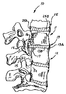
In Fig. 1, a portion of a spinal column is generally indicated by reference
character 10 and includes a pair of adjacent vertebra 11 and 12 with disk 13
interposed
therebetween and contacting each vertebra 11 and 12. Disk 13 includes annulus
13A
-4-
CA 02667140 2009-05-28
and nucleus 13B. Similarly, in Fig. 1, disk 25 includes annulus 25A and
nucleus 25B.
A disk 13 may, or may not, have a herniated nucleus, be flattened from
its normal healthy configuration, etc. The disk 13 illustrated in Fig. 1 and
treated using
the process subsequently explained below with reference to Figs. 2 to 7, is a
bulging
disk of the type illustrated in Fig. 13 wherein the bulge 13C protrudes
outwardly from
vertebra 11 and 12 toward the spinal chord. Vertebra 11 includes posterior
osteophyte
16. Vertebra 12 includes posterior osteophyte 17. The posterior longitudinal
ligament
extends along the spinal canal 14 and over osteophytes 16 and 17. Portions of
10 osteophytes 16 and 17 extend behind the vertebral bodies 11 and 12. Fig. 1
illustrates
the spinal column prior to carrying out an anterior cervical microdiskectomy.
The first step in the anterior cervical microdiskectomy procedure is to
expose the anterior cervical spine and make a rectangular incision in the
anteior
15 annulus. A one to two centimeter length or piece, indicated by arrows J in
Fig. 13, of
the anterior disk annulus is excised. The remainder of the disk annulus is
left intact
along with the portion of the posterior longitudinal ligament that runs
adjacent the
vertebrae. The posterior portion of the annulus 13A that is left intact, along
with the
posterior longitudinal ligament 15, functions to protect the Dura and spinal
cord during
drilling with the various burrs.
The second step in the microdiskectomy is to enter and remove the
nucleus of disk 13 with curettes or pituitary rongeur.
The third step in the microdiskectomy is to utilize a cutting burr to remove
the opposing end plates of vertebrae 11 and 12 and produce surfaces 11A (in
vertebra
11) and 12A (in vertebra 12). Removing the end plates allows bone material
that is
subsequently inserted intermediate vertebrae 11 and 12 to fuse more readily to
the
vertebrae 11, 12. The cutting burr typically has a spherical head with a
diameter of
about five mm. Fig. 2 illustrates the spinal column after the one to
centimeter piece
of the anterior annulus of disk 13 has been removed, after the nucleus of disk
13 has
been removed, and after the adjacent end plates of vertebra 11 and 12 have
been
removed to produce surfaces 11A and 12A. If necessary, removal of the end
plates,
of a portion(s) of the annulus, and of the nucleus can also function to create
a channel,
-5-
CA 02667140 2009-05-28
or opening, that is large enough to enable the rotatable diamond burr 40 (and
subsequently burr 50 or 60) to be inserted through the channel in the manner
illustrated
in Fig. 2 to remove portions of osteophytes 16 and 17. Diamond burr 40
typically has
a diameter of about six millimeters (mm). It is possible that the height of
the annulus
is sufficient to permit access by burr 40, and that the end plates of
vertebrae 11 and 12
need not be removed to form a tunnel or opening having a size sufficient to
allow
ingress and egress by burr 40. The end plates would still be removed to expose
cancellous bone to improve the chance of bone growth into a graft that is
interposed
between vertebra 11 and 12.
The fourth step in the microdiskectomy is to utilize a diamond burr 40 to
remove portions of osteophytes 16 and 17 in the manner illustrated in Fig. 2
while
leaving the posterior annulus and posterior longitudinal ligament 15 in place
to protect
the spinal chord.
The fifth step in the microdiskectomy is to utilize burr 50 in the manner
illustrated in Fig. 3 to remove substantially all of the remaining portions of
osteophytes
16 and 17. In Fig. 3, the shaft 53 of burr 50 is substantially horizontal. The
tunnel or
.opening formed in the manner described above by removing portions of the disk
and
vertebra enable shaft 53 to be tilted somewhat upwardly as indicated by arrow
U or
downwardly as indicated by arrow D from the horizontal to facilitate use of
the head 51
of burr 50 to remove portions of osteophytes 16 and 17 that are illustrated in
Fig. 3 and
that extend upwardly behind vertebra 11 or downwardly behind vertebra 12,
respectively. Sizing shaft 53 and the opening formed intermediate vertebrae 11
and
12 to permit the tilting of shaft 53 and head 51 is important in facilitating
the removal
of osteophytes 16 and 17.
In Fig. 4, undermining burr 50 (Fig. 9) is rotated while at least a portion
of end surface 54 and rounded peripheral edge 56 contact the annulus and the
posterior longitudinal ligament and while at least a portion of abrading
surface 52
undercuts osteophyte 16 to remove portions of osteophyte 16 that extend behind
vertebra 11. This procedure can be accomplished if the posterior annulus is
removed;
however, as noted earlier, it is preferred that the posterior annulus remain
in place
when burr 50 is utilized. Consequently, when burr 50 is used to removed
portions of
osteophytes 16 and 17, the end surface 54 can contact the posterior annulus
and will
-6-
CA 02667140 2009-05-28
not contact the posterior longitudinal ligament. Undermining burr 50 is
similarly utilized
to remove portions of osteophyte 17 that extend behind vertebra 12. When
undermining burr 50 removes portions of osteophytes 16 and 17, portions of
vertebral
bodies 11 and 12 are, as indicated by reference characters 11 and 12 in Fig.
5, also
removed. Fig. 5 illustrates spinal column 10 after osteophytes 16 and 17 have
been
substantially removed using burr 40 and using undermining burr 50. In Fig. 5,
the
posterior annulus and other remaining portion(s) of the annulus have not yet
been
removed.
Flat end surface 54 and non-cutting rounded peripheral edge 56 permit
contact with the posterior annulus and posterior longitudinal ligament with
minimal
displacement of the posterior longitudinal ligament inwardly toward the spinal
canal 14
without piercing, penetrating, or injuring the posterior annulus or posterior
longitudinal
ligament and spinal chord. This facilitates removal of osteophytes 16 and 17.
It is also
important, however, to continue moving the head of burr 50 and to not let end
surface
54 of head 51 contact and rotate on a portion of the posterior longitudinal
ligament for
any extended period of time. Allowing end surface 54 to set and rotate against
a
specific portion of the posterior annulus or posterior longitudinal ligament
allows the
rotating end surface 54 to generate frictional heat which can injure and burn
the
posterior annulus or ligament. Continuously moving head 51 and end surface 54
back
and forth over the posterior annulus or ligament, along with continuous
irrigation with
saline, avoids burning the posterior annulus or posterior longitudinal
ligament. When
burr 50 is utilized to remove osteophytes 16 and 17, care is taken to utilize
the burr 50
only in the central area of vertebra 11 and 12, and to not move burr 50 to
locations that
are too far away from plane X. This central area extends upwardly (a distance
indicated by arrows E) and downwardly (a distance indicated by arrows F) from
plane
X. As is illustrated in Fig. 5, plane X is generally normal to the plane of
the page of
paper of the drawings, extends through the annulus of disk 13, and is centered
between vertebra 11 and 12. The distance indicated by arrows E is in the range
of six
to ten mm, as is the distance indicated by arrows F. Use of burr 50 is limited
to this
central area because it is important to avoid contacting laterally a vertebral
artery that
is directly anterior to the nerve root. Extreme caution should be used if
attempts are
made to use a burr 50 for lateral decompression and/or the foraminotomy.
The sixth step in the microdiskectomy is to remove both the remaining
-7-
CA 02667140 2009-05-28
portions of the annulus 13A (including the posterior annulus), along with the
portion of
the posterior longitudinal ligament that is adjacent the opening in the
vertebral bodies
11 and 12 that is formed by removal of osteophytes 16 and 17. Curettes and
small
Kerrison surgical instruments are utilized to remove the remaining portion of
the
annulus and to remove a portion of the posterior longitudinal ligament. In
Fig. 6, the
remainder of the annulus 13A has been removed, and Kerrison instrument 18 is
about
to be utilized to remove a portion of the posterior longitudinal ligament 15.
After the remaining portions of the annulus is removed and the desired
portion of the posterior longitudinal ligament is removed, the seventh step in
the
microdiskectomy is to confirm decompression by palpating with a dental tool.
The
nerve root foramina may be checked with a nerve hook. As is illustrated in
Fig. 7,
epidural bleeding is controlled with Gelfoam (TM) 19 soaked in thrombin.
The eighth step in the microdiskectomy is to measure the interspace
defect intermediate vertebral bodies 11 and 12 and to select and insert an
implant 20,
typically an allograft. A wedge shape is recommended with the smaller end
directed
toward the spinal canal and the larger end superficial. A wedge shape placed
in this
manner decreases the potential for migration of the implant 20 into the spinal
canal and
spinal cord.
The ninth step in the microdiskectomy is to secure a plate 21 with screws
22 and 23 or other fastening means in the position illustrated in Fig. 7.
Plate 21 limits
the potential for extrusion of the graft, and immobilizes the vertebral bodies
11 and 12
to increase the potential for fusion.
In Fig. 8, rotatable diamond burr 40 includes shaft or shaft 43 and
spherical head 41 with abrading spherically shaped surface 42. Shaft 43
ordinarily is
placed in a drill or other instrument that rotates shaft 43 and head 41 about
the
longitudinal axis of shaft 43.
In Fig. 9, rotatable undermining burr 50 includes shaft 53 and
hemispherical head 51 with abrading hemispherically shaped surface 52. Head 51
also
includes smooth non-abrading non-cutting, non-piercing, non-tearing bottom
surface
54 and rounded peripheral edge 56. Surface 52 extends away from edge 56 and
-8-
CA 02667140 2009-05-28
surface 54 toward shaft 53. Smooth surface 54 can be flat, convex (as
indicated by
dashed line 54A), concave, or can undulate. Regardless, however, of the shape
of
surface 54, surface 54 is smooth and can contact and rotate over the posterior
annulus
or posteriorlongitudinal ligament without abrading, cutting, piercing and/or
tearing the
annulus or ligament. For example, if surface 54 were shaped like a cone, this
would
not be appropriate because a cone comes to a point. Such a point could easily
pierce
the annulus or longitudinal posterior ligament. Consequently, a relatively
smooth,
continuous, substantially flat surface 54, 54A is critical and is preferred in
the practice
of the invention. Similarly, the shape and dimension of the peripheral edge 56
of
undermining burr 50 is critical in the practice of the invention. Edge 56 has
a height
indicated by arrows X in Fig. 10 and functions as the bridge between smooth
surface
54 and abrasive surface 52. Edge 56 can not be sharp, can not comprise a
cutting
edge, and can not abrade. In one embodiment of the invention, the outer
surface of
edge 56 is curved and smooth. The curvature may simulate a portion of a circle
or an
ellipse or other arcuate shape. If the curvature simulates a circle, the
radius R of edge
56 (Fig. 12) should be large enough to prevent edge 56 from functioning as an
edge
that cuts the annulus or posterior longitudinal ligament when burr 50 is
utilized to
remove osteophytes. By way of example, and not limitation, the radius R is
typically at
least one thirty-second of an inch, preferably at least one-sixteenth of an
inch, and most
preferably at least three thirty-seconds of an inch. If the radius of
curvature fo edge 56
is too small, then edge 56 can, even if it is curved and smooth, function as a
cutting
edge, which is not acceptable in the practice of the invention. The abrading
surface 52
of head 51 extends outwardly away from smooth edge 56 and inwardly toward the
shaft
53. The smooth flat surface 54-smooth edge 56-outwardly and inwardly extending
abrading surface 52 combination is critical in the practice of the invention.
In another
embodiment of the invention, the edge 56 has a small or non-existent radius
and is
functional as long as the edge does not cut or pierce the posterior
longitudinal ligament
on contact.
Fig. 11 illustrates an alternate configuration of an undermining burr 60 that
can be utilized in the practice of the invention. Burr 60 includes shaft 63,
and generally
cylindrically shaped head 61. Head 61 is provided with a smooth end surface 64
or
64A, and smooth, curved peripheral surface 66 that serves as a bridge between
surface
64 and abrading surfaces 62 and 65.
-9-
CA 02667140 2009-05-28
The distal end of a shaft 53, 63 can be externally threaded and can turn
into an internally threaded opening in head 51, 61, respectively, so that when
the head
51, 61 is in the spinal column, shaft 53, 63 can be rotatably detached, and
then
reattached to head 51, 61, respectively. This permits, for example, a head 51,
61 to
first be inserted between vertebra 11 and 12 to a desired location, permits
the shaft 53,
63 to then be attached to the head 51, 61, and permits burr 50, 60 to then be
rotated
and used to remove an osteophyte. A shaft 53 can be detachably secured to a
head
51 using a hook and bayonet configuration or any other desired configuration.
In one embodiment of the invention, it is desirable to minimize the height
Z1, Z2 of the head 51, 61 of a burr 50, 60 (Figs. 10 and 11). The height Z1,
Z2 is
preferably in the range of two to eight mm, preferably two to six mm, to
facilitate ingress
and egress of head 51, 61 between vertebra 11 and 12 and the posterior
longitudinal
ligament. Such a height Z1, Z2 can advantageously be utilized in conjunction
with one
or more of the other features of the invention set forth herein.
In another embodiment of the invention, it is desirable to utilize a burr
head 51, 61 with a larger diameter or width W (Fig. 10) that is in the range
of eight to
sixteen millimeters. Such a width can advantageously be utilized in
conjunction with
one or more of the other features of the invention set forth herein. A
diameter of less
than eight mm is not preferred in the practice of the invention and tends to
defeat one
achievement of the invention, which is to facilitate removal of osteophytes
intermediate
the posterior longitudinal ligament and a vertebra 11, 12.
Having described the invention in such terms as to enable those of skill
in the art to make and practice it, and having described the presently
preferred
embodiments thereof, I Claim:
-10-