Note: Descriptions are shown in the official language in which they were submitted.
CA 02669602 2009-05-05
WO 2008/061684
PCT/EP2007/009980
ANTI-IDIOTYPE CONJUGATE AND ITS USE AS A STANDARD IN AN IMMUNASSAY
The invention comprises a conjugate and its use as a standard or positive
control in
an immunoassay for the determination of anti-drug antibodies.
Background of the Invention
Standard solid-phase immunoassays with monoclonal antibodies involve the
formation of a complex between an antibody adsorbed on a solid support
(capture
antibody), the antigen, and an antibody to another epitope of the antigen
conjugated with a detectable label (tracer antibody). Thus, a sandwich is
formed:
solid support-capture antibody-antigen-tracer antibody. In the sandwich, the
intensity of the antibody-conjugated detectable label is proportional to the
antigen
concentration in the incubation medium. The standard sandwich method is also
called double antigen bridging immunoassay because capture and tracer
antibodies
bind to different epitopes of the antigen. Hoesel, W., et al., in J. Immunol.
Methods
294 (2004) 101-110, report an anti-EPO double antigen bridging assay whereby a
mixture of immobilized rhEPO coupled to amino groups and to carbohydrate
groups was used.
Immunoassays such as the double antigen bridging ELISA are common assay types
in the investigation of an immunogenic answer of a patient to an antibody drug
(therapeutic or diagnostic antibody). Mire-Sluis, A.R., et al., in J. Immunol.
Methods 289 (2004) 1-16, summarize the recommendations for the design and
optimization of immunoassays using detection of host antibodies against
biotechnology products. According to Mire-Sluis et al. the well-known anti-
drug
antibody assay formats show considerable disadvantages. Anti-drug antibody
assays
are mentioned, for example, in WO 2005/045058; and WO 90/006515. Anti-
idiotypic antibody assays are mentioned, for example, in US 5,219,730;
WO 87/002778; EP 0 139 389; and EP 0 170 302. Wadhwa, M., et al., in J.
Immunol.
Methods 278 (2003) 1-17, report strategies for the detection, measurement and
characterization of unwanted antibodies induced by therapeutic biologicals.
Serological analysis of human anti-human antibodies is described in Ritter,
G.,
Cancer Res. 61 (2001) 6851-6859, and WO 2003/016909. The identification of
human anti-human antibodies of IgG class requires an additional step of
protein G
precipitation prior to the assay.
CA 02669602 2009-05-05
WO 2008/061684
PCT/EP2007/009980
- 2 -
Summary of the Invention
The invention comprises a conjugate comprising an anti-idiotype antibody
specifically binding to a CDR region of a parent antibody and a reference
immunoglobulin of a single immunoglobulin class.
Preferably said reference immunoglobulin is not specifically binding said anti-
idiotype antibody and said parent antibody.
The invention further comprises the use of a conjugate according to the
invention
as a standard in an immunoassay for the determination of an anti-parent-
antibody
antibody in a sample of a human being.
Preferably said conjugate is used as a standard or a positive control in an
immunological determination of an anti-parent-antibody antibody in a sample
using a sandwich immunoassay comprising a capture antibody and a tracer
antibody.
Preferably said capture antibody or said tracer antibody is said parent
antibody.
Preferably said anti-parent-antibody antibody is an anti-idiotype antibody
against
said parent antibody.
Preferably said parent antibody is a therapeutic antibody or a diagnostic
antibody.
Preferably the capture antibody is selected from the group comprising the
light
chain, the variable region of the heavy chain, a Fab, Fab', F(ab)2, or F(ab')2
fragment of said parent antibody.
Preferably conjugation of the capture antibody to the solid support is
performed by
passive adsorption.
Preferably the capture antibody is immobilized via a specific binding pair.
Preferably the capture antibody is conjugated to biotin and immobilization is
performed via immobilized Avidin or Streptavidin.
Preferably the tracer antibody is conjugated to a detectable label via a
specific
binding pair.
CA 02669602 2009-05-05
WO 2008/061684
PCT/EP2007/009980
- 3 -
Preferably the tracer antibody is conjugated to digoxigenin and linking to the
detectable label is performed via an antibody against digoxigenin.
Preferably the immunological determination is performed by surface plasmon
resonance.
Preferably conjugation of the capture and/or tracer antibody to its
conjugation
partner is performed by chemically binding it via N-terminal and/or c-amino
groups (lysine), e-amino groups of different lysines, carboxy-, sulthydryl-,
hydroxyl-, and/or phenolic functional groups of the amino acid backbone of the
antibody, and/or sugar alcohol groups of the carbohydrate structure of the
antibody.
Preferably the capture antibody is conjugated to the solid support by passive
adsorption and therefore the capture antibody conjugated to the solid support
comprises a mixture of at least two capture antibodies which are conjugated to
the
solid support via different antibody sites. Passive adsorption is, e. g.,
described by
Butler, I.E., Solid Phases in Immunoassay, In: Immunoassays, Diamandis, E.P.
and
Christopoulos, T.K. (eds.), Academic Press San Diego (1996), pp. 205-225.
In a preferred embodiment of the invention, the capture antibody is
immobilized
via a specific binding pair. Such a binding pair (first component/second
component) is, for example, Streptavidin or Avidin/biotin, antibody/antigen
(see,
for example, Hermanson, G.T., et al., Bioconjugate Techniques, Academic Press,
1996), lectin/polysaccharide, steroid/steroid binding protein, hormone/hormone
receptor, enzyme/substrate, IgG/Protein A and/or G, etc. Preferably, the
capture
parent antibody is conjugated to biotin and immobilization is performed via
immobilized Avidin or Streptavidin.
In a preferred embodiment of the invention, the tracer antibody is conjugated
to a
detectable label, preferably conjugated via a specific binding pair. Such a
binding
pair (first component/second component) is, for example, Streptavidin or
Avidin/biotin, antibody/antigen (see, for example, Hermanson, G.T., et al.,
Bioconjugate Techniques, Academic Press, 1996), lectin/polysaccharide,
steroid/steroid binding protein, hormone/hormone receptor, enzyme/substrate,
IgG/Protein A and/or G, etc. Preferably, the tracer parent antibody is
conjugated via
digoxigenin and an antibody against digoxigenin to the detectable label.
CA 02669602 2009-05-05
WO 2008/061684
PCT/EP2007/009980
- 4 -
Alternatively the tracer parent antibody is conjugated to an
electrochemiluminescent label, like a ruthenium bispyridyl complex.
Detailed Description of the Invention
The current invention comprises a conjugate comprising an anti-idiotype
antibody
specifically binding to a CDR region of a parent antibody and a reference
immunoglobulin of a single immunoglobulin class. Preferably said parent
antibody
is a therapeutic antibody. Preferably said parent antibody is a diagnostic
antibody.
The term "anti-idiotype antibody specifically binding to a CDR region of a
parent
antibody" according to the invention denotes an antibody rose against a parent
antibody in a non-human animal, i.e. a non-human antibody. Preferably said
antibody raised against a parent antibody is raised in a rodent or a macaque,
especially preferred in a mouse, a rabbit, or a cynomolgus, or obtained by a
display
technique, preferably by phage- or ribosome-display. Preferably the anti-
idiotype
antibody is a polyclonal antibody. Also preferably the anti-idiotype antibody
is a
monoclonal antibody. Immunization of the animal is performed preferably with
the parent antibody or fragments thereof. In a further step, immunoglobulin
from
said animal is isolated and purified using affinity adsorption to a/the CDR
region(s)
of the parent antibody. The term "CDR region of a parent antibody" denotes the
CDR regions of the parent antibody's light and heavy chain, i.e. comprises the
parent antibody light chain CDR1, CDR2, CDR3, the parent antibody heavy chain
CDR1, CDR2, CDR3.
The term "parent antibody" according to the invention denotes an antibody
which
can be administered as a drug (drug antibody or therapeutic antibody) or as a
diagnostic means (diagnostic antibody) to an individual, so that a sample of
said
individual is suspected to comprise said parent antibody after administration.
Within one assay performed according to the invention the parent antibody, the
capture (parent) antibody, and/or the tracer (parent) antibody comprise the
"same"
antibody molecule, e.g. recombinantly produced with the same expression vector
and comprising the same amino acid sequence. Drug antibodies (therapeutic
monoclonal antibodies) are being used widely for the treatment of various
diseases
such as oncological diseases (e.g. hematological and solid malignancies
including
non-Hodgkin's lymphoma, breast cancer, and colorectal cancer). Such antibodies
are described, for example, by Levene, A.P., et al., Journal of the Royal
Society of
Medicine 98 (2005) 146-152. Such antibodies are, for instance, antibodies
against
CA 02669602 2009-05-05
WO 2008/061684
PCT/EP2007/009980
- 5 -
CCR4, CD19, CD20, CD22, CD28, HLA-DR, CD33, CD40, CD52, CD80, CSF-1R,
CTLA-4, fibroblast activation protein (FAP), EGFR, G250, GD3, HER2/neu, HER3,
HER4, prostate-specific membrane antigen (PSMA), CD56, VEGF, VEGF2, TLSP-
R, TIE-1, TIE-2, TNF-alpha, TNF like weak inducer of apoptosis (TWEAK), CEA,
Ep-CAM, TRAIL, TRAIL-receptor 1, TRAIL-receptor 2, lymphotoxin-beta
receptor, Levis Y antigen, hepsin, melanoma-associated chondroitin sulfate
proteoglycan (MCSP), IL-1 receptor, IL-6 receptor, VEGF-receptor 1, VEGF-
receptor 2, or IGF-1 receptor. Therapeutic antibodies are also described by
Groner,
B., et al., Curr. Mol. Med. 4 (2004) 539-547, and Harris, M., Lancet Oncol. 5
(2004)
292-302.
The term "reference immunoglobulin" as used herein denotes a complete
immunoglobulin, i.e. an immunoglobulin comprising a Fab region and an Fc
region, as well as fragments of a complete immunoglobulin, such as the Fc-
region,
the CH2 domain, or the CH3 domain. The reference immunoglobulin is preferably
a
human immunoglobulin. Preferably the "reference immunoglobulin" is a human
immunoglobulin or an Fc-region of a human immunoglobulin. The "reference
immunoglobulin" is not specifically binding the anti-idiotype antibody and not
specifically binding the parent antibody of the conjugate according to the
invention. The Fc-region of an immunoglobulin is obtained by pepsin or papain
cleavage of a complete immunoglobulin. The reference immunoglobulin is of a
"single immunoglobulin class". The term "single immunoglobulin class" denotes
that the reference immunoglobulin comprises an immunoglobulin class specific
constant region amino acid sequence selected from the immunoglobulin classes
A,
E, M, and G. For immunoglobulin class specific constant region amino acid
sequences see e.g. Pink, J.R., etal., Biochem. J. 117 (1970) 33-47.
An "anti-parent-antibody antibody" is an antibody specifically binding the
parent
antibody, i.e. an antibody against a parent antibody. Likewise is an anti-anti-
IL-6R-
antibody antibody an antibody specifically binding an anti-IL-6R antibody. An
"anti-parent-antibody antibody" is an antibody directed against any region of
the
parent antibody, like the variable region, the constant region or the
glycostructure
of the parent antibody. Such anti-parent-antibody antibodies may occur during
antibody therapy as an immunogenic reaction of a patient (see Pan, Y., et al.,
FASEB J. 9 (1995) 43-49). An "anti-idiotype antibody" is an antibody
specifically
binding to a CDR region of a parent antibody. An anti-idiotype antibody
specifically binds to the light chain CDR1 region, the light chain CDR2
region, the
CA 02669602 2009-05-05
WO 2008/061684
PCT/EP2007/009980
- 6 -
light chain CDR3 region, the heavy chain CDR1 region, the heavy chain CDR2
region, or the heavy chain CDR3 region of a parent antibody.
An example (preferably monoclonal) parent antibody is an antibody against the
IL-6 receptor (anti-IL-6R antibody). Such an antibody is for example described
by
Mihara, M., et al., Clin. Immunol. 98 (2001) 319-326, Nishimoto, N., et al.,
Blood
106 (2005) 2627-2632, in clinical trial NCT00046774, or in WO 2004/096274.
An example (preferably monoclonal) parent antibody is an antibody against IGF-
1
receptor (anti-IGF-1R antibody). Such an antibody is for example described in
WO 2004/087756, or in WO 2005/005635.
The term "binding" or "specifically binding" according to the invention refers
to
binding an antigen, a constant region of an immunoglobulin, a parent antibody,
or
a CDR region of a parent antibody with a KD value of less than 10-6 M
(M=mo1/1)
(e.g. 10-12 M), more preferably by a KD value in the range of from 10-9 M to
10-15 M
in a BIAcore assay. "Non-specific binding" is found if the KD value is larger
than
10-5 M (e.g. 10-4 M).
The principles of different immunoassays are described, for example, by Hage,
D.S.,
in Anal. Chem. 71(1999) 294R-304R. Lu, B., et al., in Analyst 121 (1996) 29R-
32R,
report the orientated immobilization of antibodies for the use in
immunoassays.
Avidin-biotin-mediated immunoassays are reported, for example, by Wilchek, M.
and Bayer, E.A., in Methods Enzymol. 184 (1990) 467-469.
Antibodies, especially their constant domains, contain amino acid side chain
functionalities, i.e. chemical reactive groups, for coupling to a binding
partner like a
surface, a protein, a polymer (such as PEG, Cellulose, or Polystyrol), an
enzyme, or
a member of a binding pair. Chemical reactive groups of antibodies are, for
example, amino groups (epsilon amino groups of lysines, alpha-amino groups),
thiol groups (cystines, cysteines, and methionines), carboxylic acid groups
(aspartic
acids, glutamic acids), and sugar-alcoholic groups. Such methods are e.g.
described
by Aslam, M. and Dent, A., Bioconjugation, MacMillan Ref. Ltd. (1999) 50-100.
For conjugation of polypeptides, e.g. to solid supports, suitable chemical
protecting
agents are required. These form e.g. bonds at unprotected side chain amines
and are
less stable than and different from those bonds at the N-terminus. Many such
chemical protecting agents are known (see for example EP 0 651 761). Preferred
CA 02669602 2014-05-28
WO 2008/061684
PCT/EP2007/009980
- 7 -
chemical protecting agents include cyclic dicarboxylic acid anhydrides like
maleic
or citraconylic anhydrides.
The term "sample" includes, but is not limited to, any quantity of a substance
from
a human being. Such substances include, but are not limited to, whole blood,
serum, or plasma from an individual, which are the most widely used sources of
sample in clinical routine.
Solid supports for immunoassays according to the invention are widely
described in
the state of the art (see, e.g., Butler, J.E., Methods 22 (2000) 4-23).
The term "solid support" denotes a non-fluid substance, and includes chips,
vessels,
and particles (including microparticles and beads) made from materials such as
polymer, metal (paramagnetic, ferromagnetic particles), glass, and ceramic;
gel
substances such as silica, alumina, and polymer gels; capillaries, which may
be made
of polymer, metal, glass, and/or ceramic; zeolites and other porous
substances;
electrodes; microtiter plates; solid strips; and cuvettes, tubes or other
spectrometer
sample containers. A solid support component of an assay is distinguished from
inert solid surfaces with which the assay may be in contact in that a "solid
support"
contains at least one moiety on its surface, which is intended to interact
with the
capture antibody, either directly or indirectly. A solid support may be a
stationary
component, such as a tube, strip, cuvette, or microtiter plate, or may be non-
stationary components, such as beads and microparticles. Microparticles can
also
be used as a solid support for homogeneous assay formats. A variety of
microparticles that allow both non-covalent or covalent attachment of proteins
and
other substances may be used. Such particles include polymer particles such as
polystyrene and poly(methylmethacrylate); gold particles such as gold
nanoparticles
and gold colloids; and ceramic particles such as silica, glass, and metal
oxide
particles. See for example Martin, C.R., et al., Analytical Chemistry-News &
Features 70 (1998) 322A-327A.
A "chip" is a solid, non porous material, such as metal, glass or plastics.
The
material may optionally be coated, entirely or in certain areas. On the
surface of the
material any array of spots is present, either visible or in coordinates. On
each spot
a defined polypeptide, with or without linker or spacer to the surface of the
material, may be immobilized.
CA 02669602 2009-05-05
WO 2008/061684
PCT/EP2007/009980
- 8 -
All documents mentioned herein, both supra and infra, are hereby incorporated
herein by reference.
Chromogens (fluorescent or luminescent groups and dyes), enzymes, NMR-active
groups or metal particles, haptens (e.g. digoxigenin), are examples of
detectable
labels. The detectable label can also be a photoactivatable crosslinking
group, e.g. an
azido or an azirine group. Metal chelates which can be detected by
electrochemoluminescence are also preferred signal-emitting groups used as
detectable labels, with particular preference being given to ruthenium
chelates, such
as e.g. a ruthenium (bispyridy1)32+ chelate. Suitable ruthenium labeling
groups are
described, for example, in EP 0 580 979, WO 90/05301, WO 90/11511, and
WO 92/14138.
The invention comprises a conjugate comprising an anti-idiotype antibody
specifically binding to a CDR region of a parent antibody and a reference
immunoglobulin of a single immunoglobulin class.
The invention comprises a conjugate comprising an anti-idiotype antibody
specifically binding to a CDR region of a parent antibody and a reference
immunoglobulin of a single immunoglobulin class selected from the group
comprising human immunoglobulin E,
human immunoglobulin G,
human immunoglobulin M, or human immunoglobulin A, i.e. the reference
immunoglobulin is either of human IgE class, or of human IgG class, or of
human
IgM class, or of human IgA class.
Preferably the current invention comprises a conjugate comprising an anti-
idiotype
antibody specifically binding to a CDR region of a parent antibody and a
reference
immunoglobulin of a single immunoglobulin class not specifically binding said
anti-idiotype antibody and not specifically binding said parent antibody.
Preferably said parent antibody is a therapeutic antibody or a diagnostic
antibody.
Preferably said reference immunoglobulin is a not functionable immunoglobulin
of
a single immunoglobulin class or an Fc-region of an immunoglobulin of a single
immunoglobulin class.
The term "diagnostic antibody" denotes an antibody which is either a natural
antibody or a recombinantly produced antibody and which is used for the
detection
and visualization of its target antigen. A diagnostic antibody is used e.g. in
assay
CA 02669602 2009-05-05
WO 2008/061684
PCT/EP2007/009980
- 9 -
systems (e.g. ELISA), or for in vitro imaging. A diagnostic antibody may e.g.
be a
labeled therapeutic antibody.
Preferably said reference immunoglobulin is a human immunoglobulin or a human
immunoglobulin Fc-region.
The reference immunoglobulin provides an immunoglobulin class specific
constant
region that can be specifically bound by an anti-immunoglobulin-class
antibody,
such as an anti-human-immunoglobulin-G antibody. Thus the reference
immunoglobulin provides the conjugate according to the invention with an
immunoglobulin class specific tag, which can be specifically identified by a
tag
specific antibody. For example, if the tag is an immunoglobulin G constant
region a
tag specific antibody is an anti-immunoglobulin-G antibody.
Preferably said anti-idiotype antibody is a polyclonal antibody and said
reference
immunoglobulin is a polyclonal immunoglobulin. Also preferably said anti-
idiotype antibody is a polyclonal antibody and said reference immunoglobulin
is a
monoclonal immunoglobulin. Still preferably said anti-idiotype antibody is a
monoclonal antibody and said reference immunoglobulin is a monoclonal
immunoglobulin.
The conjugate according to the invention is obtained by chemical conjugation
of an
anti-idiotype antibody and a reference immunoglobulin.
In the conjugate according to the invention is the anti-idiotype antibody a
functionable immunoglobulin and the reference immunoglobulin a not
functionable immunoglobulin. This denotes that the anti-idiotype antibody is
specifically binding to its target antibody, whereas the reference antibody is
not
specifically binding any human antigen. The reference antibody is provided in
order to present an Fc-region as similar as possible to naturally occurring
immunoglobulins and not to bind an antigen. It is also within the scope of the
current invention that the anti-idiotype antibody and the reference antibody
are
not the same antibody, i.e. they differ by at least 20 % of the amino acid
residues,
i.e. they have an identity based on the amino acid sequence of 80 % or less.
The
reference antibody may be a polyclonal antibody or a monoclonal antibody. The
term "monoclonal" as used within the current application denotes a population
of
antibodies produced by a single cell and/or its progeny. The term denotes that
the
antibodies have the same amino acid sequence, i.e. the antibodies have the
identical
CA 02669602 2009-05-05
WO 2008/061684
PCT/EP2007/009980
- 10 -
amino acid sequence despite inadvertent mutations occurring during the
propagation of the cell(s) producing it.
The term "not functionable immunoglobulin" as used within this application
denotes an immunoglobulin that binds to a human antigen with a KD-value
(binding affinity) of 10-5 mo1/1 or higher (e.g. 10-3 mo1/1), preferably with
a KD-
value of 10-6 mo1/1 or higher. The binding affinity is determined with a
standard
binding assay, such as surface plasmon resonance technique (BIAcore ). This
binding affinity value has not to be treated as an exact value; it is merely a
point of
reference. It is used to determine and/or select immunoglobulins that show no
immunoglobulin-typical specific binding for human antigens and thus have no
therapeutic activity in humans. This does not exclude that the immunoglobulin
shows a specific binding for non-human antigens. This specific binding of a
non-
human antigen is associated with a KD-value of 10-7 mo1/1 or lower (e.g. 10-10
mo1/1),
preferably with a KD-value of 10-8 mo1/1 or lower.
Based on transfectomas, i.e. on lymphoid cells containing transfected
immunoglobulin genes obtained from immunized mice, a conjugate according to
the invention can be obtained by conjugation on the nucleic acid level.
The administration of drugs to mammals originating from outside the receiving
organism, e.g. the administration of exogenous therapeutic polypeptides,
results in
an immune response of the receiving mammal. This immune response becomes
apparent by the occurrence of anti-drug antibodies. For the evaluation of such
an
immune response a method for the detection of the antibodies against the drug
is
required.
The conjugate according to the invention can be used as standard in an
immunoassay. Thus, the invention comprises the use of a conjugate comprising
an
anti-idiotype antibody specifically binding to a CDR region of a parent
antibody
and a reference immunoglobulin of a single immunoglobulin class as a standard
in
an immunoassay for the determination of an anti-parent-antibody antibody in a
sample of a human being.
Also can the conjugate according to the invention be used as a positive
control in an
immunoassay. It enables e.g. the establishment of a detection limit. Thus, the
invention comprises the use of a conjugate comprising an anti-idiotype
antibody
specifically binding to a CDR region of a parent antibody and a reference
CA 02669602 2009-05-05
WO 2008/061684
PCT/EP2007/009980
- 11 -
immunoglobulin of a single immunoglobulin class as a positive control in an
immunoassay for the determination of an anti-parent-antibody antibody in a
sample of a human being. Preferably said conjugate is added in this embodiment
to
the sample of a human being prior to the immunoassay.
The parent antibody is preferably a therapeutic antibody. Also preferably the
parent
antibody is a murine antibody, chimeric antibody, humanized antibody, or human
antibody. Preferably the parent antibody is a chimeric antibody, humanized
antibody, or human antibody. Especially preferred is said parent antibody a
therapeutic chimeric, humanized, or human antibody. For the detection of
antibodies against a parent antibody different methods can be employed, such
as
radioimmunoassay (RIA), enzyme linked immunosorbent assay (ELISA),
immunoradiometric assays (IRMA), or surface plasmon resonance (SPR).
The term "therapeutic antibody" and grammatical equivalents thereof used
within
this application denotes an, preferably monoclonal, antibody which is intended
to
be administered to mammals, preferably humans, for use in treatment, therapy,
or
diagnosis of a disease. A therapeutic antibody is generally produced by
recombinant
means, e.g. by the cultivation of a eukaryotic cell transfected with a nucleic
acid
encoding said therapeutic antibody. Preferably the therapeutic antibody is a
chimeric antibody, or a humanized antibody, or a human antibody. The
therapeutic antibody is administered to achieve a desired effect, such as e.g.
depletion of target cells, or mediation of ADCC (antibody-dependent cell-
mediated
cytotoxicity), or mediation of CDC (complement dependent cytotwdcity). Target
cells may be e.g. cancer cells, or virus-infected cells. To mediate ADCC or
CDC the
therapeutic antibody has to bind to the target cell and is thus specific for,
i.e.
specifically binding, e.g. a tumor antigen.
A "tumor antigen," as used herein, includes the meaning known in the art,
which
includes any molecule expressed on (or associated with the development of) a
tumor cell that is known or thought to contribute to a tumorigenic
characteristic of
the tumor cell. Numerous tumor antigens are known in the art. Whether a
molecule is a tumor antigen can also be determined according to techniques and
assays well known to those skilled in the art, such as for example clonogenic
assays,
transformation assays, in vitro or in vivo tumor formation assays, gel
migration
assays, gene knockout analysis, etc. Preferably the term "tumor antigen" when
used
herein refers to a human transmembrane protein, i.e. a cell membrane protein
which is anchored in the lipid bilayer of cells. The human transmembrane
protein
CA 02669602 2009-05-05
WO 2008/061684
PCT/EP2007/009980
- 12 -
will generally comprise an "extracellular domain" as used herein, which may
bind a
ligand, a lipophilic transmembrane domain, a conserved intracellular domain,
e.g. a
tyrosine kinase domain, and a carboxyl-terminal signaling domain harboring
several tyrosine residues which can be phosphorylated. The tumor antigen
include
molecules such as EGFR, HER2/neu, HER3, HER4, EpCAM, CEA, TRAIL, TRAIL-
receptor 1, TRAIL-receptor 2, lymphotoxin-beta receptor, CCR4, CD19, CD20,
CD22, CD28, CD33, CD40, CD80, CSF-1R, CTLA-4, fibroblast activation protein
(FAP), hepsin, melanoma-associated chondroitin sulfate proteoglycan (MCSP),
prostate-specific membrane antigen (PSMA), VEGF receptor 1, VEGF receptor 2,
IGF1-R, TSLP-R, TIE-1, TIE-2, TNF-alpha, TNF like weak inducer of apoptosis
(TWEAK), or IL-1R.
Preferably the conjugate according to the invention is used as a standard in
an
immunological determination of an anti-parent-antibody antibody in a sample,
using a sandwich immunoassay comprising a capture antibody and a tracer
antibody. Especially preferred said capture or said tracer antibody is said
parent
antibody.
For example, anti-drug antibody assays are mentioned in WO 2005/045058, and
WO 90/006515, anti-idiotypic antibody assays are mentioned in US 5,219,730,
WO 87/002778, EP 0 139 389, and EP 0 170 302.
Preferably said anti-idiotype antibody is a monoclonal antibody and said
reference
immunoglobulin is a polyclonal human immunoglobulin. Also preferably said anti-
idiotype antibody is a monoclonal antibody and said reference immunoglobulin
is a
monoclonal human immunoglobulin.
The conjugate according to the invention is preferably used as a standard in
an
immunological determination of an anti-idiotype antibody against a parent
antibody in a sample. For the immunological determination a capture and a
tracer
antibody are employed. In one embodiment the capture antibody is the parent
antibody. Preferably the parent antibody used as the capture antibody is a
complete
antibody, i.e. it comprises a light and a heavy chain whereby the light chain
comprises a variable domain and a constant domain, and whereby the heavy chain
comprises a variable domain, a CH1, a CH2, a CH3, and an optional CH4 domain
and a hinge region. Preferably the capture antibody is selected from the group
comprising the light chain, the variable region of the heavy chain, a Fab,
Fab',
F(ab)2, or F(ab')2 fragment of said parent antibody, i.e. it is either the
light chain,
CA 02669602 2009-05-05
WO 2008/061684
PCT/EP2007/009980
- 13 -
the variable region of the heavy chain, the Fab, or Fab', or F(ab)2, or
F(ab')2
fragment of said parent antibody. In an other embodiment the tracer antibody
is
the parent antibody. In this embodiment e.g. the conjugate according to the
invention is bound via an immunoglobulin specifically binding the reference
immunoglobulin of the conjugate to the solid support.
The conjugation of a tracer and/or capture antibody to its conjugation partner
can
be performed by different methods, such as passive adsorption, chemical
binding,
or binding via a specific binding pair. The term "conjugation partner" as used
herein denotes e.g. solid supports, polypeptides, detectable labels, members
of
specific binding pairs. In one embodiment the conjugation of the capture
and/or
tracer antibody to its conjugation partner is performed by chemically binding
via
N-terminal and/or e-amino groups (lysine), e-amino groups of different
lysines,
carboxy-, sulthydryl-, hydroxyl-, and/or phenolic functional groups of the
amino
acid backbone of the antibody, and/or sugar alcohol groups of the carbohydrate
structure of the antibody. In one embodiment the capture and/or tracer
antibody
are/is conjugated to its conjugation partner via a specific binding pair.
Preferably
the capture antibody is conjugated to biotin and immobilization to a solid
support
is performed via solid support immobilized avidin or streptavidin. Preferably
the
tracer antibody is conjugated to digoxigenin and linking to the detectable
label is
performed via an antibody against digoxigenin. The capture antibody is in
another
embodiment conjugated to the solid support by passive adsorption. An antibody
conjugated to the solid support by passive adsorption comprises a mixture of
antibodies conjugated to the solid support via different antibody sites. Thus,
the
capture antibody conjugated to the solid support by passive adsorption is a
mixture
of two or more different conjugates wherein the conjugates differ in the
antibody
sites, i.e. the antibody residues, with which the conjugation to the solid
support is
effected. Passive adsorption is, e. g., described by Butler, J.E., in "Solid
Phases in
Immunoassay", page 205-225; Diamandis, E.P. and Christopoulos, T.K. (Editors):
Immunoassays (1996), Academic Press, San Diego.
In a preferred embodiment of the invention, the capture antibody is
immobilized
via a specific binding pair. Such a binding pair (first component/second
component) is, for example, streptavidin or avidin/biotin, antibody/antigen
(see,
for example, Hermanson, G.T., et al., Bioconjugate Techniques, Academic Press,
1996), lectin/polysaccharide, steroid/steroid binding protein, hormone/hormone
receptor, enzyme/substrate, IgG/Protein A and/or G and/or L, etc. Preferably,
the
CA 02669602 2009-05-05
WO 2008/061684
PCT/EP2007/009980
- 14 -
capture parent antibody is conjugated to biotin and immobilization is
performed
via immobilized avidin or streptavidin. In an other preferred embodiment of
the
invention, the tracer antibody is conjugated to a detectable label, preferably
conjugated via a specific binding pair. Such a binding pair (first
component/second
component) is, for example, Streptavidin or Avidin/biotin, antibody/antigen,
lectin/polysaccharide, steroid/steroid binding protein, hormone/hormone
receptor,
enzyme/substrate, IgG/Protein A and/or G and/or L, etc. Preferably, the tracer
parent antibody is conjugated via digoxigenin and an antibody against
digoxigenin
to the detectable label. Alternatively the tracer parent antibody is
conjugated to an
electrochemiluminescent label, like a ruthenium bispyridyl complex.
If the conjugate according to the invention is used as a standard in an
immunoassay
for the determination of an anti-idiotype antibody against a parent antibody
in a
sample of a human being it is preferably used in two or more different
concentrations. With the determined responses to the different concentrations
of
the standard a calibration curve is/can be calculated.
If the conjugate according to the invention is used as a standard in an
immunoassay
for the determination of an anti-idiotype antibody against a parent antibody
in a
sample of a human being an (optional) additional antibody specifically binding
said
reference immunoglobulin of said conjugate can be employed. The conjugate
according to the invention can be used as a standard alone or in combination
with
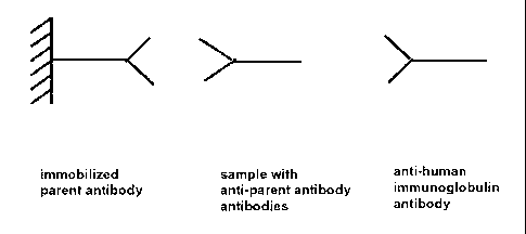
the optional additional anti-human-immunoglobulin antibody (Figures 3 and 4).
The term "standard" or "standard substance" which can be used interchangeably
within this application denotes a point of reference for an analytical method
and is
used to set up a value against which other results of the same analytical
method are
compared. The term õpositive control" as used herein denotes a standard
substance
with which, if employed in an analytical method, a response above a defined
cut-off
or threshold value will be achieved. The cut off value is in general the
average value
obtained in the analysis of samples not containing anti-drug antibodies plus
two
times, preferably three times, the standard deviation of the obtained values.
The invention further comprises a method for the use of a conjugate according
to
the invention as a standard in an immunoassay comprising the following steps
a)
contacting the conjugate according to the invention with a capture antibody,
b)
detecting the binding of said conjugate to said capture antibody. The
detection of
CA 02669602 2009-05-05
WO 2008/061684
PCT/EP2007/009980
- 15 -
the binding in step a) can be performed either directly, e.g. via a change in
the SPR
angel, or indirectly, e.g. via a tracer antibody and/or a detectable label.
The invention further comprises a method for the determination whether an anti-
drug antibody is binding to the Fc-region or the Fab-region of a parent
antibody
and which immunoglobulin class said anti-drug antibody has (see e.g. Figure
7). In
this method the binding of a sample is determined with immobilized parent
antibody, immobilized Fc-region of said parent antibody, and/or immobilized
Fab-
region of said parent antibody. This can be performed e.g. on a BIAcore chip,
on
which one of the four channels contains the immobilized parent antibody, one
contains the immobilized Fc-region of the parent antibody, one contains the
immobilized Fab-region of the parent antibody, and one contains no immobilized
parent antibody. Depending on which channels and therefore which part(s) of
the
parent antibody show binding of an anti-drug antibody from the sample the
binding region of an anti-drug antibody can be determined. The immunoglobulin
class is determined by binding of anti-human-immunoglobulin-E antibody, anti-
human-immunoglobulin-M antibody, anti-human-immunoglobulin-G antibody,
or anti-human-immunoglobulin-A antibody. The anti-human-immunoglobulin
antibodies are either used sequentially or concomitantly, preferably
sequentially.
The conjugate according to the invention can be used as a standard in this
method.
The invention further comprises a method for the determination of the
immunoglobulin class of an anti-idiotype antibody specifically binding a CDR
region of a parent antibody in a sample using a sandwich immunoassay
comprising
a capture antibody, a tracer antibody, and a conjugate according to the
invention,
comprising the following steps:
a) contacting
said sample with the capture antibody under conditions suitable
for the formation of a capture antibody/anti-idiotype antibody-complex,
b) contacting separately
i) an anti-human-immunoglobulin-A antibody,
ii) an anti-human-immunoglobulin-E antibody,
iii) an anti-human-immunoglobulin-M antibody, and
iv) an anti-human-immunoglobulin-G antibody
as tracer antibodies with said capture antibody/anti-idiotype antibody-
complex,
c)
determining the binding of each of said tracer antibodies to said complex and
thereby determining the immunoglobulin class of said anti-idiotype antibody,
CA 02669602 2009-05-05
WO 2008/061684
PCT/EP2007/009980
- 16 -
wherein a conjugate according to the invention is used as a positive control.
A scheme of this method and an exemplary response diagram are given in Figures
1
and 2.
Preferably said tracer antibodies are contacted in the order i) anti-human-
immunoglobulin-A antibody, ii) anti-human-immunoglobulin-E antibody, iii)
anti-human-immunoglobulin-M antibody, and iv) anti-human-immunoglobulin-
G antibody with said capture antibody/anti-idiotype antibody-complex, i.e. the
tracer antibodies are contacted sequentially.
Preferably said capture antibody is selected from the group comprising the
light
chain, the variable region of the heavy chain, a Fab, Fab', F(ab)2, or F(ab')2
fragment of said parent antibody.
The invention further comprises a polyclonal antibody specifically binding a
CDR
region of an anti-IL-6R antibody. A method for the preparation of a polyclonal
antibody according to the invention is described in Example la).
Preferably the method for the determination of the immunoglobulin class of an
anti-idiotype antibody according to the invention comprises the following
steps:
a) contacting said sample with the capture antibody under conditions
suitable
for the formation of a capture antibody/anti-idiotype antibody-complex,
b) contacting a tracer antibody selected from the group of tracer
antibodies
comprising anti-human-immunoglobulin-G antibody,
anti-human-
immunoglobulin-E antibody,
anti-human-immunoglobulin-M antibody,
anti-human-immunoglobulin-A antibody with said capture antibody/anti-
idiotype antibody-complex,
c) determining the binding of said tracer antibody to said complex,
d) disintegrating said capture antibody/anti-idiotype antibody-complex,
e) repeating steps a) to d) with a not previously selected tracer antibody,
f) determining the immunoglobulin class of said anti-idiotypic antibody to
be
the immunoglobulin of said tracer antibody specifically binding said capture
antibody/anti-idiotype antibody-complex.
CA 02669602 2009-05-05
WO 2008/061684
PCT/EP2007/009980
- 17 -
The tracer antibody is preferably selected from anti-human-immunoglobulin-G
antibodies, or anti-human-immunoglobulin-E antibodies, or anti-
human-immunoglobulin-M antibodies, or anti-human-immunoglobulin-A
antibodies.
The term "under conditions suitable for the formation of a [ ...] complex" and
grammatical equivalents thereof as used within this application denotes
conditions
at which an antibody/immunoglobulin of interest, e.g. an anti-idiotype
antibody,
binds to a second antibody when brought in contact with it. This does not
necessarily denote that 100 % of the antibody of interest is bound but
essentially
100 % of the antibody of interest is bound, i.e. at least 85 % of the antibody
of
interest is bound, preferably at least 90 % of the antibody of interest is
bound,
preferably at least 95 % of the antibody of interest is bound, more preferably
more
than 95 % of the antibody of interest is bound to the second antibody.
The following examples and figures are provided to aid the understanding of
the
present invention, the true scope of which is set forth in the appended
claims. It is
understood that modifications can be made in the procedures set forth without
departing from the spirit of the invention.
Description of the Figures
Figure 1
Scheme for the detection of anti-parent-antibody antibodies in a
sample with optional subsequent subclass determination.
Figure 2 BIAcore SPR diagram for the subclass determination of an anti-
idiotype antibody according to the invention. (1) Injection of
sample, (2) + (3) injection of anti-human-immunoglobulin-E
antibody, (4) injection of anti-human-immunoglobulin-G
antibody.
Figure 3 Scheme for the use of a conjugate according to the invention
as a
standard with an optional class specific antibody.
Figure 4 BIAcore SPR diagram for the use of a compound according to the
invention as standard (first response) with an optional
subsequent anti-standard immunoglobulin subclass antibody
(second response).
Figure 5
Scheme of an ELISA-determination of an anti-idiotypic antibody.
Figure 6
Standard curve of an ELISA for the determination of anti-
idiotypic antibody.
CA 02669602 2009-05-05
WO 2008/061684
PCT/EP2007/009980
- 18 -
Figure 7 Scheme of a multiple channel detection of the binding
region of
an anti-parent-antibody antibody with subsequent
immunoglobulin class determination.
The use of an antibody against the IL-6 receptor in the following examples is
only
for exemplifying the invention and does not mean to limit the scope of the
invention.
Example 1
Preparation of a composition of a polyclonal anti-idiotype antibody
specifically
binding to a CDR region of a parent antibody and a polyclonal human serum
immunoglobulin of class E, G, A, and M.
Polyclonal anti-anti-IL-6R antibody antibodies (anti-anti-IL-6R-mAb pAb)
i) Preparation of polyclonal antibodies against monoclonal anti-IL-6R antibody
Purification of polyclonal antibodies from rabbit serum
Rabbits have been immunized with monoclonal anti-IL-6R antibody according to
standard methods. In the raw serum of five immunized rabbits the lipid
components were removed by delipidation with Aerosil (1.5 To (w/v)) and the
immunoglobulins were precipitated with ammonium sulphate (1.7 M). After acid
treatment (30 min., pH 5.5) and dialysis against 15 mM potassium phosphate
buffer, supplemented with 50 mM NaC1, pH 7.0, the mixture was separated by
DEAE ion exchange chromatography at pH 7Ø The immunoglobulin G fraction
was in the flow through (= rabbit anti-anti-IL-6R-mAb pAb) and was
concentrated
to about 25 mg/ml.
Preparation of rabbit anti-anti-IL-6R-mAb antibodies specifically binding a
CDR
region of the parent antibody against IL-6R
The concentrated IgG fraction of the previous step was transferred to a buffer
system with 50 mM potassium phosphate, supplemented with 150 mM NaC1, pH
7.5 (PBS). The immunosorbent with immobilized anti-IL-6R antibody (parent
antibody), prepared by conjugation of anti-IL-6R antibody to NHS-sepharose by
state of the art techniques, was packed into a column and equilibrated with 50
mM
potassium phosphate buffer, supplemented with 150 mM NaC1, pH 7.5.
CA 02669602 2009-05-05
WO 2008/061684
PCT/EP2007/009980
- 19 -
mg concentrated immunoglobulin G fraction/ml immunosorbent were applied
to the column equilibrated with PBS. The column was washed successively with
PBS, 0.5 M NaC1 supplemented with 0.05 % (w/v) Tween 20, and 30 mM NaCl.
The IgG specifically bound to the immunosorbent was eluted with 3 mM HC1 and
5 1 M propionic acid. The eluate was dialyzed against PBS.
To eliminate antibodies with cross reactivity to the human immunoglobulin G
(IgG) constant region the affinity-purified antibodies were applied to an
affinity
column with immobilized human IgG, prepared by conjugation of unspecific
human IgG to NHS-sepharose by state of the art techniques. The column was
10 equilibrated with PBS. About 6 mg IgG/m1 immunosorbent were applied to
the
column. The desired specific polyclonal IgG-fraction is in the flow through.
After
regeneration of the column with 0.5 M NaC1 supplemented with 0.05 % (w/v)
Tween 20, 30 mM NaCl, 1 M propionic acid, and PBS. The immunosorption of
antibodies unspecifically binding to the human IgG constant region was
repeated
two times to completely eliminate antibodies with cross reactivity against
human
IgG.
The resulting purified polyclonal anti-anti-IL-6R-antibody antibody without
cross
reactivity to human IgG constant region was concentrated to about 4 mg/ml and
stored at -80 C.
ii) Conjugation of the polyclonal anti-anti-IL-6R-antibody antibody to
polyclonal
human immunoglobulin E, human immunoglobulin G, human immunoglobulin
A, and human immunoglobulin M
Preparation of a conjugate of polyclonal rabbit anti-anti-IL-6R-antibody
antibody
(aa-IL-6R pAb) with human immunoglobulin G
Step 1: Preparation of rabbit aa-IL-6R pAb-SATP
The aa-IL-6R pAb was dialyzed against 100 mM potassium phosphate buffer,
containing 150 mM NaC1, pH 7.8, and the protein solution was adjusted to a
protein concentration of about 15 mg/ml. N-succinimidy1-3-acetylthiopropionate
(SATP) was dissolved in DMSO and added to the antibody solution in a molar
ratio
of 1:5 (aa-IL-6R pAb:SATP). The pH was adjusted to pH 7.1 and the mixture was
incubated for 60 min. at 25 C. The reaction was stopped by adding L-lysine at
a
final concentration of 10 mM and the surplus of SATP was removed by dialysis
CA 02669602 2009-05-05
WO 2008/061684
PCT/EP2007/009980
- 20 -
against 10 mM potassium phosphate buffer, containing 200 mM NaC1, 1 mM
EDTA, pH 6.1.
Step 2: Preparation of human polyclonal human immunoglobulin G-MH
The polyclonal human antibody was dialyzed against 30 mM potassium phosphate
buffer, pH 7.4, and the protein solution was thereafter adjusted to a protein
concentration of about 25 mg/ml. Maleimidohexanoyl-N-hydroxysuccinimide ester
(MHS) was dissolved in DMSO and added to the antibody solution in a molar
ratio
of 1:6 (IgG:MHS). The pH was adjusted to pH 7.1 and the mixture was incubated
60 min at 25 C. The reaction was stopped by adding L-lysine to a final
concentration of 10 mM, the pH was adjusted to pH 6.2, and the surplus of MHS
was removed by dialysis against 10 mM potassium phosphate buffer, containing
200 mM NaC1, 1 mM EDTA, pH 6.1.
Step 3: Conjugation of aa-IL-6R pAb-SATP with polyclonal human
immunoglobulin G-MH
aa-IL-6R pAb-SATP was deacetylated to aa-IL-6R pAb-SH by incubation with 2 cro
(v/v) 1 M hydroxylamine, pH 7.5, and incubated for 45 min. at 25 C. The
deacetylated antibody was mixed with polyclonal human immunoglobulin G-MH
(molar ratio of IgG-SH:IgG-MH = 1:3) and diluted with 10 mM potassium
phosphate buffer, containing 200 mM NaCl, 1 mM EDTA, pH 6.1, to a final
concentration of 1.5 mg/ml aa-IL-6R pAb-SH and 4.5 mg/ml polyclonal human
immunoglobulin G-MH. The pH was adjusted to pH 7.1 and the mixture was
incubated at 25 C. The conjugation process was analyzed with an analytical
gel
filtration column (e.g. TSK 3000). The conjugation was stopped generally after
45
min. by the addition of cysteine to a final concentration of 1 mM. After a
further 30
min. incubation time N-methylmaleimide (NMM) was added to a final
concentration of 5 mM and the pH was adjusted to pH 7.5. After 60 min.
incubation at 25 C the conjugate was purified by S300 gel filtration
chromatography to eliminate non conjugated antibodies.
Preparation of a conjugate of rabbit aa-IL-6R pAb with human immunoglobulin M
Step 1: Preparation of rabbit aa-IL-6R pAb-SATP
The aa-IL-6R pAb was dialyzed against 100 mM potassium phosphate buffer,
containing 150 mM NaC1, pH 7.8, and the protein solution was adjusted to a
protein concentration of about 15 mg/ml. N-succinimidy1-3-acetylthiopropionate
(SATP) was dissolved in DMSO and added to the antibody solution in a molar
ratio
CA 02669602 2009-05-05
WO 2008/061684
PCT/EP2007/009980
- 21 -
of 1:5 (aa-IL-6R pAb:SATP). The pH was adjusted to pH 7.1 and the mixture was
incubated for 60 min. at 25 C. The reaction was stopped by adding L-lysine at
a
final concentration of 10 mM and the surplus of SATP was removed by dialysis
against 10 mM potassium phosphate buffer, containing 200 mM NaC1, 1 mM
EDTA, pH 6.1.
Step 2: Preparation of polyclonal human immunoglobulin M-MH
The polyclonal human antibody was dialyzed against 30 mM potassium phosphate
buffer, pH 7.4, and the obtained protein solution was adjusted afterwards to a
protein concentration of about 20 mg/ml. Maleimidohexanoyl-N-
hydroxysuccinimide ester (MHS) was dissolved in DMSO and added to the
antibody solution in a molar ratio of 1:50 (IgM:MHS). The pH was adjusted to
pH
7.1 and the mixture was incubated for 60 min. at 25 C. The reaction was
stopped
by adding L-lysine to a final concentration of 10 mM. The pH was adjusted to
pH
6.2 and the surplus of MHS was removed by dialysis against 10 mM potassium
phosphate buffer, containing 200 mM NaC1, 1 mM EDTA, pH 6.1.
Step 3: Conjugation of aa-IL-6R pAb-SATP with polyclonal human
immunoglobulin M-MH
aa-IL-6R pAb-SATP was deacetylated to aa-IL-6R pAb-SH by incubation with 2 %
(v/v) 1 M hydroxylamine, pH 7.5, and incubated for 45 min. at 25 C. The
deacetylated antibody was mixed with polyclonal human immunoglobulin M-MH
(molar ratio of aa-IL-6R pAb-SH:IgM-MH = 1:2) and diluted with 10 mM
potassium phosphate buffer, containing 200 mM NaC1, 1 mM EDTA, pH 6.1, to a
final concentration of about 0.9 mg/ml aa-IL-6R pAb-SH and 9.1 mg/ml
polyclonal
human immunoglobulin M-MH. The pH was adjusted to pH 7.1 and the mixture
was incubated at 25 C. The conjugation process was monitored with an
analytical
gel filtration column (e.g. TSK 5000). The conjugation reaction was stopped
generally after 60 min. by adding cysteine to a final concentration of 1 mM.
After a
further 30 min. incubation time N-methylmaleimide (NMM) was added to a final
concentration of 5 mM and the pH was adjusted to pH 7.5. After 60 min.
incubation at 25 C the conjugate was purified by S400 gel filtration
chromatography to eliminate non conjugated antibodies.
CA 02669602 2009-05-05
WO 2008/061684
PCT/EP2007/009980
- 22 -
Preparation of a conjugate of rabbit aa-IL-6R pAb with polyclonal human
immunoglobulin E
Step 1: Preparation of rabbit aa-IL-6R pAb-SATP
The aa-IL-6R pAb was dialyzed against 100 mM potassium phosphate buffer,
containing 150 mM NaC1, pH 7.8, and the protein solution was adjusted to a
protein concentration of about 15 mg/ml. N-succinimidy1-3-acetylthiopropionate
(SATP) was dissolved in DMSO and added to the antibody solution in a molar
ratio
of 1:5 (aa-IL-6R pAb:SATP). The pH was adjusted to pH 7.1 and the mixture was
incubated for 60 min. at 25 C. The reaction was stopped by adding L-lysine at
a
final concentration of 10 mM and the surplus of SATP was removed by dialysis
against 10 mM potassium phosphate buffer, containing 200 mM NaCl, 1 mM
EDTA, pH 6.1.
Step 2: Preparation of polyclonal human immunoglobulin E-MH
The polyclonal human antibody was dialyzed against 30 mM potassium phosphate
buffer, pH 7.4, and the obtained protein solution was adjusted to a final
protein
concentration of about 13 mg/ml. Maleimidohexanoyl-N-hydroxysuccinimide ester
(MHS) was dissolved in DMSO and added to the antibody solution in a molar
ratio
of 1:6 (IgE:MHS). The pH was adjusted to pH 7.1 and the mixture was incubated
for 60 min. at 25 C. The reaction was stopped by adding L-lysine to a final
concentration of 10 mM, the pH was adjusted to pH 6.2 and surplus of MHS was
removed by dialysis against 10 mM potassium phosphate buffer, containing
200 mM NaC1, 1 mM EDTA, pH 6.1.
Step 3: Conjugation of rabbit aa-IL-6R pAb-SATP with polyclonal human
immunoglobulin E-MH
aa-IL-6R pAb-SATP was deacetylated to aa-IL-6R pAb-SH by incubation with 2 %
(v/v) 1 M hydroxylamine, pH 7.5, and incubated for 45 min. at 25 C. The
deacetylated antibody was mixed with polyclonal human immunoglobulin E-MH
(molar ratio of aa-IL-6R pAb-SH:IgE-MH = 1:1.7) and diluted with 10 mM
potassium phosphate buffer, containing 200 mM NaCl, 1 mM EDTA, pH 6.1, to a
concentration of 2.9 mg/ml aa-IL-6R pAb-SH and 6.3 mg/ml polyclonal human
immunoglobulin E-MH. The pH was adjusted to 7.1 and the mixture was
incubated at 25 C. The conjugation process was monitored with an analytical
gel
filtration column (e.g. TSK 4000). The conjugation process was stopped
generally
after 90 min. by adding cysteine to a final concentration of 1 mM. After a
further 30
min. incubation time N-methylmaleimide (NMM) was added to a final
CA 02669602 2009-05-05
WO 2008/061684
PCT/EP2007/009980
- 23 -
concentration of 5 mM and the pH was adjusted to pH 7.5. After 60 min.
incubation at 25 C the conjugate was purified by S300 gel filtration
chromatography to eliminate non conjugated antibodies.
Preparation of a conjugate of rabbit aa-IL-6R pAb with polyclonal human
immunoglobulin A
Step 1: Preparation of rabbit aa-IL-6R pAb-SATP
The aa-IL-6R pAb was dialyzed against 100 mM potassium phosphate buffer,
containing 150 mM NaC1, pH 7.8, and the protein solution was adjusted to a
protein concentration of about 15 mg/ml. N-succinimidy1-3-acetylthiopropionate
(SATP) was dissolved in DMSO and added to the antibody solution in a molar
ratio
of 1:5 (aa-IL-6R pAb:SATP). The pH was adjusted to pH 7.1 and the mixture was
incubated for 60 min. at 25 C. The reaction was stopped by adding L-lysine at
a
final concentration of 10 mM and the surplus of SATP was removed by dialysis
against 10 mM potassium phosphate buffer, containing 200 mM NaC1, 1 mM
EDTA, pH 6.1.
Step 2: Preparation of polyclonal human immunoglobulin A-MH
The polyclonal human antibody is dialyzed against 30 mM potassium phosphate
buffer, pH 7.4, and the obtained protein solution is adjusted to a final
protein
concentration of about 13 mg/ml. Maleimidohexanoyl-N-hydroxysuccinimide ester
(MHS) is dissolved in DMSO and added to the antibody solution in a molar ratio
of
1:6 (IgA:MHS). The pH is adjusted to pH 7.1 and the mixture is incubated for
60
min. at 25 C. The reaction is stopped by adding L-lysine to a final
concentration of
10 mM, the pH is adjusted to pH 6.2 and surplus of MHS is removed by dialysis
against 10 mM potassium phosphate buffer, containing 200 mM NaC1, 1 mM
EDTA, pH 6.1.
Step 3: Conjugation of rabbit aa-IL-6R pAb-SATP with polyclonal human
immunoglobulin A-MH
aa-IL-6R pAb-SATP was deacetylated to aa-IL-6R pAb-SH by incubation with 2 %
(v/v) 1 M hydroxylamine, pH 7.5, and incubated for 45 min. at 25 C. The
deacetylated antibody is mixed with polyclonal human immunoglobulin A-MH
(molar ratio of aa-IL-6R pAb-SH:IgA-MH = 1:2) and diluted with 10 mM
potassium phosphate buffer, containing 200 mM NaC1, 1 mM EDTA, pH 6.1, to a
concentration of 2.9 mg/ml aa-IL-6R pAb-SH and 6.3 mg/ml polyclonal human
immunoglobulin A-MH. The pH is adjusted to 7.1 and the mixture is incubated at
CA 02669602 2009-05-05
WO 2008/061684
PCT/EP2007/009980
- 24 -
25 C. The conjugation process is monitored with an analytical gel filtration
column (e.g. TSK 4000). The conjugation process is stopped generally after 90
min.
by adding cysteine to a final concentration of 1 mM. After a further 30 min.
incubation time N-methylmaleimide (NMM) is added to a final concentration of
5 mM and the pH is adjusted to pH 7.5. After 60 min. incubation at 25 C the
conjugate is purified by S300 gel filtration chromatography to eliminate non
conjugated antibodies.
Example 2
Coupling of biotinylated anti-IL-6R antibodies to a Streptavidin coated chip
The anti-IL-6R antibody has been dialyzed against buffer (100 mM potassium
phosphate buffer, pH 8.5). Afterwards the solution was adjusted to a protein
concentration of 10 mg/ml. D-biotinoyl-aminocaproic acid-N-hydroxysuccinimide
ester was dissolved in DMSO and added to the antibody solution in a molar
ratio of
1:5. After 60 minutes the reaction was stopped by adding L-lysine. The surplus
of
the labeling reagent was removed by dialysis against 25 mM potassium phosphate
buffer supplemented with 150 mM sodium chloride, pH 7.5.
The surface of a flow cell of a CM5 chip was activated with a NHS-EDC mixture
in
the first step. After surface activation, a 100 mg/m1 solution of Neutravidin
(diluted
in buffer with pH 4.5; Pierce) was injected to allow for the formation of
covalent
bonds to the activated surface esters. Afterwards inactivation of residual
uncoupled
esters was achieved through injection of 1 M ethanolamine. Biotinylated
antibody
was injected over a single flow cell with a flow of 20 111/min and a
concentration of
20 pg/m1 for 5 minutes. This led to the immobilization of the antibody to the
flow
cell.
Example 3
Detection of IgG class anti-anti-IL-6R-antibody antibodies in human serum
The sample was diluted between 1:10 to 1:100 and injected in 20 I volumes at
a
flow rate of 20 1/min on a chip as prepared in Example 2. After sample
injection,
the anti IgG-class antibody mAb anti-human-immunoglobulin-G antibody was
injected at a flow rate of 20 1/min and a concentration of 20 [tg/ml.
Finally, one
injection of regeneration solution (100 mM H3PO4) was performed. A standard
sample was measured at the start of each measurement. 20 1 of a conjugate of
polyclonal rabbit anti-anti-IL-6R-antibody antibody (aa-IL-6R pAb) with human
CA 02669602 2009-05-05
WO 2008/061684
PCT/EP2007/009980
- 25 -
IgG as positive standard was injected over an immunogenicity chip, with an
immobilized complete antibody, F(ab')2¨fragment, and/or Fc fragment of anti-IL-
6R antibody at a flow rate of 20 1/min. After standard injection, optionally
the anti
Ig-class antibody mAb anti-human-immunoglobulin G antibody was injected at the
same flow rate.
Example 4
Detection of IgE class anti-anti-IL-6R-antibody antibodies in human serum
The sample was diluted between 1:10 to 1:100 and injected in 20 1 volumes at
a
flow rate of 20 1/min on a chip as prepared in Example 2. After sample
injection,
the anti IgE-class antibody mAb anti-human-immunoglobulin E antibody was
injected at a flow rate of 20 1/min and a concentration of 20 g/ml. Finally,
one
injection of regeneration solution (100 mM H3PO4) was performed. A standard
sample was measured at the start of each measurement. 20 1 of a conjugate of
rabbit aa-IL-6R pAb with human IgE as positive standard was injected over an
immunogenicity chip, with an immobilized complete antibody, F(ab')2¨fragment,
and/or Fc fragment of anti-IL-6R antibody at a flow rate of 20 1/min. After
standard injection, optionally the anti IgE-class antibody mAb anti-human-
immunoglobulin E antibody was injected at the same flow rate.
Example 5
Detection of IgM class anti-anti-IL-6R-antibody antibodies in human serum
The sample was diluted between 1:10 to 1:100 and injected in 20 I volumes at
a
flow rate of 20 1/min on a chip as prepared in Example 2. After sample
injection,
the anti IgM-class antibody mAb anti-human immunoglobulin M was injected at a
flow rate of 20 1/min and a concentration of 20 g/ml. Finally, one injection
of
regeneration solution (100 mM H3PO4) was performed. A standard sample was
measured at the start of each measurement. 20 I of a conjugate of rabbit aa-
IL-6R
pAb with human IgM as positive standard was injected over an immunogenicity
chip, with an immobilized complete antibody, F(ab')2¨fragment, and/or Fc
fragment of anti-IL-6R antibody at a flow rate of 20 1/min. After standard
injection, optionally the anti IgM-class antibody mAb anti-human-
immunoglobulin M antibody was injected at the same flow rate.
CA 02669602 2009-05-05
WO 2008/061684
PCT/EP2007/009980
- 26 -
Example 6
Detection of IgE, IgG, and IgM class anti-anti-IL-6R-antibody antibodies in
human serum
The sample was diluted between 1:10 to 1:100 and injected in 20 I volumes at
a
flow rate of 20 1/min on a chip as prepared in Example 2. After sample
injection,
the anti IgE-class antibody mAb anti-human-immunoglobulin E was injected at a
flow rate of 20 1/min and a concentration of 20 g/ml. After the injection
the
response was recorded. Afterwards the anti IgM-class antibody mAb anti-human-
immunoglobulin M was injected at a flow rate of 20 1/min and a concentration
of
20 g/ml. After the injection the response was recorded. Afterwards the anti
IgG-
class antibody mAb anti-human-immunoglobulin G was injected at a flow rate of
1/min and a concentration of 20 g/ml. After the injection the response was
recorded. Finally, one injection of regeneration solution (100 mM H3PO4) was
performed. The three described standard samples (conjugates) were measured at
15 the start of each measurement. Standard 1: 20 1 of a conjugate of
rabbit aa-IL-6R
pAb with human IgM as positive standard was injected over an immunogenicity
chip; Standard 2: 20 I of a conjugate of rabbit aa-IL-6R pAb with human IgE
as
positive standard was injected over an immunogenicity chip; Standard 3: 20 1
of a
conjugate of rabbit aa-IL-6R pAb with human IgG as positive standard was
injected
20 over an immunogenicity chip.
Example 7
ELISA-determination of anti-parent antibodies of the IgE class using a
standard
conjugate
In the first step biotinylated antibody against the IL-6 receptor (parent
antibody)
was bound on the surface in the wells of a Streptavidin-coated microtiterplate
(SA-
MTP). Unbound antibody was removed by washing with universal buffer.
Afterwards the samples and the reference standards (rabbit antibody against
anti-
IL-6 receptor antibody conjugated to polyclonal human IgE) were added to
different wells and incubated. Anti-parent antibody from the sample and the
reference standard, respectively, binds to the immobilized parent antibody.
After a
washing step the bound anti-parent antibody and the reference standard,
respectively, were detected with digoxigenylated antibody against human IgE
followed by incubation with a horseradish peroxidase labeled anti-digoxigenin-
antibody. The antibody-enzyme conjugate catalyzes the color reaction of ABTS
substrate. The signal is measured by ELISA reader at 405 nm wavelength
(reference
CA 02669602 2009-05-05
WO 2008/061684
PCT/EP2007/009980
- 27 -
wavelength: 490 nm). Absorbance values of each serum sample are determined in
triplicates. A positive signal was obtained for the standard and also for the
sample
in case the anti-parent antibody is of the IgE subclass (Figures 5 and 6).