Note: Descriptions are shown in the official language in which they were submitted.
# CA 02669731 2009-05-05
SPECIFICATION
ANTI-HUMAN Dlk-1 ANTIBODY SHOWING ANTI-TUMOR ACTIVITY IN VIVO
TECHNICAL FIELD
The present invention relates to anti-human Dlk-1 antibodies having anti-tumor
activity and particularly to anti-human Dlk-1 antibodies having anti-tumor
activity in
vivo. In addition, the present invention also relates to hybridomas that
produce the
aforementioned antibodies and a use of the aforementioned antibodies.
BACKGROUND ART
Human Dlk-1 (delta-like 1 homolog (Drosophila); which may be hereinafter
referred to as "hDlk-1") is a type I transmembrane (one-transmembrane-type)
protein
with a full length of 383 amino acid residues which has 6 EGF-like motifs in
its
extracellular region. The extracellular region shows homology with a
Notch/Delta/Serrate family. A hDlk-1 gene has been cloned as a molecule
expressed in
a GRP (gastrin releasing peptide)-responsive lung small cell carcinoma-derived
cell line
(Non-Patent Document 1), or as a factor for suppressing preadipocyte
differentiation
(Non-Patent Document 2). From the viewpoint of the homology of the amino acid
sequence of hDlk-1 with that of Delta that is a ligand of a Notch receptor as
a cell
differentiation regulator, such Dlk-1 is generally referred to as a gene
symbol, DLK1.
It also has several other gene symbols such as Pref-1 (Non-Patent Document 2),
pG2
(Non-Patent Document 3), SCP-1 (Non-Patent Document 4) and ZOG (Non-Patent
Document 5). However, these gene symbols basically indicate the same molecule.
Moreover, hDlk-1 is cleaved with an unidentified protease which cuts the
neighborhood of cell membrane in the extracellular region of hDlk-1, and it is
then
secreted into blood. Free hDlk-1 (hDlk-1 extracellular region) is a molecule
identical to
a glycoprotein called FA-1 (Fetal antigen-1) (Non-Patent Document 6)
consisting of 225
to 262 amino acid residues.
1
CA 02669731 2009-05-05
The hDlk-1 gene and a gene product thereof are expressed at a high level in
undifferentiated, highly proliferative, fetal cells. In particular, the hDlk-1
gene and the
gene product thereof are highly expressed in fetal liver, fetal kidney, fetal
skeletal muscle,
fetal brain and the like. After birth, however, expression of such a hDlk-1
gene and a
gene product thereof can not be observed in most of the tissues. In normal
adult tissues,
the hDlk-1 gene and the gene product thereof are localized in adrenal gland,
placenta and
hypophysis (Patent Document 1, Non-Patent Document 2).
Furthermore, even in mature tissues, expression of hDlk-1 is observed in cells
that are considered to be undifferentiated stem cells or precursor cells. For
example, it
has been reported that expression of hDlk-1 has been observed in hepatic oval
cells that
are undifferentiated and have pluripotency in adult liver (Non-Patent
Documents 7 and 8)
or in mesenchymal stem cells that are the stem cells of bone/cartilage/adipose
cells (Non-
Patent Document 9). It has been suggested that hDlk-1 is associated with the
properties
of such tissue stem cells, such as the maintenance of undifferentiation
ability.
Such an expression pattern of hDlk-1 localized in fetal cells or stem cells
and
a family of genes/gene products having EGF-like motifs (Notch-receptor, Notch
ligand
(Delta, Jagged, serrate), etc.) generally controls the growth or
differentiation of cells by
intercellular interaction via EGF-like motifs. Thus, it has been suggested
that hDlk-1
also has such functions. In fact, it has been well known that expression of
hDlk-1 is
decreased concomitant with differentiation of adipose precursor cells and that
adipose
differentiation is suppressed, if the hDlk-1 gene is forced to express in
adipose precursor
cells (Non-Patent Document 2). However, at the present time, details regarding
a
molecule (a ligand) interacting with hDlk-1 are unknown.
On the other hand, it has been reported that the hDlk-1 gene and the gene
product thereof are expressed with a high frequency in various types of
cancers or tumors.
The types of cancers, in which expression of hDlk-1 has been confirmed so far,
include:
solid cancers such as neuroendocrine tumor, neuroblastoma, glioma,
neurofibromatosis
type 1, small cell lung cancer, liver cancer, kidney cancer and ovarian cancer
(Patent
Documents 1 and 2 and Non-Patent Documents 1, 3, 10, 11, 12, 13 and 14); and
blood
2
CA 02669731 2009-05-05
cancers such as myelodysplastic syndrome (Patent Document 3 and Non-Patent
Documents 15 and 16) and acute myelocytic leukemia (Non-Patent Document 16).
It
has been reported that cell growth is accelerated if a hDlk-1 gene is
introduced into a
K562 cell that is an erythroleukemia cell line (Non-Patent Document 16) and
also that, if
such a hDlk-1 gene is introduced into glioblastomas, it causes the
disappearance of
contact inhibition of cells as well as acceleration of cell growth, so that
anchorage-
independent cell growth ability can be achieved. The relationship between hDlk-
1 and
carcinogenesis has been suggested (Non-Patent Document 17).
<Patent Documents>
Patent Document 1: W02005/052156
Patent Document 2: WO 02/081625
Patent Document 3: Japanese Patent Laid-Open No. 2001-269174
<Non-Patent Documents>
Non-Patent Document 1: Laborda, J. et al., J. Biol. Chem., vol. 268 (6), pp.
3817-3820
(1993)
Non-Patent Document 2: Smas, C. M. et al., Cell, vol. 73 (4), pp.725-734
(1993)
Non-Patent Document 3: Helman, L. J. et al., Proc. Natl. Acad. Sci. USA, vol.
84, pp.
2336-2339 (1987)
Non-Patent Document 4: Maruyama, K. et al., Unpublished, Genebank accession
number
D16847 (1993)
Non-Patent Document 5: Halder, S. K. et al., Endocrinology, vol. 139, pp. 3316-
3328
(1998)
Non-Patent Document 6 : Fay, T. N. et al., Eur. J. Obstet. Gynecol. Reprod.
Biol., vol. 29,
pp. 73-85 (1988)
Non-Patent Document 7: Tanimizu, N. et al., Gene Expression Patterns, vol. 5,
pp. 209-
218 (2004)
Non-Patent Document 8: Jensen, CH. et al., Am. J. Pathol., vol. 164 (4),
pp.1347-1359
(2004)
3
CA 02669731 2009-05-05
Non-Patent Document 9: Abdallah, B. M. et al., J. Bone Miner. Res., vol. 19
(5), pp.
841-852 (2004)
Non-Patent Document 10: Jensen, C. H. et al., Br. J. Dermatol., vol. 140 (6),
pp. 1054-
1059 (1999)
Non-Patent Document 11: Jensen, C. H. et al., Tumour Biol., vol. 20 (5), pp.
256-262
(1999)
Non-Patent Document 12 : Yin, D. et al., Int. J. Oncol., vol. 24 (4), pp. 1011-
1015 (2004)
Non-Patent Document 13 : Yin, D. et al., Oncogene, vol. 25 (13), pp. 1852-1861
(2006)
Non-Patent Document 14 : Fukuzawa, R. et al., J. Clin. Pathol., vol. 58, pp.
145-150
(2006)
Non-Patent Document 15 : Miyazato, A. et al., Blood, vol. 98, pp. 422-427
(2001)
Non-Patent Document 16: Sakajiri, S. et al., Leukemia, vol. 19 (8), pp. 1404-
1410 (2005)
Non-Patent Document 17: Yin, D. et al., Oncogene, vol. 25 (13), pp. 1852-1861
(2006)
DISCLOSURE OF THE INVENTION
As described above, in the case of normal tissues, expression of hDlk-1 is
localized in embryonic cells or stem cells. However, in the case of cancer
tissues,
hDlk-1 is expressed with a high frequency in various types of cells. Such hDlk-
1 is a
cell membrane protein/secretory protein. Based on these facts, hDlk-1 is
considered to
become a good target in the treatment of various types of tumors, etc. When
such hDlk-
1 is targeted, an anti-hDlk-1 antibody is considered to be useful.
Thus, an object of the present inveniton is to provide an anti-human Dlk- 1
antibody having anti-tumor activity and in particular, an anti-human Dlk-1
monoclonal
antibody having anti-tumor activity in vivo. Moreover, another object of the
present
inveniton is to provide a hybridoma that produces the aforementioned antibody,
a
complex of the aforementioned antibody and an agent, a pharmaceutical
composition for
diagnosing or treating a tumor, a method for detecting a tumor and a kit for
detecting or
diagnosing a tumor.
The present inventors have conducted intensive studies directed towards
4
CA 02669731 2009-05-05
achieving the aforementioned objects. As a result, the inventors have found an
antibody
that specifically reacts with human Dlk-1 (particularly, an anti-human Dlk-1
monoclonal
antibody) and has anti-tumor activity and a complex (an immunoconjugate) of
the
antibody and various types of agents. The inventors have then confirmed that
such an
antibody and a complex have anti-tumor activity in vivo. Furthermore, the
present
inventors have also found that such an antibody and a complex are useful for
the
treatment, diagnosis and detection of a tumor, thereby completing the present
invention.
That is to say, the present invention is as follows.
(1) An antibody against human Dlk-1, which has anti-tumor activity in vivo.
The above-described tumor is at least one type selected from the group
consisting of human colon cancer, human breast cancer, human liver cancer and
human
neuroblastoma, for example.
The anti-tumor activity of the antibody according to (1) above is tumor
angiogenesis-inhibiting activity, for example.
The antibody according to (1) above is a polyclonal antibody or a monoclonal
antibody, for example.
For example, the antibody according to (1) above includes: an antibody
wherein the amino acid sequences of CDRs 1 to 3 of the H chain V region are
the
amino acid sequences as shown in SEQ ID NOS: 30 to 32, respectively; and/or an
antibody wherein the amino acid sequences of CDRs 1 to 3 of the L chain V
region are
the amino acid sequences as shown in SEQ ID NOS: 33 to 35, respectively.
The antibody of the present invention includes an antibody produced by a
hybridoma, a genetically recombinant antibody, etc.
The term "hybridoma" is used herein to mean cells producing an antibody
having desired antigenic specificity, which are formed by cell fusion between
B cells
obtained by immunizing mammals other than humans with an antigen and myeloma
cells.
The genetically recombinant antibody includes antibodies produced by gene
recombination, such as a chimeric antibody (a humanized chimeric antibody), a
5
CA 02669731 2009-05-05
humanized antibody, a human antibody and an antibody fragment thereof. A
genetically recombinant antibody, which has characteristics as a monoclonal
antibody,
has low antigenecity and has a prolonged half-life in blood, is preferably
used as a
therapeutic agent. Herein, an example of the chimeric antibody is an antibody
whose
amino acid sequence of the H chain V region comprises the amino acid sequence
as
shown in SEQ ID NO: 23 and whose amino acid sequence of the L chain V region
comprises the amino acid sequence as shown in SEQ ID NO: 25.
(2)
A monoclonal antibody against human Dlk-1, which is produced by a
hybridoma having accession No. FERM BP-10707.
(3) A monoclonal antibody against human Dlk-1, which is produced by a
hybridoma having accession No. FERM BP-10899.
(4) A monoclonal antibody against human Dlk-1, which is produced by a
hybridoma having accession No. FERM BP-10900.
An example of the antibodies according to (1) to (4) above is an antibody,
which binds to (recognizes) at least a portion of a region comprising amino
acids at
positions 26 to 85, a region comprising amino acids at positions 92 to 167, or
a region
comprising amino acids at positions 131 to 244, in the amino acid sequence of
human
Dlk-1 as shown in SEQ ID NO: 2.
(5) An example of the antibodies according to (1) to (4) above is an
antibody,
which binds to a site (e.g. an epitope), to which a monoclonal antibody
produced by the
hybridoma having accession No. FERM BP-10707, FERM BP-10899, or FERM BP-
10900 binds (recognizes).
(6) An antibody fragment derived from the antibody according to any one of
(1) to
(5) above.
Examples of the antibody fragment according to (6) above include those
comprising the amino acid sequences as shown in SEQ ID NOS: 30 to 32. A
specific
example of the antibody fragment is an antibody fragment comprising the amino
acid
sequence as shown in SEQ ID NO: 23.
Examples of the antibody fragment according to (6) above include those
6
CA 02669731 2009-05-05
comprising the amino acid sequences as shown in SEQ ID NOS: 33 to 35. A
specific
example of such an antibody fragment is an antibody fragment comprising the
amino
acid sequence as shown in SEQ ID NO: 25.
(7) A hybridoma, which produces the antibody according to (1) above.
(8) A hybridoma producing a monoclonal antibody against human Dlk-1, which
has accession No. FERM BP-10707.
(9) A hybridoma producing a monoclonal antibody against human Dlk-1, which
has accession No. FERM BP-10899.
(10) A hybridoma producing a monoclonal antibody against human Dlk-1, which
has accession No. FERM BP-10900.
(11) An antibody-agent complex, which comprises the antibody according to
any
one of (1) to (5) above and a compound having anti-tumor activity and/or cell-
killing
activity.
(12) An antibody-agent complex, which comprises the antibody fragment
according
to (6) above and a compound having anti-tumor activity and/or cell-killing
activity.
In the complexes according to (11) and (12) above, the tumor is at least one
type selected from the group consisting of human colon cancer, human breast
cancer,
human liver cancer and human neuroblastoma, for example.
In the complexes according to (11) and (12) above, the anti-tumor activity is
tumor angiogenesis-inhibiting activity, for example.
(13) A pharmaceutical composition, which comprises the antibody according
to any
one of (1) to (5) above, the antibody fragment according to (6) above and the
complex
according to (11) or (12) above.
The composition according to (13) above is used in the treatment of tumor, for
example. The treatment of tumor indicates inhibition of tumor angiogenesis,
for
example. In addition, an example of the above-described composition is a
composition,
which does not cause weight reduction as a side effect.
The composition according to (13) above is used in the diagnosis of tumor, for
example.
7
CA 02669731 2009-05-05
In the composition according to (13) above, the tumor is at least one type
selected from the group consisting of human colon cancer, human breast cancer,
human
liver cancer and human neuroblastoma, for example.
(14) A tumor therapeutic agent, which comprises at least one type selected
from the
group consisting of the antibody according to any one of (1) to (5) above, the
antibody
fragment according to (6) above and the complex according to (11) or (12)
above.
An example of the therapeutic agent according to (14) above is a therapeutic
agent, which does not cause weight reduction as a side effect.
In the therapeutic agent according to (14) above, the tumor is at least one
type
selected from the group consisting of human colon cancer, human breast cancer,
human
liver cancer and human neuroblastoma, for example.
(15) A tumor angiogenesis inhibitor, which comprises at least one type
selected
from the group consisting of the antibody according to any one of (1) to (5)
above, the
antibody fragment according to (6) above and the complex according to (11) or
(12)
above.
An example of the inhibitor according to (15) above is an inhibitor, which
does
not cause weight reduction as a side effect.
In the inhibitor according to (15) above, the tumor is at least one type
selected
from the group consisting of human colon cancer, human breast cancer, human
liver
cancer and human neuroblastoma, for example.
(16) A tumor diagnostic agent, which comprises at least one type selected
from the
group consisting of the antibody according to any one of (1) to (5) above, the
antibody
fragment according to (6) above and the complex according to (11) or (12)
above.
In the diagnostic agent according to (16) above, the tumor is at least one
type
selected from the group consisting of human colon cancer, human breast cancer,
human
liver cancer and human neuroblastoma, for example.
(17) A method for detecting a tumor, which comprises: allowing at least one
type
selected from the group consisting of the antibody according to (1) to (5)
above, the
antibody fragment according to (6) above and the complex according to (11) or
(12)
8
CA 02669731 2009-05-05
above, to react with a sample collected from a living body; and detecting a
signal of the
reacted antibody.
The above-described tumor is at least one type selected from the group
consisting of human colon cancer, human breast cancer, human liver cancer and
human
neuroblastoma, for example.
(18) A method for diagnosing and/or treating a tumor, which comprises
administering to a patient, at least one type selected from the group
consisting of the
antibody according to any one of (1) to (5) above, the antibody fragment
according to (6)
above and the complex according to (11) or (12) above, or the pharmaceutical
composition according to (13) above
The above-described tumor is at least one type selected from the group
consisting of human colon cancer, human breast cancer, human liver cancer and
human
neuroblastoma, for example.
An example of the method according to (18) above is a method for treating a
tumor by inhibiting or suppressing the angiogenesis of the tumor.
(19) The antibody according to any one of (1) to (5) above, the antibody
fragment
according to (6) above, or the complex according to (11) or (12) above, which
is used in
the diagnosis and/or treatment of the tumor.
The above-described tumor is at least one type selected from the group
consisting of human colon cancer, human breast cancer, human liver cancer and
human
neuroblastoma, for example.
(20) A use of the antibody according to any one of (1) to (5) above, the
antibody
fragment according to (6) above, or the complex according to (11) or (12)
above, in
production of a pharmaceutical for diagnosing and/or treating the tumor.
The above-described tumor is at least one type selected from the group
consisting of human colon cancer, human breast cancer, human liver cancer and
human
neuroblastoma, for example.
(21) A kit for detecting, diagnosing, or treating a tumor, which comprises
at least
one type selected from the group consisting of the antibody according to any
one of (1)
9
CA 02669731 2014-04-11
30179-182
to (5) above, the antibody fragment according to (6) above and the complex
according to (11)
or (12) above.
The above-described tumor is at least one type selected from the group
consisting of human colon cancer, human breast cancer, human liver cancer and
human
neuroblastoma, for example.
Specific aspects of the invention relate to:
- an antibody against human Dlk-1, wherein the antibody has anti-tumor
activity in vivo, and wherein: the amino acid sequences of CDRs 1 to 3 of the
H chain
V region of the antibody are the amino acid sequences as shown in SEQ ID NOS:
30 to 32,
respectively; and the amino acid sequences of CDRs 1 to 3 of the L chain V
region of the
antibody are the amino acid sequences as shown in SEQ ID NOS: 33 to 35,
respectively;
- an antibody against human Dlk-1 which has anti-tumor activity in vivo, which
binds to a site to which a monoclonal antibody produced by the hybridoma
having accession
No. FERM BP-10707, FERM BP-10899, or FERM BP-10900 binds; and
- an antibody against human Dlk-1, which binds to at least a portion of a
region
comprising amino acids at positions 26 to 85, a region comprising of amino
acids at
positions 92 to 167, or a region comprising of amino acids at positions 131 to
244, in the
amino acid sequence of human Dlk-1 as shown in SEQ ID NO: 2.
BRIEF DESCRIPTION OF DRAWINGS
Figure 1 shows the results obtained by administering 3 clones Cl,(1 4C4
and 31C4) of a known anti-hDlk-1 monoclonal antibody (WO 2005/052156) to
Xenograft
Treatment models using a hDlk- 1 -expressing liver cancer cell line (Huh-7-
hDlk cells). Each
antibody was intraperitoneally administered to the model total 4 times
(indicated with the
arrows in the figures), namely, on the 1st day (Day 1), 4th day (Day 4), 7th
day (Day 7)
and 10th day (Day 10).
Figure 1A: Rat IgG (control group) (II), 1C1 (0)
CA 02669731 2014-04-11
30179-182
Figure 1B: Rat IgG (control group) (0), 4C4 (0)
Figure 1C: Rat IgG (control group) (0), 31C4 (0)
In each of Figures lA to 1C, the number of mice in each group was represented
by N and each measurement value (tumor volume) was represented by a mean value
standard error. At least 3 independent experiments were carried out in each
case. In all cases,
there was observed no anti-tumor activity to liver cancer that had been
established
subcutaneously in nude mice.
Figure 2 shows evaluation of the anti-tumor activity of a novel anti-hDlk-1
monoclonal antibody clone DI-2-14 (mouse IgG1) on Xenograft Treatment models
using
Huh-7-hDlk-cells.
Figure 2A: Tumor growth in a control group (mouse IgG) and a DI-2-14
administration group was indicated with the time elapsed (a mean value
standard error).
The arrow indicates administration of the antibody (20 mg/kg body weight,
intraperitoneal
administration). * P <0.01 (by Student's t-test)
Figure 2B: A figure obtained by plotting the tumor weight of each mouse at the
10a
CA 02669731 2009-05-05
time of the 14th day (Day 14) (the final day of the experiment) in the test of
Figure 2A
above. The numerical value described on each plot indicates a mean value
standard
error. * P < 0.01 (by Student's t-test)
Figure 2C: The results obtained by evaluating the anti-tumor activity of DI-2-
14 in another independent experiment. * P < 0.01 (by Student's t-test)
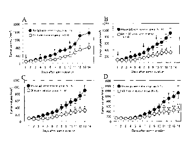
Figure 3 shows evaluation of the anti-tumor activity of A: clone 2-13 (rat
IgG2b), B: clone BA-1-3D (mouse IgG2a), C: clone DI-6 (mouse IgG1) and D:
clone
M3-1 (mouse IgG1), on Xenograft Treatment models using Huh-7-hDlk cells. The
tumor volume is indicated by a mean value standard error. The asterisk shows
the
results of a significant difference test (* P < 0.01, **P <0.05 by Student' s-
t-test).
Figure 4 shows the anti-tumor activity of an anti-hDlk-1 monoclonal antibody
(clone 2-13) on Xenograft Treatment models using hDlk-1 -expressing colon
cancer cell
line (SW480-hDlk cells). The SW480-hDlk cells were transplanted subcutaneously
in
nude mice to establish colon cancer tissues. The arrow indicates an
intraperitoneal
administration of antibody (20 mg/kg body weight) to the mice. The tumor
volume was
indicated by a mean value standard error (*P < 0.01, * *P <0.05 by Student'
s-t-test).
Figure 5 shows the reactivity of anti-hDlk-1 monoclonal antibodies with
HEK293-hDlk cells measured by flowcytometry. The number described in each
histogram indicates each clone number. The filled portions indicate the
isotype control
antibodies. The black-lined portions indicate anti-hDlk-1 monoclonal
antibodies.
Figure 6 shows the reactivity of anti-hDlk-1 monoclonal antibodies with Huh-
7-hDlk cells measured by flowcytometry. The number described in each histogram
indicates each clone number. The filled portions indicate the isotype control
antibodies.
The black-lined portions indicate anti-hDlk-1 monoclonal antibodies.
Figure 7 is a schematic view showing mutants in which each EGF-like motif
was deleted, which were produced in order to analyze epitopes recognized by
anti-hDlk-
1 monoclonal antibodies.
Figure 8 shows the results of the epitope analysis of clone DI-2-14.
Figure 8A: A figure showing the results obtained by transiently transfecting
11
CA 02669731 2009-05-05
each described hDlk-1 gene mutant into COS-7 cells by lipofection and then
performing
FACS analysis on the cells 24 to 72 hours after the gene transfection (left:
mouse IgG1 ,
right: DI-2-14). The gated portions indicate each mutant-expressing cells
recognized by
the clone DI-2-14.
Figure 8B: Photographs showing the smears of EGF (1-2)-expressing COS-7
cells, which were immunostained with a positive control (an anti-hDlk-1
polyclonal
antibody) and the clone DI-2-14. Portions stained into brownish-red color
indicate
expression of the EGF (1-2).
Figure 9 shows the results of the epitope analyses of clones DI-6, BA-1-3D and
2-13.
Figure 9A: A figure showing the results obtained by transiently transfecting
each described hDlk-1 gene mutant into COS-7 cells by lipofection and then
performing
FACS analysis on the cells 24 to 72 hours after the gene transfection. The
gated
portions indicate each mutant-expressing cells recognized by each clone.
Figure 9B: Photographs showing the smears of EGF (1-2)-expressing COS-7
cells, which were immunostained with clones DI-6, BA-1-3D and 2-13. Portions
stained into brownish-red color, which are indicated by the arrows, shows
expression of
the EGF (1-2).
Figure 10 shows the results of the epitope analysis of clone M3-1 by FACS.
The results obtained by transiently transfecting each described hDlk-1 gene
mutant into COS-7 cells by lipofection and then performing FACS analysis on
the cells
24 to 72 hours after the gene transfection. The gated portions indicate each
mutant-
expressing cells recognized by DI-2-14.
Figure 11 shows the analytical results of the internalization activity of each
anti-hDlk-1 monoclonal antibody after it bound to an antigen.
Figure 11A: HEK293-hDlk cells were allowed to react with each anti-hDlk-1
monoclonal antibody (clones M3-1, M1-290 and M3-4) (4 C, 20 minutes) and the
resultant cells were then washed with PBS 2 times. Thereafter, the cells were
incubated
at 37 C for the period of time as described in the figure. Thereafter, the
cells were
12
CA 02669731 2009-05-05
allowed to react with PE-labeled anti-mouse IgG, followed by FACS analysis.
The
results are indicated with relative values, which are obtained when the mean
fluorescence
intensity in the case of no incubation (0 minute) is defined as 100%.
Figure 11B: FITC-labeled clone M3-1 (FITC-M3-1) was allowed to react with
HEK293-hDlk cells (4 C, 20 minutes) and the resultant cells were then washed
with PBS
2 times. Thereafter, the cells were incubated at 37 C for 120 minutes. Figure
11B
shows a change in the mean fluorescence intensity obtained as a result of the
aforementioned incubation. The black column indicates a change in the mean
fluorescence intensity obtained when unlabeled M3-1 was reacted with the cells
in the
same manner as in Figure 11A above, the cells were then incubated at 37 C for
120
minutes and they were then allowed to react with PE-labeled anti-mouse IgG.
Figure 12 shows the analytical results of the internalization activity of each
rhodamine-labeled anti-hDlk-1 monoclonal antibody after it is bound to an
antigen.
Figure 12A: HEK293-hDlk cells were allowed to react with rhodamine-labeled
M3-1 (Rho-M3-1) (4 C, 20 minutes) and the resultant cells were then washed
with PBS
2 times. Immediately after the washing, a smear was prepared and localization
of Rho-
M3-1 was observed under a fluorescence microscope. Figure 12A is a photograph
showing such localization of Rho-M3-1. Orange colored portions indicate
localization
of Rho-M3-1. Localization of Rho-M3-1 into cell membrane was observed.
Figure 12B to 12E: Rho-M3-1 (B), Rho-DI-1 (C) and Rho-M1-290 (D) and
Rho-M3-4 (E) were allowed to react with HEK293-hDlk cells and the resultant
cells
were then washed with PBS 2 times, followed by incubation at 37 C for 15
minutes.
Thereafter, smears were produced and localization of each clone was observed
under a
fluorescence microscope.
Figures 12B to 12E are photographs showing such
localization of each clone. Both Rho-M3-1 and Rho-DI-1, which recognize the
same
epitopes (EGF 4-6), were incorporated into the cells and they were localized
therein in
the form of dots.
Figure 13 shows the cytotoxic activity of saporin-conjugated anti-hDlk-1
monoclonal antibodies to Huh-7-hDlk cells and SK-N-Fl cells.
13
CA 02669731 2009-05-05
Figure 13A: A figure showing the effects of a control (mouse IgG-saporin
(IgG-SAP)) and M3-1-SAP on Huh-7-hDlk cells. The longitudinal axis indicates
the
survival rate of the cells, which is indicated by a relative value obtained
when the
survival rate of cells in the case of adding no antibodies is defined as 100%
(N = 3, a
mean value standard deviation).
Figure 13B: A view showing the effects of a control (IgG-SAP), M3-1-SAP
and M1-290-SAP on SK-N-Fl cells.
In Figure 14, Figure 14A shows a change in the body weight of each mouse
and Figure 14B shows the survival rate of mice, which were obtained when mouse
IgG
(20 mg/kg body weight), M3-1-SAP (5 mg/kg body weight) and M1-290-SAP (5 mg/kg
body weight) were administered to the Xenograft models of Huh-7-hDlk cells.
The
value is indicated by a mean value standard deviation. The arrows indicate
the day in
which the antibodies were administered.
Figure 15 shows the tumor growth-inhibiting effects of mouse IgG (0; N = 8)
and M3-1-SAP (0; N = 8) obtained when these antibodies were intraperitoneally
administered to the Xenograft models of Huh-7-hDlk cells. The arrows indicate
the day
in which the antibodies were administered. The value is indicated by a mean
value
standard deviation (* P <0.01, ** P < 0.05 by Student's t-test).
In Figure 16, Figure 16A shows a tumor growth-inhibiting effect and Figure
16B shows a change in body weight, which were obtained when mouse IgG (0; N =
5)
and M3-1-SAP (0; N = 5) were intratumorally administered (40 g) to the
Xenograft
models of Huh-7-hDlk cells. Figure 16C shows a tumor growth-inhibiting effect
and
Figure 16D shows a change in body weight, which were obtained when PBS (0; N =
4)
and cisplatin (0; N = 4) were intraperitoneally administered (5 mg/kg body
weight) to
the Xenograft models of Huh-7-hDlk cells. In all the figures, the arrows
indicate the
day in which the antibodies were administered and the value is indicated by a
mean value
standard error (* P <0.01, ** P < 0.05 by Student's t-test).
Figure 17 includes photographs showing typical examples of a human colon
cancer tissue array (manufactured by Cybrdi; CC05-01-001) immunostained with
an anti-
14
CA 02669731 2009-05-05
hDlk-1 antibody. The brownish-red portions indicate cancer tissues stained
with the
anti-hDlk-1 antibody. The term "hDlk-1 negative" means a section in which no
stained
regions were observed, as in the case of section No. 48 (69-year-old male,
adenocarcinoma, Grade III). Section No. 19 (55-year-old female,
adenocarcinoma,
Grade II) was extremely strongly stained and sections stained at the same
level as section
No. 19 were defined as "hDlk-1 strongly positive." In addition, as in the case
of section
No. 25 (75-year-old male, adenocarcinoma, Grade II), a section that was
clearly
confirmed to be hDlk-1 positive and was slightly stained was defined as "hDlk-
1 weakly
positive."
Figure 18 includes photographs showing typical examples of a human breast
cancer tissue array (manufactured by Cybrdi; CC08-02-002) immunostained with
an anti-
hDlk-1 antibody. The brownish-red portions indicate cancer tissues stained
with the
anti-hDlk-1 antibody.
The upper photographs show normal mammary gland tissues contained in the
tissue array, which were stained with the anti-hDlk-1 antibody. Left: No. 07
(68-year-
old female, normal mammary gland; hDlk-1 negative), right: No. 01(43-year-old
female,
normal lobules of mammary gland; hDlk-1 weakly positive). The arrows indicate
hDlk-
1 weakly positive portions.
The lower photographs show the tissues of patients with infiltrating duct
carcinoma. Left: No. 08 (45-year-old female, Grade II; hDlk-1 negative),
center: No.
56 (28-year-old female, Grade II; hDlk-1 weakly positive), right: No. 20 (59-
year-old
female, Grade II; hDlk-1 strongly positive).
Figure 19 shows the dose-dependent anti-tumor activity of clone DI-2-14 on
Xenograft treatment models of Huh-7-dlk cells.
Figure 20 shows the dose-dependent anti-tumor activity of clone DI-2-14 on
Xenograft treatment models of SK-N-Fl cells.
Figure 20A shows tumor growth after initiation of the administration of the
antibodies. The tumor volume was indicated by a mean value + standard error
(*P <
0.01, **P <0.05 by Student's t-test).
CA 02669731 2009-05-05
Figure 20B is a graph showing the weight of the excised cancer tissues on the
23rd day (Day 23) after the administration of the antibodies.
Figure 21 shows the cDNA nucleotide sequence (SEQ ID NO: 22) of the H
chain (heavy chain) variable region (VH) of clone DI-2-14 and a putative amino
acid
sequence thereof (SEQ ID NO: 23). Signal peptides are described in italics.
The
double-lined glutamic acid (E) represents the N-terminal amino acid residue of
a mature
peptide. The CDR sequences (underlined) were provided in accordance with the
definition of Kabat et al. (Sequences of Proteins of Immunological Interests,
Fifth edition,
NIH Publication No. 91-3242, U.S. Department of Health and Human Services,
1991).
The amino acid sequences of CDRs 1 to 3 of clone DI-2-14 VH are as shown in
SEQ ID
NOS: 30 to 32, respectively.
Figure 22 shows the cDNA nucleotide sequence (SEQ ID NO: 24) of the L
chain (light chain) variable region (VL) of clone DI-2-14 and a putative amino
acid
sequence thereof (SEQ ID NO: 25). Signal peptides are described in italics.
The
double-lined aspartic acid (D) represents the N-terminal amino acid residue of
a mature
peptide. The CDR sequences (underlined) were provided in accordance with the
definition of Kabat et al. (1991; as described above). The amino acid
sequences of
CDRs 1 to 3 of clone DI-2-14 VL are as shown in SEQ ID NOS: 33 to 35,
respectively.
Figure 23 shows the nucleotide sequence (SEQ ID NO: 26) of a clone D1-2-14
VH gene and the amino acid sequence thereof (SEQ ID NO: 27). An SpeI site was
added to the 5'-terminus and a HindIII site was added to the 3'-terminus (the
two sites
were underlined). The nucleotide sequence described in italics indicates a
sequence
corresponding to an intron.
Figure 24 shows the nucleotide sequence (SEQ ID NO: 28) of a clone DI-2-14
VL gene and the amino acid sequence thereof (SEQ ID NO: 29). An NheI site was
added to the 5'-terminus and an EcoRI site was added to the 3'-terminus (the
two sites
were underlined). The nucleotide sequence described in italics indicates a
sequence
corresponding to an intron.
Figure 25 is a schematic view of a chimeric DI-2-14 gene expression vector
16
CA 02669731 2009-05-05
(pChDI-2-14). In a clockwise direction starting from the Sall site, this
vector comprises
a heavy chain translation unit starting with a human cytomegalovirus (CMV)
major
immediate early promoter and an enhancer used for initiation of the
transcription of an
antibody heavy chain gene. The CMV region then proceeds to a VH exon, the gene
sequence of a human yl heavy chain constant region (which comprises the exons
of CH1,
a hinge region, CH2 and CH3, via introns) and after CH3, a poly A portion used
for
mRNA processing. After the heavy chain gene sequence, the vector comprises a
light
chain translation unit starting with a CMV promoter and an enhancer and it
further
comprises a VL exon, the exon (CL) of a human lc chain constant region having
an intron
portion upstream thereof and the poly A signal of a lc gene. Thereafter, the
light chain
gene proceeds to a segment comprising an SV40 early promoter, an E. coli
xanthine
guanine phosphoribosyl transferase (gpt) gene and the poly A portion of SV40.
Finally,
the plasmid has a part of a pUC19 plasmid comprising the replication origin of
bacteria
and a 13- lactamase gene.
Figure 26 shows mouse DI-2-14 and chimeric DI-2-14 (ChDI-2-14), which
were developed by SDS-PAGE and were then stained with CBB. MW indicates a size
marker and the arrows indicate the molecular weights of bands (kD).
Figure 27 shows the antigen-binding activity of DI-2-14 and ChDI-2-14
measured by purified FA-1 (hDlk-1 extracellular region) solid-phase ELISA. 0
indicates a mouse IgG1 antibody (clone DI-2-14) and = indicates a chimeric
antibody
(ChDI-2-14).
Figure 28 shows the reactivity of DI-2-14 and ChDI-2-14 with HEK293-hDlk
cells measured by flowcytometry. The filled histograms indicate isotype
control
antibodies and the black-lined (opened) histograms indicate DI-2-14 and ChDI-2-
14.
Figure 29 shows the results obtained by analyzing expression of cell surface
Dlk-1 in human liver cancer cell lines by flowcytometry. The blue line
indicates mouse
IgG1 and the red line indicates an anti-human Dlk-1 antibody. These are
histograms
obtained by staining each type of cells.
Figure 30 shows the results obtained by analyzing expression of cell surface
17
CA 02669731 2014-04-11
30179-182
Dlk-1 in human breast cancer cell lines by flowcytometry. The blue line
indicates
mouse IgG1 and the red line indicates an anti-human Dlk-1 antibody. These are
histograms obtained by staining each type of cells.
Figure 31 shows the results obtained by analyzing expression of cell surface
Dlk-1 in human leukemia cell lines by flowcytometry. The blue line indicates
mouse
IgG1 and the red line indicates an anti-human Dlk-1 antibody. These are
histograms
obtained by staining each type of cells.
Figure 32 shows the immunohistostaining of Flk-1/VEGF-R2 in cancer tissues
excised from the Xenograft treatment models of Huh-7-dlk cells.
Figure 32A includes photographs in which the fresh frozen sections of cancer
tissues collected (excised) from a mouse IgG administration group (a control
group) and
a clone DI-2-14 administration group were immunostained with an anti-mouse Flk-
1NEGF-R2 antibody (objective 200-fold). In the photographs, portions indicated
by
the arrowhead (A) indicates Flk-1/VEGF-R2 positive tumor vascular endothelial
cells.
Figure 32B is a graph, which was prepared by immunostaining the fresh frozen
sections of cancer tissues collected (excised) from an IgG administration
group (2
individuals) and a DI-2-14 administration group (4 individuals) with an anti-
mouse Flk-
1/VEGF-R2 antibody, then counting the number of Flk-1/VEGF-R2 positive tumor
vascular endothelial cells in 8 to 13 visual fields (the IgG administration
group: total 21
visual fields, the DI-2-14 administration group: total 35 visual fields) under
an objective
lens of 200-fold and then showing the number of cells per visual field (*P <
0.01 by
Student's t-test).
Figure 33 shows gene expression of Flk-1/VEGF-R2 in cancer tissues excised
from the Xenograft treatment models of Huh-7-dlk cells.
Figure 33A shows an electrophoretic image, in which tumor was excised from
each of an IgG administration group (N = 7) and a DI-2-14 administration group
(N = 7),
TM
RNA was then extracted from the tumor using a Trizol reagent, 1st strand cDNA
was then
synthesized and the gene expression of mouse Flk-1/VEGF-R2 (mFlk-1) and
mouse/human GAPDH (GAPDH) was then confirmed by a PCR method (30 cycles)
18
CA 02669731 2014-04-11
30179-182
using each 1st strand cDNA as a template. With regard to the lower graph, a
band of the
amplification product obtained by PCR performed on mFlk-1 and GAPDH was
quantified by NIH image and it was then expressed in the form of a ratio (mFlk-
1/GAPDH).
Figure 33B is a figure obtained in the same manner as in Figure 33A above,
with the exception that the amplification reaction was carried out for 35
cycles by the
PCR method.
BEST MODE FOR CARRYING OUT THE INVENTION
Hereinafter, the present invention will be described in detail: The following
descriptions are not intended to limit the scope of the present invention.
Other than the
following examples, the present invention may be modified and may be carried
out, as
appropriate, within a range that does not impair the intention of the present
invention.
Japanese Patent Application No. 2006-305355 is a priority document of the
present application.
1. Summary of the present invention
As described above, human DIk-1 (delta-like 1 homolog (Drosophila); hDlk-1)
is a type I transmembrane (one-transmembrane-type) protein with a full length
of 383
amino acid residues and this protein has 6 EGF-like motifs in its
extracellular region. It
has been known that a hDlk-I gene and a gene product thereof are expressed
with a high
frequency in various types of cancer or tumor cells. In general, it is
difficult to prepare
and obtain an antibody exhibiting anti-tumor activity in vivo. Thus, even if
an anti-
hDlk-1 monoclonal antibody is produced, it has anti-tumor activity in vitro
but it does
not exhibit the activity in vivo in many cases. Moreover, the functional
domain of
19
CA 02669731 2009-05-05
=
hDlk-1 that acts on the growth of cancer cells, a ligand (or a receptor) of
hDlk-1, its
intracellular signal-transducing pathway and the like have not been clarified.
Thus, it is
substantially impossible to efficiently produce an antibody by narrowing down
its target.
Under such circumstances, in the present invention, a clone having anti-tumor
activity in
vivo has been successfully obtained by screening it from a large number of
clones.
First, based on immunohistochemistry using known anti-hDlk-1 antibodies, the
present inventors have discovered that hDlk-1 is expressed in colon cancer and
breast
cancer, in addition to the aforementioned cancers and tumor cells, in which
expression of
hDlk-1 had previously been confirmed.
Next, the present inventors have newly produced approximately 100 clones of
anti-hDlk-1 monoclonal antibodies for the purpose of producing anti-hDlk-1
antibodies
capable of killing hDlk- 1-expressing cancer cells at an individual level or
inhibiting
tumor growth, namely, anti-hDlk-l-antibodies having anti-tumor activity in
vivo.
Thereafter, the inventors have evaluated the in vivo pharmaceutical effects
(anti-tumor
action) of these clones, using tumor-bearing mice established by transplanting
various
types of cancer cell lines subcutaneously in nude mice. As a result, the
present
inventors have succeeded in obtaining several clones exhibiting significant
tumor
growth-inhibiting activity (clone name: DI-2-14, 2-13, BA-1-3D, DI-6 and M3-
1).
Moreover, among the aforementioned anti-hDlk-1 antibodies, the present
inventors have found an antibody excellent in terms of migratory ability to
move into
cells that express hDlk-1 (internalization activity) and the inventors have
produced an
antibody-agent complex, which comprises such an antibody and a compound having
anti-
tumor activity or cell-killing activity.
This complex is what is called an
"immunoconjugate," which is excellent in terms of ability to deliver agents
into tumor
cells as targets.
The present inventors have found that the aforementioned anti-hDlk-1 antibody
or antibody-agent complex having anti-tumor activity is useful for the
treatment of
various types of tumors, or for the diagnosis and detection of tumors.
CA 02669731 2009-05-05
2. Preparation of anti-hDlk-1 antibody
(1) Preparation of antigen
Information regarding the amino acid sequence (SEQ ID NO: 2) of hDlk-1 is
disclosed as "Accession number: NP 003827" at the website of NCBI (GenBank)
(http://www.ncbi.nlm.nih.gov/), for example.
Moreover, information regarding a
nucleotide sequence (SEQ ID NO: 1) encoding the amino acid sequence of hDlk-1
is
disclosed as "Accession number: NM 003836" at the same above website.
As an antigen, a polypeptide or peptide (which may be simply referred to as a
"peptide" at times) comprising at least a portion of (entire or a part of) the
amino acid
sequence of hDlk-1 can be used and preferably, a peptide comprising at least a
portion of
(entire or a part of) the amino acid sequence of the extracellular region (FA-
1) of hDlk-1
can be used. As stated above, the extracellular region of hDlk-1 comprises 6
EGF-like
motifs (EGF-1 to EGF-6). This region indicates a region comprising amino acids
at
positions 26 to 244 in the amino acid sequence as shown in SEQ ID NO: 2 and
preferably a region consisting of amino acids from "position 24" to "positions
248 to
285" (approximately 225 to 262 amino acid residues) in the amino acid sequence
as
shown in SEQ ID NO: 2.
Herein, in the case of a peptide used as an antigen, the length of the
aforementioned "at least a portion of the amino acid sequence" is not
particularly limited.
For example, a region comprising one or two or more out of the 6 EGF-like
motifs is
preferable. More preferable examples include a region comprising EGF-1 and EGF-
2
(namely, a region consisting of amino acids at positions 26 to 85 in the amino
acid
sequence as shown in SEQ ID NO: 2), a region comprising EGF-3 and EGF-4
(namely, a
region consisting of amino acids at positions 92 to 167 in the amino acid
sequence as
shown in SEQ ID NO: 2) and a region comprising EGF-4, EGF-5 and EGF-6 (namely,
a
region consisting of amino acids at positions 131 to 244 in the amino acid
sequence as
shown in SEQ ID NO: 2).
As a method for preparing a peptide used as an antigen, either a chemical
synthesis, or a synthesis by a genetic engineering means using Escherichia
coil or the
21
CA 02669731 2009-05-05
like, may be applied. Methods well known to persons skilled in the art may be
applied.
In the case of performing a chemical synthesis of peptide, such a peptide may
be synthesized by well known methods for synthesizing peptides. As such a
synthesis,
either a solid-phase synthesis method or a liquid-phase synthesis method may
be applied.
Commercially available peptide synthesizing apparatuses (e.g. PS SM-8, etc.;
manufactured by Shimadzu Corp.) may be used.
In the case of synthesizing a peptide by genetic engineering, DNA encoding the
peptide is first designed and synthesized. The designing and synthesis of the
DNA can
be carried out, for example, by a PCR method, using a vector comprising a full-
length
hDlk-1 gene or the like as a template and also using primers designed such
that a desired
DNA region can be synthesized therewith. Thereafter, the thus synthesized DNA
is
ligated to a suitable vector to obtain a recombinant vector used in expression
of a protein.
This recombinant vector is then introduced into a host such that a gene of
interest can be
expressed therein, so as to obtain a transformant (Sambrook J. et al.,
Molecular Cloning,
A Laboratory Manual, 31(1 edition, Cold Spring Harbor Laboratory Press, 2001).
As a vector, a phage or plasmid capable of autonomously replicating in host
microorganisms can be used. Further, an animal virus or insect virus vector
can also be
used. For preparation of a recombinant vector, the purified DNA may be cleaved
with
suitable restriction enzymes, the obtained DNA portion may be then inserted
into the
restriction site of suitable vector DNA, etc. and it may be then ligated to a
vector. The
type of a host used in transformation is not particularly limited, as long as
it is able to
express a gene of interest. Examples of such a host include bacteria
(Escherichia coil,
Bacillus subtilis, etc.), yeasts, animal cells (COS cells, CHO cells, etc.),
insect cells and
insects. It is also possible to use a mammal such as a goat as a host. A
method for
introducing a recombinant vector into a host is known.
The aforementioned transformant is cultured and a peptide used as an antigen
is
then collected from the culture. The term "culture" is used to mean any one of
(a) a
culture supernatant and (b) cultured cells, a cultured cell mass, or a
disintegrated product
thereof.
22
CA 02669731 2009-05-05
After completion of the culture, when a peptide of interest is produced in a
bacterial cells (bacterial bodies) or in cells, such bacterial cells or cells
are disintegrated
and a peptide is then extracted. On the other hand, a peptide of interest is
produced
outside the bacterial cell or cells, a culture solution is directly used, or
the bacterial cells
or cells are eliminated by centrifugation or the like. Thereafter, common
biochemical
methods used in isolation and purification of peptides, such as ammonium
sulfate
precipitation, gel filtration, ion exchange chromatography and affinity
chromatography,
are applied singly or in combination, so as to isolate and purify a peptide of
interest.
In the present invention, a peptide used as an antigen can also be obtained by
in
vitro translation using a cell-free synthesis system. In this case, two types
of methods,
namely, a method using RNA as a template and a method using DNA as a template
(transcription/translation) can be applied. As such a cell-free synthesis
system,
commercially available systems such as ExpresswayTM system (Invitrogen),
PURESYSTEM (registered trade mark; Post Genome Institute Co., Ltd.) and TNT
system (registered trade mark; Promega) can be used.
The thus obtained peptide may also be bound to a suitable carrier protein such
as bovine serum albumin (BSA), keyhole limpet hemocyanin (KLH), human
thyroglobulin, or chicken gamma globulin.
Furthermore, such an antigen may be a peptide, which consists of an amino acid
sequence comprising a deletion, substitution or addition of one or multiple
amino acids
with respect to the amino acid sequence of hDlk-1 (SEQ ID NO: 2) or the
aforementioned partial sequence thereof For example, there can also be used a
peptide,
which consists of an amino acid sequence comprising a deletion of one or
multiple
(preferably one or several (for example 1 to 10 and more preferably 1 to 5) )
amino acids,
a substitution of one or multiple (preferably one or several (for example 1 to
10 and more
preferably 1 to 5) ) amino acids with other amino acids, or an addition of one
or multiple
(preferably one or several (for example 1 to 10 and more preferably 1 to 5) )
amino acids,
with respect to the amino acid sequence of hDlk-1 or a partial sequence
thereof
23
CA 02669731 2009-05-05
=
In the present invention, an example of a gene to be introduced into cells or
the
like is a gene encoding a hDlk-1 protein, a partial fragment thereof, a mutant
protein
thereof, or a fragment thereof. As such a gene, a gene having the nucleotide
sequence
as shown in SEQ ID NO: 1 or a partial sequence thereof can be used, for
example.
Further, as such a gene to be introduced into cells or the like, a nucleotide
sequence, which hybridizes with a sequence complementary to the nucleotide
sequence
as shown in SEQ ID NO: 1 under stringent conditions and encodes a protein
having
hDlk-1 activity, or a partial sequence thereof can also be used.
The term "stringent conditions" is used to mean conditions applied to washing
after hybridization, which consist of a salt (sodium) concentration of buffer
between 10
and 500 mM and a temperature between 42 C and 72 C and preferably consist of
the
aforementioned salt concentration of buffer between 50 and 300 mM and a
temperature
between 55 C and 68 C.
Mutation can be introduced into a gene by known methods such as a Kunkel
method or a Gapped duplex method, using mutation introduction kits that
utilize site-
directed mutagenesis, such as GeneTailorTm Site-Directed Mutagenesis System
(manufactured by Invitrogen) or TaKaRa Site-Directed Mutagenesis System (Mutan-
K,
Mutan-Super Express Km, etc., manufactured by Takara Bio Inc.).
(2) Preparation of polyclonal antibody
The prepared antigen is administered to a mammal for immunization. The
type of such a mammal is not particularly limited. Examples of such a mammal
include
a rat, a mouse and a rabbit. Among others, a mouse is preferable.
The dose of the antigen per animal can be determined, as appropriate,
depending on the presence or absence of an adjuvant. Examples of such an
adjuvant
include a Freund's complete adjuvant (FCA), a Freund's incomplete adjuvant
(FIA) and
an aluminum hydroxide adjuvant. Immunization can be carried out by injecting
the
antigen into the vein, footpad, subcutis, abdominal cavity, etc. In addition,
immunization interval is not particularly limited. Immunization is carried out
1 to 10
24
CA 02669731 2009-05-05
times and preferably 2 or 3 times, at intervals of several days to several
weeks and
preferably at intervals of 1 week. Three to seven days after the final
immunization, an
antibody titer is measured by enzyme immunoassay (ELISA or ETA),
radioimmunoassay
(RIA), etc. On the day at which a desired antibody titer is obtained, blood is
collected
and antiserum is then obtained. In a case where an antibody should be purified
in the
aforementioned method for collecting the antibody, a suitable method is
appropriately
selected from known methods such as an ammonium sulfate salting-out method,
ion
exchange chromatography, gel filtration chromatography and affinity
chromatography, or
these methods may be used in combination, so as to purify the antibody.
Thereafter, the
reactivity of a polyclonal antibody contained in the antiserum is measured by
ELISA, etc.
(3) Preparation of monoclonal antibody
(3-1) Collection of antibody-producing cells
The type of the anti-hDlk-1 antibody of the present invention is not limited.
A
monoclonal antibody is preferable.
The prepared antigen is administered to a mammal such as a rat, a mouse or a
rabbit for immunization. The dose of the antigen per animal can be determined,
as
appropriate, depending on the presence or absence of an adjuvant. The same
adjuvants
as those described above are used herein. Also, the same immunization methods
as
described above are applied herein. One to sixty days and preferably one to
fourteen
days after the final immunization, antibody-producing cells are collected.
Examples of
such antibody-producing cells include splenic cells, lymph node cells and
peripheral
blood cells. Among others, lymph node cells and splenic cells are preferable.
(3-2) Cell fusion
In order to obtain a hybridoma (an antibody-producing cell line), cell fusion
is
carried out between antibody-producing cells and myeloma cells. As myeloma
cells to
be fused with antibody-producing cells, easily available, established cell
lines, such as
the cell lines of animals such as mice, can be used. As available cell lines,
those, which
CA 02669731 2009-05-05
have drug selectivity, cannot survive in a HAT selective medium (containing
hypoxanthine, aminopterin and thymidine) when they are in an unfused state and
can
survive therein only when they are fused with antibody-producing cells, are
preferable.
Examples of myeloma cells used herein include mouse myeloma cell lines such
as P3-X63-Ag8.653, P3-X63-Ag8(X63), P3-X63-Ag8.U1(P3U1), P3/NS I/1-Ag4-1(NS1)
and Sp2/0-Ag14(Sp2/0). Such myeloma cells can be selected, while taking into
consideration the compatibility with antibody-producing cells, as appropriate.
Subsequently, myeloma cells are fused with antibody-producing cells for cell
fusion. For such cell fusion, antibody-producing cells at a cell density of 1
x 106 to 1 x
107 cells/mL are mixed with myeloma cells at a cell density of 2 x 10⁵ to 2 x
106 cells/mL,
in a medium used for animal cells that does not contain serum, such as DMEM or
a
RPMI-1640 medium. The cell ratio between such antibody-producing cells and
such
myeloma cells (antibody-producing cells : myeloma cells) is not limited. In
general,
such a cell ratio is preferably between 1 : 1 and 10 : 1 and more preferably 3
: 1.
Subsequently, a fusion reaction is carried out in the presence of a cell
fusion promoter.
As such a cell fusion promoter, polyethylene glycol having a mean molecular
weight
between 1,000 and 6,000 daltons (D) or the like can be used, for example.
Also,
antibody-producing cells can be fused with myeloma cells using a commercially
available cell fusion device that utilizes electrical stimulation (e.g.
electroporation).
(3-3) Selection of hybridoma and cloning
A hybridoma of interest is selected from cells obtained after the cell fusion
treatment. As a selection method, a cell suspension is diluted with a fetal
bovine
serum-containing RPMI-1640 medium or the like, as appropriate and the diluted
solution
is then dispersed on a microtiter plate. A selective medium is added to each
well and
culture is then carried out while the selective medium is appropriately
exchanged with a
fresh one. As a result, cells that grow approximately 14 days after initiation
of the
culture in the selective medium can be obtained as hybridomas.
26
CA 02669731 2009-05-05
Subsequently, the presence or absence of an antibody against hDlk-1 in a
culture supernatant of the growing hybridomas is screened. Such screening of
hybridomas may be carried out in accordance with ordinary methods and thus the
type of
the screening method is not particularly limited. For example, a portion of
the culture
supernatant of the growing hybridomas contained in the well may be collected
and such
hybridomas may be then screened by ELISA, ETA, RIA, etc.
The fused cells may be cloned by limiting dilution or the like. An antibody
exhibiting strong reactivity with hDlk-1 is determined by flowcytometry or the
like and a
hybridoma that produces the antibody is selected and is established as a
clone.
(3-4) Collection of monoclonal antibody
As a method of culturing the established hybridomas and then collecting a
monoclonal antibody from the obtained culture, a common cell culture method,
an
ascites formation method, etc. can be adopted. The term "culture" is used to
mean that
a hybridoma is allowed to grow in a culture dish or culture bottle, or that a
hybridoma is
allowed to proliferate in the abdominal cavity of an animal, as described
below.
In the cell culture method, hybridomas may be cultured in an animal cell
culture
medium such as a 10% fetal bovine serum-containing RPMI-1640 medium, an MEM
medium or a serum-free medium under common culture conditions (e.g. 37 C, 5%
CO2
concentration) for 7 to 14 days and an antibody may be then obtained from the
culture
supernatant.
In the ascites formation method, hybridomas are administered at a cell density
of approximately 1 x 10⁷ cells into the abdominal cavity of an animal of the
same species
as a mammal from which myeloma cells are derived, so as to cause proliferation
of a
large amount of hybridomas. Thereafter, ascites is preferably collected 2 to 3
weeks
later.
In a case where an antibody should be purified in the aforementioned method
for collecting the antibody, a suitable method is appropriately selected from
known
methods such as an ammonium sulfate salting-out method, ion exchange
chromatography,
27
CA 02669731 2009-05-05
gel filtration and affinity chromatography, or these methods are used in
combination, so
as to purify the aforementioned antibody.
(3-5) Selection of clone having anti-tumor activity
The anti-hDlk-1 antibody of the present invention is an antibody having anti-
tumor activity in vivo.
Herein, the term "anti-tumor activity" is used to mean activity of killing
tumor
cells (cancer cells) or inhibiting tumor growth. In the present invention, as
such anti-
tumor activity, tumor angiogenesis-inhibiting activity is preferable, for
example.
Moreover, the types of human tumors (tumor cells), on which the antibody of
the present
invention is able to exhibit anti-tumor activity, include: the aforementioned
known
human tumors in which expression of hDlk-1 had been confirmed (specifically,
solid
cancers such as neuroendocrine tumor, neuroblastoma, glioma, neurofibromatosis
type 1,
small cell lung cancer, liver cancer, kidney cancer and ovarian cancer and
blood cancers
such as myelodysplastic syndrome and acute myelocytic leukemia); and human
colon
cancer and human breast cancer in which expression of hDlk-1 has been newly
confirmed by the present inventors. Of these, one or two or more types
selected from
human colon cancer, human breast cancer, human liver cancer and human
neuroblastoma
are particularly preferable.
The presence of anti-tumor activity in vivo can be confirmed by using a cancer-
bearing mouse,in which desired tumor cells have been transplanted
subcutaneously, and
then administering the obtained antibody to the mouse. In this case, the
antibody may
be administered to the mouse immediately after transplantation of the tumor
cells (a
Prevention model), or the antibody may also be administered to the mouse after
the
tumor has grown up to a desired volume after transplantation (a Treatment
model). An
administration method is not limited. For example, the antibody may be
administered
into the abdominal cavity of the mouse once every 3 days at a dose of 20 mg/kg
body
weight via intraperitoneal administration. In the case of the Prevention
model, the
presence or absence of anti-tumor activity and the level thereof can be
evaluated
28
CA 02669731 2009-05-05
depending on tumor formation frequency and tumor volume. In the case of the
Treatment model, the presence or absence of anti-tumor activity and the level
thereof can
be evaluated depending on tumor volume.
In the present invention, preferred examples of an anti-hDlk-1 antibody having
anti-tumor activity in vivo include an anti-hDlk-1 monoclonal antibody (clone
name: M3-
1) produced by a hybridoma having accession No. FERM BP-10707, an anti-hDlk-1
monoclonal antibody (clone name: DI-2-14) produced by a hybridoma having
accession
No. FERM BP-10899 and an anti-hDlk-1 monoclonal antibody (clone name: DI-6)
produced by a hybridoma having accession No. FERM BP-10900. Furthermore, an
anti-hDlk-1 monoclonal antibody with a clone name of DI-2-14 can be preferably
used as
an antibody having high anti-tumor activity in vivo.
Herein, the hybridoma having accession No. FERM BP-10707 has been
referred to as "Mouse-Mouse hybridoma: M3-1," and has been deposited with
International Patent Organism Depositary (IPOD), National Institute of
Advanced
Industrial Science and Technology (AIST Tsukuba Central 6, Higashi 1-1-1,
Tsukuba,
Ibaraki, Japan, postal code: 305-8566), on October 18, 2006. The hybridoma
having
accession No. FERM BP-10899 has been referred to as "Mouse-Mouse hybridoma DI-
2-
14," and has been deposited with the same above national institute on August
21, 2007.
The hybridoma having accession No. FERM BP-10900 has been referred to as
"Mouse-
Mouse hybridoma DI-6," and has been deposited with the same above national
institute
on August 21, 2007.
Further, preferred examples of the anti-hDlk-1 antibody of the present
invention include an anti-hDlk-1 antibody wherein the amino acid sequences of
CDRs 1
to 3 of the H chain V region are the amino acid sequences as shown in SEQ ID
NOS: 30
to 32, respectively and/or an anti-hDlk-1 antibody wherein the amino acid
sequences of
CDRs 1 to 3 of the L chain V region are the amino acid sequences as shown in
SEQ ID
NOS: 33 to 35, respectively. The aforementioned H chain V region preferably
consists
of the amino acid sequence as shown in SEQ ID NO: 23 and the aforementioned L
chain
V region preferably consists of the amino acid sequence as shown in SEQ ID NO:
25.
29
CA 02669731 2009-05-05
=
Still further, another preferred example of the anti-hDlk-1 antibody of the
present invention is an anti-hDlk-1 antibody that binds to a site (e.g. an
epitope), to
which a monoclonal antibody produced by the hybridoma having accession No.
FERM
BP-10707, FERM BP-10899 or FERM BP-10900 binds (recognizes).
(3-6) Epitope of anti-hDlk-1 antibody
An epitope (an antigenic determinant) of the anti-hDlk-1 antibody of the
present invention is not limited, as long as it is at least a portion of hDlk-
1 as an antigen.
For example, such an epitope is preferably at least a portion of a region
consisting of
amino acids at positions 26 to 85 (a region comprising EGF-1 to EGF-2 of hDlk-
1), a
region consisting of amino acids at positions 92 to 167 (a region comprising
EGF-3 to
EGF-4 of hDlk-1), or a region consisting of amino acids at positions 131 to
244 (a region
comprising EGF-4 to EGF-6 of hDlk-1), in the amino acid sequence of hDlk-1 as
shown
in SEQ ID NO: 1. Among others, a region comprising EGF-4 to EGF-6 and a region
comprising EGF-4 to EGF-6 of hDlk-1 are more preferable. A region consisting
of
amino acids at positions 92 to 120 (a region comprising EGF-3 of hDlk-1) is
particularly
preferable. An anti-hDlk-1 antibody that recognizes (binds to) such regions
has high
internalization activity into tumor cells, for example and thus it is
extremely useful as an
immunoconjugate as described later.
(4) Genetically recombinant antibody and antibody fragment
(4-1) Genetically recombinant antibody
In a preferred embodiment of the anti-hDlk-1 antibody of the present
invention,
there is provided a genetically recombinant antibody. The type of such a
genetically
recombinant antibody is not limited. Examples include a chimeric antibody, a
humanized antibody and a human antibody.
A chimeric antibody (that is, a humanized chimeric antibody) is an antibody
formed by ligating (conjugating) the variable region of a mouse-derived
antibody to the
constant region of a human-derived antibody (please refer to Proc. Natl. Acad.
Sci.
CA 02669731 2009-05-05
U.S.A. 81, 6851-6855, (1984), etc.). When such a chimeric antibody is
produced, the
thus ligated antibody can be easily constructed by a genetic recombination
technique.
As such variable regions of the mouse-derived antibody used herein, the H
chain V
region preferably consists of the amino acid sequence as shown in SEQ ID NO:
23, for
example and the L chain V region preferably consists of the amino acid
sequence as
shown in SEQ ID NO: 25, for example.
When a humanized antibody is produced, a complementarity determining
region (CDR) is transplanted from the variable region of a mouse antibody into
the
variable region of a human antibody, so as to produce a reconstructed variable
region, in
which a framework region (FR) is derived from the human and CDR is derived
from the
mouse (what is called CDR grafting (CDR transplantation)). Subsequently, the
thus
humanized, reconstructed human variable region is ligated to a human constant
region.
Such a method for producing a humanized antibody is well known in the present
technical field (please refer to Nature, 321, 522-525 (1986); J. Mol. Biol.,
196, 901-917
(1987); Queen C et al., Proc. Natl. Acad. Sci. USA, 86: 10029-10033 (1989); JP
Patent
Publication (Kohyo) No. 4-502408 A (1992) (Japanese Patent No. 2828340; Queen
et
al.), etc.) The type of a mouse-derived CDR sequence that can be used
herein for the
humanized anti-hDlk-1 antibody of the present invention is not limited. As
preferred
examples of such mouse-derived CDR sequences, the amino acid sequences as
shown in
SEQ ID NOS: 30 to 32 are preferable as the CDRs 1 to 3 of the H chain V region
(in this
order) and the amino acid sequences as shown in SEQ ID NOS: 33 to 35 are
preferable
as the CDRs 1 to 3 of the L chain V region (in this order).
In general, in the case of a human antibody (a complete human antibody), its
structure comprising a Hyper Variable region that is the antigen-binding site
of a V
region, other parts of the V region and a constant region is the same as the
structure of
the antibody of a human. However, such a Hyper Variable site may also be
derived
from other animals. A technique of producing a human antibody is publicly
known and
a method for producing gene sequences that are common in humans by genetic
engineering has been established. A human antibody can be obtained, for
example, by a
31
CA 02669731 2009-05-05
method using a human antibody-producing mouse that has human chromosomal
fragments comprising the genes of the H chain and L chain of the human
antibody
(please refer to Tomizuka, K.et al., Nature Genetics, (1977) 16, 133-143;
Kuroiwa, Y. et.
al., Nuc. Acids Res., (1998) 26, 3447-3448; Yoshida, H. et. al., Animal Cell
Technology:
Basic and Applied Aspects, (1999) 10, 69-73 (Kitagawa, Y., Matsuda, T. and
Iijima, S.
eds.), Kluwer Academic Publishers; Tomizuka, K. et. al., Proc. Natl. Acad.
Sci. USA,
(2000) 97, 722-727, etc.), or by a method of obtaining a phage display-derived
human
antibody selected from a human antibody library (please refer to Wormstone, I.
M. et. al,
Investigative Ophthalmology & Visual Science., (2002) 43 (7), 2301-8; Carmen,
S. et. al.,
Briefings in Functional Genomics and Proteomics,(2002) 1 (2), 189-203;
Siriwardena, D.
et. al., Opthalmology, (2002) 109 (3), 427-431, etc.).
In the case of the aforementioned chimeric antibody, humanized antibody and
human antibody, the N-glycoside-linked sugar chain in the antibody Fc region
is
preferably a sugar chain, in which fucose does not bind to N-acetylglucosamine
at the
reducing terminal thereof. A specific example is an antibody consisting of
genetically
recombinant antibody molecules, which has, in the Fc region of the antibody
molecules,
a sugar chain in which the position 1 of the fucose does not bind to the
position 6 of the
N-acetylglucosamine at the reducing terminal of the N-glycoside-linked sugar
chain via
an a bond. Such an antibody is able to significantly improve ADCC activity.
This
point (the characteristics of the N-glycoside-linked sugar chain in the
antibody Fc region)
is preferable also for the aforementioned polyclonal antibody and monoclonal
antibody.
(4-2) Antibody fragment
The anti-hDlk-1 antibody fragment of the present invention is included in the
antibody of the present invention. Herein, the antibody fragment of the
present
invention has binding activity to hDlk-1 and anti-tumor activity in vivo, as
in the case of
the anti-hDlk-1 antibody of the present invention.
The fragment of the antibody means a region of a portion of an anti-hDlk-1
polyclonal antibody or anti-Dlk-1 monoclonal antibody (namely, an antibody
fragment
32
CA 02669731 2009-05-05
derived from the anti-hDlk-1 antibody of the present invention). Examples of
such an
antibody fragment include peptides comprising, as at least a portion thereof,
Fab, Fab',
F(ab')2, Fv (variable fragment of antibody), a single-stranded antibody (an H
chain, an L
chain, an H chain V region and an L chain V region, etc.), scFv, diabody (scFv
dimer),
dsFy (a disulfide-stabilized V region) and a complementarity determining
region (CDR).
Fab is an antibody fragment with a molecular weight of approximately 50,000
having antigen-binding activity, which is formed by binding about a half of
the N-
terminal side of the H chain and the entire L chain via a disulfide bond,
among fragments
obtained by treating antibody molecules with a protease, papain. In addition,
it is also
possible to produce such Fab by inserting DNA encoding the Fab of an antibody
into a
prokaryote expression vector or a eukaryote expression vector and then
introducing the
vector into a prokaryote or a eukaryote so as to allow the DNA to express
therein.
F(ab')2 is an antibody fragment with a molecular weight of approximately
100,000 having antigen-binding activity, whose size is slightly greater than
Fab that
binds to Fab via disulfide bond in the hinge region, among fragments obtained
by
treating antibody molecules with a protease, pepsin. In addition, it is also
possible to
produce such F(ab')2 by the thioether bond or disulfide bond of Fab, as
described later.
Fab' is an antibody fragment with a molecular weight of approximately 50,000
having antigen-binding activity, which is formed by cleaving the disulfide
bond in the
hinge region of the aforementioned F(ab')2. In addition, it is also possible
to produce
such Fab' by inserting DNA encoding the Fab' fragment of an antibody into a
prokaryote
expression vector or a eukaryote expression vector and then introducing the
vector into a
prokaryote or a eukaryote so as to allow the DNA to express therein.
scFv is an antibody fragment having antigen-binding activity, which is a VH-P-
VL or VL-P-VH polypeptide formed by ligating a single H chain V region (VH) to
a
single L chain V region (VL) using a suitable peptide linker (P). Such scFv
can be
produced by obtaining cDNA encoding the VH and VL of an antibody, constructing
DNA encoding scFv, inserting the DNA into an expression vector for prokaryote
or an
33
CA 02669731 2009-05-05
expression vector for eukaryote and then introducing the vector into a
prokaryote or a
eukaryote so as to allow the DNA to express therein.
Diabody is an antibody fragment formed by dimerization of scFv, which has
divalent antigen-binding activities. Such divalent antigen-binding
activities may be
identical to each other, or they may also be different from each other. Such
diabody can
be produced by obtaining cDNA encoding the VH and VL of an antibody,
constructing
DNA encoding scFv such that the length of the amino acid sequence of P is 8
residues or
less, inserting the DNA into an expression vector for prokaryote or an
expression vector
for eukaryote and then introducing the vector into a prokaryote or a eukaryote
so as to
allow the DNA to express therein.
dsFy is an antibody fragment formed by binding polypeptides, in which one
amino acid residue in each of VH and VL has been substituted with a cysteine
residue, to
each other via a disulfide bond between the cysteine residues. The amino acid
residue
to be substituted with cysteine residues can be selected based on estimation
of the three-
dimensional structure of the antibody according to the method of Reiter et al.
(Protein
Engineering, 7, 697-704, 1994). Such da'v can be produced by obtaining cDNA
encoding the VH and VL of an antibody, constructing DNA encoding dsFv,
inserting the
DNA into an expression vector for prokaryote or an expression vector for
eukaryote and
then introducing the vector into a prokaryote or a eukaryote so as to allow
the DNA to
express therein.
A peptide comprising CDRs comprises at least one region of CDRs (CDRs 1 to
3) of VH or VL. A peptide multiple peptides comprising CDRs can be bound to
one
another, directly or via a suitable peptide linker. Such a peptide comprising
CDRs can
be produced by constructing DNA encoding the VH and VL of an antibody,
inserting the
DNA into an expression vector for prokaryote or an expression vector for
eukaryote and
then introducing the expression vector into a prokaryote or a eukaryote so as
to allow the
DNA to express therein. Moreover, such a peptide comprising CDRs can also be
produced by chemical synthesis methods such as a Fmoc method (a
fluorenylmethyloxycarbonyl method) and a tBoc method (a t-butyloxycarbonyl
method).
34
CA 02669731 2009-05-05
=
The antibody fragment of the present invention, as is, may be an antibody
fragment, which comprises a part of or the entire antibody Fc region in which
fucose
does not bind to N-acetylglucosamine at the reducing terminal of an N-
glycoside-linked
sugar chain. Otherwise, the antibody fragment of the present invention may
also be a
fusion protein, in which the aforementioned antibody fragment is fused with a
part of or
the entire antibody Fc region in which fucose does not bind to N-
acetylglucosamine at
the reducing terminal of an N-glycoside-linked sugar chain. Such an antibody
fragment
is able to significantly improve ADCC activity and thus it is preferable.
The type of the antibody fragment of the present invention is not limited.
Specific examples include antibody fragments comprising, as at least a portion
thereof,
the amino acid sequences as shown in SEQ ID NOS: 30 to 32 (CDRs 1 to 3 of the
H
chain V region). A specific example is an antibody fragment comprising the
amino acid
sequence as shown in SEQ ID NO: 23 (the H chain V region). Moreover, examples
of
the antibody fragments also include antibody fragments comprising, as at least
a portion
thereof, the amino acid sequences as shown in SEQ ID NOS: 33 to 35 (CDRs 1 to
3 of
the L chain V region). A specific example is an antibody fragment comprising
the
amino acid sequence as shown in SEQ ID NO: 25 (the L chain V region).
Hereinafter, in the descriptions of the present specification, the
aforementioned
antibody fragments are also included in the anti-hDlk-1 antibody of the
present invention.
3. Preparation of antibody-agent complex
As an immunoconjugate prepared using the aforementioned anti-hDlk-1
antibody of the present invention, there can be provided an antibody-agent
complex,
which comprises the aforementioned antibody and a compound having anti-tumor
activity and/or cell-killing activity. It is to be noted that a complex formed
by
previously preparing each of the aforementioned antibody molecule and the
aforementioned compound having anti-tumor activity and/or cell-killing
activity,
separately and then combining them is generally referred to as an
immunoconjugate.
On the other hand, a complex obtained by ligating a protein toxin used as such
a
CA 02669731 2009-05-05
=
compound having anti-tumor activity and/or cell-killing activity to an
antibody gene on a
gene according to a genetic recombination technique, so as to allow it to
express as a
single protein (a fusion protein), is generally referred to as an immunotoxin.
Examples of a compound having anti-tumor activity include doxorubicin,
calicheamicin, mitomycin C and Auristatin E.
Examples of a compound having cell-killing activity include saporin, lysine,
pseudomonas exotoxin and diphtheria toxin. Of these, saporin and pseudomonas
exotoxin are preferably used.
A method for producing an antibody-agent complex is not limited. For
example, a method of coupling an antibody with an agent via a disulfide bond
or a
hydrazone bond is applied.
The aforementioned anti-hDlk-1 antibody of the present invention is excellent
in terms of internalization activity into target tumor cells that express hDlk-
1. Thus, by
previously combining a compound having anti-tumor activity and cell-killing
activity
with the anti-hDlk-1 antibody, it becomes possible to allow such a compound to
directly
and highly selectively act on the tumor cells. The antibody-agent complex of
the
present invention is extremely excellent in terms of ability to deliver the
agent to the
target tumor cells.
The internalization activity into cells can be evaluated by fluorescently
labeling
an antibody with rhodamine or the like and then observing the migratory
behavior and
localization of the antibody using a fluorescence microscope or the like.
Moreover, in the present invention, in addition to the aforementioned antibody-
agent complex, there can also be provided an antibody fragment-agent complex,
in which
the aforementioned antibody fragment is used instead of an antibody. With
regard to
the details of such an antibody fragment-agent complex, the descriptions of
the
aforementioned antibody-agent complex can be applied, as appropriate.
Hereinafter, in the descriptions of the present specification, such an
antibody
fragment-agent complex is also included in the antibody-agent complex of the
present
invention.
36
CA 02669731 2009-05-05
4. Pharmaceutical composition
The anti-hDlk-1 antibody and antibody-agent complex of the present invention
are useful as active ingredients contained in a pharmaceutical composition.
The pharmaceutical composition is useful as a pharmaceutical composition for
treating and/or diagnosing a tumor. In particular, since the anti-hDlk-1
antibody of the
present invention and an antibody-agent complex comprising the aforementioned
antibody have tumor angiogenesis-inhibiting activity as such anti-tumor
activity, they are
preferably used in the treatment of tumor. That is to say, the anti-hDlk-1
antibody and
antibody-agent complex of the present invention are useful as active
ingredients
contained in a tumor therapeutic agent, a tumor angiogenesis inhibitor and a
tumor
diagnostic agent.
It is preferable to provide the pharmaceutical composition of the present
invention in the form of a pharmaceutical composition comprising the anti-hDlk-
1
antibody and/or antibody-agent complex of the present invention as active
ingredient(s)
and further comprising a pharmacologically acceptable carrier.
Target diseases (tumors), to which the pharmaceutical composition of the
present invention is applied, include: the aforementioned known human tumors,
in which
expression of hDlk-1 had previously been confirmed; and human colon cancer and
human breast cancer, in which expression of hDlk-1 has been confirmed by the
present
inventors. Among others, one or two or more types selected from colon cancer,
human
breast cancer, human liver cancer and human neurocytoma are particularly
preferable.
Such target disease may be a single disease, or two or more diseases may be
developed in
combination.
Examples of the "pharmacologically acceptable carrier" include an excipient, a
diluent, an extender, a disintegrator, a stabilizer, a preservative, a buffer,
an emulsifier,
an aromatic, a coloring agent, a sweetener, a thickener, a corrigent, a
solubilizer and
other additives. Using one or more types of such carriers, a pharmaceutical
composition can be prepared in the form of an injection, a liquid agent, a
capsule, a
37
CA 02669731 2009-05-05
suspension, an emulsion, a syrup, etc. These pharmaceutical compositions can
be
administered orally or parenterally. Another form for parenteral
administration is an
injection comprising one or more active ingredients, which is prepared by an
ordinary
method. Such an injection can be produced by dissolving or suspending the
present
antibody in a pharmacologically acceptable carrier such as a normal saline
solution or a
commercially available distilled water used for injection.
In particular, when an antibody fragment derived from the anti-hDlk-1 antibody
of the present invention (particularly, an antibody fragment with a low
molecular weight)
is administered into a living body, a colloidal dispersion system can be used
in addition
to the aforementioned components. Such a colloidal dispersion system is
anticipated to
have an effect of enhancing the stability of a compound (an antibody fragment)
in a
living body or an effect of efficiently transporting such a compound to a
specific organ,
tissue, or cell. The type of such a colloidal dispersion system is not
limited, as long as
it is commonly used. An example of such a colloidal dispersion system is a
dispersion
system comprising, as a base, polyethylene glycol, a macromolecular complex, a
macromolecular aggregate, a nanocapsule, microsphere, beads and lipids
including an oil
in water emulsifier, micelle, mixed micelle and liposome. Preferred examples
of such a
colloidal dispersion system include multiple liposomes and the vesicles of
artificial
membrane, which have an effect of efficiently transporting such a compound to
a
specific organ, tissue, or cell (Mannino et al., Biotechniques, 1988, 6, 682;
Blume and
Cevc, Biochem. et Biophys. Acta, 1990, 1029, 91; Lappalainen et al., Antiviral
Res.,
1994, 23, 119; Chonn and Cullis, Current Op. Biotech., 1995, 6, 698).
The dose of the pharmaceutical composition of the present invention differs
depending on the age, sex, body weight and symptoms of a patient, therapeutic
effects,
an administration method, a treatment time, the types of the anti-hDlk-1
antibody and
antibody-agent complex of the present invention contained in the
pharmaceutical
composition, etc. In general, the present pharmaceutical composition may be
administered within the range between 600 lig and 6,000 mg per adult per
administration.
However, the dose is not limited to the aforementioned range.
38
CA 02669731 2009-05-05
=
In a case where the pharmaceutical composition is administered in the form of
an injection, for example, it may be administered at a dose of 100 lig to 100
mg, per
administration, per body weight of a human patient, once or divided over
several
administrations, as an average daily dose. Examples of the dosage form include
intravenous injection, subcutaneous injection, intradermal injection,
intramuscular
injection and intraperitoneal injection. Of these, intravenous injection is
preferable.
In addition, such an injection may be prepared in the form of a nonaqueous
diluent (e.g.
polyethylene glycol, vegetable oil such as olive oil, alcohols such as
ethanol, etc.), a
suspension, or an emulsion. Such an injection can be sterilized by mechanical
sterilization using a filter, the mixing of a microbicide, etc. The injection
can be
produced in the form of an injection to be prepared before using. That is, a
sterilized
solid composition is prepared by a freeze-drying method or the like and the
composition
is then dissolved in sterilized distilled water used for injection or other
solvents before it
is used, so that it can be then used.
The present invention provides the use of the aforementioned anti-hDlk-1
antibody and/or antibody-agent complex of the present invention in production
of a
pharmaceutical (an agent) for treating and/or diagnosing a tumor. In addition,
the
present invention provides the aforementioned anti-hDlk-1 antibody and/or
antibody-
agent complex of the present invention, which are used for treating and/or
diagnosing
tumor.
Moreover, the present invention provides a method for treating and/or
diagnosing a tumor, which comprises using (namely, administering to patients)
the
aforementioned anti-hDlk-1 antibody and/or antibody-agent complex of the
present
invention. Furthermore, the present invention also provides the use of the
aforementioned anti-hDlk-1 antibody and/or antibody-agent complex of the
present
invention in the treatment and/or diagnosis of a tumor.
5. Method for detecting tumor
39
CA 02669731 2009-05-05
The method for detecting a tumor of the present invention is characterized in
that it comprises allowing the aforementioned anti-hDlk-1 antibody of the
present
invention to react with a sample collected from a living body (hereinafter
referred to as a
biological sample) and detecting a signal of the reacted antibody.
As described above, since hDlk-1 has been confirmed to be specifically
expressed in various types of tumor cells, hDlk-1 and particularly, free hDlk-
1 (an
extracellular region portion of hDlk-1) can be used as a marker for various
types of
tumors. In particular, such hDlk-1 can be preferably used as a marker for
human colon
cancer, human breast cancer and human liver cancer.
Thus, the anti-hDlk-1 antibody of the present invention is allowed to react
with
a biological sample and a signal of the reacted antibody is then detected, so
as to detect a
tumor. The obtained antibody signal can be used as an indicator of the amount
of an
antigen in the biological sample (that is, an hDlk-1 amount or a free hDlk-1
amount).
In detection of the tumor using the antibody of the present invention, first,
a biological
sample collected as an analyte from a subject, such as a tissue section or
blood used as a
test target, is allowed to bind to the antibody of the present invention by an
antigen-
antibody reaction. Subsequently, based on the measurement results of the
amount of
the bound antibody, the amount of an antigen of interest contained in the
biological
sample is measured. This measurement may be carried out in accordance with
known
immunoassay methods. For example, an immunoprecipitation method, an
immunoagglutination method, radioimmunoassay, immunonephelometry, a Western
blot
method, flowcytometry and the like can be used. In radioimmunoassay, a labeled
antibody is used and thus an antibody signal is expressed as the amount of the
labeled
antibody that is directly detected. Otherwise, an antibody whose concentration
or
antibody titer has been known may be used as a standard solution and thus a
signal of the
target antibody may be expressed as a relative value. That is, both the
standard solution
and the analyte may be measured using a measurement device and an antibody
signal in a
biological sample may be expressed as a value relative to the value of the
standard
solution used as a criterion. Examples of such radioimmunoassay include the
ELISA
CA 02669731 2009-05-05
method, the El method, the RIA method, fluorescence immunoassay (FIA) and
luminescence immunoassay. Of these, the ELISA method is particularly
preferable in
that it is simple and highly sensitive.
In the present invention, the state of tumor can be evaluated or diagnosed,
using
the detection result obtained by the aforementioned detection method as an
indicator.
For example, when the detection result exceeds a predetermined standard value,
the state
of tumor is defined as tumor positive and when the detection result is less
than the
predetermined standard value, it is defined as tumor negative. In the case of
tumor
positive, it is determined that a certain type of tumor could have been
developed and thus
the tumor state can be evaluated. The term "the state of tumor" is used herein
to mean
the presence or absence of the development of tumor, or the progression degree
thereof.
Thus, specific examples of the state of tumor include the presence or absence
of the
development of tumor, the progression degree thereof, the degree of
malignancy, the
presence or absence of metastasis and the presence or absence of recurrence.
In the aforementioned evaluation, as a state of tumor to be evaluated, only
one
state may be selected from the aforementioned examples, or multiple examples
may be
combined and selected. The presence or absence of tumor can be evaluated by
determining whether or not the tumor has been developed, with reference to the
predetermined standard value used as a boundary, based on the obtained
detection result.
The degree of malignancy is used as an indicator that indicates the
progression degree of
cancer. Based on the detection result, the target tumor can be classified into
a certain
disease stage and it can be evaluated. Otherwise, early cancer and advanced
cancer can
be distinguished from each other and then they can be evaluated. For example,
it is also
possible to determine the target tumor as early cancer or advanced cancer,
using the
detection result as an indicator. The metastasis of tumor can be evaluated by
determining whether or not neoplasm has appeared at a site apart from the
position of the
initial lesion, using the detection result as an indicator. The recurrence can
be evaluated
by determining whether or not the detection result has exceeded the
predetermined
standard value again after interval stage or remission.
41
CA 02669731 2009-05-05
6. Kit for detecting or diagnosing tumor
The anti-hDlk-1 antibody of the present invention can be provided in the form
of a kit for detecting or diagnosing a tumor. The kit of the present invention
comprises
a labeling substance, a solid-phase reagent on which the antibody or the
labeled antibody
has been immobilized, etc., as well as the aforementioned antibody. A labeling
substance that labels the antibody means a substance labeled with an enzyme, a
radioisotope, a fluorescent compound, a chemiluminescent compound, etc. The
kit of
the present invention may also comprise other reagents used for carrying out
the
detection of the present invention, in addition to the aforementioned
constitutional
elements. For example, when such a labeling substance is an enzyme labeling
substance, the kit of the present invention may comprise an enzyme substrate
(a
chromogenic substrate, etc.), an enzyme substrate-solving solution, an enzyme
reaction
stop solution, a diluent used for analytes, etc. Moreover, the present kit may
further
comprise various types of buffers, sterilized water, various types of cell
culture vessels,
various types of reactors (an Eppendorf tube, etc.), a blocking agent (a serum
component
such as bovine serum albumin (BSA), skim milk, or goat serum), a washing
agent, a
surfactant, various types of plates, an antiseptic such as sodium azide, an
experimental
operation manual (instruction), etc.
The kit of the present invention can be effectively used to carry out the
aforementioned detection method of the present invention and thus it is
extremely useful.
Hereinafter, the present invention will be more specifically described in the
following examples. However, these examples are not intended to limit the
scope of the
present invention.
[Example 1]
<Materials and method>
1. Cell lines
42
CA 02669731 2009-05-05
HEK-293-hDlk, 7E2-C-hDlk and Huh-7-hDlk cell lines were produced in
accordance with the descriptions of WO 2005/052156 and they were used.
Moreover,
human neuroblastoma SK-N-Fl cells were acquired from the American Type Culture
Collection (ATCC; catalog No. CRL2142).
SW480-hDlk cells were obtained by introducing an expression vector, pcDNA-
hdlk-Flag (please refer to WO 2005/052156) that contained a full-length hDlk-1
gene,
into a human colon cancer-derived cell line, SW480 (obtained from the
Laboratory of
Cell Growth and Differentiation, Institute of Molecular and Cellular
Biosciences,
University of Tokyo), then selecting cells using an antibiotic G418
(geneticin, GIBCO
BRL) and then establishing a cell line that stably expressed hDlk-1.
2. Preparation of anti-hDlk-1 polyclonal antibody
In order to construct an hDlk-1 extracellular region (FA-1) expression vector,
the following primers were designed and synthesized.
Forward (F) primer: 5'-cgcgtccgcaaccagaagccc-3' (SEQ ID NO: 3)
Reverse (R) primer: 5'-ctcgaggtgctccggctgctgcaccggc-3' (SEQ ID NO: 4)
To design the R primer, restricition enzyme sequence digested by XhoI was
added to the R primer. PCR reaction was carried out with the following
composition of
a reaction solution under the following reaction conditions using these
primers and
cDNA of hDlk-1 as a template..
43
CA 02669731 2009-05-05
<<Composition of reaction solution>>
Template DNA: 1 jiL
x PCR buffer: 5
5 2.5 mM dNTP: 4 I,
Taq DNA polymerase: 0.5 I,
F primer (10 M): 1 1AL
R primer (10 M): 1 I,
Sterilized water: 37.5 It1_,
10 Total: 50 I,
<<Reaction conditions>>
One cycle consisting of "heat denaturation/dissociation: 95 C (60 sec) -4
Annealing: 55 C (60 sec) ¨> Synthesis/elongation: 72 C (60 sec)" was repeated
35 times
(total 35 cycles).
The obtained cDNA of hDlk-1 extracellular region (human FA1) was cloned
into a pCRII vector (Invitrogen) (pCRII-hFA1). The cloned human FA1 cDNA was
confirmed by a sequencer.
An EcoRI/XhoI fragment containing the human FA1 cDNA was cut out of the
pCRII-hFA1 and it was then inserted into the EcoRI/XhoI site of a pcDNA4/Myc-
His
vector (Invitrogen) (pcDNA4-hFA1). An Myc tag and an His tag were added to the
C-
terminal of Dlk-1 protein of this expression vector and thus human FA1 was
expressed as
fusion protein with the Myc tag and the His tag. The fusion protein was used
as an
antigen and a rabbit was immunized with the antigen according to an ordinary
method, so
as to prepare an anti-hDlk-1 polyclonal antibody.
3. Preparation of EGF-like motif-deficient mutant of hDlk-1 gene
For use in the epitope analysis of the anti-hDlk-1 monoclonal antibody, EGF-
44
CA 02669731 2009-05-05
like motif-deficient mutants of the hDlk-1 gene were prepared, as described
below.
First, primers used for amplifying the target region by a PCR method were
prepared. The prepared primer sequences are as shown in the following Table 1.
To
prepare the primers, a restriction enzyme sequence digested with NotI was
added to the F
primer and a restriction enzyme sequence digested with XbaI was added to the R
primer.
However, such a restriction enzyme sequence digested with XbaI was not added
to the R
primer used for amplifying EGF (4-6) and EGF (5-6) regions. It is to be noted
that, in
the "construct" column in Table 1, the notaion such as "EGF (1-4)" means
contiguous
regions from EGF-1 to EGF-4 in the 6 EGF-like motifs (EGF-1 to EGF-6) existing
in the
FA-1 region of hDlk-1.
Table 1
Construct Primer name Primer sequence SEQ ID
NO:
EGF(1-4) Y403Not 5'-
gcggccggctgaatgcttcccggcc -3' 5
Y402Xba 5'- tctagagaggctgttggccacgatctcgc -
3' 6
EGF(1-3) Y403Not 5'-
gcggccggctgaatgcttcccggcc -3' 7
Y41 OXba 5 tctagacccgtcctttttctggcagtcc -3'
8
EGF(1-2) Y403Not 5'-
gcggccggctgaatgcttcccggcc -3' 9
Y405Xba 5'- tctagaggcccgaacatctctatcac -3'
10
EGF(4-6) Y409Not 5'-
gcggccgcaaaaaggacgggccctgtg -3' 11
Rv 5'- gcgtatagtaagctctgcgg -3' 12
EGF(5-6) Y401Not 5'-
caggcagcggccgcgagatcgtggccaac -3' 13
Rv 5'- gcgtatagtaagctctgcgg -3' 14
* With regard to each construct, the upper case indicates the F primer and the
lower
case indicates the R primer.
CA 02669731 2009-05-05
Using each of the aforementioned primers, PCR was carried out with the
following composition of a reaction solution under the following reaction
conditions.
<<Composition of reaction solution>>
Template DNA: 1 L
x PCR buffer: 5 pt
2.5 mM dNTP: 4 p,L
Taq DNA polymerase: 0.5 L
F primer (10 M): 1 L
10 R primer (10 M): 1 L
Sterilized water: 37.51,11_,
Total: 50 L
<<Reaction conditions>>
One cycle consisting of "heat denaturation/dissociation: 95 C (60 sec) ---->
Annealing: 55 C (60 sec) ¨> Synthesis/elongation: 72 C (60 sec)" was repeated
35 times
(total 35 cycles).
Each fragment amplified by the PCR method was cloned into a pCRII vector
(Invitrogen) using a TA cloning kit (Invitrogen) and the nucleotide sequence
thereof was
then confirmed. Thereafter, a fragment was obtained by cleavage with NotI/XbaI
and
this fragment was then subcloned into the NotI/XbaI site of pME18S-CFHX-FXYD
TM.
It is to be noted that the pME18S-CFHX-FXYD TM is an expression vector
(obtained
from the Laboratory of Cell Growth and Differentiation, Institute of Molecular
and
Cellular Biosciences, University of Tokyo), which has been designed such that
the
expressed protein of interest could have the signal sequence of human CD8a
(GenBank
Accession No. NM 001768) and a His tag sequence at the N-terminus thereof.
4. Preparation of anti-hDlk-1 monoclonal antibody
46
CA 02669731 2009-05-05
=
(1) Cell immunization, protein antigen immunization
The hDlk-1-expressing cell lines (HEK-293-hDlk cells, 7E2-C-hDlk cells) and
the FA-1 region of hDlk-1 (hereinafter referred to as hFA-1) prepared by the
aforementioned method were used as antigen. In the case of the hDlk-l-
expressing cell
lines, 1 x 10⁷ cells were mixed with an immunization adjuvant (complete
Freund's
adjuvant; Wako Pure Chemical Industries, Ltd.) or TiterMax Gold (Funakoshi
Corp.) at a
mixing ratio of 1 : 1. In the case of the hFA-1 protein, 20 ptg of the protein
was mixed
with an immunization adjuvant (complete Freund's adjuvant; Wako Pure Chemical
Industries, Ltd.) or TiterMax Gold (Funakoshi Corp.) at a mixing ratio of 1 :
1. Thus,
an emulsion was prepared by such mixing and it was then injected into both
footpads of
6-week-old rats (Wister) and mice (C57/BL6, Balb/c) (initial immunization).
Three
days and ten days after the initial immunization, booster was carried out. On
the day
after the final immunization, the lymph nodes of both patellas were collected
from them
and lymphocytes were then prepared therefrom. For booster, a cell suspension
formed
by suspending 5 x 10⁶ cells in PBS was used in the case of cell immunization.
In the
case of a protein antigen, 5 lig in a PBS solution was used. On the day after
the final
immunization, the lymph nodes of both patellas were collected from them and
lymphocytes were then prepared therefrom. For the booster, a cell suspension
using
PBS was used as an antigen. The thus prepared lymphocytes were mixed with a
mouse
myeloma cell line (P3-X63-Ag8.653) at a mixing ratio of 3 : 1 and cell fusion
was then
carried out by a polyethylene glycol method. A selective medium containing HAT
(aminopterin, hypoxanthine and thymidine) was used and the cells were cultured
in a 96-
well flat-bottom plate, a 5% CO2 incubator. The cells were cultured for 10 to
14 days
and a culture supernatant of the proliferating hybridomas was subjected to a
first
screening according to Cell ELISA (as described later) and was then subjected
to a
second screening according to FACS analysis using BEK-293 cells. Thereafter,
hybridoma clones that produce anti-hDlk-1 monoclonal antibodies were prepared
by a
limiting dilution method.
47
CA 02669731 2009-05-05
(2) DNA immunization
Likewise, for the purpose of preparing anti-hDlk-1 monoclonal antibodies,
using a method called a DNA immunization method, monoclonal antibodies that
recognize the three-dimensional structure of hDlk-1 and inhibit the
physiological activity
of the organism were produced. In the case of the DNA immunization method,
since an
hDlk-1 gene incorporated into the introduced expression vector is expressed in
the body
of a mouse, it becomes possible to present an antigen, while maintaining the
original
three-dimensional structure or various types of modifications after
translation (e.g. sugar
chain modification, a disulfide cross-linking, etc.). Thus, an attempt was
made to
produce a specific monoclonal antibodies that recognizes the original three-
dimensional
structure of hDlk-1 and inhibits the physiological activity of the organism,
although the
production of such a specific monoclonal antibody would be difficult when the
conventional denatured protein or synthetic peptide was used as an immunogen.
The full-length cDNA of hDlk-1 was incorporated into a tag-added mammal
expression vector. Whether or not the produced gene construct was expressed on
a cell
surface, as it had been designed, was examined before immunization, using
mammalian
cells. That is to say, the produced gene construct was transiently introduced
into
mammalian cells. The gene construct-introduced mammalian cells were cultured
in a
CO2 incubator for 24 hours and they were then used in FCM analysis. For such
FCM
analysis, an antibody that reacts with the tag added to the aforementioned
introduced
gene was added to the culture solution containing the gene-introduced cultured
cells and
the solution was then left at rest for 30 minutes. Thereafter, a fluorescently-
labeled
secondary antibody that specifically recognizes the tag was added to the
reaction solution
and the solution was then left at rest for 30 minutes. Thereafter, the
reaction solution
was used in FCM analysis. It was demonstrated that the gene construct produced
in the
present invention was expressed on the cell surface.
In order to develop anti-hDlk-1 monoclonal antibody that recognizes the three-
dimensional structure of hDlk-1 and inhibits the physiological activity of the
organism,
various types of gene constructs as constructed above were used singly or in
combination
48
CA 02669731 2014-04-11
30179-182
and they were introduced into an animal to be immunized according to various
gene
introduction methods (intramuscular injection, electroporation, or a gene gun)
(for
approximately 2 to 3 months). In order to analyze serum collected from the
immunized
animal, the aforementioned HEK293-hDlk cells were used. Serum collected from
the
animal immunized with the aforementioned introduced gene was added to a
culture
supernatant containing the HEK293-hDlk cells and the culture supernatant was
then left
at rest for 30 minutes. Thereafter, a fluorescently-labeled secondary antibody
that
specifically recognizes the immunoglobulin of the immunized animal was added
to the
reaction solution. After the solution had been left at rest for 30 minutes, it
was used in
FCM analysis. An animal which produces a specific antibody that strongly
recognizes
the HEK293-hDlk cells was anatomized and B cells were then isolated from the
animal
according to an ordinary method, so as to produce anti-hDlk-1 monoclonal
antibodies.
5. Purification of antibody
The hybridoma clones produced by the aforementioned method were
administered at a cell density of 3 x 10⁶ cells into the abdominal cavity of a
BALB/c
nude mouse, to which 2,6,10,14-tetramethylpentadecane (pristine) had
previously (7 days
before) been administered. Two weeks later, ascites was collected. Thereafter,
affinity purification was performed on this ascites using a protein G column
(HiTrapTM
protein G; GE Healthcare Biosciences) after caprylic acid precipitation, so as
to obtain an
anti-hDlk-1 monoclonal antibody produced by each hybridoma clone. The
subsequent
analyses were carried out using the thus purified anti-hDlk-1 monoclonal
antibodies.
6. Labeling of antibody
In order to classify the produced anti-hDlk-1 monoclonal antibodies based on
the epitopes or in order to evaluate internalization activity, the obtained
antibodies were
labeled. The biotin labeling of the antibody was carried out using ECL Protein
Biotinylation module (GE Healthcare Biosciences; RPN2202). The rhodamine
labeling
of the antibody was carried out using EZLabelTM Rhodamine Protein Labeling kit
49
CA 02669731 2009-05-05
=
(Pierce; 53002). The FITC labeling of the antibody was carried out using EZ-
LabelTM
Fluorescein Isothiocyanate (FITC) Protein Labeling kit (Pierce; 53004). The
manual
included with each kit was used.
7. Cell ELISA
The aforementioned 7E2-C(hdlk) cell line was seeded at a cell density of 7.5 x
103 cells/well into a gelatin-coated 96-well culture plate (Corning) and the
cells were
then cultured at 37 C for 2 days. After the cells were washed with ice-cold
PBS and
they were then fixed with a 4% paraformaldehyde solution and were then treated
with a
0.2% Triton-X-100 (product name) solution, so as to obtain a plate used for
cell ELISA.
Thereafter, an ELISA method was carried out according to an ordinary method.
Specific procedures are as follows.
First, blocking was carried out using a 1% BSA-PBS solution at room
temperature for 2 hours. Subsequently, a hybridoma supernatant was added
thereto and
the mixture was reacted at room temperature for 1 hour. Thereafter, the cells
(or plates)
was washed with a 0.1% Tween 20 (product name)-PBS solution 3 times.
Biotinylated
anti-rat IgG (Vector Laboratory) was diluted 100-fold with a 0.1% Tween 20-PBS
solution and it was used as a secondary antibody. The cells were allowed to
react with
it at room temperature for 1 hour and the cells were then washed with a 0.1%
Tween 20-
PBS solution 3 times. Thereafter, horseradish peroxidase-streptavidin (HRP;
Vector
Laboratory) diluted 1,000-fold with a 0.1% Tween 20-PBS solution was further
allowed
to react with the cells at room temperature for 1 hour and the cells was then
washed with
a 0.1% Tween 20-PBS solution 3 times. A TMB (3,3',5,5'-tetramethylbenzidine;
SIGMA) substrate solution was added to the cells to carry out a chromogenic
reaction
and 1 M sulfuric acid was then added to the reaction solution to terminate the
reaction.
Using Microplate reader Model 550 (Bio-Rad), absorbance was measured.
8. FACS analysis
The cells were removed from the culture dish by a trypsin treatment and a cell
CA 02669731 2014-04-11
30179-182
=
suspension (cell density: 5 x 10⁶ cells/mL) was then prepared. Thereafter, 0.5
jig of an
anti-human Dlk monoclonal antibody was allowed to react with 100 p.L of the
cell
suspension at 4 C for 30 minutes. After the reaction product had been washed
with
PBS, it was allowed to react with PE-labeled anti-mouse IgG or PE-labeled anti-
rat IgG
(both of which were available from BD Pharmingen) (0.5 pg) at 4 C for 30
minutes,
TM
followed by analysis using FACSCalibur (Becton, Dickinson and Company).
9. Calculation of dissociation constant (Kd value) by ELISA method
The antigen affinity (Kd value) of the produced anti-hDlk-1 monoclonal
antibody was calculated by a method using ELISA (Djavadi-Ohaniance L. et al.
(1996),
In Antibody Engineering, Chapter 4, pp. 77-97. IRL Press, Oxford).
Specifically, the purified recombinant hFA-1 protein (11.1g/mL) was added to a
96-well culture plate (Corning) to fix as an antigen (at room temperature for
1 hour).
Subsequently, the plate was washed with PBS 3 times and 2% skim milk (PBS
solution)
was added thereto for blocking (at room temperature for 1 hour). After the
plate had
been washed with PBS 2 times, an antigen-antibody complex formed by previously
mixing an antigen solution (a purified hFA-1 protein; 50, 25, 12.5, 6.25 and
3.125 nM)
with each clone of the anti-hDlk-1 monoclonal antibody (0.5 nM) and then
equilibrating
the mixture was added to the aforementioned ELISA plate for a reaction (at
room
temperature for 1 hour). After the plate had been washed with PBS 3 times, it
was
allowed to react with HRP-labeled anti-mouse IgG (final concentration: 1
u.g/mL) or
HRP-labeled anti-rat IgG (final concentration: 1 ug/mL) (both of which were
available
from GE Healthcare Biosciences), which had been diluted with a blocking
solution, at
room temperature for 1 hour.
10. Epitope analysis of anti-human Dlk-1 monoclonal antibody
The prepared approximately 100 types of anti-hDlk-1 monoclonal antibodies
were classified using epitopes recognizing them. Each of the aforementioned
expression vectors, hdlk-EGF(1-3)/pME1 8S-CFHX, hdlk-EGF (3-4)/p1VIE1 8 S-CFHX
51
CA 02669731 2014-04-11
30179-182
and hdlk-EGF (4-6)/pME18S-CFHX, was introduced into COS-7 cells. 24 to 72
hours
after the gene introduction, the cells were removed from a culture dish by a
trypsin
treatment and the type of an EGF-like motif of hDlk-1 recognized by each
antibody
clone was then examined by FACS analysis.
11. Immunohistostaining method
A human cancer tissue array (Cybrdi; colon cancer tissue array Lot: CC05-01-
001; breast cancer tissue array Lot: CC08-02-002) was subjected to a
deparaffinization
treatment. Thereafter, it was subjected to an antigen activation treatment
using an
autoclave (121 C, 5 minutes) in a 10 inM citric acid buffer (pH 6.0) and it
was then used
in staining using an anti-hDlk-1 polyclonal antibody. A chromogenic reaction
was
carried out using DAB (3,3'-diaminobenzidine) as a substrate and thereafter,
nuclear
staining was carried out using hematoxylin as counter staining. Specifically,
the
following operations were carried out.
A section, which had been fixed with neutral formalin and had been then
embedded in paraffin, was subjected to a deparaffinization treatment and then
to an
antigen activation treatment using an autoclave (121 C, 5 minutes) in a 10 mM
sodium
citrate solution. Subsequently, the resultant section was treated at room
temperature for
minutes with a solution formed by adding a hydrogen peroxide solution to
methanol
20 to a final concentration of 0.3%, so as to eliminate endogenous
peroxidase activity.
Thereafter, the section was washed with PBS at room temperature for 5 minutes
2 times
and it was then blocked for 30 minutes using a Block-AceTmreagent (Dainippon
Pharmaceutical Co., Ltd.), so as to conduct an operation to block nonspecific
binding
sites in the tissues. =
Subsequently, an anti-hDlk-1 polyclonal antibody (final
concentration: 0.5 i_tg/mL), which had been diluted with a 1/10 diluted Block-
Ace
reagent, was allowed to react with the section at room temperature for 1 hour
and it was
then washed with PBS for 5 minutes 3 times. Subsequently, a biotinylated anti-
rabbit
IgG antibody which had been diluted 100-fold with a 1/10 diluted Block-Ace
reagent,
was allowed to react with the section at room temperature for 1 hour and it
was then
52
CA 02669731 2014-04-11
30179-182
washed with PBS for 5 minutes 3 times. Thereafter, reagents in an ABC kit were
mixed
in accordance with the instruction included therewith to produce an ABC
complex and it
was then allowed to react with the section at room temperature for 30 minutes.
The
resultant was washed with PBS for 5 minutes 3 times and a chromogenic reaction
was
then carried out using a peroxidase substrate (0.02%DAB (3,3'-
diaminobenzidine), a
0.03% hydrogen peroxide solution and 50mM Tris-HCI (pH 7.5)). After
confirmation
of color development, the reaction product was washed with water for 10
minutes and the
nucleus was then stained with a Mayer's hematoxilin solution (Wako Pure
Chemical
Industries, Ltd.). Thereafter, it was dehydrated with alcohol, penetrated with
xylene
and then embedded with Entellarimnew (Merch Japan).
12. Preparation of cancer-bearing mice and evaluation of drug efficacy of anti-
hDlk-1
monoclonal antibody
(1) Prevention model
A liver cancer cell line (Huh-7-hDlk) that expresses hDlk-1 was removed by a
tryp sin treatment and it was then added to PBS to prepare a cell suspension
at a cell
density of 6 x 10⁷ cells/mL. The suspension was mixed with an equal amount of
TM
matrigel (BD Pharmingen) on ice. Using a 26 G syringe, 100 tl (3 x 10⁶ cells)
of the
mixture was subcatenously injected into the right flank of each 6-week-old
female nude
mouse (Balb/c, nu/nu). On the day of transplantation of the cancer cells, the
mice were
divided into several groups and administration of an antibody (20 mg/kg body
weight;
intraperitoneal administration) was initiated. Thereafter, the same above
administration
was carried out at intervals of once every 3 days. Anti-tumor activity was
evaluated
based on tumor formation frequency and tumor volume. Such tumor volume was
calculated using the following expression:
Tumor volume (mm3) = (minor axis)2 x (major axis) x 7t/6
(2) Treatment model
A liver cancer cell line (Huh-7-hDlk) that expresses hDlk-1 and a colon cancer
53
CA 02669731 2009-05-05
cell line (SW480-hDlk) that expresses hDlk-1 were removed by a trypsin
treatment and
each cell line was then added to PBS to prepare a cell suspension at a cell
density of 6 x
107 to 10 x 107 cells/mL. The suspension was mixed with an equal amount of
matrigel
(BD Pharmingen) on ice. Using a 26 G syringe, 100 L (3 x 10⁶ to 5 x 10⁶
cells) of the
mixture was subcutaneously injected into the right flank of each 6-week-old
female nude
mouse (Balb/c, nu/nu). Ten to fourteen days after transplantation of the
cancer cells,
mice whose tumor volume had become 50 to 150 mm3 (mean value: approximately
100
mm3) were divided into several groups. The day on which the mice were divided
was
defined as a first day (Day 1) and administration of an antibody was initiated
on that day.
The antibody was intraperitoneally administered to the mice at intervals of
once every 3
days (20 mg/kg body weight). Anti-tumor activity was evaluated by measuring
tumor
volume. A significant difference test was carried out by a Student's-t-test
and when the
obtained value was P < 0.05 was determined to be statistically significant.
13. Evaluation of internalization activity of anti-hDlk-1 monoclonal antibody
Internalization activity, by which an hDlk-1 monoclonal antibody is
incorporated into cells mediated by endocytosis after it has bound to an
antigen, depends
on an epitope recognized by the antibody. Thus, the internalization activity
of the
produced anti-hDlk-1 monoclonal antibodies were evaluated.
As a method for
evaluating internalization activity by FACS analysis, such internalization
activity was
evaluated by FACS analysis and observation under a fluorescence microscope.
Evaluation of internalization activity by FACS analysis was carried out by the
following method. An anti-hDlk-1 monoclonal antibody (0.5 g) was added to HEK-
293-hdlk cells (2 x 10⁵ cells) for reaction (4 C, 20 minutes) and the cells
were then
washed with PBS 2 times. Thereafter, the cells was suspended in a DMEM medium,
followed by incubation at 37 C (60 minutes, 90 minutes, 120 minutes and 240
minutes),
so as to promote the internalization of an antigen-antibody complex on the
cell surface.
Thereafter, the cells were centrifuged (1,000 rpm, 5 minutes) to recover them
and the
recovered cells were then allowed to react (4 C, 20 minutes) with PE-labeled
anti-mouse
54
CA 02669731 2009-05-05
(or rat) IgG (0.5 mg). Thereafter, the cells were analyzed by FACSCalibur
(Becton,
Dickinson and Company).
Such as same method, an FITC-labeled anti-hDlk-1 monoclonal antibody was
allowed to react with HEK-293-hdlk cells by the same above method and the
cells were
then washed with PBS 2 times. Thereafter, the cells were suspended in a DMEM
medium, followed by incubation at 37 C (120 minutes). The cells were then
analyzed
by FACSCalibur (Becton, Dickinson and Company).
Moreover, a rhodamine-labeled anti-hDlk-1 monoclonal antibody (0.5 lig) was
added to HEK-293-hdlk cells (2 x 10⁵ cells) for reaction (4 C, 20 minutes) and
the cells
were then washed with PBS 2 times. Thereafter, the cells were suspended in a
DMEM
medium, followed by incubation at 37 C (15 minutes, 30 minutes, 60 minutes and
120
minutes). Thereafter, a smear preparations were prepared by Cytospin (Shandon)
(800
rpm, 3 minutes) and the smear preparations were then entraped using a mounting
solution (Vector Laboratory). Thereafter, localization of an antigen-antibody
complex
was observed under a fluorescence microscope (Nikon; Eclipse E800).
14. Preparation of immunoconjugate using anti-hDlk-1 monoclonal antibody
Saporin, a plant-derived protein toxin, was conjuated with an anti-hDLk-1
monoclonal antibody clone M3-1 (mouse IgG1) having high internalization
activity after
binding to an antigen and with a clone M1-290 (mouse IgG2b) used as a control,
so as to
prepare immunoconjugates (Advanced Targeting System, San Diego).
15. Evaluation of drug efficacy of immunoconjugates using anti-hDlk-1
monoclonal
antibody
Cells were removed from a culture dish by a trypsin treatment and a cell
suspension was prepared at a cell density of 1 x 10⁵ cells in a DMEM medium,
to which
10% FBS had been added. The cell suspension was inoculated at a cell density
of 1
x104 cells/well on a 96-well flat-bottom plate coated with collagen and the
cells were
then cultured for 2 to 3 hours, so that the cells were adhered thereto.
Subsequently,
CA 02669731 2009-05-05
various types of immunoconjugates such as mouse IgG-saporin (IgG-SAP), M3-1-
saporin (M3-1-SAP) and M1-290-saporin (M1-290-SAP) were added to the cells.
Each
immunoconjugate was added thereto in concentrations of 0.1, 1, 10, 100 and
1,000
ng/mL. 48 to 72 hours after the culture, absorbance was measured by an MTT
method.
Anti-tumor activity in vivo was evaluated, using cancer-bearing mice in which
the aforementioned Huh-7-hDlk cells were used.
16. MTT method
TetraColor ONE (Seikagaku Corp.) was added to cells cultured on a 96-well
plate and they were then reacted in a 5% CO2 incubator for 3 to 4 hours. After
completion of the reaction, the 96-well plate was directly applied to a
Microplate Reader,
so as to measure the absorbance at a wavelength of 490 nm (a control
wavelength: 655
nm).
<Result s>
1. Analysis of tumor growth-inhibiting activity of known anti-hDlk-1
monoclonal
antibodies (1C1, 4C4 and 31C4) in human liver cancer cell Xenografts
(Prevention
models)
hDlk-1 is expressed on the surfaces of various types of cancer cells. If an
hDlk-1 gene is stably expressed in a human liver cancer cell line, a tumor
growth rate is
significantly promoted when the aforementioned gene is transplanted
subcutaneously in a
nude mouse (please refer to WO 2005/052156). Thus, an anti-hDlk-1 antibody is
considered to be useful as a cancer therapeutic agent. Since the gene
sequence/ amino
acid sequence of the hDlk-1 itself are known, in principle, it is possible to
obtain a
monoclonal antibody against hDlk-1 according to a known method using a
synthetic
peptide or a purified protein as an immunogen. However, in general, the
possibility of
actually producing an antibody exhibiting activity as a cancer therapeutic
agent, namely,
an antibody exhibiting anti-tumor activity at an individual level (in vivo) is
still unknown
and it cannot be estimated from the type of an antigen, an expression level, a
protein
56
CA 02669731 2009-05-05
structure, etc. Among approximately several tens of thousands types of
monoclonal
antibodies, those that have been placed on the market as therapeutic agents
for diseases
including tumors (cancers) as typical examples are only approximately 20
types. As is
clear from this fact, a majority of antibodies do not exhibit pharmaceutical
effects at an
individual level.
It has been known that known anti-hDlk-1 monoclonal antibodies (clones 1C1,
4C4 and 31C4) kill human liver cancer cells in the presence of, at least, a
complement
(please refer to WO 2005/052156). However, the pharmaceutical effects of such
antibodies in vivo have been unknown.
First, the anti-tumor activity in vivo of each of the three known clones was
examined by transplanting a liver cancer cell line (Huh-7-hDlk) that expresses
hDlk-1
subcutaneously in a nude mouse, initiating administration of the antibody at
the same
time of the transplantation and then analyzing the effect of the antibody on
the
implantation of the tumor cells subcutaneously in the nude mouse and the tumor
growth.
As shown in Table 2 below, in a case where administration of the antibody was
initiated at the same time of the cell transplantation, on the 14th day (Day
14), tumor was
formed in all 10 individuals in a control group (rat IgG administered) (mean
tumor
volume: 382.0 74.8 mm3). On the other hand, in the case of anti-hDlk-1
monoclonal
antibody administration groups, the tumor formation rate was 40% in the 1C1
administration group and it was 30% in the 4C4 administration group and 31C4
administration group. Thus, the tumor formation rate was significantly low in
the anti-
hDlk-1 monoclonal antibody administration groups. Even on the 21st day (Day
21), the
tumor formation rate was 70% in the 1C1 administration group and it was 50% in
the
4C4 administration group and 31C4 administration group. Thus, in all the
antibody
administration groups, tumor formation was inhibited by administration of the
antibody.
The volume (mean value) of the formed tumor in the antibody administration
groups was
lower than that in the control group. However, a statistically significant
difference was
not found between the two types of groups.
57
CA 02669731 2009-05-05
Table 2
Administration N Tumor formation Tumor volume
group (Number of mice) rate (mm3)
Day 14 Rat IgG 10 100%(10/10) 382.0 74.8
101 4 40%(4/10) 656.2 241.21
4C4 3 30% (3/10) 77.1 30.0
31C4 3 40% (3/10) 156.5 55.8
Day 18 Rat IgG 10 100%(10/10) 979.2 152.7
101 6 60% (6/10) 646.7 280.8
4C4 5 50% (5/10) 371.7 118.2
31C4 5 50% (5/10) 474.5 163.1
Day 21 Rat IgG 10 100%(10/10) 1464.4 207.6
101 7 70% (7/10) 899.25 308.4
4C4 5 50% (5/10) 653.5 212.8
31C4 5 50% (5/10) 770.1 216.1.8
2. Analysis of tumor growth-inhibiting activity of known anti-hDlk-1
monoclonal
antibodies (1C1, 4C4 and 31C4) in human liver cell Xenografts (Treatment
models)
In order that an anti-hDlk-1 monoclonal antibody may exhibit its
pharmaceutical effect as a cancer therapeutic antibody, it is extremely
important for the
antibody to exhibit its anti-tumor activity on the established tumor tissues.
In addition,
in the aforementioned Prevention models, the anti-tumor activity of an
antibody can be
presumed to a certain extent by making a comparison among the tumor formation
rates.
However, such tumor formation rates vary widely among individuals and thus,
anti-
tumor activity cannot be accurately evaluated.
Thus, Huh-7-hDlk cells were transplanted subcutaenously in a nude mouse and
the pharmaceutical effect of each antibody was evaluated using Treatment
models, in
which administration of the antibody was initiated at a stage where the mean
tumor
volume had become 100 mm3.
As shown in Figure 1, in the case of such Treatment models, each of 1C1 (rat
58
CA 02669731 2009-05-05
IgG1) (Figure 1A), 4C4 (rat IgG2a) (Figure 1B) and 31C4 (rat IgG2a) (Figure
1C) was
administered at a dose of 20 mg/kg body weight into the abdominal cavity of
the nude
mouse and the effect of the antibody on tumor growth was then analyzed.
However, as
a result, all clones did not exhibit significant tumor growth-inhibiting
activity.
3. Analysis of anti-tumor activity of novel anti-hDlk-1 monoclonal antibody in
vivo in
human liver cancer cell Xenografts (Treatment models)
It is essential for a cancer therapeutic antibody that targets hDlk-1 to
specifically kill tumor tissues that express hDlk-1 or to exhibit activity of
inhibiting
tumor growth in Xenograft Treatment models.
Anti-hDlk-1 monoclonal antibodies (approximately 100 clones), which were
newly produced in the invention of the present application, were evaluated
also using
Xenograft Treatment models in which Huh-7-hDlk cells were used. Among the
newly
produced approximately 100 clones, a majority of clones did not exhibit their
pharmaceutical effect in the Treatment models, as with the three known clones.
Nevertheless, several clones exhibiting significant tumor growth-inhibiting
activity, such
as clones DI-2-14, 2-13, BA-1-3D, DI-6 and M3-1, were obtained.
In the clone DI-2-14 (mouse IgG1) administration group, after administration
of the antibody, tumor growth was inhibited in all individuals (N = 8). On the
14t1 day
(Day 14) after initiation of the administration of the antibody, the tumor
volume was
175.5 46.5 mm3 (P < 0.01 by Student's t-test) in the clone DI-2-14
administration
group, whereas it was 907.7 142.8 mm3 in the control group (N = 8) (Figure
2A).
When the tumor volume at the time of initiation of the administration of the
antibody was
defined as 1.0, the tumor volume on the 14t1I day (Day 14) was 1.85 in the
clone DI-2-14
administration group, whereas it was 9.24 in the control group. The weight of
the
tumor excised was 0.15 0.04 (g) (P < 0.01 by Student's t-test) in the clone
DI-2-14
administration group, whereas it was 0.58 0.07 (g) in the control group
(Figure 2B).
As shown in Figure 2C, the anti-tumor activity of the clone DI-2-14
administered was reproduced in another independent test. In the same manner as
59
CA 02669731 2009-05-05
described above, at a stage where the mean value of the tumor volume had
reached 100
mm3 (control group: 103.8 11 mm3 (N = 7); DI-2-14 administration group:
101.4 9.5
mm3 (N = 8)), administration of the antibody was initiated. On the 14th day
(Day 14)
after initiation of the administration, the tumor volume of the clone DI-2-14
administration group was 148.83 32.65 mm3 (P <0.01 by Student's t-test),
whereas it
was 733.37 + 244.86 mm3 in the control group.
In the clone 2-13 (rat IgG2b) administration group (N = 10), it was not
perfect
but the tumor growth rate was statistically significantly inhibited. On the
14th day (Day
14) after initiation of the administration of the antibody, the tumor volume
of the clone 2-
13 administration group was 832.9 131.8 mm3 (P <0.01 by Student's t-test),
whereas it
was 1580.2 179.4 mm3 in the control group (N = 10). Thus, the clone 2-13
exhibited
a tumor growth-inhibiting activity of approximately 50% (Figure 3A).
Likewise, in the clone BA-1-3D (mouse IgG2a) administration group (N = 8)
and the clone DI-6 (mouse IgG1) administration group (N = 8), the tumor volume
was
380.8 54.4 mm3 (Figure 3B) in the BA-1-3D administration group and it was
321.0
59.6 mm3 (Figure 3C) in the clone DI-6 administration group, whereas it was
907.7
142.8 mm3 in the control group (N = 8). Thus, tumor growth was significantly
inhibited
by both types of antibodies (P <0.01 by Student's t-test).
Moreover, in the clone M3-1 (mouse IgG1) administration group (N = 8) as
well, the tumor growth rate was significantly inhibited. On the 14th day (Day
14) after
initiation of the administration, the tumor volume was 457.0 123.75 mm3 in
the clone
M3-1 administration group (P <0.05 by Student's t-test), whereas it was 1123.8
249.1
mm3 in the control group (N = 7) (please refer to Figure 3D and Table 3).
Among the aforementioned clones, a hybridoma that produces the clone M3-1
was referred to as "Mouse-Mouse hybridoma: M3-1," and it was deposited with
the
NITE Patent Microorganisms Depositary (NPMD), the National Institute of
Advanced
Industrial Science and Technology, an Independent Administrative Institution
under the
Ministry of Economy, Trade and Industry, (the AIST Tsukuba Central 6, Higashi
1-1-1,
Tsukuba, Ibaraki, Japan, postal code: 305-8566), on October 18, 2006
(accession No.
CA 02669731 2009-06-18
30179-182
FERM BP-10707).
A hybridoma that produces the clone DI-2-14 was referred to as "Mouse-
Mouse hybridoma: DI-2-14," and it was deposited with the same above national
institute
on August 21, 2007 (accession No. FERM BP-10899).
Likewise, a hybridoma that produces the clone DI-6 was referred to as "Mouse-
Mouse hybridoma: DI-6," and it was deposited with the same above national
institute on
August 21, 2007 (accession No. FERM BP-10900).
Table 3
Tumor volume
Group Growth rate (¨fold) Tumor
weight (g)
(number of mice) (mm3)
Mouse IgG
8 907.7 142.8 9.42 0.58
0.07
(control)
DI-2-14 8 *175.5 46.5 1.85 *015 004
DI-6 8 *321.0 59.6 3.41 * * 0.37
0.06
BA-1-3D 8 *380.8 54.4 4.06 *034 006
Mouse IgG 7 1123.8 249.1 9.61 n.e..
(control)
M3-1 8 * * 457.0 123.75 4.1 n.e..
Rat IgG 10 1580.2 179.4 14.5
(control)
2-13 10 *832.9 131.8 7.5 n.e..
4. Analysis of anti-tumor activity in human colon cancer cell Xenografts
(Treatment
models)
As in the case of using the aforementioned human liver cancer cell Xenograft
Treatment models, the anti-tumor activity of the clone 2-13 in human colon
cancer cell
line (SW480-hDlk) Xenograft Treatment models was analyzed (Figure 4).
In a clone 2-13 administration group, tumor growth was significantly inhibited
when compared with a control group (a rat IgG administration group). On the
16th day
61
CA 02669731 2009-05-05
(Day 16), the tumor volume was 452.71 54.97 mm3 (which corresponded to 2.87,
when
the value on the administration initiation day was defined as 1.0) in the
clone 2-13
administration group (N = 8) (P < 0.05 by Student's t-test), whereas it was
877.27 +
176.82 mm3 (which corresponded to 5.01, when the value on the administration
initiation
day was defined as 1.0) in the rat IgG administration group (N = 7) (Figure
4).
From the above results, it became clear that the anti-hDlk-1 monoclonal
antibody exhibits significant tumor growth-inhibiting activity in vivo, not
only in liver
cancer cells, but also in colon cancer cells.
5. Antigen-binding activity of anti-hDlk-1 monoclonal antibodies (clones DI-2-
14, 2-13,
BA-1-3D, DI-6 and M3-1) (FACS analysis using HEK-293-hDlk cells and
calculation of
dissociation constant by ELISA)
With regard to the anti-hDlk-1 monoclonal antibodies that exhibited
significant
anti-tumor activity in human cancer cell Xenograft models, their affinity for
hDlk-1 as an
antigen was analyzed by FACS using HEK-293-hDlk cells (Figure 5) and Huh-7-
hDlk
cells (Figure 6). As a result, it was demonstrated that all clones recognized
all cell lines
and that they recognized the three-dimensional structure of hDlk-1. Although
data was
not given, all clones did not recognize at all HEK293 cells and Huh-7 cells
that did not
express hDlk-1.
Subsequently, the affinity (dissociation constant) of these clones for an
antigen
was calculated by the aforementioned ELISA method. As a result, the Kd value
of the
clone DI-2-14 was found to be 9.26 x 10⁻⁹ (M), that of the clone M3-1 was
found to be
6.28 x 10⁻⁹ (M), that of the clone BA-1-3D was found to be 32.2 x 10⁻⁹ (M) and
that of
the clone DI-6 was found to be 10.1 x 10⁻⁹ (M). The affinity of the clone 2-13
for the
purified recombinant hFA-1 was not high and thus the Kd value thereof could
not be
calculated by the aforementioned method.
6. Epitope analyses of anti-hDlk-1 monoclonal antibodies
Next, the epitope analyses of anti-hDlk-1 monoclonal antibodies were carried
62
CA 02669731 2014-04-11
30179-182
out.
Each of expression vectors hDlk (EGF 1-2)-pME-CHFX, hDlk (EGF 1-3)-
TME-CHFX, hDlk (EGF 3-4)-p1VLE-CHFX, hDlk (EGF 4-6)-pME18-CHFX and hDlk
(EGF 5-6)-pME18-CHFX (Figure 7) was introduced into COS-7 cells and they were
then
TM
subjected to FACS analysis and the immunostaining of Cytospin specimens, so as
to
examine sites in a region containing 6 EGF-like motifs existing in the FA-1
region
(extracellular region) of hDlk-1, which were recognized by each anti-hDlk-1
monoclonal
antibody.
As a result of the FACS analysis and immunostaining, it was found that the
clone DI-2-14 recognized EGF (1-3) and EGF (3-4), but that it did not
recognize at all
EGF (1-2), EGF (4-6) and EGF (5-6) (Figure 8). It was demonstrated that the
epitope
recognized by the clone DI-2-14 may be a region containing the 3"I EGF-like
motif
(EGF-3) of hDlk-1 to the 4th EGF-like motif (EGF-4) thereof (a region
comprising amino
acids at positions 92 to 167 of hDlk-1) and that it may be EGF-3 (a region
comprising
= 15 amino acids at positions 92 to 120 of hDlk-1). It has been reported
that, among mouse
IgG isotypes, IgG2a and IgG2b have strong antibody-dependent cytotoxic
activity
(ADCC) and that the ADCC of IgG1 is low (please refer to Kipps, T.J. et al.,
1985).
The isotype of the clone DI-2-14 is mouse IgGl. Thus, it was demonstrated that
the
extremely strong tumor growth-inhibiting activity of the clone DI-2-14
inhibits the
functions of hDlk-1 at a level higher than cancer cell cytotoxic activity via
effector cells,
such as ADCC activity and thus that it exhibits anti-tumor activity. Hence, it
was
demonstrated that a region containing EGF-3 and EGF-4 of hDlk-1 and
particularly,
EGF-3 is a domain especially important for the functions of hDlk-1.
Moreover, the clones 2-13, DI-6 and BA-1-3D recognized EGF (1-2) and EGF
(1-3), but they did not recognize at all EGF (3-4) and EGF (4-6) (Figure 9) It
was
shown that the epitopes recognized by the three above clones are a region
containing the
1St EGF-like motif (EGF-1) to the 2nd EGF-like motif (EGF-2) of hDlk-1 (a
region
comprises amino acids at positions 26 to 85 of hDlk-1). Thus, it was shown
that the
region containing the EGF-1 and EGF-2 of hDlk-1 is a domain especially
important for
63
CA 02669731 2009-05-05
the functions of the hDlk-1.
Furthermore, the clone M3-1 recognized EGF(1-4) and EGF(4-6), but it did not
recognize EGF(1-2), EGF(1-3) and EGF(3-4) (Figure 10). It was shown that the
epitope recognized by the clone M3-1 is a region comprising the 4th EGF-like
motif
(EGF-4) to the 6th EGF-like motif (EGF-6) of hDlk-1 (a region comprising amino
acids
at positions 131 to 244 of hDlk-1). Thus, it was shown that the region
containing the
EGF-4, EGF-5 and EGF-6 of hDlk-1 is a domain especially important for the
functions
of the hDlk-1.
7. Internalization activity of anti-hDlk-1 monoclonal antibody
The produced anti-hDlk-1 monoclonal antibody clones were classified based on
epitopes recognized by the clones. Regarding the thus classified clones
belonging to
several groups, their internalization activity after recognition of antigens
was examined.
With regard to clone M1-290 recognizing EGF(1-2), clone M3-1 recognizing
EGF(4-6) and clone M3-4 recognizing EGF(5-6), each antibody was allowed to
react
with HEK293-hDlk cells, followed by incubation at 37 C. Thereafter, it was
allowed to
react with a PE-labeled anti-mouse antibody. As shown in Figure 11A, when the
fluorescence intensity obtained without incubation was defined as 100%, when
compared
with other clones, the fluorescence intensity of the Clone M3-1 was reduced at
a
significantly rapid rate, depending on the incubation time. This result
demonstrated that,
after completion of an antigen-antibody reaction, the amount of an antigen-
antibody
complex on the cell surface is reduced by being internalized into the cells
time-
dependently. The fluorescence intensity after each incubation time is as
follows.
60 minutes; M3-1:38.7 %, M1-290: 52.1 %, M3-4: 74.1%
90 minutes; M3-1: 36.9%, M1-290: 47.1%, M3-4: 71.15%
120 minutes; M3-1: 28.1%, M1-290: 36.3%, M3-4: 57.3%
240 minutes; M3-1: 12.2%, M1-290: 31.2%, M3-4: 41.4%
64
CA 02669731 2009-05-05
It was confirmed that the factor of reduction of a mean fluorescence intensity
after incubation is not the removal of the anti-hDlk-1 monoclonal antibody
from an
antigen, which has bound to the antigen during the incubation. The clone M3-1
was
directly labeled with FITC and it was then allowed to react with HEK293-hDlk
cells in
the same above manner. After washing with PBS, it was incubated at 37 C for
120
minutes. Thereafter, FACS analysis was carried out and the fluorescence
intensity
obtained immediately after the reaction was compared with the fluorescence
intensity
obtained 120 minutes after the incubation. As a result, there was no
significant
difference between the two types of fluorescence intensity (without
incubation: 100%;
120 minutes after the incubation: 110.9%) (Figure 11B).
Subsequently, the rhodamine-labeled antibody was allowed to react with
HEK293-hDlk cells. After washing with PBS, it was incubated at 37 C in the
same
above manner. Thereafter, a smear prepareations were prepared using Cytospin
and
localization of the fluorescence-labeled antibody was observed under a
fluorescence
microscope. As a result, as shown in Figure 12, without incubation,
localization of the
antibody into the cell membrane was observed. However, in the case of the
clones M3-
1 and DI-1 that recognize EGF(4-6), the clones were incorporated into the
cells due to
endocytosis by incubation at 37 C for only 15 minutes and they were then
incorporated
into vesicles, so that intracellular localization of the clones was observed
such as dots.
On the other hand, in the case of the clones M1-290 and M3-4, a majority of
them were
localized in the cell membrane, although localization was observed such as
dots (Figure
12).
From the aforementioned results, it was found that antibodies recognizing
EGF(4-6), such as the clone M3-1, had significantly high internalization
activity after
recognition of antigens, when compared with other antibodies recognizing other
domains.
Accordingly, since an immunoconjugate of an anti-hDlk-1 antibody such as the
clone
M3-1 and an anti-cancer agent or cytotoxin is rapidly incorporated into a
target cell, it is
considered that the immunoconjugate exhibits high pharmacological effect of
the
anticancer agent or cytotoxin and that it has a few side effects.
CA 02669731 2009-05-05
8. Cytotoxic activity of anti-hDlk-1 monoclonal antibody immunoconjugate
Saporin was allowed to bind to each of the clone M3-1 having high
internalization activity and to the clone M1-290 used as a control, so as to
prepare
immunoconjugates (M3-1-SAP and M1-290-SAP). Thus, the pharmaceutical effects
of
these immunoconjugates were evaluated and thus the effectiveness of a missile
therapy
using an immunoconjugate of the anti-hDlk-1 monoclonal antibody was analyzed.
Both
M3-1-SAP and M1-290-SAP did not exhibit at all toxicity to HEK293 cells that
did not
express hDlk-1.
Subsequently, these immunoconjugates were added to Huh7-hDlk cells and
SK-N-F 1 cells (endogenous hDlk-l-expressing neuroblastoma) that expressed
hDlk-1
and they were then cultured. As a result, a control (mouse IgG-SAP) exhibited
almost
no cytotoxic activity on all cells. However, when M3-1-SAP was added to the
culture
solution and was then cultured, the cells were damaged concentration-
dependently. In
the case of a concentration of 1 IAg/mL, the survival rate was found to be
23.3 + 1.35%
(N = 3) in the Huh-7-hDlk cells and it was found to be 9.38 2.1% (N = 3) in
the SK-N-
Fl cells. Thus, M3-1-SAP exhibited strong cytotoxic activity (Figure 13).
The cytotoxic activity of M3-1-SAP on the SK-N-F1 cells (endogenous hDlk-
1-expressing neuroblastoma) was compared with the cytotoxic activity of M1-290-
SAP
on the same above cells. As a result, as shown in Figure 13B, the activities
of the these
immunoconjugates were almost equivalent to each other (Figure 13B).
9. Tumor growth-inhibiting activity in vivo of anti-hDlk-1 monoclonal antibody
immunoconjugates
The effectiveness of M3-1-SAP as an immunoconjugate, namely, its anti-tumor
activity and side effects occurring when it is administered to individual
mice, were
evaluated using Huh-7-hDlk cell Xenograft models. M1-290-SAP was used as a
control. Anti-tumor activity was evaluated based on tumor volume in the same
manner
as that described above. Side effects were analyzed based on a change in body
weight
66
CA 02669731 2009-06-18
30179-182
and a mortality rate after administration of such immunoconjugates.
In a mouse IgG administration group (N = 8), an increase or decrease in body
weight was not observed throughout the test period (14 days). On the other
hand, in an
M3-1-SAP (5 mg/kg body weight; intraperitoneal administration) administration
group
(N = 8) and an MI-290-SAP (5 mg/kg body weight; intraperitoneal
administration)
administration group (N = 8), a decrease in body weight was observed on the
4th day
(Day 4) after initiation of the administration of each immunoconjugate (in the
case of
"M3-1-SAP," Day 1: 21.2 1.36 (g), Day 4: 18.5 1.44 (g); in the case of "M1-
290-
SAP," Day 1: 21.13 0.81 (g), Day 4: 17.9 0.85 (g)). In particular, the
toxicity of
M1-290-SAP which has low internalization acitivty was strong, two out of the
eight mice
died on the 4th day (Day 4) and the remaining six mice died on the 5th day
(Day 5).
Thus, as a result, 5 days after initiation of the administration, all the mice
died (Figure
14B).
On the other hand, all mice in the M3-1-SAP administration group survived
and their body weight was recovered after the 8th day (Day 8).
Also, as shown in Figure 15, tumor growth was strongly inhibited in the M3-1-
SAP administration group. On the 14th day (Day 14), the tumor volume in the
control
group was found to be 1123.8 245.65 mm3, whereas the tumor volume in the M3-
1-
SAP administration group was found to be 415.8 105.1 mm3 (P < 0.05 Student's
t-test)
(the immunoconjugate was administered twice, on Day 1 and Day 4).
Moreover, in order to confirm the anti-tumor activity of M3-1-SAP, M3-1-SAP
was locally administered to a tumor portion (1 mg/mL M3-1-SAP, 40 L/tumor). M3-
1-SAP and control IgG were administered twice, namely, on the time point at
which the
tumor volume became a predetermined volume (control group: 144.98 6.1 mm3 (N
=
5); M3-1-SAP group: 145.87 21.26 mm3 (N = 5)) and on the 4" day (Day 4)
after
initiation of the administration. Thereafter, the growth of tumor volume was
observed.
As a result, as shown in Figure 16A, in the M3-1-SAP administration group,
the growth of tumor was almost completely inhibited until the 7th day (Day 7)
(control
group: 584.02 137.07 mm3; M3-1-SAP group: 148.67 38.0 mm3; P < 0.01 by
67
CA 02669731 2009-05-05
Student's t-test).
Even on the 14th day, the tumor volume in the M3-1-SAP
administration group was found to be 575.75 216.61 mm3 (P <0.05 by Student's
t-test),
whereas the tumor volume in the control group was found to be 2038.66 333.17
mm3.
Thus, M3-1-SAP exhibited extremely strong anti-tumor activity.
As shown in Figure 16B, even in the case of intratumoral administration of
M3-1-SAP, after the second administration on the 4th day (Day 4), all mice
survived,
although their body weight reduction was slightly observed. After the 9th day
(Day 9),
their body weight was gradually recovered and on the 10th day (Day 10), it was
completely recovered to a normal condition before administration.
On the other hand, as shown in Figure 16C, when an existing anti-cancer agent
Cisplatin (anti-malignant tumor agent Randa Injection; Nippon Kayaku Co.,
Ltd.) was
administered, tumor growth was almost completely inhibited by administration
of 5
mg/kg of the agent (on the 16th day, control group (PBS group): 1085.36
286.30 mm3;
Cisplatin group: 77.28 15.20 mm3; P < 0.01 by Student's t-test). However, as
shown
in Figure 16D, in the Cisplatin administration group, a significant decrease
in body
weight was observed over time. On the 16th day (Day 16), the body weight of
mice in
the Cisplatin administration group was found to be 13.24 + 1.83 g (P < 0.01 by
Student's
t-test), whereas the body weight of mice in the control group was found to be
20.58
0.53 g. Thus, a significantly strong side effect (body weight reduction) was
observed.
The above results demonstrated that, when the clone M3-1 having high
internalization activity (an antibody recognizing EGF(4-6)) is used as an
immunoconjugate, it has a few side effects and high anti-tumor activity, when
compared
with other clones.
10. Expression of hDlk-1 in human colon cancer tissues and breast cancer
tissues
It has previously been reported that hDlk-1 is expressed in solid cancers such
as neuroendocrine tumor, neuroblastoma, glioma, neurofibromatosis type 1,
small cell
lung cancer, liver cancer, kidney cancer and ovarian cancer and in blood
cancers such as
myelodysplastic syndrome and acute myelocytic leukemia.
68
CA 02669731 2009-05-05
In order to examine expression of hDlk-1 in cancers other than the
aforementioned cancers, a commercially available human cancer tissue array was
immunostained with an anti-hDlk-1 antibody and expression of hDlk-1 in various
cancers was analyzed. The hDlk-1 positive rate in colon cancer was examined
using a
colon cancer tissue array (Cybrdi; lot No.CC05-01-001). Figure 17 shows
representative stained photographs. Seventy specimens of colon cancer tissues
were
examined. As a result, in the case of adenocarcinoma, 12 out of 43 specimens
(27.9%)
were strongly positive and 19 specimens thereof (44.2%) were weakly positive.
In such
adenocarcinoma cases as a whole, 31 out of 43 analytes (72.1%) were hDlk-1
positive
(Please refer to Table 4).
Moreover, in the case of 11 specimens of papillary adenocarcinoma in the same
tissue array, 6 analytes (54.5%) were strongly positive to hDlk-1 (please
refer to Table 4).
Table 4
hDlk-1 hDlk-1 hDlk-1
Adenocarcinoma
negative weakly positive strongly
positive
Grade! 0 2 1
Grade!! 4 13 8
Grade III 8 4 3
Total 12 19 12
Papillary hDlk-1 hDlk-1 hDlk-1
adenocarcinoma negative weakly positive strongly
positive
Grade! 0 3 3
Grade!! 1 2 3
Grade III 0 0 0
Total 1 5 6
The hDlk-1 positive ratio in breast cancer was examined using a breast cancer
tissue array (Cybrdi; lot No.CC08-02-002). This tissue array is composed of
total 63
sections collected from 53 specimens and 17 specimens (17 sections) thereof
were
69
CA 02669731 2009-05-05
derived from infiltrating duct carcinoma, 2 specimens (2 sections) thereof
were derived
from intraductal carcinoma, 34 specimens (44 sections) thereof were derived
from
normal tissues or non-cancerous tissues such as collagen fibers. Its stained
level,
however was extremely low, although hDlk-1 was weakly positive in the
specimens from
normal mammary gland tissues (Figure 18). On the other hand, 5 out of 17
specimens
(29%) of infiltrating duct carcinoma were strongly positive to hDlk-1 (please
refer to
Figure 18, Table 5).
Table 5
Infiltrating duct hDlk-1 hDlk-1 hDlk-1
carcinoma negative weakly positive strongly
positive
Grade I 1 0 0
Grade II 7 3 5
Grade III 0 1 0
Total 8 4 5
Intraductal carcinoma (2 analytes): hDlk-1 strongly positive (1), hDlk-1
negative (1)
Normal mammary gland, collagen fiber, etc.(34 analytes): hDlk-1 negative (24),
hDlk-1 weakly positive (10)
It became clear that hDlk-1 was strongly expressed in approximately 30% of
both colon cancer and breast cancer, as well as in the previously known hDlk-1-
expressing cancers. As described in the aforementioned example, an anti-hDlk-1
monoclonal antibody exhibited anti-tumor activity on Xenograft models of colon
cancer
cells, as well as on Xenograft models of liver cancer cells. Thus, the anti-
hDlk-1
monoclonal antibody becomes a therapeutic agent effective for colon cancer, as
well as
for liver cancer. Similarly, it also becomes an effective therapeutic agent
that targets
breast cancer or other hDlk- 1-expressing cancer cells.
[Example 2]
1. Dose-dependent anti-tumor activity of mouse anti-human Dlk-1 antibody
(clone DI-2-
14) in human Dlk-1-expressing liver cancer cell line (Huh-7-Dlk cells)
Xenograft
treatment models
CA 02669731 2009-05-05
<Purpose>
As described in Example 1, the clone DI-2-14 (mouse IgG1) that is an anti-
human Dlk-1 monoclonal antibody exhibited extremely high anti-tumor activity,
at a
dose of 20 mg/kg body weight, in the human liver cancer cell line (Huh-7-hDlk
cells)
Xenograft Treatment models. Thus, in order to further examine the anti-tumor
activity
of the clone DI-2-14, dose-dependent anti-tumor activity was evaluated.
<Method>
Anti-tumor activity was evaluated in the same manner as that described in
Example 1, except for the dose of the antibody was changed.
<Re su It s>
As shown in Figure 19, the growth of tumor was dose-dependently inhibited by
administration of the clone DI-2-14. On the 8th day (Day 8) after
administration of the
antibody, the tumor volume was found to be 522.76 107.9 mm3 in the DI-2-14
(1
mg/kg) administration group (N = 9), it was found to be 309.2 58.9 mm3 in
the DI-2-14
(2 mg/kg) administration group (N = 9) and it was found to be 285.8 38.2 mm3
in the
DI-2-14 (5 mg/kg) administration group (N = 9). In contrast, in the control
group (N =
9), the tumor volume was found to be 782.1 124.4 mm3.
2. Anti-tumor activity of mouse anti-human Dlk-1 antibody (clone DI-2-14) in
human
neuroblastoma SK-N-Flcell Xenograft treatment models
<Purpose>
As described in Example 1, among 5 types of anti-human Dlk-1- monoclonal
antibodies that exhibited anti-tumor activity in human liver cancer cell line
(Huh-7-hDlk
cells) Xenograft Treatment models, with regard to the clone DI-2-14 (mouse
IgG1)
exhibiting particularly strong anti-tumor activity, its anti-tumor activity in
the Xenograft
Treatment models of human neuroblastoma (SK-N-F 1 cells) was evaluated. Huh-7-
Dlk
cells are considered to be a cell line, in which a human Dlk-1- gene was
allowed to
71
CA 02669731 2009-05-05
extrinsically stably express in Huh-7 cells. In contrast, SK-N-F 1 cells are
considered to
be a cell line, in which Dlk-1 is endogenously expressed on the cell surface.
Accordingly, a phenomenon whereby the clone DI-2-14 exhibits anti-tumor
activity in
such an SK-N-F1 cell line Xenograft Treatment model when it is administered
thereto is
identical to a phenomenon whereby an anti-human Dlk-1 monoclonal antibody
exhibits
its pharmaceutical effect on human neuroblastoma cells. At the same time, it
can also
be said that the anti-human Dlk-1 monoclonal antibody (in particular, clone DI-
2-14) is
effective for (exhibits its pharmaceutical effect on) various types of cancer
cells, which
express Dlk-1 on the cell surface.
<Method>
Human neuroblastoma (SK-N-F1 cells; ATCC catalog No. CRL2142) that
endogenously express hDlk-1 on the cell surface was removed by a trypsin
treatment and
a cell suspension (6 x 10⁷ cells/mL) was then prepared with PBS. The cell
suspension
was mixed with an equal amount of matrigel (BD Pharmingen) on ice. Using a 26
G
syringe, 100 pt (3 x 10⁶ cells) of the mixture was injected under the slkin of
the right
flank of each 6-week-old female severe combined immunodeficiency mouse (NOD-
scid).
Ten to fourteen days after transplantation of the cancer cells, mice whose
tumor volume
had grown to 50 to 150 mm3 (mean value: 100 mm3) were divided into several
groups.
The day at which the mice were divided into several groups was defined as a
first day
(Day 1) and administration of an antibody (clone DI-2-14) was initiated. The
antibody
was intraperitoneally administered at intervals of once every 3 days (5 mg/kg
body
weight, 20 mg/kg body weight). As with Example 1, anti-tumor activity was
evaluated
by measuring tumor volume. In addition, on the final day of the experiment,
tumor was
excised by an autopsy and the tumor weight was measured and evaluated. A
significant
difference test was carried out by a Student's t-test and it was determined
that P < 0.05
was statistically significant.
<Result s>
72
CA 02669731 2014-04-11
30179-182
As shown in Figure 20A, in the case of the clone DI-2-14 (mouse IgG1)
administration group, tumor growth was significantly inhibited in both the
5mg/kg
administration group (N = 8) and the 20 mg/kg administration group (N = 7),
when
compared with the control group (N = 7). In particular, in the 20 mg/kg
administration
group (N = 7), from the following day of initiation of the administration to
the 231 day
(Day 23) at which the experiment was completed, the tumor volume relative to
the same
above day was statistically significantly small (P <0.01, by Student's t-
test).
On the 23"I day (Day 23) after initiation of the administration, the tumor
volume was found to be 333.8 6.8 mm3 (P <0.01 by Student's t-test) in the
clone DI-2-
14 (5 mg/kg body weight) administration group and it was found to be 233.0
16.4 mm3
(P <0.01 by Student's t-test) in the clone DI-2-14 (20 mg/kg body weight),
whereas it
was found to be 527.8 48.9 mm3 in the control group. Thus, the dose-
dependent anti-
tumor activity of the clone DI-2-14 was confirmed (DI-2-14 (5 mg/kg) vs DI-2-
14 (20
mg/kg), Day 23, P <0.01 by Student's t-test).
The weight of the tumor excised was found to be 0.03 0.009 (g) (P < 0.05 by
Student's t-test) in the clone DI-2-14 (5 mg/kg body weight) administration
group and it
was found to be 0.02 0.005 (g) (P <0.05 by Student's t-test) in the clone DI-
2-14 (20
mg/kg body weight) administration group, whereas it was found to be 0.07
0.04 (g) in
the control group. As in the case of tumor volume, there was a significant
difference in
tumor weight between the 5 mg/kg clone ID-2-14 administration group and the 20
mg/kg
clone DI-2-14 administration group (P < 0.05 by Student's t-test). Thus, dose-
dependent anti-tumor activity was confirmed (Figure 20B).
3. Determination of variable region sequence of antibody gene of mouse anti-
human Dlk-
1-antibody (clone DI-2-14) and construction of chimeric DI-2-14 expression
vector
Mouse anti-human Dlk-1 monoclonal antibody-producing hybridomas were
cultured in a DMEM medium containing 10% fetal bovine serum at 37 C in a 7.5%
CO2
incubator. Total RNA was extracted from 3 x 10⁶ hybridomas, using a TRIzol
reagent
(Invitrogen).
Thereafter, employing GeneRaceiTM Kit (Invitrogen), cDNA was
73
CA 02669731 2014-04-11
30179-182
synthesized according to a method included with the kit, using oligo dT
primers. A
gene encoding each of the variable regions of the H chain and L chain (VH and
VL) of
the clone DI-2-14 (mouse IgG1) was cloned by a PCR method using the
synthesized
cDNA as a template and a primer included with the GeneRacer Kit as a 5'-primer
were
used. On the other hand, with regard to a 3'-primer, a primer having a
sequence
complementary to a mouse yl constant region is used as a 3'-primer used in VH
amplification and a primer having a sequence complementary to a mouse lc
constant
region is used as a 3 '-primer used in VL amplification.
5',-primer (F primer):
5'-cgactggagcacgaggacactga-3' (SEQ ID NO: 15)
3'-primer (R primer):
VH: 5'-gccagtggatagacagatgg-3' (SEQ ID NO: 16)
VL: 5'-gatggatacagttggtgcagc-3' (SEQ ID NO: 17)
Using each of the aforementioned primers, PCR was carried out with the
following composition of a reaction solution, under the following reaction
conditions.
<<Composition of reaction solution>>
Template cDNA: 1.5 III,
10 x ThermalAce PCR buffer: 5 ,L
2 mM dNTP: 5pL
TM
ThermalAce polymerase: 0.5 pL
F primer (10 ,iN4): 3 uL
R primer (10 p,M): 1.5111_,
Sterilized water: 33.5 JAL
Total: 50 pt
74
CA 02669731 2009-05-05
<<Reaction conditions>>
One cycle consisting of "heat denaturation/dissociation: 94 C (10 sec) ¨>
Annealing: 55 C (10 sec) ¨> Synthesis/elongation: 72 C (60 sec)" was repeated
35 times
(total 35 cycles).
The cDNA of each of the synthesized VH and VL was subcloned into a
pCR4Blunt-TOPO vector (Invitrogen) and the nucleotide sequence thereof was
then
determined. The nucleotide sequences of multiple VH clones and VL clones were
determined and typical nucleotide sequences of the variable regions of mouse H
chain
and L chain were identified. Figures 21 and 22 show the consensus cDNA
sequences of
the VH and VL of DI-2-14 and the putative amino acid sequences.
Subsequently, to the VH- and VL-coding regions, mouse germ line JH- and Jic-
derived splicing donor signals, each corresponding to the aforementioned
regions, were
added, respectively. Thereafter, suitable restriction enzyme recognition
sequences used
for insertion into an animal cell expression vector were further added to both
termini.
The thus produced VH (Figure 23) and VL (Figure 24) genes having functions as
exons
were inserted into an animal cell expression vector (Figure 25) having the
constant
regions of human yl chain and lc chain, so as to produce a mouse-human
chimeric
antibody (DI-2-14 IgGl/K) expression vector (pChDI-2-14).
4. Purification of ChDI-2-14 antibody protein
The established NSO cell line stably producing a ChDI-2-14 antibody was
adapted to a serum-free medium (Hybridoma SFM, Invitrogen). Thereafter, a
culture
supernatant obtained by culturing the cells in the serum-free medium was
recovered and
an antibody was then purified by an ordinary method using a Protein A column
(GE
Healthcare).
Figure 26 shows a mouse DI-2-14 antibody and a purified ChDI-2-14 antibody,
which were applied to SDS-PAGE and were then stained with CBB. In both cases,
an
approximately 50-kD H chain and an approximately 25-kD L chain were detected
under
CA 02669731 2009-05-05
reductive conditions, so that production of a ChDI-2-14 antibody protein was
confirmed.
5. Antigen affinity of chimeric DI-2-14 antibody (ChDI-2-14)
The antigen affinity of the purified ChDI-2-14 protein was analyzed by a
method using ELISA.
ELISA was carried out in the same manner as described in Example 1, using an
ELISA plate on which the purified recombinant hFA-1 protein (0.5 to 1 1,tg/mL)
had been
immobilized. Specifically, the ELISA plate was washed with a washing buffer 3
times
and it was then blocked using a blocking solution at room temperature for 1
hour (or at
4 C overnight). Thereafter, the plate was washed with a washing buffer 2 times
and a
mouse DI-2-14 antibody and a ChDI-2-14 antibody, wherein dilution series were
produced using an ELISA buffer, were added to the plate, so that they were
allowed to
react (at 4 C overnight). Thereafter, the plate was washed with a washing
buffer 3
times and it was then allowed to react with HRP-labeled anti-mouse IgG (final
concentration: 1 lig/mL) or HRP-labeled anti-human IgG (final concentration: 1
pg/mL)
(both products manufactured by GE Healthcare), which had been diluted with a
blocking
solution. As a result, the reaction curve of mouse DI-2-14 and the reaction
curve of
ChDI-2-14 were almost overlapped and EC50 was 10 ng/mL in both cases (Figure
27).
Moreover, the binding activity of each antibody to a Dlk-1 protein expressed
on the surface of a living cell was analyzed by flowcytometry using HEK293-
hDlk cells.
As a result, as with the results of ELISA, ChDI-2-14 exhibited antigen-binding
ability
that was equivalent to that of mouse DI-2-14 (Figure 28).
The above results demonstrated that, since a chimeric DI-2-14 antibody (ChDI-
2-14) maintains antigen affinity almost equivalent to a mouse DI-2-14
antibody, the
produced chimeric DI-2-14 antibody maintains the strong anti-tumor activity in
vivo of
the mouse DI-2-14 antibody and thus that the chimeric DI-2-14 antibody can be
a
therapeutic antibody, a diagnostic antibody, or a detective antibody, which is
effective
for cancers that express Dlk-1 on the cell surface.
76
CA 02669731 2009-05-05
6. Expression of Dlk-1 on cell surfaces of human liver cancer, breast cancer
and
leukemia cell lines (FACS)
In order to examine expression of Dlk-1 in human cancer cells more in detail,
analysis by flowcytometry using an anti-human Dlk-1 antibody was performed on
liver
cancer cell lines (7 cell lines), breast cancer cell lines (10 cell lines) and
acute myelocytic
leukemia (AML) cell lines (7 cell lines).
The used cell lines as listed below were acquired from Japanese Health
Sciences Foundation (Health Science Research Resources Bank), ATCC (American
Type
Culture Collection), ECACC (European Collection of Cell Cultures) and DSMZ
(German
Collection of Microorganisms and Cell Cultures).
HL-60(ATCC), NB-4 (DSMZ), MV-4-11 (ATCC), KG-1 (ATCC), KG-la
(ATCC), TF-1 (ATCC), CMK-11-5 (Japanese Health Sciences Foundation),
HepG2 (Japanese Health Sciences Foundation), C3A/HepG2 (ATCC), Huh-7
(Japanese Health Sciences Foundation), OCUG-1 (Japanese Health Sciences
Foundation), HLE (Japanese Health Sciences Foundation), HLF (Human
Science Promotion Corporation), SK-HEP-1 (ATCC), HCC1143 (ATCC),
JIMT-1 (DSMZ), ZR-75-1 (ATCC), MDA-MB-415 (ATCC), BT549 (ATCC),
BT-474 (ATCC), MDA-MB-231 (ATCC), DU4475 (ATCC), T47D (ATCC) and
MDA-MB-468 (ATCC)
In the case of the liver cancer cell lines, expression of Dlk-1 on the cell
surface
was confirmed in all the used 7 cell lines (Figure 29). In the case of the
breast cancer
cell lines, among the used 10 types of cell lines, strong expression of Dlk-1
was
confirmed in HCC1143 cells and JIMT-1 cells (Figure 30) and even in the
remaining 8
types of cell lines, expression of Dlk-1 was confirmed on the cell surface,
although the
expression level was low (Figure 30). In the case of the AML cell lines, among
the
used 7 types of cell lines, expression of Dlk-1 on the cell surface was
confirmed in 4
types of cell lines such as CMK-11-5 cells, TF-1 cells, MV-4-11 cells and NB-4
cells
77
CA 02669731 2014-04-11
30179-182
(Figure 31).
[Example 3]
=
Angiogenesis-inhibiting effect of DI-2-14 antibody (mouse anti-human Dlk-1
monoclonal antibody, clone DI-2-14) in vivo
<Method>
(1) Immunohistostaining
Using Huh-7-hdlk cell cancer-bearing mice, cancer tissues were excised from
each of a mouse IgG administration group (a control group: 2 mice) and a DI-2-
14
administration group (4 mice). The cancer tissues were embedded into an 0.C.T
TM
compound (Tissue-Tek) and a fresh frozen section (7 1..tm) was produced. The
section
was fixed at room temperature with 2.5% glutaraldehyde/PBS for 15 minutes and
then
TM
with 0.5% Triton X-100/PBS for 3 minutes. It was then washed with PBS at room
temperature for 5 minutes, 3 times. Subsequently, the section was treated at
room
temperature for 5 minutes using a solution produced by adding a hydrogen
peroxide
solution to methanol to a final concentration of 0.3%, so as to remove
endogenous
peroxidase activity. Thereafter, the slide was washed with PBS, 0.1% Tween/PBS
and
0.02% Tween/PBS in this order, at room temperature for 5 minutes for each
washing
solution: In accordance to the protocols of M.O.M.TM Immunodetection Kit
(VECTOR),
a blocking operation was carried out using M.O.M.Tm mouse Ig Blocking Reagent,
to
block nonspecific binding sites in the tissues. Thereafter, the slide was
washed with
PBS at room temperature for 2 minutes 2 times. It was then reacted with
M.O.M.Tm
Diluent at room temperature for 5 minutes. Since tumor vessel in the formed
Huh-7-
hdlk cell-derived tumor was derived from mouse vascular endothelial cells, an
anti-
mouse Flk-1/VEGF-R2 antibody (final concentration: 2 p.g/ml) diluted with
M.O.M.Tm
mouse Ig Blocking Reagent was reacted with it at room temperature for 30
minutes and
the slide was then washed with PBS at room temperature for 2 minutes 2 times.
Subsequently, a biotinylated anti-rat IgG antibody diluted 100-fold with
M.O.M.Tm
Diluent was allowed to react with the reaction product at room temperature for
10
78
CA 02669731 2009-05-05
minutes. After the reaction product was washed with PBS for 2 minutes 2 times,
immunohistostaining was carried out according to Immunohistostaining method as
described in 11 above.
(2) RT-PCR
The term "RT-PCR" is used in the present example to mean a reaction in which
cDNA synthesis from the extracted RNA and PCR using the cDNA as a template
were
carried out, separately.
Using Huh-7-hdlk cell cancer-bearing mice, cancer tissues were extracted from
each of a mouse IgG administration group (a control group: 7 mice) and a DI-2-
14
administration group (7 mice). Thereafter, RNA was extracted from the cancer
tissues,
using a Trizol reagent (Invitrogen). Subsequently, using 1st strand cDNA
synthesis kit
(GE Healthcare), 1st strand cDNA was synthesized according to the protocols
included
with the kit.
Using the synthesized 1st strand cDNA as a template, expression of a gene of
mouse Flk-1/VEGF-R2 (Genbank accession No. X70842) used as a tumor vascular
endothelial cell marker in the cancer tissues excised from each of the mouse
IgG
administration group (the control group: 7 mice) and the DI-2-14
administration group (7
mice) was analyzed by the PCR method.
The following PCR primers were used (PCR amplification product: 336 bp).
F primer: 5'-ctt-tac-tct-ccc-cag-tta-ca-3' (SEQ ID NO: 18)
R primer: 5'-ctt-tct-att-gtc-aag-gtg-ct-3' (SEQ ID NO: 19)
Using the aforementioned primers, PCR was carried out with the following
composition of a reaction solution under the following reaction conditions.
79
CA 02669731 2009-05-05
<<Composition of reaction solution>>
Template DNA: 1 pit
x PCR buffer: 5 pt
2.5 mM dNTP: 4 ?AL
5 Taq DNA polymerase: 0.5 uL
F primer (10 M): 1 L
R primer (10 uM): 1 pit
Sterilized water: 37.5 uL
Total: 50 ptL
<<Reaction conditions>>
After performing denaturation at 95 C (3 minutes), one cycle consisting of
"heat denaturation/dissociation: 95 C (60 sec) ¨> Annealing: 55 C (60 sec) ¨>
Synthesis/elongation: 72 C (60 sec)" was repeated 35 times (total 35 cycles).
As an internal control, GAPDH (human GAPDH: NM 002046; mouse
GAPDH: NM 008084 NM 001001303, XM 001003314, XM 988853, XM 990238)
_
was used. As PCR primers used in amplification of GAPDH, the following primers
were used (PCR amplification product: 452 bp). These primers enable
amplification in
both cases of using either human GAPDH or mouse GAPDH as a template.
F primer: 5'-acc-aca-gtc-cat-gcc-atc-ac-3' (SEQ ID NO: 20)
R primer: 5'-tcc-acc-acc-ctg-ttg-ctg-ta-3' (SEQ ID NO: 21)
The amplification of GAPDH was carried out in the same PCR condition of the
above described Flk-1/VEGFR-2.
In order to quantify PCR products, the products were first separated by 1.2%
agarose gel electrophoresis and it was then stained with ethidium bromide.
Thereafter,
the obtained electrophoretic image was captured by a scanner, the PCR product
was then
CA 02669731 2014-04-11
30179-182
quantified with NIH Image and a graph was then product based on the ratio of
Flk-
1/GAPDH.
<Results>
As shown in Figure 32, the number of tumor vascular endothelial cells, whose
nucleus was confirmed by nuclear staining with hematoxylin in 8 to 13 visual
fields (an
objective lens of 200-fold) from each of an IgG administration group (20 mg/kg
body
weight) (2 mice) and a DI-2-14 administration group (20 mg/kg body weight) (4
mice),
and which were positive to Flk-1/VEGF-R2, was counted (the IgG administration
group:
total 25 visual fields; the DI-2-14 administration group: total 35 visual
fields) and the
number of tumor vascular cells per visual field was counted. As a result, the
number of
such cells was found to be 112.0 63.6 (F1k-1 positive cell number/visual
field) in the
IgG administration group, whereas the number of such cells was found to be
36.3 2.2
(Flk-1- positive cell number/visual field) in the DI-2-14 administration
group. Thus,
the number of tumor vascular cells was significantly small (P < 0.01) and thus
it was
demonstrated that tumor angiogenesis was inhibited by administration of DI-2-
14.
Moreover, in another experiment, Huh-7-hDlk-1' cell cancer-bearing mice were
also used and expression of a gene of Flk-1/VEGF-R2 acting as a marker gene
specific
for tumor vascular endothelial cells in the tumor formed from each of an IgG
administration group (20 mg/kg body weight, N = 7) and a DI-2-14
administration group
(20 mg/kg body weight, N = 7) was semi-quantitatively analyzed by RT-PCR. As a
result, as shown in Figure 33, expression of the Flk-1/VEGF-R2 gene was
decreased in
the tumor from the DI-2-14 administration group. This result demonstrated that
tumor
angiogenesis was inhibited by administration of DI-2-14.
<Consideration>
It has been known that tumor angiogenesis is essential for formation of
cancer.
As a cancer therapeutic antibody that mainly acts to inhibit such tumor
angiogenesis, an
TM
anti-VEGF antibody (Avastin) has been known. To date, information regarding
the
81
CA 02669731 2009-05-05
angiogenesis or angiogenesis-inhibiting activity of Dlk-1 and an anti-Dlk-1
antibody has
not been provided. In addition, with regard to the functions of Dlk-1, the
control of
differentiation of adipose cells and acceleration of the growth of glioma
cells or leukemia
cells by stable introduction of the Dlk-1 gene have been reported. However, it
has been
impossible to predict, based on the previous information, the fact that an
anti-Dlk-1
antibody (DI-2-14) has tumor angiogenesis-inhibiting activity. The present
example
showed at least one action mechanism of the anti-tumor activity in vivo of the
DI-2-14
antibody.
INDUSTRIAL APPLICABILITY
According to the present invention, there can be provided an anti-human Dlk-1
antibodies having anti-tumor activity and in particular, an anti-human Dlk-1
monoclonal
antibodies having anti-tumor activity in vivo. Moreover, the present invention
provides
a hybridomas that produce the aforementioned antibodies, a complex of the
aforementioned antibodies and various types of agents, a pharmaceutical
composition for
diagnosing or treating a tumor, a method for detecting tumor and a kit for
detecting or
diagnosing a tumor.
Sequence Listing Free Text
SEQ ID NO: 3 Synthetic DNA
SEQ ID NO: 4 Synthetic DNA
SEQ ID NO: 5 Synthetic DNA
SEQ ID NO: 6 Synthetic DNA
SEQ ID NO: 7 Synthetic DNA
SEQ ID NO: 8 Synthetic DNA
SEQ ID NO: 9 Synthetic DNA
SEQ ID NO: 10 Synthetic DNA
SEQ ID NO: 11 Synthetic DNA
SEQ ID NO: 12 Synthetic DNA
82
CA 02669731 2009-06-05
SEQ ID NO: 13 Synthetic DNA
SEQ ID NO: 14 Synthetic DNA
SEQ ID NO. 15 Synthetic DNA
SEQ ID NO: 16 Synthetic DNA
SEQ ID NO: 17 Synthetic DNA
SEQ ID NO: 18 Synthetic DNA
SEQ ID NO: 19 Synthetic DNA
SEQ ID NO: 20 Synthetic DNA
SEQ ID NO: 21 Synthetic DNA
SEQ ID NO: 26 Recombinant DNA
SEQ ID NO: 27 Synthetic construct (recombinant protein)
SEQ ID NO: 28 Recombinant DNA
SEQ ID NO: 29 Synthetic construct (recombinant protein)
SEQUENCE LISTING IN ELECTRONIC FORM
In accordance with Section 111(1) of the Patent Rules, this description
contains a sequence listing in electronic form in ASCII text format
(file: 30179-182 Seq 19-05-09 vl.txt).
A copy of the sequence listing in electronic form is available from the
Canadian Intellectual Property Office.
The sequences in the sequence listing in electronic form are reproduced
in the following table.
SEQUENCE TABLE
<110> LivTech, Inc.
<120> anti-hDlk-1 antibody
<130> PCT07-0041
<150> JP 2006-305355
<151> 2006-11-10
<160> 35
<170> PatentIn version 3.4
<210> 1
<211> 1532
<212> DNA
<213> Homo sapiens
83
CA 02669731 2009-06-05
<220>
<221> CDS
<222> (154)..(1305)
<400> 1
gagagcgcag cgcgcagccc ggtgcagccc tggctttccc ctcgctgcgc gcccgcgccc 60
cctttcgcgt ccgcaaccag aagcccagtg cggcgccagg agccggaccc gcgcccgcac 120
cgctcccggg accgcgaccc cggccgccca gag atg acc gcg acc gaa gcc ctc 174
Met Thr Ala Thr Glu Ala Leu
1 5
ctg cgc gtc ctc ttg ctc ctg ctg gct ttc ggc cac agc acc tat ggg 222
Leu Arg Val Leu Leu Leu Leu Leu Ala Phe Gly His Ser Thr Tyr Gly
15 20
gct gaa tgc ttc ccg gcc tgc aac ccc caa aat gga ttc tgc gag gat 270
Ala Glu Cys Phe Pro Ala Cys Asn Pro Gin Asn Gly Phe Cys Glu Asp
25 30 35
gac aat gtt tgc agg tgc cag cct ggc tgg cag ggt ccc ctt tgt gac 318
Asp Asn Val Cys Arg Cys Gin Pro Gly Trp Gin Gly Pro Leu Cys Asp
40 45 50 55
cag tgc gtg acc tct ccc ggc tgc ctt cac gga ctc tgt gga gaa ccc 366
Gin Cys Val Thr Ser Pro Gly Cys Leu His Gly Leu Cys Gly Glu Pro
60 65 70
ggg cag tgc att tgc acc gac ggc tgg gac ggg gag ctc tgt gat aga 414
Gly Gin Cys Ile Cys Thr Asp Gly Trp Asp Gly Glu Leu Cys Asp Arg
75 80 85
gat gtt cgg gcc tgc tcc tcg gcc ccc tgt gcc aac aac ggg acc tgc 462
Asp Val Arg Ala Cys Ser Ser Ala Pro Cys Ala Asn Asn Gly Thr Cys
90 95 100
gtg agc ctg gac gat ggc ctc tat gaa tgc tcc tgt gcc ccc ggg tac 510
Val Ser Leu Asp Asp Gly Leu Tyr Glu Cys Ser Cys Ala Pro Gly Tyr
105 110 115
tcg gga aag gac tgc cag aaa aag gac ggg ccc tgt gtg atc aac ggc 558
Ser Gly Lys Asp Cys Gin Lys Lys Asp Gly Pro Cys Val Ile Asn Gly
120 125 130 135
tcc ccc tgc cag cac gga ggc acc tgc gtg gat gat gag ggc cgg gcc 606
Ser Pro Cys Gin His Gly Gly Thr Cys Val Asp Asp Glu Gly Arg Ala
140 145 150
tcc cat gcc tcc tgc ctg tgc ccc cct ggc ttc tca ggc aat ttc tgc 654
Ser His Ala Ser Cys Leu Cys Pro Pro Gly Phe Ser Gly Asn Phe Cys
155 160 165
gag atc gtg gcc aac agc tgc acc ccc aac cca tgc gag aac gac ggc 702
Glu Ile Val Ala Asn Ser Cys Thr Pro Asn Pro Cys Glu Asn Asp Gly
170 175 180
gtc tgc act gac att ggg ggc gac ttc cgc tgc cgg tgc cca gcc ggc 750
Val Cys Thr Asp Ile Gly Gly Asp Phe Arg Cys Arg Cys Pro Ala Gly
185 190 195
ttc atc gac aag acc tgc agc cgc ccg gtg acc aac tgc gcc agc agc 798
Phe Ile Asp Lys Thr Cys Ser Arg Pro Val Thr Asn Cys Ala Ser Ser
200 205 210 215
84
CA 02669731 2009-06-05
ccg tgc cag aac ggg ggc acc tgc ctg cag cac acc cag gtg agc tac 846
Pro Cys Gin Asn Gly Gly Thr Cys Leu Gin His Thr Gin Val Ser Tyr
220 225 230
gag tgt ctg tgc aag ccc gag ttc aca ggt ctc acc tgt gtc aag aag 894
Glu Cys Leu Cys Lys Pro Glu Phe Thr Gly Leu Thr Cys Val Lys Lys
235 240 245
cgc gcg ctg agc ccc cag cag gtc acc cgt ctg ccc agc ggc tat ggg 942
Arg Ala Leu Ser Pro Gin Gin Val Thr Arg Leu Pro Ser Gly Tyr Gly
250 255 260
ctg gcc tac cgc ctg acc cct ggg gtg cac gag ctg ccg gtg cag cag 990
Leu Ala Tyr Arg Leu Thr Pro Gly Val His Glu Leu Pro Val Gin Gin
265 270 275
ccg gag cac cgc atc ctg aag gtg tcc atg aaa gag ctc aac aag aaa 1038
Pro Glu His Arg Ile Leu Lys Val Ser Met Lys Glu Leu Asn Lys Lys
280 285 290 295
acc cct ctc ctc acc gag ggc cag gcc atc tgc ttc acc atc ctg ggc 1086
Thr Pro Leu Leu Thr Glu Gly Gin Ala Ile Cys Phe Thr Ile Leu Gly
300 305 310
gtg ctc acc agc ctg gtg gtg ctg ggc act gtg ggt atc gtc ttc ctc 1134
Val Leu Thr Ser Leu Val Val Leu Gly Thr Val Gly Ile Val Phe Leu
315 320 325
aac aag tgc gag acc tgg gtg tcc aac ctg cgc tac aac cac atg ctg 1182
Asn Lys Cys Glu Thr Trp Val Ser Asn Leu Arg Tyr Asn His Met Leu
330 335 340
cgg aag aag aag aac ctg ctg ctt cag tac aac agc ggg gag gac ctg 1230
Arg Lys Lys Lys Asn Leu Leu Leu Gin Tyr Asn Ser Gly Glu Asp Leu
345 350 355
gcc gtc aac atc atc ttc ccc gag aag atc gac atg acc acc ttc agc 1278
Ala Val Asn Ile Ile Phe Pro Glu Lys Ile Asp Met Thr Thr Phe Ser
360 365 370 375
aag gag gcc ggc gac gag gag atc taa gcagcgttcc cacagccccc 1325
Lys Glu Ala Gly Asp Glu Glu Ile
380
tctagattct tggagttccg cagagcttac tatacgcggt ctgtcctaat ctttgtggtg 1385
ttcgctatct cttgtgtcaa atctggtgaa cgctacgctt acatatattg tctttgtgct 1445
gctgtgtgac aaacgcaatg caaaaacaat cctctttctc tctcttaatg catgatacag 1505
aataataata agaatttcat ctttaaa 1532
<210> 2
<211> 383
<212> PRT
<213> Homo sapiens
<400> 2
Met Thr Ala Thr Glu Ala Leu Leu Arg Val Leu Leu Leu Leu Leu Ala
1 5 10 15
Phe Gly His Ser Thr Tyr Gly Ala Glu Cys Phe Pro Ala Cys Asn Pro
20 25 30
Gin Asn Gly Phe Cys Glu Asp Asp Asn Val Cys Arg Cys Gin Pro Gly
35 40 45
Trp Gin Gly Pro Leu Cys Asp Gin Cys Val Thr Ser Pro Gly Cys Leu
50 55 60
CA 02669731 2009-06-05
His Gly Leu Cys Gly Glu Pro Gly Gin Cys Ile Cys Thr Asp Gly Trp
65 70 75 80
Asp Gly Glu Leu Cys Asp Arg Asp Val Arg Ala Cys Ser Ser Ala Pro
85 90 95
Cys Ala Asn Asn Gly Thr Cys Val Ser Leu Asp Asp Gly Leu Tyr Glu
100 105 110
Cys Ser Cys Ala Pro Gly Tyr Ser Gly Lys Asp Cys Gin Lys Lys Asp
115 120 125
Gly Pro Cys Val Ile Asn Gly Ser Pro Cys Gin His Gly Gly Thr Cys
130 135 140
Val Asp Asp Glu Gly Arg Ala Ser His Ala Ser Cys Leu Cys Pro Pro
145 150 155 160
Gly Phe Ser Gly Asn Phe Cys Glu Ile Val Ala Asn Ser Cys Thr Pro
165 170 175
Asn Pro Cys Glu Asn Asp Gly Val Cys Thr Asp Ile Gly Gly Asp Phe
180 185 190
Arg Cys Arg Cys Pro Ala Gly Phe Ile Asp Lys Thr Cys Ser Arg Pro
195 200 205
Val Thr Asn Cys Ala Ser Ser Pro Cys Gin Asn Gly Gly Thr Cys Leu
210 215 220
Gin His Thr Gin Val Ser Tyr Glu Cys Leu Cys Lys Pro Glu Phe Thr
225 230 235 240
Gly Leu Thr Cys Val Lys Lys Arg Ala Leu Ser Pro Gin Gin Val Thr
245 250 255
Arg Leu Pro Ser Gly Tyr Gly Leu Ala Tyr Arg Leu Thr Pro Gly Val
260 265 270
His Glu Leu Pro Val Gin Gin Pro Glu His Arg Ile Leu Lys Val Ser
275 280 285
Met Lys Glu Leu Asn Lys Lys Thr Pro Leu Leu Thr Glu Gly Gin Ala
290 295 300
Ile Cys Phe Thr Ile Leu Gly Val Leu Thr Ser Leu Val Val Leu Gly
305 310 315 320
Thr Val Gly Ile Val Phe Leu Asn Lys Cys Glu Thr Trp Val Ser Asn
325 330 335
Leu Arg Tyr Asn His Met Leu Arg Lys Lys Lys Asn Leu Leu Leu Gin
340 345 350
Tyr Asn Ser Gly Glu Asp Leu Ala Val Asn Ile Ile Phe Pro Glu Lys
355 360 365
Ile Asp Met Thr Thr Phe Ser Lys Glu Ala Gly Asp Glu Glu Ile
370 375 380
<210> 3
<211> 21
<212> DNA
<213> Artificial
<220>
<223> synthetic DNA
<400> 3
cgcgtccgca accagaagcc c 21
<210> 4
<211> 28
<212> DNA
<213> Artificial
<220>
<223> synthetic DNA
<400> 4
ctcgaggtgc tccggctgct gcaccggc 28
86
CA 02669731 2009-06-05
<210> 5
<211> 25
<212> DNA
<213> Artificial
<220>
<223> synthetic DNA
<400> 5
gcggccggct gaatgcttcc cggcc 25
<210> 6
<211> 29
<212> DNA
<213> Artificial
<220>
<223> synthetic DNA
<400> 6
tctagagagg ctgttggcca cgatctcgc 29
<210> 7
<211> 25
<212> DNA
<213> Artificial
<220>
<223> synthetic DNA
<400> 7
gcggccggct gaatgcttcc cggcc 25
<210> 8
<211> 28
<212> DNA
<213> Artificial
<220>
<223> synthetic DNA
<400> 8
tctagacccg tcctttttct ggcagtcc 28
<210> 9
<211> 25
<212> DNA
<213> Artificial
<220>
<223> synthetic DNA
<400> 9
gcggccggct gaatgcttcc cggcc 25
<210> 10
<211> 26
<212> DNA
<213> Artificial
87
CA 02669731 2009-06-05
<220>
<223> synthetic DNA
<400> 10
tctagaggcc cgaacatctc tatcac 26
<210> 11
<211> 27
<212> DNA
<213> Artificial
<220>
<223> synthetic DNA
<400> 11
gcggccgcaa aaaggacggg ccctgtg 27
<210> 12
<211> 20
<212> DNA
<213> Artificial
<220>
<223> synthetic DNA
<400> 12
gcgtatagta agctctgcgg 20
<210> 13
<211> 29
<212> DNA
<213> Artificial
<220>
<223> synthetic DNA
<400> 13
caggcagcgg ccgcgagatc gtggccaac 29
<210> 14
<211> 20
<212> DNA
<213> Artificial
<220>
<223> synthetic DNA
<400> 14
gcgtatagta agctctgcgg 20
<210> 15
<211> 23
<212> DNA
<213> Artificial
<220>
<223> synthetic DNA
88
CA 02669731 2009-06-05
<400> 15
cgactggagc acgaggacac tga 23
<210> 16
<211> 20
<212> DNA
<213> Artificial
<220>
<223> synthetic DNA
<400> 16
gccagtggat agacagatgg 20
<210> 17
<211> 21
<212> DNA
<213> Artificial
<220>
<223> synthetic DNA
<400> 17
gatggataca gttggtgcag c 21
<210> 18
<211> 20
<212> DNA
<213> Artificial
<220>
<223> synthetic DNA
<400> 18
ctttactctc cccagttaca 20
<210> 19
<211> 20
<212> DNA
<213> Artificial
<220>
<223> synthetic DNA
<400> 19
ctttctattg tcaaggtgct 20
<210> 20
<211> 20
<212> DNA
<213> Artificial
<220>
<223> synthetic DNA
<400> 20
accacagtcc atgccatcac 20
89
CA 02669731 2009-06-05
<210> 21
<211> 20
<212> DNA
<213> Artificial
<220>
<223> synthetic DNA
<400> 21
tccaccaccc tgttgctgta 20
<210> 22
<211> 411
<212> DNA
<213> Mus musculus
<220>
<221> CDS
<222> (1)..(411)
<400> 22
atg aaa tgc agc tgg gtt atc ttc ttc ctg atg gca gtg gtt aca ggg 48
Met Lys Cys Ser Trp Val Ile Phe Phe Leu Met Ala Val Val Thr Gly
1 5 10 15
gtc aat tca gag gtt cag ctg cag cag tct ggg gca gag ctt gtg aag 96
Val Asn Ser Glu Val Gin Leu Gin Gin Ser Gly Ala Glu Leu Val Lys
20 25 30
cca ggg gcc tca gtc aag ttg tcc tgc aca gct tct ggc ttc aac att 144
Pro Gly Ala Ser Val Lys Leu Ser Cys Thr Ala Ser Gly Phe Asn Ile
35 40 45
aga gac acc tat ata cac tgg gtg aag cag agg cct gag cag ggc ctg 192
Arg Asp Thr Tyr Ile His Trp Val Lys Gin Arg Pro Glu Gin Gly Leu
50 55 60
gag tgg att gga agg att gat cct ccg aat ggt aat ctt aaa tat gac 240
Glu Trp Ile Gly Arg Ile Asp Pro Pro Asn Gly Asn Leu Lys Tyr Asp
65 70 75 80
ccg aag ttc cag ggc aag gcc act ata aca gca gac aca tcc tcc aac 288
Pro Lys Phe Gin Gly Lys Ala Thr Ile Thr Ala Asp Thr Ser Ser Asn
85 90 95
aca gcc tac ctg cag ttc agc agc ctg aca tct gag gac act gcc gtc 336
Thr Ala Tyr Leu Gin Phe Ser Ser Leu Thr Ser Glu Asp Thr Ala Val
100 105 110
tat tac tgt gca agg tct gat ggt tac tcc ttt gct tac tgg ggc caa 384
Tyr Tyr Cys Ala Arg Ser Asp Gly Tyr Ser Phe Ala Tyr Trp Gly Gin
115 120 125
ggg act ctg gtc act gtc tct gca gcc 411
Gly Thr Leu Val Thr Val Ser Ala Ala
130 135
<210> 23
<211> 137
<212> PRT
<213> Mus musculus
CA 02669731 2009-06-05
<400> 23
Met Lys Cys Ser Trp Val Ile Phe Phe Leu Met Ala Val Val Thr Gly
1 5 10 15
Val Asn Ser Glu Val Gin Leu Gin Gin Ser Gly Ala Glu Leu Val Lys
20 25 30
Pro Gly Ala Ser Val Lys Leu Ser Cys Thr Ala Ser Gly Phe Asn Ile
35 40 45
Arg Asp Thr Tyr Ile His Trp Val Lys Gin Arg Pro Glu Gin Gly Leu
50 55 60
Glu Trp Ile Gly Arg Ile Asp Pro Pro Asn Gly Asn Leu Lys Tyr Asp
65 70 75 80
Pro Lys Phe Gin Gly Lys Ala Thr Ile Thr Ala Asp Thr Ser Ser Asn
85 90 95
Thr Ala Tyr Leu Gin Phe Ser Ser Leu Thr Ser Glu Asp Thr Ala Val
100 105 110
Tyr Tyr Cys Ala Arg Ser Asp Gly Tyr Ser Phe Ala Tyr Trp Gly Gin
115 120 125
Gly Thr Leu Val Thr Val Ser Ala Ala
130 135
<210> 24
<211> 396
<212> DNA
<213> Mus musculus
<220>
<221> CDS
<222> (1)..(396)
<400> 24
atg agg tgc cta gct gag ttc ctg ggg ctg ctt gtg ctc tgg atc cct 48
Met Arg Cys Leu Ala Glu Phe Leu Gly Leu Leu Val Leu Trp Ile Pro
1 5 10 15
gga gcc att ggg gat att gtg atg act cag gct gca ccc tct gta cct 96
Gly Ala Ile Gly Asp Ile Val Met Thr Gin Ala Ala Pro Ser Val Pro
20 25 30
gtc act cct gga gag tca gta tcc atc tcc tgc agg tct agt aag agt 144
Val Thr Pro Gly Glu Ser Val Ser Ile Ser Cys Arg Ser Ser Lys Ser
35 40 45
ctc ctg cat agt aat ggc aac act tac ttg tat tgg ttc ctg cag agg 192
Leu Leu His Ser Asn Gly Asn Thr Tyr Leu Tyr Trp Phe Leu Gin Arg
50 55 60
cca ggc cag tct cct cag ctc ctg ata tat cgg atg tcc aac ctt gcc 240
Pro Gly Gin Ser Pro Gin Leu Leu Ile Tyr Arg Met Ser Asn Leu Ala
65 70 75 80
tca gga gtc cca gac agg ttc agt ggc agt ggg tca gga act gct ttc 288
Ser Gly Val Pro Asp Arg Phe Ser Gly Ser Gly Ser Gly Thr Ala Phe
85 90 95
aca ctg aga atc agt aga gtg gag gct gag gat gtg ggt gtt tat tac 336
Thr Leu Arg Ile Ser Arg Val Glu Ala Glu Asp Val Gly Val Tyr Tyr
100 105 110
tgt atg caa cat gta gaa tat cca ttc acg ttc ggc tcg ggg aca aag 384
Cys Met Gin His Val Glu Tyr Pro Phe Thr Phe Gly Ser Gly Thr Lys
115 120 125
91
CA 02669731 2009-06-05
ttg gaa ata aaa 396
Leu Glu Ile Lys
130
<210> 25
<211> 132
<212> PRT
<213> Mus musculus
<400> 25
Met Arg Cys Leu Ala Glu Phe Leu Gly Leu Leu Val Leu Trp Ile Pro
1 5 10 15
Gly Ala Ile Gly Asp Ile Val Met Thr Gin Ala Ala Pro Ser Val Pro
20 25 30
Val Thr Pro Gly Glu Ser Val Ser Ile Ser Cys Arg Ser Ser Lys Ser
35 40 45
Leu Leu His Ser Asn Gly Asn Thr Tyr Leu Tyr Trp Phe Leu Gin Arg
50 55 60
Pro Gly Gin Ser Pro Gin Leu Leu Ile Tyr Arg Met Ser Asn Leu Ala
65 70 75 80
Ser Gly Val Pro Asp Arg Phe Ser Gly Ser Gly Ser Gly Thr Ala Phe
85 90 95
Thr Leu Arg Ile Ser Arg Val Glu Ala Glu Asp Val Gly Val Tyr Tyr
100 105 110
Cys Met Gin His Val Glu Tyr Pro Phe Thr Phe Gly Ser Gly Thr Lys
115 120 125
Leu Glu Ile Lys
130
<210> 26
<211> 442
<212> DNA
<213> Artificial
<220>
<223> recombinant DNA
<220>
<221> CDS
<222> (13)..(420)
<400> 26
actagtacca cc atg aaa tgc agc tgg gtt atc ttc ttc ctg atg gca gtg 51
Met Lys Cys Ser Trp Val Ile Phe Phe Leu Met Ala Val
1 5 10
gtt aca ggg gtc aat tca gag gtt cag ctg cag cag tct ggg gca gag 99
Val Thr Gly Val Asn Ser Glu Val Gin Leu Gin Gin Ser Gly Ala Glu
15 20 25
ctt gtg aag cca ggg gcc tca gtc aag ttg tcc tgc aca gct tct ggc 147
Leu Val Lys Pro Gly Ala Ser Val Lys Leu Ser Cys Thr Ala Ser Gly
30 35 40 45
ttc aac att aga gac acc tat ata cac tgg gtg aag cag agg cct gag 195
Phe Asn Ile Arg Asp Thr Tyr Ile His Trp Val Lys Gin Arg Pro Glu
50 55 60
cag ggc ctg gag tgg att gga agg att gat cct ccg aat ggt aat ctt 243
Gin Gly Leu Glu Trp Ile Gly Arg Ile Asp Pro Pro Asn Gly Asn Leu
65 70 75
92
CA 02669731 2009-06-05
aaa tat gac ccg aag ttc cag ggc aag gcc act ata aca gca gac aca 291
Lys Tyr Asp Pro Lys Phe Gln Gly Lys Ala Thr Ile Thr Ala Asp Thr
80 85 90
tcc tcc aac aca gcc tac ctg cag ttc agc agc ctg aca tct gag gac 339
Ser Ser Asn Thr Ala Tyr Leu Gln Phe Ser Ser Leu Thr Ser Glu Asp
95 100 105
act gcc gtc tat tac tgt gca agg tct gat ggt tac tcc ttt gct tac 387
Thr Ala Val Tyr Tyr Cys Ala Arg Ser Asp Gly Tyr Ser Phe Ala Tyr
110 115 120 125
tgg ggc caa ggg act ctg gtc act gtc tct gca ggtgagtcct aacttcaagc 440
Trp Gly Gln Gly Thr Leu Val Thr Val Ser Ala
130 135
tt 442
<210> 27
<211> 136
<212> PRT
<213> Artificial
<220>
<223> Synthetic Construct (recombinant protein)
<400> 27
Met Lys Cys Ser Trp Val Ile Phe Phe Leu Met Ala Val Val Thr Gly
1 5 10 15
Val Asn Ser Glu Val Gln Leu Gln Gln Ser Gly Ala Glu Leu Val Lys
20 25 30
Pro Gly Ala Ser Val Lys Leu Ser Cys Thr Ala Ser Gly Phe Asn Ile
35 40 45
Arg Asp Thr Tyr Ile His Trp Val Lys Gln Arg Pro Glu Gln Gly Leu
50 55 60
Glu Trp Ile Gly Arg Ile Asp Pro Pro Asn Gly Asn Leu Lys Tyr Asp
65 70 75 80
Pro Lys Phe Gln Gly Lys Ala Thr Ile Thr Ala Asp Thr Ser Ser Asn
85 90 95
Thr Ala Tyr Leu Gln Phe Ser Ser Leu Thr Ser Glu Asp Thr Ala Val
100 105 110
Tyr Tyr Cys Ala Arg Ser Asp Gly Tyr Ser Phe Ala Tyr Trp Gly Gln
115 120 125
Gly Thr Leu Val Thr Val Ser Ala
130 135
<210> 28
<211> 431
<212> DNA
<213> Artificial
<220>
<223> recombinant DNA
<220>
<221> CDS
<222> (13)..(420)
<400> 28
gctagcacca cc atg agg tgc cta gct gag ttc ctg ggg ctg ctt gtg ctc 51
Met Arg Cys Leu Ala Glu Phe Leu Gly Leu Leu Val Leu
1 5 10
93
CA 02669731 2009-06-05
tgg atc cct gga gcc att ggg gat att gtg atg act cag gct gca ccc 99
Trp Ile Pro Gly Ala Ile Gly Asp Ile Val Met Thr Gin Ala Ala Pro
15 20 25
tct gta cct gtc act cct gga gag tca gta tcc atc tcc tgc agg tct 147
Ser Val Pro Val Thr Pro Gly Glu Ser Val Ser Ile Ser Cys Arg Ser
30 35 40 45
agt aag agt ctc ctg cat agt aat ggc aac act tac ttg tat tgg ttc 195
Ser Lys Ser Leu Leu His Ser Asn Gly Asn Thr Tyr Leu Tyr Trp Phe
50 55 60
ctg cag agg cca ggc cag tct cct cag ctc ctg ata tat cgg atg tcc 243
Leu Gin Arg Pro Gly Gin Ser Pro Gin Leu Leu Ile Tyr Arg Met Ser
65 70 75
aac ctt gcc tca gga gtc cca gac agg ttc agt ggc agt ggg tca gga 291
Asn Leu Ala Ser Gly Val Pro Asp Arg Phe Ser Gly Ser Gly Ser Gly
80 85 90
act gct ttc aca ctg aga atc agt aga gtg gag gct gag gat gtg ggt 339
Thr Ala Phe Thr Leu Arg Ile Ser Arg Val Glu Ala Glu Asp Val Gly
95 100 105
gtt tat tac tgt atg caa cat gta gaa tat cca ttc acg ttc ggc tcg 387
Val Tyr Tyr Cys Met Gin His Val Glu Tyr Pro Phe Thr Phe Gly Ser
110 115 120 125
ggg aca aag ttg gaa ata aaa cgt aag tag act tttgcgaatt c 431
Gly Thr Lys Leu Glu Ile Lys Arg Lys Thr
130 135
<210> 29
<211> 134
<212> PRT
<213> Artificial
<220>
<223> Synthetic Construct (recombinant protein)
<400> 29
Met Arg Cys Leu Ala Glu Phe Leu Gly Leu Leu Val Leu Trp Ile Pro
1 5 10 15
Gly Ala Ile Gly Asp Ile Val Met Thr Gin Ala Ala Pro Ser Val Pro
20 25 30
Val Thr Pro Gly Glu Ser Val Ser Ile Ser Cys Arg Ser Ser Lys Ser
35 40 45
Leu Leu His Ser Asn Gly Asn Thr Tyr Leu Tyr Trp Phe Leu Gin Arg
50 55 60
Pro Gly Gin Ser Pro Gin Leu Leu Ile Tyr Arg Met Ser Asn Leu Ala
65 70 75 80
Ser Gly Val Pro Asp Arg Phe Ser Gly Ser Gly Ser Gly Thr Ala Phe
85 90 95
Thr Leu Arg Ile Ser Arg Val Glu Ala Glu Asp Val Gly Val Tyr Tyr
100 105 110
Cys Met Gin His Val Glu Tyr Pro Phe Thr Phe Gly Ser Gly Thr Lys
115 120 125
Leu Glu Ile Lys Arg Lys
130
<210> 30
<211> 5
94
CA 02669731 2009-06-05
<212> PRT
<213> Mus musculus
<400> 30
Asp Thr Tyr Ile His
1 5
<210> 31
<211> 17
<212> PRT
<213> Mus musculus
<400> 31
Arg Ile Asp Pro Pro Asn Gly Asn Leu Lys Tyr Asp Pro Lys Phe Gin
1 5 10 15
Gly
<210> 32
<211> 8
<212> PRT
<213> Mus musculus
<400> 32
Ser Asp Gly Tyr Ser Phe Ala Tyr
1 5
<210> 33
<211> 16
<212> PRT
<213> Mus musculus
<400> 33
Arg Ser Ser Lys Ser Leu Leu His Ser Asn Gly Asn Thr Tyr Leu Tyr
1 5 10 15
<210> 34
<211> 7
<212> PRT
<213> Mus musculus
<400> 34
Arg Met Ser Asn Leu Ala Ser
1 5
<210> 35
<211> 9
<212> PRT
<213> Mus musculus
<400> 35
Met Gin His Val Glu Tyr Pro Phe Thr
1 5