Note: Descriptions are shown in the official language in which they were submitted.
CA 02670273 2014-06-13
PLANT GROWTH AND IMAGING DEVICES AND RELATED METHODS
AND COMPUTER PROGRAM PRODUCTS
15
FIELD OF THE INVENTION
The present invention relates to devices for growing and/or imaging plants.
In particular embodiments, the invention relates to microscopy for imaging
plant
root or shoot portions.
BACKGROUND OF THE INVENTION
The development of a multicellidar organism is achieved by coordinated
regulation of cell division, expansion and differentiation. Within each cell,
the
genetic regulation, which controls development and physiological homeostasis,
can
be described as a network of permissive and inhibitory interactions between
molecules that communicate a biological process or cellular state. Such
networks
can be characterized by the collection of molecular nodes that are present in
the
system and by the connections of these nodes by functional interaction.
However,
the nature of cellular genetic networks is highly dynamic. These networks will
change as the cell state progresses through its ontogenic trajectory and as it
responds to a changing cellular environment. Multicellular development can
thus
be described as a system of interconnected cell networks changing over time.
1
CA 02670273 2009-05-15
WO 2008/063587
PCT/US2007/024123
Temporal and spatial gene expression regulation is a primary mechanism
that dictates the functional networks underlying physiology and development.
Determining the abundance of the RNA and protein expression products of genes
in each cell, and through the course of development, may provide quantitative
data
to model the nodes in these networks. Assigning functional connections between
nodes may necessitate additional types of mechanistic data describing the
physical
interactions between individual RNA, DNA, and protein molecular nodes [Ideker
et al. 2001, Harbison et al. 2004, Rual et al. 2005]. Functions ascribed by
gene
expression regulation at the transcriptional and post-transcriptional level
can be
achieved by multiple modes of molecular interactions. An understanding of the
functional connections regulating expression at a genomic level may include
information about transcription factors and the genes they regulate,
coordinated
regulation of epigenetic states, alternative splicing, and the extent of post-
transcriptional regulation.
The root is a plant's primary interface with the environment for nutrition
and hydration. However, the root is typically hidden from view and has
remained
an underexploited target of research in fields such as crop improvement. The
sessile nature of plants requires that a plant adapt its developmental program
to
accommodate its environment. Extensive expression analyses of whole plants or
organs exposed to abiotic stimuli have been performed, providing an indication
of
the genes mediating a response [Seki et al. 2002, Schmid et al. 2005, data
publicly
available at http://www.arabidopsis.org/info/expression/ATGenExpress.jsp].
However it
is understood that the collection of tissue types in each sample may dilute
the
expression signal from any one tissue [Birnbaum et al. 2003]. It is not
generally
well understood how each cell type in the root coordinates the genetic
response to
a change in its environment.
Green Fluorescent Protein (GFP) and other fluorescent proteins may be
used for an extensive list of in vivo experimental techniques (see reviews by
Giepmans et al. 2006, Dixit and Gilroy 2006). Microscopy images of tissues
expressing fluorescent reporters may be a rich form of experimental evidence.
Such images may yield quantifiable data for both morphology and for the
abundance of fluorescence emission [reviewed by Andrews et al. 2002]. Fusing
proteins to GFP has been used to approximate the stoichiometry of interacting
proteins in the contractile ring of the single-celled fission yeast [Wu and
Pollard
2
CA 02670273 2009-05-15
WO 2008/063587 PCT/US2007/024123
2005]. Work in the single-celled bacteria Escheria coli has demonstrated that
capturing the fluorescent activity of promoter reporters by image analysis can
predict the order of a genetic pathway and can provide kinetic parameters to
quantitatively model a transcriptional network [Kalir et al. 2001, Friedman et
al.
2005, Rosenfeld et al. 2005]. Quantitative imaging of promoter reporters in
multicellular organisms aims to extract data for each cell or tissue type;
however,
this work may be complicated= by the attenuation and scatter of fluorescence
by
imaging depth.
Quantitative fluorescence imaging in the root has been performed, such as
automating the measurement of relative fluorescence values between tissues
layers
[Lee et al. 2005, Mace et al (2006)]. However, plants that are grown for root
imaging, such as Arabidopsis, are typically transferred from the growth media
(e.g., on a Petri dish) to a glass microscopy slide. This process often
inflicts
damage to the root, and precludes the possibility of unperturbed development
upon
return to its growth media.
= SUMMARY OF EMBODIMENTS OF THE INVENTION
According to embodiments of the invention, a plant growth array device
includes an aerial growth chamber configured to receive aerial shoot portions
of a
plurality of plants. A root growth chamber is configured to receive root
portions of
the plurality of plants. A dividing member is between the aerial growth
chamber
and the root chamber and has a plurality of apertures for receiving the
plurality of
plants therein. The plurality of apertures are configured so that the root
portions
grow substantially in a common orientation.
According to further embodiments of the invention, methods of imaging a
root and/or aerial portion of a plurality of plants include growing a
plurality of
plants in a plant growth array device. The device includes an aerial growth
chamber configured to contain aerial shoot portions of the plurality of
plants. A
root growth chamber is configured to contain root portions of the plurality of
plants. A dividing member is between the aerial growth chamber and the root
chamber and has a plurality of apertures for receiving the plurality of plants
therein. The root portions and/or aerial shoot portions of the plurality of
plants in
the plant growth array device are imaged.
3
CA 02670273 2014-06-13
According to further embodiments of the present invention, computer program
product
for imaging root and/or aerial portions of a plurality of plants includes a
computer readable
medium having computer readable program code embodied therein. The computer
readable
program code includes computer readable program code configured to identify a
region of a
first image that includes a root and/or aerial portion of at least one of a
plurality of plants, and
to image the identified region to provide a second image.
According to further embodiments of the present invention, a plant growth
array device
includes an aerial growth chamber configured to receive aerial shoot portions
of a plurality of .
plants and a root growth chamber configured to receive root portions of the
plurality of plants.
A dividing member is between the aerial growth chamber and the root chamber
and has a
plurality of apertures for receiving the plurality of plants therein. A
translucent and/or
transparent imaging panel is configured to provide an imaging interface
between an imaging
device and at least one of the aerial growth chamber and the root growth
chamber.
According to another aspect, there is provided a plant growth array device
comprising:
1 5 an aerial growth chamber configured to receive aerial shoot portions of
a plurality of
plants;
a root growth chamber configured to receive root portions of the plurality of
plants; and
a dividing member between the aerial growth chamber and the root growth
chamber
and having a plurality of apertures for receiving the plurality of plants
therein, wherein the
plurality of apertures are configured so that the root portions grow
substantially in a common
orientation;
wherein the root growth chamber includes a transparent and/or translucent side
thereof
and the root portions grow substantially in the common orientation along the
transparent and/or
translucent side of the root growth chamber.
According to a further aspect, there is provided a method of imaging a root
and/or
aerial portion of a plurality of plants, the method comprising:
growing a plurality of plants in a plant growth array device, the device
comprising:
an aerial growth chamber configured to contain aerial shoot portions of the
plurality of
plants;
a root growth chamber configured to contain root portions of the plurality of
plants;
a dividing member between the aerial growth chamber and the root chamber and
having a plurality of apertures for receiving the plurality of plants therein;
a translucent and/or transparent imaging panel on at least one of the aerial
growth
chamber and the root chamber; and
4
CA 02670273 2014-06-13
imaging the root portions and/or aerial shoot portions of the plurality of
plants in the
plant growth array device, wherein imaging the root portions and/or aerial
shoot portions
includes imaging the root portions and/or aerial shoot portions via the
translucent and/or
transparent imaging panel with a microscope.
According to another aspect, there is provided a non-transitory computer
readable
medium having stored thereon computer readable program code for execution by a
computer to
perform a method of imaging root and/or aerial portions of a plurality of
plants, the computer
readable prop-am code comprising:
computer readable program code configured to identify a region of a first
image that
includes a root and/or aerial portion of at least one of a plurality of
plants; and
computer readable program code configured to image the identified region to
provide a
second image.
According to a further aspect, there is provided a plant growth array device
comprising:
an aerial growth chamber configured to receive aerial shoot portions of a
plurality of
plants;
a root growth chamber configured to receive root portions of the plurality of
plants;
a dividing member between the aerial growth chamber and the root growth
chamber
and having a plurality of apertures for receiving the plurality of plants
therein; and
a translucent and/or transparent imaging panel configured to provide an
imaging
interface between an imaging device and at least one of the aerial growth
chamber and the root
growth chamber;
wherein the plurality of apertures for receiving the plurality of plants
therein are
configured in a two-dimensional array, and the plurality of apertures are
positioned at an angle
with respect to the translucent and/or transparent imaging panel, and wherein
the root portions
of the plurality of plants are viewable from the translucent and/or
transparent imaging panel.
BRIEF DESCRIPTION OF THE DRAWINGS
The accompanying drawings, which are incorporated in and constitute a part of
the
specification, illustrate embodiments of the invention and, together with the
description, serve
to explain principles of the invention.
Figure 1 is a top perspective view of a plant growth array device according to
embodiments of the present invention;
Figure 2 is another top perspective view of the device of Figure 1 opposite
the view
shown in Figure 1;
Figure 3 is a bottom perspective view of the device of Figure 1;
Figure 4 is a top view of the device of Figure 1;
4a
CA 02670273 2014-06-13
Figure 5 is a bottom view of the device of Figure 1;
Figure 6A is a cross sectional view of the device of Figure 1;
Figure 6B is a cross sectional view of a plant growth array device according
to further
embodiments of the present invention;
Figure 6C is a cross sectional view of a plant growth array device according
to further
embodiments of the present invention;
4b
CA 02670273 2009-05-15
WO 2008/063587 PCT/US2007/024123
Figure 7 is a perspective view of the device of Figure 6C with plants
growing in apertures of the device according to embodiments of the present
invention;
Figure 8 is an enlarged, cut-away view of the device of Figure 7;
Figure 9 is a schematic drawing of methods, systems and computer
program products according to embodiments of the present invention;
Figure 10A is a schematic diagram of a typical plant root;
Figure 10B is an image of an Arabidopsis root made with fluoroscope
imaging techniques according to embodiments of the present invention;
Figure 11 is a time-lapse sequence of merged epifluorescence and
differential interface contrast microscopy images showing an Arabidopsis root
expressing a nuclear GFP reporter according to embodiments of the present
invention in which images capture dynamics of root hair growth and coordinated
nuclear migration;
Figures 12A-12B illustrate image alignment and GFP detection for root
imaging according to embodiments of the present invention;
Figure 13A is a graph of GFP fluorescence as a function of time for single
tissue layer of an Arabidopsis root according to embodiments of the present
invention;
Figure 13B is a series of confocal fluorescence images of an Arabidopsis
root illustrating pSCR:GFP activation over 12 hours in the mutant
cortex/endodermis layer, representing a subset of the images from which Figure
13A is derived, according to embodiments of the present invention;
Figures 14A-14B are schematic diagrams of devices according to
embodiments of the present invention;
Figure 14C is a schematic diagram illustrating scanning routines according
to embodiments of the present invention;
Figures 15A-15B are graphs of flow cytometry data in the epidermis and
quiescent center of a plant that may be used to calibrate tissue specific
image
quantitation according to embodiments of the present invention;
Figure 16 is a top view of a plant growth array device according to further
embodiments of the present invention;
Figure 17 is an exploded view of the plant growth array device of Figure
16; and
5
CA 02670273 2009-05-15
WO 2008/063587 PCT/US2007/024123
Figure 18 is a cross sectional view of the plant growth array device of
Figure 16.
DETAILED DESCRIPTION OF EMBODIMENTS OF THE INVENTION
Embodiments according to the present invention now will be described
hereinafter with reference to the accompanying drawings and examples, in which
embodiments of the invention are shown. This invention may, however, be
embodied in many different forms and should not be construed as limited to the
embodiments set forth herein. Rather, these embodiments are provided so that
this
disclosure will be thorough and complete, and will fully convey the scope of
the
invention to those skilled in the art.
Like numbers refer to like elements throughout. In the figures, the
thickness of certain lines, layers, components, elements or features may be
exaggerated for clarity. Broken lines illustrate optional features or
operations
unless specified otherwise.
The terminology used herein is for the purpose of describing particular
embodiments only and is not intended to be limiting of the invention. As used
herein, the singular forms "a", "an" and "the" are intended to include the
plural
forms as well, unless the context clearly indicates otherwise. It will be
further
understood that the terms "comprises" and/or "comprising," when used in this
specification, specify the presence of stated features, integers, steps,
operations,
elements, and/or components, but do not preclude the presence or addition of
one
or more other features, integers, steps, operations, elements, components,
and/or
groups thereof. As used herein, the term "and/or" includes any and all
combinations of one or more of the associated listed items. As used herein,
phrases such as "between X and Y" and "between about X and Y" should be
interpreted to include X and Y. As used herein, phrases such as "between about
X
and Y" mean "between about X and about Y." As used herein, phrases such as
"from about X to Y" mean "from about X to about Y."
Unless otherwise defined, all terms (including technical and scientific
= terms) used herein have the same meaning as commonly understood by one of
ordinary skill in the art to which this invention belongs. It will be further
understood that terms, such as those defined in commonly used dictionaries,
should be interpreted as having a meaning that is consistent with their
meaning in
6
CA 02670273 2009-05-15
WO 2008/063587 PCT/US2007/024123
the context of the specification and relevant art and should not be
interpreted in an
idealized or overly formal sense unless expressly so defined herein. Well-
known
functions or constructions may not be described in detail for brevity and/or
clarity.
It will be Understood that when an element is referred to as being "on",
"attached" to, "connected" to, "coupled" with, "contacting", etc., another
element,
it can be directly on, attached to, connected to, coupled with or contacting
the
other element or intervening elements may also be present. In contrast, when
an
element is referred to as being, for example, "directly on", "directly
attached" to,
"directly connected" to, "directly coupled" with or "directly contacting"
another
element, there are no intervening elements present. It will also be
appreciated by
those of skill in the art that references to a structure or feature that is
disposed
"adjacent" another feature may have portions that overlap or underlie the
adjacent
feature.
Spatially relative terms, such as "under", "below", "lower", "over", "upper"
and the like, may be used herein for ease of description to describe one
element or
feature's relationship to another element(s) or feature(s) as illustrated in
the figures.
It will be understood that the spatially relative terms are intended to
encompass
different orientations of the device in use or operation in addition to the
orientation
depicted in the figures. For example, if the device in the figures is
inverted,
elements described as "under" or "beneath" other elements or features would
then
be oriented "over" the other elements or features. Thus, the exemplary term
"under" can encompass both an orientation of "over" and "under". The device
may
be otherwise oriented (rotated 90 degrees or at other orientations) and the
spatially
relative descriptors used herein interpreted accordingly. Similarly, the terms
"upwardly", "downwardly", "vertical", "horizontal" and the like are used
herein for
the purpose of explanation only unless specifically indicated otherwise.
It will be understood that, although the terms "first", "second", etc. may be
used herein to describe various elements, components, regions, layers and/or
sections, these elements, components, regions, layers and/or sections should
not
be limited by these terms. These terms are only used to distinguish one
element,
component, region, layer or section from another region, layer or section.
Thus, a
"first" element, component, region, layer or section discussed below could
also be
termed a "second" element, component, region, layer or section without
departing
from the teachings of the present invention. The sequence of operations (or
steps)
7
CA 02670273 2009-05-15
WO 2008/063587 PCT/US2007/024123
is not limited to the order presented in the claims or figures unless
specifically
indicated otherwise.
The present invention is described below with reference to block diagrams
and/or flowchart illustrations of methods, apparatus (systems) and/or computer
program products according to embodiments of the invention. It is understood
that
each block of the block diagrams and/or flowchart illustrations, and
combinations
of blocks in the block diagrams and/or flowchart illustrations, can be
implemented
by computer program instructions. These computer program instructions may be
provided to a processor of a general purpose computer, special purpose
computer,
and/or other programmable data processing apparatus to produce a machine, such
that the instructions, which execute via the processor of the computer and/or
other
programmable data processing apparatus, create means for implementing the
functions/acts specified in the block diagrams and/or flowchart block or
blocks.
These computer program instructions may also be stored in a computer-
readable memory that can direct a computer or other programmable data
processing apparatus to function in a particular manner, such that the
instructions
stored in the computer-readable memory produce an article of manufacture
including instructions which implement the function/act specified in the block
diagrams and/or flowchart block or blocks.
The computer program instructions may also be loaded onto a computer or
other programmable data processing apparatus to cause a series of operational
steps to be performed on the computer or other programmable apparatus to
produce a computer-implemented process such that the instructions which
execute
on the computer or other programmable apparatus provide steps for implementing
the functions/acts specified in the block diagrams and/or flowchart block or
blocks.
Accordingly, the present invention may be embodied in hardware and/or in
software (including firmware, resident software, micro-code, etc.).
Furthermore,
embodiments of the present invention may take the form of a computer program
product on a computer-usable or computer-readable storage medium having
computer-usable or computer-readable program code embodied in the medium for
use by or in connection with an instruction execution system. In the context
of this
document, a computer-usable or computer-readable medium may be any medium
that can contain, store, communicate, propagate, or transport the program for
use
by or in connection with the instruction execution system, apparatus, or
device.
8
CA 02670273 2009-05-15
WO 2008/063587 PCT/US2007/024123
The computer-usable or computer-readable medium may be, for example,
but is not limited to, an electronic, magnetic, optical, electromagnetic,
infrared, or
semiconductor system, apparatus, device, or propagation medium. More specific
examples (a non-exhaustive list) of the computer-readable medium include, but
are
not limited to, the following: an electrical connection having one or more
wires, a
portable computer diskette, a random access memory (RAM), a read-only memory
(ROM), an erasable programmable read-only memory (EPROM or Flash memory),
an optical fiber, and a portable compact disc read-only memory (CD-ROM). Note
that the computer-usable or computer-readable medium can even be paper or
another suitable medium upon which the program is printed, as the program can
be
electronically captured, via, for instance, optical scanning of the paper or
other
medium, then compiled, interpreted, or otherwise processed in a suitable
manner, if
necessary, and then stored in a computer memory.
According to embodiments of the present invention, a plant growth array
device includes an aerial growth chamber configured to receive aerial shoot
portions of a plurality of plants and a root growth chamber configured to
receive
root portions of the plurality of plants. A dividing member may be between the
aerial growth chamber and the root growth chamber and has a plurality of
apertures
for receiving the plurality of plants therein. The plurality of apertures are
configured so that the root portions grow substantially in a common
orientation.
Accordingly, the root portions may be imaged without requiring the
removal of the root from the root growth chamber. For example, the root growth
chamber can include a transparent and/or translucent side such that the root
portions can grow substantially in the common orientation along the
transparent
and/or translucent side of the growth chamber. In some embodiments, the
transparent and/or translucent side of the growth chamber is a microscope
slide or
coverslip. The transparent side may be an array of transparent material(s),
such as
multiple pieces of coverglass arranged to accommodate more growth area on
larger
embodiments. The device can be positioned in an imaging system, such as a
microscope (for example, a confocal laser scanning microscope such as a
ZeissTM
510 confocal LSM), and the root portions may be imaged while remaining in the
root growth chamber. Images may be obtained without removing the plant or
portion of the plant from the growth environment. Therefore, a plurality of
images
can be obtained over time without disturbing the plant. Although embodiments
9
CA 02670273 2009-05-15
WO 2008/063587 PCT/US2007/024123
according to the invention are described herein with respect to root imaging,
it
should be understood that the shoots, leaves or any other plant structure may
also
be imaged.
The dividing member can be configured to maintain a gaseous growth
environment in the aerial growth chamber and a liquid growth environment in
the
root growth chamber. A gel growth media may be positioned in the plurality of
apertures, for example, to divide the gaseous growth environment in the aerial
growth chamber from the liquid growth environment in the root growth chamber,
and to provide an immobilizing substrate for the plant. For example, seeds may
be
positioned in the gel growth media and seedlings/plants may grow therein;
however, in some embodiments, plants or seedlings may be positioned in the
growth media. Various dividing member designs may be used, including a plate
with apertures or one or more layers of nylon mesh with double-sided adhesive
films of the same or varying thickness. Various materials, including gel
growth
media, can be used to immobilize the seeds/seedlings and isolate the liquid
and air
growth chambers
In particular embodiments, at least one conduit is configured to supply a
fluid to one of the aerial growth chamber and/or the root growth chamber. A
controller can be used to control a composition and/or amount of the fluid
that is
supplied to the aerial growth chamber and/or root growth chamber. The conduit
and/or controller can be used to control the environment of the aerial growth
chamber and/or the root growth chamber while the plurality of plants grow and
develop. For example, plant nutrients can be provided in the fluid, which can
be
= modified over time. Environmental pollutants may be added to or removed
from
the chambers. Gene induction or repression may be artificially controlled in
transgenic plants by chemical and/or physical means such as, for example, by a
steroid or laser.
In particular embodiments, illumination may be provided for the plants using
light sources such as incandescent or fluorescent bulbs, LEDs, or some
combination thereof to provide control of intensity and spectra of
illumination.
Light spectra, intensity, and duration may be programmed and controlled by a
controller.
= In particular embodiments, internal temperature controls may be held
constant or in a gradient across the device and regulated externally by an
electronic
CA 02670273 2009-05-15
WO 2008/063587 PCT/US2007/024123
controller.
In specific embodiments, the plurality of apertures are semi-gibbous, and
may extend at an angle, such as between 0 and 90 degrees, or about 45 degrees
with respect to the dividing member. In some embodiments, fluid flow from
conduits can be used to assist in the direct orientation of the roots, e.g.,
so that the
fluid flows in the desired direction of root growth. In some embodiments, the
apertures can be oriented at 90 degrees with respect to the dividing member
and
may have a tapered opening to the root growth chamber oriented at 90 degrees
or
less to orient the roots in a substantially common direction.
In particular embodiments, an array of micro-environmental sensors can be
included in an internal chamber of the device to directly measure the
environmental variables experienced by individual plants or root regions.
These
sensors may quantify local illumination, autofluorescence, temperature, pH,
chemical composition, movement, or biota.
An alternative utility of the present device may include the fixation and or
processing of plant tissues for light microscopy techniques, including
clearing,
chemical staining, GUS reporter staining, immunolocalization, and fluorescent
in-
situ hybridization.
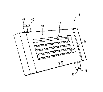
As shown in Figures 1-5, 6A-6C, and 7-8, a plant growth array device 10
includes an aerial growth chamber 12 and a root growth chamber 14. The aerial
growth chamber 12 and the root growth chamber 14 are separated by a dividing
member, such as a plate 16. The plate 16 includes apertures 20. The apertures
20
include holding members 22 that extend through the plate 16. The device 10
further includes glass plates 30, 32 (Figure 6A), which may be affixed with an
adhesive, and conduits 40, 42. In some embodiments the fluid conduits 40, 42
may
be on opposite sides of the device as shown in Figures 1-5 and 7 to bolster
gravitational effects in operation and/or to mitigate fluid leakage between
the aerial
growth chamber 12 and the root growth chamber 14 through the dividing plate
16.
However, fluid conduits 40, 42 may be positioned in any suitable location.
In some embodiments, the fluid flow may encourage root growth in a
particular direction, i.e., the direction of the fluid flow through the root
growth
chamber 14. The fluid conduits 40, 42 may be controlled by a controller (not
shown) to control and/or regulate the environment in the chambers 12, 14. In
some
embodiments, the chambers 12, 14 can includes sensors that detect
environmental
11
CA 02670273 2009-05-15
WO 2008/063587 PCT/US2007/024123
parameters (temperature, light conditions, moisture conditions, nutrient
conditions,
etc.). As shown in Figure 6A, the aerial growth chamber 12 and the root growth
chamber 14 each have a transparent and/or translucent side thereof formed by
the
respective glass plates 30, 32. As illustrated, the glass plate 30 is held by
an
adhesive; however, the glass plates 30, 32 may be held by other suitable
methods,
including slots and/or tabs.
Although embodiments according to the invention are illustrated with
respect to conduits 40, 42, it should be understood that other configurations
of
conduits for introducing fluid into the aerial growth chamber 12 and/or the
root
growth chamber 14 can be used. For example, a fluid may flow into the and out
of
the chambers 12, 14 from a plurality of ports to diffuse the fluid and provide
substantially uniform or even flow to the plants in different areas of the
array. In
some embodiments, the conduits 40,42 can be connected to a manifold chamber
(not shown). The manifold chamber can include a plurality of ports (e.g., a
perforated plate interface with one of the chambers 12, 14) to diffuse the
fluid
flowing into the chambers 12, 14.
The holding members 22 and/or apertures 20 can be positioned in any
suitable configuration. For example, as illustrated in Figure 6B, the holding
members 22A are substantially perpendicular with respect to the major plane of
the
dividing plate 16A. As shown in Figure 6C, the holding members 22B extend
away from the plate 16 and into the aerial growth chamber 12.
As shown in Figures 7-8, a plurality of plants 50, which include aerial
shoots 52 and roots 54, are received in the apertures 20B. In some
embodiments,
the plants 50 are Arabidopsis thaliana plants; however, any suitable plant may
be
used, including, but not limited to, plants with translucent roots for ease of
imaging, for example, annual crops such as maize, wheat, rice, and soybeans.
Non-transparent roots may also be used, particularly if the experimental focus
is on
external root features, such as branching architecture or mycorrhyzal
associations.
As shown, in particular, in Figures 7-8, the apertures 20B are configured to
receive the plurality of plants 50 such that the roots 54 grow substantially
in a
common orientation, such as along the glass plate 32 in the root growth
chamber
14 (see Figures 3, 5 and 7-8). In this configuration, the glass plate 32 can
be
positioned in a microscope, such as a confocal laser scanning microscope, and
images of the roots 54 may be obtained without removing the roots from the
root
12
CA 02670273 2009-05-15
WO 2008/063587 PCT/US2007/024123
growth chamber 14. Subsequent images may also be obtained at various times
during the growth of the root 54. One or more of the plants 50 may be
identified
and removed from the device to be grown on soil for propagation, which may be
useful for genetic screens.
For example, as shown in Figures 6A-6C, the apertures 20, 20A, 20B can
form a channel and be tapered in an open-ended, semi-gibbous, conical or
frusto-
conical shape, which may guide the roots 54 along a common orientation, such
as
along the glass plate 32, while maintaining sufficient space for healthy root
growth. The shape of the apertures 20, 20A, 20B may be generally spherical or
semi-gibbous to accommodate the application of a droplet of molten gel, and
which can be held in position after solidification. The apertures 20, 20A, 20B
extend at an angle (e.g., between about 0 and 90 degrees, between about 20 and
75
degrees or about 45 degrees) with respect to the plate 16. In this
configuration,
gravity may further direct the growth of the roots 54 along the common
orientation. In some embodiments, the device 10 can be oriented or re-oriented
while the plants 54 are growing to encourage root growth in a common
orientation.
The holding members 22, 22A, 22B and/or apertures 20, 20A, 20B can have the
same size, or different sizes of holding members 22, 22A, 22B and/or apertures
20,
20A, 20B may be provided on one device.
As shown in Figures 1-6A, 6B, 6C and 7-8, the device 10 is configured to
maintain a gaseous growth environment in the aerial growth chamber 12 and a
liquid growth environment in the root growth chamber 14. In some embodiments,
a gel (such as a molten gel) may be positioned in the apertures 20, and seeds
may
be held in place on the gel. The gel can include nutrients, moisture, and
other
components that may encourage seed growth. Any suitable gel can be used, such
as a low-melting temperature agarose, e.g., SeaPlaque 0 from Cambrex. In
addition, the gel can further function to separate the liquid growth
environment of
the root growth chamber 14 from the gaseous growth environment in the aerial
growth chamber 12.
In some embodiments, the conduits 40, 42 of 1-6A, 6B, 6C and 7-8 can be
connected to one or more fluid supply devices (not shown). For example, a
particular mixture of gases may be supplied to the aerial growth chamber 12
via
the conduits 40 and/or a particular mixture of liquids may be supplied to the
root
growth chamber 14 via the conduits 42. In some embodiments, the components of
13
CA 02670273 2009-05-15
WO 2008/063587
PCT/US2007/024123
the liquids and/or gases can be controlled by a controller and varied over
time (e.g.,
by using a manual or automated valve system). For example, a pollutant
introduced and/or the nutrients supplied to the chambers 12, 14 may be changed
over time and any effects of the changes may be observed by imaging the roots
as
described herein.
According to embodiments of the invention, the spatial, temporal and/or
environmental transcription pattern may be studied by imaging roots, shoot
portions and/or any other portions of the plants 54. A gene expression data
set for
a multicellular organism may provide the number of steady state transcripts
for one
or more genes in one or more individual cells and/or one or more tissues, at
one or
more time point in the cell's development. Various environmental conditions
and/or genetic backgrounds can be studied. Various methods have been used to
determine the spatial accumulation of transcripts for a single gene, such as
in situ
hybridization and an expression DNA microarray [Brown and Botstein 1999,
Yamada et al. 2003]. Other methodology to acquire spatial expression data for
a
larger number of genes by purifying cell-type specific RNA for microarray
analysis uses fluorescence activated cell sorting of discreet GFP marked cell
types
[Birnbaum et al. 2003, Birnbaum et al. 2005, Brady et al. 2007]. However,
these
methods are generally non-vital, and consequently the time component of
differential gene expression may be difficult or impossible to determine
without
using a large collection of independent samples over a time series. Transgenic
reporters of expression have been used extensively recently, due in large part
to the
popularization of experimental GFP as a qualitative measure of where and when
a
single gene might be activated [Chalfie et al. 1994; Lee et al. PNAS 2006].
In some embodiments, real time images may be obtained from live plants
without removing the plants from a growth medium. A plurality of plants may be
grown in a similar environment on a chip to provide a high throughput device.
Plant structures, including roots, shoots, and other plant structures, may be
imaged.
Embodiments of the present invention can also be used to image expression of
reporter genes such as GFP. Embodiments of the present invention can provide
quantitative expression data for a large set of genes (for example,
transcription
factor genes) at high spatial and/or temporal resolution, using a collection
of
transcriptional reporters (for example, GFP reporters). Suitable reporter
genes
include colorimetric and fluorescent reporters. The time course analyses of
14
CA 02670273 2009-05-15
WO 2008/063587 PCT/US2007/024123
expression under a spectrum of environmental conditions can be provided by
modulating stringently controlled liquid growth media in the root growth
chamber.
Cost advantages may be realized because existing confocal imaging equipment
may be used. Moreover, although embodiments of the present invention are
described with respect to the plant array growth device 10, other model
organisms
that are transformable and amenable to fluorescence imaging may be used,
including yeasts (Saccharomyces cerevisiae and Schizosaacharomyces pombe),
flies (Drosophila melanogaster), zebrafish (Danio rerio), the nematode
(Caenorhabditis elegans), moss (Physcomitrella), or cell cultures of any
suitable
organism.
Some embodiments of the invention can be used to study plant
development, including spatial and/or temporal gene expression. Gene
expression
patterns may be studied in response to external stimuli, including biological
and
abiotic stimuli.
Figure 9 is a block diagram of exemplary embodiments of data processing
systems that illustrates systems, methods, and computer program products in
accordance with embodiments of the present invention. As illustrated in Figure
9,
a system 109 includes a processor 110, memory 114, an address/data bus 138,
and
a plant imaging system 125. The memory includes application programs 154 (such
as a plant imaging module 112 and/or conduit controller module 116), data 156
(such as image data 150), I/0 device drivers 158, and an operating system 152.
The processor 110 communicates with the memory 114 via the address/data bus
148. The processor 110 can be any commercially available or custom
microprocessor. The memory 114 is representative of the overall hierarchy of
memory devices containing the software and data used to implement the
functionality of the data processing system 105. The memory 114 can include,
but
is not limited to, the following types of devices: cache, ROM, PROM, EPROM,
EEPROM, flash memory, SRAM, and DRAM.
The plant imaging system 125 can include a plant growth array, such as the
device 10 as illustrated in Figures 1-6A, 6B, 6C and 7-8, a fluid supply
system for
supplying air and/or liquid fluids to the plant growth array, and/or an
imaging
device, such as a confocal laser imaging microscope for fluorescent root
imaging.
As shown in Figure 9, the memory 114 may include several categories of
software and data used in the data processing system 105: the operating system
CA 02670273 2009-05-15
WO 2008/063587 PCT/US2007/024123
152; the application programs 154; the input/output (I/0) device drivers 158
and
the data 156. The data 156 may include image data 150 which may be obtained
from the plant imaging system 125. In some embodiments, the plant imaging
system 125 includes an automated microscope, such as a robotic microscope. The
plant imaging module 110 may control the movement of the microscope and/or
various aspects of the plant imaging system 125.
As will be appreciated by those of skill in the art, the operating system 152
may be any operating system suitable for use with a data processing system,
such
as OS/2, AIX, OS/390 or System390 from International Business Machines
Corporation, Armonk, NY, Windows CE, Windows NT, Windows95, Windows98
or Windows2000 from Microsoft Corporation, Redmond, WA, Unix or Linux or
FreeBSD, Palm OS from Palm, Inc., Mac OS from Apple Computer, LabView or
proprietary operating systems. The I/0 device drivers 158 typically include
software routines accessed through the operating system 152 by the application
programs 154 to communicate with devices such as I/0 data port(s), data
storage
156 and certain components of the memory 114 and/or the plant imaging system
125. The application programs 154 are illustrative of the programs that
implement
the various features of the data processing system 105 and preferably include
at
least one application that supports operations according to embodiments of the
present invention. Finally, the data 156 represent the static and dynamic data
used
by the application programs 154, the operating system 152, the I/0 device
drivers
158, and other software programs that may reside in the memory 114.
The plant imaging module 112 can be configured to obtain and/or control
images from the plant imaging system 125. It may be desirable to obtain
detailed
images of the roots 54 of Figures 7-8 or portions of the roots 54 (e.g., root
ends or
tips) without necessarily obtaining detailed images of the entire root growth
chamber 14. In some embodiments, the plant imaging module 112 is configured to
obtain one image that can be used to identify regions for detailed imaging.
For
example, the location of a region or regions that includes root portions of
the plants
can be obtained from an initial image. More detailed images, including images
with a higher resolution, can then be obtained at the identified locations.
The
initial image may be obtained from a microscope, camera or other imaging
device.
The initial image and the more detailed image(s) can be obtained from the same
device or from a different device. The initial image may be a concatenated two
or
16
CA 02670273 2009-05-15
WO 2008/063587 PCT/US2007/024123
three-dimensional image compiled from a series of images that capture the
entire
space of root growth. In some embodiments, the plant imaging module 112 can
control an automated or robotic microscope to image the identified regions.
The conduit controller module 116 can control a fluid supply to the growth
environment of plants, for example, via the conduits 40, 42, to provide a
particular
gaseous or liquid environment to the aerial growth chamber 12 and the root
growth
chamber 14, respectively. For example, the fluid supply may be provided by a
peristaltic pump with automated or manually operated valves. Any suitable
commercially available or customized nutrient solutions can be used to provide
a
liquid growth environment. One example is a nutrient solution having 4.3g/L
module
i1g1e6anbdeinSgkoaopgplsiaclattsio(wn (w/macro ansdinmFiicgruorneuliaens
nutrients), LapMprEecSi
sucrose.
Although embodiments of the present invention are illustrated, for
example, with reference to the plant imaging module 112 and/or conduit
controller
'aantedd by
those
those of skill in the art, other configurations may also be utilized while
still
benefiting from the teachings of the present invention. For example, the
modules
112, 114 may also be incorporated into the operating system 152, the I/0
device
drivers 158 or other such logical division of the data processing system 105.
Thus,
the present invention should not be construed as limited to the configuration
of
Figure 9, which is intended to encompass any configuration capable of carrying
out the operations described herein.
The I/0 data port can be used to transfer information between the data
processing system 105 and the plant imaging system 125 or another computer
system or a network (e.g., the Internet) or to other devices controlled by the
processor. These components may be conventional components such as those used
in many conventional data processing systems that may be configured in
accordance with the present invention to operate as described herein.
Those skilled in the art will recognize that the plant growth array device 10
of Figures 1-6A, 6B, 6C and 7-8 may take other configurations. For example, a
plant growth array device 200 is shown in Figures 16-18. The device 200
includes
an aerial shoot growth chamber 212 and a root growth chamber 214 that is
divided
by a wire mesh divider 216. The wire mesh divider 216 includes apertures 220
therein. The aerial shoot growth chamber 212 includes an adhesive film 212a, a
17
CA 02670273 2009-05-15
WO 2008/063587 PCT/US2007/024123
glass cover slip 230, and a gel 221. The adhesive film 212a separates the
divider
216 from the cover slip 230 to form the chamber 212. The root growth chamber
214 includes an adhesive film 214a and a glass cover slip 232. The adhesive
film
214a separates the divider 216 from the cover slip 232 to form the chamber
214.
The mesh divider 216 supports the gel 221. The gel 221 can immobilize seeds
and/or isolate the chambers 212, 214.
As shown in Figure 18, plants 250 grow such that shoot portions 252 of the
plants 250 extend into the aerial growth chamber 212 and root portions 254 of
the
plants 250 extend into the root growth chamber 214. The shoot portions 252
and/or the root portions 254 may be imaged as discussed herein by positioning
a
microscope or other imaging device adjacent the glass cover slip(s) 230, 232.
In particular embodiments, the adhesive film 212a forms a spacer that is
about 1-5 mm thick and the adhesive film 214a forms a spacer that is about 200-
600 p.m thick.
Fluid exchange ports (not shown) may be used to control a gaseous
environment in the aerial shoot chamber 212 and/or a liquid environment in the
root growth chamber 214. In addition, the adhesive film 214a may include a
fluidic channel pattern for directing fluid flow from a liquid exchange port.
Although embodiments according to the present invention are described
with respect to confocal laser scanning microscopy imaging devices, other
imaging
devices can be used. Various types of light microscopy, including brightfield,
dark
field and differential interference contrast microscopy, may be used.
Fluorescence
microscopy, multi-photon microscopy, optical coherence tomography and
deconvolution microscopy may be used.
Moreover, the devices described herein can be used to perform various
imaging methodologies, including, without limitation, fluorescence lifetime
imaging (FLIM), bi-molecular fluorescence complementation (BiFC), fluorescence
(Forster) resonance energy transfer (FRET), Bioluminescence Resonance Energy
Transfer (BRET), fluorescence correlation spectroscopy (FCS), calcium sensor
imaging (and other signaling sensors), auxin reporter imaging (and other
hormone
sensors and reporters), cell cycle reporter imaging, subcellular structure
reporter
imaging, chemical or physical perturbation of development, cell lineage
analysis,
laser uncaging experiments, chromophore assisted light inactivation (CALI),
18
CA 02670273 2009-05-15
WO 2008/063587 PCT/US2007/024123
chemically inducible spatial activation of gene expression, and/or chemically
inducible spatial inactivation of gene expression.
Embodiments according to the present invention will now be described
with respect to the following non-limiting examples.
Examples
Arabidopsis thaliana seedlings can be grown in the device 10 shown in
Figures 1-6A, 6B, 6C and 7-8 and the roots and/or shoots may be
imaged/monitored using the system 105 of Figure 9. The Arabidopsis root may be
used as a model system to understand the genetic control of development.
Differences in gene expression over time and/or responses to external stimuli
and
environmental conditions (such as pollutants, toxins, hormones, light,
nutrients,
oxygen, carbon dioxide and other gases, water, draught conditions and the
like)
between cells types can be detected. For example, the nature of tissue
specific gene
regulation in the root may be studied at a genomic level and quantitative gene
expression data in high temporal and spatial resolution in the root may be
obtained.
The dynamics of genome expression regulation over time during development and
in response to external stimuli may be studied. In particular, dynamic
transcription
networks and development can be studied in response to environmental stimuli,
and time-lapse three dimensional imaging of growing roots may be performed.
Responses to external stimuli and environmental conditions, such as
pollutants,
toxins, hormones, light, nutrients, oxygen, carbon dioxide and other gases,
water,
drought conditions and the like may be observed.
Non-invasive confocal imaging may be used with a large collection of
plants, each harboring a unique fluorescent expression reporter. Fluorescence
image analysis may serve as a real-time proxy for characterizing expression
dynamics. The device 10 of Figures 1-6A, 6B, 6C and 7-8 can be used to grow
the
plants in a controllable liquid growth environment.
The microscopy images using confocal or other methods may provide a
rich source of data beyond the quantification of gene expression reporters.
The
images may be used for morphological analyses of developmental and
physiological dynamics. Gene expression data can thus be correlated with
morphometric data quantifying the dimensions, volume, and arrangements in
three-
19
CA 02670273 2009-05-15
WO 2008/063587 PCT/US2007/024123
dimensional space of the organisum's subcellular components, cells, tissue
layers,
and organs.
Device Design
The design and fabrication techniques to form plant growth devices
described herein are strategically flexible allowing for simple and
inexpensive
modification to accommodate different imaging platforms or experimental goals.
In some embodiments, plant growth devices described herein can be formed of
molded silicone elastomer (polydimethylsiloxane [PDMS] Dow Corning
SYLGARD 184) and a transparent side can be provided by a microscope coverslip,
such as a coverslip having a thickness of glass (-0.15mm). Other suitable
materials may be used. Optically clear and biologically inert silicone may be
molded to contain liquid growth media between it and the glass to which it is
secured, as shown, for example, in Figure 14A. Array designs may be generated
using the SolidWorksTM three-dimensional CAD program and are exported to a
stereolithography apparatus (SLA) for fabrication using materials such as
VeroBlue FULLCURE 840 photopolymer or those available from DSM Somos,
Elgin, IL (USA) (for example Watershed 11120, NanoTool, ProtoTherm 12120).
For example, SLA can be used to generate a plastic (photopolymer) mold, in
which
a silicone array could then be cast. Alternatively, the SLA technique can be
used
to fabricate the array itself out of the photopolymer plastic or another
suitable SLA
material. Small openings molded in the silicone component may be filled by
fine
mesh or solid low melting point agar, which immobilizes the seed but allows
the
young root to grow down into the liquid growth environment and along the glass
(Figure 14B). The shallow space (-200-600 microns) where the root grows may
ensure that the root stays within the working distance of the microscope, yet
provides sufficient root interaction with the environment to sustain a healthy
plant.
The stems and leaves of the plants may be grown in a sealed volume of air to
reduce or prevent desiccation during imaging. Although the apertures in which
the
roots grow are illustrated as having an hour-glass shape, other configurations
can
be used, including frusto-conical and semi-gibbous shapes. Alternative
fabrication
techniques include techniques that can involve photolithography to generate
layers
of the device structure that can be bound to glass following plasma cleaning
[McDonald et al. 2000], or using layers of die-cut double-sided adhesive films
or
CA 02670273 2009-05-15
WO 2008/063587 PCT/US2007/024123
tapes and mesh. In some embodiments, a high density of roots can be obtained,
such as up to 96 for a 25x 75mm slide. However, larger and smaller dimensions
may be used. For example, the number of roots practical for imaging may be
limited by the scanning range of the robotic microscope stage; consequently,
more
plants can be imaged using a custom built stage and an array device of greater
dimensions.
Control of liquid and gaseous growth environments.
In some embodiments, fluids may be supplied to plant growth array
devices. For example, conduits 40, 42 of Figures 1-6A, 6B, 6C and 7-8 can be
inlet and outlet ports for a fluid and a gas, respectively, and may be
configured for
liquid media exchange. Liquid exchange may be achieved using a low flow
multichannel peristaltic pump. This peristaltic pump can be used to exchange
air,
and can be used to manipulate the aerial as well as the root growth
environment of
the plants. Manual or programmable valves operated by software, such as the
conduit controller module 116 of Figure 9, and the manual or computer-
controlled
valves can be used to change the liquid media source. An exemplary liquid
growth
media is 1% Murashige and Skoog liquid media supplemented with 1% sucrose.
= Optimization or modification of the liquid growth media, such as by
supplementary oxygenation, may be tested.
Counterstaining of cell boundaries.
Imaging cell walls of the root can be achieved by using vital concentrations
of the fluorescent stain FM-464. The potential issues of cost for this dye are
sufficiently mitigated by the low working concentrations and low volume
required
by devices according to embodiments of the invention. Optionally, staining
intensity can be automatically regulated by the image analysis programs that
control a valve mixing additional dye. An alternative method to image root
cell
boundaries involves a transgenic approach or the use of propidium iodide or
other
alternative stains.
Automated image acquisition
Strategies for high-throughput and hands-free imaging may use a Zeiss 510
confocal LSM with a robotic "x-y" stage and robotic "z". A custom high-speed
21
CA 02670273 2009-05-15
WO 2008/063587 PCT/US2007/024123
imaging platform can be developed for a confocal, spinning disk capability, or
conventional microscope. Scanning efficiency can be improved by imaging only
the regions of interest (ROI). Unlike other types of arrays where samples are
in a
predetermined position, the devices according to embodiments of the invention
can
allow roots room to grow within a somewhat restricted region (e.g.,, apertures
in
the dividing member of the array) to access to a liquid growth environment.
Sufficient space can be allowed to permit healthy plant growth. A computer
based
image recognition algorithm can be developed to allow magnification of a quick
bright-field scan of the entire array for automated determination of the "x"
and "y"
ROI coordinates, followed by high resolution confocal scanning as shown in
Figure 14C. Various methods have been proposed for finding the root coordinate
in the z-axis. A scanning auto focus routine may be used to find the top or
bottom
boundary of the root, and then approximate the median section to be 75microns
internal from that point. Alternatively a fluorescent marker for the central
cells in
the root tip can be used for an auto focus routine. Third, using the theories
behind
image deconvolution, interpretation of the out-of-focus bright field images
may
provide information to determine the distance of the root from the focal
plane. An
alternative solution to determining ROIs would be to use an imaging platform
with
simultaneous multichannel fast wide field capture to speed up the acquisition
of an
unguided tiling scan, saving data selection until after image capturing.
Techniques
described in U.S. Patent No. 6,115,111 to Korah et al. (the disclosure of
which is
hereby incorporated by reference in its entirety) may be used.
Calibration of Quantitative confocal root image analysis of GFP fluorescence
as a reporter for the activity of gene promoter regions.
The image analysis can be calibrated to independent measurements of GFP
mRNA and fluorescence for each cell type in the root. Three-dimensional
confocal
images can be used for quantitation, and the root's optical properties can be
modeled to account for attenuation and scatter of light due to depth.
Transcriptional response to external stimuli.
An experiment using a steroid inducible protein known to activate a
fluorescent transcriptional reporter can be used. The device 10 may be used in
conjunction with projects studying nutrient deprivation or toxicity and
abiotic
22
CA 02670273 2009-05-15
WO 2008/063587 PCT/US2007/024123
stress or stimulation. The dynamic response of a collection of tissue-enriched
transcription factors may be compared between environmental stimuli.
The Arabidopsis thaliana root is one of the most tractable experimental
models for development in plants. A fully sequenced genome, public gene mutant
collections, transformability, and accessibility of commercially made
expression
microarrays provide efficient tools for experiments at a genomic level
[Somerville
and Dangl 2000]. The root's simple and stereotypic anatomy makes it generally
well-suited for developmental genetics studies. The degree of rotational
symmetry,
transparency, small size, and its meristematic growth pattern distinguish the
root as
a uniquely well-suited multicellular organ for the implementation of high-
throughput automated confocal technology. A single two-dimensional (2D) image
through the median longitudinal axis is largely representative of the entire
three-
dimensional (3D) structure. Cells further from the meristematic growth center
are
progressively older and more differentiated. Consequently the same 2D image
also
represents a developmental time component as seen in Figures 10A-10B. Root
growth can be mathematically modeled over time for studies of cell expansion
and
divisions, and used to probe underlying molecular mechanisms of cell
morphogenesis and gravitropism [Beemster and Baskin 1998, Grabov et al 2005,
= Swarup et al. 20051. Quantitative morphometric analysis of confocal time
lapse
images from the shoot apical meristem may be used to model an example of
spatial
hormone signaling and a reaction-diffusion mechanism [deReuille et al. 2006,
Jonsson et al. 2005].
Root and/or shoot and/or any other portion of plants can be imaged in an
undisturbed growth environment to perform time-lapse root imaging.
Embodiments according to the invention can provide an automated or high-
throughput imaging system and may increase the power and accessibility to a
new
spectrum of detectable microscopic phenotypes for genetic and chemical
screens.
A dynamic liquid media exchange system, such as that provided by the conduits
40, 42, may allow for many types of "experiments on a chip" ranging from
investigation of nutrition, hormone biology, stress response, or engineered
gene
induction. Identifying promoter reporters that respond to specific chemicals
or
stressful environmental conditions may guide the development of transgenic bio-
sensors useful for agricultural or environmental monitoring.
23
CA 02670273 2009-05-15
WO 2008/063587 PCT/US2007/024123
A time-lapse imaging sequence of a healthy growing root expressing a
nuclear GFP reporter is show in Figures 11 and 13B. The overlay of
differential
interference contrast (DIC) and epifluorescence images captures the dynamics
of
root hair growth and coordinated movement of the nucleus within these cells.
The analysis of confocal images to quantify GFP fluorescence may be
automated [Lee et al. 2006]. Images may be transformed and aligned to fit a
template root atlas annotated by tissue type. The success of image alignment
and
GFP detection was tested using images for twenty-three transcriptional
reporters.
Correlation to the device of Figures 1-6A, 6B, 6C and 7-8 was used to
determine
expression for thirteen cell sorted tissues, and illustrates that quantitative
fluorescent reporter data may be obtained from confocal images of plants with
detectable levels of GFP. Figure 12 illustrates that an Atlas image alignment
produces relative expression data that is supported by devices according to
embodiments of the present invention. See Mace DL, Lee JY, Twigg RW, Colinas
J, Benfey PN, Ohler U. Quantification of transcription factor expression from
Arabidopsis images. Bioinformatics. 2006, 22 (14):e323-31.
Devices according to embodiments of the present invention have been
tested for the ability to capture the transcriptional promoter response to
manipulation of the liquid growth environment. Time-lapse images were captured
for a promoter reporter of the SCARECROW (SCR) gene as it is activated
following induction with Dexamethasone to rescue SHORTROOT activity. A
representative selection of five time points is shown in addition to GFP
quantitation for 13 of the 26 time points within the 12 hour experiment
(Figures
13A-13B). In addition, time-lapse imaging using artificial gene induction may
be
performed. Calibration of quantitation methods between tissues and in relation
to
empirical determination of transcriptional output may be a step towards high-
throughput application of promoter reporters as quantitative proxy for
expression
according to embodiments of the present invention.
Arabidopsis genetic background for automated imaging.
A screen for subcellular localization of GFP identified four protein
sequences that target GFP to the cell surface [Cutler et al. 2000]. Line 37-26
has
been tested and shows promise as an alternative to staining by FM4-64 or
propidium iodide. The known subcellular target sequence may be fused to an
24
CA 02670273 2009-05-15
WO 2008/063587 PCT/US2007/024123
appropriate fluorescent protein whose emission can be resolved from GFP, such
as
the mCherry RFP construct developed by [Shaner et al, 2004]. Multiple
promoters
may be tested to achieve ubiquitous expression in the root. The proposed
genetic
background for imaging may additionally express a fluorescent reporter marking
the quiescent center (QC). A transcriptional GFP reporter has been developed,
line
Q12, which strongly marks these 4-7 cells in the root tip. This promoter, or
elements derived from it, may be engineered to express a fluorescent protein
that
can be spectrally resolved from both GFP and the mCherry RFP, such as an
orange
variant [Shaner et al, 2005].
Quantitative confocal root image analysis of GFP fluorescence as a reporter
for the activity of gene promoter regions.
Fluorescent reporters can provide a read-out for the activity of promoters.
To more accurately correlate the fluorescence analysis of a confocal root
image to
the actual promoter activity, measurements and models of a system may be
performed. It may be possible to correct for predictable anomalies and account
for
noise inherent in the imaging and transgenic reporter system. Independent
measurement of transcriptional products can be quantitatively correlated to
transcriptional reporter activity.
Model attenuation of fluorescence image due to depth.
Depth may be a factor in larger, multicellular systems. Light scatter and
absorbance may occur during laser excitation and fluorescence emission as a
function of depth and the optical transparency properties of the tissues. A
data set
generated from a collection of plants, e.g., about fifteen plants, each
expressing a
tissue specific GFP, representing the entire anatomy of the root, can be used
to
indirectly test the root's optical properties.
Figures 15A-15B illustrates the flow cytometry data that can be used to
calibrate tissue specific image quantitation. For each plant line, the
distribution of
fluorescence intensity for the collection of individual cells can be
quantified and
applied to a fluorescence activated cell sorter. The flow cytometry data may
be
used to create a mathematical depth correction function for the radial axis of
the
root. The success of the depth correction may be assessed by the level of
statistically significant improvement to the correlation between image
analysis and
CA 02670273 2009-05-15
WO 2008/063587 PCT/US2007/024123
root expression map data performed by Mace DL, Lee JY, Twigg RW, Colinas J,
Benfey PN, Ohler U. Quantification of transcription factor expression from
Arabidopsis images. Bioinformatics 2006, 22(14):e323-31. Other methods may
be used to test or improve the depth correction function. For example, one
would
involve micro-injection of a fluorescent standard to a cell in each tissue
layer.
Another method would quantify GFP fluorescence using a low volume NanoDrop
fluorometer with the lysate of a single root following confocal imaging of
that
same root. A third method involves imaging a set of promoter reporters that
are
expressed ubiquitously and at comparable concentrations between tissues.
Scanning images in the Z-axis.
Choosing the number of images captured in the z-axis may be performed to
reduce the number of images captured. In particular embodiments,
quantification
of GFP in a cell is carried out by capturing array images of the cellular
compartment containing the GFP. In some embodiments, for each transcriptional
reporter, GFP is targeted to the endoplasmic reticulum (ER) to reduce or
prevent
intercellular GFP movement. ER targeting also =creates a predictable
accumulation
pattern that is comparable between the tissue types of the root meristem.
However,
images that capture anticlinal cell walls may lack GFP fluorescence. A small
set of
images in the Z axis may provide more robust= image data by representing whole
cells. A second potential benefit of using multiple images in the Z-axis is
for the
ability to reconstruct a root's three-dimensional anatomy. Certain analysis
methods
may use images that are parallel with the longitudinal axis of the root. To
the
extent that devices according to embodiments of the present invention allow
roots
to grow toward or away from the imaging plane, the ability to computationally
section a new image plane from the reconstruction of such a root may be
tested. A
training set of images can be generated from a collection of plants with a
range of
ideally oriented and less cooperative roots. This training collection may
provide a
range of Z-stack sets for a range of z-section thicknesses, and for a range of
= 30 interval distances between sections. These parameters may be
optimized as
described herein by measuring the improved correlation to a training set of
tissue
specific expression profiles. An alternative approach is to develop a 3D image
data
analysis pipeline. For each of the 2D algorithms used in data analysis, there
exists
an equivalent for three dimensions. However, the conversion to 3D may be
26
CA 02670273 2009-05-15
WO 2008/063587 PCT/US2007/024123
difficult, partly because confocal images may not be obtained much beyond the
median plane of the root. Multi-photon microscopy may provide additional
imaging depth, or the symmetry of the root to "mirror" the top half of the
root may
be used to artificially recreate the cylindrical root geometry.
Assess transgenic reporters to reflect endogenous promoter activity.
GFP may be engineered with polypeptide tags targeting ubiquitin mediated
target destruction [Downes and Vierstra. 2005, Menendez-Benito et al. 2005].
An
alternative solution would use the DsRed-E5 reporter which has predictable
changing emission spectra from green to red during its 18 hour maturation,
making
it well-suited to ratiometric emission analysis to determine both up and down-
regulation of expression [Mirabella et al. 2004]. In addition to using
standardized
settings for excitation, prior to each data acquisition routine, further
calibration can
be attained using a set of fluorescent reference standards, for example, from
Invitrogen, matched with the emission wavelength of each fluorescent protein
variant used.
Correlate fluorescence to numbers of GFP mRNA.
The GFP mRNA may be quantified by quantitative RT-PCR or any other
suitable technique. In one experiment, RNA is collected from whole roots for a
collection of, e.g., 15 GFP plant lines that are representative of the entire
root. The
correlation of GFP mRNA abundance to the quantitative image analysis described
herein may be assessed. Another experiment uses Q-RT-PCR of GFP for a 24 hour
time course at 1 hour intervals following induction of the promoter
SCARECROW:GFP reporter plant. Comparison of this data to multiple 24 hour
image acquisition series for these plants may provide information to model
both
the speed of the chemical induction and the lag time of GFP maturation. A
supporting experiment may involve microinjection of pre-determined numbers of
GFP mRNA. Data from this approach can be used to calibrate for attenuation due
to depth, measure GFP maturation rate, and correlate fluorescence to mRNA
molecule number.
Examine transcriptional response to external stimuli.
27
CA 02670273 2009-05-15
WO 2008/063587 PCT/US2007/024123
Existing methods to measure gene expression dynamics may be hindered
either by excessive cost, limited spatial or temporal information, or by the
number
of genes that can be measured. Controlled manipulation of the liquid growth
media
for a growing root may elicit a genetic and developmental response that can be
measured by image analysis. A growing collection of transcriptional reporters
exists, which can be used for plant imaging according to embodiments of the
present invention.
Capture expression activation in a gene network.
The SHORTROOT (SHR) and SCARECROW (SCR) proteins are typically
considered necessary for the proper division and differentiation of the
cortex/endodermis initial. SHR activates SCR transcription in the
cortex/endodermis initial [Cui et al, 2007]. In the shr-2 mutant background,
SCR
expression is nearly absent and this division fails to occur [Helariutta et
al, 2000].
A plant that rescues SHR expression may be generated using a steroid induction
system in the shr-2 background [Levesque et al, 2006]. Heat shock protein
90(Hsp90) sequesters a SHR:glucocorticoid receptor fusion protein in the
cytoplasm. Upon addition of the synthetic steroid, Dexamethasone, Hsp90
releases
SHR permitting it to enter the nucleus and activate SCR transcription. This
same
plant line also has a transcriptional reporter of SCARECROW, pSCR:GFP. This
induction system may be used with plant arrays according to embodiments of the
invention for time lapse imaging to study gene activation and development
simultaneously. Protein behavior may also be studied according to embodiments
of
the invention, including a split-YFP system to probe the spatial and temporal
interaction of the SCR and SHR proteins. Crosses between GFP reporter plants
and
mutants of genes known or implicated in this pathway may resolve dynamic
phenotypes for classical epistasis experiments.
Transcriptional reporters of gene expression to determine dynamic response
to environmental stimuli.
A collection of promoter reporters can be used, including a collection of
promoter reporters that has been developed for a third of all transcription
factors
genes that are expressed significantly higher in one tissue compared to four
other
tissues in the root [Lee et al. 2006]. This collection of 61 reporters can be
used to
28
CA 02670273 2009-05-15
WO 2008/063587 PCT/US2007/024123
evaluate the environmental expression response according to embodiments of the
present invention. The root tissue expression map can be compared to the
AtGenExpress Gene Atlas to implicate genes that may exhibit tissue specific
activity in response to abiotic stress conditions. An overlap comparison may
implicate genes and may show tissue specific response to the environment, and
allowing a prioritization of the creation of new promoter reporters. The
arrangement of reporter plants can be based on the standard principles of
microarray design [Draghici 2003]. These include multiple replicates for each
feature, random assignment of position, internal fluorescent standards, and
promoter-less GFP as a negative control. For quality control, available
reporter
plants can be used to detect plant stress through activation of characterized
stress
response genes such as catalase, alcohol dehydrogenase, and Hsp [Manak et al.
2005]. The dynamic developmental and transcriptional response of roots
switched
to limited media for primary and secondary nutrients can be systematically
characterized, for example using at least 6 essential micronutrients. Abiotic
stressors can be tested including salt, osmolarity, drought, oxidation,
darkness,
heat, and cold. Controlled air exchange may allow a survey of the root's
response
to gaseous pollutants and to an increased carbon dioxide environment. Results
can
be compared between environmental conditions and temporal and spatial patterns
of gene induction can be compared. The timing of gene activation may be
compared between genes using clustering methods to identity genes that may be
activated in concert. The initiation time of gene activation may be analyzed
as an
indicator of how transcription factors may be ordered in a transcription
network.
The low cost of each experiment will allow the production of a map of the
concentrations of nutrient and/or toxic chemical concentrations for each
environmental condition. The data may be used to inform the critical time
points
and chemical concentration to be used for subsequent genomic expression
analysis.
The foregoing is illustrative of the present invention and is not to be
construed as limiting thereof Although a few exemplary embodiments of this
invention have been described, those skilled in the art will readily
appreciate that
many modifications are possible in the exemplary embodiments without
materially
departing from the novel teachings and advantages of this invention.
Accordingly,
all such modifications are intended to be included within the scope of this
29
CA 02670273 2009-05-15
WO 2008/063587 PCT/US2007/024123
invention as defined in the claims. Therefore, it is to be understood that the
foregoing is illustrative of the present invention and is not to be construed
as
limited to the specific embodiments disclosed, and that modifications to the
disclosed embodiments, as well as other embodiments, are intended to be
included
within the scope of the appended claims. The invention is defined by the
following
claims, with equivalents of the claims to be included therein.
References:
Andrews PD, Harper IS, Swedlow JR. To 5D and beyond: quantitative
fluorescence microscopy in the postgenomic era. Traffic 2002, 3:29-36.
Beemster GT, Baskin TI. Analysis of cell division and elongation underlying
the
developmental acceleration of root growth in Arabidopsis thaliana. Plant
Physiology 1998, 116:1515-26.
Birnbaum K, Jung JW, Wang JY, Lambert GM, Hirst JA, Galbraith DW, Benfey
PN. Cell type-specific expression profiling in plants via cell sorting of
protoplasts from fluorescent reporter lines. Nature Methods 2005, 2:615-9.
Birnbaum K, Shasha DE, Wang JY, Jung JW, Lambert GM, Galbraith DW,
= Benfey PN. A gene expression map of the Arabidopsis root. Science 2003,
302:1956-60.
Brady SM, Orlando DA, Lee J-Y, Wang JY, Koch J, Dinneny JR, Mace D, Ohler
U, and Benfey PN (2007) A High-Resolution Root Spatiotemporal Map Reveals
Dominant Expression Patterns. Science 318: 801-806
Brown PO, Botstein D. Exploring the new world of the genome with DNA
microarrays. Nature Genetics 1999, 21 (1 Suppl):33-7.
Chalfie M, Tu Y, Euskirchen G, Ward WW, Prasher DC. Green fluorescent
protein as a marker for gene expression. Science. 1994, 263:802-5.
de Reuille PB, Bohn-Courseau I, Ljung K, Morin H, Carraro N, Godin C, Traas J.
Computer simulations reveal properties of the cell-cell signaling network at
the shoot apex in Arabidopsis. PNAS 2006, 103:1627-32.
CA 02670273 2009-05-15
WO 2008/063587 PCT/US2007/024123
Cutler SR, Ehrhardt DW, Griffitts JS, Somerville CR. Random GFP::cDNA
fusions enable visualization of subcellular structures in cells of Arabidopsis
at
a high frequency. PNAS 2000, 97:3718-23.
Dixit R, Cyr R, Gilroy S. Using intrinsically fluorescent proteins for plant
cell
imaging. Plant Journal 2006, 45:599-615.
Dmochowski IJ, Dmochowski JE, Oliveri P, Davidson EH, Fraser SE.
Quantitative imaging of cis-regulatory reporters in living embryos. PNAS
2002, 99:12895-900. Downes B, Vierstra RD. Post-translational regulation in
plants employing a diverse set of polypeptide tags. Biochemical Society
Transactions 2005, 33:393-9.
Sorin Draghici. Data Analysis Tools for DNA Microarrays. 2003. Chapman &
Hall/CRC. Boca Raton, FL.
Echeverri CJ, Perrimon N. High-throughput RNAi screening in cultured cells: a
user's guide. Nature Reviews Genetics 2006, 7:373-84.
Friedman N, Vardi S, Ronen M, Alon U, Stavans J. Precise temporal modulation
in the response of the SOS DNA repair network in individual bacteria. PLoS
Biology 2005, 3:e238.
Giepmans BN, Adams SR, Ellisman MH, Tsien RY. The fluorescent toolbox for
assessing protein location and function. Science 2006, 312:217-24.
Grabov A, Ashley MK, Rigas S, Hatzopoulos P, Dolan L, Vicente-Agullo F.
Morphometric analysis of root shape. New Phytologist 2005, 165:641-51.
Harbison CT, Gordon DB, Lee TI, Rinaldi NJ, Macisaac KD, Danford TW,
Hannett NM, Tagne JB, Reynolds DB, Yoo J, Jennings EG, Zeitlinger J, Pokholok
DK, Kellis M, Rolfe PA, Takusagawa KT, Lander ES, Gifford DK, Fraenkel E,
31
CA 02670273 2009-05-15
WO 2008/063587 PCT/US2007/024123
Young RA. Transcriptional regulatory code of a eukaryotic genome. Nature
2004, 431(7004):99-104.
Helariutta Y, Fukaki H, Wysocka-Diller J, Nakajima K, Jung J, Sena G, Hauser
MT, Benfey PN. The SHORT-ROOT gene controls radial patterning of the
Arabidopsis root through radial signaling. Cell 2000, 101:555-67.
Ideker T, Thorsson V, Ranish JA, Christmas R, Buhler J, Eng JK, Bumgarner R,
Goodlett DR, Aebersold R, Hood L. Integrated genomic and proteomic analyses
of a systematically perturbed metabolic network. Science 2001, 292:929-34.
Jonsson H, Heisler M, Reddy GV, Agrawal V, Gor V, Shapiro BE, Mjolsness E,
Meyerowitz EM. Modeling the organization of the WUSCHEL expression
domain in the shoot apical meristem. Bioinformatics 2005, 21:i232-i240.
Kakoki M, Tsai YS, Kim HS, Hatada S, Ciavatta DJ, Takahashi N, Arnold LW,
Maeda N, Smithies O. Altering the expression in mice of genes by modifying
their 3' regions. Developmental Cell 2004, 6:597-606.
Kalir S, McClure J, Pabbaraju K, Southward C, Ronen M, Leibler S, Surette MG,
Alon U. Ordering genes in a flagella pathway by analysis of expression
kinetics from living bacteria. Science 2001, 292:2080-3.
Kiger AA, Baum B, Jones S, Jones MR, Coulson A, Echeverri C, Perrimon N. A
functional genomic analysis of cell morphology using RNA interference.
Journal of Biology 2003, 2:27.
Lee JY, Colinas J, Wang JY, Mace D, Ohler U, Benfey PN. Transcriptional and
posttranscriptional regulation of transcription factor expression in
Arabidopsis roots. PNAS 2006, 103:6055-60.
Levesque MP, Vernoux T, Busch W, Cui H, Wang JY, Blilou I, Hassan H,
Nakajima K, Matsumoto N, LohmannJU, Scheres B, and Benfey PN (2006)
32
CA 02670273 2009-05-15
WO 2008/063587 PCT/US2007/024123
Whole-genome Analysis of the SHORT-ROOT Developmental Pathway in
Arabidopsis. PloS Biology 4:e143
Lucchetta EM, Lee JH, Fu LA, Patel NH, Ismagilov RF. Dynamics of Drosophila
embryonic patterning network perturbed in space and time using
microfluidics. Nature 2005, 434:1134-8.
Mace DL, Lee JY, Twigg RW, Colinas J, Benfey PN, Ohler U. Quantification of
transcription factor expression from Arabidopsis images. Bioinformatics 2006,
22(14):e323-31.
Manak MS, Paul AL, Sehnke PC, Ferl RJ. Remote sensing of gene expression in
Planta: transgenic plants as monitors of exogenous stress perception in
extraterrestrial environments. Life Support Biosphere Science 2002, 8:83-91.
McDonald JC, Duffy DC, Anderson JR, Chiu DT, Wu H, Schueller OJ, Whitesides
GM. Fabrication of microfluidic systems in
poly(dimethylsiloxane).Electrophoresis 2000, 21(1):27-40.
Menendez-Benito V, Heessen S, Dantuma NP. Monitoring of ubiquitin-
dependent proteolysis with green fluorescent protein substrates. Methods
Enzymology 2005, 399:490-511.
Mirabella R, Franken C, van der Krogt GN, Bisseling T, Geurts R. Use of the
fluorescent timer DsRED-E5 as reporter to monitor dynamics of gene activity
in plants. Plant Physiololgy 2004, 135:1879-87.
Rosenfeld N, Young =JW, Alon U, Swain PS, Elowitz MB. Gene regulation at the
single-cell level. Science 2005, 307:1962-5.
Rual JF, Venkatesan K, Hao T, Hirozane-Kishikawa T, Dricot A, Li N, Berriz GF,
Gibbons FD, Dreze M, Ayivi-Guedehoussou N, Klitgord N, Simon C, Boxem M,
Milstein S, Rosenberg J, Goldberg DS, Zhang LV, Wong SL, Franklin G, Li S,
Albala JS, Lim J, Fraughton C, Llamosas E, Cevik S, Bex C, Lamesch P, Sikorski
33
CA 02670273 2009-05-15
WO 2008/063587 PCT/US2007/024123
RS, Vandenhaute J, Zoghbi HY, Smolyar A, Bosak S, Sequerra R, Doucette-
Stamm L, Cusick ME, Hill DE, Roth FP, Vidal M. Towards a proteome-scale
map of the human protein-protein interaction network. Nature 2005,
437:1173-8.
Schmid M, Davison TS, Henz SR, Pape UJ, Demar M, Vingron M, Scholkopf B,
Weigel D, Lohmann JU. A gene expression map of Arabidopsis thaliana
development. Nature Genetics 2005, 37:501-6.
Shaner NC, Campbell RE, Steinbach PA, Giepmans BN, Palmer AE, Tsien RY.
Improved monomeric red, orange and yellow fluorescent proteins derived
from Discosoma sp. red fluorescent protein. Nature Biotechnology 2004,
22:1567-72.
Shaner NC, Steinbach PA, Tsien RY. A guide to choosing fluorescent proteins.
Nature Methods 2005, 2:905-9.
Somerville C, Dangl. Genomics. Plant biology in 2010. Science 2000, 290:2077-
8.
Swarup R, Kramer EM, Perry P, Knox K, Leyser HM, Haseloff J, Beemster GT,
Bhalerao R, Bennett MJ. Root gravitropism requires lateral root cap and
epidermal cells for transport and response to a mobile auxin signal. Nature
Cell Biology 2005, 7:1057-65.
Wu JQ, Pollard TD. Counting cytokinesis proteins globally and locally in
fission yeast. Science 2005, 310:310-4.
Yamada K, Lim J, Dale JM, Chen H, Shinn P, Palm CJ, Southwick AM, Wu HC,
Kim C, Nguyen M, Pham P, Cheuk R, Karlin-Newmann G, Liu SX, Lam B,
Sakano H, Wu T, Yu G, Miranda M, Quach HL, Tripp M, Chang CH, Lee JM,
Toriumi M, Chan MM, Tang CC, Onodera CS, Deng JM, Akiyama K, Ansari Y,
Arakawa T, Banh J, Banno F, Bowser L, Brooks S, Caminci P, Chao Q, Choy N,
Enju A, Goldsmith AD, Gurjal M, Hansen NF, Hayashizaki Y, Johnson-Hopson C,
34
CA 02670273 2009-05-15
WO 2008/063587
PCT/US2007/024123
Hsuan VW, Iida K, Karnes M, Khan S, Koesema E, Ishida J, Jiang PX, Jones T,
Kawai J, Kamiya A, Meyers C, Nakajima M, Narusaka M, Seki M, Sakurai T,
Satou M, Tamse R, Vaysberg M, Wallender EK, Wong C, Yamamura Y, Yuan S,
Shinozaki K, Davis RW, Theologis A, Ecker JR. Empirical analysis of
transcriptional activity in the Arabidopsis genome. Science 2003, 302:842-6.