Note: Descriptions are shown in the official language in which they were submitted.
CA 02670519 2009-05-25
WO 2008/075214 PCT/IB2007/053960
ENZYME DETECTION TECHNIQUES
Background
It is often desirable to determine the presence or quantity of a particular
enzyme within a test sample. In some cases, the mere presence of an enzyme
may, for example, indicate the existence of tissue or organ damage. Likewise,
abnormal enzyme concentrations may also indicate other conditions, such as a
bacterial or viral infection. For instance, proteases (e.g., aspartic
proteases) and
metallopeptidases are believed to increase the pathogenicity of Candida
albicans,
a microorganism that may cause candidal vaginitis ("yeast infection"). The
presence or concentration of an enzyme in a test sample may also serve as a
diagnostic marker for some types of cancers and other conditions. For
instance,
prostate-specific antigen (PSA) is a well-known marker for prostate cancer.
Other
examples of diagnostic markers include cathepsin B (cancer), cathepsin G
(emphysema, rheumatoid arthritis, inflammation), plasminogen activator
(thrombosis, chronic inflammation, cancer), and urokinase (cancer).
One conventional technique for detecting the presence of an enzyme is
described in U.S. Patent No. 6,348,319 to Braach-Maksvytis, et al. Braach-
Maksvytis, et al. functions by sensing the digestion of a substrate by the
enzyme.
For example, Fig. 1 of Braach-Maksvytis, et al. illustrates a device 10 that
includes
a first zone 11 and a second zone 12. The first zone 11 is provided with
polymer
beads 13 (carrier) linked to streptavidin 14 (probe) via a peptide linker 15
that is
cleavable by a protease 16. Upon addition of the protease 16, the streptavidin
14
is released and passes to the second zone 12, which includes a biosensor
membrane 17 that detects the presence of streptavidin through a change in the
impedance of the membrane. (Col. 5,11. 25-30). Unfortunately, however,
techniques such as described by Braach-Maksvytis, et al., are far too complex
and
cost prohibitive for certain types of applications, such as those requiring a
relatively
quick diagnosis by a patient (self-diagnosis or with the aid of medical
personnel).
As such, a need currently exists for a simple and inexpensive technique to
accurately detect the presence of an enzyme within a test sample.
1
CA 02670519 2009-05-25
WO 2008/075214 PCT/IB2007/053960
Summary
In accordance with one embodiment, a lateral flow assay device for
detecting an enzyme, or an inhibitor thereof, within a test sample, is
disclosed.
The device comprises a chromatographic medium defining a detection zone, a
molecular substrate, and a detectable substance capable of generating a
detection
signal. For example, the detection signal may be capable of being generated
within the detection zone to determine the presence or quantity of an enzyme
or an
inhibitor thereof. In one embodiment, a receptive material may be immobilized
within the detection zone that is capable of binding to the enzyme reaction
product
or complexes thereof. In one embodiment, a chromatographic medium may
further define a second detection zone within which a second detection signal
is
capable of being generated. For example, a second receptive material may be
immobilized within the second detection zone that is capable of binding to the
molecular substrate or complexes thereof.
In accordance with another embodiment, a method for detecting an
enzyme, or an inhibitor thereof, within a test sample, is disclosed. The
method
comprises providing a lateral flow testing device comprising a chromatographic
medium that defines a detection zone. The lateral flow device includes a
molecular substrate that is capable of undergoing a catalyzed reaction to form
a
product and a detectable substance for directly or indirectly generating a
detection
signal. The method includes contacting the chromatographic medium with a test
sample and determining the presence or intensity of a detection signal in the
detection zone. In some embodiments, the chromatographic medium can define
additional zones. For example, the chromatographic medium may define an
application area within which the test sample may contact the molecular
substrate.
In one embodiment, the chromatographic medium may define a conjugate zone
downstream of an application area and within which a detectable substance may
be diffusibly immobilized. Competitive or sandwich immunoassays may be
employed to determine the presence or concentration of the enzyme within the
test
sample.
Other features and aspects of the present disclosure are discussed in
greater detail below.
2
CA 02670519 2009-05-25
WO 2008/075214 PCT/IB2007/053960
Brief Description of the Drawings
A full and enabling disclosure of the subject matter, including the best mode
thereof, directed to one of ordinary skill in the art, is set forth more
particularly in
the remainder of the specification, which makes reference to the appended
figures
in which:
Fig. 1 is a perspective view of one embodiment of an assay device that may
be used in a lateral flow assay device;
Fig. 2 is a perspective view of another embodiment of an assay device that
may be used in a lateral flow assay device;
Fig. 3 is a perspective view of another embodiment of an assay device that
may be used in a lateral flow assay device;
Fig. 4 is a schematic illustration of one assaying technique that may be
used in one embodiment;
Fig. 5 is a schematic of another assaying technique that may be used in one
embodiment;
Fig. 6 is a schematic of another assaying technique that may be used in one
embodiment; and
Fig. 7 schematically illustrates results obtained for one embodiment of an
assay device as described herein.
Repeat use of reference characters in the present specification and
drawings is intended to represent same or analogous features or elements.
Detailed Description of Representative Embodiments
Definitions
As used herein, the term "test sample" generally refers to a material
suspected of containing an enzyme and/or enzyme inhibitor. For example, the
test
sample may be obtained or derived from a biological source, such as a
physiological fluid, including, blood, interstitial fluid, saliva, ocular lens
fluid,
cerebral spinal fluid, sweat, urine, milk, ascites fluid, mucous, synovial
fluid,
peritoneal fluid, vaginal fluid, amniotic fluid, and so forth. Besides
physiological
fluids, other liquid samples may be used such as water, food products, and so
forth, for the performance of environmental or food production assays. In
addition,
a solid material may be used as the test sample. The test sample may be used
directly as obtained from a source or following a pretreatment to modify the
3
CA 02670519 2009-05-25
WO 2008/075214 PCT/IB2007/053960
character of the sample. For example, such pretreatment may include preparing
plasma from blood, diluting viscous fluids, and so forth. Methods of
pretreatment
may also involve filtration, precipitation, dilution, distillation, mixing,
concentration,
inactivation of interfering components, the addition of reagents, etc.
Moreover, it
may also be beneficial to modify a solid test sample to form a liquid medium,
to
release the enzyme and/or enzyme inhibitor, etc.
As used herein, the term "molecular substrate" generally refers to a
molecular compound that may undergo an enzyme-catalyzed reaction to form a
product. In one embodiment, a molecule substrate may be less than about 3000
Daltons (i.e., atomic mass units, one Dalton being equivalent to 1/12 of the
atomic
mass of the most abundant carbon isotope12C). In certain embodiments, a
molecule substrate may be smaller, for instance less than about 2000 Daltons,
less than about 1000 Daltons, or less than about 500 Daltons. In one
embodiment, a molecular substrate may be free of (i.e., not bound or otherwise
attached to) secondary compounds, structures or materials that may interfere
sterically, chemically, or in any other fashion with interaction between a
molecular
substrate and an enzyme. For instance, a molecular substrate may be, in one
embodiment, free of any reporter, bead, particle, tag, or the like.
As used herein, the term "substrate conjugate" generally refers to a
molecular substrate that is bound or otherwise attached to a secondary
material
such as a probe, a particle, a bead, or the like.
Detailed Description
Reference now will be made in detail to various embodiments of the
disclosed subject matter, one or more examples of which are set forth below.
Each example is provided by way of explanation, not limitation. In fact, it
will be
apparent to those skilled in the art that various modifications and variations
may be
made in the present disclosure without departing from the scope or spirit of
the
subject matter. For instance, features illustrated or described as part of one
embodiment, may be used on another embodiment to yield a still further
embodiment. Thus, it is intended that the present disclosure covers such
modifications and variations as come within the scope of the appended claims
and
their equivalents.
The present disclosure is generally directed to a lateral flow assay device
4
CA 02670519 2009-05-25
WO 2008/075214 PCT/IB2007/053960
for detecting the presence or quantity of an enzyme or enzyme inhibitor. The
assay device utilizes a molecular substrate such as, for example, a peptide,
protein, or glycoprotein substrate, to facilitate the detection of the enzyme
or
enzyme inhibitor. The molecular substrate provides a target for an enzyme,
such
as a proteolytic enzyme. Specifically, upon contacting the molecular
substrate, a
proteolytic enzyme cleaves the molecular substrate and releases an enzyme
reaction product. The assay device also utilizes a detectable substance that
may
generate a detection signal upon reaction of an enzyme with the molecular
substrate. The signal generated by the detectable substance may then be used
to
indicate the presence or quantity of an enzyme or enzyme inhibitor within a
test
sample.
Various types of enzymes may be detected in accordance with the present
disclosure. For instance, transferases, hydrolases, lyases, and so forth, may
be
detected. In some embodiments, the enzyme of interest is a "hydrolase" or
"hydrolytic enzyme", which refers to enzymes that catalyze hydrolytic
reactions.
Examples of such hydrolytic enzymes include, but are not limited to,
proteases,
peptidases, lipases, nucleases, homo- or hetero-oligosaccharidases, homo- or
hetero-polysaccharidases, phosphatases, sulfatases, neuraminidases and
esterases. In one embodiment, for example, peptidases may be detected.
"Peptidases" are hydrolytic enzymes that cleave peptide bonds found in shorter
peptides. Examples of peptidases include, but are not limited to,
metallopeptidases; dipeptidylpeptidase I, II, or IV; and so forth. In another
embodiment, proteases may be detected. "Proteases" are hydrolytic enzymes that
cleave peptide bonds found in longer peptides and proteins. Examples of
proteases that may be detected include, but are not limited to, serine
proteases
(e.g., chymotrypsin, trypsin, elastase, PSA, etc.), aspartic proteases (e.g.,
pepsin),
thiol proteases (e.g., prohormone thiol proteases), metalloproteases, acid
proteases, and alkaline proteases. Still other enzymes are described in U.S.
Patent No. 6,243,980 to Bronstein, et al. and 2004/0081971 to Yue, et al.,
which
are incorporated herein in their entirety by reference thereto for all
purposes.
In addition to enzymes that cleave a molecular substrate, such as those
described above, the assay device may alternatively be utilized to detect the
presence of an enzyme that catalyzes the formation of a bond on a molecular
5
CA 02670519 2009-05-25
WO 2008/075214 PCT/IB2007/053960
substrate as well as enzymes that catalyze a conformational change in a
molecular
substrate. For instance, transferases, which transfer a functional group to a
substrate, ligases, which covalently bond a second molecule to a substrate,
polymerases, or isomerases may be detected. Exemplary transferases that may
be detected include kinases and methylases. For instance, kinases including
protein kinases, creatine kinases, hexokinase, and so forth may be detected
through detection of the phosphorylation of the substrate. Methylases such as
methylase II may be detected through the addition of one or more methyl groups
to
the substrate.
Likewise, any of a variety of known enzyme inhibitors may also be detected
in accordance with the present disclosure. For example, known inhibitors of
hydrolytic enzymes include, but are not limited to, inhibitors of proteases,
peptidases, lipases, nucleases, homo- or hetero-oligosaccharidases, homo- or
hetero-polysaccharidases, phosphatases, sulfatases, neuraminidases and
esterases. Protease inhibitors may include, for instance, aspartic protease
inhibitors, serine protease inhibitors, thiol protease inhibitors,
metalloprotease
inhibitors, acid or alkaline protease inhibitors, and so forth. Some specific
examples of protease inhibitors include benzamideine, indole, pepstatin,
ovomacroglobulin, haloperidol, transition state mimetics, and so forth. Some
specific examples of transferase inhibitors include ethacrynic acid, which
inhibits
glutathione S-transferase and sarasar , a benzocycloheptapyridyl Farnesyl
Transferase Inhibitor (FTI).
As stated above, molecular substrates may be used to detect the presence
or quantity of an enzyme or enzyme inhibitor. The molecular substrate may
occur
naturally or be synthetic. Some suitable molecular substrates for hydrolytic
enzymes include, for instance, esters, amides, peptides, ethers, or other
chemical
compounds having an enzymatically-hydrolyzable bond. The enzyme-catalyzed
hydrolysis reaction may, for example, result in a hydroxyl or amine compound
as
one product, and a free phosphate, acetate, etc., as a second product.
Specific
types of molecular substrates may include, for instance, proteins or
glycoproteins,
peptides, nucleic acids (e.g., DNA and RNA), carbohydrates, lipids, esters,
derivatives thereof, and so forth. For instance, some suitable molecular
substrates
for peptidases and/or proteases may include peptides, proteins, and/or
6
CA 02670519 2009-05-25
WO 2008/075214 PCT/IB2007/053960
glycoproteins, such as casein (e.g., R-casein, azocasein, etc.), albumin
(e.g.,
bovine serum albumin (BSA)), hemoglobin, myoglobin, keratin, gelatin, insulin,
proteoglycan, fibronectin, laminin, collagen, elastin, and so forth. Still
other
suitable molecular substrates are described in U.S. Patent Nos. 4,748,116 to
Simonsson, et al.; 5,786,137 to Diamond, et al.; 6,197,537 to Rao, et al.; and
6,235,464 to Henderson, et al.; 6,485,926 to Nemori, et al., which are
incorporated
herein in their entirety by reference thereto for all purposes.
Following contact of a molecular substrate with an enzyme, an enzyme
reaction product may form. The molecule substrate or the enzyme reaction
product may than interact with a detectable substance so as to directly or
indirectly
generate a detectable signal. Suitable detectable substances may include, for
instance, chromogens; luminescent compounds (e.g., fluorescent,
phosphorescent, etc.); radioactive compounds; visual compounds (e.g., latex or
metallic particles, such as gold); liposomes or other vesicles containing
signal-
producing substances; enzymes and/or substrates, and so forth. For instance,
some enzymes suitable for use as detectable substances are described in U.S.
Patent No. 4,275,149 to Litman, et al., which is incorporated herein in its
entirety
by reference thereto for all purposes. One example of an enzyme/substrate
system is the enzyme alkaline phosphatase and the substrate nitro blue
tetrazolium-5-bromo-4-chloro-3-indolyl phosphate, or derivative or analog
thereof,
or the substrate 4-methylumbelliferyl-phosphate. Other suitable detectable
substances may be those described in U.S. Patent Nos. 5,670,381 to Jou, et al.
and 5,252,459 to Tarcha, et al., which are incorporated herein in their
entirety by
reference thereto for all purposes.
In some embodiments, the detectable substance may contain a luminescent
compound that produces an optically detectable signal. The luminescent
compound may be a molecule, polymer, dendrimer, particle, and so forth. For
example, suitable fluorescent molecules may include, but are not limited to,
fluorescein, europium chelates, phycobiliprotein, rhodamine, and their
derivatives
and analogs. Other suitable fluorescent compounds are semiconductor
nanocrystals commonly referred to as "quantum dots." For example, such
nanocrystals may contain a core of the formula CdX, wherein X is Se, Te, S,
and
so forth. The nanocrystals may also be passivated with an overlying shell of
the
7
CA 02670519 2009-05-25
WO 2008/075214 PCT/IB2007/053960
formula YZ, wherein Y is Cd or Zn, and Z is S or Se. Other examples of
suitable
semiconductor nanocrystals may also be described in U.S. Patent Nos. 6,261,779
to Barbera-Guillem, et al. and 6,585,939 to Dapprich, which are incorporated
herein in their entirety by reference thereto for all purposes.
Further, suitable phosphorescent compounds may include metal complexes
of one or more metals, such as ruthenium, osmium, rhenium, iridium, rhodium,
platinum, indium, palladium, molybdenum, technetium, copper, iron, chromium,
tungsten, zinc, and so forth. Especially preferred are ruthenium, rhenium,
osmium,
platinum, and palladium. The metal complex may contain one or more ligands
that
facilitate the solubility of the complex in an aqueous or nonaqueous
environment.
For example, some suitable examples of ligands include, but are not limited
to,
pyridine; pyrazine; isonicotinamide; imidazole; bipyridine; terpyridine;
phenanthroline; dipyridophenazine; porphyrin, porphine, and derivatives
thereof.
Such ligands may be, for instance, substituted with alkyl, substituted alkyl,
aryl,
substituted aryl, aralkyl, substituted aralkyl, carboxylate, carboxaldehyde,
carboxamide, cyano, amino, hydroxy, imino, hydroxycarbonyl, aminocarbonyl,
amidine, guanidinium, ureide, sulfur-containing groups, phosphorus containing
groups, and the carboxylate ester of N-hydroxy-succinimide.
Porphyrins and porphine metal complexes possess pyrrole groups coupled
together with methylene bridges to form cyclic structures with metal chelating
inner
cavities. Many of these molecules exhibit strong phosphorescence properties at
room temperature in suitable solvents (e.g., water) and an oxygen-free
environment. Some suitable porphyrin complexes that are capable of exhibiting
phosphorescent properties include, but are not limited to, platinum (II)
coproporphyrin-I and III, palladium (II) coproporphyrin, ruthenium
coproporphyrin,
zinc(II)-coproporphyrin-I, derivatives thereof, and so forth. Similarly, some
suitable
porphine complexes that are capable of exhibiting phosphorescent properties
include, but not limited to, platinum(II) tetra-meso-fluorophenylporphine and
palladium(II) tetra-meso-fluorophenylporphine. Still other suitable porphyrin
and/or
porphine complexes are described in U.S. Patent Nos. 4,614,723 to Schmidt, et
al.; 5,464,741 to Hendrix; 5,518,883 to Soini; 5,922,537 to Ewart, et al.;
6,004,530
to Sagner, et al.; and 6,582,930 to Ponomarev, et al., which are incorporated
herein in their entirety by reference thereto for all purposes.
8
CA 02670519 2009-05-25
WO 2008/075214 PCT/IB2007/053960
Bipyridine metal complexes may also be utilized as phosphorescent
compounds. Some examples of suitable bipyridine complexes include, but are not
limited to, bis[(4,4'-carbomethoxy)-2,2'-bipyridine] 2-[3-(4-methyl-2,2'-
bipyridine-4-
yl)propyl]-1,3-dioxolane ruthenium (II); bis(2,2'bipyridine)[4-(butan-l-al)-4'-
methyl-
2,2'-bi-pyridine]ruthenium (II); bis(2,2'-bipyridine)[4-(4'-methyl-2,2'-
bipyridine-4'-yl)-
butyric acid] ruthenium (II); tris(2,2'bipyridine)ruthenium (II); (2,2'-
bipyridine) [bis-
bis(1,2-diphenylphosphino)ethylene] 2-[3-(4-methyl-2,2'-bipyridine-4'-
yl)propyl]-1,3-
dioxolane osmium (II); bis(2,2'-bipyridine)[4-(4'-methyl-2,2'-bipyridine)-
butylamine]ruthenium (II); bis(2,2'-bipyridine)[1-bromo-4(4'-methyl-2,2'-
bipyridine-
4-yl)butane]ruthenium (II); bis(2,2'-bipyridine)maleimidohexanoic acid, 4-
methyl-
2,2'-bipyridine-4'-butylamide ruthenium (II), and so forth. Still other
suitable metal
complexes that may exhibit phosphorescent properties may be described in U.S.
Patent Nos. 6,613,583 to Richter, et al.; 6,468,741 to Massey, et al.;
6,444,423 to
Meade, et al.; 6,362,011 to Masse ,; 5,731,147 to Bard, et al.; and 5,591,581
to Massey, et al., which are incorporated herein in their entirety by
reference
thereto for all purposes.
In some cases, "time-resolved" luminescent detection techniques are
utilized. Time-resolved detection involves exciting a luminescent compound
with
one or more short pulses of light, then typically waiting a certain time
(e.g.,
between approximately 1 to 100 microseconds) after excitation before measuring
the remaining luminescent signal. In this manner, any short-lived
phosphorescent
or fluorescent background signals and scattered excitation radiation are
eliminated. This ability to eliminate much of the background signals may
result in
sensitivities that are 2 to 4 orders greater than conventional fluorescence or
phosphorescence. Thus, time-resolved detection is designed to reduce
background signals from the emission source or from scattering processes
(resulting from scattering of the excitation radiation) by taking advantage of
the
characteristics of certain luminescent materials.
To function effectively, time-resolved techniques generally require a
relatively long emission lifetime for the luminescent compound. This is
desired so
that the compound emits its signal well after any short-lived background
signals
dissipate. Furthermore, a long luminescence lifetime makes it possible to use
low-
cost circuitry for time-gated measurements. For example, the detectable
9
CA 02670519 2009-05-25
WO 2008/075214 PCT/IB2007/053960
compounds may have a luminescence lifetime of greater than about 1
microsecond, in some embodiments greater than about 10 microseconds, in some
embodiments greater than about 50 microseconds, and in some embodiments,
from about 100 microseconds to about 1000 microseconds. In addition, the
compound may also have a relatively large "Stokes shift." The term "Stokes
shift"
is generally defined as the displacement of spectral lines or bands of
luminescent
radiation to a longer emission wavelength than the excitation lines or bands.
A
relatively large Stokes shift allows the excitation wavelength of a
luminescent
compound to remain far apart from its emission wavelengths and is desirable
because a large difference between excitation and emission wavelengths makes
it
easier to eliminate the reflected excitation radiation from the emitted
signal.
Further, a large Stokes shift also minimizes interference from luminescent
molecules in the sample and/or light scattering due to proteins or colloids,
which
are present with some body fluids (e.g., blood). In addition, a large Stokes
shift
also minimizes the requirement for expensive, high-precision filters to
eliminate
background interference. For example, in some embodiments, the luminescent
compounds have a Stokes shift of greater than about 50 nanometers, in some
embodiments greater than about 100 nanometers, and in some embodiments,
from about 100 to about 350 nanometers.
For example, one suitable type of fluorescent compound for use in time-
resolved detection techniques includes lanthanide chelates of samarium (Sm
(III)),
dysprosium (Dy (III)), europium (Eu (III)), and terbium (Tb (III)). Such
chelates
may exhibit strongly red-shifted, narrow-band, long-lived emission after
excitation
of the chelate at substantially shorter wavelengths. Typically, the chelate
possesses a strong ultraviolet excitation band due to a chromophore located
close
to the lanthanide in the molecule. Subsequent to excitation by the
chromophore,
the excitation energy may be transferred from the excited chromophore to the
lanthanide. This is followed by a fluorescence emission characteristic of the
lanthanide. Europium chelates, for instance, have exceptionally large Stokes
shifts
of about 250 to about 350 nanometers, as compared to only about 28 nanometers
for fluorescein. Also, the fluorescence of europium chelates is long-lived,
with
lifetimes of about 100 to about 1000 microseconds, as compared to about 1 to
about 100 nanoseconds for other fluorescent compound. In addition, these
CA 02670519 2009-05-25
WO 2008/075214 PCT/IB2007/053960
chelates have a narrow emission spectra, typically having bandwidths less than
about 10 nanometers at about 50% emission. One suitable europium chelate is N-
(p-isothiocyanatobenzyl)-diethylene triamine tetraacetic acid-Eu+3
In addition, lanthanide chelates that are inert, stable, and intrinsically
fluorescent in aqueous solutions or suspensions may also be used to negate the
need for micelle-forming reagents, which are often used to protect chelates
having
limited solubility and quenching problems in aqueous solutions or suspensions.
One example of such a chelate is 4-[2-(4-isothiocyanatophenyl)ethynyl]-2,6-
bis([N,N-bis(carboxymethyl)amino]methyl)-pyridine [Ref: Lovgren, T., et al.;
Clin.
Chem. 42, 1196-1201 (1996)]. Several lanthanide chelates also show
exceptionally high signal-to-noise ratios. For example, one such chelate is a
tetradentate [i-diketonate-europium chelate [Ref: Yuan, J. and Matsumoto, K.;
Anal. Chem. 70, 596-601 (1998)]. In addition to the fluorescent compounds
described above, other compounds that are suitable for use may be described in
U.S. Patent Nos. 6,030,840 to Mullinax, et al.; 5,585,279 to Davidson;
5,573,909 to
Singer, et al.; 6,242,268 to Wieder, et al.; and 5,637,509 to Hemmila, et al.,
which
are incorporated herein in their entirety by reference thereto for all
purposes.
As stated, a molecular substrate or a product of an enzyme catalyzed
reaction may interact with a detectable substance to generate a detectable
signal.
For instance, an enzyme reaction product may specifically bind with a compound
which in turn may bind to a detectable substance. For example, in some
embodiments, an enzyme reaction product may be a member of a specific binding
pair, i.e., two different molecules where one of the molecules chemically
and/or
physically binds to the second molecule. Immunoreactive specific binding
members may include antigens, haptens, antibodies (primary or secondary), and
complexes thereof, including those formed by recombinant DNA methods or
peptide synthesis. An antibody may be a monoclonal or polyclonal antibody, a
recombinant protein or a mixture(s) or fragment(s) thereof, as well as a
mixture of
an antibody and other specific binding members. The details of the preparation
of
such antibodies and their suitability for use as sP ecific binding members are
well
known to those skilled in the art. Other common specific binding members
include,
but are not limited to, biotin and avidin, streptavidin, neutravidin,
captavidin, or an
anti-biotin antibody; protein A and G; carbohydrates and lectins,
complementary
11
CA 02670519 2009-05-25
WO 2008/075214 PCT/IB2007/053960
nucleotide sequences (including probe and capture nucleic acid sequences used
in DNA hybridization assays to detect a target nucleic acid sequence);
complementary peptide sequences including those formed by recombinant
methods; effector and receptor molecules; hormone and hormone binding protein;
enzyme cofactors and enzymes, enzyme inhibitors and enzymes; derivatives
thereof, and so forth. Furthermore, specific binding pairs may include members
that are analogs, derivatives, and/or fragments of the original specific
binding
member. When used to indirectly generate a signal, an enzyme reaction product
that is a member of a specific binding pair may be placed into contact with a
detectable substance conjugated with another member of the specific binding
pair.
Thus, the enzyme reaction product will indirectly bind to the detectable
substance
via the specific binding pair. The signal may then be readily detected
(directly or
indirectly) using techniques well known to those skilled in the art.
Regardless of whether an enzyme reaction product or an unreacted
molecular substrate directly or indirectly binds a detectable substance, a
detectable substance may be bound to or contain particles (sometimes referred
to
as "beads" or "microbeads"). Among other things, particles enhance the ability
of
the detectable substance to travel through a chromatographic medium and
become immobilized within a detection zone, as further described below. For
instance, naturally occurring particles, such as nuclei, mycoplasma, plasmids,
plastids, mammalian cells (e.g., erythrocyte ghosts), unicellular
microorganisms
(e.g., bacteria), polysaccharides (e.g., agarose), etc., may be used. Further,
synthetic particles may also be utilized. For example, in one embodiment,
latex
particles are labeled with a fluorescent or colored dye. Although any latex
particle
may be used, the latex particles are typically formed from polystyrene,
butadiene
styrenes, styreneacrylic-vinyl terpolymer, polymethylmethacrylate,
polyethylmethacrylate, styrene-maleic anhydride copolymer, polyvinyl acetate,
polyvinylpyridine, polydivinylbenzene, polybutyleneterephthalate,
acrylonitrile,
vinylchloride-acrylates, and so forth, or an aidehyde, carboxyl, amino,
hydroxyl, or
hydrazide derivative thereof. Other suitable particles may be described in
U.S.
Patent Nos. 5,670,381 to Jou, et al. and 5,252,459 to Tarcha, et al.
Commercially
available examples of suitable fluorescent particles include fluorescent
carboxylated microspheres sold by Molecular Probes, Inc. under the trade names
12
CA 02670519 2009-05-25
WO 2008/075214 PCT/IB2007/053960
"FluoSphere" (Red 580/605) and "TransfluoSphere" (543/620), as well as "Texas
Red" and 5- and 6-carboxytetramethylrhodamine, which are also sold by
Molecular
Probes, Inc. of Eugene, Oregon. In addition, commercially available examples
of
suitable colored, latex microparticles include carboxylated latex beads sold
by
Bangs Laboratories, Inc. of Fishers, Indiana.
When utilized, the shape of the particles may generally vary. In one
particular embodiment, for instance, the particles are spherical in shape.
However,
it should be understood that other shapes are also contemplated by the present
disclosure, such as plates, rods, discs, bars, tubes, irregular shapes, etc.
In
addition, the size of the particles may also vary. For instance, the average
size
(e.g., diameter) of the particles may range from about 0.1 nanometers to about
1,000 microns, in some embodiments, from about 0.1 nanometers to about 100
microns, and in some embodiments, from about 1 nanometer to about 10 microns.
For instance, "micron-scale" particles are often desired. When utilized, such
"micron-scale" particles may have an average size of from about 1 micron to
about
1,000 microns, in some embodiments from about 1 micron to about 100 microns,
and in some embodiments, from about 1 micron to about 10 microns. Likewise,
"nano-scale" particles may also be utilized. Such "nano-scale" particles may
have
an average size of from about 0.1 to about 10 nanometers, in some embodiments
from about 0.1 to about 5 nanometers, and in some embodiments, from about 1 to
about 5 nanometers.
During use, a user may allow the test sample to contact the molecular
substrate for a certain period of time. For example, those skilled in the art
readily
recognize that the time of contact between the reactants to ensure an enzyme-
catalyzed reaction depends on the activity of the enzyme of interest, which in
turn
depends on in part on the temperature, pH, substrate concentration, the
presence
of inhibitors (competitive (binds to enzyme), uncompetitive (binds to enzyme-
substrate complex), or noncompetitive (binds to enzyme and/or enzyme-substrate
complex)), and so forth. These factors may be selectively controlled as
desired to
increase or decrease the contact time. For example, the contact time may be
greater than about 1 minute, in some embodiments from about 5 to about 50
minutes, and in some embodiments, from about 10 to about 25 minutes. Likewise,
the pH may be selectively controlled to facilitate enzyme activity. For
example,
13
CA 02670519 2009-05-25
WO 2008/075214 PCT/IB2007/053960
high levels of basic substances (e.g., amines) within a test sample may result
in a
pH that is too high for optimum activity of some enzymes, e.g., greater than
8.
Specifically, an enzyme may possess optimum activity at a pH level of from
about
3 to about 8, and in some embodiments, from about 4 to about 7. Thus, if
desired,
a buffer or other pH-altering compound may be employed to maintain the desired
pH. Similarly, the temperature may be selectively controlled using a heating
or
cooling system to facilitate the enzyme activity.
Following contact, any enzyme present within the test sample will typically
interact with at least a portion of the substrate molecules. As a result,
various
species may be formed, including enzyme reaction products, partially cleaved
complexes (e.g., enzyme-substrate complexes), unreacted substrate molecules,
and secondary reactants and products of the enzyme-catalyzed reaction. For
instance, in the case of a hydrolytic enzyme, at least two products (which may
be
the same or different) formed during the enzyme-catalyzed cleavage of the
substrate molecule will be included in the mixture. When considering an enzyme-
catalyzed reaction in which new bonds are formed on the substrate, materials
included in the mixture may include other reactants involved in the reaction
(e.g.,
ATP, methyl-donating reactants, monomers such as amino acids, and nucleotides
that may be added to the substrate by a polymerase or a ligase, etc.) as well
as
secondary products formed in the enzyme-catalyzed reaction (e.g., ADP).
Longer contact times and greater enzyme concentrations may result in a
greater concentration of enzyme reaction products in the resulting mixture,
for
instance in the case of a multiple stage enzyme reaction, a longer contact
time
may allow the multiple reactions to proceed farther to completion.
Accordingly,
some embodiments include a method for selectively controlling the contact time
of
the components of the process. For instance, following contact with the
molecular
substrate, the test sample may be contained in an area of a device according
to
any flow control means (e.g., flow restriction via physical design of a
device,
material selection of a device, and the like) so as to selectively control the
contact
time of the various components.
During and/or following a time of contact with the molecular substrate, the
test sample may contact a detectable substance that may generate a detectable
signal. For example, a detectable substance may directly or indirectly bind an
14
CA 02670519 2009-05-25
WO 2008/075214 PCT/IB2007/053960
enzyme reaction product as it is formed. Generally speaking, as enzyme
concentration begins to increase in the test sample, more enzyme reaction
product
will form in the mixture. Consequently, enzyme concentration correlates to the
quantity of the enzyme reaction product of the mixture. If the enzyme reaction
product is capable of directly binding a detectable substance to generate a
detection signal (e.g., luminescent compounds, colored dyes, etc.), the
presence
or intensity of the detection signal may be determined qualitatively,
quantitatively,
or semi-quantitatively with relative ease. For example, in one embodiment, the
amount of enzyme is directly proportional to the signal intensity of the
enzyme
product bound to the detectable substance. If desired, the signal intensity
may be
plotted versus the enzyme concentration for a range of known enzyme
concentrations to generate an intensity curve. To determine the quantity of
enzyme in an unknown test sample, the signal intensity may then be converted
to
enzyme concentration according to the intensity curve.
In some cases, it may be preferred to bring the test sample into contact with
a detectable substance following a period of time during which the test sample
interacts with the molecular substrate and any other desired reagents, e.g.,
buffers, etc. For example, it may be desired to utilize components other than
an
enzyme reaction product to determine the presence or intensity of a detection
signal. In one embodiment, a detectable substance may directly or indirectly
bind
the molecular substrate of a mixture. Accordingly, the test sample may be
brought
into contact with the detectable substance following a period of contact
during
which an enzyme in the test sample may react with the molecular substrate. In
this embodiment, the amount of enzyme may be indirectly proportional to the
signal intensity of the substrate bound to the detectable substance.
In any case, disclosed detection methods may provide a dual amplification
enzyme detection method. In particular, a method may include a first enzyme
reaction amplification followed by a second signal amplification. The two-
stage
amplification method may enhance the sensitivity and/or accuracy of detection.
Moreover, the disclosed methods may provide enzyme reaction amplification with
an effective reaction time and sample volume control scheme. In addition, the
disclosed methods may differentiate between active and non-active forms of an
enzyme in a convenient assay method without the need for a deactivating step
as
CA 02670519 2009-05-25
WO 2008/075214 PCT/IB2007/053960
is required in many previously known enzymatic assays.
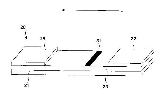
In this regard, various embodiments of an assay device that may optionally
be used to facilitate detection will now be described in more detail.
Referring to
Fig. 1, for instance, one embodiment of an assay device 20 is shown that
contains
a chromatographic medium 23 carried by a support 21. The chromatographic
medium 23 may be made from any of a variety of materials through which a fluid
is
capable of passing, such as a fluidic channel, porous membrane, etc. For
example, the chromatographic medium 23 may be a porous membrane formed
from materials such as, but not limited to, natural, synthetic, or naturally
occurring
materials that are synthetically modified, such as polysaccharides (e.g.,
cellulose
materials such as paper and cellulose derivatives, such as cellulose acetate
and
nitrocellulose); polyether sulfone; polyethylene; nylon; polyvinylidene
fluoride
(PVDF); polyester; polypropylene; silica; inorganic materials, such as
deactivated
alumina, diatomaceous earth, MgSO4, or other inorganic finely divided material
uniformly dispersed in a porous polymer matrix, with polymers such as vinyl
chloride, vinyl chloride-propylene copolymer, and vinyl chloride-vinyl acetate
copolymer; cloth, both naturally occurring (e.g., cotton) and synthetic (e.g.,
nylon or
rayon); porous gels, such as silica gel, agarose, dextran, and gelatin;
polymeric
films, such as polyacrylamide; and so forth. In one particular embodiment, the
chromatographic medium is formed from nitrocellulose and/or polyether sulfone
materials. It should be understood that the term "nitrocellulose" refers to
nitric acid
esters of cellulose, which may be nitrocellulose alone, or a mixed ester of
nitric
acid and other acids, such as aliphatic carboxylic acids having from 1 to 7
carbon
atoms. Although not required, the use of the chromatographic medium 23 for
chemical separation may provide enhanced benefits over other conventional
separation techniques, such as centrifugation. For example, the
chromatographic
medium 23 may simplify and reduce the costs of the resulting lateral flow
assay
device for many consumer applications, including those in which a disposable
kit is
desired.
The support 21 may be formed from any material able to carry the
chromatographic medium 23. Although not required, the support 21 may be
transparent so that light readily passes therethrough. In addition, it is also
generally desired that the support 21 is liquid-impermeable so that fluid
flowing
16
CA 02670519 2009-05-25
WO 2008/075214 PCT/IB2007/053960
through the medium does not leak through the support 21. Examples of suitable
materials for the support include, but are not limited to, glass; polymeric
materials,
such as polystyrene, polypropylene, polyester (e.g., Mylar film),
polybutadiene,
polyvinylchloride, polyamide, polycarbonate, epoxides, methacrylates, and
polymelamine; and so forth. As is well known the art, the chromatographic
medium 23 may be cast onto the support 21, wherein the resulting laminate may
be die-cut to the desired size and shape. Alternatively, the chromatographic
medium 23 may simply be laminated to the support 21 with, for example, an
adhesive. In some embodiments, a nitrocellulose or nylon porous membrane is
adhered to a Mylar film. An adhesive is used to bind the porous membrane to
the Mylar film, such as a pressure-sensitive adhesive. Laminate structures of
this type are believed to be commercially available from Millipore Corp. of
Bedford,
Massachusetts. Still other examples of suitable laminate structures are
described
in U.S. Patent No. 5,075,077 to Durley, III, et al., which is incorporated
herein in its
entirety by reference thereto for all purposes.
The assay device 20 may also utilize an absorbent material 28. The
absorbent material 28 generally receives fluid that has migrated through the
entire
chromatographic medium 23. As is well known in the art, the absorbent material
28 may assist in promoting capillary action and fluid flow through the medium
23.
In the embodiment illustrated in Fig. 1, the test sample may be applied
directly to conjugate pad 22. Provided reagents may include one or more
molecular substrates, co-factors, buffers, inhibitors, or other reagents
useful to
promote the enzyme reaction. For instance, for assaying a test sample that
beneficially requires a diluent, provided reagents may include a predetermined
amount of diluent in addition to other reagents. The test sample may be added
to
the diluent to initiate an enzyme reaction. The provided reagents may be
provided
together or separately, as desired. For example, a diluent may be physically
separated from other reagents. Following addition of a test sample to a
diluent, for
instance in a mixing well formed on the device, the mixture may be contacted
with
additional reagents. In the illustrated embodiment, contact is carried out
during
and following combination of the test sample with the reagents provided at the
conjugate pad.
The conjugate pad 22 is in fluid communication with the porous membrane
17
CA 02670519 2009-05-25
WO 2008/075214 PCT/IB2007/053960
23 through which the mixture may travel in the direction illustrated by arrow
"L" in
Fig. 1. Some suitable materials that may be used to form the conjugate pad 22
include, but are not limited to, nitrocellulose, cellulose, porous
polyethylene pads,
and glass fiber filter paper. The conjugate pad 22 may include a detectable
substance diffusibly immobilized thereto to which a component of the mixture
may
preferentially bind (either directly or non-directly). For example, the
conjugate pad
22 may include a diffusibly immobilized probe labeled with a detectable
substance,
the probe additionally including a specific binder for an enzyme reaction
product,
the molecular substrate, or another component of the mixture. Accordingly, a
conjugated probe including a detectable substance and a component of the
mixture may be formed. The component of the mixture, e.g., an enzyme reaction
product, may be conjugated to the probes using any of a variety of well-known
techniques, such as through covalent bonding and/or physical adsorption in a
manner such as described above. In one particular embodiment, carboxylic
groups of the probe are activated and reacted with amino groups of an enzyme
reaction product to form an amide bond. If desired, the conjugate pad 22 may
also
contain one or more assay reagents either diffusibly or non-diffusibly
immobilized
thereto, e.g., buffers, inhibitors, and the like.
Regardless, the chromatographic medium 23 defines a detection zone 31
within which the conjugated probe may be captured and detected. The manner in
which the conjugated probe is captured may depend on the nature of the probe.
For example, in some embodiments, a biological receptive material may be
immobilized within the detection zone 31 for capturing biological components.
Such biological receptive materials are well known in the art and may include,
but
are not limited to, antibodies, antigens, haptens, biotin, avidin,
streptavidin,
neutravidin, captavidin, protein A, protein G, carbohydrates, lectins,
nucleotide
sequences, peptide sequences, effector and receptor molecules, hormone and
hormone binding protein, enzyme cofactors and enzymes, enzyme inhibitors and
enzymes, and derivatives thereof.
Of course, any other suitable technique for capturing and detecting the
conjugated probes may also be used. For example, in some embodiments, non-
biological receptive materials may be immobilized within the detection zone 31
for
capturing probes. For instance, in one embodiment, the receptive material is a
18
CA 02670519 2009-05-25
WO 2008/075214 PCT/IB2007/053960
polyelectrolyte. Polyelectrolytes may have a net positive or negative charge,
as
well as a net charge that is generally neutral. Some suitable examples of
polyelectrolytes having a net positive charge include, but are not limited to,
polylysine (commercially available from Sigma-Aldrich Chemical Co., Inc. of
St.
Louis, Missouri), polyethylenimine; epichlorohydrin-functionalized polyamines
and/or polyamidoamines, such as poly(dimethylamine-co-epichlorohydrin);
polydiallyidimethyl-ammonium chloride; cationic cellulose derivatives, such as
cellulose copolymers or cellulose derivatives grafted with a quaternary
ammonium
water-soluble monomer; and so forth. In one particular embodiment, CelQuat
SC-230M or H-100 (available from National Starch & Chemical, Inc.), which are
cellulosic derivatives containing a quaternary ammonium water-soluble monomer,
may be utilized. Moreover, some suitable examples of polyelectrolytes having a
net negative charge include, but are not limited to, polyacrylic acids, such
as
poly(ethylene-co-methacrylic acid, sodium salt), and so forth. It should also
be
understood that other polyelectrolytes may also be utilized in the disclosed
methods, such as amphiphilic polyelectrolytes (i.e., having polar and non-
polar
portions). For instance, some examples of suitable amphiphilic
polyelectrolytes
include, but are not limited to, poly(styryl-b-N-methyl 2-vinyl pyridinium
iodide) and
poly(styryl-b-acrylic acid), both of which are available from Polymer Source,
Inc. of
Dorval, Canada. Further examples of polyelectrolytes are described in more
detail
in U.S. Patent App. Publication No. 2003/0124739 to Song, et al., which is
incorporated herein in it entirety by reference thereto for all purposes.
Although any polyelectrolyte may generally be utilized, the polyelectrolyte
selected for a particular application may vary depending on the nature of the
conjugated probes. In particular, the distributed charge of a polyelectrolyte
allows
it to bind to substances having an opposite charge. Thus, for example,
polyelectrolytes having a net positive charge are often better equipped to
bind with
conjugated probes (e.g., dyed particles) that are negatively charged, while
polyelectrolytes that have a net negative charge are often better equipped to
bind
to conjugated probes that are positively charged. Thus, in such instances, the
ionic interaction between these molecules allows the required binding to occur
within the detection zone 31. Nevertheless, although ionic interaction is
primarily
utilized to achieve the desired binding, it has also been discovered that
19
CA 02670519 2009-05-25
WO 2008/075214 PCT/IB2007/053960
polyelectrolytes may bind with probes having a similar charge.
According to one embodiment, an enzyme reaction product conjugated to
the probe including the detectable substance may have an affinity for the
receptive
material within the detection zone 31. In this instance, the conjugated probe
may
become immobilized within the detection zone 31 through specific binding
between
the enzyme reaction product and a receptive material so that the signal
generated
by the detectable substance may be detected. For example, the enzyme reaction
product may be bound to the probe via a first specific binding site and the
enzyme
reaction product may contain a second specific binding site that exhibits a
specific
affinity for the receptive material.
The detection zone 31 may generally provide any number of distinct
detection regions so that a user may better determine the concentration of an
enzyme within a test sample. When utilized, each region may contain the same
or
different receptive materials. For example, the detection zone 31 may include
two
or more distinct detection regions (e.g., lines, dots, etc.). The use of two
or more
distinct detection regions may provide certain benefits, such as facilitating
semi-
quantitation and/or inhibiting potential false positives due to overrunning of
the
reactive complexes or other materials. The detection regions may be disposed
in
the form of lines in a direction substantially perpendicular to the flow of
the test
sample through the chromatographic medium 23. Likewise, in some
embodiments, the detection regions may be disposed in the form of lines in a
direction substantially parallel to the flow of the test sample through the
medium
23.
For the embodiment shown in Fig., 1, as enzyme concentration increases in
a test sample, more conjugated probes are formed and become immobilized within
the detection zone 31. The increased quantity of detectable enzyme reaction
products at the detection zone 31 results in an increase in signal intensity.
From
this increase in signal intensity, the presence or concentration of the enzyme
may
be readily determined. For example, in one embodiment, the amount of enzyme is
directly proportional to the signal intensity at the detection zone 31, I1. If
desired,
the signal intensity I. may be plotted versus the enzyme concentration for a
range
of known enzyme concentrations to generate an intensity curve. To determine
the
quantity of enzyme in an unknown test sample, the signal intensity may then be
CA 02670519 2009-05-25
WO 2008/075214 PCT/IB2007/053960
converted to enzyme concentration according to the intensity curve.
It should be understood that one or more distinct regions of the detection
zone 31 may exhibit the above-described relationship between signal intensity
and
enzyme concentration; however, each distinct region need not exhibit such a
relationship. For example, in some embodiments, only one of multiple distinct
regions may exhibit a signal intensity that is directly proportional to the
concentration of the enzyme. The signal intensity of other distinct regions,
such as
those used to reduce false positives, may otherwise remain constant, or
exhibit an
increase and/or decrease in signal intensity. So long as at least one distinct
region
of the detection zone 31 satisfies the direct relationship, the signal
intensity
exhibited by the detection zone 31 is considered directly proportional to the
enzyme concentration.
Certain embodiments of the disclosed subject matter may utilize a sample
application area on the assay device. Thus, a test sample may be directly
applied
to a device upstream of the conjugate pad 22. In this regard, various
embodiments of applying a test sample to a device including a sample
application
area will now be described in more detail. Referring to Fig. 2, for example,
an
assay device 120 is shown that includes a chromatographic medium 123
positioned on a support 121, an absorbent material 128, and a sample
application
pad 124. A test sample is directly applied to the sample application pad 124.
The
sample application pad 124 may contain one or more assay pretreatment
reagents, either diffusibly or non-diffusibly immobilized thereto. For
instance, the
sample application pad 124 may contain one or more molecular substrates, co-
factors, buffers, inhibitors, or other reagents useful to promote the enzyme
reaction. The substrate and enzyme may contact one another and be allowed to
interact within the mixture formed upon the application of the test sample to
the
sample application pad 124. Thus, the sample application pad 124 may in effect
define a reaction zone on the device. In some embodiments, the contact
time/reaction time may be more specifically controlled through utilization of
a
sample application pad 124. For example, porosity of the sample application
pad
124 may be controlled to control flow rate of the mixture from the sample
application pad 124 to the adjacent sections of the device 120. In another
embodiment, the sample application pad 124 may be temporarily separated from
21
CA 02670519 2009-05-25
WO 2008/075214 PCT/IB2007/053960
adjacent areas of the device 120 through barriers. For instance, the sample
application pad may define a well within which the mixture may be formed and
held. Following the desired contact period, a temporary barrier, e.g., a gate,
may
be removed and the mixture may flow via a porous membrane, a fluidic channel,
or
the like, to a conjugate pad 122.
Probes capable of generating a detectable signal may be diffusibly
immobilized to the conjugate pad 122 that are configured to bind to a
component
of the mixture. For example, the probes may contain a detectable substance,
such
as described above. The probes may also contain particles labeled or otherwise
applied with the detectable substance. In some instances, it is desired to
modify
the probes in some manner. For example, the probes may be modified with a
specific binding member to form probes that have specific affinity for an
enzyme
reaction product, a molecular substrate, or another component of the mixture.
The
specific binding members may generally be applied to the probes using any of a
variety of well-known techniques, such as through covalent bonding and/or
physical adsorption in a manner such as described above. In one particular
embodiment, carboxylic groups on the probe surface are activated and reacted
with amino groups of the specific binding member to form an amide bond.
Regardless of its particular configuration, the assay device 120 typically
includes a detection zone 131 within which a component of the mixture, e.g.,
an
enzyme reaction product, may be captured and detected. The enzyme reaction
product may be detected within the detection zone 131 utilizing a variety of
assay
formats. In one embodiment, for example, a "sandwich" assay format is utilized
in
which the specific binding member of a probe is selected to have an affinity
for the
enzyme reaction product. The enzyme reaction product, such as antibodies,
antigens, etc., typically has two or more binding sites (e.g., epitopes). One
of
these binding sites becomes occupied by the specific binding member of the
probe
to form a conjugated probe. However, the free binding site of the enzyme
reaction
product may subsequently bind to a receptive material immobilized within the
first
detection zone 131 to form a new ternary sandwich complex. Alternatively, the
enzyme reaction product may be detected using direct or indirect "competitive"
assay formats. In such instances, the specific binding member of the probe may
be the same as or an analog of the enzyme reaction product. Thus, upon
reaching
22
CA 02670519 2009-05-25
WO 2008/075214 PCT/IB2007/053960
the detection zone 131, the detection probes and the enzyme reaction product
compete for available binding sites of the immobilized receptive material. Of
course, any other assay format is also suitable for use.
For the embodiment shown in Fig., 2, as enzyme concentration begins to
increase in the test sample, more enzyme product reaction forms in the
mixture.
Thus, if a sandwich assay format is used, more enzyme reaction product binds
to
the detectable probes form conjugated probes so that the amount of enzyme is
directly proportional to the signal intensity at the detection zone 131. On
the other
hand, if a competitive assay format is used, the amount of enzyme is
indirectly
proportional to the signal intensity at the detection zone 131. If desired,
the signal
intensity may be plotted versus the enzyme concentration for a range of known
enzyme concentrations to generate an intensity curve. To determine the
quantity
of enzyme in an unknown test sample, the signal intensity may then be
converted
to enzyme concentration according to the intensity curve.
An assay device as disclosed herein may include additional zones on the
device. For example, referring to Fig. 3, an assay device 120 is illustrated
that is
the same as the assay device 120 of Fig. 2, except that it also contains a
second
detection zone 135 positioned downstream from the detection zone 131. The
second detection zone 135 may provide one or more distinct regions (e.g.,
line,
dot, etc.), and may be positioned at any orientation relative to the flow of
the
mixture. A second receptive material is immobilized on the medium 123 within
the
second detection zone 135. The second receptive material may serve as a
stationary binding site for any detectable substance that does not become
bound
within the first detection zone 131. In one embodiment, for example, in which
a
"direct" competitive assay is employed, the first receptive material contains
an
antibody that has a specific binding affinity for both the enzyme reaction
product
and the probes (i.e., the probes have a specific binding member bound thereto
that
is the same as or an analog of the enzyme reaction product). The second
receptive material contains a polyelectrolyte that has a specific binding
affinity for
the probes. When present, the enzyme reaction product of the mixture competes
with the probes for available binding sites of the first receptive material.
Any
remaining, unbound probes travel past the first detection zone 131 to the
second
detection zone 135. Because the probes have a specific affinity for the second
23
CA 02670519 2009-05-25
WO 2008/075214 PCT/IB2007/053960
receptive material, they become immobilized within the second detection zone
135.
Likewise, in another embodiment in which an "indirect" competitive assay is
employed, the enzyme reaction product may contain a specific binding member
(e.g., biotin) and the probes may be dyed particles bound with a complementary
binding member (e.g., streptavidin) that has affinity for the enzyme reaction
product. The first receptive material contains a specific binding member that
is the
same as or an analog of the enzyme reaction product, thereby having an
affinity
for the probes. The second receptive material contains a polyelectrolyte also
having binding affinity for the probes. When present, the enzyme reaction
product
binds to the probes to form conjugated probes, thereby reducing the amount of
probes otherwise available for binding to the first receptive material.
Instead,
those conjugated probes which are complexed to the enzyme reaction product,
travel past the first detection zone 131 to the second detection zone 135.
Because
the probes have a specific affinity for the selected polyelectrolyte, they
become
immobilized within the second detection zone 135.
In the competitive assay embodiments referred to above, as the
concentration of the enzyme increases, the signal intensity at the second
detection
zone 135, 12, also begins to increase due to the presence of enzyme reaction
product in the mixture. From this increase in signal intensity, the presence
or
concentration of the enzyme may be readily determined. For example, in one
embodiment, the amount of enzyme is directly proportional to the signal
intensity at
the second detection zone 135, 12. If desired, the signal intensity 12 may be
plotted
versus the enzyme concentration for a range of known enzyme concentrations to
generate an intensity curve. To determine the quantity of enzyme in an unknown
test sample, the signal intensity may then be converted to enzyme
concentration
according to the intensity curve. It should be understood that, as discussed
above
with respect to the first detection zone 31 and/or 131, so long as one
distinct
region of the second detection zone 135 satisfies the direct relationship, the
signal
intensity exhibited by the second detection zone 135 is considered directly
proportional to the enzyme concentration.
Also, in the embodiments referenced above, an inverse relationship may
exist between the signal intensity at the detection zone 131 (I1) and the
second
24
CA 02670519 2009-05-25
WO 2008/075214 PCT/IB2007/053960
detection zone 135 (12). For example, because a predetermined amount of probes
are present, the amount captured at the second detection zone 135 is inversely
proportional to the amount captured at the detection zone 131. As a result of
this
inverse relationship, the concentration of the enzyme may, in some cases, be
more effectively measured over an extended range by comparing the signal
intensity at both detection zones. For example, in one embodiment, the amount
of
enzyme is directly proportional to the ratio of the signal intensity "IZ" to
the signal
intensity "I,." Based upon the range in which this ratio falls, the general
concentration range for the enzyme may be determined. If desired, the ratio of
12
to I1 may be plotted versus enzyme concentration for a range of known enzyme
concentrations to generate an intensity curve. To determine the quantity of
enzyme in an unknown test sample, the signal intensity ratio may then be
converted to enzyme concentration according to the intensity curve. It should
be
noted that alternative mathematical relationships between I1 and 12 may be
plotted
versus the enzyme concentration to generate the intensity curve. For example,
in
one embodiment, the value of 12 / (12 + I1) may be plotted versus enzyme
concentration to generate the intensity curve.
A device may include additional detections zones. For instance, a device
may include a detection zone within which a second component of a mixture may
be detected. A receptive material may be immobilized within this second
detection
zone that is a specific binding member for the second component of the
mixture.
For instance, an enzyme reaction product may be bound and detected in a first
detection zone, and a molecular substrate may be bound and detected in a
second
detection zone. Other zones that may be included on a device may include, for
example, control zones, for ensuring that the device is working properly, one
or
more calibration zones, for providing internal calibration capability to the
device,
and the like.
As stated above, signal intensity may be determined qualitatively,
quantitatively, and/or semi-quantitatively. In embodiments in which a
quantitative
result is desired, signal intensity may be determined using any of a variety
of
techniques known in the art. For example, in some embodiments, fluorescence
detection techniques are utilized.
The aforementioned detection techniques are described specifically in the
CA 02670519 2009-05-25
WO 2008/075214 PCT/IB2007/053960
context of enzymes. However, as stated, the presently disclosed devices are
equally suitable for detecting the presence or quantity of an enzyme inhibitor
within
a test sample. To detect the presence of an enzyme inhibitor within a test
sample,
a predetermined quantity of a corresponding enzyme may be mixed with the test
sample and allowed to incubate. In the presence of a certain amount of an
enzyme inhibitor, the enzyme-catalyzed reaction does not proceed at a
detectable
rate. Thus, the relationship between enzyme inhibitor concentration and signal
intensity will be opposite to the relationship between enzyme concentration
and
signal intensity. For example, using Fig. 1 as an illustration, an enzyme-
catalyzed
reaction will not occur in the presence of a certain amount of inhibitor.
Thus, no
enzyme reaction product will form and the detection zone 31 will fail to
generate a
detectable signal. On the other hand, as the amount of enzyme inhibitor is
reduced, the enzyme causes the enzyme reaction to form as described above.
The signal intensity generated at the detection zone 31 thus begins to
increase
due to a corresponding increase in the presence of enzyme reaction product.
Accordingly, in this particular embodiment, the amount of enzyme inhibitor
within
the test sample is inversely proportional to the signal intensity at the
detection
zone 31.
Referring to Fig. 4, one embodiment of a method for detecting the presence
of a protease using fluorescence will now be described in more detail.
Initially, a
test sample containing a protease P is applied to sample application pad 124
where it contacts molecular substrates 47 (e.g., protein or glycoprotein). The
molecular substrates 47 are allowed to contact the protease P and form a
mixture
that includes polypeptides 42 and 43 that are the enzyme reaction products of
the
enzyme-catalyzed reaction between the molecular substrates 47 and the protease
P. The mixture also includes unreacted molecular substrates 47, and protease
P.
The mixture flows to the conjugate pad 122, as indicated by the directional
arrow.
Diffusibly immobilized to the conjugate pad 122 are probes 44 that include a
detectable substance and a specific binding member for enzyme reaction product
43. Upon interaction of the mixture with the probes 44 at the conjugate pad
122,
enzyme reaction product 43 specifically bind to probes 44 to form conjugated
probes 45. As probes 44 are diffusibly immobilized to conjugate pad 122, the
mixture including the conjugated probes 45 then travels to the detection zone
131.
26
CA 02670519 2009-05-25
WO 2008/075214 PCT/IB2007/053960
Immobilized within detection zone 131 is a receptive material 90 that is
specific for
a second binding site of the enzyme reaction products 43 generated by the
enzyme-catalyzed reaction. Thus, the available binding sites in the detection
zone
131 may be bound by the conjugated probes 45.
Once captured, the signal intensity of the conjugated probes 45 may be
measured at detection zone 131. Fluorescence detection generally utilizes
wavelength filtering to isolate the emission photons from the excitation
photons,
and a detector that registers emission photons and produces a recordable
output,
usually as an electrical signal or a photographic image. One suitable
fluorescence
detector for use is a FluoroLog I II Spectrofluorometer, which is sold by SPEX
Industries, Inc. of Edison, New Jersey. Another example of a suitable
fluorescence detector is described in U.S. Patent Application Publication No.
2004/0043502 to Song, et al., which is incorporated herein in its entirety by
reference thereto for all purposes. Although the use of fluorescence is
utilized in
this particular embodiment, it should be understood that any other known
detection
technique may also be utilized. For example, other suitable optical detection
techniques may include, but are not limited to, phosphorescence, diffraction,
reflectance, transmittance, etc. The optical reader may be capable of emitting
light
and also registering a detection signal (e.g., transmitted or reflected light,
emitted
fluorescence or phosphorescence, etc.). For example, in one embodiment, a
reflectance spectrophotometer or reader may be utilized to detect the presence
of
reporters that exhibit a visual color (e.g. dyed latex microparticies). One
suitable
reflectance reader is described, for instance, in U.S. Patent App. Pub. No.
2003/0119202 to Kaylor, et al., which is incorporated herein in its entirety
by
reference thereto for all purposes.
Regardless of the technique used to measure signal intensity, the presence
or the amount of the protease P may be ascertained by the signal intensity at
the
detection zone 131.
Referring to Fig. 5, one embodiment of a method for detecting the presence
of a transferase using fluorescence via a competitive type assay will now be
described in more detail. Initially, a test sample containing a transferase T
is
applied to a sample application pad 124 that contains molecular substrates 67
(e.g., a polypeptide). The molecular substrates 67 are allowed to contact the
27
CA 02670519 2009-05-25
WO 2008/075214 PCT/IB2007/053960
transferase T for a sufficient period of time to form a mixture that includes
components 64 (e.g., ATP) that may provide the moiety (e.g., phosphorous)
targeted by the transferase. Following a contact period, which may simply be
the
period of time for flow from the sample application pad 124 to the conjugate
pad
122, the mixture may include unreacted molecular substrates 67, transferase T,
and product 68 generated by the enzyme-catalyzed reaction (e.g., a
phosphorylated polypeptide). The mixture flows to the conjugate pad 122, as
indicated by the directional arrow. Diffusibly immobilized on or in the
conjugate
pad 122 are detectable probes 70 that include the product of the enzyme
catalyzed
reaction or an analog thereof. As the mixture travels from the sample
application
pad 124 to the detection zone 131 the detectable probes 70 are picked up and
travel with the mixture. Immobilized within detection zone 131 is a receptive
material 91 that is specific for the product 68 generated by the enzyme-
catalyzed
reaction. Thus, the product 68 formed in the mixture and the detectable probes
70
compete for the available binding sites in the detection zone 131. Once
captured,
signal intensity may be measured and analyzed as described for other
embodiments described herein. Specifically, in this embodiment a larger signal
will
indicate a lower concentration of transferase T in the test sample, as more of
the
available binding sites in the detection zone 31 will be occupied with the
detectable
probes 70.
Fig. 6 illustrates another embodiment of a test device including a control
zone 136. According to this embodiment, a test sample including an enzyme E
may be applied to the device at the sample application pad 124. The sample
application pad 124 includes reagents for the enzyme catalyzed reaction
including
molecular substrates 147. The test sample combines with the reagents of the
sample application pad 124 to form a mixture within which the enzyme-catalyzed
reaction may take place to form at least one enzyme reaction product 143. The
test sample may be held at the sample application pad 124 for a period of time
as
discussed above or may travel directly to a conjugate pad 122. Diffusibly
immobilized at conjugate pad 122 are probes 144 including a detectable
substance
and a specific binding member for molecular substrates 147. Upon interaction
of
the test sample with the probes 144 at the conjugate pad 122, unreacted
molecular
substrates 147 remaining in the mixture specifically bind to probes 144 to
form
28
CA 02670519 2009-05-25
WO 2008/075214 PCT/IB2007/053960
substrate conjugates 145. As probes 144 are diffusibly immobilized to
conjugate
pad 122, the mixture including any substrate conjugates 145 then travels to
the
detection zone 131. Immobilized within detection zone 131 is a receptive
material
190 that is specific for a second binding site of the molecular substrates
147.
Thus, the available binding sites in the detection zone 131 may be bound by
the
substrate conjugates 145. Once captured, the signal intensity of the substrate
conjugates 145 may be measured at detection zone 131. For instance, a large
signal intensity may indicate a high concentration of unreacted molecular
substrates 147 in the mixture and accordingly, the presence of little or no
enzyme
in the test sample. The embodiment of Fig. 6 also includes a control zone 136
that
gives a signal to the user that the assay is performing properly. For
instance, the
control zone 136 contains an immobilized polyelectrolyte receptive material
170
within the control zone 136 for capturing probes that can include both
substrate
conjugate probes 145 and probes 144.
In one exemplary application, the assay device may be used for determining
the presence of transferase enzymes involved in the RAS protein activation
cycle.
RAS proteins function as important molecular switches for a wide variety of
signal
pathways. These pathways control processes including cytoskeleton integrity,
cell
adhesion and migration, and apoptosis. RAS proteins cycle between an activated
form (RAS-GTP) and an inactivated form (RAS-GDP).
RAS proteins are often deregulated in cancers, leading to increased
invasion and metastasis as well as decreased apoptosis. Accordingly, the assay
device may be utilized for determination of the presence of GTPase-activating
proteins (GAPs) that increase the rate of GTP hydrolysis, returning RAS-GTP to
its
inactive RAS-GDP form. For instance, a test sample containing a GAP may be
mixed with RAS-GTP molecular substrate. The molecular substrate is allowed to
contact the test sample for a sufficient period of time to form a mixture that
may
include unreacted molecular substrate (RAS-GTP), GAP, and product generated
by the enzyme-catalyzed reaction (RAS-GDP). The mixture is allowed to flow to
the conjugate pad that contains detectable probes labeled with a specific
binder for
either the molecular substrate (RAS-GTP) or the product of the enzyme-
catalyzed
reaction (RAS-GDP). For instance, the probes may be labeled with a MAP kinase
that is activated by RAS-GTP in certain metabolic pathways. Upon combination,
29
CA 02670519 2009-05-25
WO 2008/075214 PCT/IB2007/053960
unreacted molecular substrate may specifically bind to the MAP kinase-labeled
probed to form substrate conjugates, as described above, and the mixture then
travels to a detection zone. Immobilized within the detection zone is a second
receptive material that is specific for the RAS-GTP, for instance a second MAP
kinase. Thus, the available binding sites in the detection zone may be
occupied by
the substrate conjugates formed in the conjugate pad that include the
unreacted
substrate. The fluorescent particles joined to the detectable probes therefore
will
also be bound in the detection zone. Once captured, signal intensity may be
measured and analyzed as described for other embodiments described herein to
determine the presence of GAPs in the test sample.
In another embodiment, a lateral flow assay device may be utilized to
determine the presence of RAS activating proteins. For instance, the lateral
flow
assay device may be utilized to determine the presence of G exchange factors
(GEF) (e.g., CDC25, SOS1, SOS2) that catalyze the reactivation of RAS-GDP to
its active form, RAS-GTP. According to this embodiment, the molecular
substrates
may be the inactive form of the protein, RAS-GDP. Upon contact of the
molecular
substrates with the test sample containing the activating GEF, the RAS-GDP may
be activated to the RAS-GTP form. In this case, the product (RAS-GTP) may be
conjugated to a detectable substance at a conjugate pad and then captured by a
specific binder, e.g., a MAP kinase, immobilized in the detection zone to
determine
the presence or quantity of GEF in the test sample.
Another exemplary application of the lateral flow assay device may be in the
determination of the presence of angiotensin-converter enzyme (ACE) in a test
sample. ACE is an exopeptidase that catalyzes the conversion of angiotensin I
to
angiotensin II. While angiotensin I appears to exist primarily as a precursor
to
angiotensin II, angiotensin II is a potent vasoconstrictor and believed to
play a role
in conditions such as high blood pressure, heart disease and diabetic
nephropathy.
According to this particular embodiment, the lateral flow assay device may
include
a molecular substrate such as angiotensin I. Upon contact of the molecular
substrate with the test sample containing ACE, the angiotensin I may be
converted
to angiotensin II. The detection zone of the lateral flow assay device may
contain
immobilized therein a receptive material that is specific for angiotensin II,
such as
AT, or AT2 receptors, for example. The binding and detection of the detectable
CA 02670519 2009-05-25
WO 2008/075214 PCT/IB2007/053960
probes (e.g., angiotensin II conjugated probes formed at a conjugate pad) in
the
detection zone may indicate the presence of ACE in the test sample.
Lateral flow assay devices as described herein may provide a relatively
simple and cost-efficient method to quickly perform on-site testing of enzymes
or
their inhibitors. The device may provide a test result that is visible so that
it is
easily observed by the person performing the test in a prompt manner and under
test conditions conducive to highly reliable and consistent test results. The
lateral
flow assay device is also disposable so that, if desired, it may be discarded
when
the test is concluded.
The present disclosure may be better understood with reference to the
following examples.
EXAMPLE 1
To prepare a substrate, hemoglobin was biotinylated in borate buffer using
EX-LC-biotin available from Pierce Biotechnology. The biotinylated hemoglobin
was purified via dialysis.
A lateral flow device was prepared as follows:
To form the sample pad, a glass fiber pad was soaked with the purified
biotinylated hemoglobin dissolved in PBS buffer containing sucrose/Tween-20.
The glass fiber pad was then dried at 37 C.
To form the conjugate pad, a second glass fiber pad was soaked with a
solution including blue particles tagged with streptavidin available from
Bangs
Laboratories, Inc., sucrose, and Tween-20. The second glass fiber pad was then
dried.
A nitrocellulose membrane card was striped with anti-biotin antibody to form
a detection zone and also striped with polylysine to form a control zone. The
nitrocellulose membrane card was dried following application of the materials.
The conjugate pad and a cellulose pad were then laminated at either end of
the nictrocellulose membrane card with the detection zone and the control zone
therebetween. A reagent-free glass fiber pad and the hemoglobin-loaded glass
fiber sample pad were also laminated to the nitrocellulose membrane card.
Specifically, the reagent-free glass fiber pad was laminated between the
conjugate
pad and the hemoglobin-loaded sample pad to modulate the enzyme reaction
time. The assembled card was then cut into several devices, each approximately
31
CA 02670519 2009-05-25
WO 2008/075214 PCT/IB2007/053960
4mm in width.
Assays were conducted with the formed devices. Protease from S. Griseus
was diluted in PBS buffer in various concentrations and directly applied to
the
hemoglobin-loaded sample pad. The mixture thus formed flowed from the sample
pad to the reagent-free glass fiber pad and on to the conjugation pad, the
detection
zone, and the control zone, respectively.
Results for a typical run are schematically illustrated in Figure 7. Lanes 1-4
of the illustrated assay device were treated with protease at a concentration
of
30 g/mL, 0.3 g/mL, 0.03 g/mL, and a control of 0.0 g/mL, respectively.
Approximately twenty minutes following application, two strong blue bands were
observed at the detection zone in lanes 3 and 4, to which samples including
little or
no protease were applied. In contrast, a weak band or no band at all was
observed at the detection zone in lanes 1 and 2 - those samples including a
high
concentration of the protease. All lanes showed a strong blue band in the
control
zone.
While the subject matter has been described in detail with respect to the
specific embodiments thereof, it will be appreciated that those skilled in the
art,
upon attaining an understanding of the foregoing, may readily conceive of
alterations to, variations of, and equivalents to these embodiments.
Accordingly,
the scope of the present disclosure should be assessed as that of the appended
claims and any equivalents thereto.
32