Note: Descriptions are shown in the official language in which they were submitted.
CA 02672357 2009-06-11
WO 2008/071343 PCT/EP2007/010627
1
RAPID IMMUNOCHROMATOGRAPHIC DETECTION BY
AMPLIFICATION OF THE COLLOIDAL GOLD SIGNAL
The present invention relates in general to the field of diagnostics, namely
to a device for the
detection of a target in a sample. More precisely, the present invention
relates to a rapid
immunochromatographic test device especially suitable for ultra-sensitivity
detection of an
antibody and/or antigen in a sample using double sandwich immunoassay
detection for
sensitivity enhancement by signal amplification. The present invention further
refers to a
method for the production of the test device, to the uses of the test device
for the early
detection of disease infection such as HIV in a sample, as well as to a kit
comprising the test
device.
BACKGROUND OF THE INVENTION
In recent years the in vitro diagnostics (IVD) industry has made enormous
efforts to develop
immunochromatographic tests. Such tests have found applications in both
clinical and non-
clinical fields 1. A clinical utility of this test format has been shown for
more than 150
different analytes, and many of them are target now of commercially available
diagnostic
products 3. The wide range of applications for such devices has been reviewed
1 2
Rapid immunochromatographic test devices, e.g. in the form of a test strip,
are made up of a
number of components (Figure 1 a). Such a test strip 101 commonly includes a
sample pad
102, a conjugate pad 103, a membrane 104, e.g. a nitrocellulose membrane, and
an absorbent
pad 105. The membrane 104 is usually attached by means of an adhesive 106 to a
supporting
backing 107, e.g. made of plastic. In practice, the user dispense a patient
sample (usually
urine or whole blood) onto the sample pad 102. The sample then flows through
the sample
pad 102 into the conjugate pad 103, where it mixes with and releases the
detector reagent.
This mixture then flows across the membrane 104, where it binds with the test
and control
reagents located in the capture test zone 108 (sample zone) and negative
control zone 109,
respectively. When the mixture binds to the reagent that forms the test line,
a positive result is
indicated. The colour intensity of the test line is proportional to the
concentration of analyte in
CA 02672357 2009-06-11
WO 2008/071343 PCT/EP2007/010627
2
the sample. Excess sample that flows beyond the test and control zones 108,
109 is taken up
in the absorbent pad 105.
Rapid immunochromatographic test devices for diagnostic purposes are easy to
operate and
thus do not only contribute to the comfort of professional users, e.g. medical
stuff, but also
allow the operation by non-professionals users, e.g. most patients.
However, despite the wide use of rapid immunochromatographic test devices,
their suitability
is still limited with regard to certain applications. Urine, for example,
contains very low levels
of IgG, frequently around 1 mg/1. Therefore, the detection of antibodies, e.g.
directed to HIV
or HCV, require very sensitive techniques. To date, the tests for antibodies
in urine samples
are based on ELISA and Western blot techniques, which are labour-intensive,
time-
consuming and need to be carried out by qualified persons. Efforts are being
made to develop
simple and/or rapid tests for the detection of antibody to HIV in urine
specimens 4.
Oral fluid specimens consist often of saliva, which predominantly contains IgA
class
antibody, and oral mucosal transudates, which mostly contain IgG, and
therefore also have
much lower levels of IgG than serum. The levels of IgG normally found in oral
fluid
specimens (approximately 15 mg/1) are, however, higher than in urine specimens
and
innovative simple and rapid technology that has been shown to be effective for
whole blood,
serum and plasma, e.g. lateral flow through a chromatographic membrane, has
been
developed for use with these specimens 4.
Human chorionic gonadotropin (hCG) is a glycopeptide hormone produced by the
placenta
during pregnancy. The appearance and rapid increase in the concentration of
hCG in the
subject's urine makes it a good marker for confirming pregnancy. The
concentration of hCG
in urine increases steadily to a circulation peak of as much as 50,000 mIU/ml
between the
eighth and eleventh weeks.
Urine hCG levels during pregnancy are estimated to be:
1. 10-30 mIU/m17-10 days post conception.
2. 37,000-50,000 mIU/m18-11 weeks after last menstrual period.
3. <5 mIU/ml Healthy men or non-pregnant women.
CA 02672357 2009-06-11
WO 2008/071343 PCT/EP2007/010627
3
In the prior art the hCG test is a chromatographic immunoassay which uses
specific
antibodies to selectively identify hCG in urine with a high degree of
sensitivity. Elevated
levels of hCG as low as 20 mIU/ml can be detected within 3 minutes.
So far there are several tests used to detect the presence of hepatitis B
antibodies. In addition,
there are also several tests in the prior art that detect the presence of
viral antigens.
The hepatitis B surface antibody (anti-HBs) detection is one of the most
common test. Its
presence indicates previous exposure to HBV, but the virus is no longer
present and the
person cannot pass on the virus to others. The antibody also protects the body
from future
HBV infection. In addition to exposure to HBV, the antibodies can also be
acquired from
successful vaccination. This test is done to determine the need for
vaccination (if anti-HBs is
absent), or following the completion of vaccination against the disease, or
following an active
infection. Hepatitis B surface antigen (HBsAg) is a protein antigen produced
by HBV. This
antigen is the earliest indicator of acute hepatitis B and frequently
identifies infected people
before symptoms appear. HBsAg disappears from the blood during the recovery
period. In
some people (particularly those infected as children or those with a weak
immune system,
such as those with AIDS), chronic infection with HBV may occur. and HBsAg
remains
positive.
Further, testing for HIV is an essential component in the diagnosis and
treatment of persons
infected with the virus, in screening of blood for transfusion, in
surveillance and in HIV/AIDS
related research. Thus accurate and cost-effective testing is of great
importance in combating
the spread of HIV. It is imperative that tests for the diagnosis of HIV
infection be as accurate
as possible, given the serious ethical, legal and social issues that accompany
HIV infection.
The number of people living with HIV has now risen to reach its highest level
ever: close to
40 million people are living with the virus and close to 5 million people were
newly infected
with HIV in 2004 alone. Worldwide, the AIDS epidemic killed over 3 million
people last year
alone (Source: UNAIDS). Furthermore, only one in five people needing HIV
prevention
worldwide have access to basic prevention services and only one in ten people
living with
HIV has been tested for the virus.
CA 02672357 2009-06-11
WO 2008/071343 PCT/EP2007/010627
4
The HI virus is most easily transmitted to others during the initial period of
acute HIV
infection, when the viral load (quantity of HIV RNA in the blood) is
especially high and when
people are not aware of being contaminated by the virus. Most HIV infections
are transmitted
at this stage, called primary infection. Earlier detection using ultra
sensitive tests avoids
missing primary infections, enabling inunediate precautionary measures to be
taken to help
prevent the risk of HIV transmission to a non-infected partner, to an unborn
child, or through
blood donations or direct blood contact. Earlier detection of HIV infection
also ensures the
implementation of early antiretroviral therapy (ART) to slow down the
progression of HIV
infection, thereby improving patient care and quality of life.
The diagnosis of HIV infection is usually made on the basis of the detection
of HIV
antibodies and/or antigen. The diagnosis of an HIV infection can be made
indirectly, i.e.
through the demonstration of virus-specific antibodies. Besides such indirect
diagnosis based
on detection of antibodies, a direct diagnosis of HIV infection is also
possible: either through
the demonstration of infectious virus (using cell culture), viral antigens
(p24 antigen ELISA)
or viral nucleic acid (i.e. viral genome); the latter is also termed nucleic
acid testing (NAT).
One important problem of HIV antibody testing is the so-called "diagnostic
window". This is
the time period that elapses between the time of acquisition of HIV infection
until detectable
levels of antibodies are present. The switch from antibody-negative to
antibody-positive is
called "seroconversion".
The most widely used screening tests are ELISAs as they are the most
appropriate for
screening large numbers of specimens on a daily basis, e.g. blood donations.
The earliest
assays used purified HIV lysates (1 st generation assays). Improved assays
based on
recombinant proteins and/or synthetic peptides, which also enabled the
production of
combined HIV-1/HIV-2 assays, became rapidly available (2nd generation assays).
The so-
called 3rd generation or antigen-sandwich assays, which use labelled antigens
as conjugate,
are more sensitive and have reduced the diagnostic window period considerably
5 6
Thus, there is need in the prior art to provide a rapid immunochromatographic
test device
suitable for the ultra-sensitive detection of target in a sample.
CA 02672357 2009-06-11
WO 2008/071343 PCT/EP2007/010627
It is an object of the present invention to overcome the problems especially
with regard to the
applicability of rapid immunochromatographic test devices for the detection of
hCG, HBsAG,
anti-HBs, IgG, e.g. HIV antibodies, in urine, blood, serum or saliva by
enhanced sensitivity.
5 It is therefore an object to enhance the sensitivity of the rapid
immunochromatographic
detection system. Thus, it is an object of the present invention to overcome
the drawbacks of
the prior art and to provide especially a simple and rapid test device for the
ultra-sensitive
antibody and/or antigen detection by signal development and signal
amplification suitable to
be employed for the early detection of disease infections in a sample.
SUMMARY OF THE INVENTION
In one embodiment the present invention concerns a rapid immunochromatographic
test
device for the detection of a target in a sample, comprising
a) a first gold conjugate releasing pad, comprising colloidal gold conjugated
with
a first antibody or antigen, and
b) a second gold conjugate releasing pad, comprising colloidal gold conjugated
with a second antibody or antigen;
wherein both releasing pads are located at different positions within the test
device.
In a further embodiment the present invention concerns a method for the
production of a
device according to the present invention comprising the steps of
a) preparing a colloidal gold solution;
b) preparing a conjugation buffer;
c) partitioning the conjugation buffer by dividing it into a first and a
second flask;
d) adding an antibody according to the present invention to the conjugation
buffer
in the first flask;
CA 02672357 2009-06-11
WO 2008/071343 PCT/EP2007/010627
6
e) adding an antibody according to the present invention to the conjugation
buffer
in the second flask, wherein said antibody differs from the antibody used in
step d);
f) adding colloidal gold solution into each flask;
g) adding stabilizing buffer to each flask;
h) concentrating each conjugate;
i) adding a surfactant to the first conjugate and soaking glass fibre sheet
conjugate pad into the conjugate;
j) soaking another glass fibre sheet conjugate pad into the second conjugate;
k) printing sample and control lines onto the membrane;
1) laminating cards using the first gold conjugate; and
m) cutting cards into strips.
In another embodiment the present invention relates to the use of a device
according to the
present invention for the detection of a disease in at least one sample.
In a further embodiment the present invention refers to a kit for detection of
a disease
comprising the device according to the present invention and a manual.
DESCRIPTION OF THE PREFERRED EMBODIMENTS OF THE INVENTION
Before the present invention is described in more detail below, it is to be
understood that this
invention is not limited to the particular methodology, protocols and reagents
described herein
as these may vary. It is also to be understood that the terminology used
herein is for the
purpose of describing particular embodiments only, and is not intended to
limit the scope of
the present invention which will be limited only by the appended claims.
Unless defined
otherwise, all technical and scientific terms used herein have the same
meanings as commonly
understood by one of ordinary skill in the art.
Throughout this specification and the claims which follow, unless the context
requires
otherwise, the word "comprise", and variations such as "comprises" and
"comprising", will be
understood to imply the inclusion of a stated integer or step or group of
integers or steps but
not the exclusion of any other integer or step or group of integer or step.
CA 02672357 2009-06-11
WO 2008/071343 PCT/EP2007/010627
7
Several documents are cited throughout the text of this specification. Each of
the documents
cited herein (including all patents, patent applications, scientific
publications, manufacturer's
specifications, instructions, etc.), whether supra or infra, are hereby
incorporated by reference
in their entirety. Nothing herein is to be construed as an admission that the
invention is not
entitled to antedate such disclosure by virtue of prior invention.
As outlined above there is a need in the prior art to provide a new test
device suitable for the
early detection of a disease infection in at least one sample. Further, there
is a need in the
prior art to provide a new method for rapid immunochromatographic detection of
a target in a
sample for the detection of a disease or a specific condition such as
pregnancy in a subject. In
addition, there is also a need in the art for devices suitable for simple,
rapid and ultra-sensitive
detection of an antigen and/or antibody, which devices having a higher
sensitivity than
devices from the prior art.
In a first aspect the present invention provides a rapid immunochromatographic
test device for
the detection of a target in a sample, comprising
a) a first gold conjugate releasing pad, comprising colloidal gold conjugated
with a first antibody or antigen, and
b) a second gold conjugate releasing pad, comprising colloidal gold conjugated
with a second antibody or antigen;
wherein both releasing pads are located at different positions within the test
device.
The first colloidal gold conjugated with a first antibody or antigen captures
the target in the
sample and forms a complex "target-first colloidal conjugate". Preferably this
target in the
sample is an antigen and/or antibody.
In a embodiment of the device according to the present invention the first
gold conjugate
releasing pad comprises a gold conjugate that is conjugated with a first
specific antibody or
CA 02672357 2009-06-11
WO 2008/071343 PCT/EP2007/010627
8
antigen to capture the target analyte from the first site. The second gold
conjugate releasing
pad comprises a gold conjugated with a second specific antibody or antigen to
capture the
target analyte from the second site. The last mentioned conjugated antibody or
antigen is the
same antibody or antigen that is immobilized onto the nitrocellulose membrane.
In one preferred embodiment of the device according to the present invention
the first gold
conjugate releasing pad comprises a gold conjugate 201 that is conjugated with
a first specific
antibody 202 or antigen to capture the target analyte from the first site
202'. The second gold
conjugate releasing pad comprises a gold 211 conjugated with a second specific
antibody 203
or antigen to capture the target analyte from the second site 203'. The last
mentioned
conjugated antibody 203 or antigen is the same antibody or antigen that is
immobilized onto
the nitrocellulose membrane (Figure 3).
In another embodiment of the device according to the present invention the
device comprises
a test strip comprising
a) a sample pad,
b) a conjugate pad comprising the first gold conjugate pad,
c) a conjugate pad comprising the second gold conjugate pad,
d) a membrane comprising a capture test zone and a negative control zone, and
e) an absorbent pad.
In a preferred embodiment of the device according to the present invention the
capture test
zone comprises the second antibody or antigen. The antibody or antigen within
the test zone
capture the target from a site that differs from that site captured by the
first antibody
conjugated with the first colloidal gold, why both antibodies differ from each
other.
In another preferred embodiment the second specific antibody or antigen is
immobilized
within the test zone. The complex "target-first colloidal gold conjugate" will
be captured by
this second antibody or antigen and therefore kept within the test zone to
form the sandwich
detection (Figures 3 and 4). Then, the second gold conjugate releasing pad
will release its
gold conjugated with the second specific antibody or antigen to capture the
target analyte
from the second site. The second conjugate would bind with the first conjugate
from the side
of the target (Figures 3 and 4). At the same time, the other free sides of the
target will be able
CA 02672357 2009-06-11
WO 2008/071343 PCT/EP2007/010627
9
to link with their specific antibody or antigen to form more and more branched
bonds that
propagate the accumulation of colloidal gold particles onto the
capturing/sample line. This
propagation and accumulation of colloidal gold signal will amplify the signal
and highly
increase the sensitivity. This will enable us to detect very low
concentrations that are not
detectable using the same technique without signal amplification.
In one embodiment of the device according to the present invention the
membrane is attached
by means of an adhesive to a supporting backing. Preferably an acrylic
pressure sensitive
adhesive as known in the art is used.
In another embodiment of the device according to the present invention the
first and second
gold conjugate pad are laminated between the sample pad and the membrane,
wherein the two
gold conjugates are separated by a divider.
In a preferred embodiment of the device according to the present invention the
first 103.1 and
second gold conjugate pad 103.2 are laminated between the sample pad 102 and
the
membrane 104, wherein the two gold conjugates are separated by a divider 110
(Figure 1b).
Preferably, the divider is an inert divider, more preferably the divider is a
plastic divider
In another embodiment, the device according to the present invention the first
gold conjugate
pad is attached between the sample pad and the membrane while the second gold
conjugate
pad is within the upper part of the plastic housing to be released after
sample application onto
the nitrocellulose membrane directly.
In one preferred embodiment of the device according to the present invention
the supporting
backing is a plastic backing.
In another preferred embodiment of the device according to the present
invention the
membrane is nitrocellulose membrane.
In one embodiment of the device according to the present invention the first
or second
antibody is selected from the group comprising mouse anti-HIV p24, mouse anti-
HBsAg,
anti-hlgG, anti-Lipoarabinomannan, anti-H.Pylori antigen, anti-Leishmania
antigen, anti-
CA 02672357 2009-06-11
WO 2008/071343 PCT/EP2007/010627
Pneumonia antigen, anti-Malaria antigen, anti-Chlamydia antigen, anti-
Toxoplasma antigen,
anti-Schistosoma antigen, HIV 1 antibody, and HIV 2 antibody.
In a preferred embodiment of the device according to the present the first or
second antibody
5 is a monoclonal or polyclonal antibody. Preferably the first and second
antibodies are two
different monoclonal antibodies that recognize the target from two different
sites.
In another embodiment of the device according to the present invention the
first antigen is
selected from the group comprising conjugate of HIV antigen, conjugate of
hepatitis C
10 antigen, HIV 1 antigen, HIV 2 antigen, Lipoarabinomannan, H.Pylori antigen,
Toxoplasma
antigen.
In a further embodiment of the device according to the present invention the
control zone
comprises a non-specific capturing antibody and/or a non-specific antibody
capturing protein.
In one preferred embodiment of the device according to the present invention
the non-specific
antibody is selected from the group consisting of anti-mouse IgG, anti-rabbit
IgG, anti-goat
IgG, anti-donkey IgG, Anti-sheep IgG, anti-HIV p24, anti-Lipoarabinomannan,
anti-H.Pylori
antigen, anti-Leishmania antigen, anti-Pneumonia antigen, anti-Malaria
antigen, anti-
Chlamydia antigen, anti-Toxoplasma antigen, anti-Schistosoma antigen, HIV 1
antibody, and
HIV 2 antibody.
In another preferred embodiment of the device according to the present
invention the non-
specific capturing protein is either Protein A or Protein G.
In a preferred embodiment of the device according to the present invention the
device
comprises a housing comprising at least one test strip according to the
present invention.
In another preferred embodiment of the device according to the present
invention the housing
comprises two, three, four, five, six, seven, eight, nine, or ten test strips.
Preferably the
housing comprises two, three, four, or five test strips, more preferably the
housing comprises
two or three test strips.
CA 02672357 2009-06-11
WO 2008/071343 PCT/EP2007/010627
11
In one preferred embodiment of the device according to the present invention
each test strip
contains at least two antibodies or antigens, or at least one antibody and one
antigen, wherein
one of these antibodies or antigens is immobilized onto the membrane and the
other one is
conjugated with the first colloidal gold. In case of two antibodies, they have
to be different to
capture the target from two different sites.
In another aspect the present invention concerns a method for the production
of a device
according to the present invention, comprising the steps of
n) preparing a colloidal gold solution;
o) preparing a conjugation buffer;
p) partitioning the conjugation buffer by dividing it into a first and a
second flask;
q) adding an antibody according to the present invention to the conjugation
buffer
in the first flask;
r) adding an antibody according to the present invention to the conjugation
buffer
in the second flask, wherein said antibody differs from the antibody used in
step d);
s) adding colloidal gold solution into each flask;
t) adding stabilizing buffer to each flask;
u) concentrating each conjugate;
v) adding a surfactant to the first conjugate and soaking glass fibre sheet
conjugate pad into the conjugate;
w) soaking another glass fibre sheet conjugate pad into the second conjugate;
x) printing sample and control lines onto the membrane;
y) laminating cards using the first gold conjugate; and
z) cutting cards into strips.
In another aspect the present invention relates to the use of a device
according to the present
invention for the detection of a disease in at least one sample.
In one preferred embodiment of the use according to the present invention the
antibody in one
sample (e.g. specimen) and the antigen in another sample (e.g. specimen) is
detected. For
CA 02672357 2009-06-11
WO 2008/071343 PCT/EP2007/010627
12
example, in the case two test strips are used, Lipoarabinomannan-antigen can
be detected in
urine, while anti-lipoarabinomannan is detected in serum.
In another preferred embodiment of the use according to the present invention
the antibody
and antigen are detected in the same sample (specimen). For example, HIV
antibodies and the
HIV p24 antigen are detected in the same serum sample (specimen) using a
device of two
different strips.
In one embodiment of the use of the device according to the present invention
the sample is
obtained from a human.
In one preferred embodiment of the use of the device according to the present
invention the
sample is selected from the group comprising of whole blood, serum, plasma,
saliva, and
urine.
In another preferred embodiment of the use of the device according to the
present invention
the disease detected in said sample is selected from the group consisting of
HIV, Hepatitis A,
Hepatitis B, Hepatitis C, H.Pylori, Leishmania, Schistosomiasis, Malaria,
Pneumonia,
Toxoplasmosis, Tubercolosis and Chlamydial infection.
In a further aspect the present invention refers to a kit for detection of a
disease comprising
the device according to the present invention and a manual.
In one preferred embodiment of the kit according to the present invention the
kit further
comprises an assay buffer. The assay buffer can be any buffer known in the art
suitable for the
use of whole blood samples. Preferably in the case of whole blood samples Tris
buffer is
used, more preferably 0.1 M Tris buffer having a pH of 7.5 and comprising a
preservative.
Any preservative known by a person skilled in the art can be used, preferably
sodium azide
and even more preferably 0.01 M sodium azide is used.
In another embodiment the present invention relates to the use of the method
for diagnosing
and monitoring a disease or a specific condition of a subject by detecting a
target in a sample.
CA 02672357 2009-06-11
WO 2008/071343 PCT/EP2007/010627
13
The following example illustrate the present invention without, however,
limiting the same
thereto.
BRIEF DESCRIPTION OF THE DRAWING
Figure la: shows top and side views of a typical rapid-flow
immunochromatographic test
device known in the art in the form of a test strip 101 comprising a sample
pad 102, a
conjugate pad 103, a membrane 104, an absorbent pad 105, an adhesive 106, a
supporting
backing 107, a test zone 108, and a control zone 109.
Figure lb: shows top and side views of a preferred embodiment of a rapid-flow
immunochromatographic test device according to the present invention in the
form of a test
strip 101 comprising a sample pad 102, a first conjugate pad 103.1, a second
conjugate pad
103.2, a membrane 104, an absorbent pad 105, an adhesive 106, a supporting
backing 107, a
test zone 108, a control zone 109, and the conjugates divider 110.
Figure 2: shows the schematically view of a preferred embodiment of the first
and second
colloidal gold according to the present invention, wherein the first colloidal
gold 201 is
conjugated with a first specific antibody 202 and wherein the second colloidal
gold 211 is
conjugated with a second specific antibody 203. In addition, the target is
shown having two
sides 202' and 203'. The first side 202' of the target is captured by the
first antibody 202 of
the first gold conjugate 201 and the second side 203' of the target is
captured by the second
antibody 203 conjugated with the second gold conjugate 211.
Figure 3: shows a simplified scheme of a preferred embodiment of the test
device according
to the present invention. It shows the test zone 108 of the membrane 104 on
the test strip 101,
wherein the second specific antibody 203 or antigen is immobilized to the test
zone 108.
Figure 4: shows the main principle of a preferred embodiment of the signal
development
according to the present invention. By the sample flow within the rapid
immunochromatographic test device the target in the sample will be captured by
the first
specific antibody 202or antigen of the first colloidal gold 201 to form the
complex "target-
first colloidal gold". This complex flows to the test zone 108, where it will
be captured by the
CA 02672357 2009-06-11
WO 2008/071343 PCT/EP2007/010627
14
second specific antibody 203or antigen that is immobilized onto the membrane
104 to form a
sandwich detection.
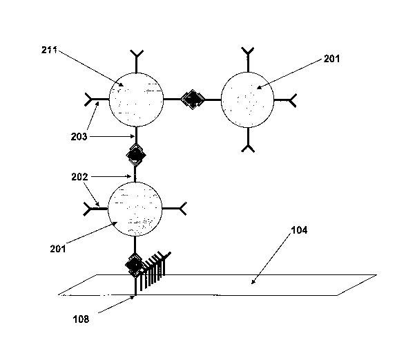
Figure 5: shows the main principle of a preferred embodiment of the signal
amplification and
multiplication according to the present invention. By the sample flow within
the rapid
immunochromatographic test device the target in the sample will be captured by
the first
specific antibody 202 or antigen that is conjugated to the first colloidal
gold 201 to form the
complex "target- first colloidal gold". This complex flows to the test zone
108, where it will
be captured by the second specific antibody 203 or antigen that is immobilized
onto the
membrane 104 of the test zone 108. Then, the second colloidal gold 211
conjugated with the
second specific antibody 203 or antigen will be released and will bind to the
target as well as
to the first colloidal gold conjugate 201 and enhance the signal by forming a
double sandwich.
EXAMPLES
The following examples illustrate the present invention without, however,
limiting the same
thereto.
Example 1: Preparation of an preferred embodiment of a test device according
to the present
invention
a) prepare 1% aqueous solution of tetrachloroauric acid at room temperature;
b) prepare 4% trisodium citrate aqueous solution at room temperature;
c) prepare 0.05 M Potassium Carbonate aqueous solution at room temperature;
d) prepare 600m1 of phosphate stabilizing buffer of pH 7.4, containing BSA,
Tween 20,
Sucrose, polyvinylpurrolidone and a preservative, e.g. sodium azide, at room
temperature;
e) prepare colloidal gold solution by reduction of 1.7 ml boiling
tetrachloroauric acid
solution (after dilution into 100ml) using lml trisodium citrate solution and
let it takes
the room temperature;
f) dilute the colloidal gold solution as 1:1 using distilled water. Adjust the
pH to 7.4
using potassium carbonate solution at room temperature;
CA 02672357 2009-06-11
WO 2008/071343 PCT/EP2007/010627
g) prepare 200m1 of phosphate conjugation buffer of pH 7.4 at room
temperature;
h) partition the 200m1 conjugation buffer by dividing it into two flasks
(100m1 of each);
i) add 1.0 mg of aqueous antibody (e.g. anti-p24 lst clone) to the conjugation
buffer in
the first flask with stirring at room temperature;
5 j) add 1.0 mg of aqueous antibody (e.g. anti-p24 2"d clone) to the
conjugation buffer in
the second flask with stirring at room temperature;
k) add 100m1 colloidal gold solution into each flask with stirring at room
temperature;
1) after about 45 minutes; add 200m1 of stabilizing buffer to each flask;
m) after about 20 minutes; concentrate each conjugate by cooled (temperature
around
10 15 C ) high speed centrifugation (10,000 rpm for one hour);
n) discard the supernatant and re-suspend the concentrated conjugates using
the
stabilising buffer at room temperature;
o) adjust the concentration for each of the two conjugates to O.D.520=2.0;
p) add 0.lml of Tween 20 to the first conjugate and soak glass fibre sheet
conjugate pad
15 into the conjugate, then heat dry at temperature around 50 C; and
q) soak another glass fiber sheet conjugate pad into the second conjugate,
then heat dry at
temperature around 50 C.
(* In case of antibodies/antigens and their specific antigens/antibodies there
is no need for
these steps of bovine serum albumin or an y other protein labelling. ** Other
proteins or
peptides could be used other than bovine serum albumin).
Additionally, print sample (e.g. anti-p24, 2"d clone) and control lines (e.g.
anti-mouse IgG)
onto nitrocellulose membrane, then heat dry at temperature around 50 C.
Finally, laminate cards according to the following procedure:
A. In case of conjulzate releasiniz site laminated within the upper side of
the device plastic
housinjz
Lamination of cards using the first gold conjugate. Laminate card components
onto the
backing material with the sequence:
1. laminate the nitrocellulose membrane nearly in the middle of the card;
CA 02672357 2009-06-11
WO 2008/071343 PCT/EP2007/010627
16
2. laminate the absorbent pad in the end of the card (overlaps from the
nitrocellulose
membrane side);
3. laminate the first conjugate pad in the other side of the nitrocellulose
membrane; and
4. laminate the sample pad.
B. In case of conjugate releasing site laminated onto the test strip itself
separated from the
first conju ag te by a divider
Laminate card components onto the backing material with the sequence (see
figure 1b):
1. laminate the nitrocellulose membrane nearly in the middle of the card;
2. laminate the absorbent pad in the end of the card (overlaps from the
nitrocellulose
membrane side);
3. laminate the first conjugate pad in the other side of the nitrocellulose
membrane ;
4. laminate the plastic divider onto the first conjugate (overlaps from the
nitrocellulose
membrane side);
5. laminate the second conjugate pad onto the divider (overlaps from the
nitrocellulose
membrane side);
6. laminate the sample pad onto the other end of the card, the sample pad will
overlaps
with the two conjugate pads; and
7. then cut cards into strips.
C. Alternatively
Lamination of the second gold conjugate could be applied within the plastic
housing itself to
ensure that the two conjugates will not propagate before release from the
releasing pad and so
stick within the releasing pad.
Example 2: Hepatitis B surface antigen (HBsAg) detection system
The first gold conjugate pad contains a conjugate of colloidal gold with a
first mouse
monoclonal anti-HBsAg , and the second gold conjugate pad contains a conjugate
of colloidal
gold with a second mouse monoclonal anti-HBsAg. The first conjugate releasing
pad 103.1 is
laminated on the test strip between the sample pad and the nitrocellulose
membrane 104 while
the second 103.2 is above the first pad separated by a divider 110 to be
released directly
CA 02672357 2009-06-11
WO 2008/071343 PCT/EP2007/010627
17
toward the nitrocellulose membrane 104 without flow through the first
conjugate pad to avoid
interact with the first conjugate before reaching the membrane (Figure 1b).
The second conjugate releasing site could be laminated within the upper side
of the device
plastic housing.
The sample line 108 is the second mouse monoclonal anti-HBsAg immobilized onto
the
nitrocellulose membrane 104. The control line 109 is anti-mouse IgG. Sample
108 and control
lines 109 turn into purple color in case of HBsAg availability in the sample;
only the control
line 109 turns into purple color in case of HBsAg free sample, see Figurelb.
The commercially available rapid tests sensitivity for Hepatitis B surface
antigen is within the
range 500-1000pg/ml while according to this system it is so simple to detect
less than 200
pg/ml.
Example 3: Human Immunodeficiency Virus (HIV) antibodies detection system
The first gold conjugate pad contains a conjugate of colloidal gold with a
first mouse anti-
human Immunoglobulin G (anti-hIgG), and the second gold conjugate pad contains
a
conjugate of colloidal gold with HIV p160. The first conjugate releasing pad
103.1 is
laminated on the test strip between the sample pad and the nitrocellulose
membrane while the
second 103.2 is above the first pad separated by a divider 110 to be released
directly toward
the nitrocellulose membrane without flow through the first conjugate pad to
avoid interact
with the first conjugate before reaching the membrane (Figure 1b).
The second conjugate releasing site could be laminated within the upper side
of the device
plastic housing.
The sample line 108 is HIV p160 antigen immobilized onto the nitrocellulose
membrane 104.
The control line 109 is anti-mouse IgG. Sample 108 and control lines 109 turn
into purple
colour in case of HIV antibodies availability in the sample; only the control
line 109 turns into
purple colour in case of HIV antibodies free sample, see Figurelb.
According to this system it is so simple to detect very low titers of HIV
antibodies.
CA 02672357 2009-06-11
WO 2008/071343 PCT/EP2007/010627
18
The features disclosed in the foregoing description, in the claims and/or in
the accompanying
drawings may, both separately and in any combination thereof, be material for
realizing the
invention in divers forms thereof.
CA 02672357 2009-06-11
WO 2008/071343 PCT/EP2007/010627
19
References
(1) J Chandler, N Robinson, and K Whiting, "Handling False Signals in Gold-
Based
Rapid Tests", IVD Technology 7, no. 2 (2001): 34-45;
http://www.devicelink.com/ivdt/archive/01 /03/002.htm1.
(2) J Chandler, T Gurmin, and N Robinson, "The Place of Gold in Rapid Tests",
IVD
Technology 6, no. 2 (2000): 37-49;
http://www.devicelink.com/ivdt/archive/00/03/004.html
(3) TC Tisone et al., "Image Analysis for Rapid-Flow Diagnostics", IVD
Technology
5, no. 5 (1999): 52-58; http://www.devicelink.com/ivdt/archive/99/09/010.html.
(4) Zaaijer, H.L., Exel-Oehlers, P.V., Kraaijeveld, T., Altena, E., Lelie,
P.N. (1992) Early
detection of antibodies to HIV-1 by third-generation assays. Lancet 340, 770-
772.
(5) Constantine, N.T., van der Groen, G., Belsey, E.M., Tamashiro, H. (1994)
Sensitivity of
HIV-antibody assays determined by seroconversion panels. AIDS 8, 1715-1720.
(6) Satten, G.A., Busch, M.P., et al. (1997) Effect of transmission route on
window period
estimates. Fourth Conference on Retroviruses and Opportunistic Infections,
Washington DC,
Abstract 122.
(7) WHO. Rapid HIV tests: Guidelines for use in HIV testing and counseling
services in
resource-constrained settings. Geneva 2004.
http://www.who.int/hiv/pub/vct/rapidhivtests/en/
(8) Holodniy M, et al. (1991) Reduction in plasma human immunodeficiency virus
ribonucleic acid following dideoxynucleoside therapy as determined by the
polymerase chain
reaction. J. Clin. Invest 88, 1755-1759.
(9) Katzenstein D.A., et al. (1994) Quantitation of human immunodeficiency
virus by culture
and polymerase chain reaction in response to didanosine after long-term
therapy with
zidovudine. J. Infect. Dis. 169, 416-419.
(10) Jackson JB, et al. (1998) Practical diagnostic testing for human
immunodeficiency virus.
Clin. Microb. Rev. 1, 124-138.
(11) Goudsmit J, et al. (1986) Expression of human Immunodeficiency virus
antigen (HIV-
Ag) in serum and cerebrospinal fluid during acute and chronic infection.
Lancet 2, 177-180.
(12) Aubuchon, J.P., Birkmeyer, J.D., Busch, M.P. (1997) Cost-effectiveness of
expanded
human immunodeficiency virus-testing protocols for donated blood. Transfusion
45, 45-51.
CA 02672357 2009-06-11
WO 2008/071343 PCT/EP2007/010627
(13) Ward, J.M., Holmberg, S.D., Allen, J.R., Cohn, D.L., et al. (1988)
Transmission of
human immunodeficiency virus (HIV) by blood transfusion screened as negative
for HIV
antibody.lV. Engl. J. Med. 8, 473-478.
(14) Alter, H.J., et al. (1990) Prevalence of human immunodeficiency virus
type 1 p24 antigen
5 in U.S. blood donors - an assessment of the efficacy of testing in donor
screening. N. Engl. J.
Med. 323, 1312-1317.
(15) Mayers, D.L. (1998) Drug-resistant HIV-1. The virus strikes back. JAMA
279, 2000-
2002.
(16) Stephenson, J. (2002) Cheaper HIV drugs for poor nations bring a new
challenge:
10 monitoring treatment. JAMA 288, 2.
(17) WHO. HIV assays: Operational characteristics (Phase 1). Report 15/
antigen/antibody
ELISAs. Geneva 2004.
http://www.who.int/diagnostics-laboratory/evaluations/hiv/en/
(18) WHO. HIV assays: Operational characteristics (Phase 1). Report 14/
simple/rapid tests.
15 Geneva 2003.
http://www.who.int/diagnostics-laboratory/evaluations/hiv/en/
(19) Meier, T, et al. (2001) Evidence for a diagnostic window in fourth
generation assays for
HIV. J. Clin. Virol. 23, 113-116.