Note: Descriptions are shown in the official language in which they were submitted.
CA 02672729 2009-06-15
WO 2008/077943 PCT/EP2007/064462
DEVICE AND METHOD FOR IMPROVING HEARING
FIELD OF THE INVENTION
The invention is in the field of implantable hearing devices, a kit, and
methods for the
implantation of said hearing devices.
BACKGROUND TO THE INVENTION
Sounds are perceived in humans by means of a mechanical-neural system
distributed
over the external ear canal, the middle ear cavity and the cochlea. Sound
waves
propagate through the external ear canal to reach and vibrate the tympanic
membrane.
The middle ear ossicles - malleus, incus and stapes - transfer the tympanic
membrane
vibrations to the footplate of the oval window that seals off the cochlea.
Footplate
vibrations set up waves of fluid motion within the fluid that is contained in
the cochlea. The
fluid motions in turn activate hair cells inside the cochlea. The hair cells
produce in
response electrical nerve impulses that are routed through the spiral ganglion
and the
auditory nerve to the brain, where they are perceived as sound. The electro-
mechanics of
the cochlear membranes and hair cells vary gradually along the length of the
cochlea,
which creates a natural spectral distribution of sensitivity along the
cochlea: high-pitch
sounds activate the hair cells near the oval window, whereas the lower pitches
activate
the hair cells further down the cochlea.
Modification and/or amplification of the energy reaching the sensory cells of
the inner ear
are the basis for treatment of conductive and sensorineural hearing losses.
First attempts
to improve hearing by making a hole in the wall of the inner ear at the level
of the lateral
semicircular canal were undertaken in 1914 in a procedure called fenestration.
In
fenestration, a trough-shaped window is made in the bony wall of the inner ear
and is
covered with transposed tympanic membrane. This connects the fluid spaces of
the
human inner ear directly to the outside world bypassing the dysfunctional
middle ear. This
procedure enables the sound energy to reach directly the membranous part of
the inner
ear and can result in an improvement of hearing by up to 30dB.
Currently, when opening of the inner ear space is necessary, other safer and
more
effective surgical techniques have been developed. In patients with
otosclerosis
(immobility of the ossicular chain due to fixation of the stapes footplate), a
small-hole
fenestration in the stapes footplate is made, and a Teflon piston is
transposed between
the incus and the opening in the footplate after removal of the stapes
superstructure. This
CA 02672729 2009-06-15
WO 2008/077943 PCT/EP2007/064462
2
procedure, albeit quite difficult technically, normalises the functional
status of the
conductive part of the middle ear and, in most cases, restores hearing to
normal or quasi-
normal.
The main drawback of the latter technique is that the fenestration of the
inner ear remains
open, which incurs the risk for inner ear infections. This may lead to
meningitis or total
hearing loss. A solution is to cover the fenestration with a piece of tissue,
however, this
has in the long term a tendency to re-ossify, which leads to diminishing
results.
Hearing improvement can also be achieved by amplification of the energy
reaching the
sensory cells of the inner ear, using a variety of hearing aids. All these
devices try to
compensate for the diminished hearing acuity by amplification of the energy
reaching the
inner ear. They either amplify air sound waves, vibrate the ossicular chain,
or vibrate the
bones of the skull. However, application of any one of these devices has a
number of
important drawbacks including lack of aesthetic appeal, poor performance of
conventional
hearing aids due to feedback and distortion, limited indications and variable
results in
implantable hearing aids.
There have also been a few devices described in the literature, which employ a
direct
energy transfer to or from the inner ear. The advantage of these systems is
that relatively
little energy is required to achieve substantial amplifications and that the
transducers can
be very small. Some of these direct energy transfer devices are described
below.
The Round Window Electromagnetic device (RWEM) realises coupling to the
cochlear
fluids through an intact round window membrane, which serves as the natural
flexible
interface between the middle and the inner ear. The RWEM uses a magnet,
surgically
placed onto the round window and an electromagnetic coil to induce vibration.
This
vibration is transmitted through an intact round window membrane to the
cochlea's fluids.
The RWEM device, however, would compromise the normal compliance of the round
window membrane, which could induce a hearing loss.
Leysieffer describes in DE 39 40 632 an implantable hearing aid with either
separate
electromechanical stimulation or separate electrical stimulation.
Money (US-PS 5,782,744) proposed an implantable microphone encapsulated in a
waterproof casing and placed at the round window in contact with the cochlear
fluid,
CA 02672729 2009-06-15
WO 2008/077943 PCT/EP2007/064462
3
immersed in the cochlear fluid or placed in the middle ear and coupled to the
inner ear
fluid by a conduction tube. Such a microphone transmits the pressure
variations induced
in the inner ear by acoustic stimulation.
A cochlear implant bypasses the mechanical signal chain altogether, and
provides direct
electrical stimulation of the auditory neural system using an elongated
electrode inserted
in and following either the scala tympani or the scala vestibuli.
Hybrid electrical-mechanical systems have been described recently that
complement the
electrical stimulation of a cochlear implant with mechanical means to induce
vibrations in
the inner ear fluid. Electrical stimulation complementary to mechanical
stimulation can be
a significant advantage to certain otological pathologies. In case of locally
damaged inner
ear structures, mechanical stimulation can be ineffective at related
frequencies. For
example in patients with presbyacousis where the sensory cells (hair cells)
for sensing the
high frequencies are damaged and no longer function, the related neural
structures are
functional and can be electrically stimulated to transfer high-frequency
acoustical signals.
There are also many pathologies other than presbyacousis pathologies with high-
frequency hearing loss. In general, electrical stimulation is necessary
whenever "dead
frequency regions" are present that cause sound distortion when only
stimulated
acoustically/mechanically.
Leysieffer (US Patent 6,697,674) describes the combination of a cochlear
electrode with
an implanted mechanical transducer that vibrates parts of the middle ear. The
middle ear
vibrations find their natural way to the inner ear via the stapes footplate in
the oval
window. Harrison (US Patent 6,754,537) describes a hybrid system for patients
with
severe high-frequency hearing loss but normal or near normal hearing for low
frequencies.
He combines a cochlear electrode that electrically stimulates the cochlea with
the high-
frequency audio content, and relies on the patient's natural hearing to pick
up the low-
frequency audio content. This low-frequency content is then provided
mechanically by
either a conventional external hearing aid, or a middle-ear mechanical
transducer.
Leysieffer describes in US Patent 6,565,503 an electrical cochlear electrode
modified with
miniature mechanical transducers distributed over the electrode's length to
generate
mechanical vibrations in the inner ear fluid.
A drawback of known hybrid electrical-mechanical devices for hearing aids is
that their
implantation is a highly invasive procedure causing irreparable damage to the
residual
CA 02672729 2009-06-15
WO 2008/077943 PCT/EP2007/064462
4
hearing the patient may still have. This is because they are configured either
as a
conventional cochlear electrode in combination with a mechanical device, or as
a cochlear
electrode modified with intra-cochlear electromechanical converters that
generate
mechanical vibrations in the inner ear fluid. Both types have an elongated
electrode that is
inserted in the scala vestibuli or scala tympani. They penetrate deep into the
cochlea
through a hole in the bony cochlea wall, thereby risking damaging the fine
features inside
and destroying whatever residual hearing the patient may still have.
Shortening and
thinning the electrodes to preserve hearing is an area of intensive research.
It is
technically challenging and experiments have yet to show conclusive and
consistent
improvements, although full coverage for speech has been demonstrated on some
patients with a 16-17 mm outer-wall electrode. More important, implanting
short electrodes
actually jeopardizes the patient's prospects for later upgrades to longer
electrodes, e.g. in
cases of progressive hearing loss. This is caused by tissue growth around the
electrodes
that tears during electrode removal and ruptures the fragile basilar membrane
with it.
The present invention aims at overcoming the problems associated with
conventional
hearing implants, by providing an effective method and device which retains
residual
hearing.
It also aims to allow the surgeon to implant an electrical and mechanical
stimulatory
hearing aid in a single procedure, in those cases where he does not have the
foreknowledge of which stimulation would be the most effective.
FIGURE LEGENDS
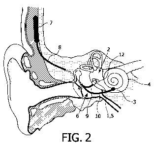
FIG. 1: A functional diagram of the ear, showing a configuration of the
present invention
whereby the proximal electrode and vibration generator are implanted in a hole
created
near to oval window for accessing the scala vestibule.
FIG. 2: A functional diagram of the ear, showing a configuration of the
present invention
whereby the proximal electrode and vibration generator are implanted in the
oval window.
FIG. 3: A functional diagram of the ear, showing the prior art arrangement of
an electrode
inserted in the scala vestibuli or scala tympani of the cochlea.
FIG. 4: A cross-section through an in situ vibration generator of the present
invention.
FIGs 5 to 18: A cross-section view depicting an in situ vibration generator
and proximal
electrode.
FIGs 19: A three dimensional view of a cochlea disposed with components of the
present
device.
CA 02672729 2009-06-15
WO 2008/077943 PCT/EP2007/064462
FIG. 20: A schematic view of a configuration of a regulating unit.
FIG.s 21, 24 to 30: Cross-section views depicting an in situ vibration
generator and
proximal electrode, where the vibration generator comprises a first and second
sub-frame
connected by an vibration-energy conducting element.
5 FIG. 22: A functional diagram of the ear, showing a configuration of the
present invention
whereby the proximal electrode implanted in a hole created near to oval window
for
accessing the scala vestibule, and vibration generator comprises a first and
second sub-
frame connected by an vibration-energy conducting element, the first sub-frame
housing
the electromechanical actuator implanted in the mastoid.
FIG. 23: A functional diagram of the ear, showing a configuration of the
present invention
whereby the proximal electrode implanted in a hole created near to oval window
for
accessing the scala vestibule, and vibration generator comprises a first and
second sub-
frame connected by an vibration-energy conducting element, the first sub-frame
housing
the electromechanical actuator incorporated in the control unit.
FIG. 31: Exploded view of a revolute joint present in a vibrational energy
conducting
element that is a hinged link.
SUMMARY OF SOME EMBODIMENTS OF THE INVENTION
One embodiment of the invention is an implantable device for improving hearing
in a
subject comprising:
- a vibration generator (5) comprising an output region (19) configured to
apply vibrational
stimulation to the inner ear (2) fluid,
- a proximal electrode (1) configured for physical attachment to a wall
enclosing the inner
ear (2) at a location proximal to the output region, and
- a separate distal electrode (3) configured to make electrical contact with
the auditory
nerve (4).
Another embodiment of the invention is an implantable device as described
above,
wherein the vibration generator comprises:
- an electromechanical actuator (20),
- a vibrating surface (25) co-operatively connected to the electromechanical
actuator (20),
which provides vibrational energy, and
- a frame (22) configured to position the vibrating surface (25) to direct
vibrational energy
therefrom to the output region (19).
CA 02672729 2009-06-15
WO 2008/077943 PCT/EP2007/064462
6
Another embodiment of the invention is an implantable device as described
above,
wherein the frame (22) is configured for physical attachment to a wall
enclosing the middle
ear (6).
Another embodiment of the invention is an implantable device as described
above,
wherein the frame (22) is configured for physical attachment to
- a wall enclosing the middle ear (6),
- a wall enclosing the inner ear (2),
- a walled interface between the middle (6) and inner ear (2),
- a walled interface between the inner ear (2) and mastoid region, or
- a wall of a cavity created in the mastoid region.
Another embodiment of the invention is an implantable device as described
above,
wherein the vibrating surface (25) is a flat surface co-operatively connected
to the
electromechanical actuator (20).
Another embodiment of the invention is an implantable device as described
above,
wherein the vibrating surface (25) is extended by an elongated member co-
operatively
connected to the electromechanical actuator (20).
Another embodiment of the invention is an implantable device as described
above,
wherein the frame (22) comprises a first sub-frame (22a) that supports the
electromechanical actuator (20) and a second sub-frame (22b) provided with the
output
region (19) wherein the vibration energy from the electromechanical actuator
(20) is
directed to the output region (19) via a vibrational-energy conducting element
(80).
Another embodiment of the invention is an implantable device as described
above,
wherein the conducting element (80) is a tube (84) adapted to contain a non-
compressible
liquid or gel (81).
Another embodiment of the invention is an implantable device as described
above,
wherein the conducting element (80) is a cable link, comprising a flexible
cable (83)
housed in a sleeve (82), which cable (83) is configured to move within the
sleeve (82),
while maintaining a coaxial relation therewith.
CA 02672729 2009-06-15
WO 2008/077943 PCT/EP2007/064462
7
Another embodiment of the invention is an implantable device as described
above,
wherein the conducting element (80) is a non-flexible, elongated rod (85).
Another embodiment of the invention is an implantable device as described
above,
wherein the conducting element (80) is an adjustable telescopic slip link
(89).
Another embodiment of the invention is an implantable device as described
above,
wherein the conducting element (80) is an adjustable hinged link (91).
Another embodiment of the invention is an implantable device as described
above,
wherein the second sub-frame (22b) forms a passage (72) having a receiving end
(70) to
receive vibrational energy from the conducting element (80), and a
transmitting end (71)
where vibrational energy is directed towards the inner ear fluid.
Another embodiment of the invention is an implantable device as described
above,
wherein the second sub-frame (22b) is disposed with the vibrating surface (25)
in the
passage (72), optionally in a region towards or at the transmitting end (71).
Another embodiment of the invention is an implantable device as described
above,
wherein the vibrating surface (25) is a flexible or flexibly suspended
membrane (73) in
sealing connection with the transmitting end (71) of the passage (72), and in
hydraulic
connection with the electromechanical actuator (20).
Another embodiment of the invention is an implantable device as described
above,
wherein the vibrating surface (25) is a flexibly suspended plate in mechanical
connection
with the electromechanical actuator (20)
Another embodiment of the invention is an implantable device as described
above,
wherein the vibrating surface (25) is formed from a sliding piston (75) in
hydraulic or
mechanical connection with the electromechanical actuator (20).
Another embodiment of the invention is an implantable device as described
above,
wherein the vibrating surface (25) comprises:
- a flexibly suspended rigid membrane (105) in sealing connection with the
transmitting
end (71) of the passage (72), and in hydraulic connection with the
electromechanical
actuator (20), and
CA 02672729 2009-06-15
WO 2008/077943 PCT/EP2007/064462
8
- a pin (101) attached to said membrane (105).
Another embodiment of the invention is an implantable device as described
above,
wherein the first sub-frame (22a) is configured for physical attachment to:
- a wall enclosing the middle ear (6) or
- a wall of a cavity created in the mastoid region.
Another embodiment of the invention is an implantable device as described
above,
wherein the first sub-frame (22a) is incorporated within the housing of a
regulating unit (7).
Another embodiment of the invention is an implantable device as described
above,
wherein the second sub-frame (22b) is configured for attachment at
- a wall enclosing the inner ear (2),
- a walled interface between the middle (6) and inner ear (2), or
- a walled interface between the inner ear (2) and mastoid region.
Another embodiment of the invention is an implantable device as described
above,
wherein the electromechanical actuator (20) is an electromagnetic,
piezoelectric,
electrostatic or magnetostrictive actuator.
Another embodiment of the invention is an implantable device as described
above,
wherein at least part of the frame (22) or at least part of the vibrating
surface (25) acts as
the proximal electrode (1).
Another embodiment of the invention is an implantable device as described
above,
wherein the proximal electrode (1) and/or the distal electrode (1) is pin-
shaped and is
configured to diverge from a longitudinal centreline of a cochlea (4) lumen.
Another embodiment of the invention is an implantable device as described
above,
wherein the proximal electrode (1), the output region (19) and/or distal
electrode (3) is
configured to sit flush or recessed with the inside wall of the inner ear (2).
Another embodiment of the invention is an implantable device as described
above,
wherein the proximal electrode (1), the output region (19) and/or distal
electrode (3) is
configured to sit flush or recessed with the inside wall of the cochlea (4)
lumen.
CA 02672729 2009-06-15
WO 2008/077943 PCT/EP2007/064462
9
Another embodiment of the invention is an implantable device as described
above, further
comprising a regulating unit (7) configured to provide electrical signals to
said electrodes
and/or vibration generator, which signals represent sound information.
Another embodiment of the invention is an implantable device as described
above,
wherein the regulating unit (7) is configured to provide full audio frequency
spectrum to the
vibration generator (5).
Another embodiment of the invention is an implantable device as described
above,
wherein the regulating unit (7) is configured to enhance or suppress one or
more bands of
audio frequency provided to the vibration generator (5).
Another embodiment of the invention is an implantable device as described
above,
wherein the regulating unit (7) is configured to translate sound information
into electrical
signals for triggering nerves to fire neural signals, which electrical signals
are provided to
the electrodes (1, 3).
Another embodiment of the invention is an implantable device as described
above,
wherein the regulating unit (7) is configured to translate full audio
frequency spectrum into
said signals.
Another embodiment of the invention is an implantable device as described
above,
wherein the regulating unit (7) is configured to enhance or suppress one or
more bands of
audio frequency and translate it into said signals.
Another embodiment of the invention is an implantable device as described
above,
wherein the regulating unit (7) is configured to split sound information into
higher
frequency signals and lower frequency signals, whereby the higher frequency
signals are
provided to the electrodes (1, 3) and the lower frequency signals are
translated and
provided to the vibration generator (5).
Another embodiment of the invention is an implantable device as described
above,
wherein the regulating unit (7) is configured to receive sound information
from an internal
microphone, an external microphone or a telecoil.
CA 02672729 2009-06-15
WO 2008/077943 PCT/EP2007/064462
Another embodiment of the invention is an implantable device as described
above,
wherein the regulating unit (7) is configured to use measurements from a
measurement
electrode for closed-loop control of electrical and/or vibrational
stimulation.
5 Another embodiment of the invention is an implantable device as described
above,
wherein the wherein the regulating unit (7) is configured to use readings from
the
electromechanical actuator (20) operating as a microphone for closed-loop
control of
electrical and/or vibrational stimulation.
10 Another embodiment of the invention is an implantable device as described
above,
wherein the wherein the regulating unit (7) is configured to generate also a
static pressure
using the vibration generator (5).
Another embodiment of the invention is an implantable device as described
above,
wherein the electromechanical actuator (20) is configured to act as a pressure
sensor.
Another embodiment of the invention is an implantable device as described
above,
wherein the wherein the regulating unit (7) is configured to control an inner
ear (2)
pressure using the vibration generator (5).
Another embodiment of the invention is an implantable device as described
above,
wherein the regulating unit (7) comprises a receiving means configured to
receive sound
information across a wireless link.
Another embodiment of the invention is an implantable device as described
above,
wherein the regulating unit (7) comprises a transmitting and/or receiving
means,
configured to exchange data with an external device across a wireless link.
Another embodiment of the invention is an implantable device as described
above,
wherein the regulating unit (7) comprises memory storage configured to store
patient-
specific data.
Another embodiment of the invention is an implantable device as described
above,
wherein the distal electrode is disposed within the regulating unit (7).
CA 02672729 2009-06-15
WO 2008/077943 PCT/EP2007/064462
11
Another embodiment of the invention is a method for improving hearing in a
subject
comprising the steps of:
- implanting a vibration generator (5), comprising an output region (19) such
that said
output region is located in a wall enclosing the inner ear, and applies
vibrational
stimulation to the inner ear fluid,
- implanting in a wall enclosing the inner ear (2), a proximal electrode (1),
which electrode
is proximal to the output region (19) of vibration generator (5),
- implanting a distal electrode (3) such that it makes electrical contact with
the cochlea (4).
Another embodiment of the invention is a method as described above, wherein
the
vibration generator further comprises:
- an electromechanical actuator (20),
- a vibrating surface (25) co-operatively connected to the electromechanical
actuator (20),
which provides vibrational energy, and
- a frame (22) configured to position the vibrating surface (25) so as to
direct vibrational
energy therefrom to the output region (19).
Another embodiment of the invention is a method as described above, wherein
the frame
(22) of the vibration generator (5) is attached to the locations defined
above.
Another embodiment of the invention is a method as described above, wherein
the frame
(22) of the vibration generator (5) is attached to a wall enclosing the middle
ear (6).
Another embodiment of the invention is a method as described above, wherein
the frame
(22) of the vibration generator (5) is attached at the interface (28) between
the middle (6)
and inner ear (2).
Another embodiment of the invention is a method as described above, wherein
the frame
(22) is embedded in a cavity machined in a bony wall enclosing the middle ear
(6), which
wall is not an interface (28) between the middle (6) and inner ear (2).
Another embodiment of the invention is a method as described above, wherein
said bony
wall enclosing the middle ear (6) is the mastoid or temporal bone.
Another embodiment of the invention is a method as described above, wherein
the frame
(22) of the vibration generator (5) is attached so as to position the output
region (19) in a
CA 02672729 2009-06-15
WO 2008/077943 PCT/EP2007/064462
12
hole drilled all the way through, or drilled partially through the interface
(28) between the
middle (6) and inner ear (2).
Another embodiment of the invention is a method as described above, wherein
the frame
(22) of the vibration generator (5) is attached so as to position the output
region (19) in a
hole drilled all the way through, or drilled partially through a wall
enclosing the inner ear
(2), preferably interface (28) between the middle (6) and inner ear (2), or
preferably the
interface between the inner ear (2) and the mastoid region.
Another embodiment of the invention is a method as described above, wherein
said hole
is in a bony part.
Another embodiment of the invention is a method as described above, wherein
the frame
comprises a first sub-frame (22a) that supports the electromechanical actuator
(20) and a
second sub-frame (22b) provided with the output region (19) as defined above.
Another embodiment of the invention is a method as described above, wherein
the first
sub-frame (22a) is attached to the locations defined above.
Another embodiment of the invention is a method as described above, wherein
the first
sub-frame (22a) is incorporated within the housing of a regulating unit (7).
Another embodiment of the invention is a method as described above, wherein
the second
sub-frame (22b) attached the locations defined above.
Another embodiment of the invention is a method as described above, wherein
the
proximal electrode (1) is implanted at the interface between the middle (6)
and inner ear
(2).
Another embodiment of the invention is a method as described above, wherein
the
proximal electrode (1) is implanted where there is a bony part.
Another embodiment of the invention is a method as described above, wherein
the
proximal electrode (1) is placed in a drilled hole in said bony part, wherein
said hole is
drilled all the way through, or drilled partially through the bony part.
CA 02672729 2009-06-15
WO 2008/077943 PCT/EP2007/064462
13
Another embodiment of the invention is a method as described above, wherein
said
proximal electrode (1) and output region (19) occupy the same said hole or
occupy
separately drilled holes.
Another embodiment of the invention is a method as described above, wherein
the
proximal electrode (1) and/or output region (19) are placed in the oval
window.
Another embodiment of the invention is a method as described above, wherein
the
proximal electrode (1) and/or distal electrode (3) is pin-shaped and is
implanted such that
a longitudinal axis of the proximal electrode (1) and/or distal electrode (3)
diverges from a
longitudinal centreline of a cochlea (4) lumen.
Another embodiment of the invention is a method as described above, wherein
the
proximal electrode (1), vibrating surface (25) and/or distal electrode (3) is
implanted such
that it is flush or recessed with the inside wall of the inner ear (2).
Another embodiment of the invention is a method as described above, wherein
the
proximal electrode (1), vibrating surface (25) and/or distal electrode (3) is
implanted such
that it is flush or recessed with the inside wall of the lumen of the cochlea.
Another embodiment of the invention is a method as described above, wherein
the distal
electrode (3) is implanted such that the electrical impedance between it and
the inner ear
fluid at 1 kHz is between 10 and 10 000 ohms.
Another embodiment of the invention is a method as described above, wherein
the distal
electrode (3) is implanted such that the electrical resistance between it and
the proximal
electrode (1) is between 10 and 10 000 ohms.
Another embodiment of the invention is a method as described above, wherein
the distal
electrode (3) is implanted such that the electrical impedance between it and
the proximal
electrode (1) at 1 kHz is between 10 and 10 000 ohms.
Another embodiment of the invention is a method as described above, further
comprising
the step of implanting a regulating unit (7), and connecting said electrodes
(1, 3) and
vibration generator (5) to said unit using one or more connecting electrical
leads.
CA 02672729 2009-06-15
WO 2008/077943 PCT/EP2007/064462
14
Another embodiment of the invention is a method as described above, wherein
the
proximal electrode, distal electrode, and vibration generator (5) are as
defined above.
Another embodiment of the invention is a kit comprising the following
components:
- at least one proximal electrode (1),
- at least one distal electrode (3),
- at least one vibration generator (5),
- one or more connecting electrical leads (8, 9, 10, 23, 24),
and optionally one or more of the following:
- a regulating unit (7),
- surgical tools, and
- instructions for use.
Another embodiment of the invention is a as described above, wherein said
connecting
electrical leads are disposed with connectors for connecting to the proximal
electrode (1),
distal electrode (3) and/or vibration generator (5).
Another embodiment of the invention is a as described above, wherein said
where in the
proximal electrode (1), distal electrode (3), and vibration generator (5) are
as defined in
above.
DETAILED DESCRIPTION OF THE INVENTION
Unless defined otherwise, all technical and scientific terms used herein have
the same
meaning as is commonly understood by one of skill in the art. All publications
referenced
herein are incorporated by reference thereto. All United States patents and
patent
applications referenced herein are incorporated by reference herein in their
entirety
including the drawings.
The articles "a" and "an" are used herein to refer to one or to more than one,
i.e. to at
least one of the grammatical object of the article. By way of example, "an
electrode"
means one electrode or more than one electrode.
Throughout this application, the term "about" is used to indicate that a value
includes the
standard deviation of error for the device or method being employed to
determine the
value.
CA 02672729 2009-06-15
WO 2008/077943 PCT/EP2007/064462
The recitation of numerical ranges by endpoints includes all integer numbers
and, where
appropriate, fractions subsumed within that range (e.g. 1 to 5 can include 1,
2, 3, 4 when
referring to, for example, a number of electrodes, and can also include 1.5,
2, 2.75 and
3.80, when referring to, for example, a measurement).
5
The present invention relates to a method and device for improving hearing of
a subject,
based on the finding by the inventors that a significant improvement in
hearing is achieved
by:
- electrically stimulating the auditory nerve 32 using two or more electrodes,
none of which
10 pass along a lumen of the cochlea (i.e. the scala tympani 42, scala
vestibuli 40 or the
scala media 41 of the cochlea), in combination with
- mechanically stimulating the inner ear, especially the cochlea.
Because the electrodes do not pass along the scala tympani 42, scala vestibuli
40 or the
15 scala media 41, the procedure is much less invasive than a traditional
cochlea electrode,
where the electrode enters and penetrates these areas.
In the present invention, a pair of electrodes can be attached anywhere near
the cochlea,
preferably outside the scala tympani 42, scala vestibuli 40 or the scala media
41 of the
cochlea, to provide electrical stimulation of the cochlea. The electrodes in
combination
with mechanical (vibrational) stimulation of the inner ear, especially the
cochlea improve
hearing, while maintaining residual natural hearing in a less invasive
surgical procedure.
The inventors have found that the electrodes can be placed in any
configuration which
provides electrical stimulation to the cochlea. In a preferred configuration,
stimulation is
achieved using a proximal electrode in physical (mechanical or actual) contact
with a wall
of the inner ear, and a distal (counter) electrode in electrical contact with
the cochlea,
more specifically the auditory nerve. Thus, a proximal electrode may be
attached to a wall
enclosing the inner ear, and a distal electrode may be attached or be
sufficiently close to
the auditory nerve to provide electrical contact.
Reference is made in the description below to the drawings which exemplify
particular
embodiments of the invention; they are not at all intended to be limiting. The
skilled person
may adapt the device and method, and substituent components and features
according to
the common practices of the person skilled in the art.
CA 02672729 2009-06-15
WO 2008/077943 PCT/EP2007/064462
16
Device
With reference to FIGs 1 and 2, one embodiment of the present invention is an
implantable device for improving hearing in a subject comprising:
- a vibration generator 5 comprising an output region 19 configured to apply
vibrational
stimulation to the inner ear fluid,
- a proximal electrode 1 configured for physical attachment to a wall
enclosing the inner
ear 2, at a location proximal to the output region 19, and
- a separate distal electrode 3 configured to make electrical contact with an
auditory nerve
32.
Proximal electrode
The proximal electrode 1 is placed proximal to the output region 19 of the
vibration
generator 5 and is configured for physical attachment to a wall enclosing the
inner ear 2.
The wall of the inner ear 2 refers to the tissues that enclose the inner ear 2
to form a fluid
filled space. The inner ear 2 includes the cochlea with its scala vestibuli,
scala typani and
the various membranes and neural elements, the vestibulum and the semi-
circular canals;
such meaning is well understood in the art. The inner ear 2 may be regarded as
the cavity
bound by the cochlea 4 and the interface between the inner ear and the middle
ear.
Preferably, the proximal electrode 1 is configured for attachment to the
outside of the wall
enclosing the inner ear, i.e. on the non-fluid-filled side of the wall.
Preferably, the proximal
electrode is configured for attachment at the interface between the middle 6
and inner ear
2; the interface may include the promontorium. Preferably, the proximal
electrode 1 is
configured for attachment at the interface between the middle 6 and inner ear
2, where
there is a bony part. Preferably, the proximal electrode 1 is configured for
attachment at
the interface between the middle 6 and inner ear 2, on the bony wall accessing
the scala
vestibuli 40 or the scala tympani 42. Preferably, the proximal electrode 1 is
configured for
attachment to an artificially drilled hole in the bony wall accessing the
scala vestibuli (FIG.
1) or to the oval window 12 (FIG. 2). The proximal electrode 1 may attach
either to the
surface of the wall, to a small hole drilled partially through the wall, or to
a small hole
drilled all the way through the wall. The proximal electrode may be configured
for
attachment to a walled interface between the inner ear (2) and mastoid region.
The
proximal electrode may be configured for attachment to a walled interface
between the
inner ear (2) and mastoid region where there is a bony part.
The shape of a proximal electrode 1 can be any that permits implanting
proximal to the
vibration generator. Examples of shapes include, but are not limited to the
following:
CA 02672729 2009-06-15
WO 2008/077943 PCT/EP2007/064462
17
- ball electrode configured for mounting onto or into the bony wall.
- cylindrical pin configured for mounting onto or into the bony wall.
- threaded pin configured for screwing into the bony wall.
The proximal 1 electrode may be provided with a measuring electrode for
measuring the
fluid or tissue voltage at the electrode interface. Such electrodes may be
provided in a
coaxial configuration whereby a tubular outer member provides the stimulation
and a
central pin measures the fluid or tissue voltage. The tubular outer member may
have a
smooth surface or may be threaded for screwing into a bony wall. An
alternative
configuration of the measuring electrode is where it is provided in the metal
wall of the
vibration generator, for example, as a pin, but electrically insulated
therefrom; the metal
wall of the vibration generator acts as the proximal electrode and stimulates
the acoustic
nerve while the pin is used to measure the fluid or tissue voltage at the
electrode interface.
Another alternative of the measuring electrode is where it is provided as part
of the
vibration generator as a coaxial arrangement with the proximal electrode; a
coaxial
electrode embedded in the metal wall of vibration generator, but electrically
insulated from
it, The outer coaxial sleeve is electrically driven to stimulate the acoustic
nerve, and where
the central pin is used to measure the fluid or tissue voltage right at the
electrode
interface.
Such a measurement can be part of a control loop that may automatically adjust
the
stimulation current on the proximal electrode to obtain a desired neural
response and/or
be used to control the vibrational stimulation. One embodiment of the
invention, therefore,
is a device as described herein, wherein the regulating unit 7 is configured
to use
measurements from a measuring electrode for closed-loop control of the
electrical and/or
vibrational stimulation.
According to one embodiment of the invention, the proximal electrode 1
penetrates a
lumen of the cochlea 4 (e.g. the scala tympani 42, scala vestibuli 40 or the
scala media
41) and contacts the fluid of the lumen. Where the electrode is pin-shaped, a
longitudinal
axis of the electrode may be divergent from a longitudinal centreline of a
cochlea 4 lumen.
In other words, a pin-shaped electrode may not lie along the passage of a
lumen of the
cochlea 4. The longitudinal axis and centreline may preferably be about
perpendicular.
This configuration is distinct from the prior art (e.g. FIG. 3) where an
electrode 40 typically
runs along the length of the passage of the scala tympani 42, scala vestibuli
40 or the
CA 02672729 2009-06-15
WO 2008/077943 PCT/EP2007/064462
18
scala media 41 such that the longitudinal axis of the electrode 40 and the
longitudinal
centreline of a cochlea lumen 41 essentially coincide or are parallel.
Where the proximal electrode 1 penetrates a lumen of the cochlea 4 (e.g. the
scala
vestubuli 40, scala media 41 or scala tympani 42) and contacts the fluid
therein, the
electrode may or may not extend into a lumen. Where it does not, the electrode
may be
flush with the inside wall of a lumen, or recessed with the inside wall. Where
it does, it
may only extend by amount so as not to damage the fragile basilar and Reissner
membranes, the spiral organ, the organ of Corti, or the sensory hair cells of
the cochlea.
According to one embodiment of the invention, the proximal electrode 1 extends
into a
lumen of the cochlea, by a distance less than or equal to 2 mm, 1.8 mm, 1.6
mm, 1.4 mm,
1.2 mm, 1 mm, 0.8 mm, 0.6 mm, 0.4 mm, 0.2 mm, 0.1 mm, 0.08 mm, 0.06 mm, 0.04
mm,
0.02 mm, or by an amount in the range between any two of the aforementioned
values.
Preferably the distance is between 0.1 and 0.5 mm.
In one embodiment of the invention, the proximal electrode is a short
intracochlear
electrode that extends into the a lumen of the cochlea 4, without damage to
the fragile
basilar and Reissner membranes, the spiral organ, the organ of Corti, or the
sensory cells
(hair cells). According to one aspect, an intracochlear electrode extends into
a lumen of
the cochlea 4 by a distance less than or equal to 15 mm, 14 mm, 12 mm, 10 mm,
8 mm, 6
mm, 4 mm, 3 mm, or by an amount in the range between any two of the
aforementioned
values. Preferably the distance is between 3 and 15 mm.
The proximal electrode 1 is configured for physical attachment to a wall
enclosing the
inner ear 2. This means it is implantable. As such, it should fulfil the
requirements for an
implant such as biocompatibility, stability, and be of suitable shape and size
for
attachment. The proximal electrode 1 may be made from any suitable
biocompatible
conducting material such as surgical steels, or platinum, iridium, titanium,
gold, silver,
nickel, cobalt, tantalum, molybdenum, or their biocompatible alloys. The
skilled person
may employ material as known in the prior art, for example as described in
Venugopalan
R. and R. Ideker, "Bioelectrodes," in Biomaterial Science - An Introduction To
Materials in
Medicine, Eds. B.D. Ratner, A.S. Hoffman, F.J. Schoen and J.E. Lemons,
Elsevier
Academic Press, ISBN 0-12-582463-7, pp. 648-657. The proximal electrode may be
coated with a substance that lowers its DC and/or AC impedance. Examples of
suitable
impedance lowering substances include porous platinum coating, titanium
nitride coating
with or without carbon, iridium coating, iridium oxide coating, titanium
nitride coating with
CA 02672729 2009-06-15
WO 2008/077943 PCT/EP2007/064462
19
iridium oxide, tantalum-based coatings. The number of proximal electrodes may
be 1, 2, 3,
4, 5, 6, 7, 8, 9, 10, or more. The number of proximal electrodes may equal the
number of
distal electrodes.
According to one aspect of the invention, a proximal electrode 1 is configured
to attach to
a wall enclosing the inner ear 2, in close proximity to the output region 19
of the vibration
generator 5. This configuration means the output region 19 and the proximal
electrode 1
are close together, so making implantation easier. The proximal electrode 1
may be
attached to the surface of the wall, adjacent to the output region 19; this
embodiment is
seen, for example, in FIGs. 8, 12 and 16. The output region 19 and proximal
electrode 1
may share the same hole; this embodiment is seen, for example, in FIGs. 5, 11
and 15.
The proximal electrode 1 may be disposed in a hole 29, adjacent to the output
region 19,
and contact the inner ear fluid; this embodiment is seen, for example, in
FIGs. 6, 13, 17,
21 and 24 to 30 where the proximal electrode 1 is disposed in a separate small
hole 21.
The proximal electrode 1 may be disposed in a hole 29, adjacent to the output
region 19,
which hole only partially penetrates the interface; this embodiment is seen,
for example, in
FIG. 7, where the proximal electrode 1 is disposed in a separate small hole
21.
Alternatively, the proximal electrode 1 may be comprised in the vibration
generator 5; this
embodiment is seen, for example, in FIG. 9 (where it is part of the frame 22),
and FIGs.
10, 14 and 18 (where it is part of the output region 19, particularly the
vibrating surface
25). According to one aspect of the invention, the output region 19 and the
proximal
electrode 1 are less than or equal to 10mm, 9.5 mm, 9.0 mm, 8.5 mm, 8.0 mm,
7.5 mm,
7.0 mm, 6.5 mm, 6.0 mm, 5.5 mm, 5.0 mm, 4.5 mm, 4.0 mm, 3.5 mm, 3.0 mm, 2.5
mm,
2.0 mm, 1.0 mm, 0.1 mm, 0.01 mm apart, or a distance apart that is in the
range between
any two of the aforementioned values. Preferably the distance is between 0.01
and 5.0
mm.
Distal electrode
The distal electrode 3 is separate from the proximal electrode 1, and is
placed apart
therefrom. The distal electrode 3 is configured to make electrical contact
with the auditory
nerve 32. It may or may not be in physical (mechanical) contact with the
auditory nerve 32
to achieve this. Where it is in physical contact with the auditory nerve 32,
it may be
attached thereto.
Where the distal electrode 3 is not in physical contact with the auditory
nerve 32, it may be
attached to a wall enclosing the cochlea 4. In which case, the distal
electrode 3 is
CA 02672729 2009-06-15
WO 2008/077943 PCT/EP2007/064462
preferably configured for attachment to the outside of the wall enclosing the
cochlea 4, i.e.
on the non-fluid-filled side of the wall. The distal electrode 3 may attach
either to the
surface of the wall, to a small hole drilled partially through the wall, or
through a small hole
drilled all the way through the wall.
5
According to one embodiment of the invention, the distal electrode 3 is
configured for
attachment at the interface between the middle 6 and inner ear 2. The distal
electrode 3
may be configured for attachment at the interface between the middle 6 and
inner ear 2,
where there is a bony part; the interface may include the promontorium. The
distal
10 electrode 3 may be configured for attachment at the interface between the
middle 6 and
inner ear 2, on the bony wall accessing the scala vestibuli or the scala
timpani. The distal
electrode 3 may be configured for attachment to an artificially drilled hole
in the bony wall
accessing the scala vestibuli or to the oval window. The distal electrode 3
may be
configured for attachment to a walled interface between the inner ear 2 and
mastoid
15 region. The distal electrode 3 may be configured for attachment to a walled
interface
between the inner ear 2 and mastoid region where there is a bony part.
According to one embodiment of the invention, the distal electrode 3
penetrates a lumen
20 of the cochlea 4 (e.g. the scala tympani 42, scala vestibuli 40 or the
scala media 41) and
contacts the fluid of the lumen. Where the electrode is pin-shaped, a
longitudinal axis of
the electrode may be divergent from a longitudinal centreline of a cochlea 4
lumen. In
other words, a pin-shaped distal electrode 3 may not lie along a passage of a
lumen of the
cochlea 4. The longitudinal axis and centreline may preferably be about
perpendicular.
This configuration is distinct from the prior art (e.g. FIG. 3) where an
electrode 40 typically
runs along the length of the passage of the scala tympani 42, scala vestibuli
40 or the
scala media 41 such that a longitudinal axis of the electrode 40 and the
longitudinal
centreline of the cochlea lumen 41 essentially coincide or are parallel.
Where the distal electrode 3 penetrates a lumen of the cochlea 4 and contacts
the fluid of
the lumen, the electrode may or may not extend into the lumen. Where it does
not, the
electrode may be flush with the inside wall of the lumen, or recessed with the
inside wall.
Where it does, it may only extend by amount not to damage the fragile basilar
and
Reissner membranes, the spiral organ, the organ of Corti, or the sensory cells
(hair cells)
inside the cochlea. According to one embodiment of the invention, the distal
electrode 3
extends into the lumen by a distance less than or equal to 2 mm, 1.8 mm, 1.6
mm, 1.4
CA 02672729 2009-06-15
WO 2008/077943 PCT/EP2007/064462
21
mm, 1.2 mm, 1 mm, 0.8 mm, 0.6 mm, 0.4 mm, 0.2 mm, 0.1 mm, 0.08 mm, 0.06 mm,
0.04
mm, 0.02 mm, or by an amount in the range between any two of the
aforementioned
values. Preferably the distance is between 0.1 and 0.5 mm.
Where the distal electrode 3 is not in physical contact with the auditory
nerve 32, it is
sufficiently close thereto to retain electrical contact with the auditory
nerve 32 or the neural
elements inside the cochlea. This means the auditory nerve or the neural
elements inside
the cochlea can be electrically stimulated by passing electrical current
between said distal
electrode 3 and proximal electrode 1. This may also mean that the electrical
impedance
between the distal electrode 3 and the inner ear fluid at 1 kHz may be less
than or equal to
100 000 ohms, 80 000 ohms, 60 000 ohms, 40 000 ohms, 20 000 ohms, 10 000 ohms,
8
000 ohms, 5 000 ohms, 2 000 ohms, 1000 ohms, 800 ohms, 600 ohms, 400 ohms, 200
ohms, 100 ohms, 50 ohms, or a value in the range between any two of the
aforementioned values. Preferably the impedance is between 10 and 10 000 ohms.
According to one aspect of the invention, the distal electrode 3 is positioned
such that the
electrical resistance between it and the proximal electrode 1 is less than or
equal to 100
000 ohms, 80 000 ohms, 60 000 ohms, 40 000 ohms, 20 000 ohms, 10 000 ohms, 8
000
ohms, 5 000 ohms, 2 000 ohms, 1000 ohms, 800 ohms, 600 ohms, 400 ohms, 200
ohms,
100 ohms, 50 ohms, or a value in the range between any two of the
aforementioned
values. Preferably the resistance is between 10 and 10 000 ohms.
According to one aspect of the invention, the distal electrode 3 is placed
such that the
electrical impedance between it and the proximal electrode 1 at 1 kHz is less
than or equal
to 100 000 ohms, 80 000 ohms, 60 000 ohms, 40 000 ohms, 20 000 ohms, 10 000
ohms,
8 000 ohms, 5 000 ohms, 2 000 ohms, 1000 ohms, 800 ohms, 600 ohms, 400 ohms,
200
ohms, 100 ohms, 50 ohms, or a value in the range between any two of the
aforementioned values. Preferably the impedance is between 10 and 10 000 ohms.
The circuit formed by the proximal electrode 1 and distal electrode 3 is shown
in FIG. 19.
In this figure, the distal electrode 3 is in proximity of the auditory nerve
32, and the
proximal electrode 1 attached to the wall of the cochlea 4. Depending on the
polarity of the
signal provided by the wires 10, 23, current may flow 30 from the proximal
electrode 1 to
the distal electrode 3 along the arrows indicated. The polarity of the signal
may equally
change, and the current flow in the opposite direction (not shown).
CA 02672729 2009-06-15
WO 2008/077943 PCT/EP2007/064462
22
According to one embodiment of the invention, the distal electrode 3 is
configured for
attachment in the vicinity of the inner ear 2. As mentioned above, it may be
in contact with
the cochlea 4, on the non-fluid-filled side of the wall. It may make contact
with the auditory
nerve. For instance, it may be implanted in a hole accessing the singular
nerve (posterior
ampullary nerve) canal that passes vestibular nerve fibres to the auditory
brain stem,
providing a low-impedance connection to the auditory nerve. Alternatively, the
distal
electrode 3 may be remote from the cochlea 4. According to one aspect of the
invention, it
may be disposed within an implanted regulating unit 7. For example, it may be
disposed
as an electrically conductive patch on the exterior housing of the regulating
unit 7.
Alternatively, the distal electrode may be the casing itself of the regulating
unit 7.
The distal electrode 3 is implantable. As such, it should fulfil the
requirements for an
implant such as biocompatibility, stability, and be of suitable shape and size
for
attachment. The distal electrode 3 may be made from any suitable biocompatible
conducting material such as surgical steels, or platinum, iridium, titanium,
gold, silver,
nickel, cobalt, tantalum, molybdenum, or their biocompatible alloys. The
distal electrode
may be coated to lower its DC and/or AC impedance; examples of suitable
coatings
include porous platinum, titanium nitride with or without carbon, iridium,
iridium oxide,
titanium nitride with iridium oxide, or tantalum-based coatings. The number of
distal
electrodes may be 1, 2, 3, 4, 5, 6, 7, 8, 9, 10 or more. The number of distal
electrodes
may equal the number of proximal electrodes.
The shape of a distal electrode 3 can be any that permits implanting to make
electrical
contact with an auditory nerve 32. Examples of shapes include, but are not
limited to the
following:
- ball electrode configured for mounting onto or into the bony wall.
- cylindrical pin configured for mounting onto or into the bony wall.
- threaded pin configured for screwing into the bony wall.
The distal electrode 3 may be provided with a measuring electrode for
measuring the
tissue voltage at the electrode interface. Such electrodes may be provided in
a coaxial
configuration whereby a tubular outer member provides the stimulation and an
central pin
measures the tissue or fluid voltage. The tubular outer member may have a
smooth
surface or may be threaded for screwing into a bony wall. An alternative
configuration of
the measuring electrode is where it is provided in the metal wall of the
vibration generator,
for example, as a pin, but electrically insulated therefrom; the metal wall of
the vibration
CA 02672729 2009-06-15
WO 2008/077943 PCT/EP2007/064462
23
generator acts as the distal electrode and stimulates the acoustic nerve while
the pin is
used to measure the tissue or fluid voltage at the electrode interface.
Another alternative
of the measuring electrode is where it is provided as part of the vibration
generator as a
coaxial arrangement with the distal electrode; a coaxial electrode embedded in
the metal
wall of vibration generator, but electrically insulated from it. The outer
coaxial sleeve is
electrically driven to stimulate the acoustic nerve, and where the central pin
is used to
measure the tissue or fluid voltage right at the electrode interface.
Vibration generator
The vibration generator 5 according to the invention comprises a vibrating
output region
19 configured to apply vibrational stimulation to the inner ear fluid.
FIGs. 4 and 21 show examples of a vibration generator 5 in situ. Typically a
vibration
generator 5 comprises a frame 22, optionally formed from two subframes 22a,
22b, which
frame is configured for attachment to a wall of the middle ear. In FIG. 4, the
frame 22 is
formed from a single elements and is attached to a hole 21 in the interface 28
between
inner ear 2 and the middle ear 6. In FIG. 21, the frame 22 comprises a first
remote sub-
frame 22a that is attached to a bony part of the middle ear or mastoid region
and a second
sub-frame 22b attached to a hole 21 in the interface 28 between inner ear 2
and the
middle ear 6. Vibrational stimulation is generated by an electromechanical
actuator 20 that
is held in place by the frame 22. Co-operatively connected (e.g. rigidly,
flexibly or semi-
flexibly) to the electromechanical actuator 20 is a vibrating surface 25 which
provides
vibrational energy. As elaborated below, the vibrating surface 25 may be
formed from a
membrane, a pin or plate-like structure, or from any suitable shaped element.
Frame 22 is
configured to position the vibrating surface 25 so as to direct the
vibrational energy
therefrom to the output region 19. Frame 22 is also configured to position the
output
region 19 to provide said vibrational stimulation to the inner ear fluid. The
frame may
comprise a housing for the electromechanical actuator 20; such housing may
protect the
actuator from exposure to fluids present in the middle ear 6.
An electrical lead 9 with lead wires 24 generally connects the
electromechanical actuator
20 to a regulating unit 7. The lead wires 24 carry processed sound information
to the
vibration generator 5. The sound information may be full audio spectrum sound.
Alternatively, the sound information may be processed, for example, low-
frequency
filtered, high-frequency filtered or multi-band processed. A vibrating surface
25 of the
electromechanical actuator 20 vibrates according to the signal on the lead
wires, and
CA 02672729 2009-06-15
WO 2008/077943 PCT/EP2007/064462
24
causes mechanical vibrations 26 that propagate in the inner ear fluid. The
mechanical
vibration generator 5 thus comprises an electromechanical actuator 20 that
converts the
electrical signals transmitted by the lead wires 24 to mechanical vibrations
26, which are
coupled to the inner ear fluid ultimately by the vibrating surface 25.
According to one aspect of the invention, a frame 22 holds the
electromechanical actuator
20 and also formed to provide an output region 19 that may be an aperture in
the frame 22
through which vibrational energy is directed. The frame 22 may be composed of
a single
element; this is shown, for example, in FIGs. 4 to 10, where the frame
encloses the
electromechanical actuator 20, and forms an aperture that provides an output
region 19.
The frame 22 of the vibration generator 5 may be configured for physical
attachment to a
wall enclosing the middle ear 6. The wall is usually solid tissue (e.g. bone).
Preferably, the
frame 22 of vibration generator 5 is configured for attachment to the outside
of the wall
enclosing the inner ear 2, i.e. on the non-fluid-filled side of the wall; this
configuration is
shown, for example, in FIGs. 4 to 14. Preferably, the frame 22 is configured
for
attachment at the interface between the middle 6 and inner ear 2; the
interface may
include the promontorium. Preferably, the frame 22 is configured for
attachment at the
interface between the middle 6 and inner ear 2, where there is a bony part.
Preferably, the
frame 22 is configured for attachment at the interface between the middle 6
and inner ear
2, on the bony wall accessing the scala vestibuli 40 or the scala tympani 42.
Preferably,
the frame 22 is configured for attachment to an artificially drilled hole in
the bony wall
accessing the scala vestibuli (FIG. 1), or to the oval window 12 (FIG. 2). The
frame 22
may attach either to the surface of the wall, to a small hole drilled
partially through the
wall, or to a small hole drilled all the way through the wall.
According to another embodiment of the invention, the frame 22 is configured
for
attachment to a wall enclosing the middle ear 6, which wall is not an
interface 28 between
the middle 6 and inner ear 2. This is exemplified in FIGs. 15 to 18, where the
wall is
adjacent to said interface 28.
According to yet another embodiment of the invention, the frame 22 is
configured for
embedding in a cavity machined in a bony wall enclosing the inner ear, e.g. in
the mastoid
or temporal bone.
CA 02672729 2009-06-15
WO 2008/077943 PCT/EP2007/064462
According to yet another embodiment of the invention, the frame 22 is
configured for
attachment at the interface between the inner ear 2 and the mastoid region.
According to
yet another embodiment of the invention, the frame 22 is configured for
attachment at the
interface between the inner ear 2 and the mastoid region where there is a bony
part.
5 According to yet another embodiment of the invention, the frame 22 is
configured for
embedding in a bony wall between the vestibule and the mastoid region. The
mastoid
region contains mastoid cells that are air-filled pockets in the mastoid
process that
connect to the middle ear. In implanting the frame 22, the mastoid cells are
removed when
a skilled practitioner e.g. surgeon carves out the mastoid to create access to
the
10 vestibulum. This surgical procedure is called a mastoidectomy. We have
recently found
that the inner-ear vestibule can be accessed surgically from behind the ear
via the
mastoid, so allowing convenient implantation.
The frame 22 is implantable. As such, it should fulfil the requirements for an
implant such
15 as being form from or coated with a biocompatible and stable material, and
be of suitable
shape and size for insertion and placement. The parts of the frame 22 in
contact with
tissue and/or fluid may be made from any suitable biocompatible material, for
example,
surgical steels, or platinum, iridium, titanium, gold, silver, nickel, cobalt,
tantalum,
molybdenum, or their biocompatible alloys.
Vibration generator - Subframes
According to another aspect of the invention, the frame 22 comprises at least
two distinct
parts; a remote, first sub-frame 22a that supports and holds in place the
electromechanical actuator 20 and a second sub-frame 22b configured for
attachment at
the interface between the middle 6 and inner ear 2, and which provides the
output region
19. The first subframe 22a is configured to position the electromechanical
actuator 20 so
as to direct the vibrational energy therefrom to the output region 19 present
in the second
sub-frame 22b. Vibration energy from the electromechanical actuator 20 is
directed to the
output region 19 via a vibrational-energy conducting element 80, which may be,
for
example, a liquid filled tube, a cable connection, or a rod link, which
conducting elements
are elaborated below. The two-part frame allows the electromechanical actuator
20
advantageously to be positioned remote from the output region 19, for example,
in
circumstances where the physiology of the subject does not allow the implant
of a single-
frame vibration generator 5.
CA 02672729 2009-06-15
WO 2008/077943 PCT/EP2007/064462
26
Vibration generator - First sub-frame
The first sub-frame 22a comprises a housing for the electromechanical actuator
20; such
housing may protect the actuator from exposure to fluids present in the middle
ear 6 or
elsewhere. According to one aspect of the invention, the first sub-frame 22a
of the
vibration generator 5 is configured for physical attachment in the middle ear
cavity.
Preferably, the first sub-frame 22a of the vibration generator 5 is configured
for physical
attachment to a supporting wall enclosing the middle ear 6 as shown, for
example, in
FIGs. 21, 24, 25, 26, 27, 28, 29 and 30. The wall is usually solid tissue
(e.g. a bony wall
of the middle ear cavity).
According to another aspect of the invention, the first sub-frame 22a of the
vibration
generator 5 is configured for placement in a cavity 100 as shown, for example,
in FIG. 22
where it is implanted in the mastoid region. According to the illustrated
embodiment, a
tube 84 carries a hydraulic connection to the output region of the second sub-
frame 22b.
According to yet another embodiment of the invention, the first sub-frame 22a
is
configured for embedding in a cavity machined in a bony wall enclosing the
inner ear, e.g.
in the mastoid or temporal bone.
According to yet another embodiment of the invention, the first sub-frame 22a
is
configured for attachment to a bony wall of a cavity created in the mastoid
region.
According to yet another embodiment of the invention, the first sub-frame 22a
is
configured for embedding in a bony wall between the vestibule and the mastoid
region.
The mastoid region contains mastoid cells that are air-filled pockets in the
mastoid
process that connect to the middle ear. In implanting the first sub-frame 22a,
the mastoid
cells are removed when a skilled practitioner e.g. surgeon carves out the
mastoid to
create access to the vestibulum. This surgical procedure is called a
mastoidectomy. As
already mentioned, we have found that the inner-ear vestibule can be accessed
surgically
from behind the ear via the mastoid, so allowing convenient implantation.
According to
another yet another aspect of the invention, the first sub-frame 22a of the
vibration
generator 5 is incorporated within the housing of the regulating unit 7, as
shown, for
example, in FIG. 23. According to the illustrated embodiment, a tube 80
carries a hydraulic
connection to the output region of the second sub-frame 22b.
CA 02672729 2009-06-15
WO 2008/077943 PCT/EP2007/064462
27
The first sub-frame 22a is implantable. As such, it should fulfil the
requirements for an
implant such as being form from or coated with a biocompatible and stable
material, and
be of suitable shape and size for insertion and placement. The parts of the
first sub-frame
22a in contact with tissue and/or fluid may be made from any suitable
biocompatible
material, for example, surgical steels, or platinum, iridium, titanium, gold,
silver, nickel,
cobalt, tantalum, molybdenum, or their biocompatible alloys.
Vibration generator - Second sub-frame
The second sub-frame 22b may be configured for attachment to a walled
interface
between the middle 6 and inner ear 2; the interface may include the
promontorium.
Preferably, the second sub-frame 22b is configured for attachment at the
interface
between the middle 6 and inner ear 2, where there is a bony part. The second
sub-frame
22b of the vibration generator 5 may be configured for physical attachment to
a walled
interface between the between the middle 6 and inner ear 2. Preferably, the
second sub-
frame 22b is configured for attachment at the interface between the middle 6
and inner
ear 2, on the bony wall accessing the scala vestibuli 40 or the scala tympani
42.
Preferably, the second sub-frame 22b may access the scala vestibule 40, the
scala
tympani 42, or the vestibulum. Preferably, the second sub-frame 22b is
configured for
attachment to an artificially drilled hole in the bony wall accessing the
scala vestibuli, or to
the oval window 12. The second sub-frame 22b may attach either to the surface
of the
wall, to a small hole drilled partially through the wall, or to a small hole
drilled all the way
through the wall. The second sub-frame 22b may be configured for attachment to
a walled
interface between the inner ear 2 and the mastoid region. The second sub-frame
22b may
be configured for attachment to a walled interface between the inner ear 2 and
the
mastoid region where there is a bony part. Preferably, the second sub-frame
22b is
configured for attachment to a bony wall of the middle ear cavity, or for
attachment to a
bony wall in the mastoid region, or for embedment in a cavity created in the
mastoid
region.
As mentioned above, the proximal electrode may be incorporated into the
vibration
generator 5; where the vibration generator 5 is formed from a multi-element-
frame as
described above, the proximal electrode 1 may be comprised in the second-sub
frame
22b or in the vibrating surface 25.
CA 02672729 2009-06-15
WO 2008/077943 PCT/EP2007/064462
28
The second sub-frame 22b is implantable. As such, it should fulfil the
requirements for an
implant such as being form from or coated with a biocompatible and stable
material, and
be of suitable shape and size for insertion and placement. The parts of the
second sub-
frame 22b in contact with tissue and/or fluid may be made from any suitable
biocompatible
material, for example, surgical steels, or platinum, iridium, titanium, gold,
silver, nickel,
cobalt, tantalum, molybdenum, or their biocompatible alloys.
The second sub-frame 22b is preferably disposed with a passage 72, essentially
cylindrical in shape, having a receiving end 70 to receive vibrational energy
from the
conducting element 80, and a transmitting end 71 where vibrational energy is
directed
towards the inner ear fluid. The passage 72 may be at least partly linear,
though other
shapes are envisaged including curved or angular. A region towards or at the
transmitting
end 71 may be disposed with the vibrating surface 25 (e.g. membrane, a plate,
piston)
that is able to vibrate responsive to vibrations generated by the
electromechanical
actuator 20 and which surface is in physical contact with the inner ear fluid.
FIGs. 21, 24,
25, 27, 28, 29 and 30 depict embodiments where the transmitting end 71 of the
passage
72 is provided with a vibrating surface 25.
In FIG. 21, the vibrating surface 25 is a flexible or flexibly suspended
membrane 73 which
seals the transmitting end 71 of the passage 72 and is hydraulically moved
forward and
backwards by fluid 81 in a tube 84 that forms the conducting element 80. By
sealing, it is
meant that a water-impermeable barrier is formed. The membrane 73 is
preferably made
from a water impermeable material. The material may be flexible i.e. will
change shape in
response to the applied hydraulic pressure. Alternatively, it may be rigid,
but connected to
the passage 72 by a flexible suspension, and the rigid membrane 73 moves
without
changing shape in response to the applied hydraulic pressure.
In FIGs. 28 to 30, the vibrating surface 25 is a formed from a rigid plate 74
which is
attached to the passage 72 of the second sub-frame 22b by a flexible
suspension. Owing
to the suspension, the plate 74 is able to vibrate responsive to vibrations
generated by the
electromechanical actuator 20 without substantial shape change. The plate 74
is moved
forward and backwards by means of a mechanical link such as a rod, a
telescopic link or
hinged link as elaborated below. Because hydraulic pressure is preferably not
used, it is
not always necessary that the plate 74 seals the passage, but sealing is not
excluded
either, for example, to prevent leakage of inner ear fluid through the passage
72 of the
second sub-frame 22b.
CA 02672729 2009-06-15
WO 2008/077943 PCT/EP2007/064462
29
FIGs. 24 and 27 depict an embodiment where the vibrating surface 25 is a
formed from a
sliding piston 75 that can move linearly along the passage 72. The piston 75
may be
extended with a pin 76. The pin may protrude from the transmitting end 71 of
the passage
72. Movements of the piston 75 may be hydraulically controlled (FIG. 24) in
which case
the piston 75 forms a seal against the passage 72 wall. The seal may be water-
tight or
may have a leakage rate that is not detrimental to the application of
hydraulic pressure. It
is noted that a water tight seal is not essential for proper functioning of
the piston. Limited
fluid leakage around the piston does not affect audio transfer, and may serve
to equalize
the static pressure in the hydraulic tube with the inner ear pressure. A water
tight seal may
be employed, for example, in circumstances when the hydraulic fluid is other
than inner
ear fluid, and mixing of the respective fluids is to be avoided.
Alternatively, the piston 75
may be controlled by a flexible cable 83 (FIG. 27), in which case a water-
tight seal is not
essential, but not excluded. A water tight seal may be included in the
instance when a
lubricant is used between the cable jacket 82 and the cable 83 to ensure
smooth
operation, and the lubricant should not mix with the inner ear fluid.
According to one
aspect of the invention, the inner ear fluid is used as a lubricant, in which
case a perfectly
sealing piston is not required. A watertight seal may then reside closer to
the receiving
end 70 of the passage 72 to avoid loss of the inner ear fluid. The vibrating
surface 25 may
be formed by the part of the pin facing the transmission end 71 of the passage
72; it may
be formed by a protrusion of the pin from the second sub-frame 22b.
FIG. 25 depicts the embodiment where the passage 72 is sealed with a flexibly
suspended
membrane 105. The flexible suspension, as mentioned above, allows a rigid
membrane to
move without changing shape in response to the applied hydraulic pressure.
Rigidly
attached to said membrane is a pin 101 that moves in concert with the membrane
100,
and which protrudes from the transmitting end 71 of the passage 72. The pin
101 is able
to vibrate responsive to vibrations generated by the electromechanical
actuator 20 via
hydraulic coupling. The vibrating surface 25 is formed by the protrusion of
the pin 101
from the second sub-frame 22b.
It is also within the scope of the invention that the passage 72 is devoid of
a vibrating
surface 25, such as the membrane 105, pin 101 or piston 75; this is depicted
in FIG. 26. In
this instance, the vibrating surface 25 is found close to the
electromechanical actuator 20,
and vibrations therefrom are propagated through the tube 84 and leave the
transmitting
end 71 of the passage 72 where they physically stimulate the inner ear fluid.
CA 02672729 2009-06-15
WO 2008/077943 PCT/EP2007/064462
Conducting elements
As already mentioned above, vibration energy generated by the
electromechanical
actuator 20 present in the first sub-frame 22a is carried to the output region
19 present in
5 the second sub-frame 22b via a conducting element 80, which may be, for
example, a
fluid containing tube, a cable connection, or a rod link; these conducting
elements are
elaborated below.
Fluid-containing tube
10 According to one aspect of the invention, the conducting element 80 is a
tube 84 adapted
to contain a fluid, which carries vibrational energy via the non-compressible
liquid medium
81. Such aspect is depicted in FIG. 21, 22, 23, 24, 25, and 26. The tube 84 is
attached at
one end to an opening in the first sub-frame 22a and at the other end to an
opening in the
second sub-frame 22b. The interior of the tube 84 is in fluid connection with
the
15 electromechanical actuator 20 of the first sub-frame 22a, and with at least
part of the
passage 72 present in the second sub-frame 22b. The tube 84 may be filled with
a fluid
that is a non-compressible liquid or gel 81. The vibrational motions are
transferred to the
vibrating surface 25 or to the output region 19 which is in vibrational
contact with the inner
ear fluid. The tube 84 is sufficiently flexible or malleable so that it can be
shaped during
20 surgery to adapt it to the anatomy of the specific patient.
The tube 84 should fulfil the requirements for an implant such as being formed
from or
coated with a biocompatible and stable material, and be of suitable shape and
size for
insertion and placement. The tube 84 is preferably made from a flexible or
malleable, non-
25 expandable, material. The tube 84 is preferably water impermeable to the
extent that it is
able to retain fluid under hydraulic pressure, without significant leakage
through the tube
detrimental to hydraulic transmission. The parts of the tube 84 in contact
with tissue
and/or fluid may be made from any suitable biocompatible material having these
properties, for example, PTFE tubing, polypropylene tubing, braid-reinforced
silicone or
30 polyimide tubing, polyketone (e.g. polyetheretherketone or PEEKTM) tubing,
or poly-
ethylene tubing.
Cable link
According to another aspect of the invention, the conducting element 80 is a
flexible cable
link, comprising a flexible cable 83 covered by a flexible sleeve 82, which
cable 83 is
configured to move within the sleeve 82, for example a rotation or a
displacement, while
CA 02672729 2009-06-15
WO 2008/077943 PCT/EP2007/064462
31
maintaining a coaxial relation with the sleeve 82. Such aspect is depicted in
FIG. 27. The
sleeve 82 is mechanically attached at one end to the first sub-frame 22a, and
at the other
end to the second sub-frame 22b, preferably such that the interior of the
sleeve 82 forms
a chamber with both the electromechanical actuator 20 and the rear side of the
vibrating
surface 25. The cable 83 passes through the sleeve 82, mechanically joining
the
electromechanical actuator 20 of the first sub-frame 22a, with the vibrating
surface 25,
more particularly, a pin 75, present in the second sub-frame 22b.
The cable 83 and sleeve 82 should fulfil the requirements for an implant such
as being
formed from or coated with a biocompatible and stable material, and be of
suitable shape
and size for insertion and placement. The cable 83 is preferably made from a
flexible, non-
stretchable material. The parts of the cable 83 in contact with tissue and/or
fluid may be
made from any suitable biocompatible material having these properties
stainless steel,
stainless steel alloy, titanium, nickel or any suitable material. The sleeve
82 is preferably
made from a flexible, non-compressible material. The parts of the sleeve 82 in
contact
with tissue and/or fluid may be made from any suitable biocompatible material
having
these properties stainless steel, stainless steel alloy, titanium, nickel,
PTFE,
polypropylene, silicone, polyimide, polyketone (e.g. polyetheretherketone or
PEEKTM), or
poly-ethylene.
Fixed length rod link
According to one aspect of the invention, the conducting element 80 is a non-
flexible
elongated member, such as a rod 85 of fixed length. Such aspect is depicted in
FIG. 28.
The rod 85 is attached at one end to the electromechanical actuator 20 by a
joint 86, and
at the other end to the vibrating surface 25, more particularly, the plate 74,
by another joint
87. The joints 85, 86 accommodate small angular misalignments between the
subframes
22a, 22b, and are preferably ball-and-socket joints. The rod 85 is preferably
made from
stainless steel, stainless steel alloy, titanium, nickel, PTFE, polypropylene,
polyimide,
polyketone (e.g. polyetheretherketone or PEEKTM), poly-ethylene or any
suitable material.
The rod 85 should fulfil the requirements for an implant such as being formed
from or
coated with a biocompatible and stable material, and be of suitable shape and
size for
insertion and placement. The rod 85 is preferably made from a rigid material,
having the
requisite compression and tensile properties i.e. able to resist compression
and stretching
in normal use. The parts of the rod 85 in contact with tissue and/or fluid may
be made
CA 02672729 2009-06-15
WO 2008/077943 PCT/EP2007/064462
32
from any suitable biocompatible material having these properties stainless
steel, stainless
steel alloy, titanium, nickel, PTFE, polypropylene, polyimide, polyketone
(i.e.
polyetheretherketone or PEEKTM), poly-ethylene or any suitable material.
Telescopic slip link
According to another aspect of the invention, the conducting element 80 is an
adjustable
telescopic slip link 89 whose length can be increased or decreased in a
telescopic manner
by the application of tensile or compression force to the ends of the link 89.
Such aspect is
depicted in FIG. 29. According to a preferred aspect of the invention, the
adjustable slip
link 89 comprises two rigid elongated members 89a, 89b each having a
longitudinal axis,
that are in slidable connection with each other along their longitudinal axes.
The length of
the slip link 89 is determined by the degree of sliding overlap of the
elongated members
89a, 89b. The desired length is adjustable, but may be locked, for example, by
applying a
spot weld or adhesive between the respective rigid elongated members 89a, 89b.
Alternatively, the length may be allowed to vary, for example, by configuring
the slidable
connection to expand or contract when a level of compression or tensile force
applied to
the ends of the slip link above a certain limit is applied; such configuration
can typically be
achieved with a frictional joint. The frictional joint thus allows
translational movements
after transplant that can absorb slow fluctuations in the sub-frame to sub-
frame distance
due to middle-ear pressure changes, anatomical changes (growth) etc.
In a preferred embodiment, the first rigid elongated member 89a comprises at
one end, an
elongated channel 98 to receive the second elongated member 89b. The channel
98 is
disposed along the longitudinal axis of the first rigid elongated member 89a,
and is
preferably dimensioned to allow a close coupling of the second elongated
member 89b.
The channel 90 is of a maximum depth that allows the shortest length of the
adjustable
slip link 89.
The slip link 89 is attached at one end to the electromechanical actuator 20
by a joint 86,
and at the other end to the vibrating surface 25, more particularly, the plate
74, by another
joint 87. Said joints 86, 87 accommodate small angular misalignments between
the
subframes 22a, 22b, and are preferably ball joints.
The slip link 89 should fulfil the requirements for an implant such as being
formed from or
coated with a biocompatible and stable material, and be of suitable shape and
size for
insertion and placement. The slip link 89 is preferably made from a rigid
material, having
CA 02672729 2009-06-15
WO 2008/077943 PCT/EP2007/064462
33
the requisite compression and tensile properties i.e. able to resist
compression and
stretching in normal use. The parts of the slip link 89 in contact with tissue
and/or fluid
may be made from any suitable biocompatible material having these properties
stainless
steel, stainless steel alloy, titanium, nickel, PTFE, polypropylene,
polyimide, polyketone
(e.g. polyetheretherketone or PEEKTM), poly-ethylene or any suitable material.
Hinged link
According to another aspect of the invention, the conducting element 80 is an
adjustable
hinged link 91 whose angle can be increased or decreased by the application of
tensile or
compression force to the ends of the link. Adjustment to the angle thus alters
the linear
distance between the ends of the link 91. Such aspect is depicted in FIG. 30.
According to
a preferred aspect of the invention, the adjustable hinged link 91 comprises
two rigid
elongated members 91 a, 91 b each having a longitudinal axis, that are joined
to each other
by a revolute joint 88. Preferably the joint 88 connects the ends or
essentially the ends of
each elongated member 91a, 91b. The desired linear distance between the link
ends is
adjustable, but may be fixed, for example, by applying a spot weld or adhesive
between
the respective rigid elongated members 91 a, 91 b, or by locking the joint 88.
Alternatively,
the length may be allowed to vary, for example, by configuring the revolute
joint 88 to
allow opening or closing of the hinge when a level of compression or tensile
force applied
to the ends of the link above a certain limit is applied; such configuration
can typically be
achieved by utilising friction in the joint. The frictional joint thus allows
movements that can
absorb slow fluctuations in the sub-frame to sub-frame distance due to middle-
ear
pressure changes, anatomical changes (growth) etc.
A controlled friction can be created by when the revolute joint 88 comprises
two surfaces
92, 93 (FIG. 31) configured to press against each other using an adjustable
force. The
force can be created by a spring 94 and nut 90a and bolt 90b arrangement for
example.
The friction characteristics of the surfaces 92, 93 can be engineered with
surface coatings
(e.g. a diamond-like carbon coating) for smooth frictional slip and high wear
resistance.
The hinged link 91 is attached at one end to the electromechanical actuator 20
by a joint
86, and at the other end to the vibrating surface 25, more particularly, the
plate 74, by
another joint 87. Said joints 86, 87 accommodate small angular misalignments
between
the subframes 22a, 22b, and are preferably a ball joints.
CA 02672729 2009-06-15
WO 2008/077943 PCT/EP2007/064462
34
The hinged link 91 should fulfil the requirements for an implant such as being
formed from
or coated with a biocompatible and stable material, and be of suitable shape
and size for
insertion and placement. The hinged link 91 is preferably made from a rigid
material,
having the requisite compression and tensile properties i.e. able to resist
compression and
stretching in normal use. The parts of the hinged link 91 in contact with
tissue and/or fluid
may be made from any suitable biocompatible material having these properties
stainless
steel, stainless steel alloy, titanium, nickel, PTFE, polypropylene,
polyimide, polyketone
(e.g. polyetheretherketone or PEEKTM), poly-ethylene or any suitable material.
The conducting element 80 of the above embodiments, will be of sufficient
length to
connect the electromechanical actuator 20 in remotely placed first sub-frame
22a with the
vibrating surface 25 or output region 19 of second sub-frame 22b. The skilled
person will
understand that the ideal position for the placement of the first sub-frame
22a will vary
from subject to subject, consequently, the length of the conducting element 80
will differ
accordingly. For example, a placement of the first sub-frame 22a in the
mastoid will
require a shorter conducting element 80 compared with its placement in the
middle ear
cavity. For guidance only, the conducting element may be of a length, or may
be
configured to connect a distance of 3 mm, 4 mm, 5 mm, 6 mm, 7 mm, 8 mm, 9 mm,
10
mm, 11 mm, 12 mm, 13 mm, 14 mm, 15 mm, 16 mm, 17 mm, 18 mm, 19 mm, 20 mm, 21
mm, 22 mm, 23 mm, 24 mm, 25 mm, 26 mm, 27 mm, 28 mm, 29 mm, 30 mm, 31 mm, 32
mm, 33 mm, 34 mm, 35 mm, 36 mm, 37 mm, 38 mm, 39 mm, 40 mm, 41 mm, 42 mm, 43
mm, 44 mm, 45 mm, 46 mm, 47 mm, 48 mm, 49 mm, 50 mm or a value in the range
between any two of the aforementioned values; preferably between 5 and 50 mm.
Vibrating surface
The vibrating surface 25 of the generator 5 provides vibrational energy to the
output
region 19. The vibrating surface 25 may be co-operatively connected (e.g.
rigidly, flexibly
or semi-flexibly) to the electromechanical actuator 20, or it may be extended
from the
electromechanical actuator 20 by a rigid, semi rigid, or fluid connection of
vibration-
transmitting material. The former configuration is shown, for example in FIGs.
4, 5, 6, 7, 8,
9, 10, and 26. The latter configuration is shown, for example, in FIGs. 11 to
18, where the
vibrating surface 25 is extended from the electromechanical actuator 20 by
means of an
elongated member co-operatively connected (e.g. rigidly, flexibly or semi-
flexibly) to the
electromechanical actuator 20; said elongate member may be rod-like,
cylindrical or any
suitable shape. The latter configuration is also shown in FIGs. 21, 24, 25, 27
to 31 where
CA 02672729 2009-06-15
WO 2008/077943 PCT/EP2007/064462
the vibrating surface 25 is extended from the electromechanical actuator 20 by
means of a
vibrational-energy conducting element 80 that is a flexible fluid-filled tube
50 (FIG. 21, 24,
25), a flexible cable rod 53 (FIG. 27), a straight (non-flexible) rod 60 (FIG.
28), a telescopic
slip link 60 (FIG. 29), or a hinged link 60 (FIG. 30). The surface 25 may have
an
5 essentially flat shape, alternatively, it may have a domed, rounded, bullet,
pin or other
suitable configuration.
Output region
The output region 19 of the vibration generator 5 transmits vibrational energy
to the fluid of
10 the inner ear. The output region 19 may enter the inner ear 2.
Alternatively it may contact
the interface 28 between the middle ear 6 and inner ear 2 e.g. stimulate a
bone in the
interface 28. Alternatively it may contact the wall of the inner ear 2 e.g.
stimulate a bone in
the wall of the inner ear.
15 The output region 19 may be an aperture in the frame 22 through which
vibrational energy
is directed. This is shown, for example, in FIGs. 4 to 10, where a single
frame encloses
the electromechanical actuator 20, the vibrating surface 25 thereof is
directed to the
aperture. In cases where the frame comprises a first and second subframe, the
first
subframe houses the output region 19 through which vibrational energy is
directed; said
20 output region may be an aperture in the first sub frame 22b as shown, for
example, in FIG.
26.
Alternatively, the output region 19 may be may the vibrating surface 25 of the
vibration
generator 5. This may be the case when the vibrating surface 25 contacts the
fluid of the
25 inner ear or contacts the interface 28 between the middle ear 6 and inner
ear 2, or contact
the wall of the inner ear 2. This is shown in FIGs. 11 to 18, where the
vibrating surface 25
is extended from the electromechanical actuator 20 by means of a rigidly-
attached
elongate member. It is also depicted in FIGs. 21, 24, 25, 27, 28, 29, and 30
where the
vibrating surface 25 is extended from the electromechanical actuator 20 by
means of a
30 hydraulic connection (FIG. 21, 24, 25) or by means of a flexible cable rod
53 (FIG. 27), a
straight (non-flexible) rod 60 (FIG. 28), a telescopic slip link 60 (FIG. 29),
hinged link 60
(FIG. 30). According to one embodiment of the invention, the vibration
generator 5 is
configured so that the output region 19 can be located in a wall enclosing the
inner ear,
and applies vibrational stimulation to the inner ear fluid. According to one
embodiment of
35 the invention at least part of the output region 19 of the vibration
generator 5 stimulates
the fluid of the inner ear, preferably through a hole in the interface 28
between the middle
CA 02672729 2009-06-15
WO 2008/077943 PCT/EP2007/064462
36
6 and inner ear 2. This hole may be drilled partially through the interface
28, or drilled all
the way through the interface 28. The output region 19 may or may not contact
the fluid of
the inner ear 2. This hole may be the same hole used to attach the frame 22 or
second
sub-frame 22b.
According to one embodiment of the invention, at least a part of the vibrating
surface 25
penetrates a lumen of the cochlea 4 (e.g. scala tympani 42, scala vestibuli 40
or the scala
media 41); this is seen for example in FIGs. 11 to 18 where the vibrating
surface 25 is
extended by an elongate member, such that the output region 19 enters a lumen
of the
cochlea 4. Where a part of the vibrating surface 25 penetrates a lumen of the
cochlea 4
and/or contacts the fluid of the inner ear, the vibrating surface 25 may or
may not extend
into the lumen. Where it does not extend, the vibrating surface 25 may be
flush with the
inside wall of the lumen, or recessed with the inside wall. Where it does
extend, it may
only extend by amount not to damage the cochlea or the intricate features
inside, e.g. the
fragile basilar and Reissner membranes, the spiral organ, the organ of Corti,
and the
sensory hair cells. According to one embodiment of the invention, the
vibrating surface 25
extends into the lumen by a distance less than or equal to 1 mm, 0.8 mm, 0.6
mm, 0.4
mm, 0.2 mm, 0.1 mm, 0.08 mm, 0.06 mm, 0.04 mm, 0.02 mm, or by an amount in the
range between any two of the aforementioned values. Preferably the distance is
between
0.1 and 0.5 mm.
The vibration generator 5 is configured for physical attachment to a wall
enclosing the
inner ear 2. This means it is implantable. As such, it should fulfil the
requirements for an
implant such as biocompatibility, stability, and be of suitable shape and size
for
attachment. The parts of the vibration generator 5 in contact with tissue
and/or fluid (e.g.
frame 22, vibrating surface 25) may be made from any suitable biocompatible
material.
Where it acts as a proximal electrode 1, the conducting parts may be made
from, for
example, surgical steels, or platinum, iridium, titanium, gold, silver,
nickel, cobalt,
tantalum, molybdenum, or their biocompatible alloys. They may also be coated
to lower
their DC and/or AC impedance; examples of suitable coatings include porous
platinum,
titanium nitride with or without carbon, iridium, iridium oxide, titanium
nitride with iridium
oxide, or tantalum-based coatings.
The electromechanical actuator 20 may be based on any electromechanical
conversion
mechanism such as electromagnetic, piezoelectric, electrostatic or
magnetostrictive.
These mechanisms are known in the art, and some are briefly elaborated below.
CA 02672729 2009-06-15
WO 2008/077943 PCT/EP2007/064462
37
An electromagnetic actuator 20 operates in a manner similar to a magnetic
loudspeaker
driver; the signal transmitted through lead wires 24 causes an electrical
current in an
actuator coil that is suspended in a magnetic field inside the actuator and
mechanically
coupled to an elastically suspended membrane or plate that has a vibrating
surface 25
that may be in contact with the inner ear 2 fluid. The coil current in the
magnetic field
produces a mechanical, so-called Lorentz force on the coil, which is
mechanically coupled
to the elastically suspended membrane or plate and which moves the vibrating
surface 25.
A piezoelectric actuator relies on the piezoelectric properties of certain
crystals which,
when subjected to an externally applied voltage, change shape by a small
amount. Many
materials like quartz, lead zirconate titanate (PZT), barium titanate, zinc
oxide, and even
certain polymers exhibit piezoelectricity, also called ferroelectricity, due
to a charge
asymmetry in the crystal structure causing a microscopic electric dipole
moment. Dipoles
near each other tend to be aligned in regions called Weiss domains. The
domains are
usually randomly oriented, but can be aligned during poling, a process by
which a strong
electric field is applied across the material, usually at elevated
temperatures. Mechanical
deformations due to piezoelectricity are typically very small, less than 0.1%.
Actual
applications often require additional mechanical arrangements, like bi-morphs,
to amplify
the deformations to more useful magnitudes. The disk bender arrangement is an
example
of such a mechanical amplifier well suited for the electromechanical actuator
in the
vibration generator 5. The disk bender comprises a thin metal plate attached
along its
perimeter to a generator housing and with its vibrating surface 25 exposed to
the inner ear
fluid. A thin piezoelectric disk is attached to the inner plate surface. One
of the lead wires
24 attaches to the thin metal plate. The other lead wire attaches to a metal
contact applied
to the inner surface of the piezoelectric disk. An electric voltage between
the metal plate
and the metal contact, applied by the implanted electronic processing unit 7
through the
lead wires 24, sets up an electric field in the piezoelectric disk and
compresses the disk
thickness. The disk bender bulges as a result, since the mechanical Poison
effect in the
piezoelectric material forces the disk to expand laterally, whereas the metal
plate does not
deform directly under the electric field. The plate bulging effect amplifies
the translation
distances. The deflection distance in the plate centre is typically orders of
magnitude
larger than the piezoelectric disk deformations.
An electrostatic actuator derives its actuation force from the electrostatic
attraction
between two plates at different voltages. A first plate may be formed by a
thin metal plate
CA 02672729 2009-06-15
WO 2008/077943 PCT/EP2007/064462
38
elastically suspended along its perimeter to a generator housing and with its
vibrating
surface 25 exposed to the inner ear fluid. A second conductive plate is held
inside the
generator housing at close distance and parallel with the first plate. One of
the lead wires
24 attaches to the first plate. The other lead wire attaches to the second
plate. The
implanted regulating unit 7 applies an electric voltage between the metal
plates through
the lead wires 24, which creates the electrostatic attraction force and moves
the first plate
with respect to the housing.
A known property of the aforementioned vibration actuators is that, besides
converting
electrical to mechanical energy, they may also perform the reverse operation,
i.e. convert
mechanical to electrical energy. That means that the vibration actuator may
also be used
as a microphone, for example, to sense inner-ear vibrations. Such a microphone
can be
part of a control loop that may automatically adjust the electrical and/or
mechanical stimuli
to obtain a desired vibration. This microphone feature may also enable the
measurement
of otoacoustic emissions directly at the cochlea producing higher fidelity
measurement
data compared to the current measurements in the external ear canal.
Otoacoustic
emissions are the acoustic response of the cochlear system to mechanical or
electrical
stimuli. They reflect the fundamental workings of the inner ear (Kemp D.T.,
"Stimulated
acoustic emissions from the human auditory system," J. Acoust. Soc. Am., vol.
64, pp.
1386-1391, 1978) and can be a powerful diagnostic and optimization tool.
Another embodiment of the invention, therefore, is a device as described
herein, wherein
the regulating unit 7 is configured to use readings from the electromechanical
actuator 20
operating as a microphone for closed-loop control of the electrical and/or
vibrational
stimulation.
Certain piezoelectric, magnetostrictive and electrostatic vibration actuators,
in casu the
actuators that can produce a static pressure, are also sensitive to static
pressure. This
feature can be important in diagnostic and treatment applications. An example
of such
application is Meniere's Disease where the inner ear develops a slowly
fluctuating static
pressure that may cause fluctuating (episodic) hearing loss, vertigo,
tinnitus, or aural
fullness (a sense of pressure in the middle ear), for reasons that are not
well understood.
This static pressure can be measured with and compensated for by the vibration
actuator
if it is able to produce static pressures.
CA 02672729 2009-06-15
WO 2008/077943 PCT/EP2007/064462
39
One embodiment of the invention, therefore, is a device as described herein,
wherein the
regulating unit 7 is configured to generate also a static pressure using the
vibration
generator 5, or more specifically the electromechanical actuator 20.
Another embodiment of the invention is a device as described herein, wherein
the
electromechanical actuator 20 is configured to act as a pressure sensor.
Yet another embodiment of the invention is a device as described herein,
wherein the
regulating unit 7 is configured to control the inner ear pressure using the
vibration
generator 5, or more specifically the electromechanical actuator 20.
Regulating unit
The device may also comprise a regulating unit 7 configured to provide
electrical signals
for the electrodes 1, 3 and/or vibration generator 5. The regulating unit 7
may receive
sound information from any type of source. These include any of the usual
sources for
external hearing aids, such as for example, through a wireless or wired
external
microphone or a Telecoil (T-coil) coupler. In one embodiment of the invention,
the sound
information is received through an implanted microphone. The sound information
is
converted by the regulating unit 7 to electrical signals for the electrodes 1,
3 and vibration
generator 5. These electrical signals may be amplified. The regulating unit 7
comprises
the necessary electronic components (e.g. integrated circuits, digital to
analogue converts,
digital signal processors, switches etc) for performing the conversion of
sound information
into electrical signals, which components and configurations thereof are known
in the art.
The regulating unit 7 may comprise a power source either directly housed in
the unit, or
electrically or magnetically connected thereto. The power source may be a
disposable
battery, preferably a long life battery (e.g. alkaline, lithium based). The
power source may
be a rechargeable battery (e.g. nickel cadmium, nickel metal hydride or
lithium based).
The battery may be recharged by externally accessible contacts, or by an
induction coil.
The power source may be an induction coil; this may be coupled with an
externally worn
complementary coil.
It is an aspect of the invention that the regulating unit 7 may incorporate
the first sub-
frame 22a of the vibration generator 5, as shown, for example, in FIG. 23.
According to
the illustrated embodiment, a tube 80 carries a hydraulic connection to the
output region of
the second sub-frame 22b.
CA 02672729 2009-06-15
WO 2008/077943 PCT/EP2007/064462
The regulating unit 7 is preferably implantable. As such, it should fulfil the
requirements for
an implant such as biocompatible and stable housing, and be of suitable shape
and size
for insertion and placement. The parts of the regulating unit 7 in contact
with tissue and/or
5 fluid may be made from any suitable biocompatible material. Where it acts as
a distal
electrode 3, the conducting parts may be made from, for example, surgical
steels, or
platinum, iridium, titanium, gold, silver, nickel, cobalt, tantalum,
molybdenum, or their
biocompatible alloys. They may also be coated to lower their DC and/or AC
impedance;
examples of suitable coatings include porous platinum, titanium nitride with
or without
10 carbon, iridium, iridium oxide, titanium nitride with iridium oxide, or
tantalum-based
coatings.
The regulating unit 7 may be configured to perform some sound processing
tasks. In one
embodiment of the invention, the regulating unit 7 processes received sound
information
15 and translates it into electrical signals carried by the proximal 1 and
distal 3 electrodes,
which are able to trigger nerves to fire neural signals (i.e. action
potentials). Although the
electrical signals are derived from sound, they do not resemble audio signals.
Electrical
signals may be, but not limited to, bursts of short bi-phasic pulses i.e.
positive current
pulse followed by an equal charge negative pulse. Typically, these pulses have
a higher
20 amplitude when the sound information is louder. They are typically 10-100ps
long with ps
edge transients, i.e. much shorter than audio signals. Such signals and
processing thereto
is known in the art, and the present method encompasses any processing tasks
which
convert sound information into signals suitable for stimulation of the
auditory nerve.
25 According to one aspect of the invention, the regulating unit 7 is
configured to translate
sound information into electrical signals able to trigger nerves to fire
neural signals, which
electrical signals are provided to the electrodes 1, 3. According to another
aspect of the
invention, the regulating unit 7 is configured to translate full audio
frequency spectrum into
said electrical signals. According to one aspect of the invention, the
regulating unit 7 is
30 configured to enhance or suppress one or more bands of frequency within
said full audio
frequency (multi-band filtering), prior to translation.
In one embodiment of the invention, the regulating unit 7 processes received
sound
information and converts it into signals for sending to the vibration
generator 5 which in
35 turn produces the corresponding mechanical vibrations in the inner ear
fluid. The signal
may be amplified. Such signals may represent full audio spectrum sound.
Alternatively,
CA 02672729 2009-06-15
WO 2008/077943 PCT/EP2007/064462
41
the regulating unit 7 processes may provide only sound in a narrow spectrum
e.g. provide
only higher (e.g. higher than 2500 Hz) frequency or lower (e.g. less than 2500
Hz)
frequency sound to the vibration generator 5, which frequency ranges are
exemplified
below.
According to one aspect of the invention, the regulating unit 7 processes
received sound
information for the vibration generator using a multi-band filtering and
processing; this
many mean the vibration generator will receive full audio spectrum whereby
certain
frequency band frequencies are be enhanced or suppressed e.g. a limited number
of high
frequency bands enhanced.
According to one aspect of the invention, the regulating unit 7 is configured
to provide full
audio frequency spectrum to the vibration generator 5. According to another
aspect of the
invention, the regulating unit 7 is configured to enhance or suppress one or
more bands of
frequency within said audio frequency spectrum (multi-band filtering).
In one embodiment of the invention, the regulating unit processes received
sound
information by splitting it into two frequency bands - one comprising higher
frequency
signals and one comprising lower frequency signals. The crossover frequency
may be
between 500 Hz and 5 kHz depending on the patient's condition. The higher
frequency
signals may be equal to or greater than 500 Hz, 600 Hz, 700 Hz, 800 Hz, 900
Hz, 1 kHz, 2
kHz, 3 kHz, 4 kHz, 5 kHz, 6 kHz, 7 kHz, 8 kHz, 9 kHz, 10 kHz, 11 kHz, 12 kHz,
13 kHz, 14
kHz, 15 kHz, 16 kHz, 17 kHz, 18 kHz, 19 kHz, 20 kHz, or a value in the range
between
any two of the aforementioned values. Preferably the higher frequency signals
are
between 2 kHz and 14 kHz. The lower frequency signals may be equal to or less
than 500
Hz, 400 Hz, 300 Hz, 200 Hz, 100 Hz, 80 Hz, 60 Hz, 40 Hz, 20 Hz, 10 Hz.
Preferably the
lower frequency signals are between 100 Hz and 500 kHz. The low-frequency
sound
information may be processed by the regulating unit 7, and provided as a
signal to the
vibration generator 5 which in turn produces the corresponding mechanical
vibrations in
the inner ear fluid. The high-frequency sound information may be processed by
the
regulating unit 7, and provided as electrical signals for triggering neuronal
signalling to the
electrodes 1, 3 for electrical stimulation of the cochlea. The high-frequency
sound
information may be processed according to techniques known in the art as
mentioned
already. This may involve signal rectification, amplitude envelope detection,
compression
and translation (i.e. translation of the band-filtered and compressed audio
into bursts of
microsecond pulses) to create electrical stimulation.
CA 02672729 2009-06-15
WO 2008/077943 PCT/EP2007/064462
42
As mentioned above, the extent of mechanical and electrical stimulation will
depend on
the condition of the subject. Some will benefit from simultaneous mechanical
and electrical
stimulation, others may only need mechanical stimulation, and others only
electrical
stimulation. Some patients will benefit from full-audio vibration stimulation,
other will
require enhancement of certain frequencies. Some patients may need complex
multi-band
audio processing. The precise requirement of each subject may be adjusted and
maintained by the regulating unit.
According to one aspect of the invention, the regulating unit 7 is
programmable so that the
sound-processing configuration (e.g. split between mechanical and electrical
stimulation,
processing algorithms are used in the mechanical and the electrical signal
path, threshold
levels, gain settings, filter parameters, compression parameters, electrode
selection etc)
can be changed depending on how the unit is programmed. The programming can be
prepared to suit the patient's condition. The unit 7 may comprise a memory
storage device
for storing such programmable configurations. The regulating unit 7 comprises
the
necessary electronic components (e.g. integrated circuits, memory chips, etc)
for
performing programmability, which components and configurations thereof are
known in
the art.
The programmable configuration may be entered into the regulating unit 7 via a
wireless
link. This wireless link can be, for example, an inductive-powering link by
means of field
modulation or backscattering, a dedicated radio link, a dedicated induction
link separate
from the powering link, or an infrared link. The regulating unit 7 comprises
the necessary
electronic components (e.g. integrated circuits, digital signal processors,
antennas, etc) for
performing the conversion of sound information into signals, which components
and
configurations thereof are known in the art.
The processing tasks, wireless capability and optional programmability
functions are
performed using an arrangement of components disposed within the regulating
unit 7.
FIG. 20 shows a possible configuration of components within the regulating
unit 7. Sound
is picked up via one or more microphones 50 and is converted into electrical
signals. The
analogue electrical sensor signals are routed to modules 51 in which they are
preprocessed, especially preamplified, and converted into digital signals
(A/D). This
preprocessing can be provided by, for example, analogue linear or nonlinear
pre-
amplification and filtering (for example, anti-aliasing filtration).
CA 02672729 2009-06-15
WO 2008/077943 PCT/EP2007/064462
43
The digitised sound information is further processed in a microcontroller 52
(pC). The
microcontroller 52 contains a read-only-memory area SO which cannot be
overwritten, in
which the instructions and parameters necessary for "minimum operation" of the
system
are stored, and storage areas S1 and S2 in which the operating software of the
intended
function or functions of the regulating unit 7 are stored. The rewriteable
program storages
S1 and S2 for storing the operating software can be based on EEPROM or on
static RAM
cells, and in the latter case, provisions may be made within the regulating
unit for this
RAM area to always be powered.
The digital output signals of the microcontroller 52 are converted using
digital-analog
converters (D/A) 53 into analogue signals and amplified and then supplied to
the
stimulating electrodes 1, 3 and the vibration generator 5.
The microcontroller 52 executes the intended function of the hearing implant.
This
includes audio signal processing described above and optionally also signal
generation in
the case of a system with additional tinnitus masker or noiser function.
Furthermore, the
microcontroller 52 may contain software modules which provide for dual control
of the
stimulating electrodes 1, 3 and the vibration generator 5 in such a manner
that the
spectral, time, amplitude- and phase-referenced transducer or stimulating
electrode signal
properties are configured such that optimum hearing success is achieved for
the pertinent
patient. These software modules can be designed to be static and dynamic. A
static
design is intended to mean that the software modules, based on scientific
findings, are
stored once in the program storage of the microcontroller 52 and remain
unchanged.
Dynamic means that these software modules are "able to learn", in order to
approach as
optimally as possible the desired hearing result in a time iterative manner.
This means that
the software modules can be designed to be adaptive, and parameter matching is
done by
training by the implant wearer and optionally using other aids such as
rehabilitation
programs. Furthermore, a software module can be provided which approximates
hearing
supply as optimum as possible based on an adaptive neural network. Training of
this
neural network can take place again by the implant wearer and/or using other
external
aids.
According to one aspect of the invention, the microcontroller 52 communicates
via a
bidirectional data bus 55 and a telemetry system (TS) 56 wirelessly (for
example, via
inductive coupling) through the closed skin indicated at 57 with an external
programming
CA 02672729 2009-06-15
WO 2008/077943 PCT/EP2007/064462
44
system (PS) 58. The programming system 58 can be a PC-based system with
corresponding programming, processing, display and administration software.
Via this
telemetry interface, the operating software of the regulating unit 7 which is
to be changed
or completely replaced is transmitted. Thus, for example, simple verification
of software
transmission can be done by a reading process via the telemetry interface
before the
operating software or the corresponding signal processing portions of this
software are
transmitted into the program storage areas S1 and S2 of the microcontroller 52
via a data
bus 55. Furthermore, the working program for the microcontroller 52 can be
changed or
replaced in whole or in part via the telemetry interface using the external
unit 58.
According to another aspect of the invention, the microcontroller 52 controls
within the
regulating unit 7, via the bidirectional data bus 60, the A/D converters 51 of
the sensor
preprocessing, the D/A converters 53 for control of the stimulating electrodes
1, 3 and the
vibration generator 5. The D/A converters 53 can also be partially or entirely
omitted when
there are digitally controlled power sources for the stimulating electrodes
and/or, in case a
vibration generator 5 is used, for example, a pulse width-modulated serial
digital output
signal of the microcontroller 52 is transmitted directly to the vibration
generator 5. Via the
data bus 60, program parts or entire software modules can also be transferred
between
an external unit and the microcontroller 52.
The regulating unit 7 may also comprise a primary or secondary battery cell 59
that
supplies the individual components with electrical operating energy.
According to one embodiment if the invention, the regulating unit 7 may have a
measurement amplifier which can read electrode voltages (distal and proximal)
which can
be used by the implant in a feedback loop to automatically adjust the
stimulation signals:
- Voltages on the stimulating electrodes during electrical stimulation allow
assessing
electrode impedance;
- Voltages on the non-stimulating electrodes during and right after electrical
stimulation
allow measuring the electrical response of the neural system;
- Voltages on the non-stimulating electrodes during and right after
vibrational stimulation
allow measuring the electrical response of the neural system;
Other components
The device may also comprise other components as would be understood by the
person
skilled in the art. For example, it may comprise electrical leads 8, 9, 10,
23, 24 that
CA 02672729 2009-06-15
WO 2008/077943 PCT/EP2007/064462
connect the electrodes 1, 3 and vibration generator 5 to a regulating unit 7.
Connectors
may be included on the electrodes 1, 3, vibration generator 5 and/or
regulating unit 7 to
allow the replacement of these components while leaving the electrical leads
8, 9, 10, 23,
24 in situ. Connectors may be included in the leads 8, 9, 10, 23, 24 to allow
easier
5 replacement of the electrodes 1, 3, vibration generator 5 and/or regulating
unit 7 while
leaving sections of the electrical leads 8, 9, 10, 23, 24 in situ.
The device may take advantage of wireless connectivity, for example, to pass
information
between the microphone and the regulating unit 7. Alternatively, or in
addition, the device
10 may also use wireless connectivity to transfer data between the regulating
unit 7 and an
external device. The external device may be capable of programming the
regulating unit 7,
receiving data from the regulating unit, or controlling the regulating unit.
The wireless link can be, for example, an inductive-powering link by means of
field
15 modulation or backscattering, a dedicated radio link, a dedicated induction
link separate
from the powering link, an infrared link or any wireless link known in the
art. It can adopt a
technical standard for data transfer such as Wi-fi, ZigBee or Bluetooth.
Configurations
20 The electrodes, vibration generator, and regulating means, described above,
can be
implement in a variety of configurations, which are within the knowledge of
the skilled
artisan. Variations include the configurations of the proximal electrode 1 and
vibration
generator 5 which are elaborated below.
25 In FIG. 5, the proximal electrode 1 is disposed in the same opening as the
vibration
generator 5. In FIG. 6, the proximal electrode 1 is disposed in an opening 29
adjacent to
the vibration generator 5, whereby the opening passes all the way through the
interface
28. In FIG. 7, the proximal electrode 1 is disposed in an opening 29 adjacent
to the
vibration generator 5, whereby the opening passes partially through the
interface 28. In
30 FIG. 8, the proximal electrode 1 is disposed on the surface 28 of the wall,
and adjacent to
the vibration generator 5. In FIG. 9, the proximal electrode 1 is disposed
within the frame
22 of the vibration generator 5. In FIG. 10, the proximal electrode 1 is
disposed on the
vibrating surface 25 of vibration generator 5.
35 In FIGs. 11, 12 and 13, the vibrating surface 25 of the vibration generator
5 is connected
to the electromechanical actuator 20 by means of a rigidly attached elongate
member; at
CA 02672729 2009-06-15
WO 2008/077943 PCT/EP2007/064462
46
least part of the output region 19 extends through the small hole 21 of the
interface 28.
The longitudinal axis of the elongated member is linear. The elongated member
may have
a cylindrical or a polygonal (e.g. 3, 4, 5, 6, 7, 8 or more sided) surface.
The elongated
member may act on an exposed lining, for example the endosteal lining of the
inner ear
fluid spaces or directly on the inner ear fluid in order to transfer the
vibration energy. The
proximal electrode 1 may be implanted in the said small hole 21 (FIG. 11). It
may be
implanted in a second small hole 29 in an inner ear part. This small hole may
be artificially
drilled through a bony wall, or it may be an oval window (FIG. 13); the hole
may pass all
the way through the interface 28 or pass partially through the interface 28.
It may be
implanted on the surface of either a bony wall, or oval window (FIG. 12).
The frame 22 is fixed to the solid tissue (e.g. bone) surrounding the said
hole 21, and
holds the vibration generator 5 and therefore the output region 19 in place
and aligned to
the small hole 21.
In FIG. 14, the vibrating surface 25 of the vibration generator 5 is extended
by an
elongated member that passes through the small hole 21; the elongated member
and
vibrating surface 25 are made out of an electrically conductive material and
also function
as a proximal electrode. The frame 22 is again fixed to the solid tissue (e.g.
bone)
surrounding the said hole 21, and holds the vibration generator 5 and output
region 19 in
place and aligned to the small hole 21.
In FIGs. 15, 16, 17 and 18 the vibrating surface 25 is extended by an
elongated member,
which longitudinal axis is not linear. In this instance, the longitudinal axis
is shaped to
allow attachment of the frame 22 of the vibration generator 5 to a structure
that is not the
interface 28. The shape of the non-linear elongated member can be any, for
example, the
longitudinal axis may be curved, angled, or have several angled joins or
curves. In FIGs.
15, 16 and 17, the frame 22 is fixed to solid tissue (e.g. bone) at some
distance from the
said hole 21, and holds the vibration generator 5 in place and aligned with
the small hole
21. The proximal electrode 1 may be implanted in the said small hole 21 (FIG.
15). It may
be implanted in a second small hole 29 in an inner ear part. The small hole
may be
artificially drilled in the bony wall, or it may be an oval window (FIG. 17).
The artificially
drilled hole may pass all the way through the interface 28 or pass partially
through the
interface 28. The proximal electrode 1 may be implanted on the surface of
either a bony
wall or an oval window (FIG. 16). In FIG. 18, the elongated member and
vibrating surface
25 are made out of an electrically conductive material to also function as the
proximal
CA 02672729 2009-06-15
WO 2008/077943 PCT/EP2007/064462
47
electrode 1. Therefore, a separate attachment of the proximal electrode 1 is
not necessary
in this embodiment.
In FIGs. 21 to 30, the frame comprises a first sub-frame 22a that supports the
electromechanical actuator 20 and a second sub-frame 22b configured for
attachment at
the interface between the middle 6 and inner ear 2, or between the mastoid and
the inner
ear 2, and which provides the output region 19, wherein the vibration energy
from the
electromechanical actuator 20 is directed to the output region 19 via a
vibrational-energy
conducting element 80. The second sub-frame 22b forms a passage 72 having a
receiving end 70 to receive vibrational energy from the conducting element 80,
and a
transmitting end 71 where vibrational energy is directed towards the inner ear
fluid,
In FIGs. 21, 22, 23, 24, 25 and 26, the conducting element 80 is depicted as a
flexible
tube 84 containing a non-compressible liquid or gel 81. In FIG. 26 the
vibrating surface 25
is disposed in the first sub-frame. In FIGs. 21, 24, 25, 27, 28, 29, and 30,
the second sub-
frame 22b is disposed with the vibrating surface 25 in the passage 72,
optionally in
connection with a region towards or at the transmitting end 71. In FIG. 21 the
vibrating
surface 25 is a flexible or flexibly suspended membrane 73 in sealing
connection with the
transmitting end 71 of the passage 72, and in hydraulic connection with the
electromechanical actuator 20. In FIG. 24 the vibrating surface 25 is formed
from a sliding
piston 75 in hydraulic connection with the electromechanical actuator 20. In
FIG. 25 the
vibrating surface 25 comprises a flexibly suspended rigid membrane 105 in
sealing
connection with the transmitting end 71 of the passage 72, and in hydraulic
connection
with the electromechanical actuator 20, and a pin 101 attached to said which
protrudes
from the transmitting end 71 of the passage 72.
In FIGs. 27, 28, 29, and 30, the conducting element 80 is a mechanical link.
In FIG. 27,
the conducting element 80 is a cable link, comprising a flexible cable 83
housed in an
essentially stationary sleeve 82, which cable 83 is configured to move within
the sleeve
82, while maintaining a coaxial relation therewith. The vibrating surface 25
is formed from
a sliding piston 75 in mechanical connection with the electromechanical
actuator 20. In
FIG. 28, the conducting element 80 is a non-flexible, elongated rod 85. The
vibrating
surface 25 is a flexibly suspended plate 74 in mechanical connection with the
electromechanical actuator 20. In FIG. 29, the conducting element 80 is an
adjustable
telescopic slip link 89. The vibrating surface 25 is a flexibly suspended
plate 74 in
mechanical connection with the electromechanical actuator 20. In FIG. 30, the
conducting
CA 02672729 2009-06-15
WO 2008/077943 PCT/EP2007/064462
48
element 80 is an adjustable hinged link 91. The vibrating surface 25 is a
flexibly
suspended plate 74 in mechanical connection with the electromechanical
actuator 20.
When electrical stimulation is applied across the distal 3 and proximal
electrodes 1, the
inner ear neural structures are stimulated. When electrical stimulation is
combined with
vibrational stimulation, there is a significant improvement in hearing
experienced by a
subject. Unlike with conventional pure electrical cochlea stimulation, or with
hybrid
stimulation using elongated electrodes inserted in the cochlea, the
improvement produced
by the present invention is complemented by no or reduced loss in residual
hearing. This
can be a significant advantage to certain otoacoustical pathologies.
The invention also allows the specialist (e.g. surgeon) to implant an
electrical and a
mechanical stimulatory hearing aid in a single procedure, when he does not
have the
foreknowledge of which stimulation would be the most effective. After the
surgery,
parameters such as the balance between mechanical and electrical stimulation,
the signal
processing algorithms and settings, can be carefully tuned to the pathology of
the specific
patient, and retuned periodically over the lifetime of the implant in cases
with progressing
hearing loss. For example, in case of locally damaged inner ear structures,
mechanical
stimulation can be greatly impaired. In patients with presbyacousis where the
sensory
cells (hair cells) for sensing the high frequencies are damaged, the
underlying neural
structures may still be functional and can be electrically stimulated to
transfer high
frequency acoustical information. Thus, the invention would provide both
electrical and
vibrational stimulation, these would be tested by the specialist (e.g.
audiologist), and the
proportions of electrical and vibrational stimulation adjusted according to
the extent of the
damage.
Kit
One embodiment of the present invention is a kit comprising one or more of the
following
components:
- at least one (e.g. 1, 2, 3, 4 or 5) proximal electrode 1,
- at least one (e.g. 1, 2, 3, 4 or 5) distal electrode 3,
- at least one (e.g. 1, 2, 3, 4 or 5) vibration generator 5,
- one or more electrical leads 8, 9, 10, 23, 24, which may or may not be
disposed with a
connector for electrical leads,
- a regulating unit 7, and
- one or more surgical tools.
CA 02672729 2009-06-15
WO 2008/077943 PCT/EP2007/064462
49
As mentioned elsewhere, the proximal electrode and vibration generator may be
comprised in a single unit.
The kit may also comprise surgical tools and instructions for use.
The kit may provide components specific to a particular size of implant.
Alternatively, it
may provide a range of different sizes, to accommodate different attachment
sites.
Method
The present invention also relates to a method for improving hearing of a
subject, by:
- electrically stimulating the cochlea using two or more electrodes none of
which pass
along the scala tympani 42, scala vestibuli 40 or the scala media 41, in
combination with
- mechanically stimulating the fluid of the inner ear.
One embodiment of the present invention is a method for improving hearing in a
subject
comprising:
- implanting a vibration generator (5) comprising an output region (19), such
that said
output region is located in a wall enclosing the inner ear, and applies
vibrational
stimulation to the inner ear fluid,,
- implanting in a wall enclosing the inner ear 2, a proximal electrode 1,
proximal to the
output region 19 of the vibration generator 5, and
- implanting a distal electrode 3 such that it makes electrical contact with
the auditory
nerve 32.
The description above in respect of the device applies also to the present
method
embodiments, and is elaborated below.
The properties of the proximal electrode 1 are described above. Preferably,
the proximal
electrode 1 is attached to the outside of the wall enclosing the inner ear,
i.e. on the non-
fluid-filled side of the wall. Preferably, the proximal electrode is attached
at the interface
between the middle 6 and inner ear 2; the interface may include the
promontorium.
Preferably, it is attached at the interface between the middle 6 and inner ear
2, where
there is a bony part. Preferably, the proximal electrode 1 is attached at the
interface
between the middle 6 and inner ear 2, the bony wall accessing the scala
vestibuli 40 or
the scala timpani 42. Preferably, the proximal electrode 1 is attached to an
artificially
CA 02672729 2009-06-15
WO 2008/077943 PCT/EP2007/064462
drilled hole in the bony wall accessing the scala vestibuli 40 (FIG. 1) or to
the oval window
12 (FIG. 2). The proximal electrode 1 may attach either to the surface of the
wall, to a
small hole drilled partially through the wall, or to a small hole drilled all
the way through the
wall.
5
According to one embodiment of the invention, the proximal electrode 1
penetrates a
lumen of the cochlea 4 (e.g. the scala tympani 42, scala vestibuli 40 or the
scala media
41) and contacts the fluid of the lumen. Where the electrode is pin-shaped, a
longitudinal
axis of the electrode may be divergent from a longitudinal centreline of a
cochlea 4 lumen.
10 In other words, a pin-shaped electrode may not lie along the passage of a
lumen of the
cochlea 4. The longitudinal axis and centreline may preferably be about
perpendicular.
This configuration is distinct from the prior art (e.g. FIG. 3) as previously
explained.
Where the proximal electrode 1 penetrates the lumen of the cochlea 4 and
contacts the
15 fluid of the lumen, the electrode may or may not extend into the lumen.
Where it does not,
the electrode may be flush with the inside wall of the lumen, or recessed with
the inside
wall. Where it does, it may only extend by amount not to damage the fragile
basilar and
Reissner membranes, the spiral organ, the organ of Corti, or the sensory cells
(hair cells)
inside the cochlea. According to one embodiment of the invention, the proximal
electrode
20 1 extends into the lumen by a distance less than or equal to 2 mm, 1.8 mm,
1.6 mm, 1.4
mm, 1.2 mm, 1 mm, 0.8 mm, 0.6 mm, 0.4 mm, 0.2 mm, 0.1 mm, 0.08 mm, 0.06 mm,
0.04
mm, 0.02 mm, or by an amount in the range between any two of the
aforementioned
values. Preferably the distance is between 0.1 and 0.5 mm.
25 The number of proximal electrodes attached may be 1, 2, 3, 4, 5, 6, 7, 8,
9, 10 or more.
The number of proximal electrodes may equal the number of distal electrodes.
According to one aspect of the invention, a proximal electrode 1 is attached
to a wall
enclosing the inner ear 2, in close proximity to the output region 19 of the
vibration
30 generator 5. This configuration means the output region 19 of the vibration
generator 5
and proximal electrode 1 are close together, so making implantation easier.
The proximal
electrode 1 may be attached to the surface of the wall, adjacent to the output
region 19 of
the vibration generator 5; this embodiment is seen, for example, in FIGs. 8,
12 and 16.
The vibration generator 5 (frame 22, or subframe 22b) and proximal electrode 1
may
35 share the same hole; this embodiment is seen, for example, in FIGs. 5, 11
and 15. The
proximal electrode 1 may be attached to the wall, adjacent to the output
region 19 of the
CA 02672729 2009-06-15
WO 2008/077943 PCT/EP2007/064462
51
vibration generator 5, and contact the inner ear fluid; this embodiment is
seen, for
example, in FIGs. 6, 13, 17, 21, and 24 to 30 where the proximal electrode 1
is disposed
in a separate small hole 29. The proximal electrode 1 may be attached to the
wall,
adjacent to the output region 25 of the vibration generator 5, in a small hole
partially drilled
through a wall enclosing the middle ear; this embodiment is seen, for example,
in FIG. 7
where the proximal electrode 1 is disposed in a second small hole 21 partially
drilled
through the interface 28. Alternatively, the proximal electrode 1 may be
comprised in the
vibration generator 5; this embodiment is seen, for example, in FIG. 9 (as
part of the frame
22) or FIGs. 10, 14 and 18 (as part of the output region 25). According to one
aspect of
the invention, the output region 19 of the vibration generator 5 and the
proximal electrode
1 are attached so as to be less than or equal to 10mm, 9.5 mm, 9.0 mm, 8.5 mm,
8.0 mm,
7.5 mm, 7.0 mm, 6.5 mm, 6.0 mm, 5.5 mm, 5.0 mm, 4.5 mm, 4.0 mm, 3.5 mm, 3.0
mm,
2.5 mm , 2.0 mm, 1.0 mm, 0.1 mm, 0.01 mm apart, or a distance apart that is in
the range
between any two of the aforementioned values. Preferably the distance is
between 0.01
and 5 mm.
The properties of the distal electrode are described above. The distal
electrode 3 is placed
apart from the proximal electrode 1, and is implanted to make electrical
contact with the
auditory nerve 32. It may or may not be in physical contact with the auditory
nerve 32 to
achieve this. Where it is in physical contact with the auditory nerve 32, it
may be attached
thereto.
Where it is not in physical contact with the auditory nerve 32, it may be
attached to a wall
enclosing the cochlea 4. In which case, the distal electrode 3 is preferably
configured for
attachment to the outside of the wall enclosing the cochlea 4, i.e. on the non-
fluid-filled
side of the wall. The distal electrode 3 may attach either to the surface of
the wall, to a
small hole drilled partially through the wall, or through a small hole drilled
all the way
through the wall.
According to one embodiment of the invention, the distal electrode 3 is
attached to the
cochlea 4 so that it penetrates a lumen of the cochlea 4 (e.g. scala tympani
42, scala
vestibuli 40 or the scala media 41) and contacts the fluid of the lumen. In
this embodiment
the longitudinal axis of the implanted electrode may be divergent from a
longitudinal
centreline of a cochlea 4 lumen. This is distinct from the prior art (e.g.
FIG. 3) as described
above.
CA 02672729 2009-06-15
WO 2008/077943 PCT/EP2007/064462
52
Where the distal electrode 3 penetrates a lumen of the cochlea 4 and contacts
the fluid of
the inner ear, the electrode may or may not extend into the lumen. Where it
does not, the
electrode may be flush with the inside wall of the lumen, or recessed with the
inside wall.
Where it does, it may only extend by amount not to damage the fragile basilar
and
Reissner membranes, the spiral organ, the organ of Corti, or the sensory cells
(hair cells)
inside the cochlea. According to one embodiment of the invention, the distal
electrode 3
extends into the lumen by a distance less than or equal to 2 mm, 1.8 mm, 1.6
mm, 1.4
mm, 1.2 mm, 1 mm, 0.8 mm, 0.6 mm, 0.4 mm, 0.2 mm, 0.1 mm, 0.08 mm, 0.06 mm,
0.04
mm, 0.02 mm, or by an amount in the range between any two of the
aforementioned
values. Preferably the distance is between 0.1 and 1.0 mm.
Where the distal electrode 3 is not in physical contact with the cochlea 4, it
is implanted so
as to retain electrical contact with the auditory nerve or the neural elements
inside the
cochlea 4. This may mean the cochlea 4 can be electrically stimulated by said
distal
electrode 3. This may also mean that the distal electrode 3 is implanted so
that electrical
impedance between the distal electrode 3 and the inner ear fluid 4 at 1 kHz is
less than or
equal to 100 000 ohms, 80 000 ohms, 60 000 ohms, 40 000 ohms, 20 000 ohms, 10
000
ohms, 8 000 ohms, 5 000 ohms, 2 000 ohms, 1 000 ohms, 800 ohms, 600 ohms, 400
ohms, 200 ohms, 100 ohms, 50 ohms, or a value in the range between any two of
the
aforementioned values. Preferably the impedance is between 10 and 10 000 ohms.
According to one aspect of the invention, the distal 3 and/or proximal 1
electrodes are
implanted so that the electrical impedance between the distal electrode 3 and
proximal
electrode 1 at 1 kHz is less than or equal to 100 000 ohms, 80 000 ohms, 60
000 ohms,
40 000 ohms, 20 000 ohms, 10 000 ohms, 8 000 ohms, 5 000 ohms, 2 000 ohms, 1
000
ohms, 800 ohms, 600 ohms, 400 ohms, 200 ohms, 100 ohms, 50 ohms, or a value in
the
range between any two of the aforementioned values. Preferably the impedance
is
between 10 and 10 000 ohms.
According to one aspect of the invention, the distal 3 and/or proximal 1
electrodes are
implanted so that the electrical resistance between the distal electrode 3 and
the proximal
electrode 1 is less than or equal to 100 000 ohms, 80 000 ohms, 60 000 ohms,
40 000
ohms, 20 000 ohms, 10 000 ohms, 8 000 ohms, 5 000 ohms, 2 000 ohms, 1 000
ohms,
800 ohms, 600 ohms, 400 ohms, 200 ohms, 100 ohms, 50 ohms, or a value in the
range
between any two of the aforementioned values. Preferably the resistance is
between 10
and 10 000 ohms.
CA 02672729 2009-06-15
WO 2008/077943 PCT/EP2007/064462
53
According to one embodiment of the invention, the distal electrode 3 is
attached in the
vicinity of the inner ear 2. As mentioned above, it may be in contact with the
cochlea 4, on
the non-fluid-filled side of the wall. It may make contact with the auditory
nerve. For
instance, it may be implanted in a hole accessing the singular nerve
(posterior ampullary
nerve) canal that passes vestibular nerve fibres to the auditory brain stem,
providing a
low-impedance connection to the auditory nerve. Alternatively, the distal
electrode 3 may
be remote from the cochlea 4. According to one aspect of the invention, the
distal
electrode 3 may be disposed within an implanted regulating unit 7 as described
above.
For example, it may be disposed as an electrically conductive patch on the
exterior
housing of the regulating unit 7. Alternatively, the distal electrode may be
the casing itself
of the regulating unit 7.
The number of distal electrodes attached may be 1, 2, 3, 4, 5, 6, 7, 8, 9, 10
or more. The
number of distal electrodes may equal the number of proximal electrodes.
The vibration generator 5 is implanted such that its' output region is located
in a wall
enclosing the inner ear, and can apply vibrational stimulation to the inner
ear fluid. The
frame 22, or second subframe 22b where present, of the vibration generator 5
is generally
attached to a wall enclosing the middle ear 6. The wall is usually solid
tissue (e.g. bone).
Preferably, the frame 22, more particularly, the second subframe 22b is
attached to the
outside of the wall enclosing the inner ear 2, i.e. on the non-fluid-filled
side of the wall.
Preferably, the frame 22, more particularly, the second subframe 22b of the
vibration
generator is attached at the interface between the middle 6 and inner ear 2.
Preferably,
the frame 22, more particularly, the second subframe 22b of the vibration
generator 5 is
attached at the interface between the middle 6 and inner ear 2, where there is
a bony part.
Preferably, the frame 22, more particularly, the second subframe 22b is
attached at the
interface between the middle 6 and inner ear 2, on the bony wall accessing the
scala
vestibuli 40 or the scala tympani 42. Preferably, the frame 22, more
particularly, the
second subframe 22b of the vibration generator 5 is attached to an
artificially drilled hole
in the bony wall accessing the scala vestibuli (FIG. 1) or to the oval window
12 (FIG. 2).
The frame 22, more particularly, the second subframe 22b may attach either to
the
surface of the wall, to a small hole drilled partially through the wall, or to
a small hole
drilled all the way through the wall. According to yet another embodiment of
the invention,
the frame 22 more particularly, the second subframe 22b is attached at the
interface
between the inner ear 2 and the mastoid region. According to yet another
embodiment of
CA 02672729 2009-06-15
WO 2008/077943 PCT/EP2007/064462
54
the invention, the frame 22 more particularly, the second subframe 22b is
attached at the
interface between the inner ear 2 and the mastoid region where there is a bony
part.
According to one embodiment of the invention, the frame 22, more particularly,
the first
subframe 22a is attached to a wall enclosing the middle ear 6, which wall is
not an
interface 28 between the middle 6 and inner ear 2. This is exemplified in
FIGs. 15 to 18,
where the wall is adjacent to said interface 28.
According to one embodiment of the invention, the frame 22 more particularly,
the first
subframe 22a, is embedded in a cavity machined in a bony wall enclosing the
middle ear
6, which wall is not an interface 28 between the middle 6 and inner ear 2,
e.g. in the
mastoid bone.
According to another aspect of the invention, the frame 22 more particularly,
the first
subframe 22a, is embedded in a cavity 100 as shown, for example, in FIG. 22
where it is
implanted in the mastoid. As already mentioned, we have found that the inner-
ear
vestibule can be accessed surgically from behind the ear via the mastoid, so
allowing
convenient implantation. According to another yet another aspect of the
invention, the first
sub-frame 22a of the vibration generator 5 is incorporated within the housing
of the
regulating unit 7, as shown, for example, in FIG. 23.
According to one embodiment of the invention, vibration generator 5 is
attached such that
at least a part of the vibrating surface 25 penetrates a lumen of the cochlea
4 (e.g. scala
tympani 42, scala vestibuli 40 or the scala media 41) and contacts the fluid
of the lumen);
this is seen for example in FIGs. 11 to 18 where the vibrating surface 25 is
extended by
an elongated member, such that the output region 19 enters a lumen of the
cochlea 4.
Where a part of the vibrating surface 25 penetrates a lumen of the cochlea 4
and contacts
the fluid of the lumen, the vibrating surface 25 may or may not extend into
the lumen.
Where it does not extend, the vibrating surface 25 may be flush with the
inside wall of the
lumen, or recessed with the inside wall. Where it does extend, it may only
extend by
amount not to damage the cochlea or the intricate features inside, e.g. the
fragile basilar
and Reissner membranes, the spiral organ, the organ of Corti, and the sensory
hair cells.
According to one embodiment of the invention, the vibrating surface 25 extends
into the
lumen by a distance less than or equal to 1 mm, 0.8 mm, 0.6 mm, 0.4 mm, 0.2
mm, 0.1
mm, 0.08 mm, 0.06 mm, 0.04 mm, 0.02 mm, or by an amount in the range between
any
two of the aforementioned values. Preferably the distance is between 0.1 and
0.5 mm.
CA 02672729 2009-06-15
WO 2008/077943 PCT/EP2007/064462
The present invention may further comprise the step of implanting a regulating
unit, and
connecting said electrodes and vibration generator to said unit using one or
more wire
cables. The properties of a regulating unit are described above, one or more
of which may
5 be implemented into the present method.
The method of the present invention includes the steps which lead to
implantation of the
configurations depicted in FIGs. 1 to 31 and which are elaborated elsewhere
herein.
10 It will be within the competence of the skilled person to carry out the
steps of method or
construct the above described device. Those skilled in the art will recognise,
or be able to
ascertain using no more than routine substitutions, many equivalents to the
specific
embodiments of the invention described herein.